characterization of venom (duvernoy’s secretion) from ...€¦ · characterization of venom...
TRANSCRIPT
Characterization of venom (Duvernoy'ssecretion) from twelve species of colubridsnakes and partial sequence of four venom
proteins
Robert E. Hill, Stephen P. Mackessy*
Department of Biological Sciences, 501 20th St., University of Northern Colorado, Greeley, CO 80639-
0017, USA
Received 25 June 1999; accepted 22 December 1999
Abstract
R.E. Hill and S.P. Mackessy. Characterization of venom (Duvernoy's secretion) fromtwelve species of colubrid snakes and partial sequence of four venom proteins. Toxicon XX,xx±yy, 2000. Ð Venomous colubrids, which include more than 700 snake speciesworldwide, represent a vast potential source of novel biological compounds. The present
study characterized venom (Duvernoy's gland secretion) collected from twelve species ofopisthoglyphous (rear-fanged) colubrid snakes, an extremely diverse assemblage of non-venomous to highly venomous snakes. Most venoms displayed proteolytic activity (casein),
though activity levels varied considerably. Low phosphodiesterase activity was detected inseveral venoms (Amphiesma stolata, Diadophis punctatus, Heterodon nasicus kennerlyi, H. n.nasicus and Thamnophis elegans vagrans ), and acetylcholinesterase was found in Boiga
irregularis saliva and venom, but no venoms displayed hyaluronidase, thrombin-like orkallikrein-like activities. High phospholipase A2 (PLA2) activity was found in Trimorphodonbiscutatus lambda venom, and moderate levels were detected in Boiga dendrophila and D. p.
regalis venoms as well as B. dendrophila and H. n. nasicus salivas. Non-reducing SDS±PAGE revealed 7±20 protein bands (3.5 to over 200 kD, depending on species) for allvenoms analyzed, and electrophoretic pro®les of venoms were typically quite distinct fromsaliva pro®les. Components from A. stolata, Hydrodynastes gigas, Tantilla nigriceps and T.
e. vagrans venoms showed protease activity when run on gelatin zymogram gels. N-terminal
0041-0101/00/$ - see front matter 7 2000 Elsevier Science Ltd. All rights reserved.
PII: S0041 -0101 (00)00091 -X
Toxicon 38 (2000) 1663±1687
www.elsevier.com/locate/toxicon
* Corresponding author. Tel.: +1-970-351-2429; fax: +1-970-351-2335.
E-mail address: [email protected] (S.P. Mackessy).
protein sequences for three 26 kD venom components of three species (H. gigas, H.torquata, T. biscutatus ) and one 3.5 kD component (T. nigriceps ) were also obtained, and
the 3.5 kD peptide showed apparent sequence homology with human vascular endothelialgrowth factor; these data represent the ®rst sequences of colubrid venom components.Protease, phosphodiesterase and PLA2 activities are also common to elapid and viperid
snake venoms, but it is apparent that numerous other (as yet undescribed) componentsmake up the majority of colubrid venom proteins. The complex nature of venoms producedby most species surveyed, and the high levels of protease or phospholipase A2 activity of
some venoms, suggest that many colubrids could become an important source of humanhealth concern as encounters with these snakes increase. 7 2000 Elsevier Science Ltd. Allrights reserved.
1. Introduction
The composition of toxic oral secretions (venoms) from snakes of the familyColubridae are largely unknown, even though this exceptionally diverse familycontains well over half of the described extant species of snakes, and perhaps halfof these produce toxic secretions from the Duvernoy's gland (Gans, 1978;Underwood, 1979; Minton, 1990, 1996). A vast body of literature, includingseveral recent reviews (see Kini, 1997; Bailey, 1998), exists on venoms fromproteroglyphous (family Elapidae) and solenoglyphous (family Viperidae) snakes(here collectively termed front-fanged snakes). In contrast, the venoms ofopisthoglyphous (rear-fanged) colubrids are rarely investigated, and no proteinsequence data have been reported. A major impediment to the study of colubridvenoms has been the (typically) low venom yields of most species and the time-intensive collection techniques required to obtain venom.
The heterogeneous assemblage of advanced snakes currently referred to thefamily Colubridae are generally considered non-venomous, but there have beenseveral cases of fatal human envenomations by Dispholidus typus (boomslang;Pope, 1958), Thelotornis capensis (formerly T. kirtlandii; twig or bird snake;FitzSimons and Smith, 1958) and Rhabdophis tigrinus (yamakagashi; Mittlemanand Goris, 1976; Ogawa and Sawai, 1986; Nomura et al., 1989); the SouthAmerican colubrid Philodryas olfersii has also been implicated in cases of severeand fatal envenomations (SalomaÄ o and di-Bernardo, 1995; de Araujo and dosSantos, 1997). The high potency and complex nature of these venoms suggest thatcolubrid venoms may contain compounds with some similarities to front-fangedsnakes' venoms, but because the elapids and viperids are distantly related to thefamily Colubridae, similar activities in colubrid venoms are likely to show novelstructural and/or speci®city motifs.
Other species of rear-fanged colubrids have not caused human deaths butproduce venoms with some characteristics similar to front-fanged snakes;examples include Hydrodynastes gigas (i.p. LD50 of 2.0 mg/kg; Glenn et al.,1992) and Boiga irregularis (i. v. LD50 of 10±80 mg/kg; Vest et al., 1991;
R.E. Hill, S.P. Mackessy / Toxicon 38 (2000) 1663±16871664
Weinstein et al., 1991, 1993). The former species is of concern due to common
occurrence in the pet trade and a recent severe envenomation (Manning et al.,
1999), and human envenomations by the latter are increasing due to increasing
abundance of this snake on Guam; over half of 94 envenomation cases during a
2-year period on Guam involved infants (Fritts et al., 1994). Many other colubrids
have also been implicated in human envenomations with less severe results
(Cowles, 1941; Taub, 1967; McKinstry, 1978; Fuller, 1981; Vest, 1981a; Morris,
1985; Vest, 1988; Minton, 1990, 1996; Ribeiro et al., 1999).
Colubrid venoms that have been studied to date appear to lack a number of
enzymatic properties that are characteristic of most front-fanged snake venoms
(Weinstein and Kardong, 1994). Phospholipase A2, hyaluronidase and L-amino
acid oxidase have been found in most elapid and viperid venoms analyzed (for
reviews see Tu, 1977; Lee, 1979; Rosenberg, 1990; Kini, 1997; Bailey, 1998);
however, most colubrid venoms apparently lack these activities (but see Mebs,
1968; Durkin et al., 1981; Vest et al., 1991; Broaders and Ryan, 1997). Two
properties common to colubrid and front-fanged snake venoms are hemorrhagic
and caseinolytic protease activities, and these activities are widely distributed
among colubrids (Grasset and Schaafsma, 1940; Robertson and Delpierre, 1969;
Kornalik et al., 1978; Hiestand and Hiestand, 1979; Vest, 1981b; Sakai et al.,
1983; Vest, 1988; Assakura et al., 1992, 1994; Glenn et al., 1992; Weinstein and
Smith, 1993; Weinstein and Kardong, 1994). Kallikrein-like protease activity has
been reported only in B. irregularis venom (Vest et al., 1991). Di�erences between
colubrid and front-fanged snake venoms are due in part to divergent evolutionary
and life histories.
Snakes contain numerous salivary and other glands lining the buccal cavity
which condition prey and assist in swallowing, and the Duvernoy's and venom
glands are the main cephalic glands producing protein-rich serous secretions which
facilitate prey capture by inducing prey quiescence/death and/or initiating
digestion. There has been some discussion of the biological role of colubrid
venoms relative to the venoms of the front-fanged snakes (Kardong, 1980, 1982;
RodrõÂ guez-Robles and Thomas, 1992; RodrõÂ guez-Robles, 1994; Kardong, 1996),
but a lack of su�cient information on colubrid venoms has made it di�cult to
address this question.
The objectives of this study were to examine the composition of venoms from a
diversity of species of opisthoglyphous colubrids and to compare colubrid venoms
with the much better characterized venoms of elapid and viperid (front-fanged)
snakes. Venoms from 12 colubrid species were analyzed using enzyme assays,
electrophoretic protease assays and molecular ®ngerprinting via gel electrophoresis
(SDS±PAGE), and four colubrid venom components were partially N-terminally
sequenced. It was predicted that due to similar roles of venoms for snakes,
colubrid venoms would have some components in common with front-fanged
snake venoms, but because of di�erences in prey bases and in phylogeny, novel
components would also be present.
R.E. Hill, S.P. Mackessy / Toxicon 38 (2000) 1663±1687 1665
2. Materials and methods
2.1. Reagents
Reagents (analytical grade or better) were purchased from Sigma BiochemicalCorp., USA, and precast electrophoretic gels and molecular weight standards(Mark 12) were acquired from Novex Inc., USA. PVDF membrane (ImmobilonP) for protein blots was obtained from Waters Corporation, USA. Proteinconcentration reagents were purchased from BioRad Inc, USA. Bovine caseinyellow (Lot ]603193) was obtained from CalBioChem Inc., USA.
2.2. Snakes
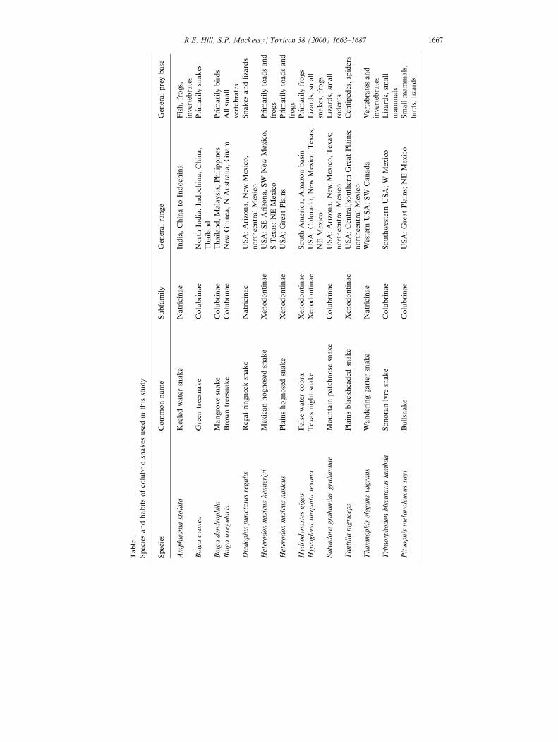
Twelve species of opisthoglyphous colubrid snakes were used in this study(Table 1); an aglyphous colubrid, Pituophis melanoleucus sayi (bullsnake), wasused as a non-venomous saliva control. Colubrid snakes native to the UnitedStates were collected in Arizona (permit ]MCKSY000221 to S.P.M.) andColorado (permit ]95-0456 to S.P.M.). Two specimens of H. gigas were on loanfrom Dr. Samuel S. Sweet and David Martin, and one specimen of T. b. lambdawas on loan from Dr. Wade Sherbrooke. Permission to extract venom from twoB. irregularis was granted by Dr. David Chiszar, and other snake species wereobtained from commercial dealers.
2.3. Extraction of venom from Duvernoy's glands
Extraction for all snakes was based on the methodology reported by Rosenberg(1992) as modi®ed by Hill and Mackessy (1997). Due to extremely low venomyields of some species and limited numbers of specimens, it was not possible torun all assays on all venoms. It should be noted that it is di�cult to obtain venomsamples completely free of saliva (and vice versa).
2.4. Protein concentration determination
Protein concentration was assayed (in triplicate) according to Bradford (1976)as modi®ed by BioRad Inc., using bovine gamma globulin as a standard. A totalvolume of 1.0 ml was used for all assays. Enzyme speci®c activities were based onprotein concentrations obtained from these assays.
2.5. Enzyme assays
Caseinolytic activity was assayed according to methods detailed by Mackessy(1993a), and speci®c activity was expressed as DA285 nm/min/mg venom protein.Due to problems with later batches of casein yellow, protease activity was alsodetermined with azocasein (Aird and da Silva, 1991), and activity was expressedas DA342/min/mg venom protein. A venom sample from Sistrurus catenatus
R.E. Hill, S.P. Mackessy / Toxicon 38 (2000) 1663±16871666
Table
1
Speciesandhabitsofcolubridsnakes
usedin
thisstudy
Species
Commonname
Subfamily
Generalrange
Generalpreybase
Amphiesm
astolata
Keeledwatersnake
Natricinae
India,Chinato
Indochina
Fish,frogs,
invertebrates
Boigacyanea
Green
treesnake
Colubrinae
NorthIndia,Indochina,China,
Thailand
Primarily
snakes
Boigadendrophila
Mangrovesnake
Colubrinae
Thailand,Malaysia,Philippines
Primarily
birds
Boigairregularis
Browntreesnake
Colubrinae
New
Guinea,N
Australia,Guam
Allsm
all
vertebrates
Diadophispunctatusregalis
Regalringnecksnake
Natricinae
USA:Arizona,New
Mexico,
northcentralMexico
Snakes
andlizards
Heterodonnasicuskennerlyi
Mexicanhognosedsnake
Xenodontinae
USA:SEArizona,SW
New
Mexico,
STexas;NEMexico
Primarily
toadsand
frogs
Heterodonnasicusnasicus
Plainshognosedsnake
Xenodontinae
USA;GreatPlains
Primarily
toadsand
frogs
Hydrodynastes
gigas
Falsewatercobra
Xenodontinae
South
America,Amazonbasin
Primarily
frogs
Hypsiglenatorquata
texana
Texasnightsnake
Xenodontinae
USA:Colorado,New
Mexico,Texas;
NEMexico
Lizards,sm
all
snakes,frogs
Salvadora
grahamiaegrahamiae
Mountain
patchnose
snake
Colubrinae
USA:Arizona,New
Mexico,Texas;
northcentralMexico
Lizards,sm
all
rodents
Tantillanigriceps
Plainsblackheaded
snake
Xenodontinae
USA:Central/southernGreatPlains;
northcentralMexico
Centipedes,spiders
Thamnophiselegansvagrans
Wanderinggarter
snake
Natricinae
Western
USA;SW
Canada
Vertebratesand
invertebrates
Trimorphodonbiscutatuslambda
Sonoranlyre
snake
Colubrinae
Southwestern
USA;W
Mexico
Lizards,sm
all
mammals
Pituophismelanoleucussayi
Bullsnake
Colubrinae
USA:GreatPlains;NEMexico
Smallmammals,
birds,lizards
R.E. Hill, S.P. Mackessy / Toxicon 38 (2000) 1663±1687 1667

edwardsi (desert massasauga rattlesnake; collected in Colorado) was also run forcomparison in both caseinolytic assays. Thrombin-like, kallikrein-like andplasmin-like proteolytic activities and arginine peptidase activity (using BAPNA)were assayed according to Mackessy (1993b). Phosphodiesterase activity wasassayed by the method of Laskowski (1980) as modi®ed by Mackessy (1988) withactivity expressed as DA400/min/mg venom protein. Phospholipase A2 activity wasassayed using 4-nitro-3-(octanoyloxy) benzoic acid as substrate (Holzer andMackessy, 1996), with activity expressed as nmol chromophore released/min/mgvenom protein (PLA2I). Phospholipase A2 activity was also assayed with egg yolkphosphatidylcholine Type IV (Sigma) following the procedure of Wells andHanahan (1969), and speci®c activity was expressed as mmol product formed/min/mg venom protein (PLA2II). L-amino acid oxidase activity was assayed accordingto Weissbach et al. (1961). Acetylcholinesterase activity was assayed as describedby Ellman et al. (1961), using forest cobra (Naja melanoleuca ) and red diamondrattlesnake (Crotalus ruber ) venoms as positive and negative controls, respectively;activity was expressed as mmol of product formed/min/mg venom protein.Hyaluronidase activity (di Ferrante, 1956) was expressed as mg hyaluronic acidhydrolyzed/min/mg venom protein.
2.6. Electrophoresis: SDS Tris±glycine gels
SDS±PAGE was used to determine the number and relative molecular weight ofproteins found in the colubrid venoms; all solutions and reagents (except gels)were prepared according to Hames (1990). Venom samples were prepared at a®nal concentration of 2.0 mg/ml crude venom in 1� SDS sample bu�er (62.5 mMTris±HCl, 10% glycerol, 1% SDS and 0.001% bromphenol blue; no b-mercaptoethanol) and were centrifuged at 6±7000 rpm for 5 min to pellet anyparticulates. Novex 14% acrylamide Tris±glycine gels were rinsed with freshreservoir bu�er, and typically 15±20 ml samples were loaded in each well; gels wererun at 100 V for 2±3 h. The gel was stained overnight in 0.1% Coomassie blue R-250 (in 30% methanol, 10% acetic acid) and then destained. After destaining, thegel was placed in 7.5% acetic acid for preservation and imaged with a CCDcamera attached to a PowerMac 7200 (with Adobe Photoshop and PageMakersoftware).
2.7. Zymogram gels
Novex zymogram gels were used to determine if the venoms had gelatin-degrading proteolytic activity (general endoprotease activity); this method alsoprovided information on the number and relative size of components withendoprotease activity (Heussen and Dowdle, 1980; Munekiyo and Mackessy,1998). Amounts of venom loaded varied between samples (typically 10±40 mg/lane). Samples were treated with SDS (but not b-mercaptoethanol) in samplebu�er and during electrophoresis; this potential inhibitor is removed during asubsequent wash in 2.5% Triton X-100 (Heussen and Dowdle, 1980).
R.E. Hill, S.P. Mackessy / Toxicon 38 (2000) 1663±16871668
2.8. Electroblot transfer
Several proteins from the venoms of H. gigas, H. t. texana, T. b. lambda and T.nigriceps were isolated via reducing SDS±PAGE (5% b-mercaptoethanol insample bu�er) followed by electroblot transfer with a Novex X-Cell (Trans-BlotElectrophoretic Transfer Cell) as described by Wilson and Yuan (1989). ThePVDF membrane was stained for 10 min with 0.1% Coomassie R-250 stain andthen destained in 50% methanol. The protein bands of interest were excised fromthe membrane and stored frozen until sequenced. The 26 kD proteins were chosenbecause of high concentration in the venoms and prevalence of this band invenoms of several species (therefore a common venom protein component). A 3.5kD protein of venom from Tantilla was chosen for analysis because speci®c toxinsfrom snake venoms are often low molecular weight peptides/proteins.
2.9. Protein sequencing
Partial N-terminal sequence was obtained from electroblots of 26 kDcomponents from H. gigas venom, H. t. texana venom and T. b. lambda venom,and the 3.5 kD T. nigriceps venom peptide. Sequencing was accomplished viaautomated Edman degradation on an ABI 473a protein sequencer(MacroMolecular Resources, Colorado State University, Fort Collins, CO, USA).
2.10. Protein sequence homologies
Sequences obtained were evaluated for sequence homology with previouslydescribed proteins via the National Center for Biotechnology Information's NRProtein Database (FASTA: Pearson and Lipman, 1988), with post-processingprovided by the Human Genome Center, Baylor College of Medicine (BEAUTY:Worley et al., 1995). All searches were conducted via the Internet (http://dot.imgen.bcm.tmc.edu:9331/seq-search/protein-search.html).
2.11. Observations of colubrid envenomations
The e�ects of envenomation on other snakes were observed for three species ofopisthoglyphous colubrids (D. punctatus, H. t. texana and T. e. vagrans ). Inaddition, the summary of a ®eld report of human envenomation by H. gigas isreported (N. Scott, personal communication, 1997).
3. Results
3.1. Protein concentrations
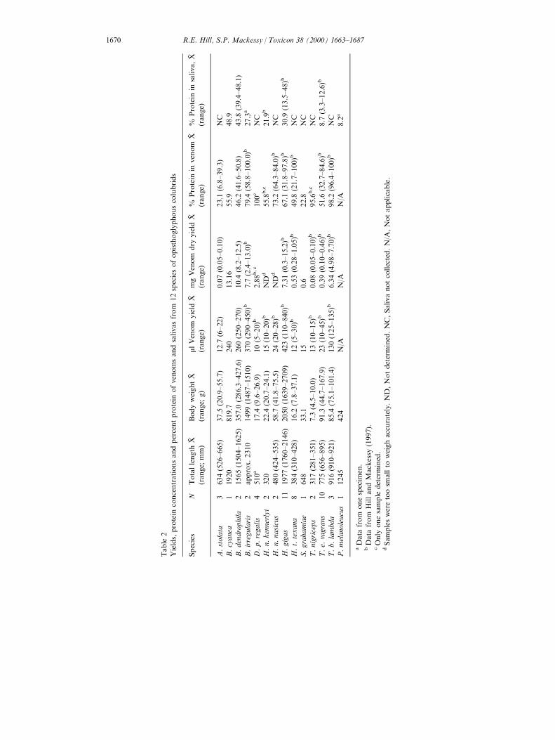
Protein concentrations of venoms (Table 2) were generally much higher thansalivas and were similar to those found in a previous study (Hill and Mackessy,
R.E. Hill, S.P. Mackessy / Toxicon 38 (2000) 1663±1687 1669
Table
2
Yields,protein
concentrationsandpercentprotein
ofvenomsandsalivasfrom
12speciesofopisthoglyphouscolubrids
Species
NTotallength
X-
(range;
mm)
BodyweightX-
(range;
g)
mlVenom
yield
X-
(range)
mgVenom
dry
yield
X-
(range)
%Protein
invenom
X-
(range)
%Protein
insaliva,X-
(range)
A.stolata
3634(526±665)
37.5
(20.9±55.7)
12.7
(6±22)
0.07(0.05±0.10)
23.1
(6.8±39.3)
NC
B.cyanea
11920
819.7
240
13.16
55.9
48.9
B.dendrophila
21565(1504±1625)
357.0
(286.3±427.6)
260(250±270)
10.4
(8.2±12.5)
46.2
(41.6±50.8)
43.8
(39.4±48.1)
B.irregularis
2approx.2310
1499(1487±1510)
370(290±450)b
7.7
(2.4±13.0)b
79.4
(58.8±100.0)b
27.3
a
D.p.regalis
4510a
17.4
(9.6±26.9)
10(5±20)b
2.88b,c
100c
NC
H.n.kennerlyi
2320
22.4
(20.7±24.1)
15(10±20)b
ND
d55.8
b,c
21.9
b
H.n.nasicus
2480(424±535)
58.7
(41.8±75.5)
24(20±28)b
ND
d73.2
(64.3±84.0)b
NC
H.gigas
11
1977(1760±2146)
2050(1639±2709)
423(110±840)b
7.31(0.3±15.2)b
67.1
(31.8±97.8)b
30.9
(13.5±48)b
H.t.texana
8384(310±428)
16.2
(7.8±37.1)
12(5±30)b
0.53(0.28±1.05)b
49.8
(21.7±100)b
NC
S.grahamiae
1648
33.1
15
0.6
22.8
NC
T.nigriceps
2317(281±351)
7.3
(4.5±10.0)
13(10±15)b
0.08(0.05±0.10)b
95.6
b,c
NC
T.e.
vagrans
10
775(656±895)
91.3
(44.7±167.9)
23(10±45)b
0.39(0.10±0.46)b
51.6
(32.7±84.6)b
8.7
(3.3±12.6)b
T.b.lambda
3916(910±921)
85.4
(75.1±101.4)
130(125±135)b
6.34(4.98±7.70)b
98.2
(96.4±100)b
NC
P.melanoleucus
11245
424
N/A
N/A
N/A
8.2
a
aData
from
onespecim
en.
bData
from
HillandMackessy
(1997).
cOnly
onesample
determined.
dSamplesweretoosm
allto
weighaccurately.ND,Notdetermined.NC,Salivanotcollected.N/A
,Notapplicable.
R.E. Hill, S.P. Mackessy / Toxicon 38 (2000) 1663±16871670
1997), indicating that secretions collected were primarily Duvernoy's glandsecretions. Total carbohydrate concentrations were determined (Dubois et al.,1956) for Boiga irregularis, H. n. kennerlyi and H. gigas venoms (0.1%, 1.0% and4.7%, respectively), and B. irregularis saliva had a carbohydrate concentration of3.7%.
3.2. Enzyme assays
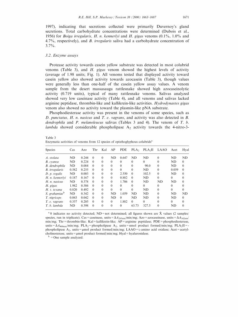
Protease activity towards casein yellow substrate was detected in most colubridvenoms (Table 3), and H. gigas venom showed the highest levels of activity(average of 1.98 units; Fig. 1). All venoms tested that displayed activity towardcasein yellow also showed activity towards azocasein (Table 3), though valueswere generally less than one-half of the casein yellow assay values. A venomsample from the desert massasauga rattlesnake showed high azocaseinolyticactivity (0.719 units), typical of many rattlesnake venoms. Salivas analyzedshowed very low caseinase activity (Table 4), and all venoms and salivas lackedarginine peptidase, thrombin-like and kallikrein-like activities. Hydrodynastes gigasvenom also showed no activity toward the plasmin-like pNA substrate.
Phosphodiesterase activity was present in the venoms of some species, such asD. punctatus, H. n. nasicus and T. e. vagrans, and activity was also detected in B.dendrophila and P. melanoleucus salivas (Tables 3 and 4). The venom of T. b.lambda showed considerable phospholipase A2 activity towards the 4-nitro-3-
Table 3
Enzymatic activities of venoms from 12 species of opisthoglyphous colubridsa
Species Cas Azo Thr Kal AP PDE PLA2 PLA2II LAAO Acet Hyal
A. stolata ND 0.244 0 0 ND 0.647 ND ND 0 ND ND
B. cyanea ND 0.224 0 0 0 0 0 0 0 ND 0
B. dendrophila ND 0.084 0 0 0 0 0 90.0 0 ND 0
B. irregularis 0.582 0.255 0 0 0 0 0 ND 0 0.059 0
D. p. regalis ND 0.003 0 0 0 2.550 0 102.5 0 ND 0
H. n. kennerlyi 0.187 0.167 0 0 0 0.882 0 ND 0 0 0
H. n. nasicus ND 0.378 0 0 0 1.706 0 ND ND ND 0
H. gigas 1.982 0.586 0 0 0 0 0 0 0 0 0
H. t. texana 0.820 0.492 0 0 0 0 0 ND 0 0 0
S. grahamiaeb ND 0.342 0 0 ND 1.059 ND ND 0 ND ND
T. nigriceps 0.043 0.042 0 0 ND 0 ND ND 0 ND 0
T. e. vagrans 0.357 0.205 0 0 0 1.882 0 0 0 0 0
T. b. lambda ND 0.398 0 0 0 0 63.73 327.5 0 ND 0
a 0 indicates no activity detected; ND=not determined; all ®gures shown are X-
values (2 samples/
species, run in triplicate). Cas=caseinase, units=DA285nm/min/mg; Azo=azocaseinase, units=DA342nm/
min/mg; Thr=thrombin-like; Kal=kallikrein-like; AP=arginine peptidase; PDE=phosphodiesterase,
units=DA400nm/min/mg; PLA2=phospholipase A2, units=nmol product formed/min/mg; PLA2II=-
phospholipase A2, units=mmol product formed/min/mg; LAAO=L-amino acid oxidase; Acet=acetyl-
cholinesterase, units=mmol product formed/min/mg; Hyal=hyaluronidase.b =One sample analyzed.
R.E. Hill, S.P. Mackessy / Toxicon 38 (2000) 1663±1687 1671

Fig. 1. Proteolytic activity of colubrid venoms and salivas toward casein yellow substrate. Activity is
expressed as DA285 nm/min/mg protein.
Table 4
Enzymatic activities of saliva from eight species of colubridsa
Species Cas Azo Thr Kal PDE PLA2 PLA2II LAAO Acet Hyal AP
B. cyanea ND 0.107 0 0 0 0 ND 0 ND 0 0
B. dendrophila ND 0.005 0 0 0.941 0 77.5 0 ND 0 0
B. irregularis 0.050 0.116 0 0 0 0 ND 0 0.064 0 0
D. p. regalis ND 0.076 0 0 0 0 ND 0 ND 0 0
H. n. nasicus ND 0.426 0 0 0 50.0 115.0 ND ND 0 0
H. gigas 0.065 0.319 0 0 0 0 0 0 0 0 0
T. e. vagrans ND 0 0 0 0 0 ND 0 ND 0 0
P. melanoleucus 0.048 0 0 0 4.412 0 ND 0 0 0 0
a 0 indicates no activity detected; ND=not determined; all ®gures shown are X-values (one sample/
species, run in triplicate). Cas=caseinase, units=DA285nm/min/mg; Azo=azocaseinase, units=DA342nm/
min/mg; Thr=thrombin-like; Kal=kallikrein-like; AP=arginine peptidase; PDE=phosphodiesterase,
units=DA400nm/min/mg; PLA2=phospholipase A2, units=nmol product formed/min/mg; PLA2II=-
phospholipase A2, units=mmol product formed/min/mg; LAAO=L-amino acid oxidase; Acet=acetyl-
cholinesterase, units=mmol product formed/min/mg; Hyal=hyaluronidase.
R.E. Hill, S.P. Mackessy / Toxicon 38 (2000) 1663±16871672
(octanoyloxy) benzoic acid substrate (63.7 nmol/min/mg; PLA2I assay, Tables 3and 4), as did Heterodon n. nasicus saliva (8.69 nmol/min/mg), but other venomsand salivas showed no apparent activity. To verify these results, a more sensitiveassay using egg yolk phosphatidylcholine was conducted on several samples(PLA2II, Tables 3 and 4). With the second assay, T. b. lambda venom, Boigadendrophila venom, D. punctatus venom, B. dendrophila saliva and H. n. nasicussaliva showed moderate activity levels. Saliva and venom from B. irregularisdisplayed low levels of activity when assayed for acetylcholinesterase (0.059 and0.062 mmol/min/mg, respectively), though it was not nearly as active as the venomfrom the forest cobra (positive control: 3.68 mmol/min/mg). Venom from the reddiamond rattlesnake (negative control) displayed no acetylcholinesterase activity.No other venoms or salivas assayed showed acetylcholinesterase activity, and noneof the venoms or salivas analyzed showed L-amino acid oxidase or hyaluronidaseactivities.
3.3. Gel electrophoresis
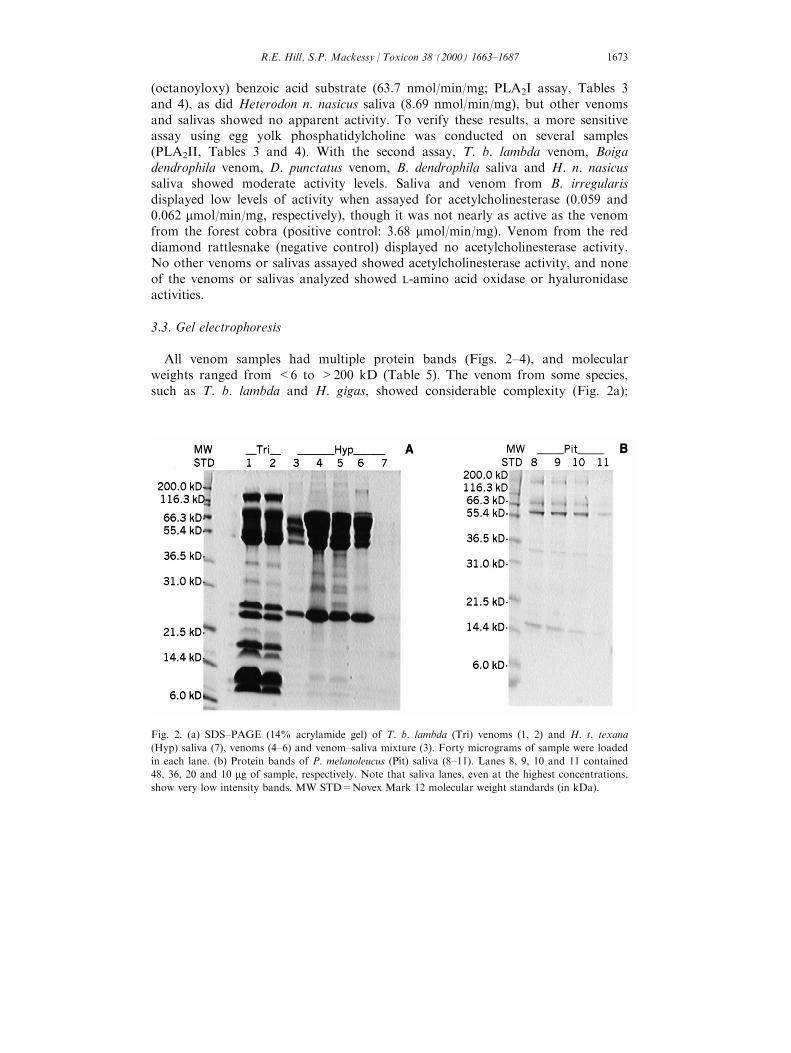
All venom samples had multiple protein bands (Figs. 2±4), and molecularweights ranged from <6 to >200 kD (Table 5). The venom from some species,such as T. b. lambda and H. gigas, showed considerable complexity (Fig. 2a);
Fig. 2. (a) SDS±PAGE (14% acrylamide gel) of T. b. lambda (Tri) venoms (1, 2) and H. t. texana
(Hyp) saliva (7), venoms (4±6) and venom±saliva mixture (3). Forty micrograms of sample were loaded
in each lane. (b) Protein bands of P. melanoleucus (Pit) saliva (8±11). Lanes 8, 9, 10 and 11 contained
48, 36, 20 and 10 mg of sample, respectively. Note that saliva lanes, even at the highest concentrations,
show very low intensity bands. MW STD=Novex Mark 12 molecular weight standards (in kDa).
R.E. Hill, S.P. Mackessy / Toxicon 38 (2000) 1663±1687 1673
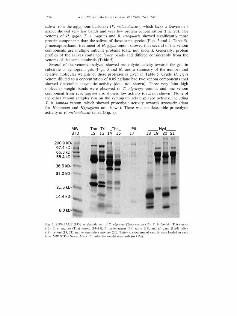
saliva from the aglyphous bullsnake (P. melanoleucus ), which lacks a Duvernoy'sgland, showed very few bands and very low protein concentration (Fig. 2b). Thevenoms of H. gigas, T. e. vagrans and B. irregularis showed signi®cantly moreprotein components than the salivas of those same species (Figs. 3 and 4; Table 5).b-mercaptoethanol treatment of H. gigas venom showed that several of the venomcomponents are multiple subunit proteins (data not shown). Generally, proteinpro®les of the salivas contained fewer bands and di�ered considerably from thevenoms of the same colubrids (Table 5).
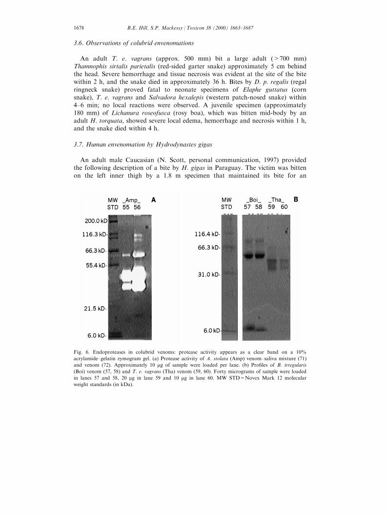
Several of the venoms analyzed showed proteolytic activity towards the gelatinsubstrate of zymogram gels (Figs. 5 and 6), and a summary of the number andrelative molecular weights of these proteases is given in Table 5. Crude H. gigasvenom diluted to a concentration of 0.05 ng/lane had two venom components thatshowed detectable enzymatic activity (data not shown). Three very faint highmolecular weight bands were observed in T. nigriceps venom, and one venomcomponent from T. e. vagrans also showed low activity (data not shown). None ofthe other venom samples run on the zymogram gels displayed activity, includingT. b. lambda venom, which showed proteolytic activity towards azocasein (datafor Heterodon and Hypsiglena not shown). There was no detectable proteolyticactivity in P. melanoleucus saliva (Fig. 5).
Fig. 3. SDS±PAGE (14% acrylamide gel) of T. nigriceps (Tan) venom (12), T. b. lambda (Tri) venom
(13), T. e. vagrans (Tha) venom (14±15), P. melanoleucus (Pit) saliva (17), and H. gigas (Hyd) saliva
(18), venom (19, 21) and venom±saliva mixture (20). Thirty micrograms of sample were loaded in each
lane. MW STD=Novex Mark 12 molecular weight standards (in kDa).
R.E. Hill, S.P. Mackessy / Toxicon 38 (2000) 1663±16871674
3.4. Protein sequencing
N-terminal sequences (5±21 residues) were obtained for four colubrid venomproteins (Table 6). The 26 kD components from H. gigas, H. t. texana and T. b.lambda venoms showed considerable sequence identity, and the 3.5 kD peptidefrom T. nigriceps venom showed no homology with the 26 kD proteins.
3.5. Protein sequence homology
High sequence identity was observed among the 26 kD components (Table 6),but there was no apparent sequence homology discovered between thesecomponents and previously reported protein sequence data (National Center forBiotechnology Information's NR Protein Database). The 3.5 kD peptide showedlimited sequence homology (<30%) with internal sequences of laminin andlaminin-type endothelial growth factor-like domain regions; this peptide alsoshowed moderate sequence identity (43%) with the N-terminal region of vascularendothelial growth factor (Table 6).
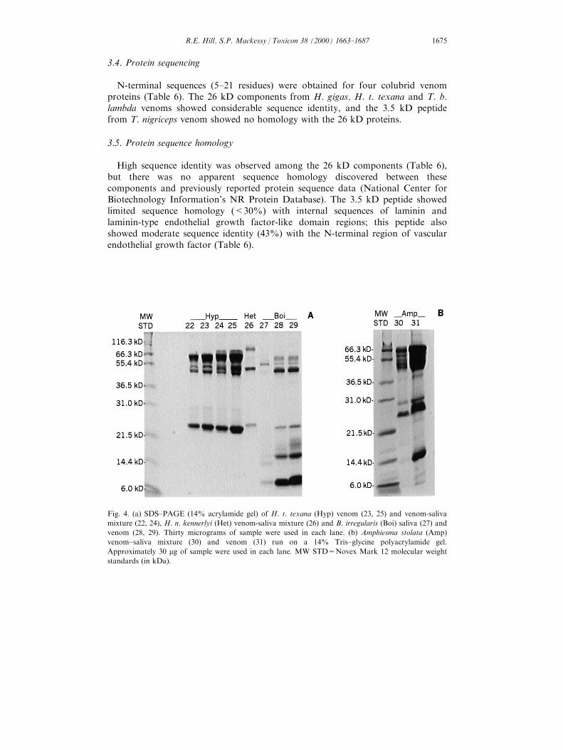
Fig. 4. (a) SDS±PAGE (14% acrylamide gel) of H. t. texana (Hyp) venom (23, 25) and venom-saliva
mixture (22, 24), H. n. kennerlyi (Het) venom-saliva mixture (26) and B. irregularis (Boi) saliva (27) and
venom (28, 29). Thirty micrograms of sample were used in each lane. (b) Amphiesma stolata (Amp)
venom±saliva mixture (30) and venom (31) run on a 14% Tris±glycine polyacrylamide gel.
Approximately 30 mg of sample were used in each lane. MW STD=Novex Mark 12 molecular weight
standards (in kDa).
R.E. Hill, S.P. Mackessy / Toxicon 38 (2000) 1663±1687 1675

Table
5
Summary
ofelectrophoreticpatternsofcolubridvenom
andsalivas:size
distributionandproteolyticactivity
Species
Number
ofbands
Sizeofprotein
bands(inDaltons)
Sizeofprotein(s)withendoproteolyticactivity
Venoms
A.stolata
7±8major;7±9minor
21,500->
200,000
Atleast
6bandsfrom
31,000->
200,000
B.cyanea
9major;5±6minor
6000->
116,300
±a
B.dendrophila
9major;5±7minor
6000->
200,000
±a
B.irregularis
8major;3±4minor
6000±66,300
Nonedetected
H.n.kennerlyi
7bands
21,500±97,400
Nonedetected
H.gigas
11bandswithatleast
1low
MW
band
<6000->
200,000
1bandat>200,000;2bandsat41,000and46,000
H.t.texana
9major;atleast
4±9minor
21,500±97,400
Nonedetected
S.grahamiae
atleast
3bands
6000±56,000
±a
T.nigriceps
atleast
10major;atleast
6minor
<4000->
200,000
3minorbandsbetween66,300±97,400b
T.e.
vagrans
7major,atleast
6minor
6000->
200,000
1bandapprox.75,000b
T.b.lambda
10major;atleast
9minor
6000±200,000
Nonedetected
Salivas
B.cyanea
9major;5±6minor
6000->
116,300
±a
B.dendrophila
9±10major;5±7minor
6000->
200,000
±a
B.irregularis
4±6major
6000±53,000
Nonedetected
H.gigas
10minor
8000±63,000
Nonedetected
T.e.
vagrans
4major;5±6minor
14,400->
200,000
Nonedetected
P.melanoleucus
5±6minor
14,400±200,000
Nonedetected
aThissample
wasnotsubjected
tozymogram
analysis.
bNotvisible
ingel
photograph.
R.E. Hill, S.P. Mackessy / Toxicon 38 (2000) 1663±16871676
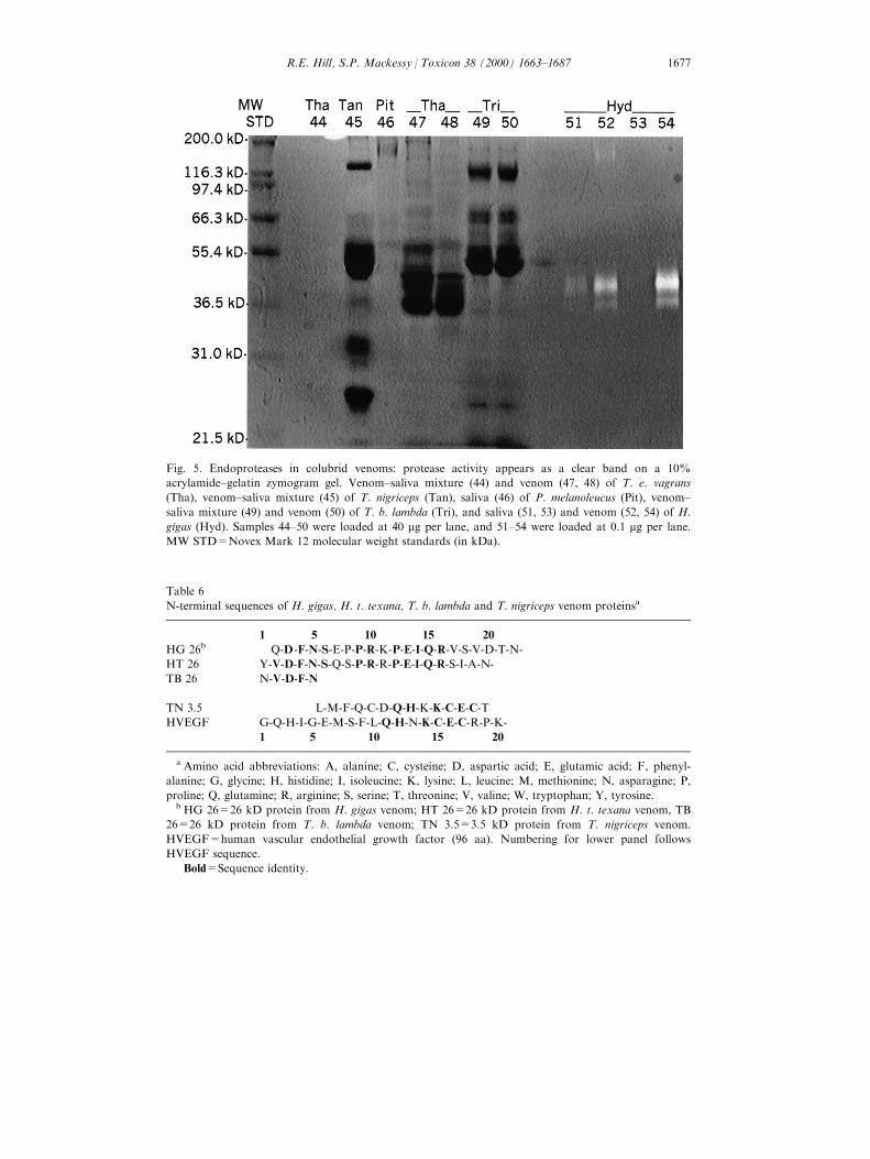
Fig. 5. Endoproteases in colubrid venoms: protease activity appears as a clear band on a 10%
acrylamide±gelatin zymogram gel. Venom±saliva mixture (44) and venom (47, 48) of T. e. vagrans
(Tha), venom±saliva mixture (45) of T. nigriceps (Tan), saliva (46) of P. melanoleucus (Pit), venom±
saliva mixture (49) and venom (50) of T. b. lambda (Tri), and saliva (51, 53) and venom (52, 54) of H.
gigas (Hyd). Samples 44±50 were loaded at 40 mg per lane, and 51±54 were loaded at 0.1 mg per lane.
MW STD=Novex Mark 12 molecular weight standards (in kDa).
Table 6
N-terminal sequences of H. gigas, H. t. texana, T. b. lambda and T. nigriceps venom proteinsa
1 5 10 15 20
HG 26b Q-D-F-N-S-E-P-P-R-K-P-E-I-Q-R-V-S-V-D-T-N-
HT 26 Y-V-D-F-N-S-Q-S-P-R-R-P-E-I-Q-R-S-I-A-N-
TB 26 N-V-D-F-N
TN 3.5 L-M-F-Q-C-D-Q-H-K-K-C-E-C-T
HVEGF G-Q-H-I-G-E-M-S-F-L-Q-H-N-K-C-E-C-R-P-K-
1 5 10 15 20
a Amino acid abbreviations: A, alanine; C, cysteine; D, aspartic acid; E, glutamic acid; F, phenyl-
alanine; G, glycine; H, histidine; I, isoleucine; K, lysine; L, leucine; M, methionine; N, asparagine; P,
proline; Q, glutamine; R, arginine; S, serine; T, threonine; V, valine; W, tryptophan; Y, tyrosine.b HG 26=26 kD protein from H. gigas venom; HT 26=26 kD protein from H. t. texana venom, TB
26=26 kD protein from T. b. lambda venom; TN 3.5=3.5 kD protein from T. nigriceps venom.
HVEGF=human vascular endothelial growth factor (96 aa). Numbering for lower panel follows
HVEGF sequence.
Bold=Sequence identity.
R.E. Hill, S.P. Mackessy / Toxicon 38 (2000) 1663±1687 1677
3.6. Observations of colubrid envenomations
An adult T. e. vagrans (approx. 500 mm) bit a large adult (>700 mm)Thamnophis sirtalis parietalis (red-sided garter snake) approximately 5 cm behindthe head. Severe hemorrhage and tissue necrosis was evident at the site of the bitewithin 2 h, and the snake died in approximately 36 h. Bites by D. p. regalis (regalringneck snake) proved fatal to neonate specimens of Elaphe guttatus (cornsnake), T. e. vagrans and Salvadora hexalepis (western patch-nosed snake) within4±6 min; no local reactions were observed. A juvenile specimen (approximately180 mm) of Lichanura roseofusca (rosy boa), which was bitten mid-body by anadult H. torquata, showed severe local edema, hemorrhage and necrosis within 1 h,and the snake died within 4 h.
3.7. Human envenomation by Hydrodynastes gigas
An adult male Caucasian (N. Scott, personal communication, 1997) providedthe following description of a bite by H. gigas in Paraguay. The victim was bittenon the left inner thigh by a 1.8 m specimen that maintained its bite for an
Fig. 6. Endoproteases in colubrid venoms: protease activity appears as a clear band on a 10%
acrylamide±gelatin zymogram gel. (a) Protease activity of A. stolata (Amp) venom±saliva mixture (71)
and venom (72). Approximately 10 mg of sample were loaded per lane. (b) Pro®les of B. irregularis
(Boi) venom (57, 58) and T. e. vagrans (Tha) venom (59, 60). Forty micrograms of sample were loaded
in lanes 57 and 58, 20 mg in lane 59 and 10 mg in lane 60. MW STD=Novex Mark 12 molecular
weight standards (in kDa).
R.E. Hill, S.P. Mackessy / Toxicon 38 (2000) 1663±16871678

unde®ned period. Three deep puncture wounds resulted, and the wounds bledprofusely. After 6 h the wound was painful and slightly swollen, but there was nodiscoloration. The thigh became very painful after 24 h, and there was edema andred discoloration at the site of envenomation. The immediate area was hard andswollen after 48 h with a slight yellow discoloration, and a burning sensationoccurred when the envenomated area was touched. The yellow discoloration andsoreness remained for 4 days after the envenomation, and after 7 days there wasno pain, swelling or discoloration. The fang puncture marks remained red, andthere was never any sign of infection.
4. Discussion
Using ketamine hydrochloride in conjunction with pilocarpine, venoms wereobtained from not only the large colubrids such as B. irregularis and H. gigas, butalso from small species such as H. t. texana and T. nigriceps. In addition,manipulation of the snakes was greatly facilitated. Although collection of venomfrom colubrids is much more labor-intensive than venom extraction from front-fanged snakes, venom su�cient for numerous analyses can be obtained from eventhe smallest species (such as T. nigriceps;05 g body weight).
Because the Duvernoy's gland is a serous gland, secretions were expected to behigh in protein concentration; the generally high protein content of venomsindicated that primarily Duvernoy's gland secretions (venom) had been collected.Salivary glands produce primarily mucopolysaccharides, and the protein contentof salivas was typically low. Some venom components are undoubtedlyglycosylated, but the results of limited carbohydrate assays in the present studysuggested that carbohydrates were minor components of B. irregularis (0.1%) andH. n. kennerlyi (1%) venoms; the carbohydrate concentration in H. gigas venom(4.7%) was similar to that of several rattlesnake venoms (S.P. Mackessy,unpublished data). Unde®ned carbohydrate±protein complexes have been reportedin the Duvernoy's glands of H. gigas (Glenn et al., 1992), but carbohydrates werenot found in the cytoplasmic granules of secretory cells from B. irregularisDuvernoy's glands (Zalisko and Kardong, 1992).
Endoproteolytic activity was common in venoms analyzed in this study, and thehighest proteolytic activities were observed in H. gigas and A. stolata venoms.Signi®cant proteolytic activity was also observed in B. irregularis, H. nasicus, H. t.texana and T. e. vagrans venoms. Based on observations of e�ects on tissues ofother snakes, some of these venoms may also contain hemorrhagic toxins, and(based on the action of viperid venom hemorrhagic proteases) these may bemetalloproteases (e.g., Takeya et al., 1990; Fox and Long, 1998; Takeya andIwanaga, 1998).
Several colubrid venoms contained components that very e�ectively digested thegelatin substrate in zymogram gels (notably H. gigas and A. stolata ), and activityfrom H. gigas crude venom components (2 bands) could readily be detected evenat concentrations as low as 0.05 ng crude venom per lane. In comparison,
R.E. Hill, S.P. Mackessy / Toxicon 38 (2000) 1663±1687 1679

rattlesnake venoms, which often have very high activity and 4+ components,usually require 0.5±1.0 mg/lane to visualize proteases (e.g., Munekiyo andMackessy, 1998). The large number of proteases in A. stolata venom (26) andtheir high activity toward the zymogram substrate were somewhat unusual for acolubrid venom and are the subject of further study. Venoms from other species,such as H. t. texana and T. b. lambda, showed proteolytic activity when assayedwith casein substrates but showed no apparent activity on zymogram gels; thesevenoms may lack gelatin-degrading proteases, or the proteases may be irreversiblydenatured following short-term exposure to SDS. Most caseinolytic and gelatin-hydrolyzing proteases from snake venoms are metalloproteases (Fox and Long,1998; Munekiyo and Mackessy, 1998; S. P. Mackessy, unpublished data, 1998),and these colubrid proteases may also be metalloenzymes.
In rattlesnake venoms, high proteolytic activity has a prominent role inpredigestion of prey tissues, facilitating digestion and allowing for broader activityand distribution patterns (e.g. Thomas and Pough, 1979; Mackessy, 1988, 1993b).Enzymatic activities of colubrid venoms, particularly proteases which promotetissue damage, are likely also correlated with prey type(s) and/or activity patterns.The wandering garter snake (Thamnophis elegans vagrans ) is very broadlydistributed in western North America, occurring at high latitude and at highelevation, and venom which is proteolytic and hemorrhagic (casein assays, e�ectson other snakes) may have provided this species with the ability to occupy thesethermally variable environments and still capitalize on bulky mammalian prey(e.g. Finley et al., 1994); a similar biological role for these components is seen inviperid venoms. Conversely, species of colubrids that feed on smaller prey itemswith high surface-to-volume ratios, such as D. punctatus regalis feeding on othersnakes and small lizards and T. nigriceps feeding on arachnids, would not likelypossess a venom with a prominent predigestive role (see Mackessy, 1988 fordetails), and protease activity in these species' venoms was quite low.
High levels of phospholipase A2 were detected in venom from B. dendrophila,D. p. regalis and T. b. lambda; this enzyme is characteristically present in venomsof most front-fanged snakes (Rosenberg, 1990; Kini, 1997) but appeared to belacking in most rear-fanged colubrid venoms (Weinstein and Kardong, 1994; butsee Broaders and Ryan, 1997). The apparent lack of this common venomcomponent in colubrid venoms is more likely due to inadequate sampling orinsensitive assays, and we predict that phospholipases will be more commonamong colubrid venoms than was previously assumed. Phospholipase A2 enzymesfrom several colubrid venoms are currently being isolated for furthercharacterization and protein sequencing.
Phosphodiesterase assays showed that approximately half of the colubridvenoms in the present study contained detectable levels of activity, and thiscomponent was also reported for several other colubrid venoms (Kornalik et al.,1978; Rosenberg et al., 1985; Vest et al., 1991). Phosphodiesterase activity iscommon to most front-fanged snake venoms, and though the exact role ofphosphodiesterases in those venoms is not known, it may involve disruption ofcAMP- and ADP-mediated events (Mackessy, 1998). It is possible that this
R.E. Hill, S.P. Mackessy / Toxicon 38 (2000) 1663±16871680
enzyme has an important functional role in colubrid venoms as well. Becausevenom enzymes showed much higher activity toward some substrates(phospholipids and thymidine 5-nitrophenyl phosphate) than toward syntheticchromogenic substrates (4-nitro-3-(octanoyloxy) benzoic acid and Ca-bis-nitrophenylphosphate), it is recommended that several di�erent substrates be usedwhen assaying colubrid venoms for a speci®c enzymatic activity before concludingthat an activity is lacking.
The low but detectable level of acetylcholinesterase activity found in B.irregularis saliva (and to a lesser degree the venom) is consistent with a recentreport of this activity in the venom of two other species of Boiga (Broaders andRyan, 1997). However, the activity in the venom sample may have been due tocontamination with saliva, since one B. irregularis venom sample assayed showedno activity.
Electrophoretic analysis demonstrated that venoms from most colubridscontained numerous protein bands and that venoms were typically quite distinctfrom salivas. At least 10±20 components, ranging in size from E4 to >200 kD,were present in most venoms, and pro®le comparisons demonstrated species-speci®c components as well as several shared components. Congeneric species,such as Boiga cyanea and B. dendrophila, showed very similar protein bandingpatterns, while non-related species patterns were quite distinct. When the crudevenoms were treated with reducing agents (b-mercaptoethanol), higher molecularweight components dissociated into smaller subunits, demonstrating higher orderorganization among some venom components, as is seen in most viperid andmany elapid venoms.
The colubrid venoms assayed in the present study lacked many of the enzymaticproperties (such as L-AAO, hyaluronidase, several serine proteases, etc.) typicallyfound in front-fanged snake venom (see also review by Weinstein and Kardong,1994). However, other components important to the biological roles of venoms,including endoproteases, phospholipases and phosphodiesterases, are present inmany colubrid and front-fanged snake venoms. Many components appear to benovel proteins or proteins unique to colubrid venoms; these proteins likely re¯ectthe divergent evolutionary histories of colubrids and convergence on an orally-delivered venom system among snakes. Non-ophidian venoms also containnumerous enzymatic activities (e.g. Ho�man, 1996), and assays based on non-ophidian venom components may help identify protein components in colubridvenoms with (at present) unknown activities.
There are considerable structural and sequence data available for viperid andelapid venom components (e.g. Shannon et al., 1989; Rosenberg, 1990; Takeya etal., 1990; Sanchez et al., 1991; Hite et al., 1992; Tu, 1991; Kini, 1997; Bailey,1998), but there are no published data for colubrid venom components. A recentstudy of a myotoxic protein from the venom of Philodryas olfersii (green snake, axenodontine colubrid) indicated that the protein was N-terminally blocked (Prado-Franceschi et al., 1998). N-terminal sequencing of the ®rst 5±21 amino acidresidues for four colubrid venom proteins represents the ®rst protein sequenceobtained for any colubrid venom components. However, in spite of the large
R.E. Hill, S.P. Mackessy / Toxicon 38 (2000) 1663±1687 1681
amount of sequence data available for front-fanged snake venom components, aswell as for non-venom proteins, no protein homologies have been discovered forthe 26 kD colubrid venom components. Regions of low homology with internalsequences of a DNA J-like protein and a glial ®brillary acidic protein likelyrepresented spurious and chance sequence homology over a short stretch of theseproteins. The level of sequence identity among the 26 kD venom components ofH. gigas, H. t. texana and T. b. lambda indicates a similar structure (and likelyfunction) in these proteins, which are prevalent venom components for the threespecies. At present, the 26 kD venom proteins remain unidenti®ed.
A low molecular weight peptide (03.5 kD) from T. nigriceps venom has nosequence homology with any published low molecular weight toxin, such asmyotoxin a (Fox et al., 1979), suggesting that it may be a novel peptide. As withthe 26 kD proteins, this peptide showed moderate homology with internalsequence of several larger proteins, such as laminin and the endothelial growthfactor domain. However, the 3.5 kD peptide also showed signi®cant sequenceidentity (6/14 residues) with the N-terminus of a 96 amino acid vascularendothelial growth factor (see Table 6). If this apparent homology is real, thepeptide from Tantilla nigriceps venom may represent another example ofconvergence of a venom peptide on the sequence of a native regulatory peptide. Acommon ``evolutionary strategy'' adopted by venomous snakes is to targethomeostatic mechanisms of prey (e.g. Stocker and Meier, 1989) and producespeci®c toxins which disrupt these mechanisms, and it is probable that colubridvenoms show a similar trend. An example are the sarafotoxins, 21-residue peptides(Kochva et al., 1982; Takasaki et al., 1988) isolated from the venoms of the molesnake Atractaspis engaddensis (formerly included in the Colubridae), which showhigh sequence and receptor homology with a group of native vasoconstrictivepeptides, the endothelins (Ambar et al., 1988; Kloog et al., 1988, 1989; Galron etal., 1991). Sarafotoxins are potent venom toxins which produce cardiotoxic andvasoconstrictive e�ects, resulting in rapid prey death (Kochva et al., 1982;Takasaki et al., 1988). We predict that the 3.5 kD peptide from T. nigricepsvenom will be a speci®c toxin, perhaps targeting a homeostatic mechanism of thearthropod prey (centipedes, spiders) of this species. It should be noted, however,that only 14 residues were available for homology searches.
Fatal envenomations of humans by colubrids are presently limited to fourspecies: Dispholidus typus, Philodryas olfersii, Rhabdophis tigrinus andThelotornis capensis; however, serious envenomations may occur from the bitesof many species. Because rear-fanged snakes lack an e�cient hollow-fangedinjection system (Kardong and Young, 1991; Weinstein and Kardong, 1994),speci®c factors of a bite, such as length of contact time by the snake, cangreatly in¯uence the severity of envenomation by colubrids. Severe systemice�ects following envenomation by H. gigas (Manning et al., 1999) likelyresulted from the long contact time (1.5 min) of the bite. Extreme careshould be exercised when handling H. gigas and A. stolata (because of thehigh level of endoproteolytic activity of their venoms) or any other rear-fanged colubrid whose venom has not been completely characterized. Lack of
R.E. Hill, S.P. Mackessy / Toxicon 38 (2000) 1663±16871682
an appropriate regard for the potential hazard from a bite of such snakes hasresulted in human fatalities even among herpetologists (Pope, 1958;FitzSimons and Smith, 1958).
In conclusion, based on the complexity of most venoms obtained, the presenceof activities which are common to front-fanged snakes and the extensivedi�erences of these venoms from other oral secretions (saliva), the Duvernoy'sgland secretions of many colubrid snakes should be considered as venomshomologous with (but not the same as) venoms of the front-fanged snakes.Previous studies have also shown that the colubrid Duvernoy's gland ismorphologically and embryonically homologous with the venom glands of front-fanged snakes (Kochva, 1965; Ovadia, 1984). Colubrid venoms, like the venoms offront-fanged snakes, are trophic adaptations which facilitate feeding (Kardong,1986; Mackessy, 1988, 1993a; Mackessy and Tu, 1993; Kardong, 1996). Diet andvenom composition are intricately interwoven for many species of front-fangedsnakes (Mackessy, 1988, 1993a; Daltry et al., 1996), and colubrid venoms likelyhave been shaped by speci®c aspects of prey. Taxa-speci®c toxins are known fromthe venom of the black widow spider (Latrodectus mactans; Grishin, 1998), andmany of the small specialized colubrids may produce analogous venom toxins.Opisthoglyphous colubrid snakes represent a vast source of unknown venomswhich deserve further investigation. The isolation, identi®cation andcharacterization of colubrid venom components will provide insight into theirfunction and biological roles and likely will produce unique molecular probes foruse in other biological systems.
Acknowledgements
Support for this work was provided by grant GM52665-01 from the NationalInstitutes of Health, National Institute of General Medical Sciences to S. P.Mackessy. The following individuals provided snakes or allowed extraction ofanimals under their care, and their assistance is appreciated: D. Chiszar, J.Hobert, D. Martin, C. Montgomery, W. Sherbrooke, S. S. Sweet and B.Tomberlin. Additional support was provided by a research incentives grant fromthe University of Northern Colorado Research Corporation. David Chiszar andJennifer Clarke provided numerous comments on an earlier version of thismanuscript, and the helpful suggestions of an anonymous reviewer are alsoacknowledged. Permission to collect several species used in this study was grantedby the Colorado Division of Wildlife and the Arizona Game and FishDepartment. Thanks go to Norm Scott for his account of a human envenomationby H. gigas and to Sean Munekiyo for his assistance with some of the enzymeassays.
R.E. Hill, S.P. Mackessy / Toxicon 38 (2000) 1663±1687 1683
References
Aird, S.D., da Silva Jr, N.J., 1991. Comparative enzymatic composition of Brazilian coral snake
(Micrurus ) venoms. Comp. Biochem. Physiol. 99B, 287±294.
Ambar, I., Kloog, Y., Kochva, E., Wollberg, Z., Bdolah, A., Oron, U., Sokolovsky, M., 1988.
Characterization and localization of a novel neuroreceptor for the peptide sarafotoxin. Biochem.
Biophys. Res. Commun. 157, 1104±1110.
Assakura, M.T., SalomaÄ o, M.G., Puorto, C., Mandelbaum, F.R., 1992. Hemorrhagic, ®brinogenolytic
and edema-forming activities of the venom of the colubrid snake Philodryas olfersii (green snake).
Toxicon 30, 427±438.
Assakura, M.T., Reichl, A.P., Mandelbaum, F.R., 1994. Isolation and characterization of ®ve
®brin(ogen)olytic enzymes from the venom of Philodryas olfersii (green snake). Toxicon 32, 819±831.
Bailey, G., 1998. Enzymes from Snake Venoms. Alaken Press, Fort Collins, CO.
Bradford, M.M., 1976. A rapid and sensitive method for the quantitation of microgram quantities of
protein utilizing the principle of protein-dye binding. Analyt. Biochem. 72, 248±251.
Broaders, M., Ryan, M.F., 1997. Enzymatic properties of the Duvernoy's secretion of Blanding's tree
snake (Boiga blandingi ) and of the mangrove snake (Boiga dendrophila ). Toxicon 35, 1143±1148.
Cowles, R.B., 1941. Evidence of venom in Hypsiglena ochrorynchus. Copeia 1941, 109.
Daltry, J.C., WuÈ ster, W., Thorpe, R.S., 1996. Diet and snake venom evolution. Nature 379, 537±540.
de Araujo, M.E., dos Santos, A.C., 1997. Cases of human envenoming caused by Philodryas olfersii
and Phylodryas patagoniensis. Rev. Soc. Bras. Med. Trop. 30, 517±519.
di Ferrante, N., 1956. Turbidimetric measurement of acid mucopolysaccharides and hyaluronidase
activity. J. Biol. Chem. 220, 303±306.
Dubois, M., Gilles, K.A., Hamilton, J.K., Rebers, P.A., Smith, F., 1956. Colorimetric method for
determination of sugars and related substances. Analyt. Chem. 28 (3), 350±356.
Durkin, J.P., Pickwell, G.V., Trotter, J.T., Shier, W.T., 1981. Phospholipase A2 (EC-2.1.1.4)
electrophoretic variants in reptile venoms. Toxicon 19, 535±546.
Ellman, G.L., Courtney, K.D., Andres Jr, V., Featherstone, R.M., 1961. A new and rapid colorimetric
determination of acetylcholinesterase activity. Biochem. Pharmac. 7, 88±95.
Finley Jr, R.B., Chiszar, D., Smith, H.M., 1994. Field observations of salivary digestion of rodent
tissue by the wandering garter snake, Thamnophis elegans vagrans. Bull. Chicago Herp. Soc. 29, 5±6.
FitzSimons, D.C., Smith, H.M., 1958. Another rear-fanged South African snake lethal to humans.
Herpetologica 14, 198±202.
Fox, J.W., Elzinga, M., Tu, A.T., 1979. Amino acid sequence and disul®de bond assignment of
myotoxin a isolated from the venom of prairie rattlesnake (Crotalus viridis viridis ). Biochemistry 18,
678±684.
Fox, J.W., Long, C. 1998. The ADAMs/MDC family of proteins and their relationships to the snake
venom metalloproteinases. In: Bailey, G.S. (Ed.), Enzymes from snake venom. Alaken Press, Fort
Collins, CO, pp. 151±178.
Fritts, T.H., McCoid, M.J., Haddock, R.L., 1994. Symptoms and circumstances associated with bites
by the brown tree snake (Colubridae: Boiga irregularis ) on Guam. J. Herpetol. 28, 27±33.
Fuller, S.R., 1981. A case of envenomation by a western hognose snake, Heterodon n. nasicus. In:
Notes from NOAH, Oct. 27, pp. 11±14.
Galron, R., Bdolah, A., Kochva, E., Wollberg, Z., Kloog, Y., Sokolovsky, M., 1991. Kinetic and cross-
linking studies indicate di�erent receptors for endothelins and sarafotoxins in the ileum and
cerebellum. FEBS 283, 11±14.
Gans, C., 1978. Reptilian venoms: some evolutionary considerations. In: Gans, C., Gans, K.A. (Eds.),
Biology of the Reptilia, 8. Academic Press, New York, pp. 1±39.
Glenn, J.L., Porras, L.W., Nohavec, R.D., Straight, R.C. 1992. Analysis of the Duvernoy's gland and
oral secretions of Hydrodynastes gigas (Dumeril, Bibron, and Dumeril) (Reptilia: Serpentes). In:
Strimple, P.D., Strimple, J.L. (Eds.), Contributions in Herpetology. Cincinnati Museum of Natural
History, Cincinnati, OH, pp. 19±26.
R.E. Hill, S.P. Mackessy / Toxicon 38 (2000) 1663±16871684

Grasset, E., Schaafsma, A.W., 1940. Studies on the venom of the boomslang (Dispholidus typus ). S.
Afr. Med. J. 14, 236±241.
Grishin, E.V., 1998. Black widow spider toxins: the present and the future. Toxicon 36, 1693±1701.
Hames, B.D. 1990. One-dimensional polyacrylamide gel electrophoresis. In: Hames, B.D., Rickwood,
D. (Eds.), Gel Electrophoresis of Proteins. Oxford University Press, New York, pp. 1±148.
Heussen, C., Dowdle, E.B., 1980. Electrophoretic analysis of plasminogen activators in polyacrylamide
gels containing sodium dodecylsulfate and copolymerized substrate. Analyt. Biochem. 102, 196±202.
Hiestand, P.C., Hiestand, R.R., 1979. Dispholidus typus (boomslang) snake venom: puri®cation and
properties of the coagulant principle. Toxicon 17, 489±498.
Hill, R.E., Mackessy, S.P., 1997. Venom yields from several species of colubrid snakes and di�erential
e�ects of ketamine. Toxicon 35, 671±678.
Hite, L.A., Shannon, J.D., Bjarnson, J.B., Fox, J.W., 1992. Sequence of a cDNA clone encoding the
zinc metalloproteinase hemorrhagic toxin e from Crotalus atrox: evidence for signal, zymogen and
disintegrin-like structures. Biochemistry 31, 6203±6211.
Ho�man, D.R., 1996. Hymenoptera venom proteins. Adv. Exp. Med. Biol. 391, 169±186.
Holzer, M., Mackessy, S.P., 1996. An aqueous endpoint assay of snake venom phospholipase A2.
Toxicon 34, 1149±1155.
Kini, R.M. 1997. In: Kini, R.M. (Ed.), Venom Phospholipase A2 Enzymes: Structure, Function and
Mechanism. Wiley, Chichester.
Kardong, K.V., 1980. Evolutionary patterns in advanced snakes. Am. Zool. 20, 269±282.
Kardong, K.V., 1982. The evolution of the venom apparatus in snakes from colubrids to viperids and
elapids. Mem. Inst. Butantan 46, 105±118.
Kardong, K.V., 1986. Predatory strike behavior of the rattlesnake, Crotalus viridis oreganus. J. Comp.
Psychol. 100, 304±314.
Kardong, K.V., 1996. Snake toxins and venoms: an evolutionary perspective. Herpetologica 52, 36±46.
Kardong, K.V., Young, B.A., 1991. Fangs and snakes: how do open grooves inject venom into
enclosed spaces? Am. Zool. 31, 51A.
Kloog, Y., Ambar, I., Sokolovsky, M., Kochva, E., Wollberg, Z., Bdolah, A., 1988. Sarafotoxin, a
novel vasoconstrictor peptide: phosphoinositide hydrolysis in rat heart and brain. Science 242, 268±
270.
Kloog, Y., Bousso-Mittler, D., Bdolah, A., Sokolovsky, M., 1989. Three apparent receptor subtypes
for the endothelin/sarafotoxin family. FEBS Lett. 253, 199±202.
Kochva, E., 1965. The development of the venom gland in the opisthoglyph snake Telescopus fallax
with remarks on Thamnophis sirtalis (Colubridae, Reptilia). Copeia 1965, 147±154.
Kochva, E., Viljoen, C.C., Botes, D.P., 1982. A new type of toxin in the venom of snakes of the genus
Atractaspis (Atractaspidinae). Toxicon 20, 581±592.
Kornalik, F., Ta borska , E., Mebs, D., 1978. Pharmacological and biochemical properties of a venom
gland extract from the snake Thelotornis kirtlandi. Toxicon 16, 535±542.
Laskowski Sr, M., 1980. Puri®cation and properties of venom phosphodiesterase. Meths Enzymol. 65,
276±284.
Lee, C.Y. (Ed.), 1979. Snake Venoms. Handbook of Experimental Pharmacology, 52. Springer, Berlin.
Mackessy, S.P., 1988. Venom ontogeny in the Paci®c rattlesnakes Crotalus viridis helleri and C. v.
oreganus. Copeia 1988, 92±101.
Mackessy, S.P., 1993a. Fibrinogenolytic proteases from the venoms of juvenile and adult northern
Paci®c rattlesnakes (Crotalus viridis oreganus ). Comp. Biochem. Physiol. 106B (1), 181±189.
Mackessy, S.P., 1993b. Kallikrein-like and thrombin-like proteases from the venom of juvenile northern
Paci®c rattlesnakes (Crotalus viridis oreganus ). J. Nat. Toxins 2 (2), 223±239.
Mackessy, S.P. 1998. Phosphodiesterases, ribonucleases and deoxyribonucleases. In: Bailey, G.S. (Ed.),
Enzymes from Snake Venom. Alaken Press, Fort Collins, CO, pp. 361±404.
Mackessy, S.P., Tu, A.T. 1993. Biology of the sea snakes and biochemistry of their venoms. In: Tu,
A.T. (Ed.), Toxin-related diseases: poisons originating from plants, animals and spoilage. Oxford/
IBH Pub. Co, New Delhi, pp. 305±351.
Manning, B., Galbo, M., Klapman, G., 1999. First report of a symptomatic South American false
water cobra envenomation. J. Toxicol.-Clin. Toxicol. 37, 613.
R.E. Hill, S.P. Mackessy / Toxicon 38 (2000) 1663±1687 1685

McKinstry, D.M., 1978. Evidence of toxic saliva in some colubrid snakes of the United States. Toxicon
16, 523±534.
Mebs, D., 1968. Analysis of Leptodeira annulata venom. Herpetologica 24, 338±339.
Minton, S.A., 1990. Venomous bites by nonvenomous snakes: a bibliography of colubrid
envenomation. J. Wild. Med. 1, 119±127.
Minton, S.A., 1996 Are there any nonvenomous snakes? An update on colubrid envenoming, Adv.
Herpetoculture 4, 127±134.
Mittleman, M.B., Goris, R.C., 1976. Death caused by the bite of the Japanese colubrid snake
Rhabdophis tigrinus (Boie). J. Herpetol. 12, 109±111.
Morris, M.A., 1985. Envenomation from the bite of Heterodon nasicus (Serpentes: Colubridae).
Herpetologica 41, 361±363.
Munekiyo, S.M., Mackessy, S.P., 1998. E�ects of temperature and storage conditions on the
electrophoretic, toxic and enzymatic stability of venom components. Comp. Biochem. Physiol. 119B,
119±127.
Nomura, T., Nagata, T., Kawamura, Y., Sawai, Y., 1989. A case of severe yamakagashi (Rhabdophis
tigrinus ) bite treated by antivenom. The Snake 21, 85±86.
Ogawa, H., Sawai, Y., 1986. Fatal bite of the yamakagashi (Rhabdophis tigrinus ). The Snake 18, 53±54.
Ovadia, M., 1984. Embryonic development of Duvernoy's gland in the snake Natrix tessellata
(Colubridae). Copeia 1984, 516±521.
Pearson, W.R., Lipman, D.J., 1988. Improved tools for biological sequence comparison. Proc. Natl.
Acad. Sci. 85, 2444±2448.
Pope, C.H., 1958. Fatal bite of captive African rear-fanged snake (Dispholidus ). Copeia 1958, 280±282.
Prado-Franceschi, J., Hyslop, S., Cogo, J.C., Andrade, A.L., Assakura, M.T., Reichl, A.P., Cruz-
HoÈ ¯ing, M.A., Rodrigues-Simioni, L., 1998. Characterization of a myotoxin from the Duvernoy's
gland secretion of the xenodontine colubrid Philodryas olfersii (green snake): e�ects on striated
muscle and the neuromuscular junction. Toxicon 36, 1407±1421.
Ribeiro, L.A., Puorto, G., Jorge, M.T., 1999. Bites by the colubrid snake Philodryas olfersii: a clinical
and epidemiological study of 43 cases. Toxicon 37, 943±948.
Robertson, S.S.D., Delpierre, G.R., 1969. Studies on African snake venoms-IV. Some enzymatic
activities in the venom of the boomslang Dispholidus typus. Toxicon 7, 189±194.
RodrõÂ guez-Robles, J.A., 1994. Are the Duvernoy's gland secretions of colubrid snakes venoms? J.
Herpetol. 28, 388±390.
RodrõÂ guez-Robles, J.A., Thomas, R., 1992. Venom function in the Puerto Rican racer, Alsophis
portoricensis (Serpentes: Colubridae). Copeia 1992, 62±68.
Rosenberg, H.I., 1992. An improved method for collecting secretion from Duvernoy's gland of colubrid
snakes. Copeia 1992, 244±246.
Rosenberg, H.I., Bdolah, A., Kochva, E., 1985. Lethal factors and enzymes in the secretion from the
Duvernoy's gland of three colubrid snakes. J. Exp. Zool. 233, 5±14.
Rosenberg, P. 1990. Phospholipases. In: Shier, W.T., Mebs, D. (Eds.), Handbook of Toxinology.
Dekker, New York, pp. 67±277.
Sakai, A., Honma, M., Sawai, Y., 1983. Studies on the pathogenesis of envenomation of the Japanese
colubrid snake yamakagashi Rhabdophis tigrinus tigrinus. 1. Study on the toxicity of the venom. The
Snake 15, 7±13.
SalomaÄ o, E.L., Di-Bernardo, M., 1995. Philodryas olfersii: uma cobra comum que mata. Caso
re®strado na a rea da 8a. delegacia regional de sau de. Arq. Socied. Brasileira Zool./Sorocaba-SP
Nos. 14/15/16.
Sanchez, E.F., Diniz, C.R., Richardson, M., 1991. The complete amino acid sequence of the
haemorrhagic factor LHFII, a metalloprotease isolated from the venom of the bushmaster snake
(Lachesis muta muta ). FEBS Lett. 282, 178±182.
Shannon, J.D., Baramova, E.N., Bjarnson, J.B., Fox, J.W., 1989. Amino acid sequence of a Crotalus
atrox venom metalloprotease which cleaves type IV collagen and gelatin. J. Biol. Chem. 264,
11,575±11,583.
Stocker, K., Meier, J., 1989. Snake venom proteins in hemostasis: new results. Folia Haematol. Int.
Mag. Klin. Morphol. Blutforsch 116, 935±953.
R.E. Hill, S.P. Mackessy / Toxicon 38 (2000) 1663±16871686
Takasaki, C., Tamiya, N., Bdolah, A., Wollberg, Z., Kochva, E., 1988. Sarafotoxins S6: several
isotoxins from Atractaspis engaddensis (burrowing asp) venom that a�ect the heart. Toxicon 26,
543±548.
Takeya, H., Iwanaga, S. 1998. Proteases that induce hemorrhage. In: Bailey, G.S. (Ed.), Enzymes from
Snake Venom. Alaken Press, Fort Collins, CO, pp. 11±38.
Takeya, H., Oda, K., Miyata, T., Omori-Satoh, T., Iwanga, S., 1990. The complete amino acid
sequence of the high molecular mass hemorrhagic protein HR1B isolated from the venom of
Trimeresurus ¯avoviridis. J. Biol. Chem. 265, 16,068±16,073.
Taub, A.M., 1967. Comparative histological studies on Duvernoy's gland of colubrid snakes. Bull.
Mus. Nat. Hist. 138, 1.
Thomas, R.G., Pough, P.H., 1979. The e�ects of rattlesnake venom on the digestion of prey. Toxicon
17, 221±228.
Tu, A.T., 1977. Venoms: Chemistry and molecular biology. Wiley, New York.
Tu, A.T., 1991. Tu, A.T. (Ed.), Handbook of Natural Toxins: Reptile Venoms and Toxins, vol. 5.
Dekker, New York.
Underwood, G., 1979. Classi®cation and distribution of venomous snakes in the world. In: Lee, C.-Y.
(Ed.), Snake Venoms. Handbook of Experimental Pharmacology, vol. 52. Springer, Berlin, pp. 15±
40.
Vest, D.K., 1981a. Envenomation following the bite of a wandering garter snake (Thamnophis elegans
vagrans ). Clin. Toxicol. 18, 573±579.
Vest, D.K., 1981b. The toxic Duvernoy's secretion of the wandering garter snake, Thamnophis elegans
vagrans. Toxicon 19, 831±839.
Vest, D.K., 1988. Some e�ects and properties of Duvernoy's gland secretion from Hypsiglena torquata
texana (Texas night snake). Toxicon 26, 417±419.
Vest, D.K., Mackessy, S.P., Kardong, K.V., 1991. The unique Duvernoy's secretion of the brown tree
snake (Boiga irregularis ). Toxicon 29, 532±535.
Weinstein, S.A., Chiszar, D., Bell, R.C., Smith, L.A., 1991. Lethal potency and fractionation of
Duvernoy's secretion from the brown tree snake, Boiga irregularis. Toxicon 29, 401±408.
Weinstein, S.A., Kardong, K.V., 1994. Properties of Duvernoy's secretions from opisthoglyphous and
aglyphous colubrid snakes. Toxicon 32, 1161±1185.
Weinstein, S.A., Smith, L.A., 1993. Chromatographic pro®les and properties of Duvernoy's secretions
from some boigine and dispholidine colubrids. Herpetologica 49, 78±94.
Weinstein, S.A., Stiles, B.G., McCoid, M.J., Smith, L.A., Kardong, K.V., 1993. Variation of lethal
potencies and acetylcholine receptor binding activity of Duvernoy's secretions from the brown tree
snake, Boiga irregularis Merrem. J. Nat. Toxins 2, 187±198.
Weissbach, H., Robertson, A.V., Witkop, B., Udenfriend, S., 1961. Rapid spectrophotometric assays
for snake venom L-amino acid oxidase based on the oxidation of L-kynurenine or 3,4-dehydro-L-
proline. Analyt. Biochem. 1, 286±290.
Wells, M.A., Hanahan, D.J., 1969. Studies on phospholipase A. I. Isolation and characterization of
two enzymes from Crotalus adamanteus venom. Biochemistry 8, 414±424.
Wilson, K.J., Yuan, P.M. 1989. Protein and Peptide Puri®cation. In: Findlay, J.B.C., Geisow, M.J.
(Eds.), Protein sequencing: a practical approach. IRL Press, Oxford, pp. 1±41.
Worley, K.C., Wiese, B.A., Smith, R.F., 1995. BEAUTY: an enhanced FASTA-based search tool that
integrates multiple biological information resources into sequence similarity search results. Genome
Res. 5, 173±184.
Zalisko, E.J., Kardong, K.V., 1992. Histology and histochemistry of the Duvernoy's gland of the
brown tree snake Boiga irregularis (Colubridae). Copeia 1992, 791±798.
R.E. Hill, S.P. Mackessy / Toxicon 38 (2000) 1663±1687 1687