characterization of two plant rhabdoviruses not previously
TRANSCRIPT

Characterization of two plant
rhabdoviruses not previously reported
in South Africa
BY
RENATE L. LAMPRECHT
Submitted in partial fulfilment of the requirements for the degree
MSc (Microbiology)
in the Faculty of Natural and Agricultural Science
University of Pretoria
Pretoria
October 2008
Supervisor: Prof L. H. Nel
Co-supervisor: Prof G. Pietersen
©© UUnniivveerrssiittyy ooff PPrreettoorriiaa

II
I declare that the thesis, which I hereby submit for the degree MSc (Microbiology) at
the University of Pretoria, is my own work and has not previously been submitted by
me for a degree at another university.
______________________________________

III
Acknowledgements
I would like to thank the following people their contributions towards
the completion of my project and thesis:
University of Pretoria for giving me the opportunity to complete an MSc
(Microbiology) degree and financial support;
My supervisor Prof Louis H. Nel, for his expertise in rhabdoviruses;
My co-supervisor Prof Gerhard Pietersen, for the discovery of the two unknown
rhabdoviruses and for his expertise in general plant virology;
Alan Hall and Chris van der Merwe for their expertise in microscopic analysis;
National Research Foundation (NRF) for financial support;
Friends and colleagues from the department of Microbiology for all the laughs;
And to my dear parents for supporting me, emotionally and financially.

IV
Abstract
Two previously uncharacterized plant rhabdoviruses, infecting Bermuda grass
(Cynodon dactylon (L.) Pers) and soybean (Glycine max) respectively, have been
found in South Africa. To determine the morphology and virion size of these viruses,
embedded ultra-thin sections of infected plant samples were observed under a
transmission electron microscope. The virion distribution within the cell, the bullet-
shaped morphology and the virion sizes indicated that both these viruses might belong
to the Rhabdoviridae family. Degenerate polymerase chain reaction (PCR) primers
were designed by alignment of the polymerase gene sequences of several plant
rhabdoviruses in order to identify conserved regions. Standard PCR and sequencing
protocols were used to determine a partial polymerase gene sequence of the viruses
that was then compared to the most closely related sequences available on Genbank.
The analysis indicated that the Cynodon rhabdovirus was most closely related to
known nucleorhabdoviruses; and the rhabdovirus-infecting soybean (Soybean
blotching mosaic virus proposed name) was closely related to other known
cytorhabdoviruses. These results indicate that both the viruses are new members to the
Nucleo- and Cytorhabdovirus genera, respectively.

V
List of abbreviations:
aa amino acid
acc. number accession number
AMV Avian Myoblastosis Virus
ARC Agricultural Research Council
BLAST Basic Local Alignment Search Tool
bp base pair
CCSV Cynodon chlorotic streak virus
CRV Cynodon rhabdovirus
cDNA complementary Deoxyribose nucleic acid
ddH2O double distilled H2O
DNA Deoxyribonucleic acid
dNTP deoxynucleic triphosphate
et al. and others
G glycoprotein
h hour
ICTV International Committee for the Taxonomy of
Viruses
IPTG Isopropyl-B-D-1-thiogalactopyranoside
ISEM Immunosorbent Electron Microsopy
L polymerase (L) protein
LB Luria-Bertani
LNYV Lettuce necrotic yellows virus
M matrix protein

VI
M Molar
mg milligram
MgCl2 Magnesium chloride
min minutes
ml millilitre
MMV Maize mosaic virus
N nucleoprotein
NCMV Northern cereal mosaic virus
ng nanogram
nm nanometer
OFV Orchid fleck virus
ORF Open reading frame
P phosphoprotein
PCR Polymerase Chain Reaction
pmol pico molar
RNA Ribonucleic Acid
RSA Republic of South Africa
RT-PCR Reverse Transcriptase PCR
RYSV Rice yellow stunt virus
SBMV Soybean blotching mosaic virus
SCV Strawberry crinkle virus
SYNV Sonchus yellows net virus
Taq Taq polymerase (Thermus aquaticus)
TaVCV Taro vein chlorosis virus
TEM Transmission Electron Microscope

VII
TMV Tobacco mosaic virus
U Unit
UHQ water Ultra high quality water
UV Ultra-Violet
µg microgram
µl microlitre
µM micro Molar
p-distance pairwise distance

VIII
List of Figures
Figure Page
1.1 Representation of the minimal rhabdovirus genome and virion. 3
1.2 Contrasts between the replication cycles of cyto- and
nucleorhabdoviruses. 9
1.3 Transmission of a plant rhabdovirus, by means of a leafhopper. 12
2.1 The symptoms associated with the unknown rhabdovirus
infecting Cynodon dactylon, found in South Africa. 25
2.2 TEM micrographs indicating the Cynodon rhabdovirus virions in
the nucleus.
(a) Viral particles that are in the process of budding through
the nuclear envelope. 31
(b) The typical bullet-shaped virion particles of the Cynodon
rhabdovirus. 31
2.3 Primer design (a, b): the L gene sequences used in this study of
plant rhabdoviruses available on Genbank were aligned to detect
relatively conserved sequences. 32
2.4 Detection of the putative new rhabdovirus in Bermuda grass by
RT-PCR. 33
2.5 The 791 bp sequenced fragment obtained from the Cynodon
rhabdovirus. 34
2.6 Phylogenetic tree of partial L gene nucleic sequences of Cynodon
rhabdovirus (CRV) and other plant rhabdoviruses. 36

IX
2.7 (a, b) TEM micrographs following serological testing. 37
3.1 The typical blotching mosaic symptoms that is caused by the
SMBV. 47
3.2 TEM micrograph of SBMV in Glycine max. 51
3.3 (a, b) Distribution of SBMV particles within the cytoplasm. 52
3.4 Detection of the putative SBMV by RT-PCR. 53
3.5 The 522 bp sequenced fragment obtained from the SBMV. 54
3.6 A neighbour-joining phylogenetic tree based on the cognate
region of 514 bp of several plant rhabdoviruses, including the
newly characterized Cynodon rhabdovirus (CRV) and Soybean
blotchy mosaic virus (SBMV). 56
4.1 The relative positions of where the degenerate primers RhabF
and RhabR will bind on to plant rhabdovirus L gene sequences
available on GenBank. 64
Appendix A: Multiple alignment of several cyto- and
nucleorhabdoviruses, including the newly characterized Cynodon
rhabdovirus (CRV) and Soybean blotchy mosaic virus (SBMV). 68

X
List of Tables
Table number Page
1.1 Properties of the rhabdovirus-encoded proteins. 3
1.2 The genera within Rhabdoviridae. 5
1.3 List of sequenced genomes of plant rhabdoviruses available on
Genbank. 10
2.1 L gene sequences of plant rhabdoviruses used for degenerate
primer design. 27
2.2 Homology matrix of nucleotide sequences of the L genes of the
South African Cynodon nucleorhabdovirus and other plant
rhabdoviruses. 35
3.1 Homology matrix of nucleotide sequences of the L genes of the
Soybean blotching mosaic virus and other plant rhabdoviruses. 55
XI
Contents
Page
Declaration II
Acknowledgements III
Abstract IV
List of Abbreviations V
List of Figures VIII
List of Tables X
Chapter 1: Literature Review 1
1.1 The family Rhabdoviridae 2
1.2 The plant rhabdoviruses: genera Cytorhabdovirus and Nucleorhabdovirus 7
1.3 Plant rhabdoviruses are arthropod-borne 11
1.4 Plant rhabdoviruses in the Republic of South Africa 13
1.5 Conclusion 15
1.6 References 16
Chapter 2: Characterization of a Nucleorhabdovirus new to South Africa 22
2.1 Abstract 23
2.2 Introduction 24
2.3 Materials and methods 25
2.3.1 Collection of samples 25
2.3.2 Electron microscopy 25
2.3.3 Isolation of RNA 26

XII
2.3.4 Oligonucleotide design 26
2.3.5 PCR amplification 27
2.3.6 Cloning of PCR products 28
2.3.7 Sequencing of PCR products and clones 29
2.3.8 Sequence analysis 29
2.3.9 Serology 30
2.4 Results 31
2.4.1 Oligonucleotide primers design 32
2.4.2 Amplification of Cynodon rhabdovirus and sequence analysis 33
2.4.3 Serology 36
2.5 Discussion 37
2.6 Acknowledgements 39
2.7 References 40
Chapter 3: Soybean blotching mosaic virus, a new Cytorhabdovirus found in South
Africa 44
3.1 Abstract 45
3.2 Introduction 46
3.3 Materials and Methods 48
3.3.1 Collection of samples 48
3.3.2 Electron microscopy 48
3.3.3 Isolation of RNA 48
3.3.4 Oligonucleotide primer design 48
3.3.5 PCR amplification 49
3.3.6 Sequencing of PCR products 49

XIII
3.3.7 Sequence analysis 50
3.4 Results 51
3.4.1 Electron microscopy 51
3.4.2 PCR amplification and sequence analysis 52
3.5 Discussion 57
3.6 Acknowledgements 58
3.7 References 59
Chapter 4: Concluding Remarks 63
Communications and Publications 67
Appendix A 68

1
Chapter 1: Literature Review
The plant rhabdoviruses

2
1.1 The family Rhabdoviridae
Rhabdoviruses, in the order Mononegavirales, family Rhabdoviridae, are viruses of
medical and agricultural (veterinary health and plant production) importance. Other
families in the order Mononegavirales include the Bornaviridae, Filoviridae and
Paramyxoviridae. The four families are viruses that have monopartite, negative sense
RNA genomes (ICTVdb 2006; Jackson et al., 2005; Pringle and Easton, 1997). The
name Rhabdoviridae is derived from the Greek word rhabdos that refers to rod, or
bullet, referring to the shape of the viral particles. Rhabdoviruses have a single-
stranded, negative-sense RNA genome, 12 - 15kb long. The rhabdovirus particle
sizes range from 45-100nm in width and 130-350nm in length. The genome encodes
for at least five functional proteins (Fig. 1.1): the nucleocapsid protein (N),
phosphoprotein (P), matrixprotein (M), glycoprotein (G) and the polymerase (L).
Different rhabdoviruses vary considerably in size. Their virions consist of a tightly
coiled nucleocapsid core that is enclosed by host-derived lipids with protruding
glycoprotein spikes. The consensus functional genes (Table 1.1) are flanked by leader
and trailer regions in the order 3’-leader-N-P-M-G-L-trailer-5’ (ICTVdb 2006;
Jackson et al., 2005).

3
Fig 1.1: Representation of the minimal rhabdovirus genome (above) and virion (below). The minimal
rhabdovirus genome (example vesiculoviruses) contains five structural protein genes: the nucleocapsid
protein (N), the polymerase complex consisting of P and L, the matrix protein (M) and the glycoprotein
(G). All of the fully sequenced plant rhabdoviruses contain one or more additional genes between P
and M. Reprinted from Publication Trends in Microbiology, Volume 11 number 5, Author(s)
Hogenhout et al, titled Plant and animal rhabdovirus host range: a bug’s view, p. 265, Fig 1(c),
Copyright (2003), with permission from Elsevier.
Table 1.1: Properties of the rhabdovirus-encoded proteins (Jackson et al., 2005; Poch
et al., 1990; Posthuma et al., 2000):
Protein Function Nucleocapsid protein (N) Assists in replication and encapsidates
the genomic RNA.
Phosphoprotein (P) It is a component of the viral
nucleocapsid core. Forms complexes with
the N and L proteins and therefore forms
part of the moveable subunit of the
polymerase.

4
Matrix protein (M) Condenses the nucleocapsid and
associates with the G protein during
morphogenesis. Responsible for the
assembly and budding of the virions.
Glycoprotein (G) It forms the glycoprotein spikes of the
virions. In vertebrate and arthropod
rhabdoviruses, the G protein functions in
host recognition by binding to host cell
receptors. Glycosylation of the G protein
is a prerequisite for viral budding.
Polymerase protein (L) Contains the polymerase and RNA
binding domains. Important for
transcription and replication.
All the fully sequenced plant rhabdoviruses (to date) contain at least one extra gene,
between the P and M genes (3’-leader-N-P-X-M-G-L-trailer-5’) indicated by X.
Therefore, all plant rhabdoviruses have at least six open reading frames (ORFs). The
function of the extra gene(s) is still however open to speculation. It has been
hypothesized is that the extra gene (or sixth protein) is required for cell-to-cell
movement in plant rhabdoviruses as it resembled other plant virus movement proteins.
Genes between P and M are absent in vertebrate-infecting rhabdoviruses (Jackson et
al., 2005; Posthuma et al., 2000).

5
Rhabdoviruses have a very broad host range, infecting vertebrates, invertebrates and
plants (Hogenhout et al., 2003; ICTVdb 2006; Jackson et al., 2005). The only other
virus family that is similar in having a large host range are the bunyaviruses
(Bunyaviridae), infecting a variety of arthropods, mammals and plants (ICTVdb
2006). There are a total of six accepted genera (Table 1.2) in Rhabdoviridae
recognized by the ICTV, separated, amongst other citeria, according to their hosts.
The mammalian genera include Ephemerovirus, Lyssavirus and Vesiculovirus.
Novirhabdovirus are rhabdoviruses that infect fish, and the plant rhabovirus genera
are the Cyto- and Nucleorhabdoviruses. Kondo et al. (2006) suggested a new genus,
Dicorhabdovirus, to be included into the Rhabdoviridae family, with Orchid fleck
virus (OFV) as the type species. This genus would include viruses that resemble
rhabdoviruses except that the genome is divided (Kondo et al., 2006). However, not
only is the genome of OFV bipartite, the viral capsid is also not enveloped (ICTVdb
2006). Therefore, despite the obvious sequence similiarities to plant rhabdoviruses,
OFV will not be able to be classified as a rhabdovirus due to its bipartite genome.
Table 1.2: The genera within Rhabdoviridae (ICTVdb 2006; Hogenhout et al., 2003):
Genus Species Hosts Insect vectors
Ephemerovirus
Bovine ephemeral fever
virus (BEFV)
Cattle Mosquitoes
Lyssavirus Rabies virus (RaV) Humans,
mammals
None (to date)
Vesiculovirus Vesicular stomatis Indiana
virus (VSINV)
Cattle and
swine
Blackflies,
sandflies
Novirhabdovirus Viral hemorrhagic Fish None

6
septicemia virus (VHSV)
Cytorhabdovirus Lettuce necrotic yellows
virus (LNYV)
Plants Planthoppers,
aphids, leafhoppers
Nucleorhabdovirus Sonchus yellow net virus
(SYNV)
Plants Planthoppers,
aphids, leafhoppers
Dicorhabdovirus* Orchid fleck virus (OFV) Plants Unknown
* New genus for Rhabdoviridae, suggested by Kondo et al. (2006).
Studies by Poch et al. (1990) revealed that the L protein of different species of
rhabdoviruses show a very high degree of homology to each other. The polymerase
gene is believed to be the most conserved among rhabdoviruses. The authors have
compared the L protein between different rhabdoviruses and also paramyxoviruses,
and found that there could be an evolutionary relationship between unsegmented
negative strand RNA viruses (Mononegavirales). It was also shown that there are six
blocks of high conservation in the L protein of the rhabdoviruses. The blocks of
conservation were designated block I – VI, of which blocks II – V are considered to
have high amino acid conservation and are located in the central region of the L gene.
Block III of the polymerase gene was found to be conserved among all RNA-
dependent RNA polymerases (Poch et al., 1990; Bouhry et al., 2005).
The phylogenetic similarity of the families in the order Mononegavirales is believed
be due to a common ancestor. Because of the wide host range of rhabdoviruses, and
the highly conserved polymerase gene, the L protein of rhabdoviruses is ideal to study
the evolution of members of negative sense RNA viruses (Poch et al., 1990; Bourhy et
al., 2005; Posthuma et al., 2000). There is also a sequence similarity of the L gene as

7
well as the N genes of viruses in the families Orthomyxoviridae and Bunyaviridae
with the families of Mononegavirales, indicating a common ancestor for all negative,
single stranded RNA viruses (ICTVdb 2006; Koonin et al., 2006; Pringle & Easton,
1997).
1.2 The plant rhabdoviruses: genera Cytorhabdovirus and Nucleorhabdovirus
Early classification of plant rhabdoviruses was simply based on electron microscopic
observations and serological cross-reactivity, without any molecular information. As a
result, about a 100 members have been characterized based on morphology alone
(Jackson et al., 2005; Kuzmin et al., 2006). Microscopy has revealed that plant
rhabdoviruses accumulate within membrane-bound vesicles and that there are two
separate replication sites within the rhabdovirus infected plant cell; the virions either
accumulate in the nuclear membranes or accumulated in the cytoplasm. Based on the
replication sites of the viruses, plant rhabdoviruses are divided into two genera,
Nucleorhabdovirus (from the latin word nux or nucis meaning “nut”) and
Cytorhabdovirus (from the greek word kytos meaning “cell”). It is not yet understood
why the two genera have two different maturation sites, but this current classification
is supported by the available genome sequence information and the cell biology
(ICTVdb 2006; Jackson et al., 2005; Hogenhout et al., 2003; Posthuma et al., 2002).
There are differences in the replication cycle of the nucleo- and cytorhabdoviruses
(Fig. 1.2). First, the rhabdoviruses gain entry into the plant host by means of an insect
vector (see later). Uncoating takes place at the endoplasmic reticulum (ER)

8
membranes and then the nucleocapsid core is released into the cytoplasm. However,
from here the two genera differ (Jackson et al., 2005):
Cytorhabdovirus:
• The newly released cores become transcriptionally active and associate with
the ER and establish viroplasms.
• The viroplasms function in transcription of viral mRNA’s and replication of
genomic and antigenomic viral RNA’s.
• Following translation, the viral proteins involved in replication accumulate in
the viroplasm.
• Viral glycoproteins are targeted to the cytoplasmic membranes.
• Maturation of cytorhabdoviruses takes place at sites of G protein accumulation
in the ER (or possibly to the outer nuclear envelope [ONE]).
• Budding occurs into proliferated ER associated with the viroplasms
Nucleorhabdovirus:
• The newly released cores are transported into the nucleus through nuclear pore
complexes (NPC).
• Following transcription and export, the viral mRNA’s are translated.
• The viral proteins are imported into the nucleus where they form large
viroplasms.
• Morphogenesis occurs near the end of active transcription and replication and
involves interactions with the M protein to coil the viral nucleocapsids and
form associations with the membrane-associated G protein.

9
• Budding occurs through intact inner nuclear envelope (INE) resulting in an
expansion of the outer nuclear envelope (ONE).
• Mature virions accumulate in the perinuclear spaces of infected cells where
they may be reacquired by insect vectors during feeding.
Fig. 1.2. Contrasts between the replication cycles of cyto- and nucleorhabdoviruses (Jackson et al.,
2005) ER: endoplasmic reticulum; ONE: outer nuclear envelope; INE: inner nuclear envelope; NPC:
nuclear pore complexes (permission for reproduction requested).

10
Additional biological, molecular and diagnostic examination needs to be performed in
order to characterize and define the relationships of known and putative plant
rhabdoviruses more clearly (Jackson et al., 2005).
It is believed that plant rhabdoviruses are globally dispersed, and there are still more
than 75 unassigned plant rhabdoviruses to be described. There are 18 recognized
plant rhabdoviruses (Jackson et al., 2005), but only 10 of these recognized plant
rhabdoviruses have been fully sequenced. Orchid fleck virus (OFV), which is still an
unclassified rhabdovirus, has also been sequenced (Table 1.3). The authors Kondo et
al. (2005) proposed that a new genus, Dicorhabdovirus, should be included into the
Rhabdoviridae family, based on OFV’s bipartite genome. However, phylogenetic
analysis based on L sequences, indicated that OFV clusters with other
nucleorhabdoviruses (see Chapter 2, Fig. 2.7, p. 38; Kondo et al., 2006). The latest
addition to the plant rhabdoviruses is the Lettuce yellow mottle virus, a new
cytorhabdovirus (Heim et al. 2008).
Table 1.3: List of sequenced genomes of plant rhabdoviruses available on Genbank
(www.ncbi.nlm.gov).
Unclassified Rhabdoviridae Acc. Number
Orchid fleck virus (OFV) NC009609
Lettuce yellow mottle virus (LYMoV) EF687738
Taro vein chlorosis virus (TaVCV) AY674964
Maize fine streak virus (MFSV) AY618417
Nucleorhabdovirus
Sonchus yellow net virus (SYNV) L32603

11
Rice yellow stunt virus (RYSV) AB001125
Maize mosaic virus (MMV) AY618418
Cytorhabdovirus
Lettuce necrotic yellows virus (LNYV) NC007642
Northern cereal mosaic virus (NCMV) NC002251
Strawberry crinkle virus (SCV) AY250986
1.3 Plant rhabdoviruses are arthropod-borne.
Insects play an important role in the horizontal transfer of most rhabdoviruses. A high
degree of vector specificity has been observed for the rhabdoviruses (Jackson et al.,
2005) Generally one or two vector species of the same genus transmit a single
rhabdovirus (Creamer & He, 1997). Five of the seven genera of Rhabdoviridae
contain viruses that are insect-transmitted and/or have insect hosts. Some
rhabdoviruses are vertically transmitted to insect progeny, but vertical transmission of
rhabdoviruses does not occur in plants or vertebrates (Hogenhout et al., 2003). This
suggests that an insect was the primary host of the rhabdovirus ancestor. The ancestor
of all ssRNA(-) viruses probably emerged for the first time around 700 million years
ago, when arthropods crawled out of the ocean to feed on plants. However, the
evolution of Rhabdoviridae continues to be unclear (Hogenhout et al., 2003; Kuzmin
et al., 2006).
The insect host transmits the rhabdoviruses in a circulative, propagative manner (Fig.
1.3). Upon feeding, the virus is acquired, internalised and actively transported across

12
several cell membranes. The virus ultimately associates with the vector’s salivary
glands to be inoculated into the host. Circulative, propagative viruses replicate in both
the arthropod as well as in the plant or vertebrate host (Gray and Banerjee, 1999) and
most rhabdovirus species have two natural hosts: either insects or plants, or insects
and vertebrates. It is thought that the vertebrate or plant host is merely a temporary
host and that it serves as a high titre source to permit the efficient infection of more
arthropod hosts (Gray and Banerjee, 1999; Jackson et al., 2005).
Fig. 1.3. Transmission of a plant rhabdovirus, by means of a leafhopper. The virus is acquired by the
leafhopper from the plant cells through the stylet from the insect’s mouthparts. From the stylet the
viruses move from the midgut lumen through the epithelial cell layer into the hemolymph, and/or nerve
cells and brain. It spreads throughout the insect and infect the salivary glands. From here they are re-
introduced into a new plant host. Reprinted from publication Trends in Microbiology, Volume 11
number 5, Author(s) Hogenhout et al, titled Plant and animal rhabdovirus host range: a bug’s view, p.
266, Fig 2(a), Copyright (2003), with permission from Elsevier.

13
Arthropod vectors play a crucial role in determining the host range of vertebrate and
plant rhabdoviruses (Jackson et al., 2005). The natural host range of plant
rhabdoviruses is determined by the vector specificity and feeding behaviours,
therefore the vector dynamics have a major influence on the distribution, evolution
and survival of the virus (Jackson et al., 2005; Gray and Banerjee, 1999). The
worldwide distribution of plant rhabdoviruses is likely to be linked to the distribution
of their arthropod vectors and their feeding behaviours (Jackson et al., 2005).
Studies have shown that plant rhabdovirus strains can be recovered after prolonged
passage in plants with a loss in their capability to be transmitted by insects. This
phenomenon could provide means for evolution of vector-less rhabdoviruses, in cases
where infections become established in vegetatively propagated hosts (Jackson et al.,
2005).
1.4 Plant rhabdoviruses in the Republic of South Africa (RSA)
Plant rhabdoviruses are globally dispersed and cause serious plant disease outbreaks
all across the world (Creamer & He, 1997; Jackson et al., 2005). Amongst the
recognized and unclassified plant rhabdoviruses, only SCV has been reported in South
Africa (Posthuma et al., 2000). It is very likely that there are more known or
unknown plant rhabdoviruses present in southern Africa. These viruses are not
observed in this country because of a lack of surveillance or funds for research in
plant rhabdoviruses. In the last two decades, reports of two probable plant
rhabdoviruses infecting soybean plants (Pietersen et al., 1998) and the other on
Bermuda grass (Pietersen, unpublished). Electron microscopy has revealed that both

14
these viruses have typical rhabdovirus morphology. The aim of this research project
was to identify and characterize these two new putative rhabdoviruses, and to provide
enough evidence to classify them within a Rhabdoviridae genus. If confirmed that
both these viruses are indeed rhabdoviruses and if they are characterized at molecular
level, it could motivate other virologists on the opportunities of describing new or
unassigned plant rhabdoviruses, not just in South Africa, but also worldwide.
1.4.1 An unknown rhabdovirus found in Cynodon dactylon
Bermuda grass (Cynodon dactylon (L.) Pers) is a perennial species of the family
Poaceae with a worldwide distribution (Hosseini et al., 2005). In South Africa
Bermuda grass is mostly used for animal food and it also makes excellent coverage
for lawns and sports fields. It plays an important role in conservation as it prevents
soil erosion. In traditional medicine it is used for indigestion, heartburn and treatment
of wounds. The Xhosa tribe developed a lotion from the grass for swellings, and the
Sotho use the grass as a charm and to ward off sorcery (Mitich, 1989).
Rhabdovirus-like particles were found to be associated with chlorotic streak
symptoms in Cynodon dactylon in the North West Province, South Africa in 1995.
Further investigation through electron microscopy revealed that the rhabdovirus
particles accumulated in the perinuclear spaces in very high numbers (Pietersen,
unpublished). This is the first report of a virus in the Nucleorhabdovirus genus in
South Africa.

15
1.4.2 An unknown rhabdovirus found in Glycine max
During a survey on soybean (Glycine max) viruses of South Africa, conducted in
1993, an unknown rhabdovirus in soybean plants was shown to be widespread in
soybean fields in the Brits/Thabazimbi region, North West Province, South Africa,
(Pietersen et al., 1998). This unknown virus showed blotchy mosaic symptoms on the
leaves of the soybean plant. The soybean rhabdovirus has not been characterized, but
electron microscopy has shown that the virions are bullet-shaped, typical of
rhabdovirus morphology (Pietersen, unpublished).
16
1.5 Conclusion
The purpose of this research was to characterise the two viruses described in the
above sections on such a level that they could be compared to other plant
rhabdoviruses towards clarifying their taxonomic status and classification. Plant
rhabdoviruses are infecting crops worldwide; several of these are serious diseases of
major economic impact. The amount of sequence information that is available in the
public domain is very limited and it is one of our objectives to add to this database of
sequence information. If more sequence data should come available, it will permit a
better understanding of the relationships among these unique viruses in the Nucleo-
and Cytorhabdovirus genera. There are still many opportunities to extend our
knowledge of plant rhabdoviruses, here in South Africa and the rest of the world.
Further characterization and proposed classification of the South African Cynodon
rhabdovirus will follow in Chapter 2. Chapter 3 will deal with the characterization
and proposed classification of the South African soybean rhabdovirus, with Chapter 4
as conclusion.

17
1.6 References
Björlund, H. V., Higman, K. H. & Kurath, G. (1996). The glycoprotein genes and
gene junctions of the fish rhabdovirus spring viremia of carp virus and hirame
rhabdovirus: analysis of relationships with other rhabdoviruses. Virus Research, 42,
65-80.
Bourhy, H., Cowly, J. A., Larrous, F., Holmes, E. C. & Walker, P. J. (2005).
Phylogenetic relationships among rhabdoviruses inferred using the L polymerase
gene. Journal of General Virology, 86, 2849–2858.
Brunt, A. A., Crabtree, K., Dallwitz, M. J., Gibbs A. J., Watson, L. & Zurcher, E. J.
(1996). Plant Viruses Online: Descriptions and Lists from the VIDE Database.
Version: 16th January 1997. URL http://biology.anu.edu.au/Groups/MES/vide/
Creamer, R. & He, X (1997). Transmission of sorghum stunt mosaic rhabdovirus by
the leafhopper vector, Graminella Sonora (Homoptera: Cicadellidae). Plant Disease,
81, 63-65.
Delarue, M., Poch, O., Tordo, N., Moras, D. & Argos, P. (1990). An attempt to unify
the structure of polymerases. Protein Engineering, 3, 461-467.

18
Dietzgen, R. G., Callaghan, B., Wetzel, T. & Dale, J. L. (2006). Completion of the
genome sequence of Lettuce necrotic yellows virus, type species of the genus
Cytorhabdovirus. Virus Research, 118, 16-22.
Fauquet, C. M., Mayo, M. A., Maniloff, J., Desselberger, U. & Ball L. A. (Eds)
(2005). Virus Taxonomy: VIIIth Report of the International Committee on Taxonomy
of Viruses. (Elsevier Academic Press).
Gray, S. M. & Banerjee, N. (1999). Mechanisms of arthropod transmission of plant
and animal viruses. Microbiology and Molecular Biology Reviews, 63, 128-148.
Heim, F., Lot, H., Delecolle, B., Bassler, A., Krezal, G. & Wetzel, T. (2008).
Complete nucleotide sequence of a putative new cytorhabdovirus infecting lettuce.
Archives of Virology, 153, 81-92.
Hogenhout, S. A., Redinbaugh, M. G. & Ammar, E. D. (2003). Plant and animal
rhabdovirus host range: a bug’s view. Trends in Microbiology, 11, 264–271.
Hosseini, A. & Izadpanah, K. (2005). Biological, serological and physicochemical
properties of Bermuda grass filamentous viruses from Iran. Parasitica, 61, 55–59.
ICTVdb – The Universal Virus Database, version 4, April 2006. URL
http:/www.ncbi.nlm.gov/ICTVdb.
Jackson, A. O., Dietzgen, R. G., Goodin, M. M., Bragg, J. N. & Deng, M. (2005).
Biology of plant rhabdoviruses. Annual Review of Phytopathology, 43, 623–660.

19
Kitajima, E. W., Kondo, H., Mackenzie, A., Rezende, J. A. M., Gioria, R., Gibbs, A.
& Tamada, T. (2001). Comparative cytopathology and immunocytochemistry of
Japanese, Australian and Brazilian isolates of Orchid fleck virus. Journal of General
Plant Pathology, 67, 231-237.
Koonin, E. V., Senkevich, T. G. & Dolja, V. V. (2006). The ancient virus world and
the evolution of cells. Biology Direct 1:1 – 27.
Kondo, H., Maeda, T., Shikaro, Y. & Tamada, T. (2006). Orchid fleck virus is a
rhabdovirus with an unusual bipartite genome. Journal of General Virology, 87, 2413-
2421.
Kuzmin, I. J., Hughes, G. J. & Rupprecht, C. E. (2006). Phylogenetic relationships of
seven previously unclassified viruses within the family Rhabdoviridae using partial
nucleoprotein gene sequences. Journal of General Virology, 87, 2323-2331.
Lockhart, B. E. L., Khaless, N., El Maataoui, M. & Lastra, R. (1985). Cynodon
chlorotic streak virus, a previously undescribed plant rhabdovirus infecting Bermuda
grass and maize in the Mediterranean area. Phytopathology, 75, 1094-1098.
Mitich, L. W. (1989). Bermudagrass. JSTOR: Weed technology, 3, 433-435.
Pietersen, G., Staples, S. M., Kasdorf, G. G. F. & Jooste, A. E. C (1998). Relative
abundance of soybean viruses in South Africa. African Plant Protection, 4, 65-70.

20
Poch, O., Blumberg, B. M., Bougueleret, L. & Tordo, N. (1990). Sequence
comparison of five polymerases (L proteins) of unsegmented negative-strand RNA
viruses: theoretical assignment of functional domains. Journal of General Virology,
71, 1153–1162.
Poch, O., Sauvaget, I., Delarue, M. & Tordo N. (1989). Identification of four
conserved motifs among the RNA-dependent polymerase encoding elements. EMBO
Journal, 8, 3867–3874.
Posthuma, K. I., Adams, A. N., Hong, Y. & Kirby, M. J. (2002). Detection of
Strawberry crinkle virus in plants and aphids by RT-PCR using conserved L gene
sequences. Plant Pathology, 51, 266-274.
Pringle, C. R. & Easton, A. J. (1997). Monopartite negative strand RNA genomes.
Seminars in Virology 8:49-57.
Redinbaugh, M. G., Seifers, D. L., Meulia, T., Abt, J. J., Anderson, R. J., Styer, W.
E., Ackerman, J., Salomon, R., Houghton, W., Creamer, R., Gordon, D. T. &
Hogenhout, S. A. (2002). Maize fine streak virus, a new leafhopper-transmitted
Rhabdovirus. Virology, 92, 1167–1173.
Reed, S. E., Tsai, C. W., Willie, K. J., Redinbaugh, M. G. & Hogenhout, S. A. (2005).
Shotgun sequencing of the negative-sense RNA genome of the rhabdovirus Maize
mosaic virus. Journal of Virological Methods, 129, 91–96.

21
Revill, P., Trihn, X., Dale, J. & Harding, R. (2005). Taro vein chlorosis virus:
characterization and variability of a new nucleorhabdovirus. Journal of General
Virology, 86, 491-499.
Tanno, F., Nakatsu, A., Toriyama, S. & Kojima, M. (2000). Complete nucleotide
sequence of Northern cereal mosaic virus and its genome organization. Archives of
Virology, 145, 1373-1384.

22
Chapter 2:
Characterization of a Nucleorhabdovirus new to
South Africa
This chapter has been published as a journal paper as follows:
R. L Lamprecht, G. Pietersen, G. G. F. Kasdorf, L. H. Nel.
Characterisation of proposed Nucleorhabdovirus new to South
Africa. European Journal of Plant Pathology (2008). DOI
10.1007/s10658-008-9339-5.

23
2.1 Abstract
A previously uncharacterized plant rhabdovirus, infecting Bermuda grass (Cynodon
dactylon (L.) Pers) in the North West Province, South Africa, has been found. To
determine the morphology and virion size of this virus, embedded ultra-thin sections
of infected plant samples were observed under a transmission electron microscope.
The virion distribution within the cell, its bullet-shaped morphology and its size (240
x 63 nanometres) indicated that this might be a rhabdovirus of the genus
Nucleorhabdovirus. Degenerate polymerase chain reaction (PCR) primers were
designed by alignment of the polymerase gene sequences of several plant
rhabdoviruses in order to identify conserved regions. Standard PCR and sequencing
protocols were used to determine a partial polymerase gene sequence of this virus
sample which was then compared to the most closely related sequences available on
Genbank. The analysis indicated that the virus was indeed most closely related to
known nucleorhabdoviruses, with the highest homologies being to Taro vein chlorosis
virus and Maize mosaic virus. Serological testing indicated that the South African
Cynodon rhabdovirus had a close serological relationship with the nucleorhabdovirus
Cynodon chlorotic streak virus.

24
2.2 Introduction
Bermuda grass (Cynodon dactylon (L.) Pers) is a perennial species of the family
Poaceae with a worldwide distribution (Hosseini et al., 2005). Rhabdovirus-like
particles were found to be associated with chlorotic streak symptoms in Cynodon
dactylon in Morocco in 1974 and the virus, named Cynodon chlorotic streak virus
(CCSV), was widely distributed in the Mediterranean area (Lockhart et al., 1985).
Similar chlorotic streak symptoms were found in Bermuda grass in the North West
Province of South Africa in 1995 and such plants also contained rhabdovirus-like
particles. The aim of the study was to characterize this virus on the basis of
antigenicity and genetic sequence comparisons. Utilizing the conserved regions of the
L gene, degenerate RT-PCR primers were designed and subsequent analysis of the
amplified sequence indicated that this virus was most closely related to the group of
viruses within the genus Nucleorhabdovirus. Antisera specific to CCSV (Lockhart et
al., 1985) reacted with the South African virus in ISEM analyses and confirmed a
close relationship between the two viruses. This report is the first to describe the
presence of a nucleorhabdovirus in South Africa.

25
2.3 Materials and Methods
2.3.1 Collection of samples.
Symptomatic Bermuda grass (Fig. 2.1), believed to be infected with an unidentified
virus, was vegetatively propagated and maintained in a greenhouse at the University
of Pretoria (Pretoria, South Africa). Fresh leaf samples were taken whenever needed.
Fig. 2.1. The symptoms associated with
the unknown rhabdovirus infecting
Cynodon dactylon, found in South Africa.
The virus causes fine, chlorotic streaking
along the younger and older blades of
Bermuda grass.
2.3.2 Electron microscopy.
To determine the morphology and virion size, embedded ultra-thin sections of
infected plant material were observed under a Philips EM301 TEM. Ultra-thin
sections of infected Bermuda grass tissue were fixed under vacuum with 2.5%
glutaraldehyde in a 0.075M phosphate buffer, pH 7.4, for 18 h at room temperature
and then rinsed three times for 10 min each using wash buffer (0.075 phosphate
buffer). The sections were then fixed in 0.5% (vv-1) aqueous osmium tetraoxide
(OsO4) for 1 h and again washed with the wash buffer. Dehydration was performed in
a successive increasing ethanol concentration series (50%, 70%, 90%, 3 x 100%, all

26
vv-1) for 10 min each. Infiltration with 30% (vv-1) Quetol 651 resin (SPI Suppliers,
Canada) in ethanol for 1 h was followed by 60% Quetol 651 resin with ethanol for
another 1 h, followed by infiltration with pure Quetol 651 resin for 4 h or longer.
Polymerisation was done with pure Quetol 651 resin for 48 h (Van der Merwe et al.,
1992). The material was then cut into ultra-thin sections (70 – 90 nm thickness with a
light gold interference) using a diamond knife fitted to an ultramicrotome (Reichert
Ultracut E, Leisa Mikrosysteme, Austria). Each section was collected on a 300 mesh
copper grid. Grids were stained for 30 min 4% (vv-1) aqueous uranyl citrate and, after
rinsing with H20, for 3 min with Reynolds’ lead citrate, (pH 12, Reynolds, 1963),
followed by a final wash step. Grids were immediately examined with a Philips
EM301 transmission electron microscope at 60 kV. Tobacco mosaic virus (TMV) was
used as an external standard to calibrate magnifications. TMV infected leaf pieces
were negatively stained in 4% (vv-1) uranyl citrate and observed with a Philips EM301
transmission electron microscope (Van der Merwe et al., 1992).
2.3.3 Isolation of RNA.
Viral RNA was extracted from infected plant material using SV Total RNA Isolation
System (Promega, Wisconsin) according to the method described by the
manufacturers.
2.3.4 Oligonucleotide primers design.
ClustalW in BioEdit version 7.0.0 (Hall, 1999) was used to perform a multiple
alignment of the L gene of various Nucleo- and Cytorhabdoviruses available on
Genbank (Table 2.1). Degenerate primers were designed to the most conserved

27
regions of the L genes and were expected to be broadly reactive to plant
rhabdoviruses.
Table 2.1: L gene sequences (GenBank) of plant rhabdoviruses used for degenerate
primer design. The abbreviation as well as the accession number of each of these
viruses is also listed (www.ncbi.nlm.nih.org.)
Nucleorhabdovirus Acc. Number
Sonchus yellow net virus (SYNV) L32603
Taro vein chlorosis virus (TaVCV) AY674964
Rice yellow stunt virus (RYSV) AB001125
Maize mosaic virus (MMV) AY618418
Cytorhabdovirus
Lettuce necrotic yellows virus (LNYV) NC007642
Northern cereal mosaic virus (NCMV) NC002251
Strawberry crinkle virus (SCV) AY250986
2.3.5 PCR amplification.
For cDNA synthesis, 10 µl Cynodon rhabdovirus RNA and 1µl RhabF (10 pmol) were
denatured at 70 °C for 5 min and cooled on ice for 2 min. A mixture of AMV buffer,
40mM dNTP’s, 50 U RNase Inhibitor and 40 U AMV-RT (Roche, Switzerland) was
added to the reaction tube, and made up to 20 µl with ultra high quality DNAse and
RNAse free water (UHQ water) and incubated at 25 °C for 10 min. The temperature
was then increased to 42 °C and incubation at this temperature was for one hour. The
AMV was then inactivated at 99°C for 5 min. PCR reactions of 50 µl reaction

28
volumes were prepared. The reaction mixture consisted of 5 µl of 10x reaction buffer
(Bioline, UK), 50 mM MgCl2, 40 mM dNTP’s, 10 pmol of each primer, and 2.5U Taq
polymerase (Bioline, UK). Finally 5 µl of the cDNA template was added, and the
volume made up to 50 ul with UHQ water. The cycle conditions, on a ABI GeneAmp
PCR System 2700 (PE Applied Biosystems, Connecticut), were as follows: one cycle
at 94 °C for 2 min, and then 30 cycles of 94 °C for 30 s, 37 °C for 30 s, 72 °C for 90 s,
followed by one final cycle at 72 °C for 7 min. Amplicons were analysed on a 1%
agarose gel with a O’GeneRuler 100bp DNA Ladder Plus (Fermentas, Vilnius
Lithuania) and viewed under a UV transilluminator (Vilber Lourmat, France). A
Strawberry crinkle virus (SCV) clone was kindly supplied by M. Goodin (University
of Kentucky, Lexington) and was used as a positive control in PCR reactions.
2.3.6 Cloning of PCR products.
PCR products were ligated into pGEM-T Easy Vector (Promega, Wisconsin) and the
recombinant plasmids introduced into JM109 High Efficiency Competent Cells
(Promega, Wisconsin). For ligation, 5 µl 2X Rapid Ligation buffer and T4 DNA
Ligase, 1 µl pGEM-T Easy Vector (50 ng), 5 µl PCR product and 1 µl of T4 DNA
Ligase (3 Weiss units-1) were mixed together, and adjusted to a final volume of 10 µl
with UHQ water. The reactions were incubated for 1 h at room temperature. For
transformation, two LB/ampicillin/IPTG/X-gal plates were prepared for each ligation
reaction according to the manufacturer’s instructions (Promega, Wisconsin). The
ligation reactions were centrifuged to collect the contents at the bottom of the tube.
Ligation reactions (2 µl each) were transferred to a sterile 1.5 ml microcentrifuge tube
on ice. Frozen competent cells were thawed for 5 min, and 100 µl added to each of the
ligation reactions. Cells were gently mixed and placed on ice for 20 min. Cells were

29
heat-shocked at 42°C for 45 – 50 s, and placed on ice for 2 min. LB broth (900 µl)
with ampicillin added to the transformants and incubated for 1.5 h at 37°C with
shaking. Cell suspensions (100 µl) were plated onto duplicate LB/amp/IPTG/X-Gal
plates and incubated overnight at 37°C (Promega, Wisconsin). Utilising blue/white
selection, colonies were picked with sterile toothpicks and dropped into test tubes
containing LB broth with 50 mg/ml ampicillin, followed by overnight incubation at
37°C, with shaking. Plasmid purification was with Wizard Plus SV Minipreps DNA
Purification System according to the manufacturers’ instructions (Promega.
Wisconsin). To verify that insertion has taken place, the purified plasmids were
digested with EcoRI (Promega, Wisconsin) and electrophoresed on a 1% agarose gel.
2.3.7 Sequencing of PCR products and clones.
Cloned inserts were sequenced in both directions using RhabF and RhabR primers
(see below). The sequencing reactions were performed using ABI Prism BigDye
Primer Cycle Sequencing Kits (PE Applied Biosystems, Connecticut), according to
the manufacturers specifications. Sequencing was with an automated fluorescent
sequencer (ABI 377 DNA Sequencer, PE Applied Biosystems, Connecticut) at the
University of Pretoria’s commercial sequencing facility.
2.3.8 Sequence analysis.
Consensus sequences from forward and reverse primers that were derived from a
single clone, were created with the ContigExpress option from Vector NTI Advance
9. The sequences were analysed by using the basic local alignment search tool
(BLAST) program available on the National Centre for Biotechnology Information
website (http://ncbi.nlm.nih.gov/). The Cynodon rhabdovirus partial L gene sequence

30
was compared with all available polymerase gene sequences of cytorhabdoviruses and
nucleorhabdoviruses by doing multiple alignments with ClustalX in BioEdit version
7.0.0 (Hall, 1999). Mega Version 3.1 (Kumar et al., 2004) was used to construct a
neighbour-joining phylogenetic tree based on the cognate sequence regions.
2.3.9 Serology.
Antiserum, raised against CCSV in rabbits, was kindly supplied by Lockhart
(Lockhart et al., 1985). Crude undiluted plant sap from infected Bermuda grass was
used as antigen in an ISEM assay as follows: for trapping of the virus particles,
different dilutions (1:100 or 1:1000 vv-1) of the CCSV antisera, in phosphate buffer
(0.02M, pH7), were placed onto dental wax. Freshly carbon coated Formvar clad
grids were placed on the drops and left for an hour at room temperature. The grids
were washed with phosphate buffer (0.02M, pH 7) and each of the grids were placed
on a drop of crude plant sap extracts and left for one hour at 37°C. After another
washing step, the grids were placed onto another set of antiserum dilutions (1:10 or
1:100 vv-1) for particle decoration, and incubated for 1 h at 37°C. Staining of the grids
with uranyl acetate for 5 min was followed with a final washing step with phosphate
buffer as well as ddH20. Pre-immunisation serum prepared in rabbits was used as a
negative control, in the same dilutions as mentioned above. The grids were viewed
under an EM 002A ABT transmission electron microscope. Viral particles were
counted to examine the enrichment of the particle trapping by the CCSV antiserum
relative to the trapping by the pre-immune serum.

31
N
2.4 Results
Vegetatively propagated Bermuda grass samples, showing fine chlorotic streaking
along the leaf veins, were sampled from the greenhouse of the University of Pretoria
(Pretoria, South Africa) for electron microscopic analysis. Transmission electron
micrographs (TEM) of ultrathin sections of these samples were used to determine the
intercell distribution as well as the morphology of this virus. The virions were found
to be present in the nucleus of infected cells and were present in high numbers (Fig.
2.2a). The viral particles were mostly bullet-shaped, and bacilliform particles were
occasionally observed. It was apparent that size variation between individual particles
was minimal, and 69 individual particles were measured, using TMV for external
calibration of the electron micrographs. The sizes of the virions were found to be
typical for members of the Rhabdoviridae family (Jackson et al., 2005) and were on
average: 240nm (σ = 26) in length and 60nm (σ = 6.5) in width [Fig. 2.2b].
(a) (b)
Fig. 2.2. TEM micrographs indicating the Cynodon rhabdovirus virions in the nucleus (N). (a).The
arrows indicate viral particles that are in the process of budding through the nuclear envelope. Scale bar
= 1µm. (b) The typical bullet-shaped virion particles of the Cynodon rhabdovirus. Nucleus (N)
indicated. Scale bar = 300nm
N

32
2.4.1 Oligonucleotide primers design
For RT-PCR, conserved areas of known plant rhabdovirus L genes were targeted and
degenerate primers were designed from blocks I and III (Fig. 2.3 a, b) of the aligned L
genes. For two or more mismatches, the site was considered degenerate and the
primers were: forward primer RhabF (5’-GGATMTGGGGBCATCC-3’), designed
from block I; reverse primer RhabR (5’- GTCCABCCYTTTTGYC-3’), designed
from block III. The expected amplicon from this primer set was approximately 900 bp
depending on rhabdovirus species detected.
Fig. 2.3. (a)
Fig. 2.3. (b)
Fig. 2.3. Primer design: the L gene sequences used in this study of plant rhabdoviruses available on
Genbank were aligned to detect relatively conserved sequences. (a) RhabF was designed from block I
of the L gene from 6 plant rhabdoviruses and (b) RhabR was designed from block III of the L gene
using the same plant rhabdoviruses.

33
2 3 M 1
3000bp
1000bp 800bp
200bp
2.4.2 Amplification of Cynodon rhabdovirus and sequence analysis.
RT-PCR was carried out with primers RhabF and RhabR on total RNA extracted from
symptomatic Bermuda grass. The amplicons obtained with this primer set represented
a ~900 bp segment from both the Cynodon sample and SCV (Fig. 2.4). The PCR
product was purified from the gel and cloned into pGEM-T Easy Vector, and
sequenced from both directions utilizing RhabF and RhabR. A contiguous sequence
was produced and yielded a 791 bp fragment (Fig. 2.5). BLAST analysis confirmed
that the cloned inserts were of viral RNA (polymerase gene) origin and that it was
most closely related to sequences of various nucleorhabdovirus polymerase genes.
The accession number for this sequence is EU650683 from GenBank.
Fig. 2.4. Detection of the putative new rhabdovirus in
Bermuda grass by RT-PCR. Samples were loaded onto a 1%
agarose gel and electrophoresed for an hour. The
O’GeneRuler 100bp DNA Ladder Plus from Fermentas (M),
infected Cynodon rhabdovirus material (1), SCV (2) and the
negative control (3) are indicated.

34
Fig. 2.5. The 791 bp sequenced fragment obtained from the Cynodon rhabdovirus (EU650683). The
sequence was obtained from a PCR fragment that was cloned in a PGEM-T-Easy vector (Promega) and
sequenced from both directions using RhabF and RhabR. BLAST analysis indicated that the Cynodon
rhabdovirus sequence was most related to nucleorhabdoviruses.

35
The 791 bp sequenced fragment of the putative new Cynodon rhabdovirus was
aligned with the L gene of known plant rhabdoviruses that were available on
Genbank. For phylogenetic analysis, a 732 bp region of this alignment of the Cynodon
rhabdovirus and selection of other plant rhabdoviruses was used. Pairwise distances
were calculated and phylogenetic trees constructed as described in Materials and
methods. The Cynodon rhabdovirus had the highest homology to the
nucleorhabdoviruses Maize mosaic virus (MMV) and Taro vein chlorosis virus
(TaVCV) [Table 2.2].
Table 2.2: Homology matrix of nucleotide sequences of the L genes of the South
African Cynodon nucleorhabdovirus (indicated CRV) and other plant rhabdoviruses
(TaVCV, LNYV, MFSV, MMV, NCMV, RYSV, SCV and the unclassified LYMoV
and OFV). Relative homologies to the Cynodon rhabdovirus are indicated in bold
print. The homology matrix was calculated using MEGA 3.1 (Kumar et al., 2004).
The abbreviations as well as the accession numbers are listed in Table 1.3, p. 10
TaVCV LNYV MFSV MMV NCMV RYSV SCV SYNV LYMoV OFV
TaVCV
LNYV 0.484
MFSV 0.436 0.511
MMV 0.299 0.511 0.44
NCMV 0.496 0.459 0.5 0.526
RYSV 0.467 0.497 0.5 0.473 0.51
SCV 0.492 0.389 0.518 0.533 0.485 0.527
SYNV 0.481 0.537 0.447 0.485 0.534 0.511 0.519
LYMoV 0.496 0.302 0.514 0.531 0.459 0.496 0.37 0.541
OFV 0.474 0.512 0.448 0.466 0.473 0.449 0.504 0.49 0.496
CRV 0.301 0.518 0.451 0.302 0.503 0.475 0.507 0.507 0.511 0.486

36
Phylogenetically the panel of viruses could be divided into two distinct clades,
correlating with the Cyto- and Nucleorhabdovirus genera (Fig. 2.6). The putative new
Cynodon rhabdovirus grouped within the nucleorhabdovirus clade, where it was close
to, but distinct from (MMV).
Fig. 2.6. Phylogenetic tree of partial L gene nucleic sequences of Cynodon rhabdovirus (CRV, blocked)
and other plant rhabdoviruses. The tree was constructed using MEGA 3.1 using the neighbour-joining
method (Kumar et al., 2004). The abbreviations as well as the accession numbers are listed in Table
1.3, p. 10.
2.4.3 Serology.
The antiserum that was raised against CCSV from Morocco reacted with the Cynodon
rhabdovirus from South Africa, and decorated the Cynodon rhabdovirus particles in a
dark halo, indicating a close serological relationship between CCSV and the South
African Cynodon rhabdovirus. Counting of the trapped viral particles (at 1:100 serum
dilutions vv-1) indicated that the CCSV antisera resulted in 23.2 (±3.96) particles
whereas the pre-immune serum only yielded particle counts of 6.6 (±2.88).
Nucleorhabdovirus
Cytorhabdovirus

37
(a) (b)
Fig. 2.7. TEM micrographs following serological testing. (a) Trapping and decorating with pre-
immunisation serum produced no reaction, and viral particle counts were low. (b) Trapping and
decorating with CCSV antiserum produced strong reactions indicating a strong serological relationship,
and particle counts were high compared to trapping and decorating with pre-immunisation serum.
2.5 Discussion
We report the presence and identification of a bacilliform virus found in high numbers
in the cell nuclei of infected Cynodon dactylon grass in South Africa. From our
morphological, serological and sequence data, we concluded that this virus is a
Nucleorhabdovirus from the family Rhabdoviridae and our report constitutes the first
record of the presence of a member of this genus in South Africa.
Sequence analysis indicated that this Cynodon rhabdovirus was most closely related to
the nucleorhabdoviruses MMV and TaVCV. From the literature, however, a
nucleorhabdovirus of Cynodon, CCSV, was reported in 1985 and was found to be
widely spread throughout the Mediterranean area (Lockhart et. al., 1985). These

38
authors also described the relatedness of CCSV and MMV. Unfortunately, CCSV has
never been analysed on sequence level and no sequence data for this virus is available
in the public domain. The source of the virus was however lost and therefore the
sequences of the two viruses could not be compared at molecular level (B. E. L.
Lockhart, personal communication). Although there appears to be slight differences in
the particle sizes we have observed (240x60nm) and that reported for CCSV
(280x80nm), our Cynodon rhabdovirus was found to be closely related to CCSV,
based on serology. Based on our cumulative evidence of morphology, pathology,
serology and phylogeny, it is likely that our Cynodon rhabdovirus is CCSV or a strain
of CCSV. Since no sequence information is available for CCSV, this suggestion is
speculative and indeed; sequence data is required to establish the true taxonomic
status of the virus known as CCSV. Since sequence data of only 10 other plant
rhabdoviruses are available in the public domain, our contribution of the first
sequence data from a Cynodon nucleorhabdovirus is also the first of any
nucleorhabdovirus from South Africa and adds to what should be a growing sequence
database for these globally disseminated plant pathogens.
Planthoppers and leafhoppers are common insect vectors for nucleorhabdoviruses,
and it is very likely that this is the case for the Cynodon rhabdovirus. MMV is
transmitted by the leafhopper Peregrinus maidis. However, the vector for TaVCV is
still unknown but is suspected that it is transmitted by the planthopper Tarophagus
proserpina (Revill et al., 2005). The degenerate primers as well as the established RT-
PCR protocol used in this study can be utilized to determine the vector species
responsible for the horizontal transfer of the Cynodon rhabdovirus. The protocols
used in this study could also be utilized as tools for detecting known, as well as new,

39
plant rhabdoviruses in field studies, broadening our understanding about the
epidemiology of these viruses and addressing the problem of inconclusive
classification and taxonomy. At present, it is not clear whether the South African
Cynodon rhabdovirus holds any threat to the significant maize farming industry of
southern Africa.
2.6 Acknowledgements
We thank B. E. L. Lockhart (Department of Plant Pathology, University of Minnesota,
USA) for kindly supplying us with the CCSV antiserum and the Virology Unit at the
ARC-Plant Protection Institute (Roodeplaat, South Africa) for supplying us with
TMV. We also like to thank A. Hall and C. van der Merwe (Department of
Microscopy and Microanalysis, University of Pretoria, Pretoria, South Africa) for
their expertise in microscopic analysis

40
2.7 References
Björlund, H. V., Higman, K. H. & Kurath, G. (1996). The glycoprotein genes and
gene junctions of the fish rhabdovirus spring viremia of carp virus and hirame
rhabdovirus: analysis of relationships with other rhabdoviruses. Virus Research, 42,
65-80.
Bouhry, H., Cowly, J. A., Larrous, F., Holmes, E. C. & Walker, P. J. (2005).
Phylogenetic relationships among rhabdoviruses inferred using the L polymerase
gene. Journal of General Virology, 86, 2849–2858.
Brunt, A. A., Crabtree, K., Dallwitz, M. J., Gibbs A. J., Watson, L. & Zurcher, E. J.
(1996). Plant Viruses Online: Descriptions and Lists from the VIDE Database.
Version: 16th January 1997. URL http://biology.anu.edu.au/Groups/MES/vide/
Delarue, M., Poch, O., Tordo, N., Moras, D. & Argos, P. (1990). An attempt to unify
the structure of polymerases. Protein Engineering, 3, 461-467.
Dietzgen, R. G., Callaghan, B., Wetzel, T. & Dale, J. L. (2006). Completion of the
genome sequence of Lettuce necrotic yellows virus, type species of the genus
Cytorhabdovirus. Virus Research, 118, 16-22.
Fauquet, C. M., Mayo, M. A., Maniloff, J., Desselberger, U. & Ball L. A. (Eds)
(2005). Virus Taxonomy: VIIIth Report of the International Committee on Taxonomy
of Viruses. (Elsevier Academic Press).

41
Hall, T. A. (1999). BioEdit: a user-friendly biological sequence alignment editor and
analysis program for Windows 95/98/NT. Nucl. Acids. Symp. Ser., 41, 95-98.
Heim, F., Lot, H., Delecolle, B., Bassler, A., Krezal, G. & Wetzel, T. (2008).
Complete nucleotide sequence of a putative new cytorhabdovirus infecting lettuce.
Archives of Virology, 153, 81-92
Hogenhout, S. A., Redinbaugh, M. G. & Ammar, E. D. (2003). Plant and animal
rhabdovirus host range: a bug’s view. Trends in Microbiology, 11, 264–271.
Hosseini, A. & Izadpanah, K. (2005). Biological, serological and physicochemical
properties of Bermuda grass filamentous viruses from Iran. Parasitica, 61, 55–59.
Jackson, A. O., Dietzgen, R. G., Goodin, M. M., Bragg, J. N. & Deng, M. (2005).
Biology of plant rhabdoviruses. Annual Review of Phytopathology, 43, 623–660.
Kitajima, E. W., Kondo, H., Mackenzie, A., Rezende, J. A. M., Gioria, R., Gibbs, A.
& Tamada, T. (2001). Comparative cytopathology and immunocytochemistry of
Japanese, Australian and Brazilian isolates of Orchid fleck virus. Journal of General
Plant Pathology, 67, 231-237.
Kondo, H., Maeda, T., Shikaro, Y. & Tamada, T. (2006). Orchid fleck virus is a
rhabdovirus with an unusual bipartite genome. Journal of General Virology, 87, 2413-
2421.

42
Kumar, S., Tamura, K. & Nei M. (2004). MEGA3: Integrated software for Molecular
Evolutionary Genetics Analysis and sequence alignment. Briefings in Bioinformatics,
5, 150-153.
Kuzmin, I. J., Hughes, G. J. & Rupprecht, C. E. (2006). Phylogenetic relationships of
seven previously unclassified viruses within the family Rhabdoviridae using partial
nucleoprotein gene sequences. Journal of General Virology, 87, 2323-2331.
Lamprecht, R. L., Pietersen, G., Kasdorf, G. G. F., Nel, L. H. (2008). Characterisation
of a proposed Nucleorhabdovirus new to South Africa. European Journal of Plant
Pathology, DOI 10.1007/s10658-008-9339-5.
Lockhart, B. E. L., Khaless, N., El Maataoui, M. & Lastra, R. (1985). Cynodon
chlorotic streak virus, a previously undescribed plant rhabdovirus infecting Bermuda
grass and maize in the Mediterranean area. Phytopathology, 75, 1094-1098.
Poch, O., Blumberg, B. M., Bougueleret, L. & Tordo, N. (1990). Sequence
comparison of five polymerases (L proteins) of unsegmented negative-strand RNA
viruses: theoretical assignment of functional domains. Journal of General Virology,
71, 1153–1162.
Poch, O., Sauvaget, I., Delarue, M. & Tordo N. (1989). Identification of four
conserved motifs among the RNA-dependent polymerase encoding elements. EMBO
Journal, 8, 3867–3874.

43
Posthuma, K. I., Adams, A. N., Hong, Y. & Kirby, M. J. (2002). Detection of
Strawberry crinkle virus in plants and aphids by RT-PCR using conserved L gene
sequences. Plant Pathology, 51, 266-274.
Redinbaugh, M. G., Seifers, D. L., Meulia, T., Abt, J. J., Anderson, R. J., Styer, W.
E., Ackerman, J., Salomon, R., Houghton, W., Creamer, R., Gordon, D. T. &
Hogenhout, S. A. (2002). Maize fine streak virus, a new leafhopper-transmitted
Rhabdovirus. Virology, 92, 1167–1173.
Reed, S. E., Tsai, C. W., Willie, K. J., Redinbaugh, M. G. & Hogenhout, S. A. (2005).
Shotgun sequencing of the negative-sense RNA genome of the rhabdovirus Maize
mosaic virus. Journal of Virological Methods, 129, 91–96.
Revill, P., Trihn, X., Dale, J. & Harding, R. (2005). Taro vein chlorosis virus:
characterization and variability of a new nucleorhabdovirus. Journal of General
Virology, 86, 491-499.
Reynolds, S. (1963). The use of lead citrate at high pH as an electron-opaque stain in
electron microscopy. Journal of Cellular Biology, 17, 208-212.
Tanno, F., Nakatsu, A., Toriyama, S. & Kojima, M. (2000). Complete nucleotide
sequence of Northern cereal mosaic virus and its genome organization. Archives of
Virology, 145, 1373-1384.
Van der Merwe, C. F. & Coetzee J. (1992). Quetol 651 for general use: a revised
formulation. Proc Electron Microsc SOC South Africa, 22, 31-32.

44
Chapter 3:
Soybean blotchy mosaic virus, a proposed new
Cytorhabdovirus found in South Africa

45
3.1 Abstract
A previously uncharacterised plant rhabdovirus, infecting soybean (Glycine max), was
collected from a soybean field in Brits, North West Province, South Africa. To
determine the morphology and virion size of this virus, embedded ultra-thin sections
of infected plant samples were observed under a transmission electron microscope.
The distribution of the virions within the cytoplasm, its bullet-shaped morphology and
its size (336 x 48 nanometres) indicated that this might be a rhabdovirus of the genus
Cytorhabdovirus. Degenerate polymerase chain reaction (PCR) primers were
designed by alignment of the polymerase gene sequences of several plant
rhabdoviruses in order to identify conserved regions. Standard PCR and sequencing
protocols were used to determine a partial polymerase gene sequence of this virus
sample that was then compared to related sequences available on Genbank. The
analysis indicated that the virus was indeed most closely related to known
cytorhabdoviruses, being the closest to Northern cereal mosaic virus but with only a
60.7% homology.

46
3.2 Introduction
Soybean (Glycine max) was introduced into South Africa in the early 1900s from
China. Soybeans are now grown commercially on average 190 000 tonnes annually in
areas the Amersfoort, Carolina, Koedoeskop, Thabazimbi, Beestekraal, Ermelo,
Middelburg, Vrede, Bergville, Frankfort, Morgenson, Vryheid, Bethal, Hendrina,
Newcastle, Warden, Brits, Hopetown, Paulpietersburg and Winterton. Of the 190 000
tonnes produced commercially, between 60 000 - 65 000 tonnes are processed for
human consumption and the remainder used mainly for animal feed
(www.sppcom.com/about_soy.htm).
A survey on soybean viruses of South Africa was conducted in 1993 to determine the
relative abundance of soybean viruses. Among those detected were soybean mosaic
potyvirus (SMV), peanut mottle potyvirus (PeMotV), cowpea aphid-borne mosaic
virus (CAMV) and alfalfa mosaic alfamovirus (AMV). During the survey an
unknown rhabdovirus in soybean plants was found to be widespread in soybean fields
in the Brits/Thabazimbi region, North West Province, South Africa, (Pietersen et al.,
1998). This unknown virus showed blotchy mosaic symptoms (Fig. 3.1) on the
younger and older leaves of the soybean plants. The soybean rhabdovirus has not been
characterized, but electron microscopy has shown that the virions are bullet-shaped –
a morphological feature that is typical to rhabdoviruses (Pietersen, unpublished).
The main aim of this research was to classify this putative soybean rhabdovirus within
a Rhabdoviridae genus on the basis of morphological and molecular data. Embedded
ultra-thin sections of the virions were viewed under a transmission electron
47
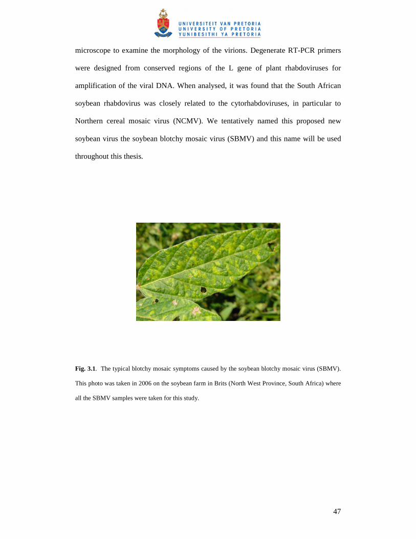
microscope to examine the morphology of the virions. Degenerate RT-PCR primers
were designed from conserved regions of the L gene of plant rhabdoviruses for
amplification of the viral DNA. When analysed, it was found that the South African
soybean rhabdovirus was closely related to the cytorhabdoviruses, in particular to
Northern cereal mosaic virus (NCMV). We tentatively named this proposed new
soybean virus the soybean blotchy mosaic virus (SBMV) and this name will be used
throughout this thesis.
Fig. 3.1. The typical blotchy mosaic symptoms caused by the soybean blotchy mosaic virus (SBMV).
This photo was taken in 2006 on the soybean farm in Brits (North West Province, South Africa) where
all the SBMV samples were taken for this study.

48
3.3 Materials and Methods
3.3.1 Collection of samples:
Soybean leaves believed to be infected by the rhabdovirus, based on symptoms, were
collected during the summer months in the beginning of 2007, at a soybean farm in
Brits, North West Province, South Africa. The infected soybean material was stored at
–70°C until required.
3.3.2 Electron microscopy:
To determine the morphology and virion size, embedded ultra-thin sections of
infected soybean material were observed under a Philips EM301 TEM as described in
section 2.3.2, p. 24. Tobacco mosaic virus (TMV) was used as an external standard to
calibrate magnifications.
3.3.3 Isolation of RNA:
Viral RNA was extracted from infected plant material using SV Total RNA Isolation
System (Promega, Wisconsin) according to the method described by the
manufacturers.
3.3.4 Oligonucleotide primers design:
Refer to section 2.3.4 and Table 2.1 (p.25) for the design of the degenerate primers.
These degenerate primers were expected to be broadly reactive to plant rhabdoviruses.

49
3.3.5 PCR amplification:
For cDNA synthesis, the reaction tube with 8.5 µl RNA and 2 µl RhabF (10 pmol)
were heat treated at 65 °C for 10 min and cooled on ice for 2 min. Then 5x Expand
Reverse Transcriptase Buffer, 100 mmol/L DTT, 10mM dNTP’s, 40 U RNase
Inhibitor and Expand Reverse Transcriptase (Roche, Switzerland) were added and
incubated at 43°C for 1 h. The reaction was inactivated on ice for 2 min. Lettuce
necrotic yellows virus (LNYV) RNA was kindly supplied by R. Dietzgen (University
of Queensland, St. Lucia, Australia) to serve as a positive control in all protocols.
cDNA synthesis of LNYV was also performed as mentioned above. LNYV was used
as a cDNA and PCR control. PCR reactions of 50 µl reaction volumes were prepared.
The reaction mixture consisted of 5 µl of 10x reaction buffer (Bioline, UK), 50 mM
MgCl2, 40 mM dNTP’s, 10 pmol of each primer, and 2.5U Taq polymerase (Bioline,
UK). Finally 5 µl of the cDNA template was added, and the volume made up to 50 ul
with UHQ water. The cycle conditions, on a ABI GeneAmp PCR System 2700 (PE
Applied Biosystems, Connecticut), were as follows: one cycle at 94 °C for 2 min, and
then 35 cycles of 94 °C for 30 s, 37 °C for 30 s, 72 °C for 90 s, followed by one final
cycle at 72 °C for 7 min. Amplicons were analysed on a 1% agarose gel with a
O’GeneRuler 200bp DNA Ladder Plus (Fermentas, Vilnius Lithuania) and viewed
under an UV transilluminator (Vilber Lourmat, France). A Strawberry crinkle virus
(SCV) clone was kindly supplied by M. M. Goodin (University of Kentucky,
Lexington) and was used as a DNA positive control in PCR.
3.3.6 Sequencing of PCR products:
PCR products of ~900bp were purified from the gels using Wizard SV gel and PCR
clean-up system (Promega, Wisconsin). The sequencing reactions were performed

50
using ABI Prism BigDye Primer Cycle Sequencing Kits (PE Applied Biosystems,
Connecticut) according to the manufacturers specifications. Sequencing was with an
automated fluorescent sequencer (ABI 377 DNA Sequencer, PE Applied Biosystems,
Connecticut) at the University of Pretoria’s commercial sequencing facility.
3.3.7 Sequence analysis:
Samples were sequenced in both directions by the forward and reverse primers and
contiguous sequences were created with the ContigExpress option from Vector NTI
Advance 9 (Invitrogen, Carlsbad). The sequences were analysed by using the basic
local alignment research tool (BLAST) program available on the National Centre for
Biotechnology Information website (http://ncbi.nlm.nih.gov/). The SBMV partial L
gene sequence was compared with the polymerase genes of all available
cytorhabdoviruses and nucleorhabdoviruses by doing a multiple alignment with
ClustalX in BioEdit version 7.0.0 (Hall T. A., 1999). The pairwise distance (p-
distance) of cognate regions of SBMV to the plant rhabdoviruses were calculated. A
neighbour-joining phylogenetic tree was constructed using the Mega Version 3.1
(Kumar S. et al., 2004).
51
3.4 Results
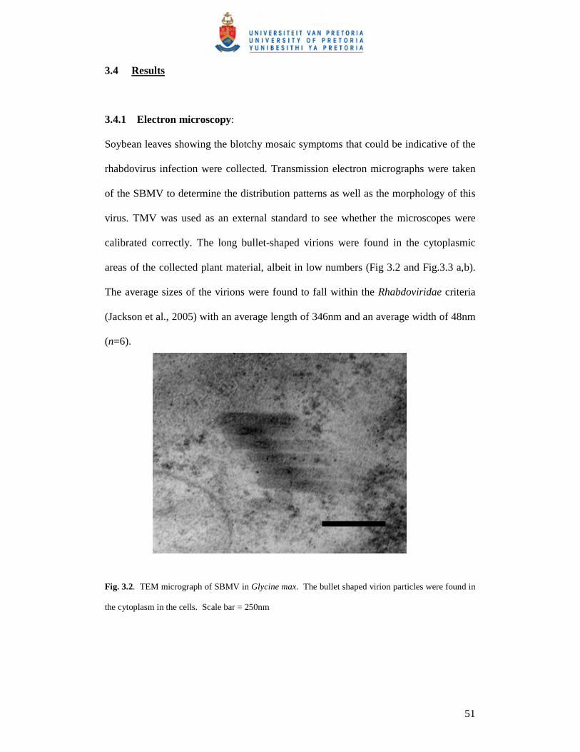
3.4.1 Electron microscopy:
Soybean leaves showing the blotchy mosaic symptoms that could be indicative of the
rhabdovirus infection were collected. Transmission electron micrographs were taken
of the SBMV to determine the distribution patterns as well as the morphology of this
virus. TMV was used as an external standard to see whether the microscopes were
calibrated correctly. The long bullet-shaped virions were found in the cytoplasmic
areas of the collected plant material, albeit in low numbers (Fig 3.2 and Fig.3.3 a,b).
The average sizes of the virions were found to fall within the Rhabdoviridae criteria
(Jackson et al., 2005) with an average length of 346nm and an average width of 48nm
(n=6).
Fig. 3.2. TEM micrograph of SBMV in Glycine max. The bullet shaped virion particles were found in
the cytoplasm in the cells. Scale bar = 250nm

52
(a)
Fig. 3.3. Distribution of SBMV particles within the cytoplasm. The cytoplasmic area is indicated (C).
Scale bar = 250nm. (a) TEM micrograph taken at a magnification of 9.8K, indicating aggregated
SBMV particles. Scale bar = 1µm. (b) The same group of virion particles as in (a) but in a higher
magnification at 75K.
3.4.2 PCR amplification and sequence analysis:
Degenerate primers RhabF and RhabR were designed and RT-PCR carried out on
total RNA extracted from symptomatic soybean leaves. The amplicon obtained with
this primer set was a ~900 bp segment from SCV, LNYV and SBMV (Fig. 3.4).
(b)

53
M 1 2 3 4
1000 bp
M 1 2 3 4
1000 bp
Fig. 3.4. Detection of the putative SBMV by RT-PCR. Samples were loaded onto a 1% agarose gel and
electrophoresed for an hour. The O’GeneRuler 200bp DNA Ladder Plus (M), SCV (1), LNYV (2),
SBMV (3) and water negative control (4) are indicated.
The PCR product was purified from the gel and sequenced from both directions
utilizing RhabF and RhabR. The PCR product was not cloned as the previous chapter
(section 2.3.6, p27), since a good clone could not be produced. A contiguous
sequence was produced and yielded a 522 bp fragment. BLAST analysis confirmed
that the PCR amplicon was of viral RNA (polymerase gene) origin and that it was
most closely related to known Cytorhabdovirus polymerase genes. The SBMV
sequence was submitted to GenBank (acc. number EU877231).

54
Fig. 3.5. The 522 bp sequenced fragment obtained from the SBMV. The sequence was obtained from a
PCR fragment sequenced from both directions using RhabF and RhabR. BLAST analysis indicated
that the SBMV sequence was most related to cytorhabdoviruses.
The 522 bp sequenced fragment of the putative SBMV was aligned with the L gene of
known plant rhabdoviruses that were available on Genbank (see Appendix A, p.68),
including the newly added Cynodon rhabdovirus, acc. number EU650683 (Lamprecht
et al., 2008). From this alignment a 410 bp region of the SBMV was compared to the
same 410 bp cognate region of other plant rhadoviruses for analysis. Pairwise
distances of SBMV to other polymerase genes of different plant rhabdoviruses were
calculated using the cognate region of 410 bp with MEGA 3.1 (Kumar et al., 2004).
The SBMV had the highest homology to NCMV with 60.7 % followed by SCV,
LYMoV and LNYV (58.5%, 56.8% and 59.7% respectively) and then followed by the
nucleorhabdoviruses (Table 3.2).

55
Table 3.1: Homology matrix of nucleotide sequences of the L genes of the Soybean
blotchy mosaic virus (SBMV) and all the other plant rhabdoviruses, including CRV.
Relative homologies to SBMV are in bold print. The homology matrix was calculated
using MEGA 3.1 (Kumar et al., 2004).
Bold print: relative homologies of plant rhabdoviruses to CRV and SBMV.
Bold and underlined: the highest relative homology for CRV and SBMV.
Blue: highest homology relationship, LNYV and LYMoV.
Pink: lowest homology relationship (most divergent sequence), LNYV and SYNV.
From Table 3.1 it can be seen, and as mentioned in previous chapters, that CRV
shared the closest sequence relationship with TaVCV, and SBMV with NCMV. Also
from Table 3.1 it can be deduced that LNYV and LYMoV (blue) shared the closest
homology (73.9 % nucleotide identitiy), whereas SYNV and LNYV (pink) were the
most divergent from all the plant rhabdoviruses, only sharing 46.6 % of their
sequence. SBMV and CRV are also divergent, sharing a homology of 48.8%
nucleotide identity in the 410 bp fragment. This indicates that these two viruses are
LNYV MMV NCMV RYSV SCV SYNV OFV TaVCV MFSV LYMoV SBMV
LNYV
MMV 0.478
NCMV 0.410 0.500
RYSV 0.493 0.437 0.463
SCV 0.312 0.49 0.437 0.490
SYNV 0.534 0.449 0.502 0.463 0.527
OFV 0.512 0.444 0.459 0.454 0.507 0.410
TaVCV 0.468 0.280 0.461 0.429 0.439 0.463 0.461
MFSV 0.483 0.417 0.463 0.473 0.459 0.407 0.415 0.415
LYMoV 0.261 0.495 0.420 0.461 0.300 0.524 0.495 0.485 0.478
SBMV 0.407 0.502 0.393 0.466 0.415 0.505 0.485 0.483 0.483 0.432
CRV 0.493 0.302 0.485 0.437 0.461 0.456 0.483 0.290 0.427 0.488 0.512

56
certainly in two different genera, and this observation is also backed by their different
maturation sites within the plant cells shown in the TEM micrographs.
A phylogenetic tree (Fig. 3.6) was constructed with the same cognate region using
Mega 3.1 (Kumar et al., 2004). The tree separated the selected plant rhabdoviruses
into two distinct clades, correlating with the Cyto- and Nucleorhabdovirus genera.
Within this analysis, the SBMV was most closely related to the cytorhabdoviruses,
especially NCMV and then to SCV. It was interesting to note that Orchid fleck virus
(OFV) clustered with the nucleorhabdoviruses, while it is still recorded as putative
bipartite rhabdovirus according to the International Committee of Taxonomy of
Viruses (ICTV) (ICTVdb, 2006). It is however highly unlikely that OFV will be
classified as a plant rhabdovirus since it has a bipartite genome and the virions are
enveloped (ICTVdb 2006).
Fig. 3.6. A neighbour-joining phylogenetic tree based on the cognate region of 410 bp of several plant
rhabdoviruses, including the newly characterized Cynodon rhabdovirus (CRV, acc. number EU600683)
and Soybean blotchy mosaic virus (SBMV, acc. number EU877231) [Blocked]. The tree formed two
distinct clades, for the two genera Nucleo- and Cytorhabdovirus. The two newly described plant
rhabdoviruses each fell into the respective genera. The abbreviations and accession numbers are listed
in Table 1.3, p. 10
Nucleorhabdovirus
Cytorhabdovirus

57
3.5 Discussion
In this study, we reported the presence and identification of a proposed new
cytorhabdovirus found in South Africa. The putative SBMV particles measured an
approximate length of 350 nm and a width of 87 nm in ultra-thin sections. The virion
particles were found in low numbers in the cytoplasm of infected Glycine max cells.
We also reported the first sequence information of SBMV. The sequenced fragment
was obtained by using degenerate primers that would amplify conserved regions
within the L gene of plant rhabdoviruses. The designed degenerate primers, RhabF
and RhabR, were also able to amplify SCV and LNYV. Sequence analysis indicated
that this SBMV is related to other cytorhabdoviruses, particularly to NCMV.
Therefore, with morphological as well as sequence data, we concluded that the SBMV
is a Cytorhabdovirus from the family Rhabdoviridae. Full or partial genome sequence
data of only 11 (including CRV) other members of plant rhabdoviruses are available
in the public domain. Of all the known plant rhabdoviruses, only SCV has been
reported in South Africa. Now that another cytorhabdovirus has been reported in
South Africa, plant rhabdoviruses should be receiving more surveillance and attention
from virologists and it is likely that more plant rhabdoviruses may be identified in
RSA and the African subcontinent at large.
Planthoppers, aphids, lacebugs and leafhoppers are common insect vectors for plant
rhabdoviruses, and it is very likely that this is the case for SBMV. It is suspected that
SBMV is transmitted by means of leafhoppers. It was investigated which leafhoppers

58
are common to the Brits area. It is suggested that the leafhoppers Austroagallia
cunneata, A. nigristerna and Peragallia carboverdensis are the potential vectors for
SBMV as they are commonly found in this area (Stiller M., personal communication).
However, transmission studies were done with the leafhopper Igerna spp (Order
Homoptera, Family Cicadellidae) and this leafhopper vector successfully transmitted
SBMV (Pietersen, unpublished). The degenerate primers as well as the established
RT-PCR protocol that were used in this study can be utilized to determine the vector
species that are responsible for the horizontal transfer of SBMV. The protocol in this
study could also be utilized for detecting known, as well as new, plant rhabdoviruses
in field studies, broadening our understanding of the epidemiology of these viruses.
3.6 Acknowledgements
We would like to thank R. Dietzgen (University of Queensland) and M. M. Goodin
(University of Kentucky) for supplying us with the LNYV RNA and SCV clone
(respectively). We thank the Virology section of the ARC-Plant Protection Research
Institute (ARC-PPRI) (Roodeplaat, South Africa) for supplying us with TMV. We
also like to thank A. Hall and C. van der Merwe (Department of Microscopy and
Microanalysis, University of Pretoria, Pretoria, South Africa) for their expertise in
microscopic analysis.

59
3.7 References
Bourhy, H., Cowley, J. A., Larrous, F., Holmes, E. C., Walker P. J. (2005).
Phylogenetic relationships among rhabdoviruses inferred using the L polymerase
gene. Journal of General Virology, 86, 2849 – 2858.
Brunt, A. A., Crabtree, K., Dallwitz, M. J., Gibbs, A. J., Watson, L. and Zurcher, E. J.
(1996). Plant Viruses Online: Descriptions and Lists from the VIDE Database.
Version: 16th January 1997. URL http://biology.anu.edu.au/Groups/MES/vide/
Delarue, M., Poch, O., Tordo, N., Moras, D., Argos, P. (1990). An attempt to unify
the structure of polymerases. Protein Engineering, 3, 461 – 467.
Dietzgen, R. G., Callaghan, B., Wetzel, T., Dale, J. L. (2006). Completion of the
genome sequence of Lettuce necrotic yellows virus, type species of the genus
Cytorhabdovirus. Virus Research, 118, 16-22.
Fauquet, C. M., Mayo, M. A., Maniloff, J., Desselberger, U. and Ball, L. A. (Eds)
(2005). Virus Taxonomy: VIIIth Report of the International Committee on Taxonomy
of Viruses. Elsevier Academic Press.
Hall, T. A. (1999). BioEdit: a user-friendly biological sequence alignment editor and
analysis program for Windows 95/98/NT. Nucl. Acids. Symp. Ser, 41, 95-98.

60
Heim, F., Lot, H., Delecolle, B., Bassler, A., Krezal, G. & Wetzel, T. (2008).
Complete nucleotide sequence of a putative new cytorhabdovirus infecting lettuce.
Archives of Virology, 153, 81-92.
Hogenhout, S. A., Redinbaugh, M. G., Ammar, E. D. (2003). Plant and animal
rhabdovirus host range: a bug’s view. Trends in Microbiology, 11, ,264 – 271.
Hogenhout, S. A. (2002). Maize fine streak virus, a new leafhopper-transmitted
Rhabdovirus. Virology, 92, 1167 – 1173.
Hosseini, A., Izadpanah, K. (2005). Biological, serological and physicochemical
properties of Bermuda grass filamentous viruses from Iran. Parasitica, 61, 55 – 59.
Jackson, A. O., Dietzgen, R. G., Goodin, M. M., Bragg, J. N., Deng, M. (2005).
Biology of plant rhabdoviruses. Annual Review of Phytopathology, 43, 623 – 660.
Kitajima, E. W., Kondo, H., Mackenzie, A., Rezende, J. A. M., Gioria, R., Gibbs, A.,
Tamada, T. (2001). Comparative cytopathology and immunocytochemistry of
Japanese, Australian and Brazilian isolates of Orchid fleck virus. Journal of General
Plant Pathology, 67, 231-237.
Kondo, H., Maeda, T., Shikaro, Y., Tamada, T. (2006). Orchid fleck virus is a
rhabdovirus with an unusual bipartite genome. Journal of General Virology, 87, 2413-
2421.

61
Kumar, S., Tamura, K., Nei, M. (2004). MEGA3: Integrated software for Molecular
Evolutionary Genetics Analysis and sequence alignment. Briefings in Bioinformatics,
5, 150-153.
Kuzmin, I. J., Hughes, G. J., Rupprecht, C. E. (2006). Phylogenetic relationships of
seven previously unclassified viruses within the family Rhabdoviridae using partial
nucleoprotein gene sequences. Journal of General Virology, 87, 2323-2331.
Lamprecht, R. L., Pietersen, G., Kasdorf, G. G. F., Nel, L. H. (2008). Characterisation
of a proposed Nucleorhabdovirus new to South Africa. European Journal of Plant
Pathology, DOI 10.1007/s10658-008-9339-5.
Pietersen, G., Staples, S. M., Kasdorf, G. G. F., Jooste, A. E. C (1998). Relative
abundance of soybean viruses in South Africa. African Plant Protection, 4, 65-70.
Poch, O., Blumberg, B. M., Bougueleret, L., Tordo, N. (1990). Sequence comparison
of five polymerases (L proteins) of unsegmented negative-strand RNA viruses:
theoretical assignment of functional domains. Journal of General Virology, 71, 153 –
1162.
Poch, O., Sauvaget, I., Delarue, M., Tordo, N. (1989). Identification of four conserved
motifs among the RNA-dependent polymerase encoding elements. EMBO Journal, 8,
3867 – 3874.

62
Posthuma, K. I., Adams, A. N., Hong, Y., Kirby, M. J. (2002). Detection of
Strawberry crinkle virus in plants and aphids by RT-PCR using conserved L gene
sequences. Plant Pathology, 51, 266-274.
Redinbaugh, M. G., Seifers, D. L., Meulia, T., Abt, J. J., Anderson, R. J., Styer, W.
E., Ackerman, J., Salomon, R., Houghton, W., Creamer, R., Gordon, D. T,
Reed, S. E., Tsai, C. W., Willie, K. J., Redinbaugh, M. G., Hogenhout, S. A. (2005).
Shotgun sequencing of the negative-sense RNA genome of the rhabdovirus Maize
mosaic virus. Journal of Virological Methods, 129, 91 – 96.
Revill, P., Trihn, X., Dale, J., Harding, R. (2005). Taro vein chlorosis virus:
characterization and variability of a new nucleorhabdovirus. Journal of General
Virology, 86, 491-499.
Reynolds, S. (1963). The use of lead citrate at high pH as an electron-opaque stain in
electron microscopy. Journal of Cellular Biology, 17, 208-212.
Tanno, F., Nakatsu, A., Toriyama, S., Kojima, M. (2000). Complete nucleotide
sequence of Northern cereal mosaic virus and its genome organization. Archives of
Virology, 145, 1373-1384.
Van der Merwe, C. F., Coetzee, J. (1992). Quetol 651 for general use: a revised
formulation. Proc Electron Microsc SOC South Africa, 22, 31-32.
www.sppcom.com/about_soy.htm

63
Chapter 4:
Concluding Remarks

64
Two unclassified plant rhabdoviruses were found in South Africa; a putative
nucleorhabdovirus infecting Cynodon dactylon, better known as Bermuda grass, and a
putative cytorhabdovirus infecting soybean, Glycine max (Jackson et al., 2005; VIDE
database, http://biology.anu.edu.au/Groups/MES/vide/). The main aim of this MSc
project was to identify and characterize these two rhabdoviruses on a morphological
and molecular level, using standard electron microsopic (EM) and RT-PCR
techniques.
Degenerate primers were designed for this study to be broadly reactive to most plant
rhabdoviruses, as it was designed from the most conserved part (block III) of the
highly conserved polymerase gene of plant rhabdoviruses (Poch et al., 1990). The
relative binding sites of RhabF and RhabR on other plant rhabdoviruses are indicated
in Fig. 4.1.
Fig. 4.1: The relative positions of where the degenerate primers RhabF and RhabR will bind on to
plant rhabdovirus L gene sequences available on GenBank (Table 4.1)[www.ncbi.nlm.gov]. The
numerals represent the base pair site where the primers would bind.
956 - 1944
1055 - 2037
947 - 1932
1079 - 2064
1097 - 2151
1058 - 2040
LNYV
MMV NCMV RYSV SYNV TaVCV

65
When tested, the primer set RhabF and RhabR was able to amplify four different plant
rhabdoviruses, Strawberry crinkle virus (SCV), Lettuce necrotic yellows virus
(LNYV), the South African Cynodon rhabdovirus (CRV) and Soybean blotchy mosaic
rhabdovirus (SBMV). There is no doubt that the protocol with the degenerate RhabF
and RhabR primers could be used as a tool to further discover or characterize new or
known plant rhabdoviruses, and even used to determine their respective insect vectors.
From this study a CRV clone was also produced and could be used as a positive
control in the future study of potential new plant rhabdoviruses.
Early researchers have classified organisms based on the differences in their
morphology and structural aspects by microscopic observations. When researchers
first started to use molecular information, in particular DNA sequences, they thought
that this system would be superior to morphology, as it had the potential for
determining the phylogeny of all life. However, it was soon realised that there are
advantages and disadvantages to using morphology and DNA data for systematics.
In virology, looking at organisms at a morphological level is still very important as it
can depict the size of the virus, the cytopathology it causes and the distribution
patterns within the cell, among others. EM techniques can still at present be more cost
effective than the molecular methods used, and therefore EM could be used for the
more classical epidemiological and surveillance studies, and for calculation of the
incidence of the disease. In this study, EM observations have revealed that the two
viruses are rhabdoviruses, based on their characteristic bullet-shaped particles, and

66
therefore it was known to which viral family degenerate primers had to be designed
for.
Studying DNA sequences could reflect ancestry and phylogenetic relationships.
Studying an organism at molecular level can give more specific and quantitative data,
depicting the probable evolutionary relationships through phylogenetic trees, rates of
evolution (molecular clock) and percentage divergence, among others. In other words,
molecular data allows for direct and quantitative comparisons. Molecular information
in this study has confirmed that the viruses were indeed related to rhabdoviruses, and
could indicate the evolutionary relationship to other plant rhabdoviruses. If the full
genomic sequences of CRV and SBMV should come available (and other new plant
rhabdoviruses) they could be compared at a much accurate and meaningful level than
comparing a partial sequence of 500-800bp.
At present it is recognized that the best classifications are based on different types of
criteria, including morphological and molecular. The PCR designed in the course of
this study has significantly increased our ability to detect and characterize plant
rhabdoviruses, and will be extremely useful in further studies to establish the
epidemiology and control strategies to these viruses. We have produced two new
sequences for two different plant rhabdoviruses; a partial L gene sequence for the
newly characterized CRV and SBMV. These viruses were described adequately to be
classified into a Rhabdoviridae genus, that the Cynodon rhabdovirus is a new addition
to the Nucleorhabdovirus genus, and SBMV is a new addition to the Cytorhabdovirus
genus.

67
Communications
Lamprecht R., Nel L. H. and Pietersen G. 2007. Characterization of two previously
unknown plant rhabdoviruses from South Africa. Molecular and Cell Biology Group
Symposium. Pretoria, South Africa. 17 October 2007.
Publications
R. L. Lamprecht, G. Pietersen, G. G. F. Kasdorf, L. H. Nel. 2008. Characterisation of
a proposed Nucleorhabdovirus new to South Africa. European Journal of Plant
Pathology (2008). DOI 10.1007/s10658-008-9339-5.

68
Appendix A
Multiple alignment of several cyto- and nucleorhabdoviruses, including the newly
characterized Cynodon rhabdovirus (CRV) and Soybean blotchy mosaic virus
(SBMV). Homologous areas are highlighted. ClustalX in BioEdit version 7.0.0
(Hall, 1999).

69