characterization of the function of a mold specific gene, ms95, in the pathogenic dimorphic
TRANSCRIPT
The University of Southern MississippiThe Aquila Digital Community
Honors Theses Honors College
5-2013
Characterization of the Function of a Mold SpecificGene, MS95, in the Pathogenic Dimorphic Fungus,Histoplasma capsulatumDanielle J. Williamson
Follow this and additional works at: http://aquila.usm.edu/honors_theses
Part of the Life Sciences Commons
This Honors College Thesis is brought to you for free and open access by the Honors College at The Aquila Digital Community. It has been acceptedfor inclusion in Honors Theses by an authorized administrator of The Aquila Digital Community. For more information, please [email protected].
Recommended CitationWilliamson, Danielle J., "Characterization of the Function of a Mold Specific Gene, MS95, in the Pathogenic Dimorphic Fungus,Histoplasma capsulatum" (2013). Honors Theses. Paper 160.
The University of Southern Mississippi
Characterization of the Function of a Mold Specific Gene, MS95, in the Pathogenic
Dimorphic Fungus, Histoplasma capsulatum
By
Danielle Jordan Williamson
A Thesis
Submitted to the Honors College of
The University of Southern Mississippi
In Partial Fulfillment
Of the Requirements for the Degree of
Bachelor of Science
In the Department of Biological Sciences
May 2013

ii

iii
Approved by
Glen Shearer Jr. Department of Biological Sciences, Thesis Director
Glen Shearer Jr. Department of Biological Sciences, Chair
David R. Davies, Dean Honors College

iv
Abstract
Histoplasma capsulatum is a dimorphic fungus that causes the respiratory disease
histoplasmosis. At 25°C, the fungus grows as a multicellular mold in soils contaminated
by bird and bat excreta. Once the soil is disturbed, spores are released and inhaled into
the lungs. The fungus shifts to a unicellular, pathogenic yeast within the lungs at 37°C.
Our laboratory’s main objective is to characterize the genes that are involved in the mold-
to-yeast dimorphism. This study focuses on the mold-specific gene, MS95. According to
The Broad Institute Histoplasma capsulatum GenBank Blastx search, MS95 has several
homologs, including the well-studied Saccharomyces cerevisiae. MS95 belongs to a gene
family of stress proteins known as DDR (DNA Damage Responsive) and is believed to
repair DNA damaged by heat stress. To better determine the function of MS95, this
project explains the creation of a MS95 knockout by allelic replacement. The MS95
knockout was confirmed by a Southern blot. Mold and yeast growth curves of the MS95
knockout did not show any significant changes in the rate of growth when compared to
the Wu27 wild type strain. Comparisons of the yeast and mold morphology in both liquid
and solid media showed no significant difference between the MS95 knockout and the
Wu27 wild type strain. These findings suggest that MS95 is not involved in dimorphism.
Future studies will include observing how MS95 knockout and Wu27 wild type strains
react to different stressors and overexpressing MS95 in the yeast phase growing
temperature to confirm that it is not involved in dimorphism.
v
Table of Contents
Chapter 1: Introduction 1
Chapter 2: Literature Review 3
Chapter 3: Methods 7
Chapter 4: Results 15
Chapter 5: Discussion 22
Primers and Acknowledgments 25
References 26

1
Chapter 1: Introduction
Parasitic fungi, such as Histoplasma capsulatum, have dimorphic capabilities and
can grow as either parasitic-unicellular yeast or saprobic-multicellular mold depending on
environmental conditions. In soil, H. capsulatum exists as a mold at 25°C. If the soil is
disturbed, mold cells release spores that can be inhaled into the lungs of passing humans
or other mammals. Within the lungs, H. capsulatum converts to yeast at 37°C. This
morphological transition enables the dimorphic, pathogenic fungi to colonize host tissues
including humans (Maresca et al., 1989).
H. capsulatum causes histoplasmosis, a systemic fungal disease. Histoplasmosis is
the most common respiratory mycotic infection among humans and other mammals. It is
prevalent in temperate, subtropical, and tropical zones such as the Mississippi and Ohio
Valleys in the United States and in similar areas in South and Central America, the
Mediterranean, Asia, Australia, and Africa. Histoplasmosis has a wide range of
symptoms and is an opportunistic fungus in immune deficient hosts. The disease is
extensive in areas where patients suffer from acquired immune deficiency syndrome and
is a growing concern in nonendemic areas due to the increasing motility of the world’s
population (Ignatov et al., 2002).
Few studies have been performed on the identification and characterization of the
genes and the gene products involved in the morphological transition of H. capsulatum.
These genes are vital for the early establishment and maintenance of the parasitic yeast
phase that invades host cells and causes histoplasmosis. One such gene under study is
MS95. It is a mold specific and belongs to a class of genes known as DNA damage
responsive (DDR) genes (Maga et al., 1986). DDR genes express increased transcription

2
in response to DNA lesions or to heat-shock stress (Treger et al., 1990). Scarce studies
have been preformed on MS95 in H. capsulatum, but many studies have been conducted
on gene DDR48 in Saccharomyces cerevisiae, a homolog of H. capsulatum. DDR48 has
also been studied in another homolog, Candida albicans, which is a dimorphic
pathogenic fungus that causes mucosal and systemic infections. Dr. Dib et al. (2008)
created a knockout of a DDR48 allele via homologous recombination. His research
concluded that DDR48 is essential for hyphal filament formation in the mold form, stress
responses, and viability of C. albicans, thus making it a prime target for antifungal drug
design. Being a homolog of H. capsulatum, DDR48 is similar in structure and
evolutionary origin to MS95 despite being from a different species. DDR genes may have
a critical function in H. capsulatum because dimorphic fungi shift between yeast and
mold forms in response to environmental changes such as temperature (Maresca et al.,
1989).
To better understand the function of MS95, this project focuses on characterizing
MS95 in the pathogenic, dimorphic fungus H. capsulatum. This goal will be achieved
through the creation of a MS95 knockout via allelic replacement. Knocking out the gene
will disrupt its normal function. The knockout will be confirmed through a Polymerase
Chain Reaction procedure (PCR) and a Southern Blot. The phenotypic appearance and
cellular response of H. capsulatum wild type and knockout will be recorded when grown
as mold at 25°C and as yeast at 37°C. Also, the wild type and knockout will be grown
under stressful conditions to determine if MS95 is involved in DNA repair. If MS95 is
responsible for initiating the shift between mold and yeast, then the cells should remain
as mold and not transform into yeast when the gene is knocked out and nonfunctional.
3
Ideally this project’s findings will be used to further expand the understanding of
the morphological transition mechanism in H. capsulatum and other dimorphic fungi and
perhaps lead to a prevention or a better treatment procedure for histoplasmosis.
Chapter 2: Literature Review
2.a Histoplasma capsulatum: Current Research
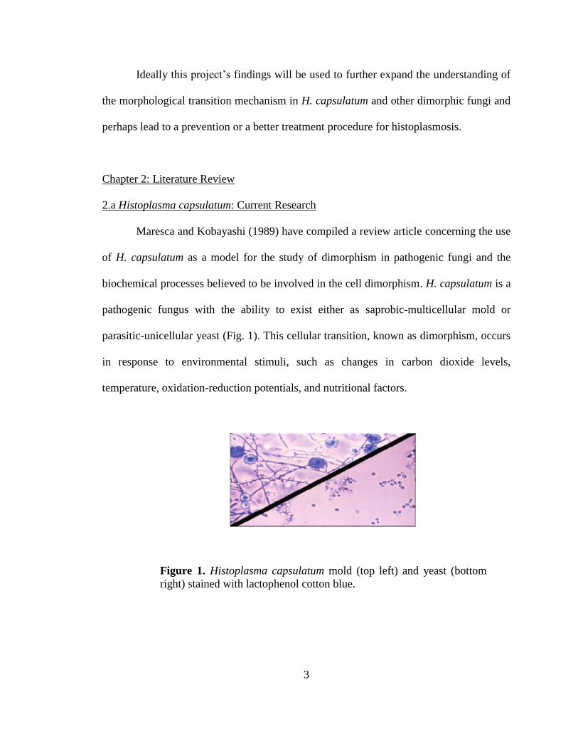
Maresca and Kobayashi (1989) have compiled a review article concerning the use
of H. capsulatum as a model for the study of dimorphism in pathogenic fungi and the
biochemical processes believed to be involved in the cell dimorphism. H. capsulatum is a
pathogenic fungus with the ability to exist either as saprobic-multicellular mold or
parasitic-unicellular yeast (Fig. 1). This cellular transition, known as dimorphism, occurs
in response to environmental stimuli, such as changes in carbon dioxide levels,
temperature, oxidation-reduction potentials, and nutritional factors.
Figure 1. Histoplasma capsulatum mold (top left) and yeast (bottom
right) stained with lactophenol cotton blue.
4
The cellular dimorphism characteristic of H. capsulatum is not essential to the cell’s life
cycle, is reversible, and can grant the cells the ability to infect host tissues (Maresca et al.,
1989).
H. capsulatum is the causative agent of histoplasmosis, a systemic fungal disease.
It has been diagnosed worldwide and is the most common respiratory mycotic infection
found in humans and animals. In the United States alone, about 500,000 infections are
diagnosed each year (Nosanchuk et al., 2008). Histoplasmosis has high prevalence in
temperate, subtropical and tropical climate, including the Mississippi and Ohio Valleys,
portions of South and Central America, the Mediterranean area, Asia, Australia, and
Africa (Ignatov et al., 2002). It is a highly opportunistic pathogen that seriously affects
humans with acquired immune deficiency syndrome (Weidenheim et al. 1992). In a study
by Brilhante et al. (2012), of patient cases reviewed, 38.9% had histoplasmosis as the first
indicator of AIDS. Histoplasmosis is becoming a greater concern due to the increasing
mobility of the world’s populations.
H. capsulatum yeast cells are ovals with 1 to 3 μm diameters, grow at 37°C, and
reproduce by budding (Maresca et al., 1989 (31)). The mold cells measure about 1.2 to
1.5 μm in diameter, grow at 25°C, and reproduce by macroconidia spores and
microconidia spores. It is believed that the microconidia are the favored infectious form
of H. capsulatum due to their small size (Maresca et. al 1989 (23, 57, 32, 63)). Yeast cells
are the only form of H. capsulatum discovered in infected host tissues, but little is known
about the transition from the microconidia mold spores to the yeast cells at 37°C.
Researchers have currently tried to understand the chromosomal characteristics of
H. capsulatum. They isolated twenty-three strains of H. capsulatum from infected human
5
tissue using restriction fragment patterns of mitochondrial DNA and ribosomal DNA.
Common strains studied in humans are G184AS, G186AS, G217B, and Downs (Maresca
et al., 1989 (167)). Researchers have also identified seven chromosome-sized bands all
exceeding one megabase (one million base pairs), except for a five hundred thousand
base pair band in one of the strains. Each band also has a unique pattern depending on the
strain (Maresca et al., 1989).
Currently, it is known that H. capsulatum yeast cells transition into mold at 22°C
to 25°C. This transition does not occur at the same time in all cells, but does begin with
the formation of a budlike structure containing large quantities of storage material. After
twenty-four to forty-eight hours, the transition is complete. (Maresca et al., 1989 (38, 44,
73)). When transitioning back into yeast, mold cells require more time. This transition is
believed to occur through an “enlarged cell” described as an oidial yeast cell. Pine and
Webster studied the transition of mold to yeast cells in H. capsulatum and discovered that
contiguous mold cells enlarge to form chains of yeastlike cells or undergo budding of
unswollen mold cells to form yeast cells (Maresca et. al 1989 (130)).
Environmental requirements that are needed to complete a successful transition
have also been studied. Salvin has shown that nutritional factors such as cysteine levels,
which contain thiol groups (sulfur-hydrogen groups), may affect the transition from mold
to yeast. He found that the addition of cysteine was necessary for the maintenance of the
yeast phase because it is required to complete the mold to yeast transition and acts as a
nutrient for the yeast cells (Maresca et al., 1989 (148)). It is clear from other research that
thiol containing compounds must be present in the culture medium to initiate the mold to
yeast transition (Maresca et al., 1989 (154)). Research conducted by Zarnowski et al.

6
(2007) demonstrates how H. capsulatum can degrade collagen and digest albumin and
casein. By degrading large proteins, H. capsulatum can acquire the nutrients, such as
thiol groups from proteins, essential for fungal growth and metabolism. Once other
conditions such as vitamins along with thiol groups are satisfied, the transition is directly
mediated by temperature changes (Maresca et al., 1989 (125, 129, 147, 154)). Transition
occurs at about 30°C, with the optimal temperature reaching 37°C. H. capsulatum can
grow as mold regardless of the incubation temperature; however, the cells require 37°C to
transform to and persist as yeast. This leads researchers to believe that mold-specific
genes express at any temperature, while yeast-specific genes can only be expressed at
higher temperatures and in the presence of thiol groups (Maresca et al., 1989). Therefore,
in order to understand the phase transition of dimorphic fungi, it is necessary to
characterize the genes and their functions in mold and yeast cells within H. capsulatum.
2.b Gene MS95: Overview
To begin to understand the role certain genes play in the transition from mold to
yeast cells in H. capsulatum, the genes must be characterized and identified as either
mold-specific or yeast-specific. Due to the lack of research on H. capsulatum, some
information pertaining to the function of certain genes must be taken from research
conducted on homologous organisms including Saccharomyces cerevisiae and Candida
glabrata.
Temperature changes within the environment are the driving force of the
dimorphic transition of H. capsulatum cells; therefore, genes that respond to heat-shock
are at the forefront of research. A class of genes known as DNA damage response genes
(DDR) respond to heat-shock and show an increased transcription in response to heat-

7
shock stress or to treatments that produce DNA lesions (Treger et al., 1990). Within H.
capsulatum, the mold-specific gene MS95 belongs to the class of DDR genes. Expression
of certain mold-specific genes is required for mold formation and maintenance, and lack
of expression allows the cells the transition into yeast (Tian et al. 2001). Therefore, it is
speculated that MS95 may be involved in initiating the transition from mold to yeast cells
in a human host. A Broad Institute gene search concluded that MS95 consists of 1499
nucleotides, codes for a protein consisting of 315 amino acids, and occurs once within the
genome. However, there is scarce research published on MS95 in H. capsulatum, so
current information must be taken from research done on the DDR48 gene of S.
cerevisiae and C. glabrata.
Chapter 3: Methods
This project focuses on expanding the knowledge of the mechanisms involved in
the transition between cellular forms of dimorphic fungus, specifically H. capsulatum.
Specifically, this project will try to determine the function of the gene MS95 by creating a
knockout.
3.a Overview of Common Procedures
Polymerase Chain Reaction (PCR) is a biochemical procedure used to generate
thousands to millions of copies of a specific DNA sequence from a few fragments of
DNA. This procedure involves three phases that are typically repeated thirty times in a
thermal cycler. Before the first phase, there is an optional step used to heat the lid of the
thermal cycler to 95°C for 60 seconds. The first phase is denaturing, or separation of the
two DNA strands. Denaturing occurs around 95°C for 10 seconds and causes the
8
hydrogen bonds between the DNA strands to break. Once the two strands of DNA
separate, the annealing phase begins. Annealing occurs around 50°C to 60°C for 30
seconds and allows the primers to adhere to the now single strand DNA. Forward and
reverse primers are designed and ordered to bind at the beginning and end respectively of
the DNA sequence that is being amplified. Due to the uniqueness of the DNA strand, the
annealing temperature depends on the primers. The final phase is elongation of the DNA
sequence. This phase requires the heat-tolerant enzyme Taq Polymerase to bind to the
primers and begin replicating the DNA strands. Taq Polymerase has an optimal
temperature around 72°C and can polymerize about a thousand base pairs per minute.
Therefore, the extension time for this cycle depends on the length of the DNA sequence
that is being replicated. Each sample to be amplified is prepared with 18μL of water,
2.5μL of Taq Polymerase buffer, 0.5μL of Taq polymerase, 0.5μL of 0.2mM dNTPs,
1.3μL of the 0.2μM forward primer, 1.3μL of the 0.2μM reverse primer, and 2–3μL of
the sample DNA. The buffer and polymerase are prepackaged in the advantage
polymerase kit from Clontech®. To ensure that the correct DNA sequence is amplified,
the DNA fragments are separated via gel electrophoresis and compared to standard
measurements.
Gel electrophoresis separates fragments of DNA based on their size. The
fragments of DNA are loaded into a 0.7% agarose gel in 1X Tris-acetate-EDTA (1X
TAE) buffer solution with pH 7.6 (48.8g/L Tris base, 11.4mL/L glacial acetic acid, 20
mL/L 0.5 M Na EDTA), and an electrical current is applied to the gel. 1X TAE is made
by mixing 50mL of 20X TAE and 950mL of water. The electric field causes the
negatively charged DNA fragments to move through the porous gel toward the positive

9
leads. The smaller fragments move faster and farther than larger ones, thus separating the
fragments based on size. DNA ladders with standard fragment sizes are also run with the
DNA samples in order to provide a “ruler” to measure the unknown fragments. The 0.7%
agarose gels range from small, medium, and large sizes. Small agarose gels contain 0.21g
of agarose in 30mL of 1X TAE. Medium gels contain 0.35g of agarose in 50mL of 1X
TAE. Large gels contain 0.42g of agarose in 60mL of 1X TAE. The solution is heated
for about one minute before adding 0.5μL of 1mg/mL ethidium bromide and being
poured into the gel mold to solidify. The ethidium bromide binds to the DNA and causes
the fragments to fluoresce under UV light. Loading dye is also added to the DNA
samples so that they are easier to see when being loaded into the gel wells.
In order for yeast cells to uptake the DNA amplified via PCR from the
environment, the cells must undergo electroporation. Electroporation increases the
permeability of a cell’s plasma membrane via an external electrical field. This process
allows the H. capsulatum yeast cells to uptake the DNA construct that will replace and
knockout the MS95 gene. To begin, 5mL of cells is placed in 15mL tubes and spun down
in a centrifuge at -25°C and 200 RCF for 2 minutes. After the supernatant is poured out,
the pellet is resuspended in 5mL of 10% mannitol. The solution is pulse vortexed to
resuspend the cells. The cells are centrifuged again at -25°C and 200 RCF for 2 minutes,
and all of the supernatant is removed with a sterile pipette. Next, 200μL of fresh 10%
mannitol is added to the tubes, and a sterile pipette is used to resuspend the cells. Once
suspended, the cells and DNA are added to a cuvette. One cuvette has cells without DNA
and is labeled as a blank. The cuvettes are placed into a BIO RAD GenePulser Xcell™,
with the following settings: V = 750; μF = 25; Ω = 600; and mm = 2. When ready, the

10
cells are pulsed with an electrical force to increase the permeability of the plasma
membrane. Once electroporated, the cells are removed from the cuvettes and plated onto
Histoplasma-macrophage media (HMM) ura (-) plates. Using plates that lack uracil will
ensure that only the cells that took up the DNA fragments will grow.
H. capsulatum cells are grown on solid or in liquid HMM. HMM is prepared by
mixing 20mL of Type 1 water and 25mL of 2X HMM stock [100ml of 2X stock is 2.14g
of F-12 HAMS powder (Sigma Chemical Co.), 3.64g of glucose (purchased from
American Type Culture Collection in Manassas, Virginia), 0.2g glutamic acid, 2mL
cysteine stock, 1.2g HEPES, pH 7.5, and filter sterilized]. The broth was supplemented
with 50μg/ml of ampicillin and 100μg/ml of streptomycin, to prevent contamination. The
solution can be added to 1.5% w/v agarose to make plates or kept in its liquid state at 4°C
for broth. Two different types of plates can be made: ones with uracil [ura (+)] and ones
without uracil [ura (-)]. Ura (+) plates are made by autoclaving 3.75g of agarose in
217mL of water. When cooled, 250mL of HMM, 33mL of 15x uracil (100μg/mL), and
500μL of amp/strep antibiotic are added to the agarose solution, and the solution is
poured into plates to solidify. Ura (-) plates are made by autoclaving 3.75g of agarose in
250mL of water. When cooled, 250mL of HMM and 500μL of amp/strep antibiotic is
added, and the solution is poured into plates to solidify. Cells that may contain a
knockout are grown on 5-FOA plates. 5-FOA plates contain 10.7g F-12 Nutrient Mix,
18.2g glucose, 1.0g glutamic acid, 0.084g cysteine, 7.5g sodium citrate, 3g 5-FOA, and
0.1g uracil. The solution is heated to 55°C, cooled, (pH) to 7.5, and filter sterilized.
Before being mixed with agarose, 2.5mL of hygromycin (150μg/mL) and 2.0mL of
amp/strep antibiotic are added to the solution.

11
Although cells may grow on the 5-FOA plates, two procedures are used to ensure
that the H. capsulatum cells underwent allelic replacement and contain a knockout: PCR
and Southern Blot. The knockout DNA sequence is larger than the wild type DNA and
can be separated and identified using PCR. However, a more inclusive procedure is a
Southern Blot. The Southern Blot involves extracting DNA from the potential knockout
colonies, cutting the DNA with specific restriction enzymes, subjecting the cut DNA to
gel electrophoresis, and then transferring the DNA bands to a Nylon N+ membrane
(Millipore). The blot is probed with P32
labeled fragment containing the MS95 open
reading frame, stripped, and re-probed with the hygromycin (Hyg) antibiotic resistant
marker. Images are taken using a Typhoon 9400 phosphoimager. For a knockout
confirmation, DNA bands must appear with the Hyg marker probe, but not with the MS95
open reading frame probe. This confirms that the Hyg antibiotic resistant marker replaced
the MS95 open reading frame in the DNA sequences, thus knocking out the gene. The
wild type DNA should have a band with the MS95 open reading frame probe, but not the
Hyg antibiotic resistant marker probe because this DNA has not been knocked out.
3.b Characterization of MS95
The characterization of MS95 will focus on determining the specific function of
the gene. Through unpublished research, MS95 has already been classified as a single
copy gene and mold-specific. This project will focus on determining if the cells can still
transition from mold into yeast if the gene is made inactive, which is known as a
knockout.
The MS95 knockout was made via fusion PCR in which a large portion of the
open reading frame was replaced with a Hyg antibiotic resistant marker and ligated into
12
the telomeric vector pRPU1 to create pDWU1. The vector has a Ura5 marker that is used
for selection after electroporation. The vector was cut with the restriction enzyme PmeI to
expose the telomeric repeats. The exposed telomeres allow the vector to mimic a
chromosome, thereby enhancing the chances of a successful electroporation into H.
capsulatum cells. This new DNA combination was electroporated into a MS95 expressing
strain, 186AS Wu27 ura –, which did not contain a Ura5 marker. The cells containing the
plasmid that harbored the Ura5 marker were streaked onto HMM ura – plates and grown
for seven days. Colonies were chosen and inoculated to 1mL of HMM and grown for
seven days under stressful conditions at 37°C to induce allelic replacement (Fig 2).
Figure 2. Genomic Replacement Map of MS95 knockout. The 677 base pair
left flanking region and the 775 base pair right flanking region were fused
with the Hyg marker via fusion PCR. During allelic replacement, the Hyg
marker replaced part of the MS95 open reading frame, which created a
knockout.
13
Stressful growing conditions encouraged recombination to occur, which causes
the cells to undergo genetic recombination and to take up the Hyg antibiotic resistant
marker into their genetic code. This process replaces a portion of the MS95 open reading
frame with the Hyg antibiotic resistant marker. After the seven days, the cells were
streaked onto 5-FOA plates that were supplemented with the antibiotic hygromycin and
grown for ten to fourteen days. This process allowed for selection against the Ura5
marker and for the Hyg antibiotic resistant marker. Of the colonies that grew, twenty-five
were chosen at random and had their DNA extracted. The DNA was analyzed with PCR
and Southern Blot to scan for a knockout.
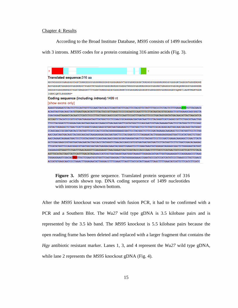
The MS95 knockout was confirmed with PCR and Southern Blot procedures. The
PCR denaturing phase occurred at 98°C for 30 seconds, and the annealing temperature
was set to 60°C for 60 seconds. The extension time was set for 6 minutes at 72°C because
the knockout DNA fragment is 5.5 kilobases, while the wild type DNA fragment is 3.5
kilobases. The primers for the PCR were designed to amplify 500 bp up and down stream
on the chromosome that is not present in the vector. For the Southern Blot, MS95
knockout and Wu27 wild type strain genomic DNA was digested with restriction
enzymes EcoRI and EcoRV. EcoRI and EcorV are non-cutters and do not cut within the
MS95 open reading frame. The blot was probed with P32
labeled fragment containing the
MS95 open reading frame, stripped, and re-probed with the Hyg antibiotic resistant
marker.
Once conformed, the mold and yeast MS95 knockout cells were compared to the
Wu27 wild type cells on various characterization studies including: phenotypic

14
comparisons, mold and yeast growth curves, and a mold and yeast stressor test with
methyl methanesulfonate (MMS).
The physical appearance of the yeast and mold cells of both MS95 and Wu27 wild
type strains were compared in liquid and solid HMM. Both strains were grown as yeast at
37°C in 50mL of HMM broth for three days. Each day, Pictures of the cells were taken
with a 150 confocal microscope (Zeiss) at 63X. MS95 knockout and Wu27 wild type
strains were also grown as mold at 25°C on HMM plates for twelve days. Pictures were
taken with an EOS 20D Canon camera for seven days.
The growth rates of MS95 knockout and Wu27 wild type strains were compared in
yeast and mold growth curves. For the yeast growth curve, MS95 knockout and Wu27
wild type cells were grown as yeast at 37°C in HMM liquid broth. The klett of each strain
was measured and recorded in triplicates every six hours for a total of thirty hours using a
Klett Colorimeter (Scienceware). For the mold growth curve, MS95 knockout and Wu27
wild type cells were grown as mold at 25°C on HMM plates. The hyphal extensions of
each sample were viewed and measured using a 10X ocular micrometer on a C2M4
Labomed microscope once a day for ten days.
To determine if the MS95 functioned in DNA repair, MS95 knockout and Wu27
wild type strains were grown on ura (+) plates on top of filters as mold at 25°C. After one
week, the filters were transferred to plates containing different concentrations of MMS:
0.005%, 0.1%, and 0.3%. The cells were grown as mold at 25°C for 12 days. Pictures
were taken with the AM4113T Dino-Lite Pro USB Digital Microscope on days 0, 4, 7,
10, and 12.
15
Chapter 4: Results
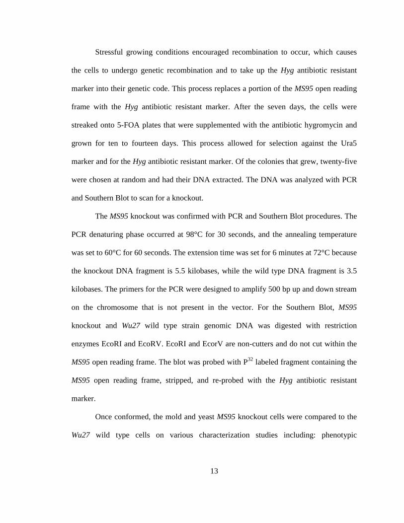
According to the Broad Institute Database, MS95 consists of 1499 nucleotides
with 3 introns. MS95 codes for a protein containing 316 amino acids (Fig. 3).
After the MS95 knockout was created with fusion PCR, it had to be confirmed with a
PCR and a Southern Blot. The Wu27 wild type gDNA is 3.5 kilobase pairs and is
represented by the 3.5 kb band. The MS95 knockout is 5.5 kilobase pairs because the
open reading frame has been deleted and replaced with a larger fragment that contains the
Hgy antibiotic resistant marker. Lanes 1, 3, and 4 represent the Wu27 wild type gDNA,
while lane 2 represents the MS95 knockout gDNA (Fig. 4).
Figure 3. MS95 gene sequence. Translated protein sequence of 316
amino acids shown top. DNA coding sequence of 1499 nucleotides
with introns in grey shown bottom.

16
The MS95 knockout was also confirmed with a Southern Blot. After the gDNA was
digested with restriction enzymes EcoRI (A) and EcoRV (B), the blot was probed with
P32
labeled fragment containing the MS95 open reading frame, stripped, and re-probed
with the Hyg antibiotic resistant marker. The results show that the MS95 open reading
frame is present in the Wu27 wild type strain, but not the MS95 knockout. They also
show that the Hyg antibiotic resistant marker is present in the MS95 knockout, but not in
Figure 4. PCR confirmation of MS95 knockout. The
knockout is represented by the 5.5 kb band in lane 2. The
Wu27 wild type is represented by the 3.5 kb band in lanes
1, 3, and 4.
1 2 3 4
17
the Wu27 wild type strain. These results were expected because the open reading frame in
the MS95 knockout was replaced with the Hyg antibiotic resistant marker, but still
remains intact in the Wu27 wild type strain (Fig. 5).
With the MS95 knockout created and cultured regularly in liquid HMM at 37°C,
different phenotypic, growth, and stressor tests were used to determine if MS95 is
involved in mold to yeast dimorphism. The first phenotypic comparison was between
yeast and mold morphological forms of MS95 knockout and Wu27 wild type strains
grown on solid HMM (Fig. 6).
Figure 5. Southern Blot confirmation of MS95 knockout. MS95
knockout and Wu27 wild type strain genomic DNA was
digested with restriction enzymes: EcoRI (A) and EcoRV (B).
The blot was probed with P32
labeled fragment containing the
MS95 ORF, stripped, and re-probed with the Hyg marker.

18
The second phenotypic comparison was between yeast and mold forms of MS95
knockout and Wu27 wild type strains grown in liquid HMM (Fig. 7). Both phenotypic
tests were used to see if MS95 functioned in controlling the phenotypical expression of
cells and regulating cellular dimorphism between mold and yeast forms.
The next analytical tests were mold and yeast growth curves. The growth curves
determined if MS95 functioned in regulating cellular growth under normal conditions. All
of the growth curves were performed in triplicates and averaged together. For the yeast
growth curve, yeast cells were grown in liquid HMM at 37°C and were measured with a
Klett Colorimeter every six hours for a total of thirty hours (Fig 8).
Figure 6. Comparison of Yeast and Mold phenotypes of MS95
knockout and Wu27 wild type strains on solid HMM. Pictures
taken on day 1 (left) and 6 (right).

19
Figure 8. Growth Curve Comparisons of MS95 knockout (KO)
and Wu27 wild type (WT) strains grown as yeast at 37°C in
liquid HMM.
0.000
50.000
100.000
150.000
200.000
250.000
300.000
350.000
400.000
450.000
500.000
0 1 2 3 4 5
Kle
t
Hours
Avg of WT
Avg of KO
Figure 7. Comparison of Yeast and Mold phenotypes of MS95
knockout and Wu27 wild type strains in liquid HMM.

20
For the mold growth curve, mold cells were grown on solid HMM plates at 25°C, and the
hyphal extension were measured with a 10X ocular micrometer on a C2M4 Labomed
microscope once a day for ten days and averaged together (Fig. 9).
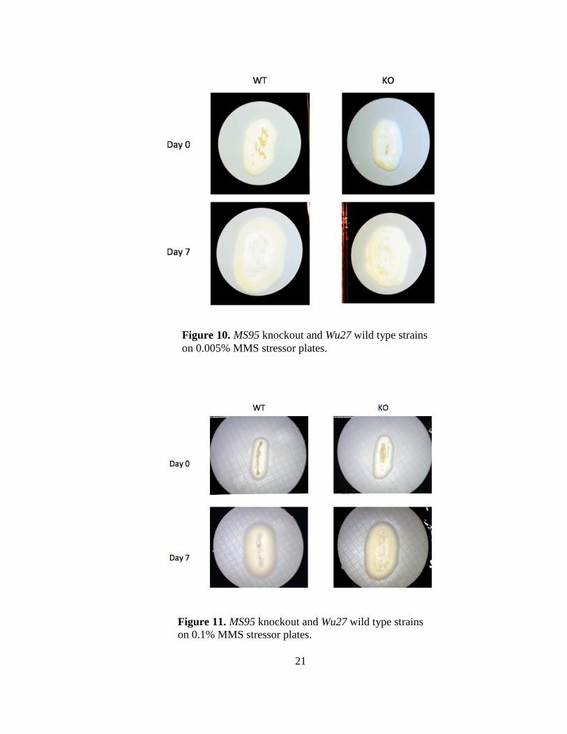
The final test was a stress response test to determine if MS95 functions in DNA repair.
MS95 knockout and Wu27 wild type strains were grown on solid HMM plates for twelve
days with three different concentrations of MMS: 0.005%, 0.1%, and 0.3%. (Figs. 10, 11,
12 respectively). Pictures were taken with the AM4113T Dino-Lite Pro USB Digital
Microscope on days 0, 4, 7, 10, and 12.
0.0000.5001.0001.5002.0002.5003.0003.5004.0004.5005.0005.5006.0006.5007.0007.500
0 1 2 3 4 5 6 7 8 9 10 11
Hy
ph
al
Le
ng
th i
n m
m
Days
WT
KO
Figure 9. Growth Curve Comparisons of MS95 knockout (KO) and
Wu27 wild type (WT) strains grown as mold at 25°C on solid
HMM
21
Figure 10. MS95 knockout and Wu27 wild type strains
on 0.005% MMS stressor plates.
Figure 11. MS95 knockout and Wu27 wild type strains
on 0.1% MMS stressor plates.
22
Chapter 5: Discussion
Histoplasma capsulatum, the dimorphic fungus, causes the respiratory disease
histoplasmosis. This dimorphic fungus grows as mold at 25°C in soil contaminated by
bird or bat excreta. If the soil is disturbed, mold spores can enter animal lungs and shift to
the pathogenic yeast at 37°C. Dramatic changes in gene regulation must occur for the
shift to be successful, which is evident in the numerous genes that are only expressed in
either the yeast or mold forms.
In order to determine which genes are involved in the dimorphic shift, differently
expressed genes are chosen and characterized. MS95 is a mold specific gene that belongs
to a gene family of stress proteins known as DDR (DNA Damage Responsive) and is
believed to repair DNA damaged by heat stress. To determine MS95 function, a knockout
Figure 12. MS95 knockout and Wu27 wild type strains on
0.3% MMS stressor plates.

23
was created via fusion PCR and tested under different conditions. The MS95 knockout
was confirmed with a PCR and Southern Blot. The PCR shows the knockout gene at 5.5
kb. This is 2 kb larger than the Wu27 wild type because the MS95 knockout contains the
Hgy antibiotic resistant marker (Fig. 4). The Southern Blot further confirms the knockout
by showing that the Hgy antibiotic resistant marker is present in the MS95 knockout
strain, but not in the Wu27 wild type strain. The blot also shows that the MS95 open
reading frame is present in the Wu27 wild type strain, but has been replaced in the
knockout strain.
With the MS95 knockout confirmed, two phenotypic tests were performed to
determine if MS95 plays a role in determining the phenotypic appearance of either mold
or yeast cells. MS95 knockout and Wu27 wild type strains were grown as yeast at 37°C in
liquid HMM (Fig. 6) and as mold at 25°C on solid HMM plates (Fig. 7). The phenotypic
comparisons of the yeast and mold cells do not indicate any significant differences in
morphology. The MS95 knockout is similar to the Wu27 wild type in size, shape, texture,
and color in both yeast and mold forms. These findings suggest that MS95 does not play a
significant role in determining phenotype. Next, two growth curves of MS95 knockout
and Wu27 wild type strains grown as yeast in liquid HMM at 37°C (Fig. 8) and as mold
on solid HMM at 25°C (Fig. 9) were compared. The klett of the yeast was measured over
thirty-six hours and averaged, while the mold hyphal extensions were measured once a
day for ten days and averaged. The two graphs show similar growth rates for both MS95
knockout and Wu27 wild type strains in both mold and yeast forms. These results suggest
that MS95 does not play a significant role in maintaining normal growth rates. Also,
hyphal extensions appeared at the same rate for both the MS95 knockout and Wu27 wild

24
types strains, suggesting that MS95 does not directly control the dimorphic shift from
yeast to mold. Finally, to determine if MS95 functions in DNA repair, MS95 knockout
and Wu27 wild type strains were grown on stressor plates containing 0.005%, 0.1%, and
0.3% concentrations of MMS for twelve days (Fig. 10, 11, 12 respectively). From the
results, it is evident that the MS95 knockout strains were not affected by the MMS due to
their similarity in color, size, and concentration of hyphal extensions to the Wu27 wild
type strains. It may be possible that the concentrations of MMS were too low and that
another experiment will need to be conducted. Nevertheless, these preliminarily results
suggest that MS95 does not play a significant role in DNA repair because the knockout
cells were able to compensate for the gene loss, remain healthy, shift from yeast to mold,
and prosper as well as the Wu27 wild type cells.
All of these results suggest that MS95 is not directly involved in the dimorphic
shift form mold to yeast. If it did play a direct role, then it would be expected that the
knockout cells could not shift from mold to yeast during the temperate change or that
some physical, structural, or growth difference would be evident between the MS95
knockout and Wu27 wild type strains. Future plans for MS95 will be forcing
overexpression of MS95 in the yeast phase growing temperature in order to confirm that
it is not involved in dimorphism.

25
Primers
Special note: there are 87 bp left of the open reading frame from the knockout that
have not been deleted. The open reading frame starts at base pair 530-1762. The MS95
knockout primers for the PCR confirmation are as follows:
MS95 forward chromosome
GGGACTTAACAAAGTGAGCGCCAATCTTTCC
MS95 reverse chromosome
CCCTATTTATATGTTTTTTCCTTGTCTTCTTTCTATTTTC
These primers are designed to amplify 500 bp up and down stream on the chromosome
that is not present in the vector.
The primers used for the Southern Blot confirmation are as follows:
MS95 open reading frame forward
CAATCTCATCTCCCTTGCTGGCCAGCCCATTCCAGTTC
MS95 open reading frame reversed
GAATAATTAGTATGCAACTTGTAGAAATGAATGGATAGTC
Acknowledgements
I would like to thank my Principle Investigator, Dr. Glen Shearer, for allowing me
to work in his lab. I would also like to thank Dr. Davida Crossley for teaching me proper
laboratory technique and for critiquing my manuscript.
This work was supported by the Mississippi INBRE and funded by grants from
the National Center for Research Resources (5P20RR016476-11) and the National

26
Institute of General Medical Sciences (8 P20 GM103476-11) from the National Institutes
of Health.
References
1. Brilhante, R., Fechine, M., Mesquita, J., Cordeiro, R., Rocha, M., Monteiro, A.,
Lima, R., Caetano, É., Pereira, J., Castelo-Branco, D., Camargo, Z., & Sidrim, J.
(2012). Histoplasmosis in HIV-positive patients in Ceará, Brazil: clinical-laboratory
aspects and in vitro antifungal susceptibility of Histoplasma capsulatum isolates.
Transactions of the Royal Society of Tropical Medicine and Hygiene 106(8), 484-488.
Retrieved from http://www.sciencedirect.com/science/article/pii/S0035920312000983
2. Broad Institute. 2010-2012. Retrieved from http://www.broadinstitute.org/
3. Dib, L., Hayek, P., Sadek, H., Beyrouthy, B., & Khalaf RA. (2008). The Candida
albicans Ddr48 protein is essential for filamentation, stress response, and confers
partial antifungal drug resistance. Med Sci Monit 14(6), 113-121. Retrieved from
http://www.ncbi.nlm.nih.gov/pubmed/18509269
4. Ignatov, A., & Keath, E. J. (2002). Molecular cell biology and molecular genetics of
Histoplasma capsulatum. International Journal of Medical Microbiology, 292(5-6),
349-361. Retrieved from
http://www.sciencedirect.com/science/article/pii/S1438422104701125
5. Maga, Janet A., Terrill A. McClanahan, & Kevin McEntee. (1986).
"Transcriptional Regulation of DNA Damage Responsive (DDR) Genes in Different
Rad Mutant Strains of Saccharomyces Cerevisiae." MGG Molecular & General
Genetics 205.2, 276-84. Print.
6. Maresca, B., & Kobayashi, G. S. (1989). Dimorphism in Histoplasma capsulatum -
A Model for the Study of Cell Differentiation in Pathogenic Fungi. Microbiological
Reviews, 53(2), 186-209. Retrieved from
http://mmbr.asm.org/content/53/2/186.full.pdf+html
7. Nosanchuk, J. & Gacser, A. (2008) Histoplasma capsulatum at the host-pathogen
interface. Microbes and Infection, 10(9), 973-977. Retrieved from
http://www.sciencedirect.com/science/article/pii/S1286457908001883
8. Tian, X. & Shearer, G. (2001). Cloning and analysis of mold-specific genes in the
dimorphic fungus Histoplasma capsulatum. Gene 271(1), 107-114. Retrieved from
http://www.sciencedirect.com/science/article/pii/S0378111901006461
9. Treger, Janet M., & Kevin McEntee. (1990). "Structure of the DNA Damage-
Inducible Gene DDR48 and Evidence for Its Role in Mutagenesis in Saccharomyces
Cerevisiae.” Molecular and Cellular Biology 10.6, 3174-184. Retrieved from
http://www.ncbi.nlm.nih.gov/pmc/articles/PMC360682/pdf/molcellb00042-0744.pdf
27
10. Weidenheim, K., Nelson, S., Kure, K., Harries, C., Biempica, L., & Dickson, D. (1992). Unusual patterns of Histoplasma capsulatum meningitis and progressive
multifocal leukoencephalopathy in a patient with the acquired immunodeficiency
virus. Human Pathology 23(5), 581-586. Retrieved from
http://www.sciencedirect.com/science/article/pii/004681779290137R
11. Zarnowski, R., Connolly, P., Wheat L., & Woods, J. (2007). Production of
extracellular proteolytic activity by Histoplasma capsulatum grown in Histoplasma-
macrophage medium is limited to restriction fragment length polymorphism class 1
isolates. Diagnostic Microbiology and Infectious Disease 59(1), 39-47. Retrieved
from http://www.sciencedirect.com/science/article/pii/S0732889307001587