characterization of radopholus similis resistance in musa … · doctoraatsproefschrift nr. 998 aan...
TRANSCRIPT
Doctoraatsproefschrift nr. 998 aan de faculteit Bio-ingenieurswetenschappen van de K.U.Leuven
CHARACTERIZATION OF RADOPHOLUS SIMILIS RESISTANCE IN MUSA SPP.
WITH EMPHASIS ON PHYTOCHEMICAL ANALYSIS
Suganthagunthalam DHAKSHINAMOORTHY
Supervisor:
Prof. D. De Waele, K.U.Leuven
Co-supervisor: Prof. A. Elsen, BDB & UGent
Members of the Examination Committee: Prof. E. Decuypere, Chairman, K.U.Leuven
Prof. A. Aertsen, K.U.Leuven
Prof. J. Coosemans, K.U.Leuven
Prof. R. Swennen, K.U.Leuven Prof. G. Gheysen, UGent
Dr. D. Hölscher, MPI-CE, Germany
Dissertation presented in partial fulfilment of the
requirements for the degree
of Doctor in Bioscience
Engineering
November 2011

© 2011 Katholieke Universiteit Leuven, Groep Wetenschap & Technologie, Arenberg
Doctoraatsschool, W. de Croylaan 6, 3001 Heverlee, België
Alle rechten voorbehouden. Niets uit deze uitgave mag worden vermenigvuldigd en/of
openbaar gemaakt worden door middel van druk, fotokopie, microfilm, elektronisch of
op welke andere wijze ook zonder voorafgaandelijke schriftelijke toestemming van de
uitgever.
All rights reserved. No part of the publication may be reproduced in any form by print, photoprint, microfilm, electronic or any other means without written permission from
the publisher.
Cover illustration: Top: Light microscopic image of Musa genotypes Long Tavoy (left)
and Yangambi km5 (right) roots showing the accumulation of phenolic phytoalexins
due to R. similis infection.
Bottom: Light microscopic image of a moulting Radopholus similis juvenile that had
killed by the uptake of phenylphenalenone type-pheytoalexin anigorufone during a
laboratory bio-assay.
ISBN 978-90-8826-216-6 Legal deposit number D/2011/11.109/49
I
ACKNOWLEDGEMENTS
My deep sense of gratitude is extended to my Promotor Prof. Dirk De
Waele for his trust in my capabilities to conduct and finish the doctoral research
as well as for critically editing this manuscript. Dirk, I deeply appreciate your
invaluable inputs and time on improving the first drafts of this thesis.
My sincere thanks to my co-promotor Prof. Annemie Elsen, who guided
me starting from the proposal of this research project with critical comments,
continuous supervision and valuable thoughts and discussions.
Financial support by the Interfaculty Council for Development Co-
operation (IRO), K.U.Leuven as a PhD fellowship and a grant from the COST
action 872 for a short term scientific mission to the Max-Planck-Institute for
Chemical Ecology are gratefully acknowledged.
I extend my sincere appreciation to the members of the examination
committee for the invaluable time and contribution to improve this text.
A special word of gratitude is extended to Prof. Rony Swennen for the
introduction and the interest on the phenylphenalenones part of this research as
well as for sharing his extensive knowledge on bananas and plantains.
My deep sense of gratitude is extended to Dr. Dirk Hölscher for
introducing me to the world of phytochemicals and metabolite profiling. My
special appreciation to Dr. Hölscher for the numerous hours of brain storming
to start, build and complete the phenylphenalenones project and for his
unconditional support remotely as well as during my stay at Max-Planck-
Institute of Chemical Ecology (MPI-CE), Jena, Germany.
My appreciation is extended to the collaborative partners at the NMR and
MS research groups of MPI-CE as well as at the Laboratory of Organic
Chemistry and Macromolecular Chemistry of the Friedrich Schiller University
in Jena, Germany. Special word of gratitude is extended to Mrs. Katrin Knop
and Ravikumar Madulla for the productive collaboration and technical support.

II Acknowledgements
Thanks to the thesis students Erwin Galon, Mariama Salifu and Els Heylen
for their dedication on their thesis. It was a challenging but also very enriching
experience to guide you all. I enjoyed working with all my colleagues of the
Laboratory of Tropical crop Improvement, K.U.Leuven. Kind assistance of
Marleen and Suzy is highly appreciated.
The Nematology group members of the LabTrop: Christine, Lieselot, Lut,
Preeti and Wim gain special thanks for the technical, moral support and
friendship. Special thanks to Christine for the warm friendship, introducing me
to many aspects of Belgian life and for the scientific discussions and for the
translation of this thesis summary to Dutch. Thanks to the sandwitch PhD
students, Tuyet, Nguyet, Thuy, Maung, Nordalyn and Pa Pa for your kind
friendship and knowledge sharing. Word of thanks is extended to the past PhD
student, Annelies Vertommen for her warm friendship and for answering all my
queries about the PhD academic procedures.
My friends in Leuven (TFL) made my leisure time full of fun, laughter and
happiness. The time that we spent together during weekends charged me to
work harder over the week.
Moral support from my parents, sister and grandmother are invaluable.
Without your support, listening ears, constant encouragements and motivation,
this work would not have been possible.
My hearty thanks and love to my husband Sudhakar for the great support,
patience and encouragements in the last phase of the PhD. Your support and
love made my writing phase more relaxed and enjoyable. Love and thanks to
the little addition to come in our family for the excellent co-operation in the last
months of my PhD.
Thank you all
நன்றி
Sugantha
November 2011, Leuven

III
TABLE OF CONTENTS
ACKNOWLEDGEMENTS............................................................................. I
TABLE OF CONTENTS ............................................................................. III
LIST OF TABLES ....................................................................................... IX
LIST OF FIGURES ..................................................................................... XI
LIST OF ABBREVIATIONS ................................................................... XVI
SUMMARY.............................................................................................XVIII
SAMENVATTING ................................................................................. XXIII
CHAPTER1GENERAL INTRODUCTION ................................................. 1
1.1. OBJECTIVES OF THE STUDY AND OUTLINE OF THE THESIS............................. 2
1.2. MUSA SPP. - SOCIAL, CULTURAL AND ECONOMIC IMPORTANCE .................. 5
1.3. PLANT-PARASITIC NEMATODES .................................................................... 6 1.3.1. BANANA NEMATODES .............................................................................. 6
1.3.1.1. The burrowing nematode Radopholus similis ......................................... 7 1.3.1.2. Radopholus similis host range and economical importance .................... 7 1.3.1.3. Radopholus similis as a major root pathogen of bananas ....................... 7 1.3.1.4. Control and management of Radopholus similis on bananas .................. 8
1.4. HOST PLANT RESISTANCE IN MUSA SPP. TO RADOPHOLUS SIMILIS .............. 9 1.4.1. HOST-NEMATODE INTERACTIONS ........................................................... 10
1.4.1.1. Localisation of the host plant by the nematode ................................... 10 1.4.1.2. Penetration and entry of the nematode in the host plant .................... 11 1.4.1.3. Nematode development and reproduction in the host......................... 12
1.4.2. POSSIBLE MECHANISMS OF PLANT RESISTANCE TO NEMATODES ............ 12 1.4.2.1. Preformed resistance mechanisms ...................................................... 12 1.4.2.2. Induced resistance mechanisms .......................................................... 13
IV 1.5. PLANT SECONDARY METABOLITES INVOLVED IN HOST PLANT RESISTANCE 15
1.5.1. PHYTOANTICIPINS ...................................................................................16 1.5.2. PHYTOALEXINS ........................................................................................16 1.5.3. PHENYLPHENALENONES ..........................................................................17
1.5.3.1. Origin and natural occurrence ..............................................................17 1.5.3.2. Biosynthesis of phenylphenalenones ....................................................19 1.5.3.3. Biosynthesis of phenylphenalenones in Musaceae................................20 1.5.3.4. Phenylphenalenones as phytoalexins and phytoanticipins ....................21 1.5.3.5. Phenylphenalenones as antibiotic compounds .....................................23 1.5.3.6. Phenylphenalenones as phytoalexins in plant-nematode interactions...24
1.5.4. PHENYLPROPANOIDS...............................................................................24 1.5.4.1. Phenylpropanoids and nematode resistance in Musa spp. ....................25
CHAPTER 2: IDENTIFICATION OF COMBINED RESISTANCE TO RADOPHOLUS SIMILIS AND MELOIDOGYNE INCOGNITA IN MUSA GERMPLASM ............................................................................................ 27
2.1. INTRODUCTION ...........................................................................................28
2.2. MATERIALS AND METHODS .........................................................................29 2.2.1. EXPERIMENTAL SET-UP ............................................................................29 2.2.2. PLANTING MATERIAL ...............................................................................29 2.2.3. NEMATODE INOCULUM AND INOCULATION ............................................31 2.2.4. EVALUATION OF THE HOST PLANT RESPONSE ..........................................32
Radopholus similis ............................................................................................32 Meloidogyne incognita .....................................................................................33
2.2.5. STATISTICAL DATA ANALYSIS ...................................................................34
2.3. RESULTS ......................................................................................................34 2.3.1. HOST RESPONSE TO R. SIMILIS .................................................................34 2.3.2. HOST RESPONSE TO M. INCOGNITA .........................................................35
2.4. DISCUSSION.................................................................................................36
2.5. CONCLUSION ...............................................................................................40
CHAPTER 3: DEVELOPMENT OF AN AUTOTROPHIC IN VITRO MODEL SYSTEM TO STUDY RADOPHOLUS SIMILIS HOST LOCATION AND PENETRATION .......................................................... 41
3.1. INTRODUCTION ...........................................................................................42
3.2. MATERIALS AND METHODS .........................................................................43 3.2.1. PLANTING MATERIAL ...............................................................................43

V
3.2.2. NEMATODE INOCULUM .......................................................................... 43 3.2.3. THE AUTOTROPHIC IN VITRO SYSTEM ..................................................... 43 3.2.4. EXPERIMENTAL SET-UP ........................................................................... 45
3.2.4.1. First experiment: attraction ................................................................. 46 3.2.4.2. Second experiment: penetration ......................................................... 46 3.2.4.3. Third experiment: attraction and penetration ...................................... 46 3.2.4.4. Fourth experiment: attraction and penetration in a two-compartment system ............................................................................................................. 47
3.2.5. STATISTICAL DATA ANALYSIS................................................................... 48
3.3. RESULTS ..................................................................................................... 48 3.3.1. FIRST (OPTIMISATION) EXPERIMENT: ATTRACTION ................................. 48 3.3.2. SECOND (OPTIMISATION EXPERIMENT): PENETRATION........................... 50 3.3.3. THIRD EXPERIMENT: ATTRACTION AND PENETRATION ............................ 52 3.3.4. FOURTH EXPERIMENT: NEMATODE ATTRACTION IN A TWO-COMPARTMENT SYSTEM ...................................................................................... 55
3.4. DISCUSSION ................................................................................................ 57
3.5. CONCLUSIONS ............................................................................................ 59
CHAPTER 4: HOST PLANT RESISTANCE IN MUSA GERMPLASM TO RADOPHOLUS SIMILIS: PRE- OR POST-INFECTIONAL? ................. 61
4.1. INTRODUCTION .......................................................................................... 62
4.2. MATERIALS AND METHODS ........................................................................ 63 4.2.1. EXPERIMENTAL SET-UP ........................................................................... 63 4.2.2. PLANTING MATERIAL .............................................................................. 63 4.2.3. NEMATODE INOCULUM .......................................................................... 63 4.2.4. FIRST EXPERIMENT: NEMATODE ATTRACTION AND PENETRATION .......... 64 4.2.5. SECOND EXPERIMENT: POST-INFECTIONAL NEMATODE DEVELOPMENT AND REPRODUCTION ........................................................................................... 64 4.2.6. STATISTICAL DATA ANALYSIS................................................................... 65
4.3. RESULTS ..................................................................................................... 65 4.3.1. FIRST EXPERIMENT: NEMATODE ATTRACTION AND PENETRATION .......... 65 4.3.2. SECOND EXPERIMENT: NEMATODE DEVELOPMENT AND REPRODUCTION.........................................................................................................69
4.4. DISCUSSION ................................................................................................ 70
4.5. CONCLUSION .............................................................................................. 72
VI
CHAPTER 5: LIGNIN AND PHENOLS INVOLVED IN THE INTERACTIONS BETWEEN RADOPHOLUS SIMILIS AND MUSA SPP. ..................................................................................................................... 73
5.1. INTRODUCTION ...........................................................................................74
5.2. MATERIALS AND METHODS .........................................................................75 5.2.1. PLANTING MATERIAL ...............................................................................75 5.2.2. NEMATODE INOCULUM...........................................................................75 5.2.3. EXPERIMENTAL SET-UP ............................................................................75 5.2.4. SAMPLING AND ASSESSMENT OF NEMATODE INFECTION ........................75 5.2.5. HISTOCHEMICAL STAINING OF ROOT CROSS SECTIONS ............................76 5.2.6. EXTRACTION AND QUANTIFICATION OF LIGNIN .......................................77 5.2.7. FOLIN-CIOCALTEU ASSAY FOR TOTAL PHENOLICS .....................................78 5.2.8. STATISTICAL ANALYSIS .............................................................................78
5.3. RESULTS ......................................................................................................79 5.3.1. ROOT AND SHOOT WEIGHT .....................................................................79 5.3.2. ROOT ANATOMY AND CELLULAR DAMAGE IN INFECTED ROOTS...............80 5.3.3. NEMATODE NUMBERS IN NECROTIC LESIONS ..........................................82 5.3.4. HISTOCHEMICAL STAINING OF MUSA ROOT CROSS SECTIONS FOR LIGNIFIED CELL WALLS ..........................................................................................83 5.3.5. LIGNIN CONTENT OF ROOT CELL WALLS...................................................87 5.3.6. HISTOCHEMICAL STAINING OF MUSA ROOT SECTIONS FOR TOTAL PHENOLS ..............................................................................................................89 5.3.7. PHENOLIC CONTENT OF MUSA ROOTS .....................................................89 5.3.8. LOCALISATION OF FLAVONOIDS ...............................................................90
5.4. DISCUSSION.................................................................................................93
5.5. CONCLUSION ...............................................................................................96
CHAPTER 6: PHENYLPHENALENONE-TYPE PHYTOALEXINS INVOLVED IN THE PLANT RESISTANCE TO PARASITIC NEMATODES ............................................................................................ 97
RATIONALE AND OUTLINE .........................................................................................98
CHAPTER 6.1: CELL-SPECIFIC LOCALISATION AND PHYTOCHEMICAL PROFILING OF PHENYLPHENALENONE-TYPE PHYTOALEXINS IN RADOPHOLUS SIMILIS-RESISTANT AND -SUSCEPTIBLE MUSA ROOTS ........................................................................... 100
6.1.1. INTRODUCTION................................................................................ 100 6.1.2. MATERIALS AND METHODS ................................................................... 101
6.1.2.1. Plants ................................................................................................. 101 6.1.2.2. Nematodes ........................................................................................ 101
VII
6.1.2.3. Experimental set-up .......................................................................... 102 6.1.2.4. Sampling ........................................................................................... 102 6.1.2.5. Extraction and analysis of phenylphanalenones ................................. 104 6.1.2.6. Thin layer chromatography (TLC) analysis .......................................... 104 6.1.2.7. Preparative HPLC (pHPLC) ................................................................. 104 6.1.2.8. Analytical HPLC (aHPLC)..................................................................... 105 6.1.2.9.
1H NMR spectroscopy ........................................................................ 105
6.1.2.10. UPLC-MS analysis ............................................................................ 105 6.1.2.11. Fixation of plant material for LDI-MSI .............................................. 106 6.1.2.12. LDI-MSI on the ultraflex III
® mass spectrometer ................................ 106
6.1.3. RESULTS ............................................................................................... 107 6.1.3.1. Root damage and root lesions ........................................................... 107 6.1.3.2. Sub-fraction weights ......................................................................... 107 6.1.3.3. Thin layer chromatography (TLC) ....................................................... 109 6.1.3.4. Purification of phenylphenalenones by HPLC ..................................... 109 6.1.3.5. Structural identification ..................................................................... 112 6.1.3.6. LDI-MSI ............................................................................................. 115
6.1.4. DISCUSSION .......................................................................................... 117 6.1.5. CONCLUSION ........................................................................................ 120
CHAPTER 6.2: ANTI-NEMATODE PROPERTIES OF THE PHENYLPHENALENONES . 122 6.2.1. INTRODUCTION .................................................................................... 122 6.2.2. MATERIALS AND METHODS .................................................................. 123
6.2.2.1. Experimental set-up .......................................................................... 123 6.2.2.2. Chemicals .......................................................................................... 123 6.2.2.3. Nematodes ....................................................................................... 123 6.2.2.4. Effect of phenylphenalenones on R. similis motility bio-assay ............ 123 6.2.2.5. Dosage effect of anigorufone (1) on R. similis motility bio-assay ........ 124 6.2.2.6. Statistical data analysis ...................................................................... 124
6.2.3. RESULTS ............................................................................................... 125 6.2.3.1. Effect of phenylphenalenones on R. similis motility bio-assay ............ 125 6.2.3.2. Dosage effect of anigorufone (1) on R. similis motility bio-assay ........ 130
6.2.4. DISCUSSION .......................................................................................... 133 6.2.5. CONCLUSIONS ...................................................................................... 136
CHAPTER 7:GENERAL CONCLUSIONS AND PERSPECTIVES....... 137
REFERENCES .......................................................................................... 142
ANNEXES ................................................................................................. 157

VIII

IX
LIST OF TABLES
Table 2.1. Characteristics of selected Musa genotypes and the reference cultivars evaluated for their host response to Radopholus similis and
Meloidogyne incognita..........................................................................30
Table 2.2. Identification of the host response of selected Musa genotypes to Radopholus similis based on a comparison with the host response of a
susceptible (Grande Naine) and a resistant (Yangambi km5) reference
cultivar...................................................................................................33
Table 2.3. Host response of selected Musa genotypes and the reference cultivars
to Radopholus similis, measured at 8 weeks after inoculation with 1,000 vermiform nematodes per plant (n = 8).................................................37
Table 2.4. Host response of selected Musa genotypes and the susceptible
reference cultivar Grande Naine to Meloidogyne incognita measured at 8 weeks after inoculation with 4,000 eggs and second-stage juveniles
per plant (n = 8).....................................................................................38
Table 3.1. Nematode attraction zones in the two-compartment autotrophic in
vitro model system.................................................................................48
Table 3.2. Fresh root and shoot weights of 4-weeks-old plants of the Musa
genotypes Saba and Yangambi km5 at 2 and 4 days after inoculation
(DAI) with 30 mature females of Radopholus similis...........................50
Table 3.3. Fresh root and shoot weights of 4-weeks-old plants of the Musa
genotypes Grande Naine and Yangambi km5 at 1 and 2 days after
inoculation (DAI) with 30 mature females of Radopholus similis........53
Table 4.1. Fresh root and shoot weights of the Musa genotypes at 4, 8 and 12
days after inoculation (DAI) with 1,000 adults and juveniles of Radopholus similis. The nematodes were inoculated around the roots of
six-weeks-old plants. (n=8)....................................................................66
Table 4.2. Fresh root and shoot weights of the Musa genotypes at 26 days after
inoculation (DAI) with 1,000 adults and juveniles of Radopholus
similis. The nematodes were inoculated around the roots of six-weeks-
old plants. (n=8).....................................................................................69
Table 4.3. Number of Radopholus similis eggs, juveniles, females, males, final
population density and reproduction factor (Rf) in the roots of the resistant (Long Tavoy, Saba, Yangambi km5) and the susceptible

X
(Grande Naine) Musa genotypes at 26 days after inoculation with 1,000
adults and juveniles of Radopholus similis. (n=8).................................70
Table 5.1. Mean fresh root and shoot weights of Radopholus similis-infected
and uninfected Musa genotypes at 3 and 6 weeks after inoculation with
1,000 adults and juveniles of R. similis.................................................79
Table 5.2. Mean number of Radopholus similis in the necrotic lesions of three
resistant (Saba, Long Tavoy & Yangambi km5) and one susceptible
(Grande Naine) Musa genotypes at 6 weeks after inoculation with 1,000 adults and juveniles of R. similis (n=4)..................................................83
Table 6.1. Weights of the sub-fractions isolated from the Musa root extracts.................................................................................................109
Table 6.2. The occurrence of identified phenylphenalenones in the resistant and
susceptible Musa cultivars based on HPLC and 1H NMR analyses....112
Table 6.3. Effect of the phenylphenalenone-type phytoalexins on the motility of
Radopholus similis...............................................................................125
Table 6.4. Percentage quiescent Radopholus similis caused by the
phenylphenalenones at two concentrations (conc.) after 24, 48 and 72 h incubation (n=3)...................................................................................127
Table 6.5. Percentage quiescent Radopholus similis caused by anigorufone (1) at six different concentrations over three days (n=6)..........................132
Table 6.6. The motility inhibitive concentrations (IC50) of anigorufone (1) on
Radopholus similis after 24, 48 and 72 h of incubation.......................133

XI
LIST OF FIGURES
Figure 1.1: Flow chart representing the outline of our study................................4
Figure 1.2: A) Food served on a banana leaf in southern India. B) A small shop
in a local market showing a high diversity of bananas for sale...............5
Figure 1.3: Host plant/pathogen relationship terminology used in nematology.............................................................................................10
Figure 1.4: Possible mechanisms of plant resistance to nematodes...................13
Figure 1.5: Host-nematode interaction and active plant responses....................14
Figure 1.6: Structure of A) phenalen, B) phenalen-1-one and C) 9-
phenylphenalen-1-one............................................................................18
Figure 1.7: Structure of A) fungal phenalenone-atrovenetin and B) plant
phenylphenalenone-haemocorin, first isolated from Penicillium herquei
and Haemodorum corymbosum, respectively…………………....……18
Figure 1.8: Biosynthetic pathway of 9-phenylphenalenone, anigorufone..........19
Figure 1.9: Structure of A) 9-phenylphenalenones commonly present in Haemodoraceae, Musaceae and other plants, B) 4-phenylphenalenones
which are more common in Musaceae and C) Ring-1: Naphthalic
anhydride type........................................................................................20
Figure 1.10: Inflorescence of A) Heamodorum sp., B) Anigozanthus flavidus, C)
Dilatris pillansii, and blood red coloured roots of D) Lachnanthes sp.
and E) Wachendorfia thyrsiflora…………………………………...…22
Figure 1.11: The core phenylpropanoid biosynthetic pathway...........................25
Figure 1.12: Phenylpropanoids and their accumulation in different plant parts in
response to abiotic and biotic stresses...................................................26
Figure 2.1: Experimental set-up of the Radopholus similis host response
experiment in the greenhouse................................................................31
Figure 2.2: Root necrosis caused by Radopholus similis at 8 weeks after
inoculation with 1,000 nematodes.........................................................35
Figure 2.3: Egg masses of Meloidogyne incognita in Musa plants stained with
phloxin B................................................................................................35

XII
Figure 3.1: Banana plants cultured in an autotrophic in vitro model system.....44
Figure 3.2: Radopholus similis inoculation spots (arrows) to study nematode attraction and penetration in an autotrophic in vitro model system.......44
Figure 3.3: Two-compartment autotrophic in vitro model system to study
nematode attraction to banana plants.....................................................47
Figure 3.4: Two-compartment autotrophic in vitro model system to study
nematode attraction to banana plants.....................................................47
Fig. 3.5: Migration of Radopholus similis females towards the roots of A)
Yangambi km5 and B) Saba at 1 h after inoculation.............................49
Figure 3.6: Attraction of Radopholus similis females (expressed as a percentage
of 30 inoculated mature females) towards the roots of 4-weeks-old plants of the Musa genotypes Yangambi km5 and Saba at 1, 2, 4 and 24
h after inoculation..................................................................................49
Figure 3.7: A) Penetration of Radopholus similis females (expressed as a percentage of 30 inoculated mature females) in the roots of 4-weeks-old
plants of the Musa genotypes Yangambi km5 and Saba at 2 and 4 days
after inoculation. B) Number of eggs laid by the penetrated R. similis females at 4 days after inoculation.........................................................51
Figure 3.8: Penetration and egg laying of Radopholus similis females in banana roots grown in an autotrophic in vitro system. Roots of Saba...............52
Figure 3.9: Attraction of Radopholus similis females (expressed as a percentage
of 30 inoculated mature females) towards the roots of 4-weeks-old plants of the susceptible Musa genotype Grande Naine and the resistant
Musa genotype Yangambi km5 at 1, 3, 4 and 6 h after inoculation......53
Figure 3.10: Penetration of Radopholus similis females (expressed as a
percentage of 30 inoculated mature females) in the roots of 4-weeks-old
plants of the susceptible Musa genotype Grande Naine and the resistant Musa genotype Yangambi km5 at 1 and 2 days after inoculation.........54
Figure 3.11: Migration tracks of Radopholus similis females near banana roots in an autotrophic in vitro system observed at 24 h after inoculation.....55
Figure 3.12: Percentages of Radopholus similis females present in the attraction
zones of the two-compartment autotrophic in vitro system at 30 min, 1, 2, 3 and 6 h after inoculation of 30 mature females at the inoculation
spot (I)....................................................................................................56
XIII
Figure 4.1: Root damage caused by Radopholus similis....................................67
Figure 4.2: Females of Radopholus similis penetrating the roots of A) Grande Naine and B) Long Tavoy at 4 days after inoculation and eggs laid by
the penetrated females at 12 days after inoculation...............................67
Figure 4.3: Number of Radopholus similis that had penetrated the roots of the resistant (Long Tavoy, Saba, Yangambi km5) and the susceptible
(Grande Naine) Musa genotypes at 4, 8 and 12 days after inoculation
with 1,000 adults and juveniles of Radopholus similis..........................68
Figure 4.4: Number of Radopholus similis eggs observed in the roots of the
resistant (Long Tavoy, Saba, Yangambi km5) and the susceptible (Grande Naine) Musa genotypes at 12 days after inoculation with 1,000
adults and juveniles of Radopholus similis............................................68
Figure 4.5: Number of adult and juvenile Radopholus similis in the roots of resistant (Long Tavoy, Saba, Yangambi km5) and susceptible (Grande
Naine) Musa genotypes at 26 days after inoculation with 1,000 adults
and juveniles of Radopholus similis......................................................70
Figure 5.1: Anatomical root structures of Musa spp. cv. Saba (cross
sections).................................................................................................81
Figure 5.2: Necrotic root cross sections of Radopholus similis-resistant and
susceptible Musa genotypes at 6 weeks after inoculation with R. similis.....................................................................................................82
Figure 5.3: Lignifications in uninfected Musa root cells at 3 and 6 weeks........84
Figure 5.4: Lignifications in Radopholus similis-infected Musa root cells at 3
and 6 weeks after inoculation with 1,000 adults and juveniles of R.
similis.....................................................................................................85
Figure 5.5: Tissue localisation of lignin in the root cross sections of Radopholus
similis-resistant Musa genotype Yangambi km5 at 6 weeks after inoculation with 1,000 adults and juveniles of R. similis......................86
Figure 5.6: Tissue localisation of syringyl units of lignin in the root cross sections of Radopholus similis-resistant Musa genotype Long Tavoy at
6 weeks after inoculation with 1000 adults and juveniles of R.
similis.....................................................................................................86
Figure 5.7: Lignin content (mg/g of fresh root weight) in root cell walls of
Radopholus similis-infected and uninfected plants of resistant and
susceptible Musa genotypes at 3 and 6 weeks after inoculation with 1,000 adults and juveniles of R. similis.................................................88

XIV
Figure 5.8: Tissue localisation of total phenols by toluidine blue staining in root
cross sections of Musa genotypes at 6 weeks after inoculation with
1,000 adults and juveniles of Radopholus similis..................................89
Figure 5.9: Total phenol contents (mg TAE/g roots) in the Radopholus similis-
infected and uninfected roots of resistant and susceptible Musa
genotypes at 3 and 6 weeks after inoculation with 1,000 adults and juveniles of R. similis.............................................................................91
Figure 5.10: Fluorescence of phenolic compounds in root cross sections of Radopholus similis-infected susceptible and resistant Musa genotypes at
3 and 6 weeks after inoculation (WAI)..................................................92
Figure 6.1: Musa plants arranged in a randomized block design in the
greenhouse to study the phytoalexins synthesis in response to
Radopholus similis infection................................................................102
Figure 6.2: Scheme followed for the isolation, cellular distribution and
structural identification of phenylphenalenones..................................103
Figure 6.3: The root system of Musa cultivar Grande Naine. A) Uninfected
healthy roots. B) Radopholus similis-infected roots at 12 weeks after
infection...............................................................................................108
Figure 6.4: Root necrosis caused by Radopholus similis infection in susceptible
(Grande Naine) and resistant (Yangambi km5) Musa cultivars.........108
Figure 6.5: TLC chromatogram of chloroform subfractions............................109
Figure 6.6: Structure of all the isolated phenylphenalenone-type phytoalexins.........................................................................................110
Figure 6.7: Analytical HPLC chromatograms of chloroform sub-fractions separated from the ethanol extracts of Musa spp.............................…111
Figure 6.8: Identification of anigorufone (1) based on the comparison of 1H
NMR spectrum of authentic compounds.............................................113
Figure 6.9: The 1H NMR spectrum of anigorufone (1)....................................113
Figure 6.10: The LDI mass spectra of Musa roots...........................................115
Figure 6.11: Mass images of the necrotic lesions on Yangambi km5 caused by
Radopholus similis infection................................................................116
XV
Figure 6.12: The heat profile of LDI-MSI for the m/z 271 in the necrotic lesions
of Yangambi km5 roots showing the distribution of anigorufone only in
the necrotic lesion................................................................................116
Figure 6.13: Mass images of the necrotic lesions on Grande Naine caused by
Radopholus similis infection................................................................117
Figure 6.14: Light microscopic images of Radopholus similis during the bio-
assay with anigorufone (1)...................................................................128
Figure 6.15: Light microscopic images of all life stages of Radopholus similis
that had died because of ingesting anigorufone (1) during the bio-
assays...................................................................................................129
Figure 6.16: Light microscopic images of Radopholus similis from the negative
control, 1% ethanol..............................................................................130
Figure 6.17: Percentage quiescent Radopholus similis caused by a gradient of
six different concentrations of anigorufone (1) after 72 h
exposure...............................................................................................131
Figure 6.18: Percentage quiescent Radopholus similis observed during the
motility bio-assay on a concentration gradient of anigorufone after 24, 48 and 72 h of incubation....................................................................132
.

XVI Abbreviations
LIST OF ABBREVIATIONS
13C carbon 13
1H NMR proton nuclear magnetic resonance
Acetone- d6 deuterated acetone aHPLC analytical HPLC
ANOVA analysis of variance
AU absorbance units C carbon
CHCl3 chloroform
CO2 carbon dioxide CRD completely randomized design
cv. cultivar
DAD diode array detector
DAI days after inoculation DBCP 1,2-dibromo-3-chloropropane
DD dichloropropane-dichloropropene
DNA deoxyrinonucleic acid DPBA diphenylboric acid 2-aminoethyl ester
EDB ethylene dibromide
EPPO European and Mediterranean Plant Protection Organization
FITC fluorescein isothiocyanate G. Naine Grande Naine
GN Grande Naine
H2O water ha hectare
HCl hydrochloric acid
HPLC high performance liquid chromatography HR hyper sensitive response
HSD honestly significant difference
IAA indole acetic acid
IC50 inhibitive concentration affecting 50% of nematode population ITC International Transit Centre, K.U.Leuven
ITO indium tin oxide
J2 second-stage juveniles KMnO4 potassium permanganate
LC-MS liquid chromatography coupled with mass spectrometry
LDI-MSI laser desorption/ionisation mass spectrometry imaging LTGA lignothioglycolic acid
m/z mass-to-charge ratio
MALDI matrix assisted laser desorption/ionisation mass spectrometry
imaging MS mass spectrometry
Abbreviations XVII
n number of repetitions
Na2CO3 sodium carbonate
NaCl sodium chloride
NaOCl sodium hypochlorite NaOH sodium hydroxide
Nd:YAG neodymium-doped yttrium aluminium garnet
NH3 ammonia NMR nuclear magnetic resonance spectroscopy
PAL phenylalanine ammonia lyase
Pf final nematode population density pHPLC preparative HPLC
Pi initial nematode population density
ppm parts per million
PR pathogenesis related PTFE polytetrafluoroethylene
RBD randomized block design
Rf reproduction factor Rf retention factor
Ri T-DNA root-inducing transferred-DNA
RNA ribonucleic acid Rr reproduction ratio
Rt retention time
SAR systemically acquired resistance
SDS sodium dodecyl sulphate TAE tannic acid equivalents
TFA trifluoroacetic acid
TLC thin layer chromatography TMS tetramethylsilane
TRIS tris(hydroxymethyl)aminomethane
UPLC-MS ultra performance liquid chromatography-mass spectrometry UV ultraviolet
UV-Vis ultraviolet-visible spectroscopy
v/v volume per volume w/v weight per volume
WAI weeks after infection
WAT weeks after transplantation
YKm5 Yangambi Km5

XVIII Summary
SUMMARY
The burrowing nematode, Radopholus similis (Cobb, 1893) Thorne, 1949
is considered as the most damaging nematode species in commercial banana
plantations. Nematicides have been intensively used to control plant-parasitic
nematodes. However, many effective nematicides have been withdrawn from
the market due to their adverse effects on the environment, non-target
organisms and the accumulation of toxic residues in the food chain.
The use of resistant cultivars is an efficient and economical alternative
approach for controlling nematode populations. Unraveling the mode(s) of
action or gaining in-depth knowledge on the mechanism(s) of host resistance to
a nematode may provide information that can be used either to improve the
efficacy of the use of the resistance or to assist in the faster selection or
breeding of resistant cultivars. The main objective of our study was to
characterise the mechanism(s) of resistance to R. similis in Musa spp.
To achieve this objective, seven newly reported R. similis-resistant Musa
genotypes were selected and their response to R. similis infection was verified
under greenhouse conditions. The host responses were compared with the well-
known R. similis-resistant reference cultivars Yangambi km5, Pisang Jari Buaya
and the susceptible cultivar Grande Naine. The host response of these Musa
genotypes to the root-knot nematode Meloidogyne incognita (Kofoid and
White, 1919) Chitwood, 1949 infection was also evaluated to examine if the R.
similis-resistant genotypes are also resistant to M. incognita.
Four Musa genotypes Long Tavoy, Saba, Pisang Mas and Pora Pora
expressed resistance to R. similis. Marau expressed a partial resistance to R.
similis. The host response of Kokopo was inconclusive and Gia Hui was
susceptible to R. similis. We consider the use of tissue culture-derived plants
and highly pathogenic population of R. similis as the reasons for susceptibility
of the previously reported R. similis-resistant genotype Gia Hui. For the first
time, Vudu Papau and Pisang Mas were identified as resistant to M. incognita
based on the final nematode population density. Pisang Mas is identified as a
Musa genotype with combined resistance to both R. similis and M. incognita.
This is the first time that a Musa genotype has been found as resistant to these
two major banana root pathogens. Three R. similis resistant Musa genotypes
Long Tavoy, Saba and Yangambi km5 were shortlisted for characterization of
R. similis resistance in comparison with the susceptible reference cultivar
Grande Naine.

Summary XIX
Our first major specific objective was to find out at which phase of the
nematode-plant interactions (i.e. pre- or post-infection) the resistance to R.
similis was active in the Musa genotypes. To achieve this specific objective, an
autotrophic in vitro model system was developed to compare R. similis
attraction, migration towards and penetration of the R. similis-resistant and
susceptible Musa genotypes. A novel two-compartment system was developed
to examine the attraction of R. similis to either Grande Naine or Yangambi km5
when both Musa genotypes were present.
Plant root growth in the autotrophic in vitro system was good with well-
developed secondary and tertiary roots. The autotrophic in vitro system was an
advantageous model system to study nematode attraction towards and
penetration in the roots of banana. This include good plant and root growth in
the autotrophic system and ability to observe the living plant roots under
microscope without compromising the easy handling, short duration and aseptic
conditions of strict in vitro model systems. No significant differences were
observed in the percentage of R. similis migrated towards the roots of the
Grande Naine (susceptible) and Yangambi km5 (resistant) at 3, 4 and 6 h after
inoculation. The percentages of females that had penetrated the roots of
Yangambi km5 and Grande Naine were not significantly different from each
other at 1 and 2 days after inoculation. The results of the two-compartment
system showed no significant differences in the percentage of R. similis females
migrated towards the roots of resistant and susceptible genotypes when they
could access both roots at the same time. Hence, R. similis females showed no
preference to migrate towards the roots of either the resistant or susceptible
Musa genotype when a choice was given.
Two greenhouse experiments were conducted on the penetration,
development and reproduction of R. similis on three resistant (Long Tavoy,
Saba and Yangambi km5) and one susceptible (Grande Naine) Musa genotypes.
Radopholus similis penetration rate was checked at 4, 8 and 12 days after
inoculation. The nematode development and reproduction were checked at 26
days after inoculation.
No significant differences were observed between the numbers of
nematodes that had penetrated the four Musa genotypes at 4 and 8 days after
inoculation. Eggs were observed in Grande Naine but not in any of the three
resistant Musa genotypes at 8 days after inoculation. Significantly lower
number of eggs was observed in the roots of a resistant genotype Long Tavoy at
12 days compared to Grande Naine. The number of eggs, males, females,
number of adults and juveniles and final population density were significantly
higher in the susceptible genotype Grande Naine compared to the three R.
XX Summary
similis-resistant Musa genotypes at 26 days after inoculation. The post-
infectional nematode development and reproduction were severely impaired in
the resistant Musa genotypes. Hence, it appears that the mechanism of
resistance in the investigated Musa genotypes to R. similis is induced after
nematode penetration and that preformed host resistance factors do not function
strongly against the nematode attraction and migration towards the roots, and
penetration of the roots.
The second major specific objective of our study was to identify the
phytochemicals involved in the resistance to R. similis in the resistant Musa
genotypes. A preliminary phytochemical profiling was performed to assess the
involvement of lignin and phenols in the resistance of the three R. similis-
resistant Musa genotypes in comparison with Grande Naine.
The uninfected plants of all Musa genotypes sampled at 6 weeks after
inoculation showed intense lignifications in their vascular bundle compared to
the plants sampled at 3 weeks after infection. This showed that the older roots
are more extensively lignified in the secondary cell walls than the younger
roots. Nematode infection has significantly increased the lignin content of
Yangambi km5 roots at 6 weeks after inoculation. In all four Musa genotypes,
lignification started at the endodermis, extended to the peripheral tissues of
vascular system especially the xylem walls, xylem-accompanying parenchyma
cells and eventually progress to the vascular parenchyma (sclerenchyma) cells
in the centre. The cortex and aerenchyma cells were lignified to a very small
extent. Extensive lignification is not associated with the cortex cells that are
directly involved in the defense to R. similis. This shows that the increased
lignification is only a general defense response to protect the vascular bundle to
reduce damage to the plants. Hence it appears that the lignification is more
associated with the plant‟s tolerance to R. similis preventing damage to the
plants than resistance to nematode development, reproduction and
multiplication. Histochemical localisation of total phenols by staining with
toluidine blue showed no preformed phenolic cells in the cortex of the R.
similis-resistant and susceptible Musa genotypes. Phenolic substances were
major constituents of the nematode infected necrotic cells. The Folin-Ciocalteu
assay showed that the nematode infection has almost doubled the total phenols
contents in all Musa genotypes at 3 weeks after infection and in the resistant
genotypes at 6 weeks after inoculation. The enhanced synthesis of phenols
could be due to the biosynthesis or accumulation of secondary metabolites such
as phytoalexins in the nematode infection sites.

Summary XXI
In the final part of characterizing the nematode resistance in Musa
genotypes, a combination of analytical techniques namely high performance
liquid chromatography (HPLC), Proton nuclear magnetic resonance
spectroscopy (1H NMR) and ultra performance liquid chromatography coupled
with mass spectrometry (UPLC-MS) was used to identify the secondary
metabolites that are induced in nematode infection sites. The secondary
metabolites were localised to study their distribution using matrix-free laser
desorption/ionisation mass spectrometric imaging (LDI-MSI) techniques.
The HPLC and 1H NMR analysis structurally identified nine
phenylphenalenone-type phytoalexins and the UPLC-MS analysis identified
one additional phenylphenalenone-type phytoalexin from R. similis-infected
necrotic tissues of the banana roots. Our results provided clear evidence for the
induction of phenylphenalenone-type secondary metabolites in Musa spp. in
response to R. similis infection. These compounds were absent in the uninfected
control roots. The phenylphenalenones showed a highly localised presence only
in the nematode-infected necrotic regions of the banana roots. Observation of
the lesions also showed that, in Yangambi km5, the lesions are small,
discontinuous and non-expanding as in hypersensitive lesions. But, in Grande
Naine, the lesions were large, tunnel-like and covered larger areas of the
infected roots. Anigorufone was identified as the most abundant
phenylphenalenone-type secondary metabolite present in the necrotic lesions of
R. similis-resistant and susceptible cultivars. But, the concentration of
anigorufone per unit area was very high in Yangambi km5 compared to Grande
Naine. The higher concentration of anigorufone and other phenylphenalenone-
type phytoalexins localised in few cells in the R. similis-resistant cultivar can
create a more toxic cellular environment to the nematode compared to Grande
Naine.
The anti-nematode properties of the identified phenylphenalenones were
assessed by in vitro bio-assays on nematode motility inhibition and nematode
mortality. Eleven out of thirteen tested phenylphenalenones were inhibitive to
R. similis motility. Our results are the first evidence for the anti-nematode
properties of phenylphenalenones.
Anigorufone, 4-hydroxy-2-methoxy-9-phenylphenalenone and
isoanigorufone were remarkably powerful anti-nematode compounds tested in
our study in that they caused quiescence to more than 75% of R. similis
juveniles and adults. Hydroxyanigorufone and monohydroxyanigorootin were
also highly inhibitive to R. similis motility starting from 24 h of incubation and
the effect was either consistent or increased over time. Anigorufone was the
most powerful anti-nematode compound assayed. The motility inhibition
XXII Summary
concentration of anigorufone required to cause quiescence to 50% of the tested
R. similis (IC50) was only 23 ppm at 72 h of incubation. At higher
concentrations of anigorufone such as 50, 100 and 150 ppm, nematodes
ingested large amounts of the compound causing mortality of the nematodes.
The ingested anigorufone was found accumulated in the nematode guts of all
life stages of nematodes. Elaborated future bio-assays using different
combinations of the identified compounds will enhance the understanding on
the synergism or antagonism of the compounds.
Future studies could explore the possibilities of enhancing the cellular
concentration and improved localisation of anigorufone and other
phenylphenalenones in the roots of R. similis-susceptible commercially
successful banana cultivars such as Grande Naine.

Summary XXIII
SAMENVATTING
De tunnels gravende nematode Radopholus similis (Cobb, 1893) Thorne,
1949 wordt beschouwd als de meest schadelijke soort in commerciële
bananenplantages. Nematiciden werden intensief gebruikt om plantenparasitaire
nematoden te bestrijden. Vele effectieve nematiciden zijn echter uit de markt
gehaald wegens hun kwalijke effecten op het milieu en niet-doelorganismen en
de accumulatie van toxische residuen in de voedselketen.
Het gebruik van resistente cultivars vormt een efficiënte en voordelige
alternatieve benadering voor de controle van nematodenpopulaties. Het
ontrafelen van de werkingsmechanismen en een dieper inzicht in de
mechanismen van gastheerresistentie tegenover nematoden kan informatie
aanreiken die gebruikt kan worden om de efficiëntie van resistentie te
verbeteren, of bijdragen aan de snellere selectie en kweek van resistente
cultivars. Het hoofddoel van onze studie was dan ook om de
werkingsmechanismen van de resistentie tegenover R. similis in Musa spp. te
karakteriseren.
Om dit doel te bereiken, werden zeven Musa genotypes geselecteerd
waarvan de resistentie tegen R. similis recent gerapporteerd werd, en hun
respons tegenover R. similis infectie werd geverifieerd onder serrecondities. De
gastheerrespons werd vergeleken met die van de welgekende R. similis-
resistente referentiecultivars Yangambi km5, Pisang Jari Buaya en met het
gevoelige cultivar Grande Naine. De gastheerrespons van deze Musa genotypes
tegen infectie door de wortelknobbelnematode Meloidogyne incognita (Kofoid
and White, 1919) Chitwood, 1949 werd eveneens geëvalueerd, om te
onderzoeken of de R. similis-resistente genotypes ook resistent zijn tegen M.
incognita.
Vier Musa genotypes Long Tavoy, Saba, Pisang Mas and Pora Pora
vertoonden resistentie tegen R. similis. Marau vertoonde een gedeeltelijke
resistentie tegen R. similis. De gastheerrespons van Kokopo was onduidelijk en
Gia Hui was gevoelig voor R. similis. Het gebruik van planten afkomstig van
weefselcultuur en de hoge pathogeniciteit van de R. similis populatie kunnen
aangehaald worden als redenen voor de gevoeligheid van het genotype Gia Hui,
dat eerder gerapporteerd werd als zijnde R. similis-resistent. Vudu Papau en
Pisang Mas werden voor het eerst geïdentificeerd als resistent tegen M.
incognita, gebaseerd op de finale densiteit van de nematodenpopulatie. Pisang
Mas werd geïdentificeerd als Musa genotype met gecombineerde resistentie

XXIV Summary
tegen zowel R. similis als M. incognita. Het is de eerste keer dat een Musa
genotype gevonden werd dat resistent bleek tegen deze twee belangrijke
wortelpathogenen van banaan. Drie R. similis resistente Musa genotypes Long
Tavoy, Saba en Yangambi km5 werden weerhouden voor karakterisatie van de
R. similis resistentie in vergelijking met het gevoelige referentiecultivar Grande
Naine.
Het eerste specifieke hoofddoel was om te ontdekken op welke fase van de
nematode-planteninteractie (i.e. pre- of post-infectioneel) de resistentie tegen R.
similis geactiveerd wordt in de Musa genotypes. Om dit specifieke hoofddoel te
bereiken, werd een autotroof in vitro model ontwikkeld om R. similis attractie,
migratie en penetratie in de R. similis resistente en gevoelige Musa genotypes te
vergelijken. Een nieuw bicompartimenteel systeem werd ontwikkeld om de
attractie van R. similis naar Grande Naine of Yangambi km5 te vergelijken
wanneer beide Musa genotypes tegelijk aanwezig waren.
De planten in het autotrofe in vitro systeem vertoonden een goede
wortelgroei, met goed ontwikkelde secundaire en tertiaire wortels. Het autotrofe
in vitro systeem was een goed modelsysteem om de nematodenattractie en
wortelpenetratie in banaan te bestuderen met diverse voordelen. Deze omvatten
een goede plantengroei en wortelgroei in het autrotrofe systeem, de
mogelijkheid om levende plantenwortels te observeren onder de microscoop, en
de korte duur en aseptische condities van een strikt in vitro systeem. Er werden
geen significante verschillen geobserveerd in het percentage R. similis
nematoden die migreerden naar de wortels van Grande Naine (gevoelig) en
Yangambi km5 (resistent) op 3, 4 and 6h na inoculatie. Het percentage
vrouwelijke nematoden dat de wortels van Yangambi km5 en Grande Naine
binnendrong was niet significant verschillend op 1 en 2 dagen na inoculatie. De
resultaten van het bicompartimenteel systeem vertoonden geen significante
verschillen in het percentage vrouwelijke R. similis dat migreerde naar de
wortels van resistente of gevoelige genotypes, wanneer beide tegelijk konden
bereikt worden. Vrouwelijke R. similis vertoonden dus geen voorkeur voor
migratie in de richting van de wortels van resistente of gevoelige Musa
genotypes wanneer zij de keuze kregen.
Twee serre-experimenten werden uitgevoerd om de penetratie,
ontwikkeling en reproductie van R. similis te bestuderen op drie resistente
(Long Tavoy, Saba en Yangambi km5) en één gevoelig (Grande Naine) Musa
genotype. De penetratiegraad van R. similis werd bepaald op 4, 8 en 12 dagen
na inoculatie. De nematodenontwikkeling – en reproductie werden bepaald op
26 dagen na inoculatie.
Summary XXV
Er werden geen significante verschillen geobserveerd tussen het aantal
nematoden dat de vier Musa genotypes gepenetreerd had op 4 en 8 dagen na
inoculatie. Acht dagen na inoculatie werden eieren geobserveerd in Grande
Naine maar niet in de drie resistente Musa genotypes. Het aantal eieren in de
wortels van het resistente genotype Long Tavoy op 12 dagen was significant
lager dan dat in Grande Naine. Het aantal eieren, mannetjes, vrouwtjes, het
aantal volwassen nematoden en juvenielen en de finale populatiedensiteit waren
significant hoger in het gevoelige genotype Grande Naine in vergelijking met
de drie R. similis resistente Musa genotypes op 26 dagen na inoculatie. De post-
infectionele nematodenontwikkeling en reproductie waren ernstig verhinderd in
de resistente Musa genotypes. Het lijkt er dus op dat het resistentiemechanisme
tegen R. similis in de onderzochte Musa genotypes geïnduceerd wordt na
nematodenpenetratie en dat vooraf gevormde gastheerresistentiefactoren geen
grote rol spelen in nematodenattractie, migratie naar de wortels, en
wortelpenetratie.
Het tweede specifieke hoofddoel van deze studie was de identificatie van
plantenchemicaliën in de resistente Musa genotypes die betrokken zijn bij de
resistentie tegen R. similis. Een preliminaire studie werd uitgevoerd om de rol
van lignine en fenolen in de resistentie van de drie R. similis-resistente Musa
genotypes te bepalen, in vergelijking met Grande Naine.
De niet-geïnfecteerde planten van alle Musa genotypes waarvan stalen
werden genomen op 6 weken na inoculatie vertoonden intense lignificatie in
hun vaatbundels in vergelijking met de planten waarvan stalen werden genomen
op 3 weken na infectie. Dit toonde aan dat de oudere wortels meer
gelignificeerd waren in hun secundaire celwanden dan de jongere wortels.
Nematodeninfectie verhoogde significant het ligninegehalte van Yangambi km5
wortels 6 weken na inoculatie. In alle vier Musa genotypes startte de lignificatie
in de endodermis, om vervolgens uit te breiden naar de perifere weefsels van de
vaatbundels met in het bijzonder de xyleemwanden en de parenchymcellen rond
het xyleem en uiteindelijk naar het vasculaire parenchym (sclerenchym) in het
centrum. De cortex en aerenchymcellen waren slechts beperkt gelignificeerd.
Uitgebreide lignificatie is niet gerelateerd met de cortexcellen die direct
betrokken zijn bij de defensie tegen R. similis. Dit toont aan dat de verhoogde
lignificatie slechts een algemene defensierespons is ter bescherming van de
vaatbundels en dient om schade aan de plant te verminderen. Lignificatie lijkt
dus meer geassocieerd met tolerantie van de plant voor R. similis door schade te
voorkomen, dan met resistentie tegen nematodenontwikkeling en reproductie.
Histochemische lokalisatie van het totale fenolgehalte door kleuring met
toluidineblauw toonde geen voorgevormde fenolische cellen in de cortex van de

XXVI Summary
R. similis-resistente en gevoelige Musa genotypes. Necrotische cellen
geïnfecteerd door nematoden bevatten voornamelijk fenolische componenten.
De Folin-Ciocalteu test toonde aan dat de nematodeninfectie verantwoordelijk
was voor een verdubbeling van het totale fenolengehalte in alle Musa genotypes
op 3 weken na infectie, en in de resistente genotypes op 6 weken na infectie. De
verhoogde fenolsynthese kan het gevolg zijn van de biosynthese of accumulatie
van secundaire metabolieten zoals fytoalexines in de
nematodeninfectieplaatsen.
In het laatste deel van de karakterisatie van de nematodenresistentie in
Musa genotypes, werd een combinatie van analytische technieken gebruikt om
de secundaire metabolieten te identificeren die geïnduceerd werden in de
nematodeninfectieplaatsen, namelijk "high performance liquid
chromatography” (HPLC), “Proton nuclear magnetic resonance spectroscopy”
(1H NMR) en “ultra performance liquid chromatography coupled with mass
spectrometry” (UPLC-MS). De secundaire metabolieten werden gelokaliseerd
om hun distributie te bestuderen met behulp van “matrix-free laser
desorption/ionisation mass spectrometric imaging” (LDI-MSI) technieken.
De HPLC en 1H NMR analyses identificeerden op structurele manier
negen fytoalexines van het fenylfenalenone-type, en de UPLC-MS analyse
identificeerde nog een additioneel fytoalexine van het fenylfenalenone-type in
het R. similis-geïnfecteerd necrotisch weefsel van bananenwortels. De
resultaten leveren het duidelijke bewijs voor de inductie van secundaire
metabolieten van het fenylfenalenone-type in Musa spp. als reactie op een R.
similis infectie. Deze componenten werden niet teruggevonden in niet-
geïnfecteerde controlewortels. De aanwezigheid van de fenylfenalenones was
uitsluitend gelokaliseerd in de nematoden-geïnfecteerde necrotische regio‟s van
de bananenwortels. Observatie van de lesies toonde ook aan dat de lesies in
Yangambi km5 klein zijn, niet-continu en niet uitbreidend tot hypersensitieve
lesies. In Grande Naine echter waren de lesies groot, tunnelvormig en bedekten
zij grotere delen van de geïnfecteerde wortels. Anigorufone werd
geïdentificeerd als meest abundante secundaire metaboliet van het
fenylfenalenone-type met aanwezigheid in de necrotische lesies van R. similis-
resistente en gevoelige cultivars. Maar de concentratie van anigorufone per
gebiedseenheid was veel hoger in Yangambi km5 in vergelijking met Grande
Naine. De hogere concentratie van anigorufone en andere fytoalexines van het
fenylfenalenone-type in enkele cellen van het R. similis-resistente cultivar kan
een meer toxische omgeving creëren voor de nematode dan in Grande Naine.
Summary XXVII
De anti-nematode eigenschappen van de geïdentificeerde fenylfenalenones
werden bestudeerd door middel van in vitro biotesten voor inhibitie van
nematodenbeweeglijkheid en letaliteit. Elf van de dertien geteste
fenylfenalenones hadden een inhiberend effect op de beweeglijkheid van R.
similis. Deze resultaten vormen het eerste bewijs voor de anti-nematode
eigenschappen van fenylfenalenones.
Anigorufone, 4-hydroxy-2-methoxy-9-phenylphenalenone en
isoanigorufone waren opmerkelijk krachtige anti-nematode componenten en
veroorzaakten quiescentie in meer dan 75% van de R. similis juvenielen en
volwassenen. Hydroxyanigorufone en monohydroxy-anigorootin hadden ook
een sterk inhiberend effect op R. similis beweeglijkheid na 24h incubatie, en het
effect was ofwel stabiel ofwel verhoogde het in de tijd. Anigorufone was de
meeste krachtige anti-nematode component die getest werd. De vereiste
concentratie van anigorufone om 50% quiescentie te veroorzaken (IC50)
bedroeg slechts 23ppm na 72h incubatie. Bij hogere concentraties van
anigorufone, zoals 50, 100 en 150ppm, namen nematoden grote hoeveelheden
van de component op in hun lichaam, wat leidde tot hun dood. Accumulatie van
het opgenomen anigorufone werd gevonden in het darmkanaal van de
nematoden van alle levensstadia. Uitgebreide biotesten waarbij gebruik
gemaakt wordt van verschillende combinaties van de geïdentificeerde
componenten zullen in de toekomst onze kennis vergroten van synergie of
antagonisme tussen deze componenten.
Verdere studies zouden de mogelijkheden kunnen onderzoeken om de
cellulaire concentratie van anigorufone en andere phenylphenalenones te
verhogen en hun lokalisatie te verbeteren in de wortels van R. similis-gevoelige
bananencultivars die commercieel succesvol zijn, zoals Grande Naine.


CHAPTER 1
GENERAL INTRODUCTION

2 Chapter 1
1.1. OBJECTIVES OF THE STUDY AND OUTLINE OF THE
THESIS
Nematodes are the second-most important limiting biotic factor of banana1
production after the black Sigatoka leaf streak disease caused by the fungus
Mycosphaerella fijiensis (Gowen et al., 2005). The burrowing nematode,
Radopholus similis (Cobb, 1893) Thorne, 1949 is considered as the most
damaging nematode species in commercial banana plantations (Sarah, 2000).
Plant-parasitic nematodes are notoriously challenging pests to control.
Complete eradication of R. similis in infested banana plantations requires a 5-
years-crop free fallow, as the nematodes can survive on alternative weeds
(EPPO, 2008).
Nematicides have been intensively used to control plant-parasitic
nematodes. However, many effective nematicides have been withdrawn from
the market due to their adverse effects on the environment, non-target
organisms and the accumulation of toxic residues in food chain. The use of
resistant cultivars is an efficient and economical alternative approach for
controlling nematode populations (Fuller et al., 2008; Zasada et al., 2010).
Musa cultivars resistant to R. similis have been identified. However, the
commercially successful Cavendish-type dessert bananas are highly susceptible
to R. similis. Unraveling the mode(s) of action or gaining in-depth knowledge
of the precise mechanism(s) of host resistance to a nematode may provide
information that can be used either to improve the efficacy of the use of the
resistance or to assist in the faster selection or breeding of resistant cultivars.
The overall objective of our study was to characterise the mechanism(s) of
resistance to R. similis in Musa spp. To achieve this objective, the well-known
R. similis-resistant Musa cultivar Yangambi km5 along with two recently
identified Musa resistance sources, Long Tavoy and Saba (Dochez et al.,
2006), were studied.
The R. similis resistance of the recently identified new resistant Musa
genotypes was first verified and compared with the resistance of Yangambi
km5 under greenhouse conditions. The host response of these Musa genotypes
to Meloidogyne incognita (Kofoid and White, 1919) Chitwood, 1949 infection
was also evaluated to examine if the R. similis-resistant genotypes are also
resistant to M. incognita, a root-knot nematode species which is increasingly
reported as a major nematode associated with bananas in the absence of R.
1 Bananas and plantains will alternately be referred to either as banana(s) or Musa spp.,
and where used as an adjective, they may be referred to simply as Musa (i.e. Musa
cultivars).
Introduction 3 similis in certain geographical areas (Chapter 2). In this thesis, the mechanisms
of Radopholus similis resistance in Musa genotypes are studied in the further
chapters. If a genotype is found to be resistant to both Radopholus similis and
Meloiogyne incognita, the same mechanisms could operate against both
nematodes. This can be highly interesting for future studies.
The first major specific objective of our study was to find out at which
phase of the nematode-plant interactions (i.e. pre- or post-infection), the
resistance to R. similis in the four Musa genotypes was active. To achieve this
specific objective, an autotrophic in vitro model system was developed
(Chapter 3) and greenhouse experiments were conducted to compare R. similis
attraction, migration, penetration, development and reproduction in Long
Tavoy, Saba, Yangambi km5 and in the well-known R. similis-susceptible
Musa cultivar Grande Naine (Chapter 4).
The second major specific objective of our study was to identify the
phytochemicals involved in the R. similis resistance of the Musa genotypes. A
preliminary phytochemical profiling was performed to localise and quantify
lignin and total phenols in the Musa genotypes (Chapter 5). A more detailed
phytochemical profiling of the roots of Yangambi km5 and Grande Naine was
carried out to precisely identify the secondary metabolites synthesized in the R.
similis-infected cells. A combination of analytical techniques namely high
performance liquid chromatography (HPLC), proton nuclear magnetic
resonance spectroscopy (1H NMR) and ultra performance liquid
chromatography coupled with mass spectrometry (UPLC-MS) and matrix-free
laser desorption/ionisation mass spectrometric imaging (LDI-MSI) technique
was used for the identification and localisation of phenylphenalenone-type
phytoalexins present in the Musa roots (Chapter 6.1). The anti-nematode
properties of the phenylphenalenones found in the Musa roots were assessed by
in vitro bio-assays on nematode motility inhibition and nematode mortality
(Chapter 6.2).
Finally, general conclusions and perspectives for further study are
presented in Chapter 7. The outline of our study is shown in Figure 1.1.

4 Chapter 1
Figure 1.1: Flow chart representing the outline of our study.
Verification of R. similis resistance in Musa
spp. and screening for M. incognita resistance
(Chapter 2)
Occurrence of resistance in Musa
genotypes: Pre- or post-
infectional?
Phytochemical characterization
of resistance in Musa spp.
Autotrophic in vitro systems to test
R. similis attraction and penetration
in Musa spp. (Chapter 3)
Greenhouse studies to test R. similis
penetration, development and
reproduction in Musa spp.
(Chapter 4)
Phenylphenalenones profiling of
Musa root system (Chapter 6.1)
Anti-nematode properties of
phenylphenalenones (Chapter 6.2)
General conclusions and
perspectives (Chapter 7)
Mechanisms of resistance in bananas to R. similis
Lignin and total phenols profiling of
Musa root system (Chapter 5)
Introduction 5
1.2. MUSA SPP. - SOCIAL, CULTURAL AND
ECONOMIC IMPORTANCE
Bananas and plantains, (Musa spp.), the largest herbaceous flowering
plants of the world, originated in Southeast Asia. Their natural distribution is
mainly restricted to the humid and sub-humid tropics of Asia, Africa and the
Americas (Heslop-Harrison & Schwarzacher, 2007). Bananas are a highly
valued commodity due to their social, cultural and economic importance. In the
Indian sub-continent, bananas are considered as divine plants as almost all
plant parts are used for human consumption. Apart from the fruits, the
pseudostem and the male flower buds are consumed as vegetables. Uniquely,
banana leaves possess cultural importance in many Asian countries as they are
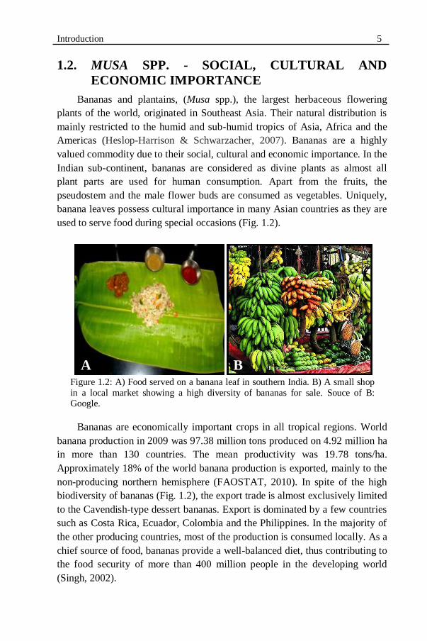
used to serve food during special occasions (Fig. 1.2).
Figure 1.2: A) Food served on a banana leaf in southern India. B) A small shop
in a local market showing a high diversity of bananas for sale. Souce of B: Google.
Bananas are economically important crops in all tropical regions. World
banana production in 2009 was 97.38 million tons produced on 4.92 million ha
in more than 130 countries. The mean productivity was 19.78 tons/ha.
Approximately 18% of the world banana production is exported, mainly to the
non-producing northern hemisphere (FAOSTAT, 2010). In spite of the high
biodiversity of bananas (Fig. 1.2), the export trade is almost exclusively limited
to the Cavendish-type dessert bananas. Export is dominated by a few countries
such as Costa Rica, Ecuador, Colombia and the Philippines. In the majority of
the other producing countries, most of the production is consumed locally. As a
chief source of food, bananas provide a well-balanced diet, thus contributing to
the food security of more than 400 million people in the developing world
(Singh, 2002).
A B

6 Chapter 1
The production of bananas is often restricted by diseases and pests.
Nematodes are ranked as the second-most important limiting biotic
factor of banana production after the black Sigatoka leaf streak disease
caused by the fungus M. fijiensis (Gowen et al., 2005).
1.3. PLANT-PARASITIC NEMATODES
Nematodes are unsegmented, thread-like worms. Their natural habitats
include terrestrial and aquatic ecosystems ranging from mountains to oceans.
However, for these animals it is crucial that their body is always surrounded by
a thin film of water for their mobility and survival (Perry, 1996).
Nematodes are highly adaptive and diverse. They are the most numerous
metazoans on earth: four out of every five animals are nematodes (Bird &
Kaloshian, 2003). Recently, a nematode species (Halicephalobus mephisto) has
been found in the Earth‟s terrestrial deep subsurface, expanding the known
metazoan biosphere (Borgonie et al., 2011). Nematodes are either free-living
or parasites of plants and animals. Some plant-parasitic nematode species can
damage the aboveground plant parts but the majority of the plant-parasitic
nematode species are root pathogens. Most of the plant-parasitic nematodes are
smaller than 1 mm.
Based on their life cycle and feeding habit, plant-parasitic nematodes can
be classified as 1) ectoparasites that live completely outside the plant and feed
on the outer cell layers of the roots with the aid of a stylet, 2) semi-
endoparasites that enter the roots partially and feed on the inner cell layers of
the roots while a part of the nematode‟s body remains outside in the soil, 3)
endoparasites that penetrate the root completely and feed on the cortical and/or
vascular cells of the roots. Endoparasitic nematodes can be further sub-divided
into two types: a) sedentary endoparasitic nematodes of which the females
become sessile after inducing specialized feeding cells on which they
exclusively feed and b) migratory endoparasitic nematodes of which the
juveniles and adults remain mobile and continuously feed on the cortical cells
of the roots as they migrate inside the roots (Speijer & De Waele, 1997;
Siddiqi, 2000).
1.3.1. BANANA NEMATODES
Although approximately 150 different nematode species were found
associated with Musa spp., the following nematode species are considered the
most important nematodes of this crop (Speijer & De Waele, 1997; Gowen et
al., 2005): the burrowing nematode R. similis, the root-lesion nematodes

Introduction 7 Pratylenchus goodeyi Sher & Allen, 1953 Pratylenchus coffeae (Zimmermann,
1898) Filipjev & Schuurmans Stekhoven, 1941 and the spiral nematode
Helicotylenchus multicinctus (Cobb, 1893) Golden, 1956. Root-knot
nematodes (Meloidogyne spp.) have also been often found associated with
Musa spp. (Gowen et al., 2005). For our study, we have used R. similis as this
species is considered the most important nematode species of banana
worldwide.
1.3.1.1. The burrowing nematode Radopholus similis
More than a century ago, R. similis was first isolated from banana roots in
the Fiji Islands (Cobb, 1893). As a migratory endoparasitic nematode, all life
stages of R. similis remain mobile and are infective. Radopholus similis can be
found worldwide on bananas in all tropical and subtropical regions with a
mean annual temperature of 24-32 °C. The optimum temperature for its
reproduction is about 30 °C. Survival of R. similis in temperate regions is
limited only to greenhouses as the reproduction stops below 16-17 °C (as well
as above 33 °C) (Sarah et al., 1996). A diagram of the Radopholus similis is
provided in the annexes.
1.3.1.2. Radopholus similis host range and economical importance
Although the best known hosts of R. similis are bananas, black pepper and
citrus, this nematode species has been found associated with hundreds of
different plants (Holdeman, 1986) including arecanut, avocado, coconut,
coffee, ginger, palm, sugarcane, tea, turmeric and ornamentals belonging to the
families Araceae and Marantaceae (Moens & Perry, 2009). Radopholus similis
is grouped among the 10 most damaging plant-parasitic nematode species
around the world (EPPO, 2008). It causes severe yield losses by causing the
“toppling” disease on bananas, “yellows” disease on black pepper and
“spreading decline” on citrus.
1.3.1.3. Radopholus similis as a major root pathogen of bananas
Radopholus similis is considered as the most damaging nematode species
in intensive commercial banana plantations, especially of Cavendish-type
dessert bananas (Sarah, 2000). This nematode continuously feeds on the
cortical cells. Collapse of the root cell walls ultimately leads to cavities and
tunnels due to necrosis of the damaged root cells. This damage to the roots
reduces water and nutrient uptake and translocation, causing impaired plant
8 Chapter 1 growth and development, a prolongation of the vegetative stage and thus crop
cycle, loss of bunch weight and poor root anchorage. Severe root damage may
cause the whole plant to topple under strong windy conditions (Sarah, 2000;
Gowen et al., 2005).
The average yield loss in bananas by R. similis was estimated as 20%
(Sasser & Freckman, 1987). However, yield losses of bananas up to 75% over
a 3-years cropping period caused by R. similis have been reported (Sarah,
1989). Nematicidal treatment in R. similis-infested plots doubled plantain yield
compared to untreated plots in which about 50% of the plants toppled over a 2-
years cropping period (Fogain, 2000a). Together with H. multicinctus, R.
similis caused about 71% of plantains to die at flowering of the first ratoon
crop (Coyne et al., 2005).
1.3.1.4. Control and management of Radopholus similis on bananas
Conventionally, large amounts of nematicides were used in the
commercial banana plantations to limit yield losses caused by R. similis.
However, because of environmental concerns, several efficient fumigant-
nematicides such as DD (dichloropropane-dichloropropene), EDB (ethylene
dibromide), DBCP (1,2-dibromo-3-chloropropane) and methyl bromide which
were highly used until the 1980s have been phased out and withdrawn from the
market (Quénéhervé, 2009). As a result, chemical nematode control currently
depends upon some organophosphate and carbamate nematicides such as
Aldicarb (Temik), Etoprop (Mocap), Fenamifos (Nemacur), Furadan
(carbofuran) and Terbufos (Counter) which are applied 2-4 times a year
(Villanueva, 2003; Quénéhervé, 2009). However, the intensive application of
these organophosphate and carbamate nematicides continues to pose a threat to
the environment and the health of agricultural workers (Atkinson et al., 2004).
Moreover, strict regulations are exercised in many countries on the use of these
nematicides that are still in use (Villanueva, 2003; Quénéhervé, 2009). Cultural
practices such as fallowing, crop rotation, etc. are also used to control
nematodes but are not applicable in banana fields. Also, they are often not
adequate enough as a sole practice to control nematodes (Lilley et al., 2007).
Complete eradication of R. similis from infested banana plantations requires a
5-years-crop free fallow as the nematode can survive on alternative weeds
(EPPO, 2008). Using resistant cultivars to manage nematode populations in
severely infested fields offers an efficient and economical alternative nematode
management strategy (Fuller et al., 2008; Zasada et al., 2010).
Introduction 9
1.4. HOST PLANT RESISTANCE IN MUSA SPP. TO
RADOPHOLUS SIMILIS
In nematology, resistance is defined as the ability of a host plant to
prevent or suppress nematode multiplication while in a susceptible host plant
nematodes can multiply. Tolerance is independent of resistance and is defined
as the ability of a host plant to suffer little damage even when quite heavily
infected with nematodes while a sensitive host plant will suffer much injury
even when relatively lightly infected with nematodes (Bos & Parleviet, 1995).
This terminology is illustrated in Figure 1.3.
Using natural host plant resistance for the management of plant-parasitic
nematodes offers a sustainable, ecologically-friendly and cost-effective
alternative to the use of pesticides (Roberts, 1992; Fuller et al., 2008).
However, most known resistance sources are specific only to certain nematode
species. Also, resistance sources to the predominant nematode species have not
been identified yet in many agricultural crops (Young, 1992; Atkinson et al.,
2003; Lilley et al., 2007).
In bananas, numerous studies were carried out around the world to
identify Musa genotypes with natural resistance to R. similis (Quénéhervé et
al., 2008b). Wehunt et al. (1978) were the first to report that Musa cultivars
belonging to the subgroup Pisang Jari Buaya (AA genome) were resistant to R.
similis. This discovery intensified the search for additional resistant sources
(see for instance Price, 1994; Fogain & Gowen, 1998; Stoffelen et al., 2000;
Viaene et al., 2003; Dochez et al., 2005, 2006; Quénéhervé et al., 2008a,
2008b, Dizon et al., 2010; Herradura et al., 2011). Yangambi km5 (AAA) was
another R. similis-resistant source that was studied elaborately (Price, 1994;
Fogain & Gowen, 1998).

10 Chapter 1
Figure 1.3: Host plant/pathogen relationship terminology used in
nematology. Source: Roberts, 2002.
1.4.1. HOST-NEMATODE INTERACTIONS
An in-depth understanding of the interactions between a host plant and its
pathogen(s) is crucial for the development of a successful management
strategy. Plant infection by nematodes is a continuous and dynamic process. In
the nematode infection cycle the following phases can be distinguished:
1) localisation of the host plant by the nematode (including migration
by the nematode towards the host plant)
2) penetration and entry of the nematode in the host plant
3) nematode development
4) nematode reproduction.
The host-nematode interactions can be either successful or unsuccessful
for the nematode. In the first case the plant is susceptible. In the second case
the plant is resistant or a non-host.
1.4.1.1. Localisation of the host plant by the nematode
Plant-released cues are exploited by plant-parasitic nematodes to guide
themselves towards the roots of their host plants. Plant roots release
chemically-rich exudates and diffusates. Additionally, several other gradients
existing in the rhizosphere, such as a CO2 gradient, microbial density, redox
potential or an electric field, can serve as guiding signals for nematodes to
locate their host plants (Perry, 1996; Spence et al., 2008). Nematodes possess
Susceptible Non-host Resistant Tolerant host
Intolerant host
Terminology
Host growth
Nematode population increase
Introduction 11 sensory receptors in their head region. The pair of amphids is considered as a
primary chemosensory receptor along with inner and outer labial sensilla
which are considered as vital in nematode host finding and identification of
suitable penetration sites (Perry, 1996). Host localisation or nematode
attraction takes place over a distance of several centimeters. Nematodes sense
the long-distance, short-distance and local attractants operating at several
centimeters, in the close proximity of roots and at the direct contact of the host
plant, respectively (Spence et al., 2008).
Long-distance cues are, in general, non-specific plant signals such as a
CO2 gradient (Robinson, 1995). Short-distance cues are usually host-specific.
Nematodes with a broader host range, such as Meloidogyne spp., seem to
respond to short-distance but non-specific plant signals (Robinson, 2002).
Nematode host finding can be inhibited by blocking either the release of host-
signaling molecules or the nematode‟s chemoreceptors (Zuckerman, 1983).
Lectins were shown to inhibit the attraction of R. similis towards the banana
roots in vitro (Wuyts et al., 2003). However, inconsistent effects were observed
as treating R. similis with lectins resulted in enhanced root penetration by the
nematode (Kaplan & Davis, 1991). Certain nematicides, such as aldicarb,
impair nematode orientation at low concentration without affecting nematode
movement (Perry, 1996).
1.4.1.2. Penetration and entry of the nematode in the host plant
Once nematodes have found the host roots, they invade the roots using
their specialised feeding apparatus, the stylet and pharyngeal gland secretions.
The stylet is a thin, hollow, needle-like tube located at the anterior end of the
nematodes. Nematodes attack the plant‟s epidermal cells by repeated stylet
thrusts to pierce the plant cell wall. In addition to physical damage, they also
produce pharyngeal gland secretions to dissolve the cell wall. Radopholus
similis possesses three large pharyngeal glands secreting different enzymes
including cell wall degrading enzymes such as endo-1,4-β-glucanases
(Haegeman et al., 2008) and endoxylanase (Haegeman et al., 2009). These two
enzymes aid R. similis in penetrating the host and in migrating inside the host
as they degrade the major cell wall components cellulose and xylan,
respectively. Radopholus similis is capable of invading the plant root at any
place albeit that they seem to prefer root tips and tender roots (Sarah et al.,
1996). In a successful penetration, nematodes breach the first-line physical and
chemical barriers of the host plant and penetrate it. In an incompatible
interaction, plants are able to block nematode penetration by early defence
12 Chapter 1 responses such as cell wall strengthening by lignifications or pre-existing
specialised cell layers, etc.
Biochemicals that may be involved in resistance of plants to nematodes
are discussed later in this chapter, in the section 1.5.
1.4.1.3. Nematode development and reproduction in the host
Following penetration, R. similis migrates through the cortex both inter-
and intra-cellularly and continuously feeds on the cortical cells by sucking and
digesting the cytoplasm using enzymatic secretions. Radopholus similis keeps
migrating to new cortical cells to feed on (Sarah et al., 1996; Gowen et al.,
2005). Juveniles develop into further developmental stages while females start
laying one to six eggs per day (Haegeman et al., 2010). Resistant plants
prevent nematode development and reproduction through hypersensitive
responses, anti-nematode toxins or by inhibiting nematode feeding.
1.4.2. POSSIBLE MECHANISMS OF PLANT RESISTANCE TO
NEMATODES
Natural plant resistance mechanisms towards nematodes (and other
pathogens) can be classified into a) preformed resistance mechanisms and b)
induced resistance mechanisms (Fig. 1.4).
1.4.2.1. Preformed resistance mechanisms
Pre-existing (passive) structural features such as root thickness, waxiness
of the cuticle, degree of secondary wall thickenings, vascular structure, etc.,
have been reported to contribute to plant resistance to pathogens including
plant-parasitic nematodes (Hutcheson, 1998). For instance, coffee clones
resistant to the root-lesion nematode P. coffeae possessed more hairy roots,
thicker epidermal and endodermal cell walls, and higher total polyphenol
contents than susceptible clones (Toruan-Mathius et al., 1995).

Introduction 13
Figure 1.4: Possible mechanisms of plant resistance to nematodes. HR: hypersensitive
response.
Constitutive antimicrobial secondary metabolites present in the plants may
contribute to the preformed chemical mechanisms of nematode resistance. The
polythienyl compounds extracted from Tagetes spp. are well documented as
anti-nematode compounds. A phytochemical, thiophene α-terthienyl isolated
from T. erecta plants was reported as toxic to the potato cyst nematode
Globodera rostochiensis, the wheat seed gall nematode Anguina tritici and the
stem and bulb nematode Ditylenchus dipsaci. In soil, the isothiocyanates and
glucosinolates released by plants belonging to the family Brassicaceae are
active against a variety of nematodes and insects (Chitwood, 2002)
1.4.2.2. Induced resistance mechanisms
Induced resistance mechanisms include active, energy-requiring responses
of plants after infection by a pathogen. The triggering of these strong responses
requires specific recognition of certain molecules from the invading pathogen
which are collectively known as elicitors. Experiments have shown that in the
absence of the pathogen, elicitors are capable of initiating the active plant
defence response (Garcia-Brugger et al., 2006; Sanchez-Estrada et al., 2009).
When resistant plants recognise matching elicitors, intracellular signal
transduction pathways are activated resulting in an integrated series of plant
responses leading to eventual neutralization of the invading pathogen (Fig. 1.5)
(Dixon et al., 1994; Williamson & Hussey, 1996; Das et al., 2008; Fuller et al.,
2008).
Resistant mechanisms
Preformed
(Passive)Infection-Induced
(Active)
Structural barriers e.g. waxy cuticle, branched roots
Chemical barriers
e.g.phytoanticipins
Hypersensitive responses (HR)
Antibiotics, toxins production
- Phytoalexins
Systemic acquired responses
Structural e.g. cell wall
strengthening by lignin, suberin
Other Induced phytochemicals
e.g.
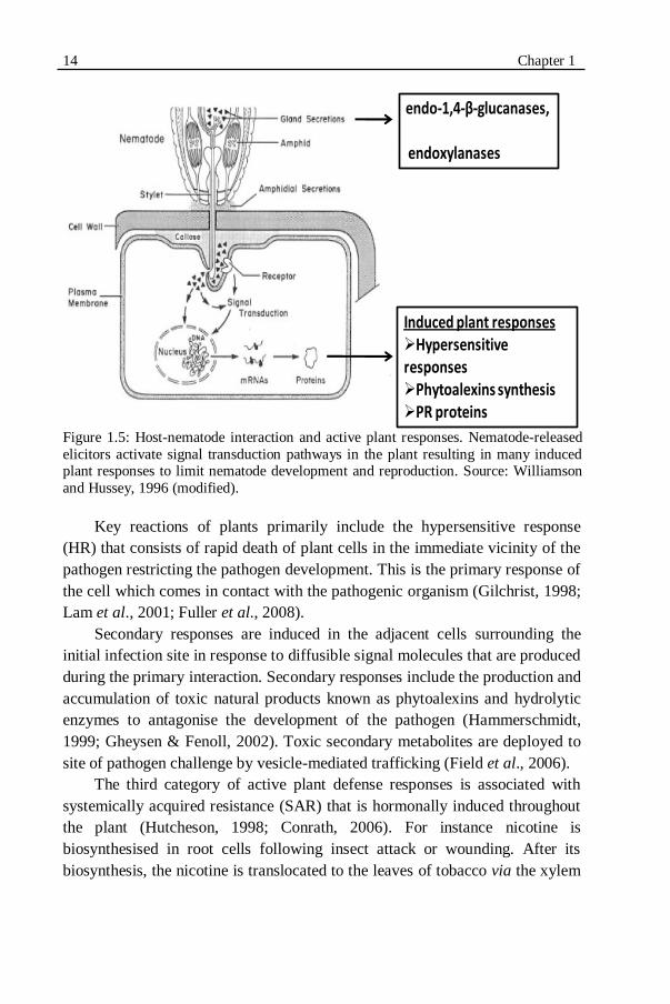
14 Chapter 1
Figure 1.5: Host-nematode interaction and active plant responses. Nematode-released
elicitors activate signal transduction pathways in the plant resulting in many induced plant responses to limit nematode development and reproduction. Source: Williamson
and Hussey, 1996 (modified).
Key reactions of plants primarily include the hypersensitive response
(HR) that consists of rapid death of plant cells in the immediate vicinity of the
pathogen restricting the pathogen development. This is the primary response of
the cell which comes in contact with the pathogenic organism (Gilchrist, 1998;
Lam et al., 2001; Fuller et al., 2008).
Secondary responses are induced in the adjacent cells surrounding the
initial infection site in response to diffusible signal molecules that are produced
during the primary interaction. Secondary responses include the production and
accumulation of toxic natural products known as phytoalexins and hydrolytic
enzymes to antagonise the development of the pathogen (Hammerschmidt,
1999; Gheysen & Fenoll, 2002). Toxic secondary metabolites are deployed to
site of pathogen challenge by vesicle-mediated trafficking (Field et al., 2006).
The third category of active plant defense responses is associated with
systemically acquired resistance (SAR) that is hormonally induced throughout
the plant (Hutcheson, 1998; Conrath, 2006). For instance nicotine is
biosynthesised in root cells following insect attack or wounding. After its
biosynthesis, the nicotine is translocated to the leaves of tobacco via the xylem
Induced plant responsesHypersensitive responsesPhytoalexins synthesis PR proteins
endo-1,4-β-glucanases,
endoxylanases
Introduction 15 and accumulated in the leaves‟ vacuoles as a defensive toxin against herbivores
(Yazaki et al., 2008).
Possible biochemical mechanisms of resistance and the secondary
metabolites involved in resistance in Musa spp. to R. similis are discussed later
in this chapter, in the sections 1.5.3 and 1.5.4.
1.5. PLANT SECONDARY METABOLITES
INVOLVED IN HOST PLANT RESISTANCE
Secondary metabolites are chemical compounds of metabolism that are
not essential for photosynthesis, respiration, growth, development and
reproduction of an organism but play a role in the interaction of the organism
with its environment, ensuring the adaptation and survival of the organism in
its ecological niche (Verpoorte, 2000). The importance of these natural
products can be illustrated by the fact that approximately 25% of the known
Arabidopsis thaliana genes are implicated in natural product synthesis and
more than 170 natural products from seven major classes of compounds have
now been reported in A. thaliana (Ben Field & Osbourn, 2006). Secondary
metabolites are extremely diverse comprising more than 200,000 structures
(Hartmann, 2007). Their classification is complex. Major groups of plant
secondary metabolites are a) phenolics such as simple phenols, polyphenols,
xanthones, phenylpropanoids, stilbenes and glycosides, b) isoprenoid-like
compounds such as terpenes, steroids and its glycosides, c) alkaloids such as
caffeine and nicotine, and d) amino acids such as alliin or canavanine (Dixon,
2001).
Originally, secondary metabolites were thought to be metabolic sinks.
Their functions were largely unknown and hence ignored (Hartmann, 2007).
However, these secondary metabolites are increasingly attracting the attention
of scientists due to the discovery of their role in plants as protectants from
pathogens, as attractants for pollinators and seed dispersers, as allelopathic
agents and UV protectants. Besides, they also form valuable products such as
dyes, fibres, glues, waxes, oils, drugs, perfumes, flavouring agents, etc.
(Crozier et al., 2006). Plants adapted different strategies to protect themselves
from pathogens using secondary metabolites. Based on whether a plant
accumulates secondary compounds as a preparation to face attacks by
pathogens or secondary compounds are produced in response to attacks by
pathogens, defence-related compounds are classified either as phytoanticipins
or phytoalexins.

16 Chapter 1
1.5.1. PHYTOANTICIPINS
Phytoanticipins are low molecular weight, antimicrobial compounds that
are either present in plants before they are challenged by micro-organisms or
they are produced after infection solely from pre-existing constituents
(VanEtten et al., 1994). The distinction between a phytoanticipin and a
phyoalexin is mainly based on when it is biosynthesized in the host, not on its
chemical structure. In the cases where a constitutive metabolite is produced in
larger amounts after infection, its status as a phytoanticipin or phytoalexin will
depend on whether or not the constitutive concentrations were sufficient to
cause antibiotic effects (Dixon, 2001).
Two different strategies have been adapted by plants in constitutive
defense using phytoanticipins: a) constitutive accumulation of defense
metabolites in specialised cells and b) constitutive accumulation of preformed
defense compounds and respective enzymes in well-separated spatial
compartments. Pathogenic attack could de-compartment them and release the
active defense compound. For example, in A. thaliana, glucosinolates can be
stored in the vacuoles of specialised cells known as S-cells (Koroleva et al.,
2000) while the enzymes that activate them (myrosinases) are sequestered in
myrosin cells (Rask et al., 2000). Tissue damage caused by pathogen attack
leads to breakdown of cell compartments followed by hydrolysis of
glucosinolates to unstable aglucones by myrosinases. These unstable
intermediates are then converted to toxic products such as isothiocyanates and
nitriles (Ben Field & Osbourn, 2006). Baldridge et al. (1998) investigated the
level of defense response isoflavonoids in alfalfa roots with different levels of
resistance to the root-lesion nematode Pratylenchus penetrans. Constitutive
levels of the phytoanticipin medicarpin were highest in the roots of the two
most resistant plants.
1.5.2. PHYTOALEXINS
Müller (1961) defined phytoalexins as "compounds produced after
infection under the influence of two metabolic systems, that of the host and
that of the parasite, and inhibitory to the parasite." Since then, the term
phytoalexin has undergone several conceptual changes becoming more
inclusive of their production in response to physiological stress. Hence the new
definition is "phytoalexins are low molecular weight antimicrobial/antibiotic
compounds which are synthesized and accumulated in plants after exposure to
pathogenic organisms or stress" (Paxton, 1980).

Introduction 17 A long list of compounds can elicit the biosynthesis of phytoalexins (Kuć,
1995). This list includes the inorganic salts, oligoglucans, ethylene, fatty acids,
polypeptides, some fungicides, low temperature and ultraviolet radiation. Also
plant constituents released after injury or infection can function as elicitors.
A vast diversity of phytoalexins has been reported so far. More than 350
phytoalexins have been chemically characterized from approximately 30 plant
families (Kuć, 1995). Due to their great diversity, there is no evident
relationship between their chemical structure and their antibiotic property.
Generally speaking, phytoalexins are found to be lipophilic, localised at and
around infection sites (Kuć, 1995). However, each plant family possesses a
certain unique set of secondary compounds equipping the plant to adapt to its
own ecological niche (Hartmann, 2007). For example, sulphur-containing
compounds such as glucosinolates are main phytoalexins in Brassicaceae
(Morissey & Osbourn, 1999), isoflavonoids in the Leguminosae and
sesquiterpenes in the Solanaceae (Dixon, 2001). But in contrast, the
phenylpropanoid-type phytoalexins are present in many plant families such as
Leguminosae, Solanaceae, Convolvulaceae, Umbelliferae, Gramineae,
Cucurbitaceae and Musaceae (Dixon, 2001).
Phenolic compounds play an important role in the interactions of plants
with their environment. They serve as phytoanticipins, phytoalexins, structural
barriers, modulators of pathogenicity and activators of plant defense genes
(Hammerschmidt, 2005). Two major groups of phenolic secondary
metabolites, phenylphenalenones and phenylpropanoids, are reported to be
involved in the defense response of Musa spp. to R. similis.
1.5.3. PHENYLPHENALENONES
1.5.3.1. Origin and natural occurrence
Phenylphenalenones are aromatic, naturally occurring secondary
metabolites first isolated as natural pigments about 55 years ago. The term
phenylphenalenone derives from the carbon skeleton of a phenalene-body, one
keto group and an additional phenyl ring (Fig. 1.6).
The first phenalenone was isolated almost at the same time from a higher
plant family, Haemodoraceae, and from fungi belonging to the Hyphomycetes
and Discomycetes (Cooke & Segal, 1955b; Harman et al., 1955)

18 Chapter 1
Figure 1.6: Structure of A) phenalen, B) phenalen-1-one and C) 9-
phenylphenalen-1-one.
. However, the structures of the phenalenones produced by higher plants
are significantly different from those produced by fungi i.e. the fungal
phenalenones were lacking the additional phenyl group and are derived from
an unrelated biosynthetic pathway (Fig. 1.7).
Haemocorin, a constituent of the rhizomes of Haemodorum corymbosum,
was the first phenylphenalenone derivative to be characterized from plants
(Cooke & Segal, 1955a,b). Following this description, many
phenylphenalenones and related compounds were described from the same
plant family, Haemodoraceae (Edwards & Weiss, 1974; Cooke & Dagley,
1979; Hölscher & Schneider, 1997).
Figure 1.7: Structure of A) fungal phenalenone-atrovenetin and B) plant
phenylphenalenone-haemocorin, first isolated from Penicillium herquei and
Haemodorum corymbosum, respectively. Source: A: Harman et al., 1955; B:
Cooke and Segal, 1955b.
Only during the 1990s, phenylphenalenones were reported to be present in
other plant families. But so far, their occurrence and distribution seem to be
limited to some monocotyledonous families of the plant kingdom namely
Haemodoraceae, Musaceae (Luis et al., 1993), Pontederiaceae (Della Greca et
al., 1993) and Strelitziaceae (Hölscher & Schneider, 2000).
A) B) C)
B) A) B) A) B)
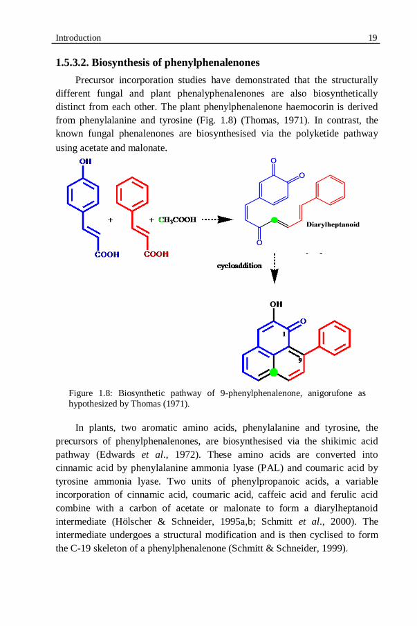
Introduction 19
1.5.3.2. Biosynthesis of phenylphenalenones
Precursor incorporation studies have demonstrated that the structurally
different fungal and plant phenalyphenalenones are also biosynthetically
distinct from each other. The plant phenylphenalenone haemocorin is derived
from phenylalanine and tyrosine (Fig. 1.8) (Thomas, 1971). In contrast, the
known fungal phenalenones are biosynthesised via the polyketide pathway
using acetate and malonate.
Figure 1.8: Biosynthetic pathway of 9-phenylphenalenone, anigorufone as
hypothesized by Thomas (1971).
In plants, two aromatic amino acids, phenylalanine and tyrosine, the
precursors of phenylphenalenones, are biosynthesised via the shikimic acid
pathway (Edwards et al., 1972). These amino acids are converted into
cinnamic acid by phenylalanine ammonia lyase (PAL) and coumaric acid by
tyrosine ammonia lyase. Two units of phenylpropanoic acids, a variable
incorporation of cinnamic acid, coumaric acid, caffeic acid and ferulic acid
combine with a carbon of acetate or malonate to form a diarylheptanoid
intermediate (Hölscher & Schneider, 1995a,b; Schmitt et al., 2000). The
intermediate undergoes a structural modification and is then cyclised to form
the C-19 skeleton of a phenylphenalenone (Schmitt & Schneider, 1999).
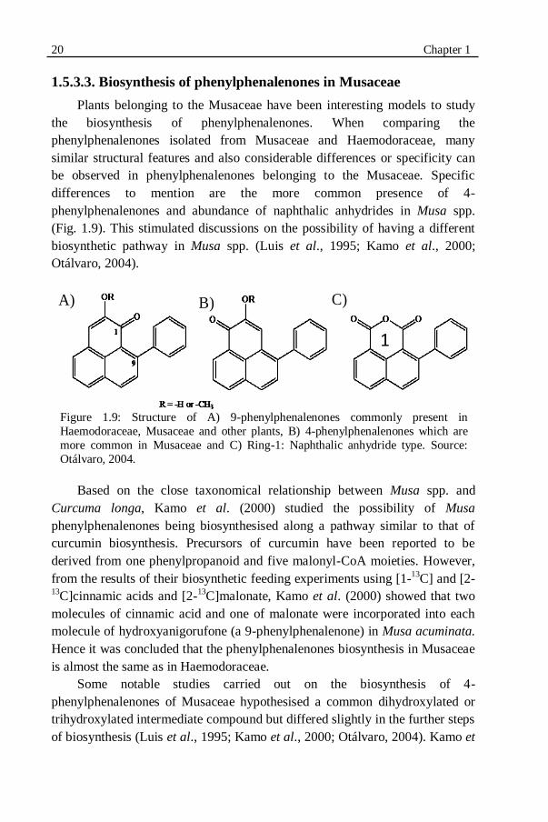
20 Chapter 1
1.5.3.3. Biosynthesis of phenylphenalenones in Musaceae
Plants belonging to the Musaceae have been interesting models to study
the biosynthesis of phenylphenalenones. When comparing the
phenylphenalenones isolated from Musaceae and Haemodoraceae, many
similar structural features and also considerable differences or specificity can
be observed in phenylphenalenones belonging to the Musaceae. Specific
differences to mention are the more common presence of 4-
phenylphenalenones and abundance of naphthalic anhydrides in Musa spp.
(Fig. 1.9). This stimulated discussions on the possibility of having a different
biosynthetic pathway in Musa spp. (Luis et al., 1995; Kamo et al., 2000;
Otálvaro, 2004).
Figure 1.9: Structure of A) 9-phenylphenalenones commonly present in
Haemodoraceae, Musaceae and other plants, B) 4-phenylphenalenones which are
more common in Musaceae and C) Ring-1: Naphthalic anhydride type. Source:
Otálvaro, 2004.
Based on the close taxonomical relationship between Musa spp. and
Curcuma longa, Kamo et al. (2000) studied the possibility of Musa
phenylphenalenones being biosynthesised along a pathway similar to that of
curcumin biosynthesis. Precursors of curcumin have been reported to be
derived from one phenylpropanoid and five malonyl-CoA moieties. However,
from the results of their biosynthetic feeding experiments using [1-13
C] and [2-13
C]cinnamic acids and [2-13
C]malonate, Kamo et al. (2000) showed that two
molecules of cinnamic acid and one of malonate were incorporated into each
molecule of hydroxyanigorufone (a 9-phenylphenalenone) in Musa acuminata.
Hence it was concluded that the phenylphenalenones biosynthesis in Musaceae
is almost the same as in Haemodoraceae.
Some notable studies carried out on the biosynthesis of 4-
phenylphenalenones of Musaceae hypothesised a common dihydroxylated or
trihydroxylated intermediate compound but differed slightly in the further steps
of biosynthesis (Luis et al., 1995; Kamo et al., 2000; Otálvaro, 2004). Kamo et
A) B) C)
1

Introduction 21 al. (2000) suggested that 9-phenylphenalenones were converted to 4-
phenylphenalenones through a carbonyl transposition process. But, the other
two postulations (Luis et al., 1995; Otálvaro, 2004) suggest a diversification of
the biosynthetic pathway after the synthesis of the common intermediate
compound leading to the formation of 4-phenylphenalenones. The presence of
a 4-phenylphenalenone from Anigozanthos preissii (Hölscher & Schneider,
1997) and the isolation of a 5-methoxy-7-phenylphenalenone in Dilatris spp.
(Hölscher & Schneider, 2007) recommend further studies to prove the
hypotheses of unique presence of 4-phenylphenalenones in Musaceae.
1.5.3.4. Phenylphenalenones as phytoalexins and phytoanticipins
Haemodoraceae and Musaceae are the two major plant families in which a
rich occurrence of phenylphenalenones has been found. Originally,
phenylphenalenones were characterized as naturally occurring pigments in the
Haemodoraceae in the genera Anigozanthos, Conostylis, Dilatris,
Haemodorum, Lachnanthes, Xiphidium and Wachendorfia (Cooke & Edward,
1981; Dora et al., 1993; Hölscher & Schneider, 1997; Opitz & Schneider,
2002). Phenylphenalenones are constitutively present in the cells of plants
belonging to these genera (Fig. 1.10).
In Musa spp., phenylphenalenones are produced as phytoalexins after the
plant is being infected by pathogens (bacteria, fungi and nematodes) or
chemical elicitors. Two phenylphenalenones, irenolone and
hydroxyanigorufone, were first reported as phytoalexins in Musa spp. by Luis
et al. (1993; 1995) following their biosynthesis in the leaves of the cultivar
Grande Naine due to infection by M. fijiensis. Phenylphenalenones were also
found to be produced and accumulated in banana fruits after infection by
Colletotrichum musae (Kamo et al., 1998; 2001) and in rhizomes and roots
after infection by Fusarium oxysporum f. sp. cubense (Luis et al., 1994, 1996,
1997).

22 Chapter 1
Figure 1.10: Inflorescence of A) Heamodorum sp., B) Anigozanthus flavidus, C)
Dilatris pillansii, and blood red coloured roots of D) Lachnanthes sp. and E)
Wachendorfia thyrsiflora. Source of C and E: Dr. Hölscher, Max-Planck-Institute for Chemical Ecology. A, B and D: Google.
Although most reports show that phenylphenalenones are biosynthesised
in Musa spp. only after elicitation, rarely phenylphenalenones were also
reported to be present in low concentrations in the healthy tissues of Musa spp.
It was proposed that they function as phytoanticipins in these healthy tissues
due to their occurrence in the Musa hybrid SH-3481 (Luis et al., 1999). This
hybrid was reported earlier as resistant to F. oxysporum f. sp. cubense races 1
and 4, and to M. fijiensis. Concentration of constitutive hydroxyanigorufone in
banana fruits increased greatly upon wounding without infection whereas all
other phenylphenalenones were biosynthesised only after infection with
conidial spores of C. musae (Kamo et al., 2000).
E) D)
A) B)
C)
Introduction 23
1.5.3.5. Phenylphenalenones as antibiotic compounds
Phenylphenalenones being synthesized as phytoalexins, their function as
antimicrobial or antibiotic compounds, their mode of action and relationship
between the structure of the compounds and their activity have become a major
study field. Antibiotic properties of phenalenones were reported more than
three decades ago. Phenalen-1-one was toxic to green algae, blue-green algae,
and diatoms. With green algae and diatoms, this toxicity was strongly
wavelength-dependent showing greater toxicity to green algae in white light
(Winters et al., 1977).
Antifungal properties of the phenylphenalenones, especially against plant
pathogens, were reported. Phenylphenalenones isolated from Musa spp.
inhibited the germination of F. oxysporum f. sp. cubense race 4 and moderately
inhibited germination of Alternaria sp. (Luis et al., 1996, 1998). Kamo et al.
(1998) studied the antifungal activity of 14 different phenylphenalenones and
their derivatives by the thin layer chromatography (TLC) autobiography
method. Six compounds namely anigorufone, irenolone, 2-hydroxy-4-(4´-
methoxyphenyl)phenalen-1-one, (+) cis 2,3-dihydro-2,3-dihydroxy-4-(4´-
hydroxyphenyl)phenalen-1-one, 2-(4´-hydroxyphenyl)-1-methoxycarbonyl-8-
naphthalenecarboxylic acid and 2-(4´-hydroxyphenyl)-8-methoxycarbonyl-1-
naphthalenecarboxylic acid, were shown to inhibit the spore germination of C.
musae. These results lead the authors to conclude that a phenolic-hydroxyl
group is essential to have this antifungal activity and 4-phenylphenalenones
have stronger anti-fungal properties than 9-phenylphenalenones.
Systematic bioassays conducted using nine phenylphenalenones against
M. fijiensis, the causal agent of black Sigatoka leaf streak disease on bananas,
showed that the natural phytoalexins anigorufone, isoanigorufone,
hydroxyanigorufone and irenolone inhibited mycelial growth and spore
germination of this fungus albeit in very low intensity compared to the
synthetic phenalenones, perinaphthenone. Perinaphthenone being an efficient
singlet oxygen sensitiser, it was speculated that the high antifungal activity of
this compound could have been due to the production of singlet oxygen among
other possible complex biochemical interactions (Quiñones et al., 2000).
Similarly, Lazzaro et al. (2004) attributed the enhanced antifungal activity of
phenylphenalenones against F. oxysporum f. sp. cubense to the production of
reactive oxygen species. Recently, two new phenylphenalenones, 2-hydroxy-
1H-phenalen-1-one and 2-methoxy-1H-phenalen-1-one, isolated from the
banana cultivar Yangambi km5, have shown inhibitory effect on the mycelial
growth and spore germination of M. fijiensis (Otálvaro et al., 2007).

24 Chapter 1 Antibacterial properties were also reported for these secondary metabolites.
Populations of Sarcina lutea and Escherichia coli were inhibited by 2-methyl-
9-(4´-hydroxyphenyl)-phenalen-1-one (Qin et al., 2006).
1.5.3.6. Phenylphenalenones as phytoalexins in plant-nematode
interactions
Anigorufone was the first phenylphenalenone identified to be produced de
novo in the roots of the banana cultivar Pisang Sipulu (group: Pisang Jari
Buaya) in response to infection by R. similis (Binks et al., 1997). Since
phenylphenalenones are produced as phytoalexins in response to nematode
damage, it has been suggested that they play a significant role in the defense
system of the plant (Luis et al., 1998). But, no information is available on the
anti-nematode properties of these phenylphenalenones and on the role of
phenylphenalenones in resistance or defense mechanism of Musa spp. to
nematodes.
1.5.4. PHENYLPROPANOIDS
The phenylpropanoids are a diverse group of organic compounds that are
synthesised from the amino acid phenylalanine through an enzymatic reaction
mediated by the key enzyme phenylalanine ammonia lyase (PAL). Though
structurally diverse, the name phenylpropanoids is derived from the core
structure with six-carbon aromatic phenyl groups and three carbon propene
tails of cinnamic acid (Fig. 1.11). Cinnamic acid is a common precursor of all
phenylpropanoids and also the first product derived from phenylalanine by a
nonoxidative de-amination in the core phenylpropanoid pathway (Dixon &
Paiva, 1995). From the core pathway, specific pathways branch out leading to
the synthesis of diverse compounds, such as coumarins, chlorogenic acid,
isoflavonoids, flavonoids, suberin, lignin and other wall-bound phenolics.
As mentioned earlier, phenylpropaniods are one of the rare groups of
compounds that are distributed throughout the plant kingdom. The
phenylpropanoid pathway compounds have attracted the attention of plant
physiologist, biochemists and pathologists due to their inducibility at the
transcriptional level and their diverse functions. In plants, they function inter
alia as flower pigments, UV protectants, antibiotic compounds (phytoalexins
or anticipins) and signaling molecules in plant-microbe interactions. They are
also vital for the structural integrity of plants as the plant cell walls are
composed of polymeric phenylpropanoids such as lignin, lignans and suberins
(Hahlbrock & Scheel, 1989).
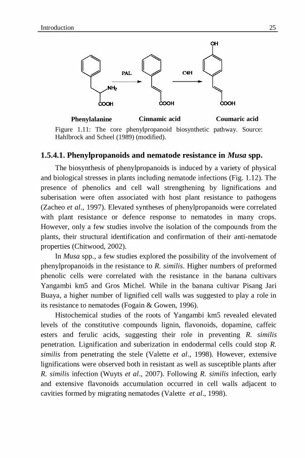
Introduction 25
Figure 1.11: The core phenylpropanoid biosynthetic pathway. Source: Hahlbrock and Scheel (1989) (modified).
1.5.4.1. Phenylpropanoids and nematode resistance in Musa spp.
The biosynthesis of phenylpropanoids is induced by a variety of physical
and biological stresses in plants including nematode infections (Fig. 1.12). The
presence of phenolics and cell wall strengthening by lignifications and
suberisation were often associated with host plant resistance to pathogens
(Zacheo et al., 1997). Elevated syntheses of phenylpropanoids were correlated
with plant resistance or defence response to nematodes in many crops.
However, only a few studies involve the isolation of the compounds from the
plants, their structural identification and confirmation of their anti-nematode
properties (Chitwood, 2002).
In Musa spp., a few studies explored the possibility of the involvement of
phenylpropanoids in the resistance to R. similis. Higher numbers of preformed
phenolic cells were correlated with the resistance in the banana cultivars
Yangambi km5 and Gros Michel. While in the banana cultivar Pisang Jari
Buaya, a higher number of lignified cell walls was suggested to play a role in
its resistance to nematodes (Fogain & Gowen, 1996).
Histochemical studies of the roots of Yangambi km5 revealed elevated
levels of the constitutive compounds lignin, flavonoids, dopamine, caffeic
esters and ferulic acids, suggesting their role in preventing R. similis
penetration. Lignification and suberization in endodermal cells could stop R.
similis from penetrating the stele (Valette et al., 1998). However, extensive
lignifications were observed both in resistant as well as susceptible plants after
R. similis infection (Wuyts et al., 2007). Following R. similis infection, early
and extensive flavonoids accumulation occurred in cell walls adjacent to
cavities formed by migrating nematodes (Valette et al., 1998).
Coumaric acid Phenylalanine Cinnamic acid
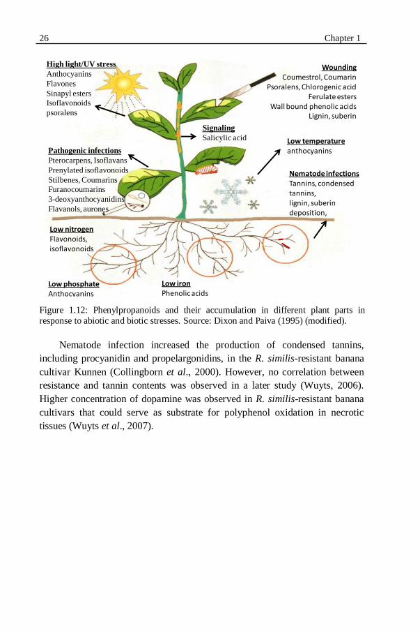
26 Chapter 1
Figure 1.12: Phenylpropanoids and their accumulation in different plant parts in
response to abiotic and biotic stresses. Source: Dixon and Paiva (1995) (modified).
Nematode infection increased the production of condensed tannins,
including procyanidin and propelargonidins, in the R. similis-resistant banana
cultivar Kunnen (Collingborn et al., 2000). However, no correlation between
resistance and tannin contents was observed in a later study (Wuyts, 2006).
Higher concentration of dopamine was observed in R. similis-resistant banana
cultivars that could serve as substrate for polyphenol oxidation in necrotic
tissues (Wuyts et al., 2007).
High light/UV stress
Anthocyanins
Flavones
Sinapyl esters
Isoflavonoids
psoralens
Pathogenic infections
Pterocarpens, Isoflavans
Prenylated isoflavonoids
Stilbenes, Coumarins
Furanocoumarins
3-deoxyanthocyanidins
Flavanols, aurones
WoundingCoumestrol, Coumarin
Psoralens, Chlorogenic acidFerulate esters
Wall bound phenolic acidsLignin, suberin
Low temperatureanthocyanins
Low phosphateAnthocyanins
Low ironPhenolic acids
Low nitrogenFlavonoids, isoflavonoids
Nematode infectionsTannins, condensed tannins, lignin, suberin deposition,
Signaling
Salicylic acid

CHAPTER 2
IDENTIFICATION OF COMBINED RESISTANCE TO RADOPHOLUS SIMILIS AND MELOIDOGYNE INCOGNITA
IN MUSA GERMPLASM2
2 The results presented in this chapter were published as: SUGANTHAGUNTHALAM, D.,
ELSEN, A. AND DE WAELE, D. (2010). Identification of combined resistance to
Radopholus similis and Meloidogyne incognita in Musa germplasm. International
Journal of Nematology 20:19-24.

28 Chapter 2
2.1. INTRODUCTION
Burrowing and root-knot nematodes are root pathogens that can cause
serious yield losses to Musa spp. (Gowen et al., 2005). Worldwide, the
burrowing nematode Radopholus similis (Cobb, 1893) Thorne, 1949 is
considered as the most damaging nematode species in Musa-based cropping
systems (Sarah et al., 1996; Gowen et al., 2005). However, under field
conditions, usually several nematode species co-exist in the roots of bananas
and plantains (De Waele & Elsen, 2007), and in the absence of R. similis, other,
less important nematode species such as the root-knot nematode Meloidogyne
incognita (Kofoid and White, 1919) Chitwood, 1949 can become more
damaging to Musa spp. (De Waele & Davide, 1998; De Waele, 2000; Brentu et
al., 2004; Cofcewicz et al., 2005; Van den Bergh et al., 2006).
The use of host plant resistance is considered as an economical and
environmental-friendly nematode management strategy in the low-input
cropping systems prevailing in developing countries (Roberts, 1992). This
strategy also ensures maximal land use efficiency without changing the existing
cropping systems (Atkinson et al., 2003). In Musa, a relatively high number of
studies have been carried out to identify natural sources of resistance to R.
similis resulting in the identification of some potential R. similis-resistant Musa
genotypes (see for instance, Wehunt et al., 1978; Fogain & Gowen, 1998;
Stoffelen et al., 2000; Viaene et al., 2003; Dochez et al., 2005, 2006;
Quénéhervé et al., 2008a, 2008b; Herradura et al., 2011). However, the
efficient use of natural sources of resistance to nematodes is limited due to
several reasons (Roberts, 1992). A major limitation is that, in most cases, the
resistance is restricted to one nematode species or a few pathotypes of the same
nematode species. Planting of species-specific or pathotype-specific resistant
cultivars under field conditions has resulted in a shift in nematode population
composition causing less important nematode species to become more
damaging and, sometimes, even a major problem (Young, 1992). To overcome
this loss of efficiency, identification of plant genotypes with resistance to more
than one nematode species or pathotype is necessary. Moreover, if these plant
genotypes can be used in breeding programmes, their identification could lead
to the development of cultivars with multiple nematode resistances.
The objective of this part of our study was to evaluate the host response of
selected Musa genotypes to both R. similis and M. incognita. The Musa
genotypes studied were selected based on their resistant host response to R.
similis as reported by Dochez et al. (2006). For their experiments, Dochez et al.
(2006) used sucker-derived planting material. Some studies have demonstrated
Combined resistance to R. similis and M. incognita 29
that tissue culture-derived Musa planting material can be more susceptible to R.
similis compared to sucker-derived planting material (Blomme et al., 2004;
Viaene et al., 2003) apparently due to their more fragile root system. In spite of
this, tissue culture-derived plants are preferred as planting material for the
initiation of new or the rejuvenation of existing commercial plantations and also
for research purposes because of their homogeneity, availability throughout the
year and because they are free of pathogens. In the first part of our study we re-
confirmed the host response of the R. similis-resistant Musa genotypes reported
by Dochez et al. (2006) using this time tissue culture-derived plants. In the
second part of our study, we examined the host response of the same Musa
genotypes to M. incognita, also using tissue culture-derived plants.
2.2. MATERIALS AND METHODS
2.2.1. EXPERIMENTAL SET-UP
Two experiments were carried out in the greenhouse at 27 and 20 °C day
and night temperatures, respectively, 12 h photoperiod and 80% relative
humidity. In the first experiment, seven Musa genotypes were examined for
their host response to R. similis. This experiment was carried out in two
separate batches. The host response of each genotype was compared to the host
response of a susceptible (Grande Naine) and two resistant (Yangkambi km5,
Pisang Jari Buaya) reference cultivars (Speijer & De Waele, 1997). In the
second experiment, eight Musa genotypes were examined for their host
response to M. incognita. In this experiment, Grande Naine was included as the
susceptible reference cultivar (Speijer & De Waele, 1997). There was no
resistant reference cultivar included as no Musa genotypes with resistance to M.
incognita were known. In both experiments, all treatments were replicated eight
times and a randomized block design was used. Some important characteristics
of the selected genotypes and reference cultivars are listed in Table 2.1.
2.2.2. PLANTING MATERIAL
All Musa genotypes were initially obtained from the Musa germplasm
collection maintained at the International Transit Centre (ITC), K.U.Leuven,
Belgium. The plant material was proliferated, regenerated and rooted in test
tubes on Murashige and Skoog medium including vitamins, 30 g/l sugar, 10
mg/l ascorbic acid and 2 g/l gelrite with pH 6.12 (Murashige & Skoog, 1962).
Proliferation was obtained by adding 10-6
M indole-3-acetic acid (IAA) and 10-5
M 6-benzylaminopurine (BAP). For rooting, 0.05% (w/v) active charcoal was

30 Chapter 2
added (Banerjee & De Langhe, 1985). No plant growth regulators were added.
The plantlets were grown in growth chambers at 28 °C and 16 h photoperiod.
Table 2.1. Characteristics of selected Musa genotypes and the reference cultivars*
evaluated for their host response to Radopholus similis and Meloidogyne
incognita.
ITC: International Transit Centre, K.U.Leuven, Belgium.
S: susceptible; R: resistant.
Eight-weeks-old rooted tissue culture plantlets were planted in 1 l pots
filled with sand and potting soil (2:1). The potted plants were maintained under
greenhouse conditions (Fig. 2.1) and fertilized at 10 days intervals throughout
the experiments.
Host response
Genotype
(subgroup)
ITC Genome Use R. similis M. incognita
Gia Hui
(Pisang Awak)
1143
ABB
cooking
-
-
Kokopo 1243 AA cooking/
dessert
- -
Long Tavoy
(Burbannica)
0283
AA
wild
-
-
Marau 0772 AAA
(AAB)
cooking - -
Pisang Mas
(Sucrier)
0653
AA
dessert
-
-
Pora Pora 0868 AA cooking/
dessert
- -
Saba 1138 ABB cooking - -
Vudu Papau 0590 AA dessert - -
Grande Naine*
(Cavendish)
1256
AAA
dessert
S
S
Yangambi km5*
(Ibota)
1123
AAA
dessert
R
-
Pisang Jari Buaya* 0312 AA dessert R -
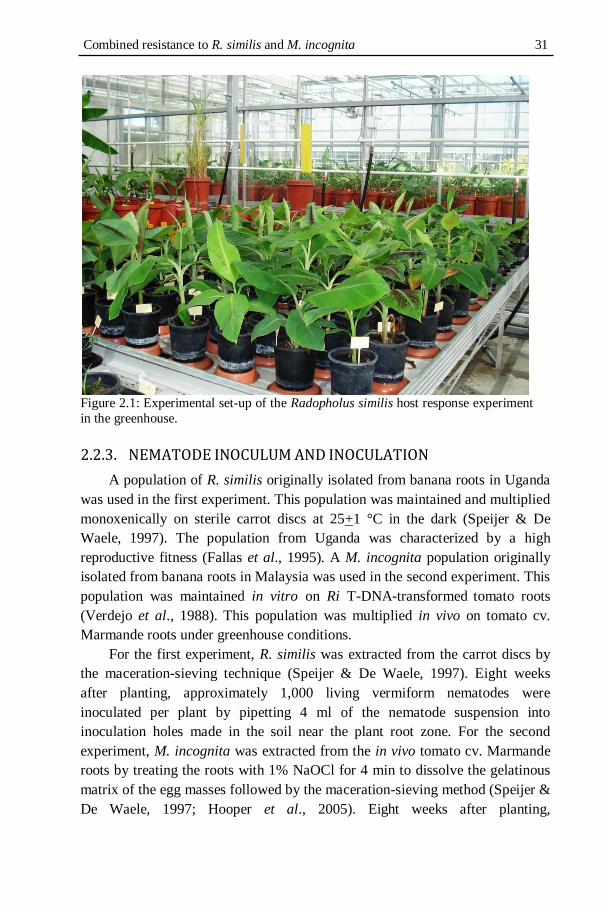
Combined resistance to R. similis and M. incognita 31
Figure 2.1: Experimental set-up of the Radopholus similis host response experiment
in the greenhouse.
2.2.3. NEMATODE INOCULUM AND INOCULATION
A population of R. similis originally isolated from banana roots in Uganda
was used in the first experiment. This population was maintained and multiplied
monoxenically on sterile carrot discs at 25+1 °C in the dark (Speijer & De
Waele, 1997). The population from Uganda was characterized by a high
reproductive fitness (Fallas et al., 1995). A M. incognita population originally
isolated from banana roots in Malaysia was used in the second experiment. This
population was maintained in vitro on Ri T-DNA-transformed tomato roots
(Verdejo et al., 1988). This population was multiplied in vivo on tomato cv.
Marmande roots under greenhouse conditions.
For the first experiment, R. similis was extracted from the carrot discs by
the maceration-sieving technique (Speijer & De Waele, 1997). Eight weeks
after planting, approximately 1,000 living vermiform nematodes were
inoculated per plant by pipetting 4 ml of the nematode suspension into
inoculation holes made in the soil near the plant root zone. For the second
experiment, M. incognita was extracted from the in vivo tomato cv. Marmande
roots by treating the roots with 1% NaOCl for 4 min to dissolve the gelatinous
matrix of the egg masses followed by the maceration-sieving method (Speijer &
De Waele, 1997; Hooper et al., 2005). Eight weeks after planting,
32 Chapter 2
approximately 4,000 eggs and juveniles were inoculated per plant by pipetting 4
ml of the nematode suspension into inoculation holes made in the soil near the
plant root zone.
2.2.4. EVALUATION OF THE HOST PLANT RESPONSE
Eight weeks after nematode inoculation (i.e. 16 weeks after planting),
plants were uprooted and gently washed free of soil under running tap water.
The fresh root and shoot weights, root damage and nematode reproduction were
assessed.
Radopholus similis
Estimation of the percentage root necrosis caused by R. similis was based
on the scoring of five randomly selected, 10-cm-long, longitudinally sliced
functional primary roots (Speijer & De Waele, 1997). Then, the same roots
were cut into 1-cm-long pieces, weighed and, if necessary, additional roots
were sampled to make up 15 g of roots for nematode extraction by the
maceration-sieving technique (Speijer & De Waele, 1997). The roots were
macerated in a kitchen blender for three periods of 10 sec each separated by a 5
sec interval. The macerated suspension was passed through a series of 250, 100,
40 and 25 µm aperture sieves. Nematodes were collected from the 40 and 25
µm aperture sieves and pooled. Eggs, juveniles, females and males were
counted separately from a 2 ml aliquot out of the homogenised suspension. The
final nematode population densities were determined by calculating the sum of
all developmental stages of the nematodes present in the total root system. The
reproduction factor (Rf) was calculated as the final nematode population
density divided by the inoculum. The host response of each genotype to R.
similis was determined based on a comparison of the final nematode population
density of each genotype with the reference cultivars as explained in Table 2.2.

Combined resistance to R. similis and M. incognita 33
Table 2.2. Identification of the host response of selected Musa genotypes to Radopholus similis based on a comparison with the host response of a susceptible
(Grande Naine) and a resistant (Yangambi km5) reference cultivar.
Significance was measured according to the Dunnett‟s test (P < 0.05).
Meloidogyne incognita
Roots were cut into 1- to 2-cm-long pieces; a sample of 5 g roots was taken
at random and stained with phloxine B for visual observation of the egg masses
(Hooper et al., 2005). The red stained egg masses were counted as egg-laying
females using a stereoscopic microscope. After counting the egg-laying
females, the same roots were used to extract the eggs and juveniles by
solubilising the gelatinous matrix with 1% NaOCl followed by the maceration-
sieving technique (Speijer & De Waele, 1997). The roots were macerated in a
kitchen blender for three periods of 10 sec each separated by a 5 sec interval.
The macerated suspension was passed through a series of 250, 100, 40 and 25
µm aperture sieves. Eggs and juveniles retained on the 40 and 25 µm aperture
sieves were collected, pooled and counted in a 2 ml aliquot out of the
homogenised suspension using a stereoscopic microscope. The final M.
incognita population density (Pf) was calculated by the sum of eggs and
(mostly) second-stage juveniles (J2) present in the whole root system. The
reproduction factor (Rf) was calculated as the final number of eggs and J2
divided by the inoculum. The host response of each genotype to M. incognita
was determined as follows: the genotypes with a significantly lower final
nematode population density compared to the susceptible reference cultivar
Grande Naine were categorised as resistant while the genotypes with a final
nematode population density which was not significantly different compared to
the susceptible reference cultivar Grande Naine were categorised as susceptible.
Comparison of the final nematode
population density
Host response
with Grande Naine with Yangambi km5
significantly lower significantly lower highly resistant
significantly lower insignificant resistant
significantly lower significantly higher partial resistant
insignificant significantly higher susceptible
significantly higher significantly higher highly susceptible
insignificant insignificant inconclusive
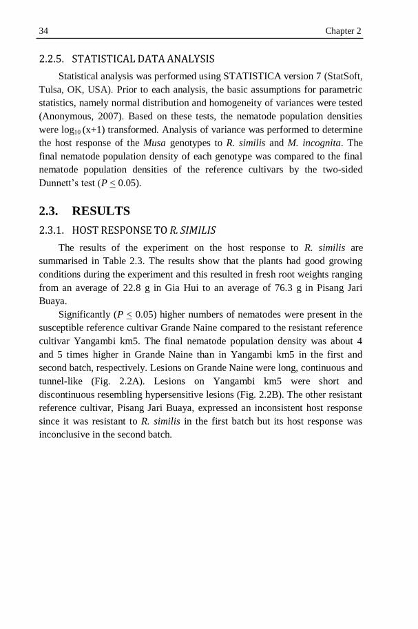
34 Chapter 2
2.2.5. STATISTICAL DATA ANALYSIS
Statistical analysis was performed using STATISTICA version 7 (StatSoft,
Tulsa, OK, USA). Prior to each analysis, the basic assumptions for parametric
statistics, namely normal distribution and homogeneity of variances were tested
(Anonymous, 2007). Based on these tests, the nematode population densities
were log10 (x+1) transformed. Analysis of variance was performed to determine
the host response of the Musa genotypes to R. similis and M. incognita. The
final nematode population density of each genotype was compared to the final
nematode population densities of the reference cultivars by the two-sided
Dunnett‟s test (P < 0.05).
2.3. RESULTS
2.3.1. HOST RESPONSE TO R. SIMILIS
The results of the experiment on the host response to R. similis are
summarised in Table 2.3. The results show that the plants had good growing
conditions during the experiment and this resulted in fresh root weights ranging
from an average of 22.8 g in Gia Hui to an average of 76.3 g in Pisang Jari
Buaya.
Significantly (P < 0.05) higher numbers of nematodes were present in the
susceptible reference cultivar Grande Naine compared to the resistant reference
cultivar Yangambi km5. The final nematode population density was about 4
and 5 times higher in Grande Naine than in Yangambi km5 in the first and
second batch, respectively. Lesions on Grande Naine were long, continuous and
tunnel-like (Fig. 2.2A). Lesions on Yangambi km5 were short and
discontinuous resembling hypersensitive lesions (Fig. 2.2B). The other resistant
reference cultivar, Pisang Jari Buaya, expressed an inconsistent host response
since it was resistant to R. similis in the first batch but its host response was
inconclusive in the second batch.
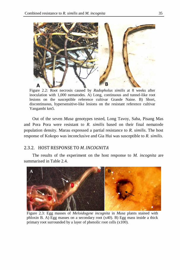
Combined resistance to R. similis and M. incognita 35
Figure 2.2: Root necrosis caused by Radopholus similis at 8 weeks after
inoculation with 1,000 nematodes. A) Long, continuous and tunnel-like root
lesions on the susceptible reference cultivar Grande Naine. B) Short,
discontinuous, hypersensitive-like lesions on the resistant reference cultivar
Yangambi km5.
Out of the seven Musa genotypes tested, Long Tavoy, Saba, Pisang Mas
and Pora Pora were resistant to R. similis based on their final nematode
population density. Marau expressed a partial resistance to R. similis. The host
response of Kokopo was inconclusive and Gia Hui was susceptible to R. similis.
2.3.2. HOST RESPONSE TO M. INCOGNITA
The results of the experiment on the host response to M. incognita are
summarised in Table 2.4.
Figure 2.3: Egg masses of Meloidogyne incognita in Musa plants stained with phloxin B. A) Egg masses on a secondary root (x40). B) Egg mass inside a thick
primary root surrounded by a layer of phenolic root cells (x100).
A B
A)
B)
A)

36 Chapter 2
The results show that the plants had good growing conditions during the
experiment and this resulted in fresh root weights ranging from an average of
11.6 g in Kokopo to an average of 45.2 g in Pisang Mas. Although
characteristic galls were not very conspicuous for direct observation in our
study, many egg-laying females and egg masses were observed during counting
(Fig. 2.3). The susceptible reference cultivar Grande Naine had a high number
of egg-laying females per 5 g roots and the highest final nematode population
density per 5 g roots and per root system. Out of the eight Musa genotypes
tested, Vudu Papau and Pisang Mas were resistant to M. incognita based on
their final nematode population density. The number of egg-laying females per
5 g roots was low in Pisang Mas but in Vudu Papau as high as in Grande Naine.
Nevertheless, the final nematode population density per 5 g roots and per root
system was about 5 to 6 times lower in Vudu Papau than in Grande Naine. The
other genotypes tested were susceptible to M. incognita.
2.4. DISCUSSION
The resistance to R. similis of Long Tavoy, Saba, Pisang Mas and Pora
Pora observed when using sucker-derived plants (Dochez et al., 2006) was
confirmed by our study with tissue culture-derived plants. However, in our
study Gia Hui was susceptible to R. similis while in the study by Dochez et al.
(2006) this genotype was resistant. Our study confirms other reports (De Waele
et al., 1998; Stanton, 1999; Viaene et al., 2003; Blomme et al., 2001, 2003,
2004) that tissue culture-derived plants of some Musa genotypes could be
susceptible and sensitive to R. similis while sucker-derived plants of the same
genotype are resistant. It is possible that the R. similis resistance genes are not
yet expressed by tissue-cultured plants at the time of infection while they are
being expressed at the same time in sucker-derived plants. Previously, the host
response of Pisang Mas to R. similis was reported as less susceptible (Binks &
Gowen, 1997) and partially resistant (Marin et al., 2000). Conversely, another
study (Stoffelen et al., 1999) reported Pisang Mas as susceptible to R. similis.
These observations underline the necessity to confirm the resistant host
response observed in Musa genotypes to R. similis under a range of
experimental conditions, including different types of planting material
(Herradura, 2009).

Combined resistance to R. similis and M. incognita 37
Table 2.3. Host response of selected Musa genotypes and the reference cultivars† to Radopholus similis, measured at 8 weeks after inoculation
with 1,000 vermiform nematodes per plant (n = 8). Plants were inoculated at 8 weeks after planting.
Genotype Fresh root
weight
Pf Rf Comparison of Pf/root system with Host
response (g) per g roots per root system Grande Naine Yangambi km5
Batch 1
Long Tavoy
62.2
86
5331
5.3
*
n.s.
R Long Tavoy 62.2 86 5,331 5.3 * n.s. R Saba 61.2 151 9,521 9.5 * n.s. R
Pisang Mas 53.6 277 15,079 15.1 * n.s. R Marau 43.5 375 15,457 15.5 * * PR Grande Naine† 54.7 485 26,499 26.5 - * S
Yangambi km5† 56.4 134 7,332 7.3 * - R Pisang Jari Buaya† 76.3 150 11,350 11.4 * n.s. R
Batch 2
Pora Pora
69.7
558
37958
38.0
*
n.s.
R Pora Pora 69.7 558 37,958 38.0 * n.s. R
Kokopo 49.9 811 38,404 38.4 n.s. n.s. I Gia Hui 22.8 3,008 62,115 62.1 n.s. * S Grande Naine† 45.2 1,543 69,444 69.4 - * S Yangambi km5† 64.7 245 13,885 13.9 * - R
Pisang Jari Buaya† 65.7 631 40,748 40.7 n.s. n.s. I
Pf: final nematode population density; Rf: reproduction factor (Pf/Pi; Pi: initial nematode inoculum density).
*: significantly different; n.s.: not significantly different according to Dunnett‟s test (P < 0.05).
R: resistant; PR: partially resistant; S: susceptible; I: inconclusive.

38 Chapter 2 Table 2.4. Host response of selected Musa genotypes and the susceptible reference cultivar Grande Naine to Meloidogyne incognita measured
at 8 weeks after inoculation with 4,000 eggs and second-stage juveniles per plant (n = 8). Plants were inoculated at 8 weeks after planting.
Rf: reproduction factor (Pf/Pi; Pi: initial nematode inoculum density).
*: significantly different; n.s.: not significantly different according to Dunnett‟s test (P < 0.05). R: resistant; S: susceptible.
Genotype Fresh root
weight
Egg-
laying
females
Final nematode population
density
(Pf)
Rf Comparison of Pf per root system
with Grande Naine
Host
response
(g) per 5 g
roots
per 5 g
roots
per root
system
Vudu Papau 17.8 8 390 1,390 0.35 * R
Pisang Mas 45.2 2 207 1,874 0.47 * R
Kokopo 11.6 7 1,123 2,606 0.65 n.s. S
Long Tavoy 34.2 4 386 2,643 0.66 n.s. S
Saba 40.3 4 590 4,755 1.19 n.s. S
Gia Hui 17.3 12 1,590 5,501 1.38 n.s. S
Pora Pora 26.1 3 821 4,285 1.07 n.s. S
Marau 18.3 9 1,634 5,979 1.49 n.s. S
Grande Naine 22.5 8 1,990 8,954 2.24 - -

Combined resistance to R. similis and M. incognita 39
Despite the resistance to R. similis shown by Long Tavoy, Saba, Pisang
Mas, Pora Pora and Yangambi km5, the final nematode population densities
were high in their root systems. This indicates that these resistant genotypes are
not immune but significantly less susceptible to R. similis compared to the
susceptible reference cultivar Grande Naine. In the case of complete immunity,
the reproduction ratio would have been less than one as reviewed by Bos and
Parlevliet (1995).
The final R. similis population density of Pisang Jari Buya, the other
resistant reference cultivar, was significantly (P ≤ 0.05) lower in the first batch
compared to Grande Naine but not in the second batch. This observation
confirms previous observations (Herradura, 2009) that the resistance to R.
similis in Pisang Jari Buya in experiments using tissue culture-derived plants
can be more easily overcome by R. similis compared to Yangambi km5. Pisang
Jari Buaya was also reported as showing only partial resistance to R. similis in
another study (Binks & Gowen, 1997). It indicates that Yangambi km5 is a
more reliable resistant reference cultivar than Pisang Jari Buaya. However, care
should be taken with this observation.
The final M. incognita population densities of Vudu Papau and Pisang Mas
were significantly (P ≤ 0.05) lower compared to the susceptible reference
cultivar Grande Naine. Vudu Papau had on average 80.4 and 84.5%, and Pisang
Mas on average of 89.6 and 79.1% less nematodes per 5 g roots and per root
system, respectively, compared to Grande Naine. However, the numbers of egg-
laying females on these two genotypes were not significantly different from
Grande Naine. Although the mean root fresh weight of Pisang Mas was high at
the end of the experiment, this did not lead to a high number of M. incognita
per root system. This can be illustrated by the observation that the root fresh
weight of Pisang Mas was 2 times higher than Grande Naine, but the final
nematode population density per root system was 4.8 times lower. This
observation indicates that Pisang Mas might be a good source of resistance to
M. incognita but this need to be confirmed under field conditions. Care should
be taken since the degree of resistance exhibited by these genotypes is hard to
categorize due to the lack of resistant reference cultivars in Musa. In future
experiments, Pisang Mas could be included as a resistant reference cultivar to
compare M. incognita host responses among Musa genotypes. In this thesis, the
mechanisms of Radopholus similis resistance in Musa genotypes are studied in
the further chapters (Chapter 5 and 6). In Pisang mas, the same mechanisms
could operate against the resistance of Meloidogyne incognita also.This can be
highly interesting for future studies.

40 Chapter 2
In spite of a relatively high number of studies (Stoffelen et al., 1999, 2000;
Van den Bergh et al., 2006; Quénéhervé et al., 2008b), resistance to M.
incognita has not been found in Musa genotypes except by Davide and
Marasigan (1985), and Dizon et al. (2010). Davide and Marasigan (1985)
identified nine out of 90 Musa genotypes evaluated as resistant to M. incognita.
However, the host plant response of these genotypes was only based on the root
gall index, not on nematode reproduction. Dizon et al. (2010) examined the host
response to M. incognita of 32 Philippine banana cultivars and identified three
banana cultivars as resistant to M. incognita based on nematode reproduction.
2.5. CONCLUSION
Our study identified Pisang Mas as a Musa genotype with combined
resistance to both R. similis and M. incognita. This is the first time that a Musa
genotype has been found as resistant to these two major banana root pathogens.
Resistance of Long Tavoy, Saba, Pisang Mas and Pora Pora to R. similis was
confirmed. Three R. similis resistant Musa genotypes, Long Tavoy, Saba,
Yangambi km5 were shortlisted for characterization of R. similis resistance in
comparison with the susceptible reference cultivar Grande Naine. These three
genotypes were selected based on the low final R. similis population density in
their roots.

CHAPTER 3
DEVELOPMENT OF AN AUTOTROPHIC IN VITRO MODEL SYSTEM TO STUDY RADOPHOLUS SIMILIS
HOST LOCATION AND PENETRATION

42 Chapter 3
3.1. INTRODUCTION
Some nematode management strategies such as the use of resistant host
plants aim at the disruption of the life and infection cycles of plant-parasitic
nematodes. Understanding how and when resistant plants interfere with the
attraction and migration of plant-parasitic nematodes towards roots, and with
nematode penetration, development and reproduction inside the roots may lead
to practices that increase the efficacy of the nematode management strategy or
the development of novel nematode management strategies (Sheridan et al.,
2004).
All plant-parasitic nematodes attacking roots are soil-borne. Studying the
nematode‟s behaviour in soil has been very difficult because it is in most cases
impossible to view the nematodes in situ because of their microscopic size,
transparent body and the opaque soil habitat (Spence et al., 2008). To study the
behaviour of plant-parasitic nematodes in the soil, model systems are necessary.
In autotrophic in vitro systems, plant shoots are exposed to open air
allowing the plant to perform active photosynthesis through atmospheric carbon
exchange. At the same time, the root system and the nematodes are maintained
under strict in vitro conditions in a Petri dish. Due to the horizontal root growth
in the Petri dishes, this system enables direct observation of the roots and the
nematodes using a stereoscopic microscope. A successful autotrophic in vitro
system was developed by Van der Veken (2010) to study the bio-protective
effect of Rhizobium etli against Meloidogyne incognita on common bean
(Phaseolus vulgaris). In a strict in vitro system, the whole plant is grown inside
a culture tube hence the plant cannot perform active photosynthesis by
atmospheric carbon exchange. The culture medium supplies carbon source to
these plants in the form of sucrose. The results obtained from this model system
are applicable to the field conditions as the banana plants in the field are
composed of young and old roots. Banana plants produce fresh roots
continuously until flowering (Swennen et al., 1986) and especially nematodes
prefer to enter the root system through the young roots and the root tips (Sarah
et al., 1996).
In this part of our study we report the development of an autotrophic in
vitro model system to study the host location and penetration behaviour of
Radopholus similis (Cobb, 1893) Thorne, 1949 on bananas. The basic set-up of
our system was based on the autotrophic in vitro system developed by Koffi et
al. (2009) to study banana mycorrhization.

Autotrophic in vitro model systems 43
3.2. MATERIALS AND METHODS
3.2.1. PLANTING MATERIAL
Tissue-cultured banana (Musa acuminata) plants of the cultivars Grande
Naine (Musa acuminata AAA, Cavendish subgroup, ITC 1256), Yangambi
km5 (Musa acuminata AAA, Ibota subgroup, ITC 1123) and Saba (Musa spp.
ABB, Saba subgroup, ITC 1138), were initially obtained from the Musa
germplasm collection maintained at the International Transit Centre (ITC),
K.U.Leuven, Belgium. The plant material was proliferated, regenerated and
rooted as explained in Chapter 2 (2.2.2.). Shoots of well-developed plantlets
were transferred to sterile culture boxes (8 x 5.5 x 10 cm) containing 150 ml of
Murashige and Skoog medium including vitamins, 10 mg/l ascorbic acid, 30 g/l
sugar and 2 g/l gelrite with pH 6.12 to allow horizontal root growth (Fig. 3.1A).
The plantlets in the sterile boxes were grown in growth chambers at 28 °C and
16 h photoperiod. After 3 weeks, plantlets with developing secondary roots
were transferred to the autotrophic in vitro system.
3.2.2. NEMATODE INOCULUM
A population of R. similis originally isolated from banana roots in Uganda
was used in the experiments. The population from Uganda was characterized by
a high reproductive fitness (Fallas et al., 1995). This population was maintained
and multiplied monoxenically on sterile alfalfa callus tissues at 25+1 °C in the
dark (Elsen et al., 2001). To obtain inoculum, nematodes were collected from
the callus tissues or from the medium by rinsing with sterile distilled water.
Only 6-weeks-old nematode cultures were used to prepare the inoculum.
3.2.3. THE AUTOTROPHIC IN VITRO SYSTEM
For the autotrophic in vitro system, sterile Petri dishes with a diameter of
14.5 cm were used. The dishes were filled with 150 ml of Murashige and Skoog
medium including vitamins, 10 mg/l ascorbic acid and 2 g/l gelrite with pH 6.12
but lacking sugar. A 1-cm-diameter perforation was made on the edge of the
Petri dish lid using a heated cork borer. Well-rooted, 3-weeks-old plantlets from
the culture boxes (Fig. 3.1A) were transferred carefully to the Murashige and
Skoog medium at an edge of the Petri dishes. Placing the plants in the edge did
not affect the growth of the plants as previously tested with mulching the
banana plants in either side of the root system (Swennen, 1984).
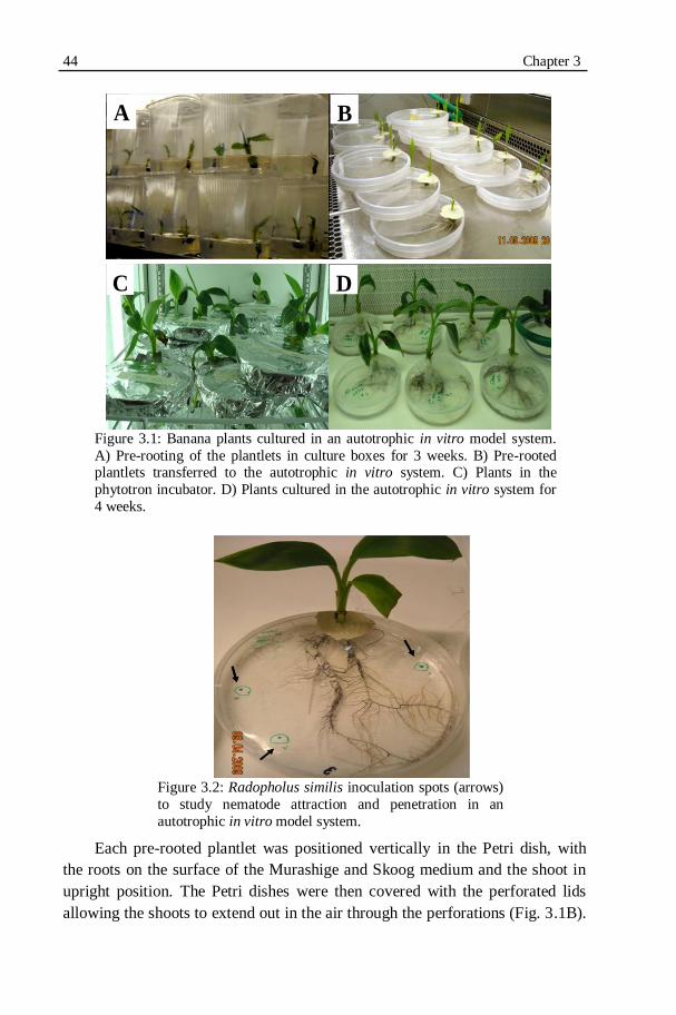
44 Chapter 3
Figure 3.1: Banana plants cultured in an autotrophic in vitro model system.
A) Pre-rooting of the plantlets in culture boxes for 3 weeks. B) Pre-rooted plantlets transferred to the autotrophic in vitro system. C) Plants in the
phytotron incubator. D) Plants cultured in the autotrophic in vitro system for
4 weeks.
Figure 3.2: Radopholus similis inoculation spots (arrows)
to study nematode attraction and penetration in an
autotrophic in vitro model system.
Each pre-rooted plantlet was positioned vertically in the Petri dish, with
the roots on the surface of the Murashige and Skoog medium and the shoot in
upright position. The Petri dishes were then covered with the perforated lids
allowing the shoots to extend out in the air through the perforations (Fig. 3.1B).
A B
C D
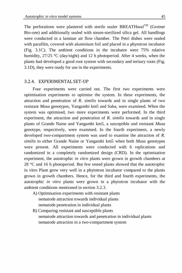
Autotrophic in vitro model systems 45
The perforations were plastered with sterile sealer BREATHseal
TM (Greiner
Bio-one) and additionally sealed with steam-sterilized silica gel. All handlings
were conducted in a laminar air flow chamber. The Petri dishes were sealed
with parafilm, covered with aluminium foil and placed in a phytotron incubator
(Fig. 3.1C). The ambient conditions in the incubator were 75% relative
humidity, 27/25 °C (day/night) and 12 h photoperiod. After 4 weeks, when the
plants had developed a good root system with secondary and tertiary roots (Fig.
3.1D), they were ready for use in the experiments.
3.2.4. EXPERIMENTAL SET-UP
Four experiments were carried out. The first two experiments were
optimisation experiments to optimise the system. In these experiments, the
attraction and penetration of R. similis towards and in single plants of two
resistant Musa genotypes, Yangambi km5 and Saba, were examined. When the
system was optimised, two more experiments were performed. In the third
experiment, the attraction and penetration of R. similis towards and in single
plants of Grande Naine and Yangambi km5, a susceptible and resistant Musa
genotype, respectively, were examined. In the fourth experiment, a newly
developed two-compartment system was used to examine the attraction of R.
similis to either Grande Naine or Yangambi km5 when both Musa genotypes
were present. All experiments were conducted with 6 replications and
randomized in a completely randomized design (CRD). In the optimisation
experiment, the autotrophic in vitro plants were grown in growth chambers at
28 °C and 16 h photoperiod. But few tested plants showed that the autotrophic
in vitro Plant grew very well in a phytotron incubator compared to the plants
grown in growth chambers. Hence, for the third and fourth experiments, the
autotrophic in vitro plants were grown in a phytotron incubator with the
ambient conditions mentioned in section 3.2.3.
A) Optimisation experiments with resistant plants
nematode attraction towards individual plants
nematode penetration in individual plants
B) Comparing resistant and susceptible plants
nematode attraction towards and penetration in individual plants
nematode attraction in a two-compartment system
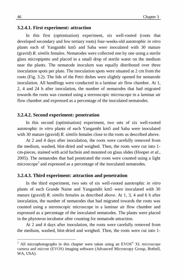
46 Chapter 3
3.2.4.1. First experiment: attraction
In this first (optimisation) experiment, six well-rooted (roots that
developed secondary and few tertiary roots) four-weeks-old autotrophic in vitro
plants each of Yangambi km5 and Saba were inoculated with 30 mature
(gravid) R. similis females. Nematodes were collected one by one using a sterile
glass micropipette and placed in a small drop of sterile water on the medium
near the plants. The nematode inoculum was equally distributed over three
inoculation spots per plate. The inoculation spots were situated at 2 cm from the
roots (Fig. 3.2). The lids of the Petri dishes were slightly opened for nematode
inoculation. All handlings were conducted in a laminar air flow chamber. At 1,
2, 4 and 24 h after inoculation, the number of nematodes that had migrated
towards the roots was counted using a stereoscopic microscope in a laminar air
flow chamber and expressed as a percentage of the inoculated nematodes.
3.2.4.2. Second experiment: penetration
In this second (optimisation) experiment, two sets of six well-rooted
autotrophic in vitro plants of each Yangambi km5 and Saba were inoculated
with 30 mature (gravid) R. similis females close to the roots as described above.
At 2 and 4 days after inoculation, the roots were carefully removed from
the medium, washed, blot-dried and weighed. Then, the roots were cut into 1-
cm-pieces, stained with acid fuchsin and mounted on glass slides (Hooper et al.,
2005). The nematodes that had penetrated the roots were counted using a light
microscope3 and expressed as a percentage of the inoculated nematodes.
3.2.4.3. Third experiment: attraction and penetration
In the third experiment, two sets of six well-rooted autotrophic in vitro
plants of each Grande Naine and Yangamibi km5 were inoculated with 30
mature (gravid) R. similis females as described above. At 1, 3, 4 and 6 h after
inoculation, the number of nematodes that had migrated towards the roots was
counted using a stereoscopic microscope in a laminar air flow chamber and
expressed as a percentage of the inoculated nematodes. The plants were placed
in the phytotron incubator after counting for nematode attraction.
At 2 and 4 days after inoculation, the roots were carefully removed from
the medium, washed, blot-dried and weighed. Then, the roots were cut into 1-
3 All microphotographs in this chapter were taken using an EVOS® XL microscope
camera and micron (EVOS) imaging software (Advanced Microscopy Group, Bothell,
WA, USA).

Autotrophic in vitro model systems 47
cm-pieces, stained with acid fuchsin and mounted on glass slides (Hooper et al.,
2005). The nematodes that had penetrated the roots were counted using a light
microscope and expressed as a percentage of the inoculated nematodes.
3.2.4.4. Fourth experiment: attraction and penetration in a two-
compartment system
In the fourth experiment, six well-rooted autotrophic in vitro plants each of
Grande Naine and Yangambi km5 were connected with a small transparent
bridge at the start of the experiment (Fig. 3.3).
Figure 3.3: Two-compartment autotrophic in vitro model system to study nematode
attraction to banana plants. R1, R2 and R3 represent the attraction zones of the
resistant plant. S1, S2 and S3 represent the attraction zones of the susceptible plant.
Figure 3.4: Two-compartment autotrophic in vitro model system to study nematode attraction to banana plants.
The bridge was 7 cm long, 1 cm wide and contained approximately 4.5 ml
of 1% sterile water agar. The middle of the bridge was selected as the
inoculation spot. Three attraction zones were marked off at each side of the
bridge based on their distance from the inoculation spot (Table 3.1). The zones

48 Chapter 3 at the side of Yangambi km5, the resistant genotype, are denoted as R1, R2 and
R3 while the zones at the side of Grande Naine, the susceptible genotype, are
denoted as S1, S2 and S3 (Fig. 3.3). To connect the Petri dishes to the bridge, 1-
cm-diameter openings were made on the edge of the Petri dishes using a heated
forceps. The bridges were secured firmly using transparent sticky tapes (Fig.
3.4).
Table 3.1. Nematode attraction zones in the two-compartment
autotrophic in vitro model system.
Zone Distance from the inoculation spot
1 0-1.0 cm
2 1-2.0 cm
3 2-3.5 cm
At 3 h after the two compartments were connected via the bridge, 30
mature (gravid) R. similis females were inoculated in a small depression made
at the inoculation spot. At 30 min, 1, 2, 3 and 6 h after inoculation, the number
of nematodes that had migrated to each attraction zone was counted using a
stereoscopic microscope in a laminar air flow chamber and expressed as a
percentage of the inoculated nematodes.
3.2.5. STATISTICAL DATA ANALYSIS
Data with normal distribution and homogeneous variances were subjected
to analysis of variances using STATISTICA version 9 (StatSoft, Tulsa, OK,
USA). Percentages (percentage nematode attraction and penetration) were
arcsin (x/100) transformed prior to the analysis. When significant differences (P
≤ 0.05) were observed, Tukey‟s HSD (Honestly Significant Difference) test was
applied for multiple comparisons of group means.
3.3. RESULTS
3.3.1. FIRST (OPTIMISATION) EXPERIMENT: ATTRACTION
Migration of the nematodes towards the roots was observed (Fig. 3.5).
Plants had good growth in the absence of sugar due to active photosynthesis. At
1 h after inoculation, a significantly (P ≤ 0.05) higher percentage of R. similis
had migrated towards the roots of Saba compared to Yangambi km5 (52.6 vs
6.7%)

Autotrophic in vitro model systems 49
Fig. 3.5: Migration of Radopholus similis females towards the roots of A) Yangambi
km5 and B) Saba at 1 h after inoculation. Magnification: 40x
Figure 3.6: Attraction of Radopholus similis females (expressed as a percentage of
30 inoculated mature females) towards the roots of 4-weeks-old plants of the Musa
genotypes Yangambi km5 and Saba at 1, 2, 4 and 24 h after inoculation. Means are
presented between parentheses. Means within a same time (h) followed by the same
letter are not significantly (P ≤ 0.05) different from each other according to Tukey‟s
HSD test. Error bars represent confidence intervals. (n=6).
At 2, 4 and 24 h after inoculation, no significant differences in migration
towards the roots of both Musa genotypes were observed anymore. The
difference between the two genotypes decreased at each observation time and at
24 h after inoculation, more than 80% of the inoculated nematodes had
migrated towards the roots of Saba as well as Yangambi km5 (Fig. 3.6).
A B

50 Chapter 3
3.3.2. SECOND (OPTIMISATION EXPERIMENT): PENETRATION
Plant growth in the autotrophic in vitro system was good (Table 3.2). A
small decrease in fresh root and shoot weights was observed at 4 days after
inoculation (DAI) compared to 2 DAI but the differences were not statistically
significant. No significant differences were observed between the percentages
of nematodes that had penetrated the roots of Saba and Yangambi km5 at 2 and
4 DAI. At 2 DAI, 30 and 25.7% of the 30 inoculated females had penetrated the
roots of Saba and Yangambi km5, respectively (Fig. 3.7).
Table 3.2. Fresh root and shoot weights of 4-weeks-old plants of the Musa
genotypes Saba and Yangambi km5 at 2 and 4 days after inoculation (DAI) with
30 mature females of Radopholus similis.
Genotypes Fresh root weight (g) Fresh shoot weight (g)
2 DAI 4DAI 2 DAI 4DAI
Saba 0.88 0.64 1.26 1.18
Yangambi km5 0.58 0.44 1.60 1.38
Means within the same columns followed by the same letter are not significantly
(P ≤ 0.05) different from each other according to Tukey‟s HSD Test. Data were
log10 (x+1) transformed prior to statistical analysis.
Although more than 80% of the females had been attracted and migrated
towards the roots (Fig 3.6), only about 25 to 30% of the females had penetrated
the roots at 2 DAI (Fig. 3.7A). Penetration of a single root by many females at
the same site was observed (Fig. 3.8B). Penetrated adult females were laying
eggs at 4 DAI in both Saba and Yangambi km5 (Fig. 3.8). At 4 DAI, no
significant differences were observed in the number of eggs laid in Saba and
Yangambi km5 (Fig. 3.7B). As no increase in penetration was observed at 4
DAI compared with 2 DAI, the observation time to examine nematode
penetration was limited to 2 DAI in the next experiment.
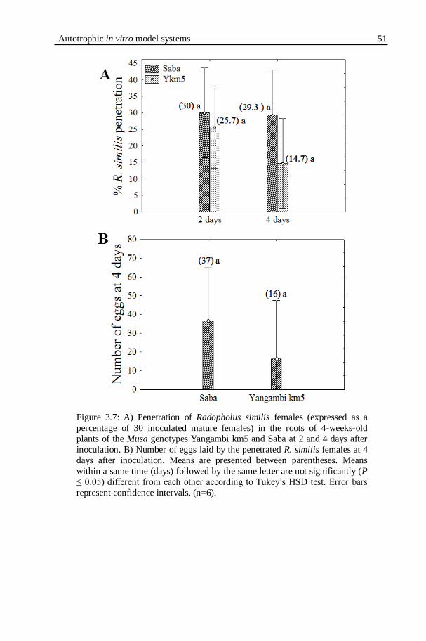
Autotrophic in vitro model systems 51
Figure 3.7: A) Penetration of Radopholus similis females (expressed as a
percentage of 30 inoculated mature females) in the roots of 4-weeks-old
plants of the Musa genotypes Yangambi km5 and Saba at 2 and 4 days after
inoculation. B) Number of eggs laid by the penetrated R. similis females at 4
days after inoculation. Means are presented between parentheses. Means
within a same time (days) followed by the same letter are not significantly (P
≤ 0.05) different from each other according to Tukey‟s HSD test. Error bars
represent confidence intervals. (n=6).
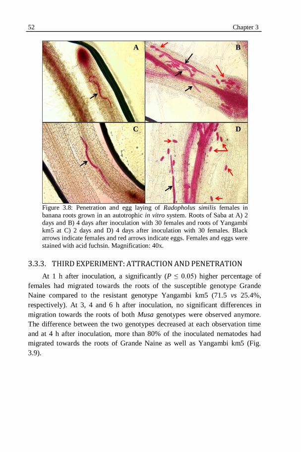
52 Chapter 3
Figure 3.8: Penetration and egg laying of Radopholus similis females in
banana roots grown in an autotrophic in vitro system. Roots of Saba at A) 2
days and B) 4 days after inoculation with 30 females and roots of Yangambi
km5 at C) 2 days and D) 4 days after inoculation with 30 females. Black
arrows indicate females and red arrows indicate eggs. Females and eggs were
stained with acid fuchsin. Magnification: 40x.
3.3.3. THIRD EXPERIMENT: ATTRACTION AND PENETRATION
At 1 h after inoculation, a significantly (P ≤ 0.05) higher percentage of
females had migrated towards the roots of the susceptible genotype Grande
Naine compared to the resistant genotype Yangambi km5 (71.5 vs 25.4%,
respectively). At 3, 4 and 6 h after inoculation, no significant differences in
migration towards the roots of both Musa genotypes were observed anymore.
The difference between the two genotypes decreased at each observation time
and at 4 h after inoculation, more than 80% of the inoculated nematodes had
migrated towards the roots of Grande Naine as well as Yangambi km5 (Fig.
3.9).
A B
C D

Autotrophic in vitro model systems 53
Figure 3.9: Attraction of Radopholus similis females (expressed as a
percentage of 30 inoculated mature females) towards the roots of 4-weeks-
old plants of the susceptible Musa genotype Grande Naine and the resistant
Musa genotype Yangambi km5 at 1, 3, 4 and 6 h after inoculation. Means are presented between parentheses. Means within a same time (h) followed by
the same letter are not significantly (P ≤ 0.05) different from each other
according to Tukey‟s HSD test. Error bars represent confidence intervals.
(n=6).
Plant growth in the autotrophic in vitro system was good (Table 3.3). The
percentages of females that had penetrated the roots of Yangambi km5 and
Grande Naine were not significantly different from each other at both 1 and 2
DAI (Fig. 3.10).
Table 3.3. Fresh root and shoot weights of 4-weeks-old plants of the Musa genotypes
Grande Naine and Yangambi km5 at 1 and 2 days after inoculation (DAI) with 30
mature females of Radopholus similis.
Genotype Fresh root weight (g) Fresh shoot weight (g)
1 DAI 2 DAI 1 DAI 2 DAI
Grande Naine 5.0 4.8 5.2 4.6
Yangambi km5 2.9 3.3 3.1 3.8
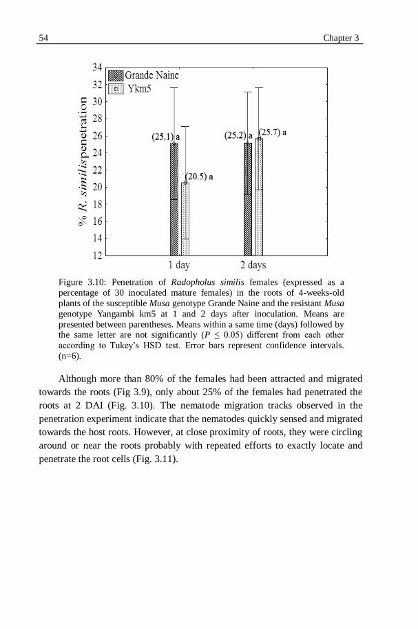
54 Chapter 3
Figure 3.10: Penetration of Radopholus similis females (expressed as a
percentage of 30 inoculated mature females) in the roots of 4-weeks-old
plants of the susceptible Musa genotype Grande Naine and the resistant Musa
genotype Yangambi km5 at 1 and 2 days after inoculation. Means are
presented between parentheses. Means within a same time (days) followed by the same letter are not significantly (P ≤ 0.05) different from each other
according to Tukey‟s HSD test. Error bars represent confidence intervals.
(n=6).
Although more than 80% of the females had been attracted and migrated
towards the roots (Fig 3.9), only about 25% of the females had penetrated the
roots at 2 DAI (Fig. 3.10). The nematode migration tracks observed in the
penetration experiment indicate that the nematodes quickly sensed and migrated
towards the host roots. However, at close proximity of roots, they were circling
around or near the roots probably with repeated efforts to exactly locate and
penetrate the root cells (Fig. 3.11).

Autotrophic in vitro model systems 55
Figure 3.11: Migration tracks of Radopholus similis females near banana
roots in an autotrophic in vitro system observed at 24 h after inoculation. A)
and B) Grande Naine. C) Yangambi km5. At the beginning of the
experiment, 30 mature females were placed at 2 cm distance from the roots.
R: roots. Scale bar: 200µm
3.3.4. FOURTH EXPERIMENT: NEMATODE ATTRACTION IN A TWO-COMPARTMENT SYSTEM
The results of this experiment are shown in Fig. 3.12. At 30 min and 1 h
after inoculation, the majority of the inoculated R. similis females had remained
at the inoculation point except a small percentage (about 2%) of the nematodes
that had migrated to the first attraction zones (R1 & S1).
R
R
R
A
B C
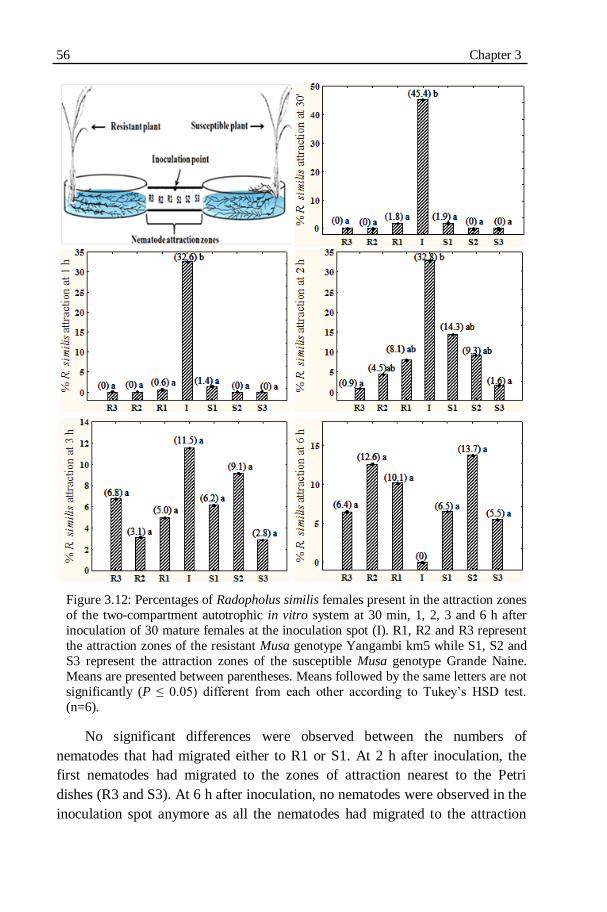
56 Chapter 3
Figure 3.12: Percentages of Radopholus similis females present in the attraction zones
of the two-compartment autotrophic in vitro system at 30 min, 1, 2, 3 and 6 h after
inoculation of 30 mature females at the inoculation spot (I). R1, R2 and R3 represent
the attraction zones of the resistant Musa genotype Yangambi km5 while S1, S2 and
S3 represent the attraction zones of the susceptible Musa genotype Grande Naine.
Means are presented between parentheses. Means followed by the same letters are not
significantly (P ≤ 0.05) different from each other according to Tukey‟s HSD test.
(n=6).
No significant differences were observed between the numbers of
nematodes that had migrated either to R1 or S1. At 2 h after inoculation, the
first nematodes had migrated to the zones of attraction nearest to the Petri
dishes (R3 and S3). At 6 h after inoculation, no nematodes were observed in the
inoculation spot anymore as all the nematodes had migrated to the attraction
Autotrophic in vitro model systems 57
zones. No significant differences were observed between the numbers of
nematodes that had migrated towards the different attraction zones at 3 and 6 h.
About 30% of the females had been attracted to the zones of the resistant
genotype Yangambi km5 while about 25% of the females had been attracted to
the zones of the susceptible Grande Naine.
3.4. DISCUSSION
The autotrophic in vitro system proved to be a very good model system
with notable advantages over the strict in vitro model systems to study
nematode attraction towards and penetration in the roots of banana. First, in
case of the strict in vitro systems, banana plants are usually grown inside glass
tubes or jars. As a result their roots are not suitable for non-destructive
microscopic observation. In an autotrophic in vitro system, the plant roots
develop in a thin gel layer in a Petri dish. This enables direct observation of the
nematodes using a stereoscopic microscope while the plant is developing intact
and undisturbed. Second, the sucrose-free medium and the exposure of the
shoots to open air allowed the plants to perform active photosynthesis through
atmospheric carbon exchange. Thus, this system simulates more of the plant‟s
natural conditions when compared to the strict in vitro systems. Third, the
autotrophic in vitro system does not compromise the advantages of the strict in
vitro systems such as providing controlled experimental conditions, limited
space requirements, reduced experimental duration and easy handling (Elsen et
al., 2002). Hence, large batches of plants can be studied in a short time. This
system could also be used to screen banana plants for nematode resistance.
Banana plants were grown successfully for about 11 weeks in this system
(Koffi et al., 2009).
However, the autotrophic in vitro system demands continuous monitoring
as the vigorous growth of the shoots disturbs the silica gel which is sealing the
perforation on the lid. This makes the system sensitive to microbial
contamination. Contaminations were prevented by re-applying the sterile silica
gel whenever there is a possibility of exposure. As the vigorous root growth
also depletes the medium, it is necessary to refill the medium especially in long
duration experiments such as screening for nematode resistance. Careful
extrapolation of the results to field situation should be done as nematode
movement in the two-dimentional agar based medium is unlike the three-
dimentional natural soil environments (Spence et al., 2008).
Results from our study show that the R. similis females were attracted to
both the resistant and susceptible Musa genotypes included in our experiments
58 Chapter 3 within the first hour after infection. The low percentage of nematode attraction
observed towards Yangambi km5 roots at 1 h after inoculation might be due to
reduced secondary and tertiary root growth compared to Saba and Grande
Naine. Nematode attraction is halted when the root‟s growth is stopped or
limited (Perry, 1996). In general, no differences were observed in host location
of resistant and susceptible plants by R. similis females when tested separately.
Furthermore, in the two-compartment system, R. similis females showed no
preference to migrate towards the roots of either the resistant or susceptible
Musa genotype when a choice was given.
Concentrated, circling nematode migration tracks were observed at close
proximity of the banana roots in our study. This may be because of a high
concentration of root exudates or attraction cues near the roots making it
difficult for the nematodes to exactly locate the roots. The nematodes seemed to
make repeated attempts to penetrate the roots.
A general drop in the percentage nematode penetration was observed after
initial penetration. Though insignificant, this trend is also observed in the
greenhouse experiments reported in the following chapter (4.3.1 & Fig. 4.3).
Emigration of certain nematodes could be a probable reason. Drastic drop in
nematode penetration after initial penetration of tobacco roots by M. incognita
was reported previously. It was attributed to nematodes emigrated after
unsuccessful attempts to establish feeding sites (Schneider, 1991). Windham &
Williams (1994) reported probable emigration of J2 from resistant and
susceptible corn genotypes. Emigration of M. arenaria juveniles were reported
from resistant and susceptible myrobalan plum cultivars (Voisin et al., 1999).
Penetration of same root by several nematodes was observed in our study.
Secretions from nematode-damaged plant cells or pheromones from the
nematode were reported as possible reasons (Wang et al., 2009)
No differences were observed in the penetration of the resistant and
susceptible Musa genotypes included in our experiments by R. similis females.
Despite the fact that most of the inoculated females migrated to the plant roots
within a few hours after inoculation, a relatively small fraction penetrated the
roots successfully. Data on the attraction and penetration of R. similis are
scarce. Studies on root-knot nematodes (Meloidogyne spp.) report a similar
ratio of nematode penetration in other crops. For instance, less than 10% of the
inoculated M. incognita were reported to penetrate susceptible as well as
resistant genotypes of cotton (Faske & Starr, 2009), M. arenaria, M. hapla, M.
incognita and M. javanica in cucumber (Walters et al., 2006), and M. arenaria
in soybean (Pedrosa et al., 1996). Fogain (2000b) reported no difference in the
penetration rates of R. similis on Yangambi km5 and French Sombre, a

Autotrophic in vitro model systems 59
susceptible plantain. Similarly, no differences were reported in the penetration
rates of R. similis in susceptible and resistant Musa cultivars based on an in
vitro study but using excised roots, not intact plants (Dochez, 2004).
Conversely, a higher rate of R. similis penetration was reported in the roots of
the susceptible Musa genotype Poyo compared to Yangambi km5 at 24 h after
inoculation (Valette et al., 1997). It is generally accepted that plant-parasitic
nematodes are attracted by the host roots and that the sensitivity of the
nematodes for this is very high. Generally, the attraction does not appear to be
selective (Prot, 1980; Perry, 1996).
3.5. CONCLUSIONS
The autotrophic in vitro model system was a very good system with
notable advantages over the strict in vitro systems to study nematode attraction
and penetration. No differences were observed in host location of the resistant
and susceptible Musa genotypes included in our experiments by R. similis
females except at 1h after inoculation. Furthermore, R. similis females showed
no preference to migrate towards the roots of either the resistant or susceptible
Musa genotypes when a choice was given to migrate towards both plant roots at
the same time. Also no differences were observed in the penetration of the
resistant and susceptible Musa genotypes included in our experiments by R.
similis females.
Greenhouse experiments should confirm the results observed in vitro.
Therefore a greenhouse study was conducted and the results of this study on
nematode penetration, development and reproduction of R. similis on resistant
and susceptible Musa genotypes are presented in Chapter 4.


CHAPTER 4
HOST PLANT RESISTANCE IN MUSA GERMPLASM TO RADOPHOLUS SIMILIS: PRE- OR POST-INFECTIONAL?

62 Chapter 4
4.1. INTRODUCTION
The successful infection of susceptible Musa genotypes by Radopholus
similis (Cobb, 1893) Thorne, 1949 involves many compatible nematode-plant
interactions eventually leading to damage and yield loss. Although these
interactions are dynamic and continuous, specific phases can be distinguished.
The first phase is the location of the host plant and migration towards the roots
of the host plant which are based, among many factors, on cues released by the
host plant such as root exudates (Perry, 1996; Spence et al., 2008). The root and
root cap exudates are rich in ions, enzymes, mucilage, secondary metabolites
such as IAA, volatile compounds as well as peptides and plant primary
metabolites (Curtis 2008; Wang et al., 2011). Next, the nematodes penetrate the
root cell walls with the aid of a stylet and cell wall degrading enzymes
(Haegeman et al., 2008, 2009). Then, after entering the roots, the nematodes
feed continuously and develop into further life stages, and finally reproduce.
In contrast, resistant plants interfere in the nematodes‟ life and infection
cycles ultimately resulting in an incompatible plant-nematode interaction. This
process also involves many host responses limiting the nematodes (reviewed by
Fuller et al., 2008). Host plant resistance to nematodes may occur either at the
pre- or post-infectional level (Kaplan & Keen, 1980).
When the resistance operates at the pre-infectional level, the nematodes‟
attraction and/or migration towards the roots of the host plant or penetration
into the roots of the host plant is hampered. The host plant can release cues such
as metabolites or peptides that block the nematodes‟ sensory perception
disrupting the orientation of the nematodes towards the roots (Wang et al.,
2011). The host plant can resist penetration of the root cell walls by constitutive
or induced production or deployment of structural defence barriers such as
lignin and suberin (Valette et al., 1998).
In association with or apart from these early host plant responses, a
stronger defence response can occur at the post-infectional stage when the
nematodes have breached the first line of physical and chemical defences. Post-
infectional resistance mechanisms are usually characterized by impaired
nematode development and reproduction caused by various mechanisms
(Zacheo et al., 1997; Fuller et al., 2008). These resistance mechanisms include
the increased production of anti-nematode products or toxins such as
phytoalexins, oxidised phenolics, lipid peroxides, hydrolytic enzymes and
proteinases-inhibitors in the feeding cells, and hypersensitive responses.

R. similis penetration, development and reproduction 63
The objective of this part of our study was to enhance our basic
understanding of the resistance mechanism(s) in Musa spp. to R. similis. To
achieve this objective, penetration, development and reproduction of R. similis
in three resistant Musa genotypes, Long Tavoy (Musa acuminate AA,
Burmannica subgroup, ITC 0283), Saba (Musa spp. ABB, Saba subgroup, ITC
1138) and Yangambi km5 (Musa acuminata AAA, Ibota subgroup, ITC 1123),
and one susceptible Musa genotype, Grande Naine (Musa acuminata AAA,
Cavendish subgroup, ITC 1256), were examined and compared. The resistant
Musa genotypes were selected based on the results reported in Chapter 2. They
showed good growth under the experimental conditions and expressed a strong
resistance to R. similis.
4.2. MATERIALS AND METHODS
4.2.1. EXPERIMENTAL SET-UP
Two independent greenhouse experiments were conducted. In the first
experiment, the attraction and penetration of R. similis towards and in the roots
of three R. similis-resistant Musa genotypes (Long Tavoy, Saba, Yangambi
km5) and one susceptible Musa genotype (Grande Naine) were examined. In
the second experiment, the development and reproduction of R. similis was
examined in the same four Musa genotypes. In the greenhouse, the ambient day
and night temperatures were 27 and 20 ºC, respectively, the relative humidity
was 80% and the photoperiod was 12 h.
4.2.2. PLANTING MATERIAL
All Musa genotypes were initially obtained from the Musa germplasm
collection maintained at the International Transit Centre (ITC), K.U.Leuven,
Belgium. The plant material was proliferated and rooted in test tubes as
explained in Chapter 2 (2.2.2). The plantlets were grown in growth chambers at
28 °C and 16 h photoperiod.
4.2.3. NEMATODE INOCULUM
A population of R. similis originally isolated from banana roots in Uganda
was used in the experiments. This population was maintained and multiplied
monoxenically on sterile carrot discs at 25+1 °C in the dark (Speijer & De
Waele, 1997). The population from Uganda was characterized by a high
reproductive fitness (Fallas et al., 1995). To obtain the inoculum, R. similis was
64 Chapter 4
extracted from the carrot discs by the maceration-sieving method (Speijer & De
Waele, 1997).
4.2.4. FIRST EXPERIMENT: NEMATODE ATTRACTION AND
PENETRATION
Six-weeks-old rooted tissue culture plantlets were planted in 200 ml cups
filled with sand and potting soil (2:1). The plants were maintained under
greenhouse conditions and fertilized at 10 days intervals throughout the
experiment.
Six weeks after planting, approximately 1,000 living vermiform nematodes
were inoculated per plant by pipetting 4 ml of the nematode suspension into
inoculation holes made in the soil near the plant root zone. At 4, 8 and 12 days
after inoculation, plants were uprooted and gently washed free of soil under
running tap water. The fresh root and shoot weights were measured. The
nematodes were extracted from the whole root systems by the maceration-
sieving method (Speijer & De Waele, 1997) and counted as described in
Chapter 2 (2.2.4). One replication of each genotype at every harvest time was
stained with acid fuchsin to visualise and photo-document the nematodes that
had penetrated the roots. All treatments (four genotypes at three harvest times)
were replicated eight times and a randomized block design (RBD) was used.
The experiment was repeated once to confirm the results.
4.2.5. SECOND EXPERIMENT: POST-INFECTIONAL NEMATODE
DEVELOPMENT AND REPRODUCTION
Six-weeks-old rooted tissue culture plantlets were planted in 200 ml cups
filled with sand and potting soil (2:1). The plants were maintained under
greenhouse conditions and fertilized at 10 days intervals throughout the
experiment.
Six weeks after planting, each plant was inoculated with a 4 ml suspension
containing 1,000 living vermiform nematodes pipetted into inoculation holes
made in the soil near the plant root zone. At 12 days after inoculation, plants
were uprooted and gently washed free of soil under running tap water to limit
nematode penetration to a maximum of 12 days. Then the plants were
transplanted in 1 l pots filled with the sand and potting soil (2:1).
At 26 days after inoculation (i.e. 14 days after transplanting), plants were
uprooted and gently washed free of soil under running tap water. The fresh root
and shoot weights were measured. The nematodes were extracted from the
whole root systems by the maceration-sieving method (Speijer & De Waele,

R. similis penetration, development and reproduction 65
1997) and counted as described in Chapter 2 (2.2.4). The final nematode
population densities were determined by calculating the sum of all
developmental stages of the nematodes extracted from the whole root systems.
The reproduction factor (Rf) was calculated as the final nematode population
density divided by the inoculum. All treatments (four genotypes) were
replicated eight times and a randomized block design was used.
4.2.6. STATISTICAL DATA ANALYSIS
Data with normal distribution and homogeneous variances were subjected
to analysis of variances using STATISTICA version 9 (StatSoft, Tulsa, OK,
USA). Nematode numbers were log10(x+1) transformed prior to the analysis.
When significant (P ≤ 0.05) differences were observed, Tukey‟s HSD
(Honestly Significant Difference) test was applied for multiple comparisons of
group means.
4.3. RESULTS
4.3.1. FIRST EXPERIMENT: NEMATODE ATTRACTION AND
PENETRATION
The fresh root and shoot weights of the Musa genotypes at 4, 8 and 12
days after inoculation (DAI) are presented in Table 4.1.
Lesions were observed on the secondary and tertiary feeder roots of all
genotypes on 4 DAI. No visible damages were observed on the primary roots of
the resistant and susceptible Musa genotypes at 4 DAI. Visibly large lesions
were observed on the primary and secondary roots of Grande Naine at 8 and 12
DAI (Figs 4.1A & B). Much smaller lesions were visible on the primary roots
of the resistant Musa genotypes at 8 and 12 DAI (Figs 4.1C & D).
The inoculated adults and juveniles had penetrated the roots of both
resistant and susceptible Musa genotypes starting from the first observation at 4
DAI (Figs 4.2A & B). Eggs were observed next to the penetrated females in the
roots of Grande Naine and Long Tavoy (Figs 4.2.C & D). Phenolic cells were
observed in Yangambi km5 always at the nematode infection sites (Fig. 4.2E).
Few nematodes were found invading the stele of Yangambi km5 especially in
the secondary roots or at root junctions. No nematodes were observed in the
stele of the other genotypes.

66 Chapter 4
Table 4.1. Fresh root and shoot weights of the Musa genotypes at 4, 8 and 12 days after
inoculation (DAI) with 1,000 adults and juveniles of Radopholus similis. The
nematodes were inoculated around the roots of six-weeks-old plants. (n=8).
Genotypes Root fresh weight (g) Shoot fresh weight (g)
4 DAI 8 DAI 12 DAI 4 DAI 8 DAI 12 DAI
Long Tavoy 5.9a 7.4b 10.7b 20.1b 24.2b 25.7b
Saba 3.7a 5.4ab 8.1a 10.8a 17.4a 16.2a
Yangambi km5 4.5a 5.0ab 8.2ab 17.5b 18.2ab 20.6ab
Grande Naine 4.1a 4.1a 7.6a 16.3ab 17.2a 20.0ab
Means within a same column followed by the same letter are not significantly (P ≤
0.05) different according to Tukey‟s HSD test.
No significant differences were observed between the numbers of
nematodes that had penetrated the four Musa genotypes at 4 and 8 DAI (Fig.
4.3). At 12 DAI, a significantly (P ≤ 0.05) lower number of nematodes had
penetrated the roots of Long Tavoy compared to Grande Naine. No significant
differences were observed between the number of nematodes that had
penetrated Saba, Yangambi km5 and Grande Naine at 12 DAI (Fig. 4.3). A
decrease in the number of penetrated nematodes was observed in all genotypes
at 12 DAI.
Eggs were laid by the females that had penetrated in Grande Naine at 8
DAI but not in any of the three resistant Musa genotypes at 8 DAI. At 12 DAI,
eggs were also observed in the roots of Long Tavoy but the number of eggs was
significantly (P ≤ 0.05) lower than in Grande Naine. No eggs were observed in
Saba and Yangambi km5 (Fig. 4.4).

R. similis penetration, development and reproduction 67
Figure 4.1: Root damage caused by Radopholus similis. A) and B) Lesions on the
primary and secondary roots of Grande Naine at 8 days and 12 days after
inoculation, respectively. C) Small lesions on the primary and secondary roots of
Saba at 8 days after inoculation. D) Small lesions on the primary roots of Long
Tavoy at 12 days after inoculation. Arrows indicate the lesions on the primary roots.
Figure 4.2: Females of Radopholus similis penetrating the roots of A) Grande
Naine and B) Long Tavoy at 4 days after inoculation. Eggs (arrows) laid by the
penetrated females at 12 days after inoculation in C) Grande Naine and D) Long
Tavoy. E) Phenolic cells (black arrows) observed only around the nematode (red
arrow) infection sites in Yangambi km5. Magnification: 40x
A B
C D
A
C D E
B

68 Chapter 4
Figure 4.3: Number of Radopholus similis that had penetrated the roots of the resistant (Long Tavoy, Saba, Yangambi km5) and the susceptible (Grande Naine) Musa
genotypes at 4, 8 and 12 days after inoculation with 1,000 adults and juveniles of
Radopholus similis. Data were log10(x+1) transformed prior to statistical analysis. The
original means are presented between parentheses. Means within a same time (DAI)
followed by the same letter are not significantly (P ≤ 0.05) different from each other
according to Tukey‟s HSD test. Error bars represent confidence intervals. (n=8).
Figure 4.4: Number of Radopholus similis eggs observed in the roots of the resistant
(Long Tavoy, Saba, Yangambi km5) and the susceptible (Grande Naine) Musa
genotypes at 12 days after inoculation with 1,000 adults and juveniles of Radopholus
similis. Means are presented between parentheses. Means followed by the same letter
are not significantly (P ≤ 0.05) different from each other according to Tukey‟s HSD test.
Error bars represent confidence intervals. (n=8).
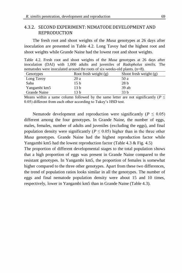
R. similis penetration, development and reproduction 69
4.3.2. SECOND EXPERIMENT: NEMATODE DEVELOPMENT AND
REPRODUCTION
The fresh root and shoot weights of the Musa genotypes at 26 days after
inoculation are presented in Table 4.2. Long Tavoy had the highest root and
shoot weights while Grande Naine had the lowest root and shoot weights.
Table 4.2. Fresh root and shoot weights of the Musa genotypes at 26 days after
inoculation (DAI) with 1,000 adults and juveniles of Radopholus similis. The
nematodes were inoculated around the roots of six-weeks-old plants. (n=8).
Genotypes Root fresh weight (g) Shoot fresh weight (g)
Long Tavoy 20 a 50 a
Saba 15 b 28 b
Yangambi km5 13 b 39 ab
Grande Naine 13 b 33 b
Means within a same column followed by the same letter are not significantly (P ≤
0.05) different from each other according to Tukey‟s HSD test.
Nematode development and reproduction were significantly (P ≤ 0.05)
different among the four genotypes. In Grande Naine, the number of eggs,
males, females, number of adults and juveniles (excluding the eggs), and final
population density were significantly (P ≤ 0.05) higher than in the three other
Musa genotypes. Grande Naine had the highest reproduction factor while
Yangambi km5 had the lowest reproduction factor (Table 4.3 & Fig. 4.5)
The proportion of different developmental stages to the total population shows
that a high proportion of eggs was present in Grande Naine compared to the
resistant genotypes. In Yangambi km5, the proportion of females is somewhat
higher compared to the three other genotypes. Apart from these two differences,
the trend of population ratios looks similar in all the genotypes. The number of
eggs and final nematode population density were about 15 and 10 times,
respectively, lower in Yangambi km5 than in Grande Naine (Table 4.3).

70 Chapter 4
Table 4.3. Number of Radopholus similis eggs, juveniles, females, males, final
population density and reproduction factor (Rf) in the roots of the resistant (Long
Tavoy, Saba, Yangambi km5) and the susceptible (Grande Naine) Musa genotypes at 26
days after inoculation with 1,000 adults and juveniles of Radopholus similis. (n=8).
Genotype Mean number of Radopholus similis/root system
Eggs Juveniles Females Males Final
population
density
Rf
Long Tavoy 129 b 187 b 246 b 6 a 568 b 0.57
Saba 99 b 210 b 238 b 3 a 549 b 0.55
Yangambi km5 38 a 47 a 101 a 1 a 188 a 0.19
Grande Naine 555 c 480 b 820 c 34 b 1,890 c 1.89
Means within a same column followed by the same letters are not significantly (P ≤
0.05) different from each other according to Tukey‟s HSD test. Rf: reproduction factor
= final nematode population density/initial inoculum density.
Figure 4.5: Number of adult and juvenile Radopholus similis in the roots of
resistant (Long Tavoy, Saba, Yangambi km5) and susceptible (Grande
Naine) Musa genotypes at 26 days after inoculation with 1,000 adults and
juveniles of Radopholus similis. Error bars represent standard error. (n=8).
4.4. DISCUSSION
This part of our study clearly demonstrates that the three R. similis-
resistant Musa genotypes impair the post-infectional development and
reproduction of R. similis. The pre-infectional resistance factors, on the other
hand, do not impair the nematodes‟ attraction towards and penetration in the
roots. No significant differences were observed in the number of nematodes that
had penetrated the resistant and susceptible Musa genotypes until 8 days after
inoculation (DAI).
In a previous study, Mateille (1992) found that the penetration of R. similis
was initially slower in the roots of the resistant Musa genotype Gros Michel
than in the susceptible Musa genotype Poyo. However, 2 weeks later, an equal
439 450
150
1334
0
200
400
600
800
1000
1200
1400
1600
Long Tavoy Saba YKm5 Grande
Naine
Total number of adults and juveniles
R. similis penetration, development and reproduction 71
number of nematodes had invaded both the resistant and susceptible Musa
genotype. No differences were observed in the penetration of R. similis in
Yangambi km5 and French Sombre, a susceptible plantain (Fogain, 2000b).
Similarly, no differences were reported in the penetration of R. similis in
resistant and susceptible Musa cultivars based on an in vitro study using excised
roots (Dochez, 2004).
In contrast, significant differences were observed in the penetration of R.
similis in the susceptible Musa genotype Poyo and Yangambi km5 at 24 h after
inoculation in an in vitro study (Valette et al., 1997). In our study, R. similis
penetration was lower in only one resistant Musa genotype, Long Tavoy, than
in Grande Naine at 12 DAI. However, already at 8 DAI the second generation
of nematodes (eggs) were observed in Grande Naine as the inoculum consisted
mostly of gravid females. This could have influenced the higher number of
nematodes observed in Grande Naine at 12 DAI. The second generation did not
appear in Yangambi km5 and Saba until 12 DAI. Though eggs were found in
Long Tavoy at 12 DAI, the number was significantly lower than the number of
eggs found in Grande Naine.
In our study, few nematodes invaded the stele of Yangambi km5 especially
in the secondary roots or at root junctions. This might have been due to the
absence of lignifications in the young roots. A previous study reported the
invasion of the vascular region by R. similis in the susceptible Musa genotype
Poyo (Valette et al., 1997).
Results from our nematode development and reproduction experiment
showed a significant increase in the numbers of eggs, adults and the final
nematode population density in Grande Naine compared to all three resistant
Musa genotypes at 26 DAI. This shows that the resistance mechanisms of the R.
similis-resistant Musa genotypes examined are strongly influencing the post-
infectional nematode development and reproduction of R. similis. In a previous
study, the resistant Musa genotype Gros Michel significantly reduced the
reproduction factor of R. similis compared to the susceptible Musa genotype
Poyo at 2 months after inoculation (Mateille, 1992). Investigations on the
resistance mechanisms against the migratory endoparasite R. similis are scarce.
Investigations on the sedentary endoparasitic root-knot nematodes
(Meloidogyne species) showed that initial root penetration by Meloidogyne
incognita was not impaired in resistant genotypes of cotton (Faske & Starr,
2009) and soybean (Fourie, 2005). Meloidogyne arenaria, M. incognita, M.
javanica and M. hapla readily penetrated resistant and susceptible cucumber
plants leading to the conclusion that reduced nematode development and

72 Chapter 4
reproduction are clearly one of the mechanisms of resistance involved (Walters
et al., 2006).
Based on the many reported investigations on sedentary and some
migratory endoparasitic nematodes, it was stated that these nematodes freely
penetrate the roots of resistant and susceptible plants and that incompatibility at
this stage may occur but is rare. Preformed morphological and chemical barriers
do not appear to be effective against penetration of plant tissues by these
nematodes (Kaplan & Keen, 1980). Published reports showed that many
resistance host responses, particularly those involving biotrophic pathogens, are
governed by specific interactions between a pathogen avirulent gene and a plant
resistance gene (Dangl & Jones, 2001) and less due to preformed factors.
Moreover, most of the identified and cloned nematode resistance genes act via a
localized necrosis or hypersensitive response influencing the post-infectional
migration, development and reproduction of the nematodes (Williamson &
Hussey, 1996; Das et al., 2008).
4.5. CONCLUSION
No significant differences were observed in the number of R. similis
penetrating the resistant and susceptible Musa genotypes included in our study
except in the resistant genotype Long Tavoy at 12 days after inoculation. Post-
infectional nematode development and reproduction were impaired in the
resistant Musa genotypes. On the basis of these observations it appears that the
mechanism of resistance in the investigated Musa genotypes to R. similis is
induced after nematode penetration and that preformed host resistance factors
do not function strongly against the nematode attraction and migration towards
the roots, and penetration of the roots.

CHAPTER 5
LIGNIN AND PHENOLS INVOLVED IN THE INTERACTIONS BETWEEN RADOPHOLUS SIMILIS AND
MUSA SPP.
74 Chapter 5
5.1. INTRODUCTION
Plants produce more than 100,000 diverse, low molecular weight
secondary metabolites (Dixon, 2001). Secondary metabolites play a major role
in the plants‟ interaction with the environment ensuring successful adaptation
and survival (Verpoorte, 2000). Phenolic secondary metabolites such as lignin
and phenols were associated with nematode resistance in bananas (Valette et
al., 1998; Wuyts et al., 2007).
Lignin is the second most abundant natural biopolymer after cellulose,
found in all vascular plants (Ferrer et al., 2008). Lignin is estimated to comprise
about 20% of the global biomass (Chen et al., 2000). Lignin deposition
strengthens the cell walls. The plant cell wall is vital for the structural integrity
of plants, plant growth, intercellular communication, water movement and
pathogen defense (Aquije et al., 2010).
The resistance of lignin to microbial degradation was thought to provide a
hard barrier to invading pathogens (Tronchet et al., 2010). Presence of a higher
number of lignified cell walls in the vascular bundles of the Musa spp. cv.
Pisang Jari Buaya was associated with its resistance to Radopholus similis
(Fogain & Gowen, 1996). Lower root dry matter was correlated with lower
lignin content resulting in the susceptibility of the Musa spp. cv. Poyo to R.
similis (Fogain & Gowen, 1996). Higher levels of constitutive lignin contents
were observed in R. similis-resistant Musa cultivars than in the susceptible
cultivar Grande Naine (Wuyts et al., 2007).
A higher number of preformed phenolic cells was related to the resistance
of Musa spp. cv. Yangambi km5 to R. similis (Fogain & Gowen, 1996).
Constitutive presence of lignin, dopamine, flavonoids, caffeic and ferulic acids
in Yangambi km5 roots was associated with the resistance to R. similis and
hypothesized that they act as chemical barriers for R. similis penetration and
colonization (Valette et al., 1998).
In this part of our study, preliminary phytochemical studies were carried
out to assess the involvement of lignin and phenols in the resistance of the
newly identified R. similis-resistant Musa genotypes Long Tavoy (Musa
acuminate AA, Burmannica subgroup, ITC 0283) and Saba (Musa spp. ABB,
Saba subgroup, ITC 1138). The well known reference cultivars Yangambi km5
(Musa acuminata AAA, Ibota subgroup, ITC 1123) (resistant) and Grande
Naine (Musa acuminata AAA, Cavendish subgroup, ITC 1256) (susceptible)
were also included in the study as control. The lignin and total phenols were
localised by histochemical staining in root cross sections of R. similis-infected
and uninfected plants. Quantitative assessments were performed on the root

Lignin and Phenols involved in plant-nematode interactions 75
extracts of all the plants. The results are discussed comparing R. similis-infected
and uninfected plants of susceptible and resistant Musa genotypes.
5.2. MATERIALS AND METHODS
5.2.1. PLANTING MATERIAL
All Musa genotypes were initially obtained from the Musa germplasm
collection maintained at the International Transit Centre (ITC), K.U.Leuven,
Belgium. The plant material was proliferated, regenerated and rooted in test
tubes as explained in Chapter 2 (2.2.2). The plantlets were grown in growth
chambers at 28 °C and 16 h photoperiod.
5.2.2. NEMATODE INOCULUM
A population of R. similis originally isolated from banana roots in Uganda
was used in the experiments. This population was maintained and multiplied
monoxenically on sterile carrot discs at 25±1 °C in the dark (Speijer & De
Waele, 1997). The population from Uganda was characterized by a high
reproductive fitness (Fallas et al., 1995). To obtain the inoculum, R. similis was
extracted from the carrot discs by the maceration-sieving technique (Speijer &
De Waele, 1997).
5.2.3. EXPERIMENTAL SET-UP
Six-weeks-old rooted tissue culture plantlets were planted in 1 l pots filled
with sand and potting soil (2:1). Eight weeks after planting, two sets of eight
plants of each Musa genotype were inoculated with approximately 1,000 living
vermiform nematodes by pipetting 4 ml of the nematode suspension into
inoculation holes made in the soil near the plant root zone. Two sets of eight
uninfected plants of each Musa genotype were included as control plants. The
plants were placed in the greenhouse according to a randomized block design.
The plants were maintained under greenhouse conditions and fertilized at
10 days intervals throughout the experiment. In the greenhouse, the ambient day
and night temperatures were 27 and 20 ºC, respectively, relative humidity was
80% and the photoperiod was 12 h.
5.2.4. SAMPLING AND ASSESSMENT OF NEMATODE INFECTION
One set of eight R. similis-infected and one set of eight uninfected plants
were uprooted at 3 weeks after inoculation. The second sets were harvested at 6
76 Chapter 5 weeks after inoculation. At both sampling times, the root systems were
carefully washed free of soil (without destroying the root epidermis) under
running tap water. The fresh root and shoot weights of the whole root systems
were measured. About half of the fresh roots were used for histochemical
staining and nematode extraction. The remaining roots were snap frozen in
liquid nitrogen and stored at -80 ºC for quantitative assays of lignin and total
phenols. Necrotic cells from R. similis-infected roots (~0.5 g) were manually
dissected under a stereoscopic microscope for nematode extraction. Nematodes
were extracted from the micro-dissected lesions by opening the roots with a
needle in a thin film of water and the nematodes were counted under a
stereoscopic microscope.
5.2.5. HISTOCHEMICAL STAINING OF ROOT CROSS SECTIONS
Thin, free hand-cut root cross sections of R. similis-infected and uninfected
Musa genotypes were used for the histological staining of lignin and total
phenols. The Wiesner reagent was used for the localisation of lignin (syringyl
and guaiacyl units) and Mäule reagent was used for the localisation of the
syringyl units of lignin.
For Wiesner staining, hand-cut root cross sections were fixed in 2%
phloroglucinol in ethanol/water (95/5, v/v) for 10 min and mounted in 37% HCl
on glass slides. Wiesner reagent reacts with cinnamaldehyde end groups in the
lignin resulting in the cationic chromophore which appears as a burgundy-red
compound (Vermerris & Nicholson, 2006).
For Mäule‟s staining, hand-cut root cross sections were immersed in 1%
KMnO4 for 30 min, rinsed with distilled water and destained using 20% HCl for
2 min. The stained sections were mounted in 10% NH3 on glass slides. The
presence of syringyl units results in the development of a deep red colour while
the absence of syringyl units results in the development of a yellow colour
(Vermerris & Nicholson, 2006).
For the localisation of total phenols, fresh root cross sections were treated
with 0.5% (v/v) toluidine blue in 2.5% Na2CO3 at pH 9 for 10 min and rinsed
with 70% ethanol. Stained root sections were observed under a bright field
transmitted light microscope. Phenolic cells stain as deep blue (Valette et al.,
1997).
Fluorescent flavonoids and lignified cell walls were detected by Neu‟s
reagent (Valette et al., 1998). Fresh root cross sections were stained with
freshly prepared 0.25% (w/v) diphenylboric acid 2-aminoethyl ester (DPBA) in
MilliQ water with 0.02% (v/v) triton-x-100 for 2 min. Sections were observed
Lignin and Phenols involved in plant-nematode interactions 77
immediately under an epifluorescence microscope with a DAPI filter (excitation
340-380 nm, suppression LP 430 nm) and a FITC filter (excitation 450-490 nm,
suppression LP 520 nm). Photography of the root sections was done using a
SPOT RT CCD camera and SPOT RT software version 3.5.5 (Diagnostic
Instruments, Inc., USA)
5.2.6. EXTRACTION AND QUANTIFICATION OF LIGNIN
Lignin contents of isolated cell wall samples were determined by using the
thioglycolic acid derivatised lignin method as described by Lange et al. (1995).
To isolate the cell walls, 500 mg of frozen root samples were ground in liquid
nitrogen using a mortar and pestle without letting the sample thaw. Ground root
samples were first suspended in 1.5 ml methanol and vigorously stirred for 1 h.
The suspension was centrifuged for 5 min at 16,000 x g. Pellets were suspended
in 1.5 ml of the following seven solutions one after the other by vigorously
stirring for 15 min and each time the suspension was centrifuged for 5 min at
16,000 x g: (a) methanol, (b) 1 M NaCl, (c) 1% SDS (w/v), (d) twice with
MilliQ water, (e) ethanol, (f) chloroform/methanol (1:1, v/v) and (g) tert-butyl
methyl ether. The resultant pellets are the purified cell walls. The purified cell
wall pellets were freeze-dried overnight.
Approximately 15 mg of the cell wall preparations were taken and
suspended in a mixture of 1 ml HCl (2 M) and 200 µl thioglycolic acid.
Suspensions were incubated for 4 h in a water bath at 95+2 ºC. After cooling
down to room temperature, samples were centrifuged for 10 min at 16,000 x g.
Pellets were washed three times with MilliQ water, suspended in 1 ml NaOH
(0.5 M) and vigorously shaken overnight to extract the lignothioglycolic acid
(LTGA). Samples were centrifuged for 10 min at 16,000 x g and the
supernatants were collected. The pellets were washed with an additional 500 µl
of NaOH (0.5 M). The combined supernatants (alkali extracts) were acidified
with 300 µl concentrated HCl and incubated for 4 h at 4 ºC to precipitate the
LTGA from the alkali extracts. Samples were centrifuged for 10 min at 16,000
x g and pellets were dried in a SpeedVac centrifuge. Dry brown pellets were
dissolved in 1 ml NaOH (0.5 M) to measure the absorbance.
Absorbance of the samples was measured against the blank at 280 nm in a
NanoDrop spectrophotometer using the UV-Vis high absorbance mode (ND-
1000, NanoDrop Technologies, Inc., Wilmington, USA). The lignin content
(LTGA) was expressed as mg lignin per g fresh root weight using a calibration
curve of alkali lignin (Sigma-Aldrich, Inc., Borem, Belgium).
78 Chapter 5
5.2.7. FOLIN-CIOCALTEU ASSAY FOR TOTAL PHENOLICS
This method determines the total soluble phenolic content based on the
reduction of the phospho-molybdene/phospho-tungstate present in the Folin–
Ciocalteu reagent. One g of frozen root samples were ground in liquid nitrogen
using a mortar and pestle without letting the sample thaw. Ground root samples
were extracted with 5 ml of 50% methanol in test tubes and secured with screw
caps. Samples were shaken at 4 ºC for 60 min. Each extract was passed through
a 0.45 µm PTFE filter (Merck) and the filtrate was kept on ice. For the analysis
of total phenols using the Folin-Ciocalteu method (Makkar et al., 1993), 100 µl
of the extract was mixed with 400 µl of distilled water in glass test tubes. To
each of the samples 250 µl of 1 N Folin-Ciocalteu reagent and 1,250 µl of 20%
sodium carbonate solution were added and vortexed. The samples were kept in
the dark at room temperature for 40 min. Absorbance was measured at 725 nm
against a blank using a Novaspec II spectrophotometer. A standard calibration
curve of tannic acid was established and total phenolic content was expressed as
tannic acid equivalents per g of fresh root weight (TAE/g FRW).
5.2.8. STATISTICAL ANALYSIS
Statistical analysis was performed using STATISTICA version 9 (StatSoft,
Tulsa, OK, USA). Data on fresh root and shoot weights and lignin content were
analysed using analysis of variance (ANOVA). When significant differences (P
≤ 0.05) were observed, Tukey‟s HSD test was applied for multiple comparisons
of group means. Nematode numbers and total phenolic content data were
analysed using Kruskal-Wallis analysis of variance by ranks. The non
parametric equivalent of ANOVA, Kruskal-Wallis analysis of variance by
ranks, was applied because the data set did not meet the basic assumptions of
the parametric tests namely normal distribution and homogeneous variances.
When the Kruskal-Wallis analysis of variance by ranks was significant,
multiple comparisons between treatments were calculated as described by
Siegel and Castellan (1988).
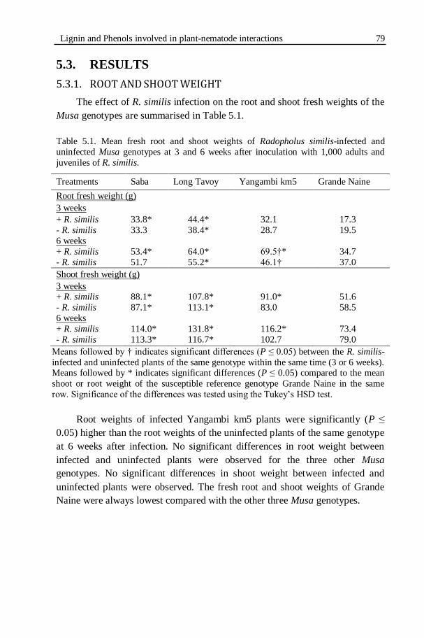
Lignin and Phenols involved in plant-nematode interactions 79
5.3. RESULTS
5.3.1. ROOT AND SHOOT WEIGHT
The effect of R. similis infection on the root and shoot fresh weights of the
Musa genotypes are summarised in Table 5.1.
Table 5.1. Mean fresh root and shoot weights of Radopholus similis-infected and
uninfected Musa genotypes at 3 and 6 weeks after inoculation with 1,000 adults and
juveniles of R. similis.
Treatments Saba Long Tavoy Yangambi km5 Grande Naine
Root fresh weight (g)
3 weeks
+ R. similis 33.8* 44.4* 32.1 17.3
- R. similis 33.3 38.4* 28.7 19.5
6 weeks
+ R. similis 53.4* 64.0* 69.5†* 34.7
- R. similis 51.7 55.2* 46.1† 37.0
Shoot fresh weight (g)
3 weeks + R. similis 88.1* 107.8* 91.0* 51.6
- R. similis 87.1* 113.1* 83.0 58.5
6 weeks
+ R. similis 114.0* 131.8* 116.2* 73.4
- R. similis 113.3* 116.7* 102.7 79.0
Means followed by † indicates significant differences (P ≤ 0.05) between the R. similis-
infected and uninfected plants of the same genotype within the same time (3 or 6 weeks). Means followed by * indicates significant differences (P ≤ 0.05) compared to the mean
shoot or root weight of the susceptible reference genotype Grande Naine in the same
row. Significance of the differences was tested using the Tukey‟s HSD test.
Root weights of infected Yangambi km5 plants were significantly (P ≤
0.05) higher than the root weights of the uninfected plants of the same genotype
at 6 weeks after infection. No significant differences in root weight between
infected and uninfected plants were observed for the three other Musa
genotypes. No significant differences in shoot weight between infected and
uninfected plants were observed. The fresh root and shoot weights of Grande
Naine were always lowest compared with the other three Musa genotypes.
80 Chapter 5
5.3.2. ROOT ANATOMY AND CELLULAR DAMAGE IN INFECTED
ROOTS
Thin, fresh, hand-cut root cross sections were made from all the Musa
genotypes to compare the anatomical structures of the uninfected and R. similis-
infected plants. In the infected plants, nematode damage and lesion patterns
were studied. The anatomical root structures in the uninfected Musa root cross
sections are shown in Figure 5.1. The anatomy of the healthy roots was similar
to previous descriptions of Musa roots (Acquarone, 1930; Wuyts, 2006).
The outermost layer is the root epidermis lining the root cortex. Few
outermost cortical layers are thick cork cells. The outer cortex layers are
composed of large cells, arranged in irregular radial rows with small
intercellular spaces. The inner side of the outer cortex continues to form
aerenchyma previously described as lacunae by Acquarone (1930). The
aerenchyma is wide and the cavities are lined by radial cortical cell plates. The
inner cortex follows the aerenchyma internally. The inner cortical cells are
smaller, cylindrical and arranged in more regular radial rows with well defined
larger intercellular spaces (Fig. 5.1A). The inner cortical cells become smaller
for every inner row. The innermost cortex layer is a layer of very small, well
packed cells called endodermis. Suberin deposition thickens the lateral sides of
endodermal cells.
The pericycle is the outermost layer of the vascular bundle made of a
single layer of cells lying just below the endodermis. As a monocotyledon root,
the vascular bundle is a polyarch type and radial in arrangement. The outer
protoxylem vessels lie against the pericycle. The metaxylem vessels are
strikingly large. Each xylem vessel is lined by a layer of sclerenchyma (wood
parenchyma). The peripheral and central phloem strands are found in between
the xylem vessels (Fig. 5.1B).

Lignin and Phenols involved in plant-nematode interactions 81
Figure 5.1: Anatomical root structures of Musa spp. cv. Saba (cross sections).
A) Epidermis, inner and outer cortex, aerenchyma and vascular bundle (stele)
(x40). B) The vascular bundle with its different structures (x100); en:
endodermis; mx: metaxylem; pc: pericycle; ph: phloem cells; px: protoxylem.
The necrotic lesions in R. similis-infected root cross sections were
observed under a compound, transmitted light microscope. The images of the
lesions are presented in Figure 5.2. The necrosis started from the epidermis and
extended into the outer cortex, aerenchyma and inner cortex but it was more
extensive in the aerenchyma of all the four Musa genotypes (Fig. 5.2C).
Necrosis was also observed in the endodermis, extending into the outer layers
of vascular bundle of Yangambi km5 at 3 weeks after infection. Observation of
the lesions showed that the cells were dark brown coloured in Yangambi km5
and Saba (Figs 5.2B, E & F) and reddish brown coloured in Grande Naine (Fig.
5.2A).
epidermis
Outer cortex
Aerenchyma
Inner cortexStele
pc
mxpx
en
ph
A
B

82 Chapter 5
Figure 5.2: Necrotic root cross sections of Radopholus similis-resistant and susceptible
Musa genotypes at 6 weeks after inoculation with R. similis.
A) Young lesions extending from the cortex to the aerenchyma cells of Grande Naine.
B) Dark brown necrosis in the cortex of Saba. C) Lesions in the aerenchyma cells of
Long Tavoy. D) Phenolic compounds starting to accumulate in a newly developing
lesion in Long Tavoy. E) Developing lesions with high phenol contents (dark brown
cells) in the necrotic areas of Yangambi km5. F) Well developed lesions in Yangambi
km5. Magnification of the images except D: 100x; D: 400x
5.3.3. NEMATODE NUMBERS IN NECROTIC LESIONS
The numbers of R. similis in the necrotic lesions of the resistant and
susceptible Musa genotypes at 6 weeks after infection are summarised in Table
5.2. The mean number of nematodes extracted from the necrotic lesions of
susceptible Musa genotype Grande Naine was significantly (P ≤ 0.05) higher
A
FE
DC
B

Lignin and Phenols involved in plant-nematode interactions 83
than the number of nematodes extracted from the resistant Musa genotypes
Saba, Long Tavoy and Yangambi km5.
Table 5.2. Mean number of Radopholus similis in the necrotic lesions of three resistant
(Saba, Long Tavoy & Yangambi km5) and one susceptible (Grande Naine) Musa
genotypes at 6 weeks after inoculation with 1,000 adults and juveniles of R. similis
(n=4).
Genotype Nematodes/g of necrotic lesion
Saba 583 a
Long Tavoy 254 a
Yangambi km5 255 a
Grande Naine 1057 b
Means within the same column followed by the same letter are not
significantly (P ≤ 0.05) different from each other according to the Kruskal-
Wallis analysis of variance by ranks.
5.3.4. HISTOCHEMICAL STAINING OF MUSA ROOT CROSS SECTIONS
FOR LIGNIFIED CELL WALLS
Lignified cell walls fluoresce under UV light when stained with DPBA.
When the root section is viewed through a FITC filter, the syringyl units
fluoresce in green and the guaiacyl units fluoresce in bright yellow colour. The
uninfected plants of all Musa genotypes sampled at 6 weeks showed intense
fluorescence (Figs 5.3B, D, F, G & I) compared to the plants sampled at 3
weeks after infection (Figs 5.3A, C, E & H) i.e. older roots showed more
extensive secondary wall thickening due to lignin deposition. The uninfected
plants of the R. similis-resistant genotype Saba showed distinct lignification of
aerenchyma with syringyl units (Fig. 5.3D).
Radopholus similis-infected plants (Fig. 5.4) showed much intense
fluorescence in the vascular bundles and endodermis compared to the
uninfected plants of the same age (Fig. 5.3). The R. similis-infected plants of
Long Tavoy showed the most extensive secondary wall lignification of vascular
parenchyma (sclerenchyma) cells (Figs 5.4I & J) compared to all other
genotypes. Lignification starts at the endodermis (Fig. 5.4E), extending to the
peripheral tissues of vascular system especially the xylem walls, xylem-
accompanying parenchyma cells (Fig. 5.4H) and eventually the lignification
progress to the vascular parenchyma (sclerenchyma) cells in the centre (Figs
5.4I & J).

84 Chapter 5
Figure 5.3: Lignifications in uninfected Musa root cells at 3 and 6 weeks.
Fresh, hand-cut root cross sections were stained with DPBA and observed under
epifluorescence microscope using FITC (all images except G) and DAPI filter
(G). A) Vascular bundle of Grande Naine at 3 weeks showing no cell wall
lignification and B) initial lignification of protoxylem cells of Grande Naine at 6
weeks. C) Initial lignifications in the xylem-accompanying vascular parenchyma
cells of Saba at 3 weeks and D) lignified aerenchymatic cells at 6 weeks in Saba.
E) Initial lignifications of parenchyma cells surrounding xylem cells of Long
Tavoy at 3 weeks and F) & G) lignified vascular cell (sclerenchyma) walls of
Long Tavoy at 6 weeks. H) Initial lignifications of xylem cells of Yangambi km5 at 3 weeks and I) strong lignifications progressing to the centre of the vascular
bundles of Yangambi km5 at 6 weeks. Arrows are pointing at the lignified cell
walls. Magnification of the images: 100x.
A B
C D
F GE
IH
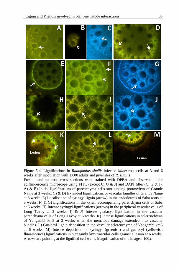
Lignin and Phenols involved in plant-nematode interactions 85
Figure 5.4: Lignifications in Radopholus similis-infected Musa root cells at 3 and 6 weeks after inoculation with 1,000 adults and juveniles of R. similis
Fresh, hand-cut root cross sections were stained with DPBA and observed under
epifluorescence microscope using FITC (except C, G & J) and DAPI filter (C, G & J).
A) & B) Initial lignifications of parenchyma cells surrounding protoxylem of Grande
Naine at 3 weeks. C) & D) Extended lignifications of vascular bundles of Grande Naine
at 6 weeks. E) Localisation of syringyl lignin (arrow) in the endodermis of Saba roots at
3 weeks. F) & G) Lignifications in the xylem accompanying parenchyma cells of Saba
at 6 weeks. H) Intense syringyl lignifications (arrows) in the peripheral vascular cells of
Long Tavoy at 3 weeks. I) & J) Intense guaiacyl lignification in the vascular
parenchyma cells of Long Tavoy at 6 weeks. K) Intense lignifications in sclerenchyma
of Yangambi km5 at 3 weeks when the nematode damage extended into vascular bundles. L) Guaiacyl lignin deposition in the vascular sclerenchyma of Yangambi km5
at 6 weeks. M) Intense deposition of syringyl (greenish) and guaiacyl (yellowish
fluorescence) lignifications in Yangambi km5 vascular cells against a lesion at 6 weeks.
Arrows are pointing at the lignified cell walls. Magnification of the images: 100x.
A B C D
FE G
JIH
MLK
Lesion
Lesion
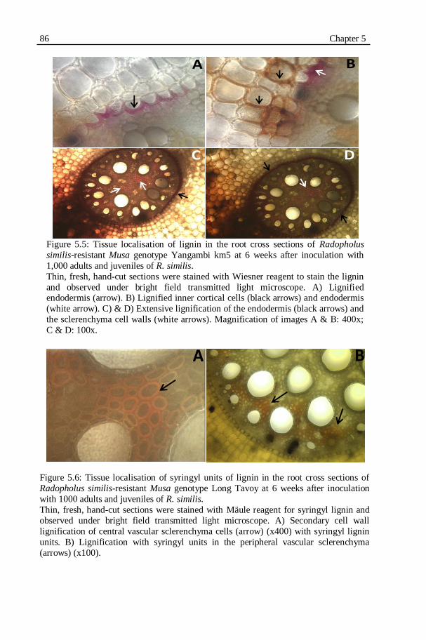
86 Chapter 5
Figure 5.5: Tissue localisation of lignin in the root cross sections of Radopholus
similis-resistant Musa genotype Yangambi km5 at 6 weeks after inoculation with
1,000 adults and juveniles of R. similis.
Thin, fresh, hand-cut sections were stained with Wiesner reagent to stain the lignin
and observed under bright field transmitted light microscope. A) Lignified endodermis (arrow). B) Lignified inner cortical cells (black arrows) and endodermis
(white arrow). C) & D) Extensive lignification of the endodermis (black arrows) and
the sclerenchyma cell walls (white arrows). Magnification of images A & B: 400x;
C & D: 100x.
Figure 5.6: Tissue localisation of syringyl units of lignin in the root cross sections of
Radopholus similis-resistant Musa genotype Long Tavoy at 6 weeks after inoculation
with 1000 adults and juveniles of R. similis.
Thin, fresh, hand-cut sections were stained with Mäule reagent for syringyl lignin and
observed under bright field transmitted light microscope. A) Secondary cell wall
lignification of central vascular sclerenchyma cells (arrow) (x400) with syringyl lignin
units. B) Lignification with syringyl units in the peripheral vascular sclerenchyma
(arrows) (x100).
A
C D
B
A B
Lignin and Phenols involved in plant-nematode interactions 87
Cell wall lignifications in the Musa genotypes were confirmed by
histochemical staining of the root cross sections with lignin specific dyes. When
reacted with the Wiesner reagent, the lignified cells develop burgundy-red
colour. With the Mäule reagent, the syringyl units of lignin develop bright or
deep red colour.
Endodermis of all Musa genotypes became deep red coloured when treated
with Wiesner reagent indicating the endodermal cell wall lignification
especially after infection with R. similis (Figs 5.5C & D). Distinct pink
colouration was observed in the endodermis of Yangambi km5 (Figs 5.5A &
B). The reactions with Maule‟s reagents showed high syringyl units of lignin
present in the central (Fig. 5.6A) and peripheral (Fig. 5.6B) vascular
parenchyma (sclerenchyma) of R. similis-infected plants.
5.3.5. LIGNIN CONTENT OF ROOT CELL WALLS
The total lignin contents in the isolated roots cell walls of R. similis-
infected and uninfected Musa genotypes are shown in the Figure 5.7. The
results show that R. similis infection significantly (P ≤ 0.05) increased the
lignin content of Yangambi km5 roots at 6 weeks after inoculation. The root
lignin content in the uninfected Yangambi km5 was lower than the root lignin
content in uninfected Grande Naine at 6 weeks after inoculation (Fig. 5.7B).
The post infection lignin content in Yangambi km5 was significantly (P ≤ 0.05)
higher than the post infection lignin content in Grande Naine at 6 weeks after
inoculation.

88 Chapter 5
Figure 5.7: Lignin content (mg/g of fresh root weight) in root cell walls of Radopholus similis-infected and uninfected plants of resistant and susceptible
Musa genotypes at 3 and 6 weeks after inoculation with 1,000 adults and
juveniles of R. similis.
Mean lignin contents are presented between parentheses. Means followed by †
indicate a significant difference (P ≤ 0.05) between the R. similis-infected and
uninfected plant of the same genotype. Means followed by * indicate a
significant difference (P ≤ 0.05) compared to the susceptible reference genotype
Grande Naine. Error bars represent confidence intervals. (n=8).
G. Naine: Grande Naine; Ykm5: Yangambi km5.
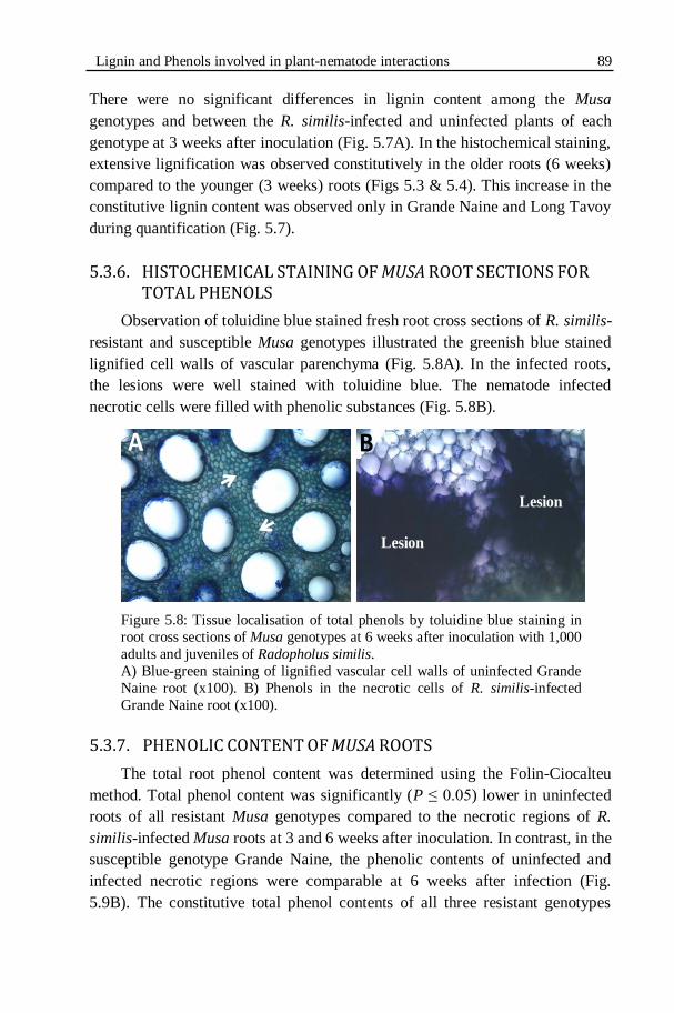
Lignin and Phenols involved in plant-nematode interactions 89
There were no significant differences in lignin content among the Musa
genotypes and between the R. similis-infected and uninfected plants of each
genotype at 3 weeks after inoculation (Fig. 5.7A). In the histochemical staining,
extensive lignification was observed constitutively in the older roots (6 weeks)
compared to the younger (3 weeks) roots (Figs 5.3 & 5.4). This increase in the
constitutive lignin content was observed only in Grande Naine and Long Tavoy
during quantification (Fig. 5.7).
5.3.6. HISTOCHEMICAL STAINING OF MUSA ROOT SECTIONS FOR
TOTAL PHENOLS
Observation of toluidine blue stained fresh root cross sections of R. similis-
resistant and susceptible Musa genotypes illustrated the greenish blue stained
lignified cell walls of vascular parenchyma (Fig. 5.8A). In the infected roots,
the lesions were well stained with toluidine blue. The nematode infected
necrotic cells were filled with phenolic substances (Fig. 5.8B).
Figure 5.8: Tissue localisation of total phenols by toluidine blue staining in
root cross sections of Musa genotypes at 6 weeks after inoculation with 1,000
adults and juveniles of Radopholus similis.
A) Blue-green staining of lignified vascular cell walls of uninfected Grande
Naine root (x100). B) Phenols in the necrotic cells of R. similis-infected
Grande Naine root (x100).
5.3.7. PHENOLIC CONTENT OF MUSA ROOTS
The total root phenol content was determined using the Folin-Ciocalteu
method. Total phenol content was significantly (P ≤ 0.05) lower in uninfected
roots of all resistant Musa genotypes compared to the necrotic regions of R.
similis-infected Musa roots at 3 and 6 weeks after inoculation. In contrast, in the
susceptible genotype Grande Naine, the phenolic contents of uninfected and
infected necrotic regions were comparable at 6 weeks after infection (Fig.
5.9B). The constitutive total phenol contents of all three resistant genotypes
A B
Lesion
Lesion
90 Chapter 5 were significantly (P ≤ 0.05) lower than the constitutive phenol content in
Grande Naine at 6 weeks after inoculation. No significant differences were
observed between the total root phenol contents of the resistant and susceptible
genotypes at 3 weeks after inoculation.
5.3.8. LOCALISATION OF FLAVONOIDS
Observations of DPBA treated root sections under an epifluorescence
microscope with a FITC filter showed various fluorescing compounds. Green,
bright yellow, yellow-gold, and orange fluorescence were observed in cells
surrounding the necrosis in the cortex and aerenchyma of the root sections (Fig.
5.10). These fluorescent compounds are indicative of lignin and certain
flavonoids according to the literature. After DPBA staining, lignin shows
similar green or bright yellow fluorescence when observed under an
epifluorescence microscope with FITC filter. Identification of the compounds
based on the fluorescence is highly ambiguous. The fluorescing compounds
were always observed in the cells and the intercellular spaces surrounding the
necrotic lesions. They were never observed in the lesions.

Lignin and Phenols involved in plant-nematode interactions 91
Figure 5.9: Total phenol contents (mg TAE/g roots) in the Radopholus similis-
infected and uninfected roots of resistant and susceptible Musa genotypes at 3 and
6 weeks after inoculation with 1,000 adults and juveniles of R. similis.
Mean total phenol contents are presented between parentheses. Means followed by
† indicate a significant differences (P ≤ 0.05) between the R. similis-infected and
uninfected plants of the same genotype. Means followed by * indicate a significant
differences (P ≤ 0.05) compared to the susceptible reference genotype Grande
Naine. Error bars represent confidence intervals. G. Naine: Grande Naine; Ykm5:
Yangambi km5.
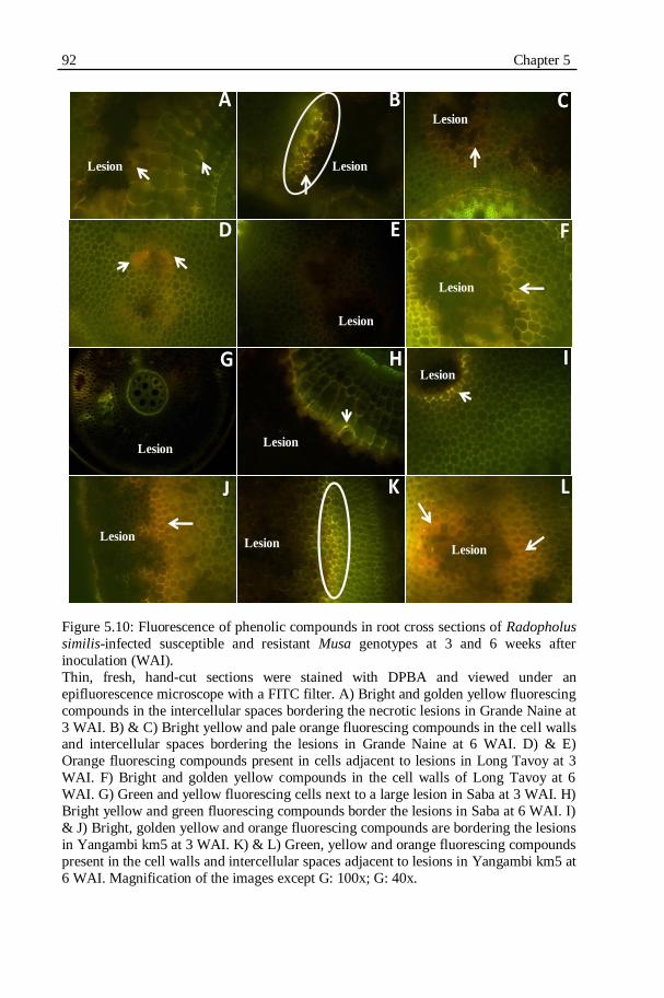
92 Chapter 5
Figure 5.10: Fluorescence of phenolic compounds in root cross sections of Radopholus
similis-infected susceptible and resistant Musa genotypes at 3 and 6 weeks after
inoculation (WAI).
Thin, fresh, hand-cut sections were stained with DPBA and viewed under an
epifluorescence microscope with a FITC filter. A) Bright and golden yellow fluorescing
compounds in the intercellular spaces bordering the necrotic lesions in Grande Naine at
3 WAI. B) & C) Bright yellow and pale orange fluorescing compounds in the cell walls and intercellular spaces bordering the lesions in Grande Naine at 6 WAI. D) & E)
Orange fluorescing compounds present in cells adjacent to lesions in Long Tavoy at 3
WAI. F) Bright and golden yellow compounds in the cell walls of Long Tavoy at 6
WAI. G) Green and yellow fluorescing cells next to a large lesion in Saba at 3 WAI. H)
Bright yellow and green fluorescing compounds border the lesions in Saba at 6 WAI. I)
& J) Bright, golden yellow and orange fluorescing compounds are bordering the lesions
in Yangambi km5 at 3 WAI. K) & L) Green, yellow and orange fluorescing compounds
present in the cell walls and intercellular spaces adjacent to lesions in Yangambi km5 at
6 WAI. Magnification of the images except G: 100x; G: 40x.
A
J K L
G H I
D E F
B C
LesionLesion
Lesion
Lesion
LesionLesion
Lesion
Lesion
Lesion
Lesion
Lesion

Lignin and Phenols involved in plant-nematode interactions 93
5.4. DISCUSSION
Lignin and phenols were tissue localised by histochemical analysis and
quantitatively assessed in the R. similis-resistant and susceptible Musa roots to
understand their involvement in the R. similis-resistance of Musa spp. The
effect of R. similis infection on lignin and phenols accumulation was studied by
comparing their constitutive levels in uninfected plants.
In our study, nematode infection was associated with a significant increase
in the root weight of the resistant reference genotype Yangambi km5. Talwana
et al. (2006) observed higher number of roots in the R. similis-infected banana
genotypes Nabusa, Pisang Awak and Sukali Ndizi compared to the uninfected
plants. This stimulation in root growth is a mechanism to compensate for the
nematode damage by the dormant root primordia (Talwana et al., 2006).
Moreover, the nematode infection has increased the lignin content of Yangambi
km5. The increase in root weight has been associated with an increase in the
root secondary cell wall lignification (Fogain & Gowen, 1996). However, the
increased root damage by nematodes results in reduced root system and loss of
root weight and anchorage upon completion of the many nematode life cycles
in the host roots.
No difference in the general structural root anatomy was found between
the R. similis-resistant and susceptible Musa genotypes. The anatomical
structures were similar to the previous descriptions of Musa roots (Acquarone,
1930; Wuyts, 2006). The nematode damage and necrosis were extensive in the
outer cortex and aerenchyma. The necrosis has been extended to the
endodermis and vascular bundle of a Yangambi km5 plant at early stage.
Invasion of the vascular bundles by R. similis in Musa spp. has been
occasionally reported (Mateille, 1994; Sarah et al., 1996; Valette et al., 1998).
Poor lignification in the young roots might be the reason for this vascular
invasion by the nematodes
Based on the histochemical staining, it is observed in our study that lignin
was a major constituent of the cell walls of vascular bundles and endodermis.
The cortex and aerenchyma cells were lignified to a very small extent. Similar
observations were made previously (Fogain & Gowen, 1996; Wuyts et al.,
2007). This shows that the increased lignification is only a general defense
response to protect the vascular bundle. Therefore, the damage to the plant can
be reduced. Extensive lignification is not associated with the cortex cells that
are directly involved in the defense to R. similis. Hence lignification is more
likely to be associated with the plant‟s tolerance to R. similis preventing damage
94 Chapter 5 to the plants and less likely to be associated with the resistance to nematode
development, reproduction and multiplication.
Histochemical studies indicated a more extensive lignification of vascular
bundles in older plants than in younger plants. Increased cell wall lignification
with increase in age is documented in Musa spp. (Wuyts et al., 2007) and in
other crops. For instance, the proximal roots of wheat contain more lignin than
the distal younger roots (Rengel et al., 1994). The higher levels of indole acetic
acid (IAA) in younger root tissues inhibit phenylalanine ammonia lyase activity
and lignin deposition (Burnell, 1988). However, in our study the quantitative
assessment of lignin content based on the thioglycolic acid assay did not
confirm extensive lignification of older roots of all genotypes.
Histochemical staining showed more intense secondary cell wall
lignifications in the endodermis, xylem-accompanying cells and the vascular
parenchyma of R. similis-infected plants compared to the uninfected plants of
all Musa genotypes. Stronger lignification in the endodermis and vascular
bundle is considered as a general defence response to nematode infection
creating a physical and chemical barrier to the vascular invasion (Keen, 1992;
Zacheo et al., 1997; Valette et al., 1998; Wuyts et al., 2007).
Varying levels of lignin contents were observed in the infected and
uninfected plants at 3 weeks after infection. However, the infection of R. similis
increased the cell wall lignin content of Yangambi km5 plants at 6 weeks after
infection. The post-infection increase in the lignin content of Yangambi km5
was 7 times higher than in the uninfected plants. In Grande Naine, the extended
cellular damage due to nematode infection might have caused a reduction in
total lignin content compared to the uninfected plants. A prominent increase in
lignin contents due to R. similis-infection was reported in R. similis-resistant
Musa genotypes Pisang Jari Buaya and Calcutta 4 as well as in the susceptible
genotype Grande Naine. However, a reduction in lignin content was in
Yangambi km5 after R. similis infection (Wuyts et al., 2007).
Histochemical localisation of total phenols by staining with toluidine blue
showed that the phenolic substances are major constituents of the nematode
infected necrotic cells. Toluidine blue staining showed the lignified cell walls of
vascular bundles in the roots. No preformed phenolic cells were observed in the
R. similis-resistant and susceptible Musa genotypes as previously reported in
the R. similis-resistant Musa genotypes Yangambi km5 and Gros Michel
(Fogain & Gowen 1996).
Lignin and Phenols involved in plant-nematode interactions 95
The results of the quantitative assay of total phenols by the Folin-Ciocalteu
assay corroborate the histochemical localisation. The nematode infection has
almost doubled the total phenols contents in all Musa genotypes at 3 weeks
after infection. This drastic increase in total phenols was observed in the R.
similis-resistant Musa genotypes at 6 weeks after infection. Infected regions
were sampled for this phenolic assay. Hence the increase in total phenolic
contents is due to the phenols accumulation in the lesions. The enhanced
synthesis of phenols could be due to the biosynthesis or accumulation of
phytoalexins in the nematode feeding cells with anti-nematode properties.
However, the Folin-Ciocalteu assay is a very general test for phenols. Further
studies using analytical techniques such as chromatography, NMR spectroscopy
and mass spectrometry can facilitate the structural identification of the
individual compounds. In our further study we focused on identifying the
secondary metabolites accumulated or biosynthesised in the necrotic lesions.
The high constitutive phenols content in Grande Naine is probably due to high
polyphenolic content in Grande Naine or increased polyphenol content due to
tissue damage during sampling and handling. The older in vitro Grande Naine
roots in tissue cultures also show intense browning and secrete dark brown
compounds in the medium without any infection or physical injury.
Histochemical staining of R. similis-infected roots with Neu‟s reagent
showed the presence of fluorescing compounds bordering the lesions in all the
Musa genotypes. These fluorescing compounds could be lignin accumulation
induced by the nematode infection to protect the adjacent cells from nematode
invasion. These fluorescing compounds were also reported as candidates of
certain flavonoids (Valette et al., 1998; Wuyts et al., 2007). However, it is
highly ambiguous to identify the secondary metabolites based on the
fluorescence. These compounds were mainly present in the cell walls and
intercellular spaces suggesting their role as defence related signalling
molecules. Phenolic compounds were suggested to play a key role in internal
signalling in plants (Ferrer et al., 2008). No striking differences were observed
between the susceptible and resistant genotypes in the presence of constitutively
fluorescing compounds as previously reported (Valette et al., 1998). The
phenols in the lesions were non-fluorescent. Our further study (Chapter 6.1)
reports the results using analytical techniques for the unambiguous
identification of the secondary metabolites in the nematode infected root
tissues.
96 Chapter 5
5.5. CONCLUSION
Radopholus similis infection has increased the root secondary wall
lignification of the endodermis and vascular bundles of resistant and susceptible
Musa genotypes. This increased lignification is a general defense response to
protect the vascular bundle to reduce damage to the plant. Extensive
lignification is not associated with the cortex cells that are directly involved in
the defense with nematodes. Hence lignification is more likely to be associated
with the plant‟s tolerance to R. similis damage than with resistance to nematode
development, reproduction and multiplication.
Preformed phenolic cells were not found to be related with nematode
resistance in the studied Musa genotypes. The nematode infection has
dramatically increased the total phenol contents of resistant plants. Phenolic
secondary metabolites are major constituents of the nematode infected necrotic
cells. Detailed analytical studies are essential to identify the secondary
metabolites that are induced in nematode infection sites. Further studies are
necessary for the structural identification of these secondary metabolites
involved in the necrotic lesions.
CHAPTER 6
PHENYLPHENALENONE-TYPE PHYTOALEXINS INVOLVED IN THE PLANT RESISTANCE TO PARASITIC
NEMATODES
98 Chapter 6
RATIONALE AND OUTLINE
"Phytoalexins are low molecular weight antibiotic compounds, which are
synthesized and accumulated in plants after exposure to pathogens or stress"
(Paxton, 1980). Biosynthesis of phytoalexins is well accepted as a plant‟s
defense strategy to fight biotic stress (Bednarek & Osbourn, 2009). However,
the real contribution of phytoalexins to plant‟s resistance to nematodes remains
elusive as only few secondary metabolites were isolated and identified from
plants and examined for nematicidal activity (Chitwood, 2002; Wuyts, 2006).
Moreover, the majority of the studies were based on histochemical staining
lacking analytical studies for proper identification (Wuyts et al., 2007; Svatoš,
2010).
Phenylphenalenones are phenolic secondary metabolites originally
identified as pigments in plants belonging to the monocot family
Haemodoraceae (Cooke & Edwards, 1981; Dora et al., 1993; Hölscher &
Schneider, 1997; Opitz & Schneider, 2002). These compounds show an organ-
specific distribution such as in apical meristems, root caps, cortex and
epidermis (Opitz et al., 2003) and in the secretary cavities of leaves and flowers
(Hölscher & Schneider, 2007) of Haemodoraceae. Phenylphenalenones are
found to act as phytoalexins, elicited by pathogenic infections in plants
belonging to the Musaceae family (Luis et al., 1993, 1995). In Musa spp.,
phenylphenalenones are elicited in the roots and rhizomes by the fungus
Fusarium oxysporum f. sp. cubense causing Panama disease (Luis et al., 1994;
1996) and by the burrowing nematode Radopholus similis (Cobb, 1893)
Thorne, 1949 (Binks et al., 1997), in the aerial parts by the fungus
Mycosphaerella fijiensis causing Black Sigatoka leaf streak disease (Luis et al.,
1993, 1994; Otálvaro et al., 2007) and in the fruits by the fungus
Colletotrichum musae causing anthracnose (Kamo et al., 1998, 2001). Report
on phenylphenalenones elicitation in response to nematode infection is limited
to the isolation and identification (Binks et al., 1997). Systematic studies
involving tissue localisation, isolation and identification of the compounds in
resistant and susceptible cultivars, followed by examination of the anti-
nematode properties of the compounds have not been performed so far.
Molecular level information on the host-nematode interactions of
migratory endoparasitic nematodes is only emerging recently and scarce
compared to the wealth of information available for sedentary endoparasitic
nematodes (Haegeman et al., 2010). Some studies are dedicated to the
investigation of phytochemicals in R. similis-resistant and susceptible Musa
cultivars (Fogain & Gowen, 1996; Valette et al., 1998; Collingborn et al., 2000;

Phenylphenalenones in the plant resistance to nematodes 99
Wuyts et al., 2007). The majority of these studies used non-specific tests for
phenols and histochemical staining suggesting the involvement of
phenylpropanoids in Musa-R. similis interactions (Collingborn et al., 2000). In
our study, many analytical techniques such as liquid chromatography, mass
spectrometry, mass spectrometry imaging and nuclear magnetic resonance
spectroscopy were combined to precisely identify the phytoalexins involved in
the Musa-R. similis interactions. The anti-R. similis properties of the isolated
compounds were also evaluated. The objective of our study was to identify and
quantify the phytoalexins followed by assessment of the anti-nematode
properties of the phytoalexins elicited in the R. similis-resistant and susceptible
Musa cultivars. It was also our objective to develop a strategy that could be
applicable to study other host-nematode interactions by combining an array of
technologies.
Our results are reported in this chapter in two sections. The first section
presents the results of tissue localisation and profiling of phenylphenalenones in
a R. similis-resistant and a susceptible Musa genotypes. The second section
presents the results of the in vitro bio-assays evaluating the anti-R. similis
properties of the phenylphenalenones.

100 Chapter 6
CHAPTER 6.1: CELL-SPECIFIC LOCALISATION
AND PHYTOCHEMICAL PROFILING OF
PHENYLPHENALENONE-TYPE PHYTOALEXINS IN
RADOPHOLUS SIMILIS-RESISTANT AND -
SUSCEPTIBLE MUSA ROOTS
6.1.1. INTRODUCTION
Plants use a wide range of phytochemicals to defend themselves from
invading pathogens as they cannot move to escape from the pathogens (Field et
al., 2006). The interaction between a host plant and parasitic nematodes affects
the molecular and physiological pathways in host plants (Grunewald et al.,
2009). It also induces many host responses including metabolite expressions
such as production of phytoalexins (Gheysen & Fenoll, 2002). Little
information is available on the changes in plant metabolites due to interactions
with migratory endoparasitic nematodes. The Musa cultivar Yangambi km5 has
been identified and used as a reference cultivar, resistant to R. similis (Speijer &
De Waele, 1997; Fogain & Gowen, 1998). This cultivar is also resistant to
Black Sigatoka leaf streak disease (Fouré et al., 1990; Otálvaro et al., 2007).
Based on the results of our previous experiments (Chapters 3 & 4), it is clear
that in resistant Musa cultivars the post-infectional development and
reproduction of R. similis is inhibited. However, the mechanism of this
nematode resistance is elusive.
Phenylphenalenones were reported as phytoalexins produced in response
to R. similis infection in Musa roots (Binks et al., 1997). A recent study
(Otálvaro et al., 2007) suggested the involvement of phenylphenalenones in the
resistance of Yangambi km5 to the Black Sigatoka leaf streak disease. It is
anticipated that phenylphenalenone-type phytoalexins might also be involved in
the resistance of Yangambi km5 to R. similis. Investigations on
phenylphenalenone profiles of R. similis-infected and uninfected plants of
Yangambi km5 and Grande Naine, a reference Musa cultivar susceptible to R.
similis, form the main focus of this part of the present chapter.
The matrix-free laser desorption/ionisation mass spectrometric imaging
(LDI-MSI) technique was successfully used in Hypericum sp. and the model
plant Arabidopsis thaliana for cell-specific localisation of UV-absorbing-
secondary metabolites (Hölscher et al., 2009). In our study, LDI-MSI is used on
Musa roots to specifically map the secondary metabolites elicited in the root
cells after nematode infection. The knowledge on the cellular localisation will
deepen the understanding on the function of the secondary metabolites. LDI-

Phenylphenalenones in the plant resistance to nematodes 101
MSI provides information on spatial distribution of the secondary metabolites
and the m/z (mass-to-charge ratio) values of the compounds. The secondary
metabolites corresponding to the mass were identified and confirmed by a
multidimensional approach, combining the analytical techniques such as high
performance liquid chromatography (HPLC), proton nuclear magnetic
resonance spectroscopy (1H NMR) and ultra performance liquid
chromatography coupled with mass spectrometry (UPLC-MS). NMR
techniques are less sensitive and amplification techniques such as commonly
used for DNA and RNA are not available for metabolites (Hölscher et al.,
2009). However, when sufficient amounts of the metabolites are available,
NMR analysis is the most powerful tool to obtain structural information of
secondary metabolites. The UPLC-MS technique provides information about
mass of a metabolic signal and the modern day MS techniques are highly
sensitive. The combination of analytical techniques namely LDI-MSI, HPLC, 1H NMR and UPLC-MS were used to localise and identify the secondary
metabolites involved in the Musa-R. similis interactions.
6.1.2. MATERIALS AND METHODS
6.1.2.1. Plants
The R. similis-susceptible cultivar Grande Naine (Musa acuminata AAA,
Cavendish subgroup, ITC 1256) and the R. similis-resistant cultivar Yangambi
km5 (Musa acuminata AAA, Ibota subgroup, ITC 1123) were initially obtained
from the Musa germplasm collection maintained at the International Transit
Centre (ITC), K.U.Leuven, Belgium. The plant material was proliferated,
regenerated and rooted in test tubes as explained in Chapter 2 (2.2.2). The
plantlets were grown in growth chambers at 28 °C and 16 h photoperiod. Eight-
weeks-old rooted tissue culture plantlets were planted in 8 l pots filled with
sand and potting soil (2:1). The plants were maintained under greenhouse
conditions and fertilized at 10 days intervals throughout the experiment. In the
greenhouse, the ambient day and night temperatures were 27 and 20 ºC,
respectively, relative humidity was 80% and the photoperiod was 12 h.
6.1.2.2. Nematodes
A population of R. similis originally isolated from banana roots in Uganda
was used in the experiment. This population was maintained and multiplied
monoxenically on sterile carrot discs at 25+1 °C in the dark (Speijer & De
Waele, 1997). The population from Uganda was characterized by a high
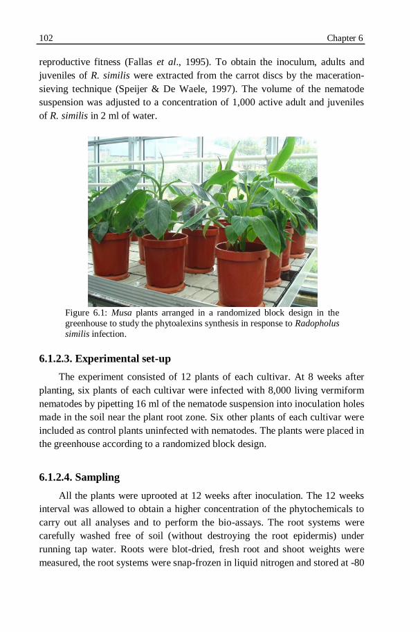
102 Chapter 6
reproductive fitness (Fallas et al., 1995). To obtain the inoculum, adults and
juveniles of R. similis were extracted from the carrot discs by the maceration-
sieving technique (Speijer & De Waele, 1997). The volume of the nematode
suspension was adjusted to a concentration of 1,000 active adult and juveniles
of R. similis in 2 ml of water.
Figure 6.1: Musa plants arranged in a randomized block design in the
greenhouse to study the phytoalexins synthesis in response to Radopholus
similis infection.
6.1.2.3. Experimental set-up
The experiment consisted of 12 plants of each cultivar. At 8 weeks after
planting, six plants of each cultivar were infected with 8,000 living vermiform
nematodes by pipetting 16 ml of the nematode suspension into inoculation holes
made in the soil near the plant root zone. Six other plants of each cultivar were
included as control plants uninfected with nematodes. The plants were placed in
the greenhouse according to a randomized block design.
6.1.2.4. Sampling
All the plants were uprooted at 12 weeks after inoculation. The 12 weeks
interval was allowed to obtain a higher concentration of the phytochemicals to
carry out all analyses and to perform the bio-assays. The root systems were
carefully washed free of soil (without destroying the root epidermis) under
running tap water. Roots were blot-dried, fresh root and shoot weights were
measured, the root systems were snap-frozen in liquid nitrogen and stored at -80
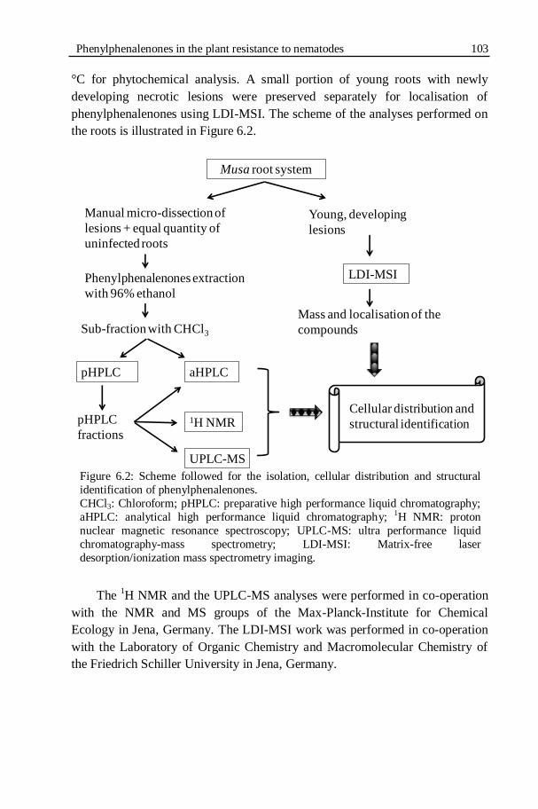
Phenylphenalenones in the plant resistance to nematodes 103
°C for phytochemical analysis. A small portion of young roots with newly
developing necrotic lesions were preserved separately for localisation of
phenylphenalenones using LDI-MSI. The scheme of the analyses performed on
the roots is illustrated in Figure 6.2.
Figure 6.2: Scheme followed for the isolation, cellular distribution and structural
identification of phenylphenalenones.
CHCl3: Chloroform; pHPLC: preparative high performance liquid chromatography;
aHPLC: analytical high performance liquid chromatography; 1H NMR: proton
nuclear magnetic resonance spectroscopy; UPLC-MS: ultra performance liquid
chromatography-mass spectrometry; LDI-MSI: Matrix-free laser
desorption/ionization mass spectrometry imaging.
The
1H NMR and the UPLC-MS analyses were performed in co-operation
with the NMR and MS groups of the Max-Planck-Institute for Chemical
Ecology in Jena, Germany. The LDI-MSI work was performed in co-operation
with the Laboratory of Organic Chemistry and Macromolecular Chemistry of
the Friedrich Schiller University in Jena, Germany.
Musa root system
Young, developing
lesions
Manual micro-dissection of
lesions + equal quantity of
uninfected roots
Phenylphenalenones extraction
with 96% ethanol
Sub-fraction with CHCl3
pHPLC aHPLC
pHPLC
fractions
1H NMR
UPLC-MS
LDI-MSI
Mass and localisation of the
compounds
Cellular distribution and
structural identification

104 Chapter 6
6.1.2.5. Extraction and analysis of phenylphanalenones
From the nematode-infected plants of Yangambi km5 and Grande Naine,
necrotic root tissues were manually micro-dissected and weighed. The same
amounts of healthy roots were sampled from the uninfected plants of each
cultivar as control. Samples were ground in liquid nitrogen. This was followed
by an immediate extraction using 96% ethanol. The extract was filtered by
using filter paper. This crude extract was evaporated using a rotary evaporator
(Rotavapor R-114, waterbath B-480, Büchi Labortechnik AG, Flawil,
Switzerland; Pump system: Vaccubrand Diaphragm Vaccum Pump type MZ
2C, Vaccubrand, Wertheim, Germany) to remove the solvent. The resultant was
sub-fractioned between chloroform/H2O, followed by partition between ethyl
acetate/H2O and n-butanol/H2O using a separating funnel. All three sub-
fractions obtained from each sample were collected separately, filtered and
evaporated to remove the solvents and stored in small vials at room
temperature.
6.1.2.6. Thin layer chromatography (TLC) analysis
TLC was used to obtain a preliminary idea about the compounds in the
sub-fractions prior to using other analytical methods. The chloroform sub-
fraction from Grande Naine was developed in the TLC system along with the
chloroform sub-fraction of Strelitzia reginae roots as a standard for comparison
(Hölscher & Schneider, 2000). About 15 µl of the samples were loaded on the
TLC sheets (TLC aluminium sheets, 20 x 20 cm, silica gel 60 F254, Merck,
Darmstadt, Germany). The TLC developing system consisted of 3:2 toluene and
acetone. The retention factors (Rf) of the separated compounds from Grande
Naine were compared with the Rf of known compounds from S. reginae for a
tentative identification.
6.1.2.7. Preparative HPLC (pHPLC)
Purifications of the sub-fractions were done on a Merck-Hitachi
LiChrograph preparative chromatography system with an L-6200A gradient
pump, L-4250 UV-VIS detector (Merck-Hitachi, Darmstadt, Germany). The
preparative HPLC runs were performed on a reverse phase RP18 column
(LiChrospher® 100 RP18; 250 x 10 mm, 10 μm) at a constant flow rate of 3.5
ml/min using a binary solvent system: solvent A, water with 0.1%
trifluoroacetic acid (TFA) and solvent B, acetonitrile. The HPLC gradient
system was started with 15% B and linearly increased to 95% B at 120 min and

Phenylphenalenones in the plant resistance to nematodes 105
held for 5 min, then brought back to the 15% B initial condition at 135 min and
held for 5 min for the column re-equilibration for the next injection.
Wavelength studied at 284 nm. All the peaks individually collected and purified
using pHPLC were subjected to analytical HPLC, 1H NMR and UPLC-MS to
detect phenylphenalenones.
6.1.2.8. Analytical HPLC (aHPLC)
Tentative identification and purification of phenylphenalenones from the
Musa root extracts were carried out by analyzing the sub-fractions using an
analytical HPLC system (Agilent series HP1100; binary pump G1312A; auto-
sampler G1313A; diode array detector (DAD) G1315B). The aHPLC was
performed using a reverse phase analytical column, LiChrospher® 100 RP18
(Merck, Darmstadt, Germany). Each sub-fraction was dissolved in methanol
mixture at a concentration of 10 mg/ml. In each run, 25 μl of the sub-fraction
was separated at room temperature at a constant flow rate of 0.8 ml/min using a
binary solvent system: solvent A, water with 0.1% TFA and solvent B,
acetonitrile. The chloroform sub-fractions were separated using the aHPLC
gradient started with 20% of B and linearly increased to 50% B at 60 min, 95%
B at 65 min and back to 20% B initial condition at 70 min for the column re-
equilibration for the next injection. The diode array detector (Agilent diode
array detector (DAD) G1315B) was used to record the online spectra at 254,
270, 284, 360 and 450 nm wavelengths.
6.1.2.9. 1H NMR spectroscopy
1H NMR spectra were recorded on a Bruker Avance DRX 500 NMR
spectrometer (Bruker, Karlsruhe, Germany) in acetone-d6 at 24 °C. The
resonance frequency was 125.13 MHz for 1H. Chemical shifts () are given
relative to tetramethylsilane (TMS), which was used as an internal standard for
referencing 1H NMR spectra. The spectrometer was equipped with a broad-
band probe (5 mm, z-gradient, automatic tuning and matching). XwinNMR 3.1
software (Bruker-Biospin) was used to control the spectrometer and data
processing.
6.1.2.10. UPLC-MS analysis
The LC-MS system consisted of an Ultimate 3000 series RSLC (Dionex,
Sunnyvale, California) system and orbitrap mass spectrometer (Thermo Fisher
Scientific, Bremen, Germany). HPLC was performed using Acclaim C18

106 Chapter 6
Column, (150 x 2.1 mm, 2.2 μm, Dionex) at a constant flow rate of 300 μl/min
using a binary solvent system: solvent A, water with 0.1% formic acid and
solvent B, acetonitrile with 0.1% formic acid. The HPLC gradient system
started with 5% B, linearly increased to 98% B at 28 min and held for 3 min,
then brought back to the 5% B initial condition and held for 5 min for the
column re-equilibration for the next injection. Full scan mass spectra were
generated using 30,000 resolving power.
6.1.2.11. Fixation of plant material for LDI-MSI
Initially, the cryo-sectioning and fixation performed at -25 °C produced
root cross sections of 60 m thickness (Cryostat CM 1850, Leica
Microsystems, Nußloch, Germany). However, the fixative used in cryo-
sectioning obstructed the LDI-MSI procedure. Alternatively, thin, small
longitudinal sections of root epidermis at the region of nematode infection were
performed using a razor blade. The root sections were fixed on a carbon
conductive adhesive tape (Plano, Wetzlar, Germany). The sample on the
conductive tape was fixed on the conductive, indium tin oxide (ITO) coated
glass slides (Bruker Daltonic, Bremen, Germany). A gel-liner (Staedtler triplus
gel-liner, silver, 0.4 mm; Staedtler-Mars, Nürnberg, Germany) was used to
place marks surrounding the sections to define their position. The gel marked
points were used as reference points or guidelines to teach the LDI-MSI system
to position the regions of root samples to be analyzed.
6.1.2.12. LDI-MSI on the ultraflex III®
mass spectrometer
An Ultraflex III® 421 (Bruker Daltonics, Bremen, Germany) was used for
the analysis. The instrument was equipped with a Nd:YAG laser with an
emitting wavelength of 355 nm. All spectra were measured in the negative
reflectron mode. For measuring the pixels, the minimum laser focus setting was
used (corresponding to a diameter of about 10 µm laser, under usage of
oversampling for the measurement with a raster of 20 x 20 µm). For each raster
point, a spectrum was accumulated with 100 laser shots, a shot rate of 40 Hz
and fixed laser intensity. For image reconstruction, the FlexImaging v.2.0
software (Bruker Daltonics, Bremen, Germany) was used. Within all prepared
mass images, the signal intensity is represented by the corresponding color
intensity for the represented mass.

Phenylphenalenones in the plant resistance to nematodes 107
6.1.3. RESULTS
6.1.3.1. Root damage and root lesions
The root damage caused by R. similis infection was severe in the
susceptible Musa cultivar Grande Naine compared to the resistant Musa cultivar
Yangambi km5 (Figs 6.3 & 6.4).
Visual observation of R. similis-infected roots of Yangambi km5 showed
that the young developing as well as the old lesions were small, discontinuous
and dark coloured and non-expanding as in hypersensitive reactions (Figs 6.4E-
G). The lesions were large, continuous and tunnel-like in Grande Naine (Figs
6.4A & B). Moreover, the root damage was extended to the root bases in the
Grande Naine corms (Fig. 6.4C). No corm infection was observed in Yangambi
km5.
6.1.3.2. Sub-fraction weights
The mean weights of each sub-fraction isolated from the root extracts of
Yangambi km5 and Grand Naine are listed in Table 6.1. Higher amounts of all
the sub-fractions were isolated from the nematode-infected plants of both
cultivars. The chloroform sub-fraction extracted from the nematode-infected
Yangambi km5 roots weighed about 2.7 times of the weight of chloroform sub-
fraction of uninfected plants. In Grande Naine, the chloroform sub-fraction of
nematodes infected roots weighed 1.6 times of the weight of the chloroform
sub-fraction from uninfected plants. A striking difference was observed in the
colour intensity between the ethanol extracts of R. similis-infected and
uninfected plants (Fig. 6.4D). The chloroform sub-fractions of both cultivars
showed signals of candidate phenylphenalenones in TLC and HPLC analyses.

108 Chapter 6
Figure 6.3: The root system of Musa cultivar Grande Naine. A) Uninfected
healthy roots. B) Radopholus similis-infected roots at 12 weeks after
infection.
Figure 6.4: Root necrosis caused by Radopholus similis infection in susceptible
(Grande Naine) and resistant (Yangambi km5) Musa cultivars. A) Well
developed, tunnel-like, dark brown, older lesions on Grande Naine. B) Young,
developing, reddish brown lesions on Grande Naine. C) Lesions on the root bases of a Grande Naine corm. D) The ethanol extracts of uninfected (left) and
R. similis-infected (right) Grande Naine roots. E) & F) Small, discontinuous,
hypersensitive-like, dark brown, older lesions on Yangambi km5. G) Small,
young, developing reddish brown to dark brown lesions on Yangambi km5.
A B
A
FE
B C
G
D

Phenylphenalenones in the plant resistance to nematodes 109
Table 6.1. Weights of the sub-fractions isolated from the Musa root extracts.
Details Weight of the sub-fractions (mg/g of roots)
Chloroform Ethyl acetate n-Butanol
Grande NaineS
Infected region 1.97 0.85 2.58
Uninfected plant 1.25 0.30 2.08
Yangambi km5R
Infected region 4.67 1.65 2.55
Uninfected plant 1.74 0.31 2.42 S Susceptible Musa cultivar; R Resistant Musa cultivar; Infected: Radopholus similis-
infected.
6.1.3.3. Thin layer chromatography (TLC)
The chloroform sub-fraction separated from the ethanol extracts of the R.
similis-infected Grande Naine roots were analysed by TLC for the occurrence
of phenylphenalenones. A chloroform sub-fraction isolated from S. reginae
plants was used as a standard to compare the retention factor (Rf) of the known
phenylphenalenones. The known phenylphenalenones anigorufone and
hydroxyanigorufone were tentatively identified in the chloroform sub-fraction
of Grande Naine (Fig. 6.5).
Figure 6.5: TLC chromatogram of chloroform subfractions.
GN CHCl3: Grande Naine chloroform sub-fraction; SR CHCl3: S. reginae
chloroform sub-fraction. 1: anigorufone and 2: hydroxyanigorufone.
6.1.3.4. Purification of phenylphenalenones by HPLC
The chloroform sub-fractions of R. similis-infected and uninfected root
extracts of Yangambi km5 and Grande Naine were purified by pHPLC.
Nineteen fractions from Yangambi km5 and 16 fractions from Grande Naine
were eluted from the chloroform sub-fractions of R. similis-infected plants
during the pHPLC runs.
GN CHCl3 SR CHCl3
1 1
22

110 Chapter 6
All these fractions were individually collected and subjected to aHPLC,
1H
NMR and UPLC-MS analyses for identification. Nine phenylphenalenones-type
phytoalexins were structurally identified based on 1H NMR analyses. The
structures of the identified compounds are illustrated in Figure 6.6. The
compounds will be numbered throughout the chapter as in Figure 6.6 for the
ease of identification.
Figure 6.6: Structure of all the isolated phenylphenalenone-type phytoalexins. (1)
anigorufone; (2) hydroxyanigorufone; (3) (2S,3R)-2,3-dihydro-2,3-dihydroxy-9-
phenylphenalen-1-one; (4) isoanigorufone; (5) irenolone; (6) methlyirenolone; (7) 4-(4-
hydroxyphenyl)-1H,3H-benzo[de]isochromene-1,3-dione; (8) 4-phenyl-1H,3H-
benzo[de]isochromene-1,3-dione; (9) anigorootin. The numbers between the
parentheses corresponding to each compound will be used for identification throughout
the chapter.
The identified peaks are marked with the corresponding compound
numbers in the aHPLC chromatograms (Fig. 6.7B & D). The 1H NMR analysis
of other peaks of the aHPLC run did not clearly identify further metabolites but
mainly indicated mixtures or suggested few unknown natural products. The
chromatograms of the uninfected plants were comparable to the solvent
(methanol) run (Figs 6.7A & C). The aHPLC results clearly exhibited the
differences between the nematode-infected regions and uninfected plants. Many
R. similis-infection-induced signals were observed in the chromatograms of the
chloroform sub-fractions (Figs 6.7B & D).
OO OHO
(7)
O
OH(1)
O
OH
HO
(2)
OH
O
(4) OH
OHO
(5) OH
OH3CO
(6)
(8)OO O
O
O
O
O
HO
H OH
H
(9)
9-Phenylphenalenone type
4-Phenylphenalenone type
2-Phenyl naphthalic anhydrides Other phenylphenalenones related
O OH
OH(3)

Phenylphenalenones in the plant resistance to nematodes 111
Figure 6.7: Analytical HPLC chromatograms of chloroform sub-fractions
separated from the ethanol extracts of Musa spp. A) & B) Yangambi km5.
C) & D) Grande Naine. A) Chromatogram from the uninfected roots. B)
Chromatogram of Radopholus similis-infected root necrotic lesions. C)
Chromatogram from the uninfected roots. D) Chromatogram of R. similis-
infected root necrotic lesions. Each peak is numbered with the
corresponding compound number as in Figure 6.6.
010 20 30 40 50 70 min
100
400
mAUA
B
60
0
200
300
100
0
200
300
400
7
3
5, 2, 8
1 4
9
0
10 20 30 40 50 70 min
200
400
100
300
500
0
200
400
100
300
500
0
mAU C
D
60
600
6
7
1
5, 2, 8

112 Chapter 6
The identified compounds and their occurrence in Yangambi km5 and
Grande Naine are listed corresponding to their aHPLC retention time (Table
6.2).
Table 6.2. The occurrence of identified phenylphenalenones in the resistant and
susceptible Musa cultivars based on HPLC and 1H NMR analyses.
Rt (min) Name of the compound Ykm5 GN
23.35 4-(4-Hydroxyphenyl)-1H,3H-benzo[de]isochromene-1,3-
dione (7)
X
X
29.19 (2S,3R)-2,3-dihydro-2,3-dihydroxy-9-phenylphenalen-1-
one (3)
X
-
36.87 Irenolone (5) X X
37.59 Hydroxyanigorufone (2) X X
37.59 4-Phenyl-1H,3H-benzo[de]isochromene-1,3-dione (8) X X
51.65 Anigorufone (1) X X 52.67 Isoanigorufone (4) X -
52.93 Methylirenolone (6) - X
66.75 Anigorootin (9) X -
The Rt listed in the table is based on the aHPLC analysis. Rt: retention time; Ykm5:
Yangambi Km5; GN: Grande Naine; X: present; -: absent.
6.1.3.5. Structural identification
Nine secondary metabolites were isolated and identified from the R.
similis-infected Musa plants. All of them were identified as phenylphenalenone-
type compounds based on 1H NMR spectroscopy. The
1H NMR spectra of all
these compounds showed proton signals of phenylphenalenones-type
characteristics. The compounds were unambiguously identified by comparing
the chemical shifts (δ, ppm) and spin-spin couplings of the aromatic, aliphatic
and methoxy signals from the 1H NMR spectrum of each fraction (e.g. Fig.
6.8B) with the 1H NMR spectrum of authentic phenylphenalenones previously
reported from Musa spp. and other plant sources (e.g. Fig. 6.8A).

Phenylphenalenones in the plant resistance to nematodes 113
Figure 6.8: Identification of anigorufone (1) based on the comparison of 1H
NMR spectrum of authentic compounds.
A) The 1H NMR spectrum of authentic compound anigorufone (1).
B) 1H NMR spectra of the pHPLC peak corresponding to the aHPLC peak at
the Rt 51.65 min.
Figure 6.9: The 1H NMR spectrum of anigorufone (1). The 1H signals are
numbered corresponding to their positions in the structure of the anigorufone
(1).
A)
B)
O
OH
13
4
5
67
8
9
2’
4’ 6’
6.46.66.87.07.27.47.67.88.08.28.48.68.8 ppm
7
6 45
8
2’–6’
3
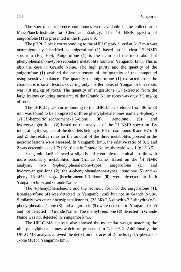
114 Chapter 6
The spectra of reference compounds were available in the collection at
Max-Planck-Institute for Chemical Ecology. The 1H NMR spectra of
anigorufone (1) is presented in the Figure 6.9.
The pHPLC peak corresponding to the aHPLC peak eluted at 51.7 min was
unambiguously identified as anigorufone (1) based on its clear 1H NMR
spectrum (Fig. 6.9). Anigorufone (1) is the main and the most abundant
phenylphenalenone-type secondary metabolite found in Yangambi km5. This is
also the case in Grande Naine. The high purity and the quantity of the
anigorufone (1) enabled the measurement of the quantity of the compound
using sensitive balance. The quantity of anigorufone (1) extracted from the
characteristic small lesions covering only smaller areas of Yangambi km5 roots
was 7.8 mg/kg of roots. The quantity of anigorufone (1) extracted from the
large lesions covering most area of the Grande Naine roots was only 3.9 mg/kg
of roots.
The pHPLC peak corresponding to the aHPLC peak eluted from 36 to 38
min was found to be comprised of three phenylphenalenones namely 4-phenyl-
1H,3H-benzo[de]isochromene-1,3-dione (8), irenolone (5) and
hydroxyanigorufone (2) based on the analysis of the 1H NMR spectrum. By
integrating the signals of the doublets belong to H4 of compound 8 and H7 of 5
and 2, the relative ratio for the amount of the three metabolites present in the
necrotic lesions were assessed. In Yangambi km5, the relative ratio of 8, 5 and
2 was determined as 1.7:1.6:1.0 but in Grande Naine, the ratio was 1.0:1.3:3.5.
Yangambi km5 showed a slightly different phytochemical profile with
more secondary metabolites than Grande Naine. Based on the 1H NMR
analysis, two 9-phenylphenalenone-types: anigorufone (1) and
hydroxyanigorufone (2), the 4-phenylphenalenone-types: irenolone (5) and 4-
phenyl-1H,3H-benzo[de]isochromene-1,3-dione (8) were detected in both
Yangambi km5 and Grande Naine.
The 4-phenylphenalenone and the isomeric form of the anigorufone (1),
isoanigorufone (4) was detected in Yangambi km5 but not in Grande Naine.
Similarly two other phenylphenalenones, (2S,3R)-2,3-dihydro-2,3-dihydroxy-9-
phenylphenalen-1-one (3) and anigorootin (9) were detected in Yangambi km5
and not detected in Grande Naine. The methylirenolone (6) detected in Grande
Naine was not detected in Yangambi km5.
The UPLC-MS analysis also showed the molecular weight matching the
nine phenylphenalenones which are presented in Table 6.2. Additionally, the
UPLC-MS analysis allowed the detection of traces of 2-methoxy-1H-phenalen-
1-one (10) in Yangambi km5.

Phenylphenalenones in the plant resistance to nematodes 115
6.1.3.6. LDI-MSI
LDI-MSI made it possible to detect the localised presence of the
phenylphenalenones in the necrotic regions. The LDI mass spectra showed
prominent signals for the presence of the UV-absorbing-secondary metabolites
in the necrotic lesions of Grande Naine (Fig. 6.10A) and Yangambi km5 (Fig.
6.10C). The regions of necrotic lesions were strikingly different from the
healthy, uninfected surrounding regions for the presence of secondary
metabolites. The LDI mass spectra of uninfected regions showed no prominent
signal for the presence of the phenylphenalenones (Figs 6.10B & D).
Figure 6.10: The LDI mass spectra of Musa roots. A) The Radopholus similis-
infected region of Grande Naine. B) Uninfected region of Grande Naine. C) The R.
similis-infected region of Yangambi km5. D) Uninfected region of Yangambi km5.
The LDI mass spectrum of the uninfected regions of Yangambi km5
showed high background effects (Fig. 6.10D). The heat profile for the m/z value
of 271 as an arbitrarily chosen example verified the high intensity of the m/z
value of 271 in the region of the necrotic lesions in opposite to the surrounding
healthy tissue regions (Fig. 6.12). The m/z values 271, 273, 287, 289, 301 were
detected in Yangambi km5 (Fig. 6.11) and in Grande Naine (Fig. 6.13).
A
C
B
D
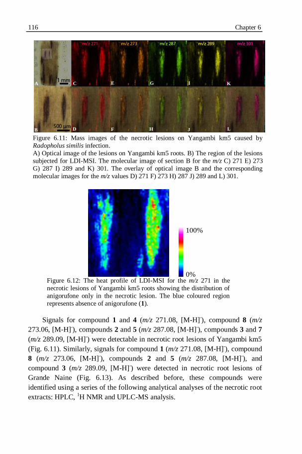
116 Chapter 6
Figure 6.11: Mass images of the necrotic lesions on Yangambi km5 caused by
Radopholus similis infection.
A) Optical image of the lesions on Yangambi km5 roots. B) The region of the lesions
subjected for LDI-MSI. The molecular image of section B for the m/z C) 271 E) 273
G) 287 I) 289 and K) 301. The overlay of optical image B and the corresponding
molecular images for the m/z values D) 271 F) 273 H) 287 J) 289 and L) 301.
Figure 6.12: The heat profile of LDI-MSI for the m/z 271 in the
necrotic lesions of Yangambi km5 roots showing the distribution of
anigorufone only in the necrotic lesion. The blue coloured region
represents absence of anigorufone (1).
Signals for compound 1 and 4 (m/z 271.08, [M-H]-), compound 8 (m/z
273.06, [M-H]-), compounds 2 and 5 (m/z 287.08, [M-H]
-), compounds 3 and 7
(m/z 289.09, [M-H]-) were detectable in necrotic root lesions of Yangambi km5
(Fig. 6.11). Similarly, signals for compound 1 (m/z 271.08, [M-H]-), compound
8 (m/z 273.06, [M-H]-), compounds 2 and 5 (m/z 287.08, [M-H]
-), and
compound 3 (m/z 289.09, [M-H]-) were detected in necrotic root lesions of
Grande Naine (Fig. 6.13). As described before, these compounds were
identified using a series of the following analytical analyses of the necrotic root
extracts: HPLC, 1H NMR and UPLC-MS analysis.
B
C
D
E
F
A G
H
I
J
K
L
0%
100%
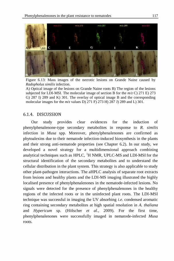
Phenylphenalenones in the plant resistance to nematodes 117
Figure 6.13: Mass images of the necrotic lesions on Grande Naine caused by
Radopholus similis infection.
A) Optical image of the lesions on Grande Naine roots B) The region of the lesions
subjected for LDI-MSI. The molecular image of section B for the m/z C) 271 E) 273
G) 287 I) 289 and K) 301. The overlay of optical image B and the corresponding
molecular images for the m/z values D) 271 F) 273 H) 287 J) 289 and L) 301.
6.1.4. DISCUSSION
Our study provides clear evidences for the induction of
phenylphenalenone-type secondary metabolites in response to R. similis
infection in Musa spp. Moreover, phenylphenalenones are confirmed as
phytoalexins due to their nematode infection-induced biosynthesis in the plants
and their strong anti-nematode properties (see Chapter 6.2). In our study, we
developed a novel strategy for a multidimensional approach combining
analytical techniques such as HPLC, 1H NMR, UPLC-MS
and LDI-MSI
for the
structural identification of the secondary metabolites and to understand the
cellular distribution in the plant system. This strategy is also applicable to study
other plant-pathogen interactions. The aHPLC analysis of separate root extracts
from lesions and healthy plants and the LDI-MS imaging illustrated the highly
localised presence of phenylphenalenones in the nematode-infected lesions. No
signals were detected for the presence of phenylphenalenones in the healthy
regions of the infected roots or in the uninfected plant roots. The LDI-MSI
technique was successful in imaging the UV absorbing i.e. condensed aromatic
ring containing secondary metabolites at high spatial resolution in A. thaliana
and Hypericum sp. (Hölscher et al., 2009). For the first time,
phenylphenalenones were successfully imaged in nematode-infected Musa
roots.
A
B
C
D
E
F
G
H
I
J
K
L
M
N
118 Chapter 6
Anigorufone (1) was identified as the most abundant phenylphenalenone-
type secondary metabolite present in the lesions of Yangambi km5.
Furthermore, it is well localised in the necrotic lesions. Interestingly,
anigorufone (1) was also the most active compound against R. similis in the in
vitro bio-assays causing the highest percentage nematode motility inhibition
(see Chapter 6.2). Nematode mortality was observed at high concentrations (see
Chapter 6.2). The anti-nematode property was positively correlated with the
concentration of the phenylphenalenones (see Chapter 6.2). It is evident that the
anigorufone (1) plays a key role in inhibiting R. similis reproduction in
Yangambi km5. Anigorufone (1) was previously reported as a constitutive
compound in Anigozanthos spp. (Cooke & Thomas 1975; Hölscher &
Schneider, 1997) and as phytoalexin in the R. similis-resistant Musa cultivar
Pisang Jari Buaya (Binks et al., 1997). Infection with fungal pathogens has
shown to induce anigorufone (1) synthesis in Musa spp. (Kamo et al., 1998,
2000; Otálvaro et al., 2007; Jitsaeng, 2009).
Anigorufone (1) was also found as a main phenylphenalenone in Grande
Naine in our study. However, the quantity of anigorufone (1) in Grande Naine
was only about half the quantity in Yangambi km5. Observation of the lesions
demonstrated the highly concentrated accumulation of the secondary
metabolites in small lesions of Yangambi km5; Accumulation of phytoalexins
in larger necrotic areas resulting in lower concentration in Grande Naine.
Hence, apart from higher quantity, the concentration per unit area is far higher
in Yangambi km5 compared to Grande Naine. The high quantity and high
concentration could create a more toxic cellular environment to the nematodes
in Yangambi km5.
The localised synthesis of the phytoalexins at high concentration in the
nematode-infected cells demonstrates the high degree of regulation in
delivering the nematode toxins to the target cells. This tight, highly regulated
synthesis and localised accumulation of the toxic metabolite in a very limited
area at high concentration in the nematode-infected cells can be the key
mechanism by Yangambi km5 to limit R. similis reproduction. The high
concentrations of anigorufone (1), isoanigorufone (4) and hydroxyanigorufone
(2) were shown to inhibit the nematode motility (see Chapter 6.2). The reduced
motility and mortality probably limits the spread of the nematode to only a
small area. Future studies could aim at finding the mode of action of
anigorufone (1) and other active phenylphenalenones on nematode motility
inhibition and toxicity.
Phenylphenalenones in the plant resistance to nematodes 119
Secondary metabolites are often found to accumulate in specific plant
tissues at high concentrations (Opitz et al., 2003; Hölscher & Schneider, 2007;
Wuyts et al., 2007; Bednarek & Osbourn, 2009). This accumulation in such
compartments should be regulated in a highly sophisticated manner, because
some secondary metabolites are phytotoxic if they are mislocalised (Yazaki et
al., 2008). This tightly regulated expression only at the nematode-infected
tissues or feeding sites shows their role in the plant‟s defense against
nematodes.
Disease resistance is largely determined by the rapidity and magnitude of
the synthesis or accumulation of the defence products instead of their presence
or absence in the plants (Kuć, 1995; Wuyts et al., 2007). The speed of the
phytoalexins accumulation and specific localisation at the infection sites of
resistant plants result in concentrations far in excess of inhibitory to the
invading pathogens (Mert-Türk, 2002). Phytoalexins were also shown to be
specifically deployed to the sites of pathogenic attacks by vesicle-mediated
trafficking (Field et al., 2006; Boller & He, 2009). The efficient cargo of
phytoalexins is progressively studied as a key factor in making the plant
resistant to diseases (Field et al., 2006; Cheung & De Vries, 2008; Kwon et al.,
2008). It was reported that the most spectacular manifestation of plant
resistance is the synthesis of phytoalexins in a limited area at the site of
infection (Tronchet et al., 2010).
Phenylphenalenones were reported as inducible compounds of Musa spp.
by pathogenic fungal infections such as M. fijiensis on leaves (Luis et al., 1993;
1995; Otálvaro et al., 2007), F. oxysporum in roots and rhizomes (Luis et al.,
1994; 1996; 1997), C. musae on fruits (Kamo et al., 1998, 2001; Otálvaro et al.,
2007) and by the nematode R. similis in roots (Binks et al., 1997). However,
few reports showed that phenylphenalenones such as 4‟-dehydroxyirenolone
(Luis et al., 1999) and hydroxyanigorufone (2) (Kamo et al., 2000) were
present in the healthy tissues of Musa spp. at very low concentrations. They
possibly play a role as phytoanticipins. Moreover these phytoanticipin
concentrations were shown to increase after infection with pathogens (Kamo et
al., 2000).
The other phenylphenalenones isolated in our study were previously
reported from Musa. Irenolone (5) and hydroxyanigorufone (2) were isolated as
phytoalexins from the leaves of Grande Naine infected with M. fijiensis (Luis et
al., 1993) and in rhizomes infected with F. oxysporum (Luis et al., 1996). 4-(4-
hydroxyphenyl)-1H,3H-benzo[de]isochromene-1,3-dione (7) was reported in
unripe fruits infected by C. musae (Hirai et al., 1994). (2S,3R)-2,3-dihydro-2,3-
dihydroxy-9-phenylphenalen-1-one (3), isoanigorufone (4), methylirenolone (6)
120 Chapter 6
and anigorootin (9) were reported in Musa spp. as natural compounds or in
response to fungal infections (Luis et al., 1995; Kamo et al., 1998; Otálvaro et
al., 2007; Jitsaeng et al., 2010).
Methoxyanigorufone was reported as the major compound in Yangambi
km5 rhizomes (Otálvaro et al., 2007). The two perinaphthenone type
(phenalenone) compounds, 2-hydroxy-1H-phenalenone and 2-methoxy-1H-
phenalenone (10) were previously reported from Yangambi km5 rhizomes
(Otálvaro et al., 2007). With the exception of traces of 2-methoxy-1H-
phenalenone (10) detectable by UPLC-MS technique, the other main
compounds were not isolated from the nematode-infected roots in our study.
The growing conditions and nematode infection in our study might be the
reason for the different phytochemical profile of the Yangambi km5 roots.
Phenylphenalenones show a high potential in protecting monocot plants
from biotic stresses. However they are relatively less explored. Treating in vitro
banana roots with non-pathogenic yeast cultures of Sporobolomyces
salmonicolor has been shown to induce phenylphenalenone-type compounds in
the roots (Jitsaeng & Schneider, 2010). Chemical elicitors such as kanamycin
and jasmonic acid also induced the synthesis of phenylphenalenones in Musa
(Luis et al., 1993, Jitsaeng, 2009). The underlying reason for the enhanced
tolerance to diseases due to endophytic colonization may also be attributed to
enhanced phenylphenalenones biosynthesis. Future studies on the possibility of
enhancing the cellular concentration and improved localisation of anigorufone
(1) and other phenylphenalenones in Grande Naine roots will be highly
fascinating to see if these changes may make Grande Naine also resistant to R.
similis reproduction. Although metabolic engineering of secondary metabolites
pathway for enhancing the disease resistance could be very challenging, a
feasible strategy for is previously presented and discussed (Dixon, 2001).
6.1.5. CONCLUSION
Our study provides clear evidence for the induction of phenylphenalenone-
type secondary metabolites in Musa spp. in response to R. similis infection.
Nine phenylphenalenone-type phytoalexins were structurally identified from the
necrotic tissues of the banana roots. The phenylphenalenones showed a highly
localised presence in the nematode-infected necrotic regions of the banana
roots. The phenylphenalenones were not detected in the healthy regions.
Anigorufone (1) was identified as the most abundant phenylphenalenone-type
secondary metabolite present in the necrotic lesions. It is evident that
anigorufone (1) plays a key role in limiting R. similis reproduction in the

Phenylphenalenones in the plant resistance to nematodes 121
resistant Musa cultivar Yangambi km5. Anigorufone (1) was also a major
compound in the susceptible Musa cultivar Grande Naine. However, the
concentration per unit area was very high in Yangambi km5 compared to
Grande Naine. This may have created a more toxic cellular environment to the
nematode. Phenylphenalenones show a high potential in protecting monocot
plants from biotic stresses.

122 Chapter 6
CHAPTER 6.2: ANTI-NEMATODE PROPERTIES OF
THE PHENYLPHENALENONES
6.2.1. INTRODUCTION
Studies on identifying phytochemicals with anti-nematode properties have
been gaining interest among nematologists (Akthar & Mahmood, 1994;
Chitwood, 2002; Wuyts et al., 2006). Phytochemical-based nematode
management strategies could provide economically viable and environmentally
safer alternatives to the conventional nematicides or they could serve as models
for developing new nematicides (Chitwood, 2002).
Phytoalexins elicited in nematode-resistant plants are generally anticipated
to have anti-nematode properties. However, knowledge on the mode of action,
the biologically active concentrations and the effective exposure time are vital
to determine the efficiency and feasibility of practical applications of
phytochemicals. Phenylphenalenones biosynthesis is elicited in Musa plants in
response to biotic stresses (Luis et al., 1993; Kamo et al., 1998). Biosynthesis
of phenylphenalenones has become evident as a response to nematode infection
in resistant as well as susceptible Musa cultivars (see Chapter 6.1). Hence it is
our primary interest to discover the role of these nematode infection-site-
specific compounds in the plant‟s defense against nematodes.
The antimicrobial properties of phenalenones were reported previously
(Winters et al., 1977; Qin et al., 2006). Antifungal properties of
phenylphenalenones against the major fungal pathogens of Musa spp. such as
Fusarium oxysporum f. sp. cubense, Colletotrichum musae and Mycosphaerella
fijiensis were also reported (Luis et al., 1996, 1998; Kamo et al., 1998;
Quiñones et al., 2000; Lazzaro et al., 2004; Otálvaro et al., 2007). But to our
knowledge, there is no study performed so far on the anti-nematode properties
of the phenylphenalenones.
The objective of this part of our study was to identify the anti-nematode
properties of the phenylphenalenone-type phytoalexins using in vitro bio-assays
on the motility of R. similis as well as on the toxicity effect of the compounds
on R. similis.
Phenylphenalenones in the plant resistance to nematodes 123
6.2.2. MATERIALS AND METHODS
6.2.2.1. Experimental set-up
Two series of in vitro bio-assay experiments were carried out. The first
series of experiments were conducted with 13 different phenylphenalenones,
each at two concentrations (50 and 100 ppm) to test their effects on R. similis
motility. In the second experiments, the dosage effect of anigorufone (1) on R.
similis motility was tested using a gradient of six concentrations (10, 20, 40, 50,
100 and 150 ppm). All experiments were repeated twice to confirm the results.
6.2.2.2. Chemicals
All phenylphenalenones used in the bio-assays were extracted and purified
using pHPLC from Musa spp. and Anigozanthos spp. at the Max-Planck-
Institute for Chemical Ecology. Compounds were dissolved in aqueous ethanol
(1%). Stock solutions of 200 μg/ml (ppm) were prepared aseptically and stored
in small portions at -20 °C.
6.2.2.3. Nematodes
A population of R. similis originally isolated from banana roots in Uganda
was used in the experiments. This population from Uganda was characterized
by a high reproductive fitness (Fallas et al., 1995). This population was
maintained and multiplied monoxenically on sterile alfalfa callus tissues at
25+1 °C in the dark (Elsen et al., 2001). To obtain inoculum, nematodes were
collected from the callus tissues or from the medium by rinsing with sterile
distilled water. Only 6-weeks-old nematode cultures were used to prepare the
inoculum of adults and juveniles of R. similis.
6.2.2.4. Effect of phenylphenalenones on R. similis motility bio-assay
Thirteen different phenylphenalenones were tested for their anti-nematode
properties using in vitro bio-assays. The molecular structures of the compounds
tested in the bio-assays are provided in the annexes. Nematode motility was
assayed in two concentrations (50 and 100 ppm). Each treatment was replicated
three times and for each compound, the assay was repeated at least twice. The
experiment was performed in 24-well-plates under aseptic conditions. The
negative control consisted of the compound solvent, 1% ethanol. A sterile
distilled water control was also included to test the solvent effects on motility.
At the onset of experiment, an aliquot of 100 μl sterile nematode
suspension containing 80-100 adults and juveniles of R. similis was added to
124 Chapter 6
each well prior to the addition of the compounds. The exact numbers of motile
and immotile nematodes was counted in each well using an inverted compound
microscope (40×). The stock solution (200 ppm) of the compounds and sterile
distilled water were added into each well according to their concentration.
Plates were incubated in the dark at 25+1 °C. The numbers of quiescent
(immotile) nematodes were counted after 24, 48 and 72 h of incubation.
The percentages of quiescent nematodes were calculated relative to the
number of actively moving and immotile nematodes at the onset of the
experiment. Compounds were considered motility inhibitive when significantly
(P ≤ 0.05) more nematodes have become quiescent than in the solvent control at
any assayed concentration.
6.2.2.5. Dosage effect of anigorufone (1) on R. similis motility bio-
assay The motility bio-assay was performed with a gradient of six different
concentrations of anigorufone (1): 10, 20, 40, 50, 100 and 150 ppm. Each
treatment was replicated six times and the bio-assay was repeated twice. The
experimental methods were the same as the motility assay (6.2.2.4). Dose-
dependent effects allowed the calculation of the IC50 values at 24, 48 and 72 h
of incubation.
The IC50 value is defined as the concentration of a compound required to
cause 50% of the total nematodes to become quiescent. Toxicity of the
anigorufone (1) was tested when a minimum of 10% or more nematodes
become quiescent in a treatment than in the solvent control. Nematicidal effects
were checked by transferring the quiescent nematodes to sterile distilled water
after 72 h incubation in anigorufone (1) followed by counting the number of
live and dead nematodes under a fluorescence microscope at 96 h (Forge &
MacGuidwin, 1989) combined with poking the nematodes.
6.2.2.6. Statistical data analysis
Statistical analysis was performed using STATISTICA version 9 (StatSoft,
Tulsa, OK, USA). Data on motility assay were analyzed using Kruskal-Wallis
analysis of variance by ranks. When the Kruskal-Wallis analysis of variance by
ranks was significant, each group mean was compared with the negative control
by multiple comparisons of mean ranks.
Data on dosage effect of anigorufone (1) on R. similis motility were
analyzed by analysis of variance (ANOVA) provided the conditions of
ANOVA (normal distribution and homogeneity of variance) were met. The

Phenylphenalenones in the plant resistance to nematodes 125
percentage quiescent nematodes were arcsin (x/100) transformed prior to the
analysis. When significant differences (P ≤ 0.05) were observed, Tukey‟s HSD
test was applied for multiple comparisons of group means. The IC50 values were
obtained by interpolating the corresponding percentage of quiescent nematodes
vs. anigorufone (1) concentration in scatter plots.
6.2.3. RESULTS
6.2.3.1. Effect of phenylphenalenones on R. similis motility bio-assay
The effects of the 13 assayed phenylphenalenones on R. similis motility are
listed in Table 6.3.
Table 6.3. Effect of the phenylphenalenone-type phytoalexins on the motility of
Radopholus similis.
Compound Effect on
motility
Anigorufone (1) I
Anigorootin (9) I
(2R,3R)-2,3-Dihydro-2,3-dihydroxy-9-phenylphenalenone I
Dihydroxyanigorootin -
Hydroxyanigorufone (2) I
4-Hydroxy-2-methoxy-9-phenylphenalenone I
4-(4-Hydroxyphenyl)-1H,3H-benzo[de]isochromene-1,3-dione (7) I
Irenolone (5) I
Isoanigorufone (4) I
Methoxyanigorufone I
Methylirenolone (6) -
Monohydroxyanigorootin I
Perinaphthenone I
A compound was considered motility inhibitive (I) when it caused significantly (P ≤
0.05) high percentage of nematode quiescence compared to the negative control.
Significance was tested according to Kruskal-Wallis analysis.
I: Inhibitory effect; -: No effect.
Eleven out of 13 compounds inhibited the nematode motility in at least one
concentration at any of the observed time points. Only dihydroxyanigorootin
and methylirenolone (6) did not show any significant inhibition on nematode
motility at any assayed concentrations.
The extended results of the nematode motility bio-assay are presented in
Table 6.4. Specific to mention are anigorufone (1), 4-hydroxy-2-methoxy-9-
phenylphenalenone and isoanigorufone (4) that caused about 75% of R. similis
to become quiescent. Anigorufone (1), hydroxyanigorufone (2) and
monohydroxyanigorootin were causing nematode quiescence in all tested

126 Chapter 6
concentrations starting from 24 h of incubation and the effect was either
consistent or increased over time.
The anti-nematode activity displayed by anigorufone (1) was the strongest
causing 89% of R. similis to become quiescent at 72 h of incubation in the
compound. 4-Hydroxy-2-methoxy-9-phenylphenalenone, 4-(4-hydroxyphenyl)-
1H,3H-benzo[de]isochromene-1,3-dione (7) and isoanigorufone (4) also caused
persistent and strong nematode quiescence at 100 ppm concentration. However,
at a lower concentration (50 ppm), a significantly (P ≤ 0.05) high percentage of
nematode quiescence was observed only starting from 48 h of incubation
compared to the negative control.
Anigorootin (9), (2R,3R)-2,3-dihydro-2,3-dihydroxy-9-phenylphenalenone,
irenolone (5), methoxyanigorufone and perinaphthenone showed moderate anti-
nematode activity as they caused a non-persistent nematode quiescence at only
one of the tested concentrations. The solvent 1% ethanol exhibited no
significant effect on nematode movement compared to the sterile distilled water.
A notable observation was made while counting the quiescent nematodes.
The nematodes ingested anigorufone (1) in such large amounts that the ingested
anigorufone (1) molecules were clearly visible inside the nematodes under light
microscopic observation (Figs 6.14 & 6.15). This is due to the natural colour of
anigorufone (1) and the natural transparency of nematode cuticle. No coloured
spots were observed in the nematodes in the controls (Fig. 6.16).
Due to this interesting observation, the bio-assay with anigorufone (1) was
repeated many times. The percentages of nematodes ingesting visible amounts
of anigorufone (1) were variable from one experiment to the other. However, all
the nematodes that ingested visibly large amounts of anigorufone (1) molecules
were always dead (Fig. 6.15).
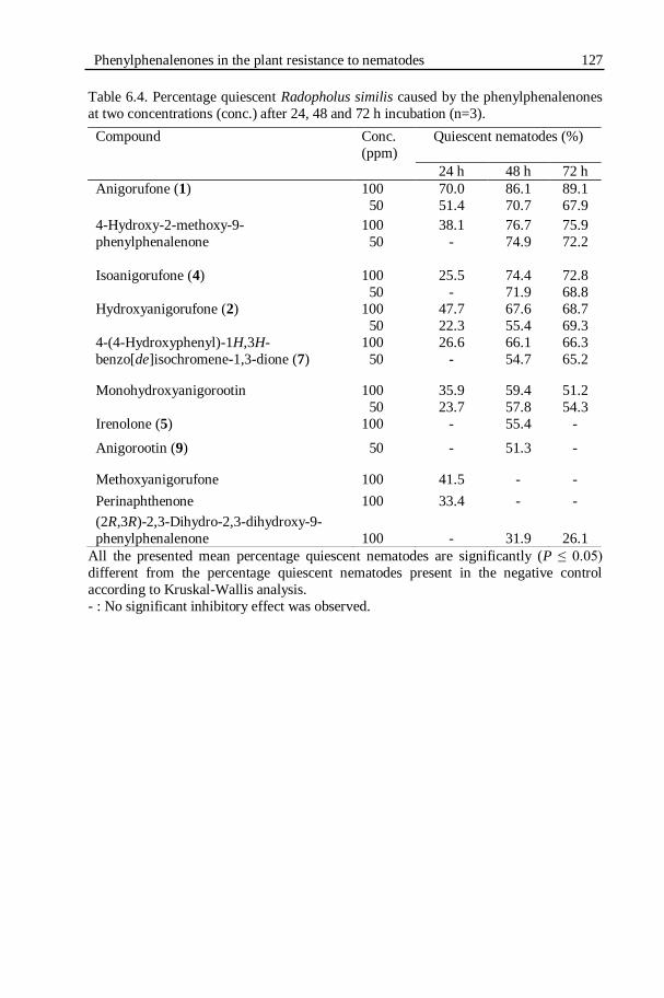
Phenylphenalenones in the plant resistance to nematodes 127
Table 6.4. Percentage quiescent Radopholus similis caused by the phenylphenalenones
at two concentrations (conc.) after 24, 48 and 72 h incubation (n=3).
All the presented mean percentage quiescent nematodes are significantly (P ≤ 0.05)
different from the percentage quiescent nematodes present in the negative control
according to Kruskal-Wallis analysis.
- : No significant inhibitory effect was observed.
Compound Conc.
(ppm)
Quiescent nematodes (%)
24 h 48 h 72 h
Anigorufone (1) 100 70.0 86.1 89.1
50 51.4 70.7 67.9
4-Hydroxy-2-methoxy-9-
phenylphenalenone
100 38.1 76.7 75.9
50 - 74.9 72.2
Isoanigorufone (4) 100 25.5 74.4 72.8
50 - 71.9 68.8
Hydroxyanigorufone (2) 100 47.7 67.6 68.7
50 22.3 55.4 69.3
4-(4-Hydroxyphenyl)-1H,3H-
benzo[de]isochromene-1,3-dione (7)
100 26.6 66.1 66.3
50 - 54.7 65.2
Monohydroxyanigorootin 100 35.9 59.4 51.2
50 23.7 57.8 54.3
Irenolone (5) 100 - 55.4 -
Anigorootin (9) 50 - 51.3 -
Methoxyanigorufone 100 41.5 - -
Perinaphthenone 100 33.4 - -
(2R,3R)-2,3-Dihydro-2,3-dihydroxy-9-
phenylphenalenone
100
-
31.9
26.1

128 Chapter 6
Figure 6.14: Light microscopic images of Radopholus similis during the bio-
assay with anigorufone (1). A) Quiescent nematodes resulted from 24 h
incubation in anigorufone (1). B) & D) Live, active nematodes present in the
negative control, 1% ethanol after 24 h of incubation. C) Dead nematodes
after ingesting anigorufone (1) molecules during the bio-assay; ingested
molecules (yellow coloured in the gut) stored along the entire length of the
nematode gut. Scale bar: C & D: 20 µm; A & B: 40 x magnification.
A
C
B
D

Phenylphenalenones in the plant resistance to nematodes 129
Figure 6.15: Light microscopic images of all life stages of Radopholus similis that had
died because of ingesting anigorufone (1) during the bio-assays. A) Intense
accumulation of the compound in a moulting juvenile. B) & F) Accumulation of
anigorufone (1) in small vacuoles throughout the body of a juvenile. C) & D) Adult females; C) shows that the compound is present in the digestive tract not in the
pseudocoelum. E) Male nematode. G) Female nematodes showing anigorufone (1)
accumulated near the ovary. H) Anigorufone (1) accumulation at the end of the
digestive tract but not in the tail. Scale bar: A: 25µm; B: 40µm; C, D & E): 20µm.
A B C D E
F G H

130 Chapter 6
Figure 6.16: Light microscopic images of Radopholus similis from the negative
control, 1% ethanol. Nematodes were temporarily fixed for imaging purpose. A)
The head & B) the tail of a R. similis showing normal digestive tract. C) Juvenile
& D) adult female R. similis after 72 h incubation in the negative control.
6.2.3.2. Dosage effect of anigorufone (1) on R. similis motility bio-
assay The dosage effects of anigorufone (1) on R. similis motility after 24, 48
and 72 h of incubation are listed in Table 6.5 and shown in the Figure 6.18. A
sharp increase in the effect of concentration on nematode motility was observed
after an initial lag phase (Fig. 6.17). At the lowest concentration of 10 ppm,
A B
C D
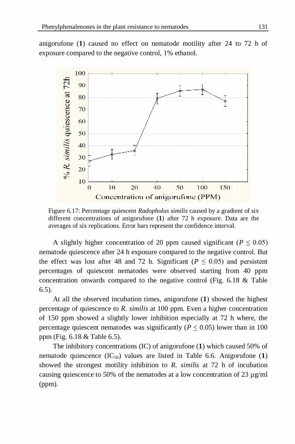
Phenylphenalenones in the plant resistance to nematodes 131
anigorufone (1) caused no effect on nematode motility after 24 to 72 h of
exposure compared to the negative control, 1% ethanol.
Figure 6.17: Percentage quiescent Radopholus similis caused by a gradient of six
different concentrations of anigorufone (1) after 72 h exposure. Data are the
averages of six replications. Error bars represent the confidence interval.
A slightly higher concentration of 20 ppm caused significant (P ≤ 0.05)
nematode quiescence after 24 h exposure compared to the negative control. But
the effect was lost after 48 and 72 h. Significant (P ≤ 0.05) and persistent
percentages of quiescent nematodes were observed starting from 40 ppm
concentration onwards compared to the negative control (Fig. 6.18 & Table
6.5).
At all the observed incubation times, anigorufone (1) showed the highest
percentage of quiescence to R. similis at 100 ppm. Even a higher concentration
of 150 ppm showed a slightly lower inhibition especially at 72 h where, the
percentage quiescent nematodes was significantly (P < 0.05) lower than in 100
ppm (Fig. 6.18 & Table 6.5).
The inhibitory concentrations (IC) of anigorufone (1) which caused 50% of
nematode quiescence (IC50) values are listed in Table 6.6. Anigorufone (1)
showed the strongest motility inhibition to R. similis at 72 h of incubation
causing quiescence to 50% of the nematodes at a low concentration of 23 µg/ml
(ppm).

132 Chapter 6
Table 6.5. Percentage quiescent Radopholus similis caused by anigorufone (1) at
six different concentrations over three days (n=6).
Concentration of anigorufone
(ppm)
Quiescent nematodes (%)
24h 48h 72h
10 25.9 a† 30.2 a† 32.7 a†
20 31.4 a 28.6 a† 35.9 a†
40 50.2 b 68.6 b 79.3 bc
50 63.8 c 79.3 c 85.6 c
100 74.3 d 85.0 c 86.8 c
150 70.1 cd 82.8 c 77.0 b
1% ethanol 17.6 21.0 27.3
Means within each column followed by the same letters are not significantly (P ≤
0.05) different from each other according to Tukey‟s HSD test. Means followed by † are not significantly different from the mean percentage quiescent
nematodes present in the negative control (1% ethanol).
Figure 6.18: Percentage quiescent Radopholus similis observed during the
motility bio-assay on a concentration gradient of anigorufone after 24, 48 and
72 h of incubation. Error bars represent the confidence interval (n=6).
24h 48h 72h10
20
30
40
50
60
70
80
90
% R
. si
mil
is q
uie
scen
ce
150 PPM 100PPM 50 PPM 40 PPM 20 PPM 10 PPM
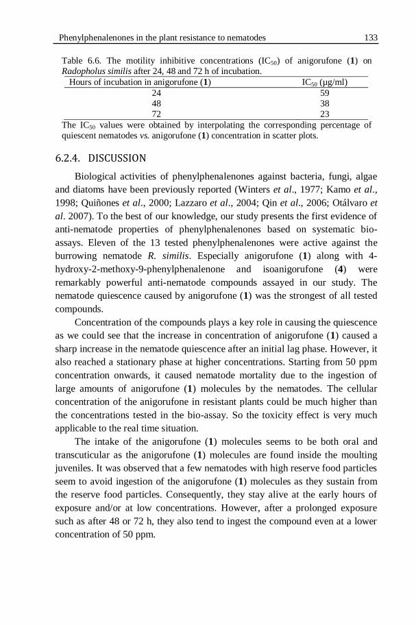
Phenylphenalenones in the plant resistance to nematodes 133
Table 6.6. The motility inhibitive concentrations (IC50) of anigorufone (1) on
Radopholus similis after 24, 48 and 72 h of incubation.
Hours of incubation in anigorufone (1) IC50 (µg/ml)
24 59
48 38
72 23
The IC50 values were obtained by interpolating the corresponding percentage of
quiescent nematodes vs. anigorufone (1) concentration in scatter plots.
6.2.4. DISCUSSION
Biological activities of phenylphenalenones against bacteria, fungi, algae
and diatoms have been previously reported (Winters et al., 1977; Kamo et al.,
1998; Quiñones et al., 2000; Lazzaro et al., 2004; Qin et al., 2006; Otálvaro et
al. 2007). To the best of our knowledge, our study presents the first evidence of
anti-nematode properties of phenylphenalenones based on systematic bio-
assays. Eleven of the 13 tested phenylphenalenones were active against the
burrowing nematode R. similis. Especially anigorufone (1) along with 4-
hydroxy-2-methoxy-9-phenylphenalenone and isoanigorufone (4) were
remarkably powerful anti-nematode compounds assayed in our study. The
nematode quiescence caused by anigorufone (1) was the strongest of all tested
compounds.
Concentration of the compounds plays a key role in causing the quiescence
as we could see that the increase in concentration of anigorufone (1) caused a
sharp increase in the nematode quiescence after an initial lag phase. However, it
also reached a stationary phase at higher concentrations. Starting from 50 ppm
concentration onwards, it caused nematode mortality due to the ingestion of
large amounts of anigorufone (1) molecules by the nematodes. The cellular
concentration of the anigorufone in resistant plants could be much higher than
the concentrations tested in the bio-assay. So the toxicity effect is very much
applicable to the real time situation.
The intake of the anigorufone (1) molecules seems to be both oral and
transcuticular as the anigorufone (1) molecules are found inside the moulting
juveniles. It was observed that a few nematodes with high reserve food particles
seem to avoid ingestion of the anigorufone (1) molecules as they sustain from
the reserve food particles. Consequently, they stay alive at the early hours of
exposure and/or at low concentrations. However, after a prolonged exposure
such as after 48 or 72 h, they also tend to ingest the compound even at a lower
concentration of 50 ppm.
134 Chapter 6
At initial observation, the yellow coloured spots were distributed in small
putative lipid-like globules. However, with time, the globules joined with each
other and formed a channel of yellow globules accupying a large space of the
nematode body volume. This might also imposs a large constraint in nematode
body affecting the nematode viability. The possibility of anigorufone forming a
complex with the nematode‟s putative lipid molecules could be of interest for
future studies. Lipids are reported in the free-living nematode Caenorhabditis
elegans (Klapper et al., 2011). The reason why the nematodes did not excrete
but accumulate the anigorufone remains to be studied further. Biogenic amines
and trace amines such as tyramine and octopamine were shown to affect the
behaviour of free living nematode Caenorhabditis elegans. These amines were
shown to inhibit the aversive response of C. elegans to 100% octanol (Wragg et
al., 2007). Possibility of such behavioural modulation by the nematode-ingested
phytoalexins influencing the behaviour of R. similis could be of interest to study
in future. Lipophilic compounds are highly membrane disruptive (Cowan,
1999). Thus, there is also a possibility that the ingestion of the lipophilic
anigorufone (1) by nematodes could be associated with the toxicity.
Bio-assays conducted by Quiñones et al. (2000) and Otálvaro et al. (2007)
showed that anigorufone (1) was also a notable antifungal compound which
significantly inhibited the mycelial growth but, moderately inhibited the spore
germination of M. fijiensis, the causal agent of Black Sigatoka leaf disease in
Musa spp. Anigorufone (1) was also active against F. oxysporum in dark but
inactive in light (Lazzaro et al., 2004).
In previous reports, isoanigorufone (4) showed strong inhibition of F.
oxysporum in the dark but was less active in light (Lazzaro et al., 2004).
However, the same compound showed only mild and non-persistent inhibition
of M. fijiensis (Quiñones et al., 2000).
Hydroxyanigorufone (2), 4-(4-hydroxyphenyl)-1H,3H-
benzo[de]isochromene-1,3-dione (7), and monohydroxyanigorootin were the
next notably active anti-nematode compounds in our study. Antifungal
properties of hydroxyanigorufone (2) were reported against key pathogens of
Musa spp. A moderate to low inhibition of M. fijiensis (Quiñones et al., 2000;
Otálvaro et al., 2007), a significant inhibition of F. oxysporum in light as well
as in dark (Lazzaro et al., 2004) and a moderate inhibition of C. musae (Kamo
et al., 1998) were caused by the hydroxyanigorufone (2). The photoactive
toxicity of phenylphenalenones was shown to be caused by the production of
singlet oxygen (Lazzaro et al., 2004).
Phenylphenalenones in the plant resistance to nematodes 135
Based on the results of a study on the anti-fungal activity of 14 different
phenylphenalenones and their derivatives, Kamo et al. (1998) concluded that a
phenolic-hydroxyl group is essential for this anti-fungal activity. Our study is
not in agreement with this conclusion because the highly active anti-nematode
compounds such as anigorufone (1), isoanigorufone (4) possess a phenolic
group without any hydroxyl group. Further studies are required with structurally
diverse compounds to understand the relationship between the structural
similarities and anti-nematode property. The same study by Kamo et al. (1998)
also suggested that the 4-phenylphenaelnone types could be more biologically
active than the 9-phenylphenalenones types based on their observation that
irenolone (5) inhibited the spore germination of C. musae more than
hydroxyanigorufone (2). In contrast, in our study, the 9-phenylphenalenone,
anigorufone (1) was more active against R. similis than the 4-
phenylphenalenone, isoanigorufone (4). Similarly, hydroxyanigorufone (2) was
more active than irenolone (5).
Irenolone (5), anigorootin (9) and methoxyanigorufone caused moderate
but non-persistent quiescence of R. similis. Irenolone (5) was also less active
against M. fijiensis and C. musae (Quiñones et al., 2000; Kamo et al., 1998).
Methoxyanigorufone was highly active against F. oxysporum in the presence of
light but inactive in the dark and moderately active against M. fijiensis
(Otálvaro et al., 2007). Perinaphthenone was reported as a powerful anti-fungal
compound: which inhibited the M. fijiensis significantly more than the
fungicide benomyl. As perinaphthenone is an efficient singlet oxygen sensitizer,
it was suggested that the high anti-fungal activity could be a result of the
production of singlet oxygen (Quiñones et al., 2000). In contrast, in our study,
perinaphthenone displayed very low activity against R. similis and it remained
effective only until 24 h incubation.
The suggested mechanisms conferring the antibiotic properties of
phenylphenalenones are DNA intercalation, radical formation and oxygen
activation (Winters et al., 1977; Kamo et al., 1998; Quiñones et al., 2000).
Lazzaro et al. (2004) provided evidence for photoactive toxicity of
phenylphenalenones due to the production of singlet oxygen. However, light-
induced toxicity could not function against nematodes due to the fact that
nematodes live in dark and the phenylphenalenones are elicited in roots under
dark conditions. The phenylphenalenones such as anigorufone (1),
isoanigorufone (4) and hydroxyanigorufone (2) which displayed anti-fungal
activity under the dark experimental conditions (Lazzaro et al., 2004) were the
most active anti-nematode compounds in our study and the reverse (i.e. those
displayed high anti-fungal activity in light are less active or inactive against

136 Chapter 6
nematodes) is also true. Further investigations are needed to explain the
mechanism and conditions required for the anti-nematode properties of the
phenylphenalenones.
Laboratory in vitro bio-assays may have a limited relevance to the field or
real time situations (Spence et al., 2008). In real time plant-nematode
interactions, enzymatic oxidation and polymerisation of individual compounds
could occur resulting in increased or reduced toxicity. Nevertheless, the
laboratory bio-assays are a simplified and practical version of the field
conditions (Spence et al., 2008). In our study, we focused mainly on the
individual compound effects not on the whole extract and insufficient amount
of compounds obtained from the plants limited us from performing extended
toxicity, reversibility and dosage effect studies of all the interesting compounds.
The individual compounds in the whole root extract could interact with each
other synergistically or antagonistically (Chitwood, 2002). Elaborated future
bio-assays using different combinations of the identified compounds will
enhance the understanding on the synergism or antagonisms of the compounds.
Several practical applications could result from the identification of the
anti-nematode phytochemicals. The compounds could be developed as plant-
based nematicides or they could serve as models for developing nematicides
based on the relationship between the chemical structure and the biological
activity. However, extreme caution must be exercised in this application as
certainly not all phytochemicals are safe for the environment, non-target
organisms or humans (Chitwood, 2002). The most appealing way forward is to
exploit the plant-nematode interactions to enhance the cellular synthesis and
localisation of the most active anti-nematode compounds in the target cells of
the susceptible plant roots.
6.2.5. CONCLUSIONS
From this study, we offered first evidence for the anti-nematode properties
of phenylphenalenones. Anigorufone (1) was the most powerful anti-nematode
compound assayed. At higher concentrations such as 50, 100 and 150 ppm of
anigorufone (1), nematodes ingested large amounts of the compound causing
mortality of the nematodes. 4-Hydroxy-2-methoxy-9-phenylphenalenone and
isoanigorufone (4) are remarkably powerful anti-nematode compounds assayed
in our study. Further studies could aid in developing these highly active anti-
nematode phytochemicals for use in practical applications for nematode
management.

CHAPTER 7
GENERAL CONCLUSIONS AND PERSPECTIVES

138 Chapter 7
In our study, we evaluated and characterized the mechanism(s) of
resistance to Radopholus similis (Cobb, 1893) Thorne, 1949, a migratory endo-
parasitic nematode, in a selection of Musa genotypes. For this purpose, seven
recently identified R. similis-resistant Musa genotypes were verified for their R.
similis resistance under greenhouse conditions (Chapter 2). The well-known R.
similis-resistant cultivars Yangambi km5 and Pisang Jari Buaya, and the R.
similis-susceptible cultivar Grande Naine were included as references for
comparison. The host response of these Musa genotypes to Meloidogyne
incognita infection was also evaluated to examine if the R. similis-resistant
genotypes are also resistant to the root-knot nematode M. incognita (Chapter 2).
Resistance of Long Tavoy, Saba, Pisang Mas and Pora Pora to R. similis was
confirmed. The Musa genotypes Long Tavoy, Saba and Yangambi km5 were
shortlisted for characterization of the R. similis resistance. The Musa genotypes
Vudu Papau and Pisang Mas were identified as resistant to M. incognita. Our
study identified Pisang Mas as a Musa genotype with combined resistance to
both R. similis and M. incognita. This is the first time that a Musa genotype has
been found that is resistant to both these two major banana root pathogens.
The first major specific objective of our study was to find out at which
phase of the nematode-plant interactions (i.e. pre- or post-infection) the
resistance to R. similis was active. To achieve this objective, an autotrophic in
vitro model system was developed to facilitate the study of host location and
penetration behaviour of R. similis on the selected R. similis-resistant and
susceptible Musa genotypes (Chapter 3). In autotrophic in vitro systems, plant
shoots are exposed to open air allowing the plant to perform active
photosynthesis through atmospheric carbon exchange. At the same time, the
root system and the nematodes are maintained under strict in vitro conditions in
a Petri dish. Due to the horizontal root growth in the Petri dishes, this system
enables direct observation of the roots and the nematodes using a stereoscopic
microscope. The autotrophic in vitro model system proved to be a very good
model system and advantageous compared to the strict in vitro systems to study
nematode attraction and penetration. Our direct microscopic observation of
nematode attraction and migration showed no differences in host location of the
R. similis-resistant genotypes Saba and Yangambi Km5, and the susceptible
Musa genotype Grande Naine by R. similis females. Our novel two-
compartment autotrophic in vitro set-up proved that the R. similis females
showed no preference to migrate towards the roots of either the resistant
(Yangambi km5) or susceptible (Grande Naine) Musa genotypes when a choice
was given to migrate towards both plant roots at the same time. Also no
differences were observed in penetration of the resistant and susceptible Musa

Conclusions and Perspectives 139
genotypes by R. similis females under autotrophic in vitro conditions. In
conclusion, our studies using the autotrophic in vitro systems showed that no
preformed resistance factors inhibit the nematode attraction, migration towards
and penetration of the resistant Musa roots compared to the susceptible
genotype Grande Naine.
Greenhouse experiments were conducted on the penetration, development
and reproduction of R. similis on resistant and susceptible Musa genotypes. Our
objective of this study was to understand whether the R. similis resistance
operates pre-infectionally towards the nematode attraction and penetration or
post-infectionally towards nematode development and reproduction (Chapter
4). No significant differences were observed in the number of R. similis
penetrating the resistant Musa genotypes Long Tavoy, Saba and Yangambi
km5, and the susceptible genotype Grande Naine in our study. But, the post-
infectional nematode development and reproduction were highly impaired in
the resistant Musa genotypes. The number of eggs, juveniles, males, females
and the final population density were significantly higher in Grande Naine than
in the R. similis-resistant Musa genotypes. On the basis of these observations, it
appears that the mechanism of resistance in the investigated Musa genotypes to
R. similis is induced after nematode penetration and that the preformed host
resistance factors do not function strongly against nematode attraction,
migration towards the roots, and penetration of the roots.
The second major specific objective of our study was to identify the
phytochemicals involved in the resistance to R. similis in Musa genotypes. A
preliminary phytochemical profiling was performed to localise and quantify
lignin and total phenols in the R. similis-resistant and susceptible Musa
genotypes (Chapter 5). Our results showed that R. similis infection increased the
root secondary wall lignification of the endodermis and vascular bundles of
resistant and susceptible Musa genotypes. This increased lignification is a
general defense response to protect the vascular bundle to reduce damage to the
plant. Extensive lignification was not found associated with the cortex cells that
are directly involved in the defense with nematodes. Hence, lignification does
not seem to be associated with resistance to nematode development,
reproduction and multiplication. Preformed phenolic cells were not found to be
related with nematode resistance in the studied Musa genotypes. The nematode
infection has dramatically increased the total phenol contents of resistant plants.
Phenolic secondary metabolites are major constituents of the nematode infected
necrotic cells.

140 Chapter 7
In the final part of our study to characterize the nematode resistance in
Musa genotypes, more detailed analytical studies were carried out to identify
the secondary metabolites that are induced in nematode infection sites (Chapter
6.1). Phytochemical profiling of Yangambi km5 and Grande Naine root extracts
was carried out using a combination of analytical techniques namely HPLC, 1H
NMR, UPLC-MS and LDI-MSI analysis. The results of this study provided
clear evidence for the induction of phenylphenalenone-type secondary
metabolites in Musa spp. in response to R. similis infection. Nine
phenylphenalenone-type phytoalexins were isolated and structurally identified
from R. similis-infected necrotic tissues of the banana roots. The
phenylphenalenones showed a highly localised presence only in the nematode-
infected necrotic regions of the banana roots. The phenylphenalenones were not
detected in the healthy regions. Anigorufone (1) was identified as the most
abundant phenylphenalenone-type secondary metabolite present in the necrotic
lesions. It is evident that anigorufone (1) plays a key role in limiting R. similis
reproduction in the resistant Musa cultivar Yangambi km5. Anigorufone (1)
was also a major compound in the susceptible Musa cultivar Grande Naine.
However, the concentration of anigorufone (1) per unit area was very high in
Yangambi km5 compared to Grande Naine. This higher concentration of
anigorufone localised in few cells in the R. similis-resistant cultivar can create a
more toxic cellular environment to the nematode compared to Grande Naine.
Phenylphenalenones show a high potential in protecting monocot plants from
biotic stresses. Hence we conclude that the presence of the phenylphenalenone-
type phytoalexins at high concentrations localised in the hypersensitive-like
necrotic cells of Yangambi km5 is highly associated with the mechanism of R.
similis resistance.
The anti-nematode properties of the identified phenylphenalenones were
assessed by in vitro bio-assays on nematode motility inhibition and nematode
mortality (Chapter 6.2). Our results offered the first evidence for the anti-
nematode properties of phenylphenalenones. Eleven out of thirteen tested
phenylphenalenones were inhibitive to R. similis motility. Anigorufone (1), 4-
hydroxy-2-methoxy-9-phenylphenalenone and isoanigorufone (4) are
remarkably powerful anti-nematode compounds tested in our study causing
quiescence of more than 75% of the nematodes. Hydroxyanigorufone (2) and
monohydroxyanigorootin were also highly inhibitive to R. similis motility
starting from 24 h of incubation and the effect was either consistent or increased
over time. Anigorufone (1) was the most powerful anti-nematode compound
assayed. At higher concentrations such as 50, 100 and 150 ppm of anigorufone
(1), nematodes ingested large amounts of the compound causing mortality of

Conclusions and Perspectives 141
the nematodes. The ingested anigorufone was found accumulated in the
nematode guts of all life stages of nematodes. Elaborated future bio-assays
using different combinations of the identified compounds will enhance the
understanding on the synergism or antagonisms of the compounds.
The identification of these anti-nematode phytochemicals offers many
practical applications for nematode management. Though there are the options
to either develop the phytochemicals as plant-based nematicides or to use them
as models for developing nematicides, extreme caution must be exercised in
this application as certainly not all phytochemicals are safe for the environment,
non-target organisms or humans (Chitwood, 2002). The most appealing way
forward is to exploit the plant-nematode interactions to enhance the cellular
synthesis and localisation of the most active anti-nematode compounds in the
target cells of the susceptible plant roots.
Future studies could explore the possibilities of enhancing the cellular
concentration and improved localisation of anigorufone (1) and other
phenylphenalenones in the roots of R. similis-susceptible commercially
successful banana cultivars such as Grande Naine. A feasible strategy for
metabolic engineering of secondary metabolites pathways for enhancing disease
resistance has been previously discussed by Dixon (2001).

142 References
REFERENCES
Acquarone, P. 1930. The roots of Musa sapientum L. United Fruit Company Bulletin No. 26. Pp. 36.
Akthar, M. and Mahmood, I. 1994. Potentiality of phytochemicals in nematode
control: a review. Bioresource Technology 48: 189-201.
Anonymous, 2007. Statsoft Inc. Statistica package 7.1. USA, Tulsa. Aquije, G.M.D.V., Zorzal, P.B., Buss, D.S., Ventura, J.A., Fernandes, P.M.B.
and Fernandes, A.A.R. 2010. Cell wall alterations in the leaves of
fusariosis-resistant and susceptible pineapple cultivars. Plant Cell Reports 29: 1109-1117.
Atkinson, H.J., Urwin, P.E. and McPherson, M.J. 2003. Engineering plants for
nematode resistance. Annual Review of Phytopathology 41: 615-639.
Atkinson, H.J., Griwwood, S., Johnston, K. and Green, J. 2004. Prototype demonstration of transgenic resistance to the nematode Radopholus
similis conferred on banana by a cystatin. Transgenic Research 13:
135–142. Baldridge, G.D., O'Neill, N.R. and Samac, D.A. 1998. Alfalfa (Medicago sativa
L.) resistance to the root-lesion nematode, Pratylenchus penetrans:
defense-response gene mRNA and isoflavonoid phytoalexin levels in roots. Plant Molecular Biology 38: 999-1010.
Banerjee, N. and De Langhe, E. 1985. A tissue culture technique for rapid
clonal propagation and storage under minimal growth conditions of
Musa (banana & plantain). Plant Cell Reports 4: 351-354. Bednarek, P. and Osbourn, A. 2009. Plant-microbe interactions: chemical
diversity in plant defense. Science 324: 746-748.
Binks, R.H., Greenham, J.R., Luis, J.G. and Gowen, S.R. 1997. A phytoalexin from roots of Musa acuminata var. Pisang Sipulu. Phytochemistry 45:
47-49.
Binks, R.H. and Gowen, S.R. 1997. Early screening of banana and plantain
varieties for resistance to Radopholus similis. International Journal of Nematology 7: 57-61.
Bird, D. and Kaloshian, I. 2003. Are roots special? Nematodes have their say.
Physiological and Molecular Plant Pathology 62: 115-123. Blomme, G., De Beule, H., Swennen, R.L., Tenkouano, A. and De Waele, D.
2001. Horizontal distribution of root nematode damage in six Musa
spp. genotypes. African Plant Protection 7: 73-80. Blomme, G., Swennen, R.L. and De Waele, D. 2003. Multivariate analysis of
nematode population densities and root damage parameters, and their
relationship to plant growth of in vitro- and sucker-derived plants of six
Musa spp. genotypes. International Journal of Nematology 13: 139-150.

References 143
Blomme, G., De Beule, H., Swennen, R., Tenkouano, A. and De Waele, D.
2004. Effect of nematodes on root and shoot growth of in vitro-
propagated and sword sucker-derived plants of six Musa spp.
genotypes. Nematology 6: 593-604. Boller, T. and He, S.Y. 2009. Innate immunity in plants: an arms race between
pattern recognition receptors in plants and effectors in microbial
pathogens. Science 324: 742-744. Borgonie, G., Garcia-Moyano, A., Litthauer, D., Bert, W., Bester, A., Van
Heerden, E., Moller, C., Erasmus, M. and Onstott, T.C. 2011.
Nematoda from the terrestrial deep subsurface of South Africa. Nature 474: 79-82.
Bos, L. and Parlevliet, J.E. 1995. Concepts and terminology on plant/pest
relationships: towards consensus in plant pathology and crop
protection. Annual Review of Phytopathology 33: 69-102. Brentu, C.F., Speijer, P.R., Green, K.R., Hemeng, B.M.S., De Waele, D. and
Coyne, D.L. 2004. Micro-plot evaluation of the yield reduction
potential of Pratylenchus coffeae, Helicotylenchus multicinctus and Meloidogyne javanica on plantain cv. Apantu-pa (Musa spp., AAB-
group) in Ghana. Nematology 6: 455-462.
Burnell, J.N. 1988. The biochemistry of manganese in plants. In: Graham, R.D., Hannam, R.J., Uren, N.C. (Eds). Manganese in soils and plants.
Dordrecht. Kluwer Academic Publishers. Pp: 125–137.
Chen, M.H., Sommer, A.J. and McClure, J.W. 2000. Fourier Transform–IR
determination of protein contamination in thioglycolic acid lignin from radish seedlings, and improved methods for extractive-free cell wall
preparation. Phytochemical Analysis 11: 153-159.
Cheung, A.Y. and De Vries, S.C. 2008. Membrane trafficking: intracellular highways and country roads. Plant Physiology 147: 1451–1453.
Chitwood, D.J. 2002. Phytochemical based strategies for nematode control.
Annual Review of Phytopathology 40: 221-249.
Cobb, N.A. 1893. Nematodes, mostly Australian and Fijian. F. Cunninghame & Co., Sydney.
Cofcewicz, E.T., Carneiro, R.M.D.G., Randig, O., Chabrier, C. and
Quénéhervé, P. 2005. Diversity of Meloidogyne spp. on Musa in Martinique, Guadeloupe and French Guiana. Journal of Nematology
37: 313-322.
Collingborn, F.M., Gowen, S.R. and Mueller-Harvey, I. 2000. Investigations into the biochemical basis for nematode resistance roots of three Musa
cultivars in response to Radopholus similis infection. Journal of
Agriculture and Food Chemistry 48: 297-301.
Conrath, U. 2006. Systemic acquired resistance. Plant Signalling and Behaviour 1: 179-184.

144 References Cooke, R.G. and Edwards, J.M. 1981. Naturally occurring phenalenones and
related compounds. Progress in the Chemistry of Organic Natural
Products 40: 153-190.
Cooke, R.G. and Segal, W. 1955a. Colouring matters of Australian plants. V. Haemocorin - the chemistry of the aglycone. Australian Journal of
Chemistry 8: 413-421.
Cooke, R.G. and Segal, W. 1955b. Colouring matters of Australian plants. VI. Haemocorin - a unique glycoside from Haemodorum corymbosum
Vahl. Australian Journal of Chemistry 8: 107-113.
Cooke, R.G. and Dagley, I.J. 1979. Colouring matters of Australian plants. 21. naphthoxanthenones in the Haemodoraceae. Australian Journal of
Chemistry 32: 1841-1847.
Cooke, R.G. and Thomas, R.L. 1975. Coloring matters of Australian plants. 18
constituents of Anigozanthos rufus. Australian Journal of Chemistry 28: 1053-1057.
Cowan, M.M. 1999. Plant products as antimicrobial agents. Clinical
Microbiology Reviews 12: 564-582. Coyne, D.L., Rotimi, O., Speijer, P., De Schutter, B., Dubois, T., Auwerkerken,
A., Tenkouano, A. and De Waele, D. 2005. Effect of nematode
infection and mulching on the yield of plantain (Musa spp., AAB-group) ratoon crops and plantation longevity in Southeastern Nigeria.
Nematology 7: 531-541.
Crozier, A., Jaganath, I.B. and Clifford, M.N. 2006. Phenols, polyphenols and
tannins: an overview. In: Crozier, A., Clifford, M.N. and Ashihara, H. (Eds). Plant secondary metabolites: occurrence, structure and role in
the human diet. Willey-Blackwell Publishing, Oxford, UK.
Curtis, R.H.C. 2008. Plant-nematode interactions: environmental signals detected by the nematode‟s chemosensory organs control changes in the
surface cuticle and behaviour. Parasite 15: 310-316.
Dangl, J.L. and Jones, J.D.G. 2001. Plant pathogens and integrated defence
responses to infection. Nature 411: 826-833. Das, S., DeMason, D.A., Ehlers, J.D., Close, T.J. and Roberts, P.A. 2008.
Histological characterization of root-knot nematode resistance in
cowpea and its relation to reactive oxygen species modulation. Journal of Experimental Botany 59: 1305–1313.
Davide, R.G. and Marasigan, L.Q. 1985. Yield loss assessment and evaluation
of resistance of banana cultivars to the nematodes Radopholus similis and Meloidogyne incognita. Philippine Agriculturist 68: 335-349.
De Waele, D. 2000. Nematode pathogens: Root-knot nematodes. In: Jones,
D.R. (Ed.). Diseases of banana, abaca and ensete. CABI Publishing,
Singapore. Pp. 307-314. De Waele, D. and Davide, R.G. 1998. The root-knot nematodes of banana.
Musa Pest Factsheet No. 2. The International Network for the
Improvement of Banana and Plantain, Montpellier, France. Internet:
References 145
http://www.bioversityinternational.org/Publications/index.asp
De Waele, D. and Elsen, A. 2007. Challenges in tropical plant nematology.
Annual Review of Phytopathology 45: 457-485.
De Waele, D., Swennen, R. and Boonen, E. 1998. Nematode susceptibility and sensitivity of in vitro propagated “Valery” bananas under field
conditions in Costa Rica. In: Galan Sauco, V. (Ed.). Proceedings of
International Symposium on Banana in Subtropics, Acta Horticulturae 490: 361-367.
Della Greca, M., Molinaro, A., Monaco, P. and Previtera, L. 1993. Degraded
phenalene metabolites in Eichhornia crassipes. Natural Product Letters 1: 233-238.
Dixon, R.A. 2001. Natural products and plant disease resistance. Nature 411:
843-847.
Dixon, R.A. and Paiva, N.L. 1995. Stress-induced phenylpropanoid metabolism. The Plant Cell 7: 1085-1097.
Dixon, R.A., Harrison, M.J. and Lamb, C.J. 1994. Early events in the activation
of plant defense responses. Annual Review of Phytopathology 32: 479-501.
Dizon, T.O., Pinili, M.S., dela Cruz, F.S., Damasco, O.P., Van Den Bergh, I.
and De Waele, D. 2010. Response of Philippine banana (Musa spp.) cultivars to Radopholus similis (Thorne) and Meloidogyne incognita
Chitwood, under Greenhouse conditions. Philippine Journal of Crop
Sciences 35: 36-51.
Dochez, C. 2004. Breeding for resistance to Radopholus similis in East African highland bananas (Musa spp.). PhD dissertation. Katholieke
Universiteit Leuven, Belgium.
Dochez, C. Whyte, J. Tenkouano, A. Ortiz, R. and De Waele, D. 2005. Response of East African highland bananas and hybrids to Radopholus
similis. Nematology 7: 655-666.
Dochez, C. Dusabe, J. Whyte, J. Tenkouano, A. Ortiz, R. and De Waele, D.
2006. New sources of resistance to Radopholus similis in Musa germplasm from Asia. Australasian Plant Pathology 35: 481-485.
Dora, G., Xie, X.Q. and Edwards, J.M. 1993. Two novel phenylphenalenones
from Dilatris viscosa. Journal of Natural Products 56: 2029-2033. Edwards, J.M. and Weiss, U. 1974. Phenalenone pigments of the root system of
Lachnanthes tinctoria. Phytochemistry 13: 1597-1602.
Edwards, J.M., Schmitt, R.C. and Weiss, U. 1972. Biosynthesis of a 9-phenylperinaphthenone by Lachnanthes tinctoria. Phytochemistry 11:
1717-1720.
Elsen, A., Lens, K., Nguyet, D.T.M., Broos, S., Stoffelen, R. and De Waele, D.
2001. Aseptic culture systems of Radopholus similis for in vitro assays on Musa spp. and Arabidopsis thaliana. Journal of Nematology 33:
147–151.
146 References Elsen, A., Stoffelen, R., Tuyet, N.T., Baimey, H., Dupré de Boulois, H. and De
Waele, D. 2002. In vitro screening for resistance to Radopholus
similis in Musa spp. Plant Science 163: 407-416.
European and Mediterranean Plant Protection Organization (EPPO) Diagnostics Report. 2008. Radopholus similis. EPPO Bulletin 38: 374–378.
Fallas, G.A., Sarah, J.L. and Fargette, M. 1995. Reproductive fitness and
pathogenicity of eight Radopholus similis isolates on banana plants (Musa AAA cv. Poyo). Nematropica 25: 135-141.
FAOSTAT. 2010. Food and Agriculture Organization of United
Nations://faostat.fao.org/ Faske, T.R. and Starr, J.L. 2009. Mechanism of resistance to Meloidogyne
incognita in resistant cotton genotypes. Nematropica 39: 281-288.
Ferrer, J.L., Austin, M.B., Stewart, Jr.C. and Noel, J.P. 2008. Structure and
function of enzymes involved in the biosynthesis of phenylpropanoids. Plant Physiology and Biochemistry 46: 356-370.
Field, B., Jordan, F. and Osbourn, A. 2006. First encounters – deployment of
defence-related natural products by plants. New Phytologist 172: 193-207.
Fogain, R. 2000a. Effect of Radopholus similis on plant growth and yield of
plantains (Musa, AAB). Nematology 2: 129-133. Fogain, R. 2000b. Evaluation of Musa spp. for susceptibility to nematodes and
study of resistance mechanisms. In: Craenen, K., Ortiz, R., Karamura,
E.B. and Vuylsteke, D.R. (Eds). Proceedings of the First International
Conference on Banana and Plantain for Africa, Kampala, Uganda, 14-18 October 1996. Acta Horticulturae 540: 215-224.
Fogain, R. and Gowen, S.R. 1996. Investigations on possible mechanisms of
resistance to nematodes in Musa. Euphytica 92: 375-381. Fogain, R. and Gowen, S.R. 1998. “Yangambi km5” (Musa AAA, Ibota
subgroup): a possible source of resistance to Radopholus similis and
Pratylenchus coffeae. Fundamental and Applied Nematology 21: 75-80.
Forge, T.A. and MacGuidwin, A.E. 1989. Nematode autofluorescence and its use as an indicator of viability. Journal of Nematology 21: 399-403.
Fouré, E., Moulium, P.A. and Mourichon, X. 1990. Etude de la sensibilité
variétale des bananiers et plantains à Mycospharella fijiensis Morelet au Cameroon. Caractérisation de la résistance au champ de bananiers
appartenant à divers groupes génétiques. Fruits, 45: 339-345.
Fourie, H. 2005. In vivo evaluation of resistance to Meloidogyne incognita race 2 (Nematode: Tylenchida) and identification of genetic markers for this
trait in soybean (Glycine max). PhD thesis. K.U. Leuven, Belgium.
Fuller, V.L., Lilley, C.J. and Urwin, P.E. 2008. Nematode resistance. New
Phytologist 180: 27-44. Garcia-Brugger, A., Lamotte, O., Vandelle, E., Bourque, S., Lecourieux, D.,
Poinssot, B., Wendehenne, D. and Pugin, A. 2006. Early signaling
events induced by elicitors of plant defenses. Molecular Plant-Microbe

References 147
Interactions 19: 711–724.
Gheysen, G. and Fenoll, C. 2002. Gene expression in nematode feeding sites.
Annual Review of Phytopathology 40:191–219.
Gilchrist, D.G. 1998. Programmed cell death in plant disease: The purpose and promise of cellular suicide. Annual Review of Phytopathology 36: 393–
414.
Gowen, S. R., Quénéhervé, P. and Fogain, R. 2005. Nematode parasites of bananas and plantains. In: Luc, M., Sikora, A., and Bridge, J. (Eds).
Plant Parasitic Nematodes in Subtropical and Tropical Agriculture.
Oxfordshire, UK, CABI Publishing. Pp. 611-644. Grunewald, W., Cannoot, B., Friml, J. and Gheysen, G. 2009. Parasitic
nematodes modulate PIN-mediated auxin transport to facilitate
infection. Plos Pathogens 5: e1000266.
Doi:10.1371/journal.ppat.1000266. Haegeman, A., Jacob, J., Vanholme, B., Kyndt, T. and Gheysen, G. 2008. A
family of GHF5 endo-1,4-beta-glucanases in the migratory plant-
parasitic nematode Radopholus similis. Plant Pathology 57: 581–590. Haegeman, A., Vanholme, B. and Gheysen, G. 2009. Characterization of a
putative endoxylanase in the migratory plant-parasitic nematode
Radopholus similis. Molecular Plant Pathology 10: 389-401. Haegeman, A., Elsen, A., De Waele, D. and Gheysen, G. 2010. Emerging
molecular knowledge on Radopholus similis, an important nematode
pest of banana. Molecular Plant Pathology 11: 315-323. ublishing Ltd Hahlbrock, K. and Scheel, D. 1989. Physiology and molecular biology of
phenylpropanoid metabolism. Annual Review of Plant Physiology and Plant Molecular Biology 40: 347-369.
Hammerschmidt, R. 1999. Phytoalexins: what have we learned after 60 years?
Annual Review of Phytopathology 37: 285-306. Hammerschmidt, R. 2005. Phenols and plant–pathogen interactions: The saga
continues. Physiological and Molecular Plant Pathology 66: 77–78.
Harman, R.E., Cason, J., Stodola, F.H. and Adkins, A.L. 1955. Structural features of herqueinone, a red pigment from Penicillium herquei.
Journal of Organic Chemistry 20: 1260-1269.
Hartmann, T. 2007. From waste products to ecochemicals: fifty years research
of plant secondary metabolism. Phytochemistry 68: 2831-2846. Herradura, L. 2009. Host-plant response of Eumusa varieties to a Radopholus
similis population from the Philippines. PhD dissertation. Katholieke
Universiteit Leuven, Leuven, Belgium. Herradura, L.E., Lobres, Ma.A., De Waele, D., Davide, R.G. and Van den
Bergh, I. 2011. Host response of Southeast Asian Musa genotypes to
Radopholus similis. International Journal of Nematology 21: 225-233. Heslop-Harrison, J.S., Schwarzacher. T. 2007. Domestication, genomics and
the future for banana. Annals of Botany 100: 1073-1084.
Hirai, N., Ishida, H. and Koshimizu, K. 1994. A phenalenones-type phytoalexin
148 References
from Musa acuminata. Phytochemistry 37: 383-385.
Holdeman, Q.L. 1986. The burrowing nematode Radopholus similis sensu lato.
Sacramento, CA: Nematology Publication, California Department of
Food and Agriculture, Division of Plant Industry. Pp: 52. Hölscher, D. and Schneider, B. 1995a. The biosynthetic origin of the central
one-carbon unit of phenylphenalenones in Anigozanthos preissii.
Natural Product Letters 7: 177-182. Hölscher, D. and Schneider, B. 1995b. A diarylheptanoid intermediate in the
biosynthesis of phenylphenalenones in Anigozanthos preissii. Journal
of the Chemical Society, Chemical Communications 525-526. Hölscher, D. and Schneider, B. 1997. Phenylphenalenones from root cultures of
Anigozanthos preissii. Phytochemistry 45: 87-91.
Hölscher, D. and Schneider, B. 2000. Phenalenones from Strelitzia reginae.
Journal of Natural Products 63: 1027-1028. Hölscher, D. and Schneider, B. 2007. Laser microdissection and cryogenic
nuclear magnetic resonance spectroscopy: an alliance for cell type-
specific metabolite profiling. Planta 225: 763-770. Hölscher, D., Shroff, R., Knop, K., Gottschaldt, M., Crecelius, A., Schneider,
B., Heckel, D.G., Schubert, U.S. Svatoš, A. 2009. Matrix-free UV-laser
desorption/ionization (LDI) mass spectrometric imaging at the single-cell level: distribution of secondary metabolites of Arabidopsis thaliana
and Hypericum species. The Plant Journal 60: 907–918.
Hooper, D.J., Hallman, J. and Subbotin, S. 2005. Methods for extraction,
processing and detection of plant and soil nematodes. In: Luc, M., Sikora, R.A. and Bridge, J. (Eds). Plant-parasitic Nematodes in
Subtropical and Tropical Agriculture. 2nd edition. Oxfordshire, UK,
CABI Publishing. Pp. 53-86. Hutcheson, S.W. 1998. Current concepts of active defense in plants. Annual
Review of Phytopathology 36: 59-90.
Jitsaeng, K. 2009. Phytochemical and molecular studies on Musa plants and
related species. PhD Dissertation. Friedrich-Schiller-Universität Jena. Jena, Germany. Pp 103.
Jitsaeng, K. and Schneider, B. 2010. Metabolic profiling of Musa acuminata
challenged with Sporobolomyces salmonicolor. Phytochemistry Letters 3: 84-87.
Jitsaeng, K., Paetz, C. and Schneider, B. 2010. Phenylphenalenones from Musa
cv. „Thepanom‟ (BBB). Records of Natural Products 4: 26-30. Kamo, T., Kato, N., Hirai, N., Tsuda, M., Fujioka, D. and Ohigashi, H. 1998.
Phenylphenalenones-type phytoalexins from unripe Bungulan banana
fruit. Bioscience Biotechnology and Biochemistry 62: 95-101.
Kamo, T., Hirai, N., Tsuda, M., Fujioka, D. and Ohigashi, H. 2000. Changes in the content and biosynthesis of phytoalexins in banana fruit. Bioscience
Biotechnology and Biochemistry 64: 2089-2098.
References 149
Kamo, T., Hirai, N., Iwami, K., Fujioka,D. and Ohigashi, H. 2001. New
phenylphenalenones from banana fruit. Tetrahedron 57: 7649-7656.
Kaplan, D.T. and Davis, E.L. 1991. A bioassay to estimate root penetration by
nematodes. Journal of Nematology 23: 446-450. Kaplan, D.T. and Keen, N.T. 1980. Mechanisms conferring plant
incompatibility to nematodes. Revue de Nématologie 3: 123-134.
Keen, N.T. 1992. The molecular biology of disease resistance. Plant Molecular Biology 19: 109-122.
Klapper, M., Ehmke, M., Palgunow, D., Böhme, M., Matthäus, C., Bergner, G.,
Dietzek, B., Popp, J. and Döring, F. 2011. Fluorescence-based fixative and vital staining of lipid droplets in Caenorhabditis elegans reveal fat
stores using microscopy and flow cytometry approaches. Journal of
Lipid Research 52: 1281- 1293.
Koffi, M.C., De La Providencia, I.E., Elsen, A. and Declerck, S. 2009. Development of an in vitro culture system adapted to banana
mycorrhization. African Journal of Biotechnology 8: 2750-2756.
Koroleva, O.A., Davies, A., Deeken, R., Thorpe, M.R., Tomos, D.A. and Hedrich, R. 2000. Identification of a new glucosinolate-rich cell type in
Arabidopsis flower stalk. Plant Physiology 124: 599–608.
Kuć, J. 1995. Phytoalexins, stress metabolism, and disease resistance in plants. Annual Review of Phytopathology 33: 275-297.
Kwon, C., Bednarek, P. and Schulze-Lefert, P. 2008. Secretory pathways in
plant immune responses. Plant Physiology 147: 1575–1583.
Lam, E., Kato, N. and Lawton, M. 2001. Programmed cell death, mitochondria and the plant hypersensitive response. Nature 411: 848-853.
Lange, B.M., Lapierre, C. and Sandermann, H. 1995. Elicitor-induced spruce
stress lignin. Plant Pathology 108: 1277-1287. Lazzaro, A., Corominas, M., Martí, C., Flors, C., Izquierdo, L.R., Grillo, T.A.,
Luis, J.G. and Nonell, S. 2004. Light- and singlet oxygen-mediated
antifungal activity of phenylphenalenone phytoalexins. Photochemical
and Photobiological Sciences 3: 706-710. Lilley, C.J., Bakhetia, M., Charlton, W.L. and Urwin, P.E. 2007. Recent
progress in the development of RNA interference for plant parasitic
nematodes. Molecular Plant Pathology 8: 701-711.
Luis, J.G., Echeverri, F., Quiñones, W., Brito, I., López, M., Torres, F.,
Cardona, G., Aguiar, Z., Pelaez, C. and Rojas, M. 1993. Irenolone and
emenolone - 2 new types of phytoalexin from Musa paradisiaca. Journal of Organic Chemistry 58: 4306-4308.
Luis, J.G., Fletcher, W.Q., Echeverri, F. and Grillo, T.A. 1994. Phenalenone-
type phytoalexins from Musa acuminata. Synthesis of 4-phenyl-phenalenones. Tetrahedron 50: 10963-10970.
Luis, J.G., Fletcher, W.Q., Echeverri, F., Grillo, T.A., Perales, A. and Gonzalez,
J.A. 1995. Intermediates with biosynthetic implications in de-novo

150 References
production of phenylphenalenone-type phytoalexins by Musa
acuminata revised structure of emenolone. Tetrahedron 51: 4117-4130.
Luis, J.G., Quiñones, W., Echeverri, F., Grillo, T.A., Kishi, M.P., Garcia-
Garcia, F., Torres, F. and Cardona, G. 1996. Musanolones: Four 9-
phenylphenalenones from rhizomes of Musa acuminate.
Phytochemistry 41: 753-757. Luis, J.G., Lahlou, E.H., Andrés, L.S., Echeverri, F. and Fletcher, W.Q. 1997.
Phenylphenalenonic phytoanticipins - new acenaphtylene and dimeric
phenylphenalenones from the resistant Musa selected hybrid SH-3481.
Tetrahedron 53: 8249-8256. Luis, J.G. 1998. Phenylphenalenone-type phytoalexins and phytoanticipins
from susceptible and resistant cultivars of Musa species. Its potencial
for engineering resistance to fungi and nematodes into banana . In: Sauco,G.V. (Ed.). Proceedings of the International Symposium on
Banana in the Subtropics. Acta Horticulturae 490: 425-431.
Luis, J.G., Lahlou, E.H. and Andres, L.S. 1999. 4'-dehydroxy-irenolone. A new phytoanticipin from the resistant Musa selected hybrid SH-3481.
Natural Product Letters 13: 299-304.
Makkar, H.P.S., Blüemmel, M., Borowy, N.K. and Becker, K. 1993.
Gravimetric determination of tannins and their correlation with chemical and protein precipitation methods. Journal of the Science of
Food and Agriculture 61: 161-165.
Marin D.H., Barker K.R., Kaplan D.T., Sutton T.B. and Opperman C.H. 2000. Development and evaluation of a standard method for screening for
resistance to Radopholus similis in bananas. Plant Disease 84: 689-693.
Mateille, T. 1992. Comparative development of three banana-parasitic nematodes on Musa acuminata (AAA Group) cvs Poyo and Gros
Michel vitro-plants. Nematologica 38: 203-214.
Mateille, T. 1994. Biologie de la relation plantes-nématodes: perturbations
physiologiques et mechanisms de défense des plantes. Nematologica 40: 276-311.
Mert-Türk, F. 2002. Phytoalexins: defence or just a response to stress? Journal
of Cell and Molecular Biology 1: 1-6. Moens, M. and Perry, R.N. 2009. Migratory plant endoparasitic nematodes: a
group rich in contrasts and divergence. Annual Review of
Phytopathology 47: 313-332.
Morrissey, J.P. and Osbourn, A.E. 1999. Fungal resistance to plant antibiotics as a mechanism of pathogenesis. Microbiology and Molecular Biology
Reviews 63: 708-724.
Müller, K.O. 1961. The phytoalexin concept and its methodological significance. Recent Advances in Botany 1: 396-400.
Murashige, T. and Skoog, F. 1962. A revised medium for rapid growth and bio
assays with tobacco tissue cultures. Physiologia Plantarum 15: 473-497.
References 151
Opitz, S. and Schneider, B. 2002. Organ-specific analysis of
phenylphenalenone-related compounds in Xiphidium caeruleum.
Phytochemistry 61: 819-825.
Opitz, S., Schnitzler, J.P., Hause, B. and Schneider, B. 2003. Histochemical analysis of phenylphenalenone-related compounds in Xiphidium
caeruleum (Haemodoraceae). Planta 216: 881-889.
Otálvaro, L.F. 2004. Strucutral, phytopathological and biosynthesis aspects of phenylphenalenones from Musa. Ph.D. dissertation. Faculty of Exact
and Natural Sciences, Institute of Columbia, Medellin, Columbia.
Otálvaro, F., Nanclares, J., Vásquez, L.E., Quiñones, W., Echeverri, F.,
Arango, R. and Schneider, B. 2007. Phenalenone-type compounds from
Musa acuminata var. "Yangambi km 5" (AAA) and their activity
against Mycosphaerella fijiensis. Journal of Natural Products 70: 887-890.
Paxton, J.D. 1980. Phytoalexins - A working redefinition. Phytopathologische
Zeitschrift – Journal of Phytopathology 101: 106-109. Pedrosa, E.M.R., Hussey, R.S. and Boerma, H.R. 1996. Penetration and post-
infectional development and reproduction of Meloidogyne arenaria
races 1 and 2 on susceptible and resistant soybean genotypes. Journal
of Nematology 28: 343-351. Perry, R.N. 1996. Chemoreception in plant parasitic nematodes. Annual Review
of Phytopathology 34: 181-199.
Price, N.S. 1994. Field trial evaluation of nematode susceptibility within Musa. Fundamental and Applied Nematology 17: 391-396.
Prot, J.C. 1980. Migration of plant-parasitic nematodes towards plant roots.
Revue de Nématologie 3: 305-318. Qin, B., Shao, Z.Y., Zeng, W., Wang, H.Q. and Zhu, D.Y. 2006. Musellactone,
a new lactone from Musella lasiocarpa. Journal of the Chinese
Chemical Society 53: 475-478.
Quénéhervé, P. 2009. Integrated management of banana nematodes. In: Ciancio, A. and Mukerji, K.G. (Eds). Integrated management of fruit
crops and forest nematodes. Springer Science. Pp: 3-61.
Quénéhervé, P., Salmon, F., Topart, P. and Horry, J.P. 2008a. Nematode resistance in bananas: screening results on some new Mycosphaerella
resistant banana hybrids. Euphytica. Doi: 10.1007/s10681-008-9774-6.
Quénéhervé, P., Valette, C., Topart, P., Tezenas du Montcel, H. and Salmon, F.
2008b. Nematode resistance in bananas: screening results on some wild and cultivated accessions of Musa spp. Euphytica. Doi:
10.1007/s10681-008-9773-7.
Quiñones, W., Escobar, G., Echeverri, F., Torres, F., Rosero, Y., Arango, V.,
Cardona, G. and Gallego, A. 2000. Synthesis and antifungal activity of
Musa phytoalexins and structural analogs. Molecules 5: 974-980.

152 References Rask, L., Andréasson, E., Ekbom, B., Eriksson, S., Pontoppidan, B. and Meijer,
J., 2000. Myrosinase: gene family evolution and herbivore defense in
Brassicaceae. Plant Molecular Biology 42: 93–113.
Rengel, Z., Graham, R.D. and Pedler. J.F. 1994. Time-course of biosynthesis of phenolics and lignin in roots of wheat genotypes differing in
manganese efficiency and resistance to take-all fungus. Annals of
Botany 74: 471–477. Roberts, P.A. 1992. Current status on the availability, development, and use of
host plant resistance to nematodes. Journal of Nematology 24: 213-227.
Roberts, P.A. 2002. Concepts and consequences of resistance. In: Starr, J.L. Cook, R.J. and Bridge, J. (Eds). Plant resistance to parasitic
nematodes. Oxfordshire, UK, CABI Publishing, Pp. 23-43.
Robinson, A.F. 1995. Optimal release rates for attracting Meloidogyne
incognita, Rotylenchulus reniformis, and other nematodes to carbon dioxide in sand. Journal of Nematology 27: 42-50.
Robinson, A.F. 2002. Host finding by plant-parasitic nematodes. In: Lewis,
E.E., Campbell, J.F. and Sukhdeo, M.V.K. (Eds). The Behavioral Ecology of Parasites. New York: CABI. Pp: 89–110.
Sánchez-Estrada, A., Tiznado-Henández, M., Ojeda-Contreras, A., Valenzuela-
Quintanar, A.L. and Troncoso-Rojas, R. 2009. Induction of enzymes and phenolic compounds related to the natural defence response of
netted melon fruit by a bio-elicitor. Journal of Phytopathology 157: 24–
32.
Sarah, J.L. 1989. Banana nematodes and their control in Africa. Nematropica 19: 199-215.
Sarah, J.L. 2000. Nematode pathogens: burrowing nematode. In: Jones, D.R.
(Ed.), Diseases of Banana, Abaca and Ensete. CABI, Wallingford. Pp: 295–303.
Sarah, J.L., Blavignac, F., Sabatini, F., and Boisseau, M. 1992. Une méthode de
laboratoire pour le criblage variétal des bananiers vis-à-vis de la
résistance aux nématodes. Fruits 47: 559-564. Sarah, J.L., Pinochet, J. and Stanton, J. 1996. The burrowing nematode of
bananas, Radopholus similis Cobb. Musa Pest Fact Sheet No.1.
Montpellier, France, INIBAP. Sasser, J.N., Freckman, D.W. 1987. A world prospective in Nematology: the
role of the society. In: Veech, J.A. and Dickson, D.W. (Eds). Vistas
on Nematology, Hyattsville, U.S.A., Society of Nematologists Inc. Pp. 7-14.
Schmitt, B. and Schneider, B. 1999. Dihydrocinnamic acids are involved in the
biosynthesis of phenylphenalenones in Anigozanthos preissii.
Phytochemistry 52: 45-53. Schmitt, B., Hölscher, D. and Schneider, B. 2000. Variability of
phenylpropanoid precursors in the biosynthesis of phenylphenalenones
in Anigozanthos preissii. Phytochemistry 53: 331-337.

References 153
Schneider, S.M. 1991. Penetration of susceptible and resistant tobacco cultivars
by Meloidogyne juveniles. Journal of Nematology 23: 225-228.
Sheridan, J.P., Miller, A.J. and Perry, R.N. 2004. Early responses of resistant
and susceptible potato roots during invasion by the potato cyst nematode Globodera rostochiensis. Journal of Experimental Botany
55: 751-760.
Siddiqi, M.R. 2000. Tylenchida: Parasites of Plants and Insects. Wallingford: CABI Publishing. pp:833.
Siegel, S. and Castellan, N.J. 1988. Nonparametric statistics for the behavioral
sciences. McGraw-Hill Book Co., New York, USA. Singh, H.P. 2002. Indian banana issues and strategies. Global Conference on
Banana and Plantain. 28-31 October 2002, UAS, Bangalore, India.
Abstract: 1.
Speijer, P.R. and De Waele, D. 1997. Screening of Musa germplasm for resistance and tolerance to nematodes. INIBAP Technical Guidelines 1.
Montpellier, France, INIBAP. Website:
http://www.bioversityinternational.org/publications/publications/search.html
Spence, K.O., Lewis, E.E. and Perry, R.N. 2008. Host-finding and invasion by
entomopathogenic and plant-parasitic nematodes: evaluating the ability of laboratory bioassays to predict field results. Journal of Nematology
40: 93-98.
Stanton, J.M. 1999. Assessment of resistance and tolerance of in vitro-
propagated banana plants to burrowing nematode, Radopholus similis. Australian Journal of Experimental Agriculture 39: 891-895.
Stoffelen, R., Thanh Tam, V.T., Swennen, R. and De Waele D. 1999. Host
plant response of banana (Musa spp.) cultivars from Southeast Asia to nematodes. International Journal of Nematology 9: 130-136.
Stoffelen, R., Verlinden, R., Pinochet, J., Swennen, R.L. and De Waele, D.
2000. Host plant response of Fusarium wilt resistant Musa genotypes to
Radopholus similis and Pratylenchus coffeae. International Journal of Pest Management 46: 289-293.
Suganthagunthalam, D., Elsen, A. and De Waele, D. 2010. Identification of
combined resistance to Radopholus similis and Meloidogyne incognita in Musa germplasm. International Journal of Nematology 20: 19-26
Svatoš, A. 2010. Mass spectrometric imaging of small molecules, Trends in
Biotechnology 28: 425-434. Swennen, R., 1984. A physiological study of the suckering behaviour in
plantain. PhD dissertation. Katholieke Universiteit Leuven, Leuven,
Belgium.
Swennen, R., De Langhe, E., Janssen, J. and Decoene, D. 1986. Study of the root development of some Musa cultivars in hydroponics. Fruits 41:
515-524,

154 References Talwana, H.L., Speijer, P.R., Gold, C., Swennen, R. and De Waele, D. 2006.
Effect of nematode infection and damage on the root system and plant
growth of three Musa cultivars commonly grown in Uganda.
Nematology 8: 177-189. Thomas, R. 1971. The biosynthesis of the plant phenalenone Haemocorin.
Journal of Chemical Society D: Chemical Communications 14: 739-
740. Toruan-Mathius, N., Pancoro, A., Sudarmadji, D., Mawardi, S. and Hutabarat,
T. 1995. Root characteristics and molecular polymorphisms associated
with resistance to Pratylenchus coffeae in Robusta coffee. Menara Perkebunan 63:43-51.
Tronchet, M., Balague, C., Kroj, T., Jouanin, L. and Roby, D. 2010. Cinnamyl
alcohol dehydrogenases-C and D, key enzymes in lignin biosynthesis,
play an essential role in disease resistance in Arabidopsis. Molecular Plant Pathology 11: 83–92.
Valette, C. Nicole, M., Sarah, J.L., Boisseau, M., Boher, B., Fargette, M. and
Geiger, J.P. 1997. Ultrastructure and cytochemistry of interactions between banana and the nematode Radopholus similis. Fundamental
and Applied Nematology 20: 65-77.
Valette, C., Andary, C., Geiger, J.P., Sarah, J.L. and Nicole, M. 1998. Histochemical and cytochemical investigations of phenols in roots of
banana infected by the burrowing nematode Radopholus similis.
Phytopathology 88: 1141-1148.
Van den Bergh, I., Nguyet, D.T.M., Tuyet, N.T., Nhi, H.H. and De Waele, D. 2006. Influence of Pratylenchus coffeae and Meloidogyne spp. on plant
growth and yield of banana in Vietnam. Nematology 8: 265-271.
Van der Veken, L. 2010. Potential of multipurpose intercrops for the management of pathogenic nematodes and beneficial arbuscular
mycorrhizal fungi and root nodulating rhizobacteria in banana-based
cropping systems. PhD dissertation. Katholieke Universiteit Leuven,
Leuven, Belgium. VanEtten, H.D., Mansfield J.W., Bailey J.A. and Farmer E.E. 1994. Two
classes of plant antibiotics: phytoalexins versus phytoanticipins. The
Plant Cell 6: 1191 -1192. Verdejo, S., Jaffee, B.A. and Mankau, R. 1988. Reproduction of Meloidogyne
javanica on plant roots genetically transformed by Agrobacterium
rhizogenes. Journal of Nematology 20: 599-604. Vermerris, W. and Nicholson, R. 2006. Phenolic compound biochemistry.
Springer-Verlag, USA. Pp: 276.
Verpoorte, R. 2000. In Verpoorte, R. and Alfermann, A.W. (Eds). Metabolic
Engineering of Plant Secondary Metabolism. Kluwer Academic Publishers, Dordrecht, the Netherlands, Pp. 1-29.
References 155
Viaene, N., Durán, L.F., Rivera, J.M., Dueńas, J., Rowe, P. and De Waele, D.
2003. Responses of banana and plantain cultivars, lines and hybrids to
the burrowing nematode Radopholus similis. Nematology 5: 85-98.
Villanueva, L.M. 2003. Status of nematode problem affecting banana in the Philippines. In: Dela Cruz, F.S., Van den Bergh, I., De Waele, D.,
Hautea, D.M. and Molina, A.B. (Eds). Towards management of Musa
nematodes in Asia and the Pacific. INIBAP publication. Pp: 51-59. Walters, S.A. Wehner, T.C. Daykin, M.E. and Barker, K.R. 2006. Penetration
rates of root-knot nematodes into Cucumis sativus and C. metuliferus
roots and subsequent histological changes. Nematropica 36: 231-242. Wang, D., Jones, L.M., Urwin, P.E. and Atkinson, H.J. 2011. A synthetic
peptide shows retro- and anterograde neuronal transport before
disrupting the chemosensation of plant-pathogenic nematodes. Plos
One 6: e17475. Doi:10.1371/journal.pone.0017475 Wehunt, E.J., Hutchinson, D.J. and Edwards, D.I. 1978. Reaction of banana
cultivars to the burrowing nematode (Radopholus similis). Journal of
Nematology 10: 368-370. Williamson, V.M. and Hussey, R.S. 1996. Nematode pathogenesis and
resistance in plants. The Plant Cell 8: 1735-1745.
Winters, K., Batterton, J.C. and Van Baalen, C. 1977. Phenalen-1-one: Occurrence in a fuel oil and toxicity to microalgae. Environmental
Science and Technology 11: 270-272.
Wragg, R.T., Hapiak, V., Miller, S.B., Harris, G.P., Gray, J., Komuniecki, P.R.
and Komuniecki, R.W. 2007. Tyramine and octopamine independently inhibit serotonin-stimulated aversive behaviors in Caenorhabditis
elegans through two novel amine receptors. The Journal of
Neuroscience 27:13402–13412 Wuyts, N. 2006. Interactions between plant parasitic nematodes and plant
secondary metabolisms, with emphasis on phenylpropanoids in roots.
PhD dissertation. Katholieke Universiteit Leuven, Leuven, Belgium.
Wuyts, N., Elsen, A., Van Damme, E., Peumans, W., De Waele, D., Swennen, R. and Sagi, L. 2003. Effect of plant lectins on the host-finding
behaviour of Radopholus similis. Nematology 5: 205-212.
Wuyts, N., De Waele, D. and Swennen, R. 2006. Activity of phenylalanine ammonia-lyase, peroxidase and polyphenol oxidase in roots of banana
(Musa acuminata AAA, cvs Grande Naine and Yangambi km5) before
and after infection with Radopholus similis. Nematology 8: 201-209. Wuyts, N., Lognay, G., Verscheure, M., Marlier, M., De Waele D. and
Swennen, R. 2007. Potential physical and chemical barriers to infection
by the burrowing nematode Radopholus similis in roots of susceptible
and resistant Banana (Musa spp.). Plant Pathology 56: 878-890. Yazaki, K., Sugiyama, A., Morita, M. and Shitan, N. 2008. Secondary transport
as an efficient membrane transport mechanism for plant secondary
metabolites. Phytochemistry Reviews 7: 513–524.

156 References Young, L.D. 1992. Problems and strategies associated with long-term use of
nematode resistant cultivars. Journal of Nematology 24: 228-233.
Zacheo, G., Bleve-Zacheo, T. and Melillo, M.T. 1997. Biochemistry of plant
defense responses to nematode infection. In: Fenoll, C., Grundler, F.M.W. and Ohl. S.A. (Eds). Cellular and Molecular Aspects of Plant–
Nematode Interactions. Dordrecht, The Netherlands, Kluwer
Publishing. Pp. 201-213. Zasada, I.A., Halbrendt, J.M., Kokalis-Burelle, N., LaMondia, J., McKenry,
M.V. and Noling, J.W., 2010. Managing nematodes without methyl
bromide. Annual Review of Phytopathology 48: 311-328. Zuckerman, B.M. 1983. Hypotheses and possibilities of intervention in
nematode chemoresponses. Journal of Nematology15: 173–182.

Annexes 157
ANNEXES
Annex 1: IUPAC name / trivial name and chemical structure of the
phenylphenalenones tested in in vitro bio-assays.
2-Hydroxy-9-phenyl-1H-phenalen-1-one / Anigorufone
7b,14b-Dihydro-7a,14a-dihydroxy-6,13-diphenyl-(7H,14H)-
diphenalen[2,3,3a,4-b,c,d:2,3,3a,4-g,h,i]pyrano[4,3-c]pyran-7,14-dione
/ Anigorootin
(2R,3R)-2,3-Dihydro-2,3-dihydroxy-9-phenylphenalen-1-one /
Musanolone C
7b,14b-Dihydro-7a,14a-dihydroxy-6,13-di-(4-hydroxyphenyl)-(7H,14H)-
diphenalen[2,3,3a,4-b,c,d:2,3,3a,4-g,h,i]pyrano[4,3-c]pyran-7,14-dione
/ Dihydroxyanigorootin
O
OH
O
O
O
O
HO
H OH
H
O
OH
OH

158 Annexes
2-Hydroxy-9-(4-hydroxyphenyl)-1H-phenalen-1-one /
Hydroxyanigorufone
4-Hydroxy-2-methoxy-9- phenalen-1-one
4-(4-Hydroxyphenyl)-1H,3H-benzo[de]isochromene-1,3-dione
2-Hydroxy-4-(4-hydroxyphenyl)-1H-phenalen-1-one / Irenolone
O
O
O
O
HO
H OH
HHO
OH
O
OH
HO
O
OH
OCH3
OO OHO
OH
OHO
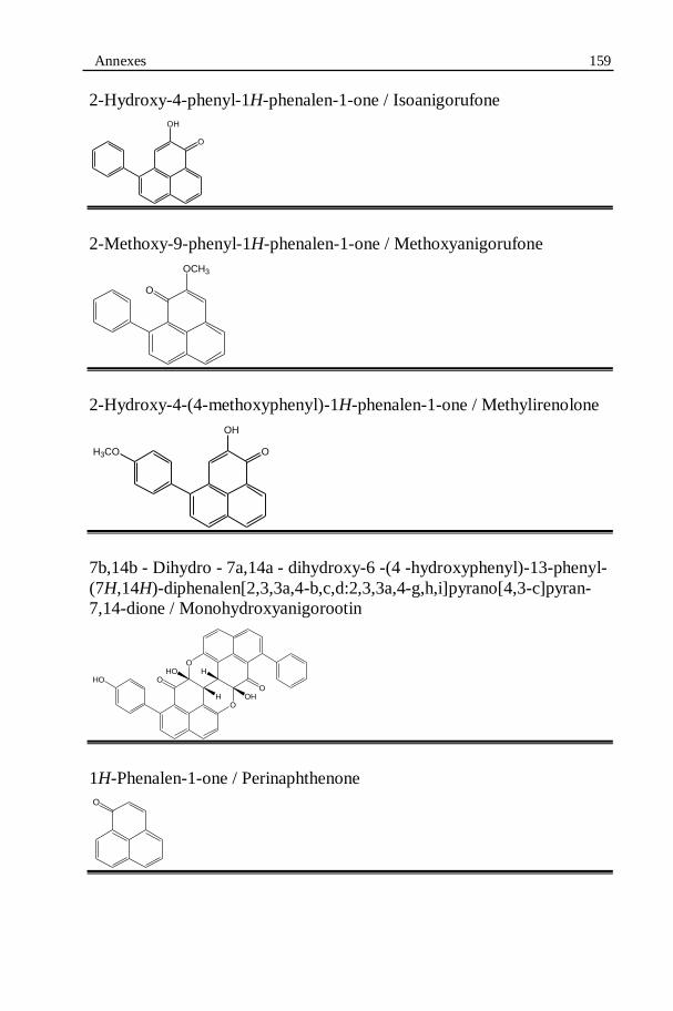
Annexes 159
2-Hydroxy-4-phenyl-1H-phenalen-1-one / Isoanigorufone
2-Methoxy-9-phenyl-1H-phenalen-1-one / Methoxyanigorufone
2-Hydroxy-4-(4-methoxyphenyl)-1H-phenalen-1-one / Methylirenolone
7b,14b - Dihydro - 7a,14a - dihydroxy-6 -(4 -hydroxyphenyl)-13-phenyl-
(7H,14H)-diphenalen[2,3,3a,4-b,c,d:2,3,3a,4-g,h,i]pyrano[4,3-c]pyran-
7,14-dione / Monohydroxyanigorootin
1H-Phenalen-1-one / Perinaphthenone
OH
O
O
OCH3
OH
OH3CO
O
O
O
O
HO
H OH
HHO
O
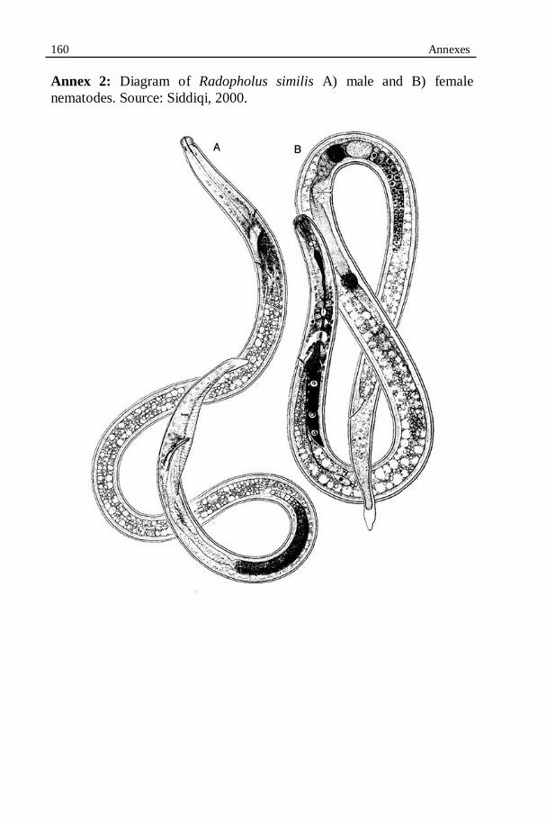
160 Annexes
Annex 2: Diagram of Radopholus similis A) male and B) female
nematodes. Source: Siddiqi, 2000.
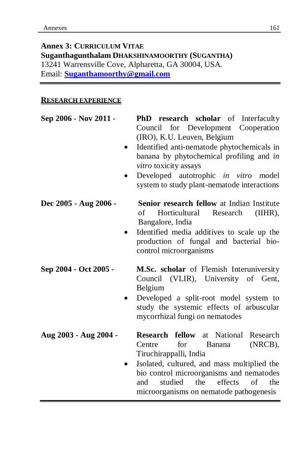
Annexes 161
Annex 3: CURRICULUM VITAE
Suganthagunthalam DHAKSHINAMOORTHY (SUGANTHA)
13241 Warrensville Cove, Alpharetta, GA 30004, USA.
Email: [email protected]
RESEARCH EXPERIENCE
Sep 2006 - Nov 2011 - PhD research scholar of Interfaculty
Council for Development Cooperation
(IRO), K.U. Leuven, Belgium
Identified anti-nematode phytochemicals in
banana by phytochemical profiling and in
vitro toxicity assays
Developed autotrophic in vitro model
system to study plant-nematode interactions
Dec 2005 - Aug 2006 - Senior research fellow at Indian Institute
of Horticultural Research (IIHR),
Bangalore, India
Identified media additives to scale up the
production of fungal and bacterial bio-
control microorganisms
Sep 2004 - Oct 2005 - M.Sc. scholar of Flemish Interuniversity
Council (VLIR), University of Gent,
Belgium
Developed a split-root model system to
study the systemic effects of arbuscular
mycorrhizal fungi on nematodes
Aug 2003 - Aug 2004 - Research fellow at National Research
Centre for Banana (NRCB),
Tiruchirappalli, India
Isolated, cultured, and mass multiplied the
bio control microorganisms and nematodes
and studied the effects of the
microorganisms on nematode pathogenesis
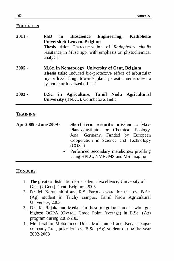
162 Annexes
EDUCATION
2011 - PhD in Bioscience Engineering, Katholieke
Universiteit Leuven, Belgium Thesis title: Characterization of Radopholus similis
resistance in Musa spp. with emphasis on phytochemical
analysis
2005 - M.Sc. in Nematology, University of Gent, Belgium
Thesis title: Induced bio-protective effect of arbuscular
mycorrhizal fungi towards plant parasitic nematodes: a
systemic or localized effect?
2003 - B.Sc. in Agriculture, Tamil Nadu Agricultural
University (TNAU), Coimbatore, India
TRAINING
Apr 2009 - June 2009 - Short term scientific mission to Max-
Planck-Institute for Chemical Ecology,
Jena, Germany. Funded by European
Cooperation in Science and Technology
(COST)
Performed secondary metabolites profiling
using HPLC, NMR, MS and MS imaging
HONOURS
1. The greatest distinction for academic excellence, University of
Gent (UGent), Gent, Belgium, 2005
2. Dr. M. Karunanidhi and R.S. Paroda award for the best B.Sc.
(Ag) student in Trichy campus, Tamil Nadu Agricultural
University, 2003
3. Dr. K. Rajukannu Medal for best outgoing student who got
highest OGPA (Overall Grade Point Average) in B.Sc. (Ag)
program during 2002-2003
4. Mr. Ibrahim Mohammed Doka Mohammed and Kenana sugar
company Ltd., prize for best B.Sc. (Ag) student during the year
2002-2003

Annexes 163
5. Meenashi ammal Medal for highest OGPA in agricultural botany
courses from I to VI semester, 2002
6. Tmt. R. Santha medal for having obtained highest OGPA in all
agronomy courses in B.Sc. (Ag) 2002-2003
7. AVR Sundaram trust Salem award for having obtained highest
OGPA in horticultural courses in B.SC. (Ag) during the year
2002-2003
8. 1995-96 batch students endowment medal for best Student in
agricultural economics in B.Sc. (Ag), during the year 2002-2003
9. Western electronic and scientific works prize award for highest
OGPA I to VI semester in B.Sc. (Ag), 2002
LIST OF PUBLICATIONS
A. Articles
1. Suganthagunthalam, D., Elsen, A. and De Waele, D. 2010.
Identification of combined resistance to Radopholus similis and
Meloidogyne incognita in Musa germplasm. International
Journal of Nematology 20, 19-26.
2. Suganthagunthalam, D., Galon, E. J., Elsen, A. and De Waele, D.
2009. Host plant resistance in Musa spp. to the burrowing
nematode: Pre or Post-infectional? Communications in Applied
and Biological Sciences 74: 39-43.
3. Suganthagunthalam, D., Galon, E. J., Elsen, A. and De Waele, D.
2009. Pathogenesis of Radopholus similis in an incompatible
interaction with Musa spp. Communications in Applied and
Biological Sciences 74: 541-544.
4. Suganthagunthalam, D., Swennen, R., De Waele, D. and Elsen,
A. 2008. Host plant response of burrowing nematode resistant
Musa genotypes to the root-knot nematode, Meloidogyne
incognita. Communications in Applied and Biological Sciences
73: 219-222.
B. Abstracts/Proceedings
1. Knop, K., Hölscher, D., Suganthagunthalam, D., Elsen, A.,
Crecelius, A., Heckel, D., Schneider, B. and Schubert, U. 2009.
LDI imaging of phenylphenalenones in infected banana tissues
and pest. Proceedings of 18th
International Mass Spectrometry
Conference, Bremen, Germany, 30 August to 04 September 2009.

164 Annexes
2. Suganthagunthalam, D., Elsen, A. Swennen, R., De Waele, D.
and Hölscher, D. 2009. Phytoalexins involved in incompatible
plant-nematode interactions. Third annual meeting of COST 872,
“Nemagenics – Exploiting genomics to understand plant-
nematode interactions”. Toledo, Spain, 25-28 May 2009.
3. Suganthagunthalam, D., Galon, E. J., Elsen, A. and De Waele, D.
2009. Pathogenesis of Radopholus similis in an incompatible
interaction with Musa spp. 61st International Symposium on Crop
Protection, Gent, Belgium, 19th May 2009.
4. Suganthagunthalam, D., Galon, E. J., Elsen, A. and De Waele, D.
2009. Host plant resistance in Musa spp. to the burrowing
nematode: Pre or Post-infectional? 15th
PhD Symposium on
Applied Biological Sciences, Leuven, Belgium, 6th November
2009.
5. Suganthagunthalam D., Elsen A. and De Waele D. 2008. Detailed
evaluation of in vitro-propagated Musa spp. for resistance against
the burrowing nematode, Radopholus similis. In: Book of
abstracts of 9th
International Congress of Plant Pathology,
Torino, Italy, 24-29 August 2008. (Eds). Porta-Puglia A. &
Gonthier P. Journal of plant pathology 90, S2.345.
6. Suganthagunthalam D., Swennen R., De Waele D. and Elsen A.
2008. Host plant response of burrowing nematode resistant Musa
genotypes to the root-knot nematode, Meloidogyne incognita. 14th
PhD symposium on applied biological sciences, Gent, Belgium,
15th September 2008.
7. Suganthagunthalam D., Elsen A. and De Waele D. 2007.
Characterization of resistance in Musa against the burrowing
nematode, Radopholus similis. First annual meeting of COST
872, “Nemagenics – Exploiting genomics to understand plant-
nematode interactions”. La Colle-Sur-Loup, France, 9-11 May
2007.

Annexes 165
GRADUATE / UNDERGRADUATE STUDENTS’ THESIS SUPERVISION
1. Els Heylen. (Belgium) Studies on burrowing nematode resistant
Musa genotypes to understand their resistant mechanisms.
Bioscience and Engineering, Katholieke University of Leuven,
Belgium 2009-10.
2. Katemani Mdili, (Tanzania) Characterization of transgenic banana
shoots with RNAi constructs against Radopholus similis. M.Sc.
Nematology, University of Gent, 2009-10.
3. Erwin Jamilo Galon. (Philippines) Occurrence of host plant
resistance in Musa genotypes to the burrowing nematode: Pre or
post-infectional? M.Sc. Nematology, University of Gent, Belgium
2008-09.
4. Kahpui Mariama Salifu. (Cameron) Biochemical and histochemical
studies on the burrowing nematode resistant Musa genotypes – A
step towards understanding the mechanisms of nematode resistance.
M.Sc. Nematology, University of Gent, 2008-09.