characterization of minchinia sp spores (ascetospora
TRANSCRIPT

W&M ScholarWorks W&M ScholarWorks
Dissertations, Theses, and Masters Projects Theses, Dissertations, & Master Projects
1990
Characterization of Minchinia sp Spores (Ascetospora: Characterization of Minchinia sp Spores (Ascetospora:
Haplosporidiidae) Infecting Teredo navalis L and Placopecten Haplosporidiidae) Infecting Teredo navalis L and Placopecten
magellanicus Von Martens (Mollusca: Teredinidae) in the Western magellanicus Von Martens (Mollusca: Teredinidae) in the Western
North Atlantic North Atlantic
Elizabeth Robinson McGovern College of William and Mary - Virginia Institute of Marine Science
Follow this and additional works at: https://scholarworks.wm.edu/etd
Part of the Animal Diseases Commons, Fresh Water Studies Commons, and the Oceanography
Commons
Recommended Citation Recommended Citation McGovern, Elizabeth Robinson, "Characterization of Minchinia sp Spores (Ascetospora: Haplosporidiidae) Infecting Teredo navalis L and Placopecten magellanicus Von Martens (Mollusca: Teredinidae) in the Western North Atlantic" (1990). Dissertations, Theses, and Masters Projects. Paper 1539617622. https://dx.doi.org/doi:10.25773/v5-3jk9-fm91
This Thesis is brought to you for free and open access by the Theses, Dissertations, & Master Projects at W&M ScholarWorks. It has been accepted for inclusion in Dissertations, Theses, and Masters Projects by an authorized administrator of W&M ScholarWorks. For more information, please contact [email protected].
CHARACTERIZATION OF Minchinia sp. SPORES (ASCETOSPORA: HAPLOSPORIDIIDAE)
INFECTING Teredo navalis L. AND Teredo furcifera VON MARTENS
(MOLLUSCA: TEREDINIDAE) IN THE WESTERN NORTH ATLANTIC
A Thesis
Presented to
The Faculty of the School of Marine Science
The College of William and Mary in Virginia
In Partial Fulfillment
of the Requirement for the Degree of
Master of Arts
by
Elizabeth Robinson McGovern
APPROVAL SHEET
This thesis is submitted in partial fulfillment
the requirements for the degree of
Masters of Arts
Elizabeth R. McGovern
Approved, January, 1990
Euge;;ene M. Burreson, Ph.D.Chairman
»hn E. Olney, M.A.
v̂ . /Ernest Warinner, III, M.A.
Pl/O-WWolfga^g^/K. ^ogel^in, M.S.
Jusan E. Ford, Ph.Rutgers University Shellfish Laboratory
P.O. Box 687 Port Norris, New Jersey 08349

TABLE OF CONTENTS
Page
ACKNOWLEDGMENTS........................................................iv
LIST OF F I G U R E S ..................................................... v
A B S T R A C T ................................................ viii
INTRODUCTION ....................................................... 2
MATERIALS AND METHODS .............................................. 5
Specimen collection ......................................... 5Paraffin histology ............................................ 5Rabbit immunization ......................................... 6Immunogold Silver Staining Assay ........................... 7Scanning Electron Microscopy ................................ 11Transmission Electron Microscopy ............................ 12
R E S U L T S ................................................................. 14
Immunogold Silver Staining Assay ........................... 15Electron Microscopy ......................................... 22
D I S C U S S I O N ............................................................ 28
LITERATURE CITED .................................................. 39
V I T A ................................................................... 45
i i i

ACKNOWLEDGMENTS
I would like to recognize a number of people -for their
contributions to this thesis. First, I thank my major professor, Gene
Burreson, for his guidance over the past few years and his red pen when
it came to editing manuscripts, abstracts, talks and this thesis. I'd
also like to thank my committee members, Susan Ford, Ernie Warinner,
Wolfgang Vogelbein and John Olney, for their critical review of this
thesis. I extend special thanks to Wolfgang for teaching me the wonders
of electron microscopy and to John for adding a systematist's point of
view. I thank Mike Castagna and his people at the Wachapreague Lab for
supplying the shipworm infested planks. Additional thanks goes to Nita
Walker for assistance with histology and Patrice Mason for EM
assistance.
I'd especially like to thank my family, my husband Jack, my
brother Bert and my parents,‘for their friendship, love and support.

LIST OF FIGURES
Figure Page
1. Diagrammatic representation of immunogold silverstaining assay illustrating antigen, primary antibody (rabbit anti-haplosporidan spore from T. navalis) and colloidal gold conjugated secondary antibody ............ 10
Plate 1 ................................................................. 18
2. Histological section of T. navalis gill lamellae showing sporocysts in the blood spaces, deterioration of gill epithelium and sporocysts in the water tubules.
3. Histological section of heavily infected T. navalis gill showing sporocysts in afferent branchial vein and blood spaces.
Plate I I ...............................................................19
4. Diagrammatic illustration of Minchinia sp . spore illustrating position of epispore extensions.
5. Light micrograph of live Minchinia sp. spores showing epispore extensions.
Plate III...............................................................20
6. MSX spores in digestive diverticula of C. virginica.
7. Spores in gills of T. navalis.
8. Teredo navalis gills treated with rabbit anti-HS serum.
9. Teredo navalis gills treated with rabbit pre-inoculation serum.
10. Crassostrea virginica treated with rabbit anti-HS serum.
11. Crassostrea virginica treated with rabbit pre-inoculation serum.
V

Plate IV 21
12.
13.
14.
15.
16.
Plate
17.
18.
19.
20.
Plate
21.
2 2 .
23.
Haplosporidium louisiana spores in P. herbstii assayed with rabbit anti-HS serum.
Haplosporidium louisiana spores assayed with rabbit pre-inoculation serum.
Haplosporidium costale spores in C. virginica assayed with rabbit anti-HS serum.
Haplosporidium costale spores assayed with rabbit pre- inoculation serum.
Haplosporidan spores from Barnegat Bay Teredo spp. assayed with rabbit anti-HS serum.
V ................................................................. 25
SEM micrograph of transverse section through shipworm gill lamellae showing abundant sporocysts, symbiotic ciliates, a food groove and the afferent branchial vein.
SEM micrograph of sporocysts from shipworm gill lamellae illustrating individual sporocysts within epispore cytoplasm.
Transmission electron micrograph of immature spore within epispore cytoplasm.
Enlarged view of the same spore as shown in Fig. 19 illustrating the microtubule-like structures in the epispore cytoplasm and the microfilament-like structures within the sporoplasm.
V I ...............................................................26
SEM micrograph of spores with extensions observed through a tear in the sporocyst membrane.
SEM micrograph of individual spore isolated by needle puncture of sporocysts showing three of the four extensions.
Electron micrograph illustrating a longitudinal section through the base of an extension showing spore wall, disentegrating epispore cytoplasm and microtubule-like structures.
Vi

Plate VII 27
24. Electron micrograph of base of opercular extension showing that microtubule-like structures and membrane are not attached to spore wall.
25. Spores isolated by disintegration of shipworm tissue illustrating loosely fitting epispore membrane and extensions.
26. Spores isolated by disintegration of shipworm tissue illustrating loosely fitting epispore membrane and extensions.
27. Unornamented spores following complete lysis of epispore membrane.

ABSTRACT
Spores of a haplosporidan infecting Teredo navalis and T. furcifera have been described as morphologically indistinguishable from spores of Haplosporidium nelsoni, the oyster pathogen commonly referred to as MSX.A colloidal gold immunoassay was used to test the hypothesis that H. nelsoni and the haplosporidan infecting Teredo spp. are conspecific. Additionally, antigenic characteristics of spores of the haplosporidan found in Teredo spp. were compared to spores of other local haplosporidan species, H. costale infecting Crassostrea virginica and H. louisiana infecting Panopeus herbstii. The immunoassay demonstrated that the haplosporidan infecting Teredo spp. is not conspecific with H. nelsoni.H. costale or H. louisiana.
Electron microscopy was utilized to further characterize spores of the haplosporidan infecting Teredo spp. and revealed four distinct membrane-bound extensions, one apical, opposite the'opercular hinge, one terminal and two opposing lateral extensions. These extensions were not continuous with the spore wall, but contained microtubule-like structures and degrading epispore cytoplasm. Parasites in the family Haplosporidiidae are separated based on the type of epispore ornamentation into two genera, Haplosporidium and Minchinia: however, there has been some debate in the literature over the correct assignment of species to these genera. At present, species whose spores are ornamented by spore wall filaments and those ornamented by wrappings are placed in the genus Haplosporidium. Haplosporidan species with epispore cytoplasm extensions and species with unornamented spores are assigned to Minchinia. Therefore, the haplosporidan infecting Teredo spp. is placed in the genus Minchinia based on the possession of four epispore cytoplasm extensions with similar composition to the extensions found on spores of the type species, M. chitonis.

CHARACTERIZATION OF Minchinia sp. SPORES
(ASCETOSPORA: HAPLOSPORIDIIDAE)
INFECTING Teredo navalis L. AND Teredo furcifera VON MARTENS
(MOLLUSCA: TEREDINIDAE) IN THE WESTERN NORTH ATLANTIC
1

INTRODUCTION
A haplosporidan has been reported to infect three species of
shipworms, Teredo navalis L . , T. bartschi Clapp and T. furcifera von
Martens from Barnegat Bay, New Jersey (Hillman 1978, 1979, 1980;
Hillman et al. 1982). This parasite was discovered while analyzing
the effects of outflow from the Oyster Creek Nuclear Generating
Station on local shipworm populations in Barnegat Bay, New Jersey.
From 1975 through 1980, monthly prevalences of infection determined by
histological examination were recorded and pooled for all stations
sampled. Infected Teredo spp. were found throughout the year with
prevalence peaks in the fall of each year. The most commonly
occurring species of shipworm in the 20 stations sampled throughout
Barnegat Bay was Bankia gouldi: however, this species was never found
to be infected by the haplosporidan (Hillman et al. 1982). Of the
three species of Teredo found, two of them, T. bartschi and T.
furcifera are subtropical species, probably introduced to the area by
wooden boats and able to survive in the warm water effluent of the
power station. Hillman et al. (1982) hypothesized that T. bartschi
may have developed some disease resistance to the haplosporidan
parasite, as evidenced by increasing numbers of individuals through
1980 and decreasing parasite prevalence. Alternatively, the
population of T. furcifera declined through the sampling period.
Teredo furcifera was the most abundant Teredo species in 1974
2

(Hoagland and Turner 1980) but the number of individuals collected
decreased through 1980 perhaps due to mortality caused by high
parasite prevalence.
Based on light microscopy, Hillman (1979) assigned the organism
parasitizing Teredo spp. to the family Haplosporidiidae (Phylum
Ascetospora, Class Stellatosporea, Order Balanosporida) and discussed
similarities in size and shape of its spores to those of
Haplosporidium nelsoni Haskin, Stauber and Mackin, the oyster pathogen
commonly referred to as MSX. MSX has been implicated in mass
mortalities of Crassostrea virginica Gmelin in both Delaware and
Chesapeake Bays over the last thirty years, yet the life cycle of this
parasite is unknown. Investigators have suggested existence of a
reservoir host, a species other than C. virginica which serves as a
source of MSX from which oysters become infected, because of the lack
of correlation between disease severity and oyster abundance (Ford and
Haskin 1982, Andrews 1984). In addition, spores of H. nelsoni are
rarely seen in adult oysters; however, a recent study by Kanaley and
Barber (1989) indicates that MSX spores are more common in oyster spat
(36% of 234 spat examined June 1988 were in some stage of
sporulation). At present, the possibility of a reservoir host for MSX
still cannot be ruled out. Hillman (1979) acknowledged the
unlikelihood of Teredo spp. being a reservoir host for MSX since few
Teredo spp. are found in MSX endemic areas; however, similar spore
morphologies and the abundance of the spore stage in infections of
Teredo spp. warranted further investigation.
3

Therefore, a study was undertaken to examine the hypothesis that
the haplosporidan infecting Teredo spp. is conspecific with the oyster
pathogen H. nelsoni and to compare the haplosporidan infecting Teredo
spp. to other local species of the family Haplosporidiidae. The
objectives of this study were as follows:
1. Compare antigenic characteristics of spores of the
haplosporidan infecting Teredo spp. to spores of
Haplosporidium nelsoni.
2. Compare antigenic characteristics of spores of the
haplosporidan infecting Teredo spp. to spores of H. costale
infecting the oyster Crassostrea virginica and to H.
louisiana infecting the mudcrab Panopeus herbstii.
3. Examine spore morphology of the haplosporidan infecting
Teredo spp. with paraffin histology, scanning electron
microscopy and transmission electron microscopy.
4. Determine generic assignment of the haplosporidan infecting
Teredo spp.
4

MATERIALS AND METHODS
Specimen collection:
In April of 1987 and 1988, pine planks for collection of
shipworms were submerged beneath the dock at the Virginia Institute of
Marine Science laboratory on the Atlantic coast in Wachapreague,
Virginia. Teredo navalis were collected in October 1987 and October
1988 from planks exposed for six months and in February 1989 from
planks exposed for ten months. Teredo furcifera were collected in
October 1988 from planks exposed for six months and in February 1989
from planks exposed for ten months. Additionally, planks suspended
from the dock on 24 April 1988 were sampled monthly from June 1988
through February 1989 to ascertain prevalence and intensity of
infection.
Shipworms were removed from planks with scalpel and forceps. An
effort was made to remove intact worms because pallets are necessary
for species identification (Turner 1966); however, in some cases
infected worms were obtained without pallets. Smears of Teredo spp.
gills were examined by light microscopy to determine presence of
haplosporidan spores.
Paraffin histology:
Pieces of infected T. navalis tissue were preserved in Davidson's
AFA (30% (v/v) 95% EtOH, 20% (v/v) formalin, 20% (v/v) acetic acid and
5
20% (v/v) glycerin) for 24-48 hours. Tissues were then dehydrated in
a graded ethanol series and embedded in Tissue Prep paraffin (Fisher
Scientific, Fair Lawn, NJ) using an automatic tissue processor (Auto-
Technicon, Technicon Corp., Tarrytown, NY). Tissue blocks were cut at
6 um on a rotary microtome (American Optical, Buffalo, NY). Resulting
paraffin ribbons were floated on a warm water bath and picked up on
glass slides coated with Szombathy's adhesive (0.01% (w/v) gelatin,
0.15% (v/v) glycerin in distilled water). Slides were allowed to dry
overnight in a 45°C oven and then stained with Harris' Haematoxylin
and Eosin (HH&E) according to routine staining procedures.
Rabbit Immunization:
Spore suspensions for rabbit immunization were obtained by
placing pieces of infected T. navalis collected in October 1987 in
beakers of high salinity water (32 ppt). Supernatant was changed
daily for approximately seven days until all T. navalis tissue
decayed. Prior to immunization, spores were incubated one hour in a
saturated solution of N-Acetyl-L-Cysteine to dissolve all remaining
tissue. Spores were washed in 0.22 um filtered sea water, sonicated
gently with a Sonifier Cell Disruptor (Heat Systems-Ultrasonics, Inc.,
Plainview, NY) to remove clumps, followed by fixation for one hour in
Davidson's AFA. Fixative was removed by two additional washings in
0.22 um filtered sea water. An emulsion of 2 x 10^ intact spores in
1.0 ml RAS (Ribi adjuvant system: Monophosphoryl lipid A, Trehalose
dimycolate and cell wall skeleton, Ribi ImmunoChem Research, Inc.,
6
Hamilton, MT) was injected into a New Zealand white rabbit (SUJO
Rabbit Farm, Gloucester, VA) according to the following schedule:
Day 0 - 5.0 ml blood taken for pre-inoculation serum via heart
puncture, 0.25 ml RAS emulsion injected subcutaneously
(sc) in each of four sites along back.
Day 10- Boosted with 0.25 ml RAS emulsion sc in each of four
sites along back.
Day 20- Boosted as on Day 10.
Day 29- 10.0 ml blood taken via heart puncture.
Blood was allowed to clot one hour at room temperature and overnight
at 5°C. Serum was removed and aliquots of 0.10 ml were frozen at
-18°C until time of assay.
IGSS assay:
Paraffin blocks of P. herbstii infected with H. louisiana and C.
virginica infected with H. nelsoni or H. costale were obtained from
the oyster disease archive at VIMS. These tissues had been stored in
paraffin for variable periods of time. Panopeus herbstii infected
with H. louisiana was preserved as described for T. navalis but stored
in paraffin blocks for five years prior to immunoassay. Two of the H.
nelsoni infected oysters had been preserved for 48 h in Woods ' AFA
(2.5% (v/v) acetic acid, 6.5% (v/v) formalin, 48.5% (v/v) 95% EtOH)
and held in paraffin for 12 years. A third oyster was preserved
exactly as described for T. navalis and stored in paraffin for six
months prior to immunoassay. Of the three H. costale infected oysters
7
assayed, two had been stored in paraffin for four years and the third
was held in paraffin for six years.
Tissue blocks were cut as previously described (see "paraffin
histology"); however, slides with sections were left unstained for
immunoassay.
Auroprobe LM Immunogold Silver Staining (Janssen Life Science
Products, Piscataway, NJ) was used as described by the manufacturer.
Rabbit polyclonal antiserum made specific for spores of the Teredo
spp. haplosporidan was applied to paraffin sections of spores to be
tested (Fig. 1). If the spore in the section under consideration had
the same antigenic properties as the Teredo spp. haplosporidan, the
antibody molecules attached to the antigenic determinants on the spore
coat. These antigen-antibody complexes were then tagged by the
addition of goat anti-rabbit IgG coated on 5 run colloidal gold. The
signal was enhanced by precipitation of metallic silver on the gold
particles yielding a dark brown to black signal at the site of each
antigen-antibody complex when viewed with light microscopy.
Initially, five dilutions (1/50, 1/100, 1/200, 1/400 and 1/800 in
PBS with 0.5% (w/v) bovine serum albumin and 0.5% (w/v) sodium azide)
of rabbit antiserum to haplosporidan spores from T. navalis (primary
antiserum, hereafter referred to as rabbit anti-HS) were tested
against infected T. navalis and C. virginica infected with MSX.
Sections were subsequently incubated one hour in a 1/40 dilution of
secondary antibody, affinity purified goat anti-rabbit IgG conjugated
to 5 ran colloidal gold particles (AuroProbe LM), and eight minutes in
8

silver enhancement reagents (IntenSE II, supplied with kit). Pre
treatment with Lugol's iodine and modifications in incubation times of
primary antiserum and silver enhancement reagents were attempted to
increase contrast between positively reacting spores and background
(DeMey et a l . 1986). A 30-minute incubation of the 1/100 dilution of
rabbit anti-HS was chosen as optimal because background was minimal
and a positive reaction was easily seen. Subsequent assays were
therefore performed at this concentration. Lugol's iodine was not
used except on an initial sample because pretreatment with Lugol's
increased background thereby decreasing contrast.
Cross sections through the gill regions of five infected T.
navalis and the visceral mass of one infected T. navalis were tested
in duplicate against rabbit anti-HS using IGSS. In addition, three
oysters infected with H. nelsoni. three oysters infected with H.
costale and one mudcrab infected with H. louisiana were assayed in
duplicate. As negative controls, sections from each organism were
assayed substituting rabbit pre-inoculation serum and commercial
normal rabbit serum (Cooper Biomedical, West Chester, PA) as primary
antisera. Sections of uninfected T. navalis and C. virginica were
also assayed with rabbit pre-inoculation serum and rabbit anti-HS
serum. In addition, paraffin sections of infected Teredo spp. from
Barnegat Bay, New Jersey provided by R. E. Hillman were tested against
rabbit anti-HS to confirm that the haplosporidan infecting T. navalis
in Wachapreague is the same as that described by Hillman (1978). A
negative control of Hillman's infected Teredo spp. using rabbit pre-
inoculation serum as primary antiserum was not performed due to lack
9
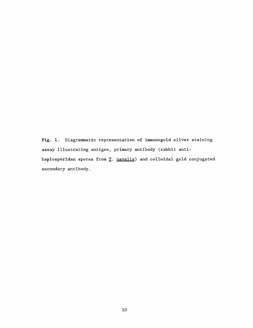
Fig. 1. Diagrammatic representation of immunogold silver staining
assay illustrating antigen, primary antibody (rabbit anti-
haplosporidan spores from T. naval is-) and colloidal gold conjugated
secondary antibody.
10

Legend
antigen
primary antibody
secondary antibody/colioidal gold conjugate

of tissue.
SEM:
Sections through gill regions of infected worms were preserved
for four hours in 2.5% (v/v) glutaraldehyde in 0.1 M sodium cacodylate
buffer (pH = 7.4) for SEM of sporocysts. Preserved cross-sections
were washed three times in 0.1 M sodium cacodylate buffer and post
fixed for two hours in 1% (w/v) OsO^ in 0.1 M sodium cacodylate buffer
at 5°C. Following three additional washes in 0.1 M sodium cacodylate
buffer, samples were dehydrated in a graded series of EtOH and
transferred to 100% acetone for critical point drying in liquid .
Once dried, tissue sections were mounted on support stubs using
colloidal graphite in isopropanol and coated with gold-palladium (60%
: 40% (w/w)) by vacuum evaporation.
For SEM study of epispore ornamentation, spores were separated
from sporocysts, prior to fixation, by three different methods. In
October 1987, spores were obtained from decayed T. navalis tissue as
described for rabbit immunization. These spores were then washed,
sonicated gently and resuspended in filtered sea water. In method
two, gills from heavily infected T. navalis and T. furcifera collected
in October 1988 were dissected and teased to release sporocysts.
Sporocysts were then disrupted by gentle sonication to yield a
suspension of spores. No effort was made to separate spores from the
two species of shipworms. In the third method, sporocysts teased from
11

gills of T. navalis and T. furcifera were punctured with needle probes
to release spores. There was no sonication in the third method.
Spores were prepared for SEM by adhesion to poly-1-lysine coated
12 mm round glass coverslips. Spores obtained by all three methods
were washed three times in 0.22 um filtered sea water and fixed in
2.5% (v/v) glutaraldehyde in 0.1 M sodium cacodylate (pH = 7.4) for
one hour at room temperature (RT). Spore suspensions were added as a
puddle onto each coated coverslip and allowed to settle for one hour.
Coverslips were placed in snap cap vials, washed in three 10 minute
changes of 0.1 M sodium cacodylate buffer and post-fixed in 1% (w/v)
OsO^ in 0.1 M sodium cacodylate buffer for two hours at RT. Samples
were again washed in sodium cacodylate buffer followed by dehydration
in EtOH. Coverslips were then transferred to 100% acetone, critical
point dried, mounted on stubs and coated as previously described.
TEM:
Minced gills of heavily infected T. navalis and T. furcifera
were prepared for TEM by three hour fixation in 0.1 M sodium
cacodylate (pH = 7.4) buffered 3% (v/v) glutaraldehyde at 5°C.
Following three 20 minute washes in 0.1 M sodium cacodylate buffer,
tissue was post-fixed for two hours in 1% (w/v) 0s0^ buffered in 0.1 M
sodium cacodylate at 5°C. Samples were again washed in buffer,
dehydrated through a graded series of EtOH and infiltrated over six
days with Spurr's low viscosity embedding media (Spurr 1969). Blocks
12

were polymerized overnight in a 58°C vacuum oven at -15 psi. Tissue
sections were cut at approximately 800 run, stained 20 min with
saturated uranyl acetate in 50% (v/v) EtOH and stained five minutes
with Reynolds' lead citrate (Reynolds 1963). In addition, spores
obtained in October 1988 by needle puncture of sporocysts were
negatively stained with uranyl acetate. Spores were settled onto
Formvar-coated grids, fixed 45 seconds in a 2% (w/v) OsO^ chamber and
stained for five minutes in 0.8% (w/v) uranyl acetate. Sections and
whole mounts were viewed with a Zeiss OEM 902 transmission electron
microscope.
13

RESULTS
Teredo spp. with haplosporidan infections were found from early
September 1988 through February 1989, with heaviest infections
occurring in October. Since boards were initially exposed in April,
an accurate assessment of haplosporidan prevalence in May through
August was not made. Very few shipworms were found in boards sampled
prior to September, because it takes approximately two months for
shipworms to colonize newly exposed boards and grow to a size easily
detectable during board dissection. Shipworm boards should be set out
several times during the year so that newly exposed boards are not
relied on for haplosporidan prevalence estimates.
Shipworms with heavy infections were characterized by numerous
white and brown pinpoints throughout the gills and mantle which were
visible to the unaided eye. Light microscopy revealed that these
pinpoints were sporocysts. The spore stage of the haplosporidan
infecting Teredo spp. was the most commonly occurring stage of the
parasite. Sporocysts were present in gills of infected Teredo spp. in
the blood spaces, efferent branchial vein, afferent branchial vein,
water tubules, epibranchial cavity and mantle cavity (Plate I, Fig.
2,3). Deterioration of the gill epithelium because of heavy
haplosporidan infection was observed and is presumed to be responsible
for release of sporocysts into the water tubules and mantle
cavity. Sporocysts ruptured by slight coverslip pressure released
14
numerous spores, 6-7 um in length, ornamented by four epispore
extensions (Plate II, Fig. 4,5).
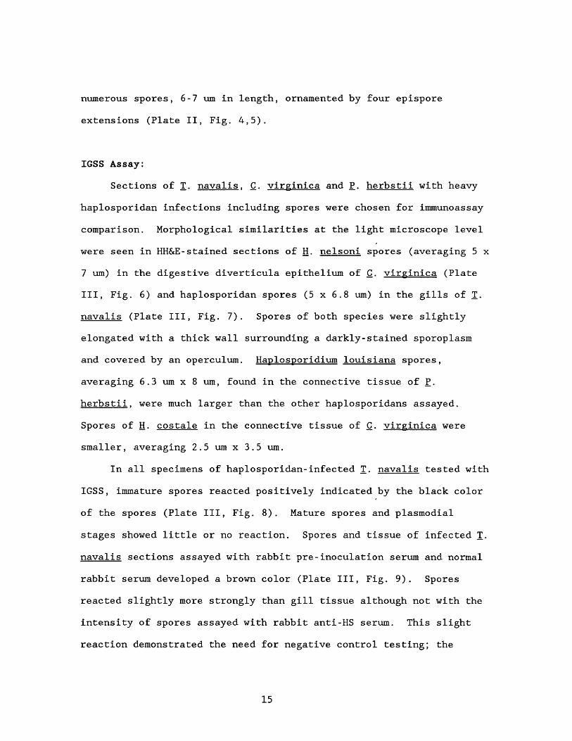
IGSS Assay:
Sections of T. navalis. C. virginica and P. herbstii with heavy
haplosporidan infections including spores were chosen for immunoassay
comparison. Morphological similarities at the light microscope level
were seen in HH&E-stained sections of H. nelsoni spores (averaging 5 x
7 um) in the digestive diverticula epithelium of C. virginica (Plate
III, Fig. 6) and haplosporidan spores (5 x 6.8 um) in the gills of T.
navalis (Plate III, Fig. 7). Spores of both species were slightly
elongated with a thick wall surrounding a darkly-stained sporoplasm
and covered by an operculum. Haplosporidium louisiana spores,
averaging 6.3 um x 8 um, found in the connective tissue of P.
herbstii. were much larger than the other haplosporidans assayed.
Spores of H. costale in the connective tissue of C. virginica were
smaller, averaging 2.5 um x 3.5 um.
In all specimens of haplosporidan-infected T. navalis tested with
IGSS, immature spores reacted positively indicated by the black color
of the spores (Plate III, Fig. 8). Mature spores and plasmodial
stages showed little or no reaction. Spores and tissue of infected T.
navalis sections assayed with rabbit pre-inoculation serum and normal
rabbit serum developed a brown color (Plate III, Fig. 9). Spores
reacted slightly more strongly than gill tissue although not with the
intensity of spores assayed with rabbit anti-HS serum. This slight
reaction demonstrated the need for negative control testing; the
15

amount of color development seen with pre-inoculation serum was
regarded as negative background. Uninfected T. navalis showed no
positive reaction when assayed with rabbit anti-HS or pre-inoculation
serum. The negative background that developed in the immunoassay of
infected T. navalis tissue was also seen in the assay of uninfected
tissue.
Haplosporidium nelsoni spores from C. virginica did not react
with rabbit anti-HS serum (Plate III, Fig. 10). Assayed sections of
oyster tissue contained both mature and immature spores. Brown color
development of the spores was the same as that seen for sections
assayed with rabbit pre-inoculation serum and normal rabbit serum and
therefore dismissed as negative background (Plate III, Fig. 11). In
one of the oysters preserved in 1976 a seemingly positive reaction of
MSX spores which stained dark brown to black when assayed with rabbit
anti-HS serum was accompanied by dark brown to black-stained oyster
tissue. This apparent reaction of MSX spores was regarded as negative
because there was no difference between spores and oyster tissue in
intensity of the reaction. The same oyster yielded similar results
when assayed with rabbit pre-inoculation serum indicating that the
reaction seen when assayed with rabbit anti-HS serum was unusually
high background. Uninfected C. virginica showed no reaction.
A high background was seen in sections of P. herbstii infected
with H. louisiana when assayed with rabbit antiserum or pre-
inoculation serum (Plate IV, Fig. 12, 13). Nuclei of digestive tubule
epithelial cells and some immature spores reacted with both types of
serum producing a dark brown color. These reactions were regarded as
16

negative because the cell nuclei and spores reacted with the same
intensity to both the antiserum and control pre-inoculation serum.
Spores of H. costale stained dark brown when exposed to rabbit
anti-HS serum (Plate IV, Fig. 14). Spores in sections exposed to pre
inoculation serum also developed a brown color (Plate IV, Fig. 15),
though not as dark as those reacted with antiserum.
Sections of infected Teredo spp. provided by R. E. Hillman were
tested only against rabbit anti-HS serum due to lack of sufficient
material (Plate IV, Fig. 16). Spores stained dark brown; however,
this reaction was not as intense as the black-stained spores seen when
infected T. navalis from Wachapreague was assayed with rabbit anti-HS
serum. In addition, nuclei of some unidentifiable cells along the
periphery of gill lamellae in Hillman's samples reacted with a strong
black signal. No such reaction was seen in Wachapreague samples.
17

PLATE I
Fig. 2,3. Harris' Haematoxylin and Eosin stained paraffin sections of
Teredo navalis. 2. Histological section of T. navalis gill lamellae
showing sporocysts (s) in blood spaces (b), deterioration of gill
epithelium (e) and sporocysts (s) in the water tubules (w). Bar = 22
um. 3. Histological section of heavily infected T. navalis gill
showing sporocysts (s) in the afferent branchial vein (a) and blood
spaces (b). Bar = 50 um.
18
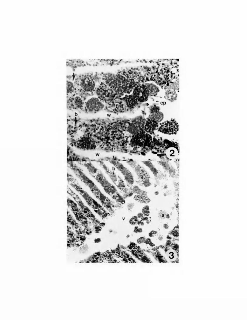

PLATE II
Fig. 4,5. Position of epispore cytoplasm extensions on Minchinia s p .
spores. 4. Diagrammatic illustration of Minchinia s p . spore
illustrating position of epispore extensions. Bar = 1 um. 5. Light
micrograph of live Minchinia sp. spores showing epispore extensions.
Bar = 5 um.
19


PLATE III
Fig. 6-11. IGSS of haplosporidan spores (arrows: M=mature,
I=immature) in C. virginica and T. navalis. (The bar in Fig. 11
represents 12 um and applies to all figures in this plate.) Figures
6, 7 were stained with HH&E. Figures 8-11 were assayed by IGSS and
counterstained with Fast Green. 6. MSX spores in digestive
diverticula of C. virginica. 7. Spores in gills of T. navalis. 8.
Teredo navalis gills treated with rabbit anti-HS serum. 9. Teredo
navalis gills treated with rabbit pre-inoculation serum. 10.
Crassostrea virginica treated with rabbit anti-HS serum. 11.
Crassostrea virginica treated with rabbit pre-inoculation serum.
20
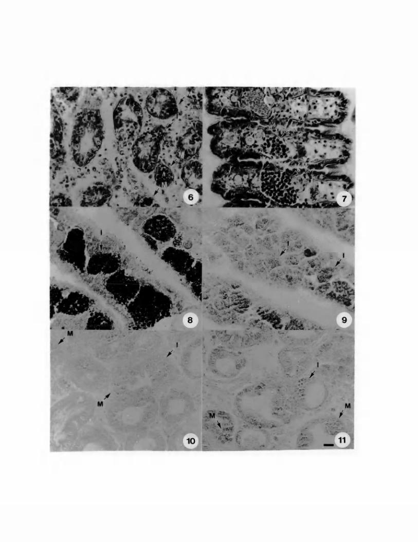

PLATE IV
Fig. 12-16. Haplosporidan species compared to Minchinia s p . by
immunogold silver staining. (The bar in Fig. 16 represents 12 urn and
applies to all figures in this plate.) 12. Hanlosporidium louisiana
spores (arrows) in P. herbstii assayed with rabbit anti-HS serum. 13.
Haplosporidium louisiana spores (arrows) assayed with rabbit pre-
inoculation serum. 14. Haplosporidium costale spores (arrows) in C.
virginica assayed with rabbit anti-HS serum. 15. Haplosporidium
costale spores (arrows) assayed with rabbit pre-inoculation serum.
16. Haplosporidan spores from Barnegat Bay Teredo spp. assayed with
rabbit anti-HS serum.
21


Electron Microscopy:
When viewed with SEM, numerous closely packed sporocysts,
approximately 30-60 urn in diameter, were evident within the blood
spaces adjacent to the food groove of Teredo spp. (Plate V, Fig. 17,
18). Additionally, sporocysts emerging from the water tubules between
lamellae were seen along the gill ventral surface. Spores were
represented by bulges of the opaque membrane bounding each sporocyst.
Thin sections through sporocysts revealed developing spores
within membrane-bound epispore cytoplasm (Plate V, Fig. 19). Spore
wall formation was initiated as nodes of wall material evenly spaced
around the sporoplasm. These nodes gradually merged to form a
continual wall, five to seven layers thick in mature spores (Plate VI,
Fig. 23; Plate VII, Fig. 24). The spore orifice was covered by a
hinged operculum composed of wall material. Within the sporoplasm of
these developing spores, the spherulosome, nucleus, mitochondria and
formative inclusions containing haplosporosomes were clearly visible
(Plate V, Fig. 19). Near the sporoplasm membrane adjacent to the
spore wall, a single layer of microfilament-like structures was
evident (Plate V, Fig. 20). The epispore cytoplasm of developing
spores contained microtubule-like structures immediately beneath the
epispore membrane and degenerating mitochondria. (Without an
immunoassay using anti-tubulin serum, it cannot be said definitively
that these structures are microtubules and therefore, "microtubule
like structures" is used throughout). These microtubule-like
structures, approximately 25 nm in diameter, were present around the
22

spore at 35-50 ran intervals, usually forming an irregular band (Plate
V, Fig. 20).
Several sporocysts with tears in the sporocyst membrane 10-40 um
in length were found in gill cross sections (Plate VI, Fig. 21).
Spores with intertwined extensions were visible through these tears.
SEM examination of individual spores isolated by needle puncture of
sporocysts (Plate VI, Fig. 22) and by decay of shipworm tissue (Plate
VII, Fig. 25) revealed four distinct epispore cytoplasm extensions
from 10-30 um in length, one apical, opposite the opercular hinge, one
terminal and two opposing lateral extensions (Plate II, Fig. 4).
Cross-sections through these extensions demonstrated that they were
membrane-bound and contained microtubule-like structures and
degenerating epispore cytoplasm (Plate VI, Fig. 23). The number of
microtubule-like structures comprising the extensions varied from
approximately 135 near the base of the extension to 55 near the tip.
Thin sections of spores at varying stages of development revealed
the structure of epispore cytoplasm extension formation. The band of
microtubule-like structures present within epispore cytoplasm of
immature spores appeared to coalesce as spores developed accompanied
by progressive degradation of the cytoplasm. Mature spores were
therefore surrounded by a complete membrane covering a thin layer of
tightly-packed microtubule-like structures adjacent to the spore wall
(Plate VI, Fig. 23; Plate VII, Fig. 24). The membrane and
microtubule-like structures extended to form the tapering extensions
in four distinct locations around the spore. Because of the
impervious nature of the spore wall, the sporoplasm of mature spores
23

did not fix and infiltrate well. The sporoplasm was therefore pulled
out during sectioning leaving a hole surrounded by spore wall,
epispore membrane and extensions; however, these structures were well-
fixed and demonstrated that the extensions were neither composed of
spore wall material nor firmly attached to the wall. These findings
were supported by the negatively stained whole mounts which did not
reveal any periodic substructure of the extensions.
In spores collected in October 1987 that were held in sea water
for seven days, the epispore membrane and extensions appeared as thin,
often loose coverings sometimes partially lysed and pulled away from
the spore at the operculum as if being shed (Plate VII, Fig. 25, 26).
Spores with no extensions or epispore membrane were observed in the
same preparation (Plate VII, Fig. 27).
24

PLATE V
Fig. 17-20. Minchinia sp. from Teredo spp. 17. SEM micrograph of
transverse section through shipworm gill lamellae showing abundant
sporocysts (s), symbiotic ciliates (c), a food groove (f) and the
afferent branchial vein (v). Bar = 100 um. 18. SEM micrograph of
sporocysts from shipworm gill lamellae illustrating individual spores
within the sporocysts. Bar = 10 um. 19. Transmission electron
micrograph of immature spore within epispore cytoplasm (e). Visible
within the sporoplasm are the spherulosome (sp), nucleus (n) and
haplosporosome formative inclusions (h). The epispore cytoplasm
contains a supporting substructure of microtubule-like structures (m).
Bar = 500 nm. 20. Enlarged view of the same spore as shown in Fig.
19 illustrating the microtubule-like structures (m) in the epispore
cytoplasm and the microfilament-like structures (f) within the
sporoplasm. Bar = 250 nm.
25

msgfm

Plate VI
Fig. 21-23. Minchinia sp. spores from Teredo spp. 21. SEM
micrograph of spores with extensions observed through a tear in the
sporocyst membrane. Bar = 5 um. 22. SEM micrograph of individual
spore isolated by needle puncture of sporocysts showing three of the
four extensions. Bar = 1 um. 23. Electron micrograph illustrating a
longitudinal section through the base of an extension showing spore
wall (w) , disintegrating epispore cytoplasm (e) and microtubule-like
structures (m). Also seen are an extension in transverse section (t)
showing microtubule-like structures surrounded by a thin membrane, and
the tip of an operculum (o) with sheath composed of microtubule-like
structures (m). Bar = 350 nm.
26

A'*,’ .*
Plate VII
Fig. 24-27. Minchinia sp. spores with degrading epispore cytoplasm.
24. Electron micrograph of base of opercular extension showing that
microtubule-like structures and membrane are not attached to spore
wall (arrows). Bar = 350 nm. 25,26. Spores isolated by
disintegration of shipworm tissue illustrating loosely fitting
epispore membrane and extensions. Bar — 1 um. 27. Unornamented
spores following complete lysis of epispore membrane. Bar = 1 um.
27


DISCUSSION
In order to understand the results of the immunogold silver
staining assay, it is important to understand antiserum specificity
and cross-reactivity. Antiserum specificity results from the action
of a population of individual antibody molecules directed against
different determinants on the antigen molecule (Roitt, Brostoff and
Male 1985). Therefore, antiserum raised against spores of the
haplosporidan infecting Teredo spp. reacted specifically with several
antigenic sites on the same spores. Additionally, spores of another
species may have some shared antigenic sites with the Teredo spp.
haplosporidan spores and thus the antiserum to Teredo spp.
haplosporidan spores may cross-react by binding only to the sites that
the two species have in common. In an immunoassay, the specific
binding of an antiserum produces a strong positive reaction. In the
case of immunogold silver staining, the specific reaction is a black
reaction because of the number of antibody molecules bound and hence
the amount of gold available for silver enhancement. Cross-reactivity
is expressed by brown color development because fewer antibody
molecules bind and there is less color development.
The fact that the antiserum raised against spores from T. navalis
reacted specifically with spores of T. navalis and did not react with
spores of H. nelsoni indicates that these two haplosporidan species
are antigenically distinct and thus different species. These results
28

demonstrate that Teredo spp. is not a reservoir ho^t for H. nelsoni.
Additionally, the lack of reaction of H. louisiana spores with the
rabbit anti-HS serum indicates that the haplosporidan infecting Teredo
spp. and H. louisiana are also antigenically distinct. The negative
background seen in sections of infected C. virginica and P. herbstii
assayed with rabbit pre-inoculation serum is attributable to cross
reaction of naturally occurring rabbit antibodies with spores and
tissue.
The dark brown color of H. costale spores when assayed with
rabbit anti-HS serum can be explained by cross-reactivity.
Immunoassay results indicate that spores of the Teredo spp.
haplosporidan have some antigenic sites in common with spores of H.
costale. It would seem logical that spores of different haplosporidan
species would have some shared antigenic determinants; however, spores
of H. louisiana and H. nelsoni did not cross-react with the antiserum
to spores of the Teredo spp. haplosporidan. Therefore, the shared
antigenic sites between SSO spores and spores of the Teredo spp.
haplosporidan may be due to environmental conditions since both
species are found on the Eastern Shore of the Chesapeake Bay. The
important result in this immunoassay is that there was no specific
reaction of the rabbit anti-HS serum and SSO spores. Haplosporidium
costale is a separate species from the haplosporidan found in Teredo
spp. These results are supported by light microscopy where
differences are seen in spore size and in location of sporulation,
considered by Andrews (1984) to be species specific. Sporulation of
H. costale occurs in the connective tissue and yields smaller spores
29
than those of the Teredo spp. haplosporidan found in the blood spaces
of the gill. Additionally, when viewed with TEM, H. costale spores
were described by Perkins (1969) as possessing spore wall wrappings.
Such wrappings are distinct from the extensions found in the
haplosporidan infecting Teredo spp.
The fact that rabbit anti-HS serum reacted only with immature
spores in paraffin sections of infected T. navalis indicates that the
antiserum is specific for immature spores. This may be attributed to
the presence in immature spores of membrane-bound epispore cytoplasm
which degenerates later in spore development revealing naked spores
(Burreson and Robinson 1988) as seen in SEM preparations of spores
held in sea water. Antiserum made to spores with intact epispore
cytoplasm would therefore not react with spores in which the cytoplasm
had lysed and disappeared. Scanning electron microscopy preparations
of the same material used for rabbit immunization showed spores with
intact epispore cytoplasm and naked spores. Immunoassay results of
the parasite stage specificity of the rabbit anti-HS serum indicate
that the immature spores were greater in number or simply more
strongly antigenic to the rabbit.
The antiserum to spores of the haplosporidan infecting Teredo
spp. from Wachapreague did not react specifically with spores of the
haplosporidan infecting Teredo spp. from Barnegat Bay. This may
possibly be explained by differences in fixation or age of sections as
Hillman's samples were preserved in Bouin's fluid from 1975-1980.
Wachapreague samples were preserved in Davidson's AFA and embedded in
paraffin six months prior to immunoassay. Fresh samples from Barnegat
30
Bay need to be preserved in Davidson's AFA and embedded in paraffin
using the same techniques as described for the Wachapreague samples.
The discovery of spores with four epispore cytoplasm extensions
further supports the immunoassay results (McGovern and Burreson 1989)
that the haplosporidan infecting T. navalis is not conspecific with H.
nelsoni whose spores are ornamented by wrappings. Perkins (1968,
1979) described spore wall ornamentation in H. nelsoni as developing
in the epispore cytoplasm. According to Perkins (1968, 1979), the
cytoplasm then dispersed as spores matured leaving threads or ribbons
attached to the wall. Ornamentation of the Teredo spp. haplosporidan
is clearly distinct from that of H. nelsoni spores. Rather than
developing within epispore cytoplasm, the extensions of the Teredo
spp. haplosporidan are composed of cytoplasm which has degraded during
spore maturation causing coalescence of microtubule-like structures.
These microtubule-like structures probably add support to the
extensions and epispore membrane which surround the spore. It is
clear from the EM micrographs that these extensions, the microtubule
like structures and membrane are at no time continuous with the spore
wall. In fact, as spores develop further, the membrane and extensions
are shed yielding unornamented spores.
Spores of the haplosporidan infecting Teredo spp. have epispore
ornamentation that is thus far unique to any species in the
Balanosporida. The spore extensions are similar to those of Minchinia
chitonis (Lankester) Labbe (Labbe 1896, 1899); however, spores from
Teredo spp. possess four extensions while spores of M. chitonis have
only two extensions. The extensions of spores of both species are
31

composed of epispore cytoplasm and microtubule-like structures. Ball
(1980) described short microtubule-like structures strengthening the
epispore cytoplasm and extensions of M. chitonis. In mature spores,
he observed a coalescence of these microtubules similar to that
discovered in spores of the Teredo spp. haplosporidan. Unornamentated
spores have not been described for M. chitonis: however, Ball (1980,
1981) only studied spores within host tissue.
Spores of Urosnoridium i iroveci (Order Balanosporida, Family
Anurosporidiidae) possess a single epispore cytoplasm extension with
supporting microtubules similar in longitudinal section to those of
the haplosporidan infecting Teredo spp. (Ormieres et al. 1973).
However, Urosporidium spp. spores differ from the operculated
Haplosporidiidae spores in that the former possess an internal flap or
lingula for closure of the spore orifice (Perkins and van Banning
1981).
Haplosporidan parasites of the family Haplosporidiidae have been
traditionally separated into two genera, Haplosporidium Caullery and
Mesnil and Minchinia Labbe. Species of both genera have spores with
an orifice closed by a hinged operculum that overhangs the spore wall
except along the hinge (Perkins 1989) and ornamentation consisting of
a wide variety of structures variously described by different authors
as wrappings, ribbons, threads, filaments or tails. The nature of the
ornamentation has recently been determined for many species of
Minchinia and Haplosporidium through electron microscopy and has led
to conflicting definitions of structures and confusion as to the
proper generic allocation of many species.
32
Sprague (1982) characterized the genus Haplosporidium including
such species as H. nelsoni and H. costale Wood and Andrews by threads
(wrappings) wound around the spore coat while Minchinia spores,
exemplified by M. chitonis and M. armoricana van Banning, were defined
by anterior and posterior extensions. Sprague (1982) did not specify
the origin of these threads or extensions.
In his description of H. parisi spores, Ormieres (1980) presented
alternative criteria for distinguishing these two genera. Extensions
of the spore wall such as the two long filaments arising from the
posterior region of spores of H. parisi were differentiated from
extensions of epispore cytoplasm, a distinction which Sprague (1982)
did not recognize. Based on his interpretation of the original type
species descriptions of M. chitonis and H. scolopli Caullery and
Mesnil (Caullery and Mesnil 1905), Ormieres (1980) described Minchinia
spores as possessing tails defined as extensions of epispore cytoplasm
and Haplosporidium spores as possessing filaments, defined as
extensions of the spore wall that persist after degradation of
epispore cytoplasm. Ormieres' (1980) definition of filaments appears
to have been based on a sketch by Caullery and Mesnil (1905) of H.
scolopli spores with two posterior extensions. In the description of
this figure, Caullery and Mesnil (1905) referred to "a delicate
external membrane which is often only recognizable in some debris".
Based on present knowledge, this discussion seems to pertain to
membrane-bound epispore cytoplasm. It is not clear whether the
extensions of H. scolopli are a part of this membrane and therefore
33
should be classified as epispore cytoplasm tails or are spore wall
derived filaments surrounded by the membrane.
Spores with paired posterior filaments similar to those of H.
uarisi have been described for H. lusitanicum Azevedo (Azevedo 1984)
and possibly H. comatulae La Haye et al. (La Haye et al. 1984) and H.
tumefacientis Taylor (Taylor 1966). This group of species with spore
wall extensions is clearly distinct morphologically from spores with
epispore cytoplasm extensions such as are found in the Teredo spp.
haplosporidan and M. chitonis.
Perkins (1988, 1989) generally agreed with Sprague's (1982)
generic distinctions and grouped spores with prominent extensions,
either epispore cytoplasm tails or spore wall filaments, in the genus
Minchinia and spores lacking such extensions in Haplosporidium.
Spores with epispore cytoplasm tails like those of M. chitonis were
considered congeneric with M. armoricana ornamented by anterior and
posterior extensions and M. narisi (= H. parisi) possessing posterior
paired spore wall filaments; however, Perkins (1988) generic
reallocations of H. parisi and H. lusitanicum to Minchinia may not be
valid since the type species of the genus Minchinia. M. chitonis,
possesses spore ornaments that are composed entirely of epispore
cytoplasm and are not attached to the spore wall (Lankester 1885;
Labbe 1896, 1899; Ball 1981).
Lauckner (1983) in a long footnote to a discussion of M.
chitonis. stated that Minchinia is a nomen nudum because lifecycle
stages of two different organisms were included in the original
description. Labbe (1896, 1899) combined spore stages of a
34

haplosporidan in Lenidochiton cinereus with sporozoan stages of a
coccidian (Pseudoklossia chitonis Debaisieux) from Acanthochiton
fascicularis. Therefore Lauckner (1983) placed all species in
Haplosnoridium. However, the spore Labbe (1896) described was clearly
a haplosporidan enabling subsequent species, with spore morphology
similar to that of M. chitonis, to be assigned to the genus Minchinia.
Thus Lauckner's (1983) conclusion has not gained wide acceptance.
Morphology of the M. armoricana extensions and subsequent generic
assignment of this species have been a source of confusion in the
literature. In his original description of M. armoricana. van Banning
(1977) described anterior and posterior extensions of the epispore
cytoplasm. Pichot et al. (1979) described, but did not name, a
haplosporidan from Ostrea edulis that resembled van Banning's (1977)
description of M. armoricana except that the spores were ornamented by
filaments arising from the spore wall in extensions of the epispore
cytoplasm. Perkins and van Banning (1981), studying spores held in
sea water for one year, reported the presence of anterior and
posterior filaments on M. armoricana consisting of bundles of fibers
originating from several points on the spore surface. It is possible
that the filaments described by Pichot et al. (1979) and Perkins and
van Banning (1981) are either developing spore wall filaments
surrounded by epispore cytoplasm or are supporting structures for
epispore cytoplasm tails similar to the microtubule-like structures
described here for the haplosporidan infecting Teredo spp. Bachere et
al. (1987) referred to a haplosporidan from Ostrea angasi as
35

Haplosporidium sp. and compared it to H. armoricana but did not
provide clear evidence for the composition of the epispore extensions.
In recent papers (Bachere and Grizel 1983; Desportes and Nashed
1983; Bachere et al. 1987; Chagot et al. 1987), the generic
distinctions proposed by Ormieres (1980) and his definitions of tails
and filaments have been followed; however, the term wrappings is still
not clearly defined. According to Perkins (1968), the wrappings of H.
costale are formed in the epispore cytoplasm as tubular elements and
are left in contact with the spore wall after lysis of the cytoplasm.
The spore wrappings of H. louisiana were described by Perkins (1975)
as forming in vacuoles of the epispore cytoplasm. These ornaments are
not formed until the spore wall is complete around the sporoplasm
(Perkins and van Banning 1981). Following degradation of the
cytoplasm, these strands were found either fused to the spore wall or
wrapped loosely around it. The wrappings of H. costale and H.
louisiana seem to be distinct from the spore wall filaments of H.
parisi and H. lusitanicum which are attached to the wall at a single
point and are formed as the spore wall is forming prior to lysis of
the epispore cytoplasm (Ormieres 1980; Azevedo 1984); however, species
possessing spore wall filaments and those ornamented by wrappings are
presently placed in the genus Haplosporidium. Further research into
the composition of wrappings is necessary to determine if they are
more similar to the ornamentation of H. scolonli. the type species of
the genus Haplosporidium. or to M. chitonis. the type species of
Minchinia.
36

Haplosporidan spore ornamentation should be placed in three
categories: spore wall filaments, epispore cytoplasm extensions and
wrappings. Filaments, as found on spores of H. parisi. are composed
of wall material and are formed as the spore wall is forming.
Epispore cytoplasm extensions are more ephemeral and may be shed after
spores are released from the host as has been shown herein for the
Teredo spp. haplosporidan. Wrappings, exemplified-by spores of H.
costale, are formed in epispore cytoplasm and adhere to the spore wall
following lysis of the cytoplasm.
An additional problem in classification of the Haplosporidiidae
is the lack of accurate type species descriptions. The type species
of the genus Haplosporidium. H. scolopli has not been studied with
electron microscopy and the origin of its epispore extensions is
uncertain.
Spores of haplosporidans should be more closely studied at all
stages of development in order to better define the genera of this
family. Minchinia dentali (Arvy) (Desportes and Nashed 1983) and M.
tapetis (Chagot et a l . 1987) spores have been described as
unornamented. In light of the present study in which epispore
cytoplasm and tails are shed at some stage of development, it seems
correct to assign mature spores without ornaments to the genus
Minchinia. Further research into the morphology of these unornamented
spores could reveal some type of epispore cytoplasm ornamentation at
an earlier stage of development.
Based upon the present understanding of the taxonomy of the
Haplosporidiidae and the morphology of M. chitonis, the genus
37

Minchinia contains those species whose spores possess epispore
cytoplasm extensions. Spores of the haplosporidan infecting Teredo
spp. bear four extensions composed of epispore cytoplasm supported by
microtubule-like structures enabling placement of this organism in the
genus Minchinia.
38
LITERATURE CITED
Andrews, J. D. 1984. Epizootiology of diseases of oysters
(Crassostrea virginica') , and parasites of associated organisms in
eastern North America. Helgolander Meeresunters.. 37:149-166.
Azevedo, C. 1984. Ultrastructure of the spore of Haplosporidium
lusitanicum sp. n. (Haplosporida, Haplosporidiidae), parasite of
a marine mollusc. J. Parasitol., 70:358-371.
Bachere, E. and Grizel, H. 1983. Mise en evidence d'Haplosporidium
sp. (Haplosporida-Haplosporidiidae) parasite de l'huitre plate
Ostrea edulis L. Rev. Tray. Inst. Peches Marit.. 46:226-232.
Bachere, E. , Chagot, D . , Tige, G. and Grizel, H. 1987. Study of a
haplosporidian (Ascetospora), parasitizing the Australian flat
oyster Ostrea angasi. Aquaculture. 67:266-268.
Ball, S. J. 1980. Fine structure of the spores of Minchinia
chitonis. (Lankester, 1885) Labbe, 1896 (Sporozoa: Haplosporida),
a parasite of the chiton Lepidochiton cinereus. Parasitol.,
81:169-176.
Ball, S. J. 1981. Spore structure of Minchinia chitonis. M a r . Fish.
R ev., 43:5-8.
Burreson, E. M. and Robinson, M. E. 1988. An SEM study of
haplosporidan spores from Teredo navalis. J. Shellfish R es.,
7:215.
39

Caullery, M. and Mesnil, F. 1905. Recherches sur les Haplosporidies.
Arch. Zool. Exp. Gen.. 4:101-181.
Chagot, D. , Bachere, E . , Ruano, F . , Comps, M. and Grizel, H. 1987.
Ultrastructural study of sporulated instars of a haplosporidian
parasitizing the clam Ruditapes decussatus. Aquaculture. 67:262-
263.
De Mey, J., Hacker, G . , De Waele, M. and Springall, D. 1986. Gold
probes in light microscopy. In: Polak, J. and van Noorden, S.
(ed.), Immunocytochemistry, 2nd ed., Wright-PSG, Bristol,
England, pp. 71-88.
Desportes, I. and Nashed, N. 1983. Ultrastructure of sporulation in
Minchinia dentali (Arvy), an haplosporean parasite of Dentalium
entale (Scaphopoda, Mollusca); taxonomic implications.
Prostistologica. 19:435-460.
Ford, S. E. and Haskin, H. H. 1982. History and epizootiology of
Haplosporidium nelsoni (MSX), an oyster pathogen in Delaware Bay,
1957-1980. J. Invert. Pathol.. 40:118-141.
Hillman, R. E. 1978. The occurrence of Minchinia s p . (Haplosporida,
Haplosporidiidae) in species of the molluscan borer Teredo from
Barnegat Bay, New Jersey. J. Invert. Pathol.. 31:265-266
Hillman, R. E. 1979. Occurence of Minchinia sp . in species of the
molluscan borer Teredo. M a r . Fish. Rev.. 41:21-24.
Hillman, R. E. 1980. Life cycle stages of Minchinia s p . in Teredo
navalis. Amer. Zool.. 20:961.
Hillman, R. E . , Maciolek, N. J., Lahey, J. I. and Belmore, C. I.
1982. Effects of a haplosporidian parasite Haplosporidium sp. on
40

species of the molluscan woodborer Teredo in Barnegat Bay, New
Jersey. J. Invert. Pathol.. 40:307-319.
Hoagland, K. E. and Turner, R. D. 1980. Range and extensions of
teredinids (shipworms) and polychaetes in the vicinity of a
temperate-zone nuclear generating station. M a r . Biol., 58:55-64.
Holgate, C., Jackson, P., Cowen, P. and Bird, C. 1983. Immunogold
silver staining: new method of immunostaining with enhanced
sensitivity. J. Histochem. Cvtochem.. 31:938-944.
Kanaley, S. and Barber, R. 1989. Recent observations on the
sporulation of Haplosporidium nelsoni (MSX) ii> the American
oyster Crassostrea virginica. NJAES Publ. No. K-32901-1-89.
p . 74.
Labbe, A. 1896. Recherches zoologiques, cytologiques et biologiques
sur les coccidies. Arch. Zool. Exp. Gen.. 4:533-608.
Labbe, A. 1899. Sporozoa. In: Das Tierrich. Friedlander, Berlin,
5:1-180.
La Haye, C. A., Holland, N. D. and McLean, N. 1984. Electron
microscopic study of Haplosporidium comutalae n. sp . (Phylum
Ascetospora: Class Stellatosporea), a haplosporidian endoparasite
of an Australian Crinoid, Oligometra seripinna (Phylum
Echinodermata). Protistologica. 20:507-515.
Lankester, E. R. 1885. Protozoa. In: Encyclopaedia Britannica, 9th
ed. Encyclopaedia Britannica, London, 19:830-866.
Lauckner, G. 1983. Diseases of Mollusca: Amphineura. In: Kinne, 0.
(ed.), Diseases of Marine Animals, Vol. II: Introduction,
41

Bivalvia to Scaphopoda. Biologische Anstalt Helgoland, Hamburg,
963-975.
McGovern, E. R. and Burreson, E. M. 1989. Immunoassay comparison of
haplosporidan spores from Teredo navalis and Haplosporidium
nelsoni spores from Crassostrea virginica. J. Protozool.,
36:289-292.
Ormieres, R. 1980. Haplosporidium parisi n. s p ., Haplosporidie
parasite de Serpula vermicularis L. etude ultrastrueturale de la
spore. Protistologica. 16:467-474.
Ormieres, R . , Sprague, V. and Bartoli, P. 1973. Light and electron
microscope study of a new species of Urosporidium (Haplosporida),
hyperparasite of trematode sporocysts in the clam Abra ovata. J.
Invert. Pathol.. 21:71-86.
Perkins, F. 0. 1968. Fine structure of the oystef pathogen Minchinia
nelsoni (Haplosporida, Haplosporidiidae). J . Invert. Pathol.,
10:287-307.
Perkins, F. 0. 1969. Electron microscope studies of sporulation in
the oyster pathogen, Minchinia costalis (Sporozoa: Haplosporida).
J. Parasitol.. 55:897-920.
Perkins, F. 0. 1975. Fine structure of Minchinia sp. (Haplosporida)
sporulation in the mud crab Panopeus herbstii. M a r . Fish. Re v .,
37:46-60.
Perkins, F. 0. 1979. Cell structure of shellfish pathogens and
hyperparasites in the genera Minchinia. Urosporidium.
Haplosporidium and Marteilia--taxonomic implications. M a r . Fish.
Rev., 41:25-37.
42

Perkins, F. 0. 1988. Parasite morphology, strategy and evolution:
structure of protistan parasites found in bivalve molluscs. A m .
Fish. Soc. Spec. Publ.. 18:93-111.
Perkins, F. 0. 1989. The Haplosporidia. In: Margulis, L . , Corliss,
J. D . , Melkonian, M. and Chapman, D . , (ed.). Handbook of
Protoctista. Jones and Bartlett, Boston, pp.' 19-29.
Perkins, F. 0. and van Banning, P. 1981. Surface ultrastructure of
spores in three genera of Balanosporida, particularly in
Minchinia armoricana van Banning, 1977--the taxonomic
significance of spore wall ornamentation in the Balanosporida.
J. Parasitol.. 67:866-874.
Pichot, Y. Comps, M. and Deltreil, J. 1979. Recherches sur
Haplosporidium sp . (Haplosporida--Haplosporidiidae) parasite de
l'huitre plate Ostrea edulis L. Rev. Trav. Inst. Peches Marit.,
43:405-408.
Reynolds, E. S. 1963. The use of lead citrate at high pH as an
electron opaque stain in electron microscopy. J. Cell Biol.,
17:208-212.
Roitt, I., Brostoff, J. and Male, D. 1985. Immunology. The C. V.
Mosby Company, St. Louis.
Sprague, V. 1982. Ascetospora. In: Parker, S. (ed.), Synopsis and
Classification of Living Organisms. McGraw-Hill, New York, 599-
601.
Spurr, A. R. 1969, A low-viscosity resin embedding medium for
electron microscopy. J. Ultrast. Res., 26:31-43.
43

Taylor, R. L. 1966. Haplosporidium tumefacientis sp. n . , the
etiologic agent of a disease of the California Sea Mussel,
Mvtilus californianus Conrad. J. Invert. Pathol.. 8: 109-121.
Turner, R. D. 1966. A survey and illustrated catalogue of the
Teridinidae (Mollusca: Bivalvia). The Museum of Comparative
Zoology, Cambridge, 265p.
van Banning, P. 1977. Minchinia armoricana sp. nov. (Haplosporida),
a parasite of the European flat oyster, Ostrea edulis. J.
Invert. Pathol.. 30:199-206.
44

VITA
Elizabeth Robinson McGovern
Born in Jacksonville, Florida, 24 October 1962. Graduated from
Wilton High School, Wilton, Connecticut in 1980. Earned B.A. in
Biology from Lafayette College, Easton, Pennsylvania in December 1983.
Entered Masters program at College of William and Mary, School of
Marine Science in 1986. Hired as Senior Laboratory Specialist at the
Virginia Institute of Marine Science in 1988. Married John Clarke
McGovern August 1988.
45