characterization of fungi from palm kernel cake (pkc) and
TRANSCRIPT

Pertanika J. Trop. Agric. Sci. 41 (1): 115 - 128 (2018)
ISSN: 1511-3701 © Universiti Putra Malaysia Press
TROPICAL AGRICULTURAL SCIENCEJournal homepage: http://www.pertanika.upm.edu.my/
E-mail addresses: [email protected] (Razali, S. M.),[email protected] (Lee, H. Y.),[email protected] (Jinap, S.),[email protected] (Mahyudin, N. A.) * Corresponding author
Article history:Received: 28 February 2016Accepted: 08 November 2017
ARTICLE INFO
Characterization of Fungi from Palm Kernel Cake (PKC) and the Effect of Storage Temperature on Fungi Growth
Razali, S. M.1,2, Lee, H. Y.3, Jinap, S.1,2 and Mahyudin, N. A.1* 1Faculty of Food Science and Technology, Universiti Putra Malaysia, 43400 UPM, Serdang, Selangor, Malaysia2Food Safety and Food Integrity (FOSFI), Institute of Tropical Agriculture and Food Security, Universiti Putra Malaysia, 43400 UPM, Serdang, Selangor, Malaysia3Tropical Infectious Diseases Research and Education Centre (TIDREC), Universiti Malaya, 50603 Kuala Lumpur, Malaysia
ABSTRACT
The widespread contamination of animal feed with mycotoxin is not a new issue worldwide. Apart from economic loss, mycotoxin can have adverse health effects on humans due to the carcinogenicity, teratogenicity and mutagenicity potential of the toxins. Palm kernel cake (PKC) is the largest animal feed production in Malaysia. PKC is a by-product of palm kernel oil processing and it has been exported as animal feed. The purpose of this study was to isolate and characterise toxigenic fungi cultured in three different media, Dichloran Rose Bengal Chloramphenicol (DRBC) Agar, Dichloran 18% Glycerol (DG18) Agar and Malt Extract Agar (MEA), derived from PKC that is stored under three different temperatures, 4°C, 25°C and 60°C. Identification of fungi was carried out based on macroscopy and microscopy as well as molecular identification. Four mycotoxigenic fungi were found: Aspergillus niger, Aspergillus flavus, Aspergillus fumigatus and Penicilium citrinum. In order to characterise polymorphism of the isolates, RAPD assay was performed using OPA 3 as the primer. The software resulted in a constructed dendrogram that revealed the percentage of similarities between the typable isolates (A. fumigatus, A. niger and P. citrinum) within range from 20% to 80%. The effect of storage temperature on the strains’
enumeration is reported in this work. The distributing strains are influenced by the storage temperature of the PKC matrices. The findings clearly show that Aspergillus species profused at 25°C PKC storage, while it was restricted at low and high temperature.
Keywords: Mycotoxin, PKC, fungi, media, RAPD
assay, temperature

Razali, S. M., Lee, H. Y., Jinap, S. and Mahyudin, N. A.
116 Pertanika J. Trop. Agric. Sci. 41 (1): 115 - 128 (2018)
INTRODUCTION
Mycotoxins are secondary metabolites produced by mycotoxigenic fungi in conditions that are favourable for fungal growth. Mycotoxin contamination of agricultural crops and animal feed is a serious issue since it has undesirable consequences on human and animal health. Aspergillus spp., Penicillium spp. and Fusarium spp. are among the common types of mycotoxigenic fungi. Some of the common mycotoxins found in animal feed are aflatoxins and fumonisins.
Agriculture commodities are easily contaminated by fungi, in particular Aspergillus spp, when their storage conditions are favourable for fungi growth. This is a serious concern since Aspergillus spp. are capable of producing aflatoxins. Aflatoxins are produced by a number of strains of A. flavus and A. parasiticus in a variety of commodities that include cereals, figs, oilseeds, nuts and tobacco (Diener et al., 1987). Mycotoxin contamination of animal feed results in a reduction of its nutritional value; this is highly undesirable as it has unfavourable consequences on human and animal health.
The l i terature pertaining to the evaluation of protocols and methods used to define the relationship between isolates has increased significantly over the years. Randomly amplified polymorphic DNA (RAPD) has been highlighted by many researchers as the most convenient method for this purpose (Williams et al., 1990; Meyer et al., 1991). RAPD involves the
application of short primers followed by PCR analysis using a large template of genomic DNA. These short primers will or will not amplify the DNA template. This process is dependent on the positions that are complementary to the sequence of the primers.
Malaysia is one of the major producers of palm oil and palm oil products in the world. The increasing demand for palm oil over the years has led to the increase in the production of palm kernel cake (PKC). In 2009, Malaysia produced 17.56 million tonnes of palm oil, from which 2.31 million tonnes of PKC were produced on 4.69 million hectares of planted land, and it is projected that these values will increase over the years (MPOB, 2009). In 2002, almost 4 million metric tonnes of PKC were generated all over the world (Atasie & Akinhanmi, 2009) and in 2006, Malaysia contributed 2.20 million tonnes of PKC and exported about 2.12 million tonnes of it (Ong et al., 2004). As little is known about fungi profiles isolated from PKC, the aim of this study was to isolate and identify fungi isolated from PKC and to determine the effect of media and temperature on fungal growth.
MATERIALS AND METHOD
Sample Collection
Samples of PKC (2 kg per pack) were collected from four different manufacturers in Selangor, Malaysia. The samples were kept at 4°C prior to being transported to the lab for further analysis.

Characterization of Fungi from Palm Kernel Cake (PKC)
117Pertanika J. Trop. Agric. Sci. 41 (1): 115 - 128 (2018)
Statistical analysis. The results were analysed using Analysis of Variance (ANOVA) by Minitab Statistical Software v.14 (Minitab Inc., State College, Pa., U.S.A.). One-way ANOVA was used to evaluate the mean differences in a) the culture medium of DRBC at the temperatures of 4°C, 25°C and 60°C (p=0.794); b) the culture medium of DG18 at the temperatures of 4°C, 25°C and 60°C, and; c) the culture medium of MEA at the temperatures of 4°C, 25°C and 60°C. The significance between the differences were determined using Tukey’s multiple comparisons test.
Macroscopic and Microscopic Analysis
M a c r o s c o p i c d e s c r i p t i o n . T h e morphological description of fungi was conducted according to the procedure given by Gao et al. (2007) with some modification. This procedure was carried out after five days of incubation at 28°C. The characteristics measured and observed by the naked eye were the diameter, colour, texture and underside (reverse) colour of the colonies. The micromorphological and structural characteristics of the colonies were measured using a dissecting microscope, namely the size of the metulae, shape of the conidial heads, the length, width and colour of the stipes, the shape, size and seriation of the vesicles and foot cell colour as well as the size of the phialides.
Microscopic observation. Taxonomic identification of the genera and species was performed based on microscopic
Sample Preparation
A mass of 2 kg of samples were milled and divided into three parts. They were incubated at three temperatures, 4°C, 25°C and 60°C, for two weeks.
Isolation and Quantification of Fungi
Frequency of isolation of fungal species and total fungal count in palm kernel cake (PKC). Isolation and quantification of fungi were carried out in accordance with the procedure described by West (2009). The DRBC, DG18 and MEA culture media were used for comparative study of viable fungi-collecting media. A mass of 10 g of a PKC sample was weighed, after which sterile peptone water 0.1% (w/v) was added to it. The solution was homogenised using Stomacher® for 5 min and then put through serial dilution (10-1, 10-2, 10-3 and 10-4). It should be kept in mind that the elapsed time between the cessation of mixing in the dilution tube and the withdrawal of the sample using a pipette should not be more than a few seconds. Following this, 100 µl of each dilution was spread onto the surface of the DRBC, DG18 and MEA solid culture media using the spread plate method. The DRBC, DG18 and MEA plates were then incubated at 28°C. Each plate was prepared in triplicate. After five days of incubation, the fungal colonies were counted using a colony counter and expressed as the number of colonies forming units per gram (CFU/g). The isolates of Aspergillus spp. and Penicillium spp. were further sub-cultured onto potato dextrose agar (PDA).

Razali, S. M., Lee, H. Y., Jinap, S. and Mahyudin, N. A.
118 Pertanika J. Trop. Agric. Sci. 41 (1): 115 - 128 (2018)
characteristics of the fungi by referring to the standard key (Pitt & Hocking, 1997). The mass of hydrophobic conidia from harvested mycelia were removed using ethanol 90% (v/v) and stained with lactophenol blue solution and observed microscopically.
Molecular Analysis
DNA extraction. Genomic DNA of fungi was extracted according to the procedure described by Gonzalez-Mendoza et al. (2010) with some modification. Mycelia along with their agars were harvested and each mixed with 200 µl of Tris-EDTA (TE) buffer solution in a microcentrifuge tube. Following this, purified water was added into the microcentrifuge tube until the volume of the solution was 100 mL. The homogenised mixture was centrifuged at 13,200 rpm for 5 min until two layers formed in the microcentrifuge tube. The first layer was collected and discarded. The suspension left was slowly mixed with 0.2 mL phenol:chloroform (1:1) and incubated at 65°C for 5 min. The remaining suspension in the microcentrifuge tube was slowly mixed with 0.2 mL of phenol:chloroform (1:1) and incubated at 65°C for 5 min. Following this, 0.4 mL of isopropanol was added and mixed, and the mixture was then incubated at 20°C for 20 min. The mixture was then centrifuged at 10,000 rpm for 10 min. The supernatant was discarded and the pellet was washed twice with 75% ethanol. The suspension was centrifuged at 13,200
rpm for 5 min and the pellet was suspended with 50 µl of TE buffer consisting of 100 mL of Tris-HCl, 20 mL of 0.5 M EDTA and 880 mL of purified water. The prepared DNA of the fungi was kept in a freezer at the temperature -20°C until further use.
Randomly amplified polymorphic DNA polymerase chain reaction (PCR) amplification. Polymerase chain reaction (PCR) assays were carried out in a DNA thermal cycler (Applied Biosystems 2720 thermal cycler, USA). The PCR amplification protocol was programmed as follows: (1) initial denaturation at 94°C for 5 min; (2) 45 cycles of denaturation at 94°C for 1 min; (3) annealing at 36°C for 1 min; (4) extension at 72°C for 1 min; and (5) final extension at 72°C for 10 min. The 25 µl mixture of PCR amplification consisted of 5 µl of PCR buffer, 3 µl of 2 mM MgCl2, 0.5 µl of dNTP, 0.3 µl of Taq polymerase, 1.0 µl of primers, 4 µl of DNA template and 11.2 µl of sterile double distilled water. PCR kits were purchased from Promega (Madison, WI, USA) and the primers were synthesised by 1st BASE Laboratories Sdn. Bhd., Selangor.
The PCR products were analysed on 1 % agarose gel in 0.5 X TBE buffer (54 g of Tris base, 27.5 g of Boric acid, 2.92 g of EDTA). The DNA ladder was used as the molecular size marker. DNA bands were stained with ethidium bromide and viewed under UV light. Thirteen primers were randomly screened as shown in Table 1.

Characterization of Fungi from Palm Kernel Cake (PKC)
119Pertanika J. Trop. Agric. Sci. 41 (1): 115 - 128 (2018)
RAPD analysis software. The DNA product was analysed by means of computer generated software, GelCompare 4.2 (Applied Maths, Belgium). A dendrogram was created using GelCompare 4.2 to
observe the relatedness and similarities between the fungi isolates. A similarity coefficient curve was drawn based on Pearson correlation and dice band based with 1 % position tolerance. The Unweighted Pair Group Linkage Analysis method (UPGMA) was chosen for dendrogram construction.
RESULTS AND DISCUSSION
Isolation of Fungi
The growth of fungi in the PKC samples was analysed in the different temperatures used for PKC storage of 4°C, 25°C and 60°C using three different types of culture media, which were DG18, DRBC and MEA. In order to see the range of suitable storage temperatures for PKC in the industry, the stated temperatures were selected. The occurrence and quantification of fungi by mean in various selected temperatures and media are summarised in Table 1.
Table 1 Screened primers for rapd-pcr characterisation of fungi isolates
Code SequenceOPA3 AGT CAG CCACOPA8 GTG ATC GCAGOPA10 CAG CAC CCACOPA17 GAC CGC TTGTOPA18 AGG TGA CCGTOPA20 GTT GCG ATCCOPB3 CAT CCC CCTGOPB8 GTC CAC ACGGOPB10 CTG CTG GGACOPB13 TTC CCC CGCTOPB17 AGG GAA CGAGOPB18 CCA CAG CAGTOPB20 GGA CCC TTAC
Table 2 Effect of culture medium and storage temperature on the frequency of fungi in the pkc samples (log cfu/g)s
Palm Oil Mill Culture Medium Temperature4°C 25°C 60°C
1 DG18 3.79 3.68 2.64DRBC 2.10 3.17 3.83MEA 2.87 2.93 2.62
2 DG18 1.28 2.77 1.53DRBC 1.53 3.92 3.69MEA 1.20 3.00 1.62
3 DG18 0.00 0.00 0.00DRBC 1.43 1.43 0.00MEA 1.16 1.49 0.00
4 DG18 0.00 4.00 2.00DRBC 1.00 1.00 1.00MEA 2.10 0.00 0.00
Note: 1-4 values are means of the four different types of palm oil mills located in Klang. DRBC is Rose Bengal Chloramphenicol, DG18 is Dichloran 18 % glycerol and MEA is Malt Extract Agar

Razali, S. M., Lee, H. Y., Jinap, S. and Mahyudin, N. A.
120 Pertanika J. Trop. Agric. Sci. 41 (1): 115 - 128 (2018)
In the present study, total filamentous fungi counts were in line with the value reported in cereal and mixed feed (Dalcero et al., 1997). In general, the total fungi counts obtained in this study did not exceed the log 5 CFU/g level proposed as a feed quality limit (Chelkowski, 1991). It can be seen that a storage temperature of 25°C resulted in the highest frequency of fungi in the PKC samples, whereas the lowest frequency of fungi was obtained for the storage temperature of 60°C. In addition, the DG18 culture medium gave the highest occurrence of fungi compared to the DRBC and MEA culture media.
The one-way ANOVA statistical analysis showed that there were no significant differences (p>0.05) between a) the culture medium of DRBC at the temperatures of 4°C, 25°C and 60°C (p=0.794); b) the culture medium of DG18 at the temperatures of 4°C, 25°C and 60°C, and; c) the culture medium of MEA at the temperatures of 4°C, 25°C and 60°C where the p-values were equal to 0.794, 0.785 and 0.612, respectively. In addition, the statistical analysis also proved that there were no significant differences between a) the temperatures of 4°C, 25°C and 60°C at which the DRBC culture medium was stored; b) the temperatures of 4°C, 25°C and 60°C at which the DG18 culture medium was stored, and; c) the temperatures of 4°C, 25°C and 60°C at which the MEA culture media was stored where the p-values were equal at 0.678, 0.487 and 0.587, respectively.
The distribution of fungal species differred from one palm oil mill to another,
which may have been due to the fact that each palm oil mill had its own method of storing PKC. Some palm oil mills may not have dedicated storage facilities for PKC and unfavourable storage conditions might have promoted the growth of fungi in the PKC, particularly that of the Aspergillus species.
The findings demonstrated that the fungi were readily isolated using the three media of DRBC, DG18 and MEA. However, it can be observed that the use of DRBC and DG18 culture media maximised the collection of fungi from the PKC samples. Moreover, the fungal colonies on DG18 and DRBC were clearer and the growth of the fungal colonies were slower in these media compared with their growth in the MEA culture medium. This finding was similar to the results of Al-Gabr et al. (2013); the outcome can be attributed to the ingredients in the culture media i.e. dichloran and chloramphenicol. Dichloran functions as an anti-fungal agent that prevents fungi from overgrowing, whereas the chloramphenicol in the DRBC and DG18 culture media restricted the growth of bacteria from the environment from contaminating the PKC samples. According to King et al. (1979), the DRBC culture medium is recommended for enumeration of yeasts and moulds. In addition, photodegradation of the DRBC culture medium resulted in a reactive oxygen species that inhibited the growth of Saccharomyces (Chilvers et al., 1999) and possibly other yeasts. The result, however, contradicted what had been defined previously, as the study represented

Characterization of Fungi from Palm Kernel Cake (PKC)
121Pertanika J. Trop. Agric. Sci. 41 (1): 115 - 128 (2018)
the optimum growth of fungi appearring in DG18, perhaps due to the development of yeast colonies inhibited in the medium (Deak et al., 2001). Based on the results obtained in this study, it can be deduced that DG18 is an excellent culture medium for the isolation of fungi from PKC samples and the storage temperature i.e. incubation temperature that results in the highest frequency of fungi is 25°C.
Macroscopic and Microscopic Characteristics of Fungi
Mycological analysis of PKC genotypes reported the existence of four principal genera filamentous fungi considered as the most important from a toxicological perspective. Mycological examination of PKC indicated three species of Aspergillus and one species of Penicillium. A total of 30 strains were isolated from the PKC matrices.
Each of these species is discussed in the following sub-sections.
Aspergillus fumigatus.
Macromorphology of A. fumigatus. The diameter of the A. fumigatus isolates inoculated at three points on the PDA culture medium was within a range of 20-55 mm after five days of incubation. It could be observed that the fungal colonies did not overlap each other, as shown in Figure 1. Referring to the conidia of the isolates in Figure 1(a), it could be seen that the colour of the fungal colonies varied from bluish green to turquoise, whereas the texture of the fungal colonies appeared dense and smooth. From the underside, it could be seen that the colour of the fungal colonies varied from white to pale yellow, as shown in Figure 1(b).
Figure 1. Image of A. fumigatus on PDA: (a) top view; (b) underside view
14
underside, it could be seen that the colour of the fungal colonies varied from white to pale
yellow, as shown in Figure 1(b).
(a) (b)
Figure 1. Image of A. fumigatus on PDA: (a) top view, (b) underside view.
Micromorphology of A. fumigatus. Figure 2 shows an image of A. fumigatus as viewed
under a microscope with 40× magnification. It could be observed that the conidial heads
ranged from spatulate to pyriform. The length and width of the stipes were found to be
within the range of 10-25 µm and 0.5-1.0 µm, respectively. The stipes and foot cells of
the A. fumigatus isolates appeared colourless. The shape of the vesicles was subglobose.
The seriation of the vesicles varied from radiate to columnar for most of the isolates,
while some were biseriate and uniseriate, as shown in Figure 2.
Micromorphology of A. fumigatus. Figure 2 shows an image of A. fumigatus as viewed under a microscope with 40× magnification. It could be observed that the conidial heads ranged from spatulate to pyriform. The length and width of the stipes were found
to be within the range of 10-25 µm and 0.5-1.0 µm, respectively. The stipes and foot cells of the A. fumigatus isolates appeared colourless. The shape of the vesicles was subglobose. The seriation of the vesicles varied from radiate to columnar for most of

Razali, S. M., Lee, H. Y., Jinap, S. and Mahyudin, N. A.
122 Pertanika J. Trop. Agric. Sci. 41 (1): 115 - 128 (2018)
the isolates, while some were biseriate and uniseriate, as shown in Figure 2.
Aspergillus niger
Macromorphology of A. niger. The diameter of A. niger isolates was within a range of 30-55 mm. The colour of the outer area of the fungal colonies was white and the fungal colonies did not overlap one another. Referring to the conidia of the isolates as seen in Figure 3(a), the colour of the fungal colonies was dark brown while their texture was rough, fluffy and less dense. In contrast, the colour of the fungal colonies on the reverse side varied from pale yellow to yellow at the centre and the fungal colonies were colourless near the edges, as shown in Figure 3(b).
Figure 2. Image of A. fumigatus as viewed under a microscope with 40× magnification
15
Figure 2. Image of A. fumigatus as viewed under a microscope with 40× magnification.
Aspergillus niger.
Macromorphology of A. niger. The diameter of A. niger isolates was within a range of
30-55 mm. The colour of the outer area of the fungal colonies was white and the fungal
colonies did not overlap one another. Referring to the conidia of the isolates as seen in
Figure 3(a), the colour of the fungal colonies was dark brown while their texture was
rough, fluffy and less dense. In contrast, the colour of the fungal colonies on the reverse
side varied from pale yellow to yellow at the centre and the fungal colonies were
colourless near the edges, as shown in Figure 3(b).
Figure 3. Image of A. niger on PDA: (a) top view, (b) underside view
16
(a) (b)
Figure 3. Image of A. niger on PDA: (a) top view, (b) underside view.
Micromorphology of A. niger. Figure 4 shows an image of A. niger, as viewed under a
microscope with 40× magnification. It could be observed that the shape of the conidial
heads of A. niger varied from radiate to columnar. The length and width of the stipes
were found to be within a range of 15-35 µm and 0.5-1.0 µm, respectively. The stipes
were colourless, whereas the colour of the foot cells was pale brown. The shape of the
vesicles was spherical, with a diameter ranging from 1.0 to 2.5 µm. The seriation of the
vesicles varied from radiate to columnar for the A. niger isolates, while some were
biseriate.
Micromorphology of A. niger. Figure 4 shows an image of A. niger, as viewed under a microscope with 40× magnification. It could be observed that the shape of the conidial heads of A. niger varied from radiate to columnar. The length and width of the stipes were found to be within a range of 15-35 µm and 0.5-1.0 µm, respectively. The stipes were colourless, whereas the colour of the foot cells was pale brown. The shape of the vesicles was spherical, with a diameter ranging from 1.0 to 2.5 µm. The seriation of the vesicles varied from radiate
to columnar for the A. niger isolates, while some were biseriate.
Figure 4. Image of A. niger as viewed under a microscope with 40× magnification
17
Figure 4. Image of A. niger as viewed under a microscope with 40× magnification.
Aspergillus flavus.
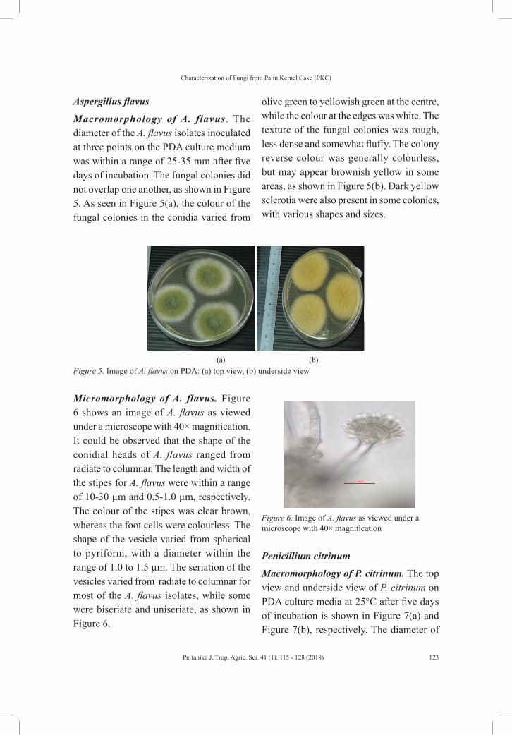
Macromorphology of A. flavus. The diameter of the A. flavus isolates inoculated at three
points on the PDA culture medium was within a range of 25-35 mm after five days of
incubation. The fungal colonies did not overlap one another, as shown in Figure 5. As
seen in Figure 5(a), the colour of the fungal colonies in the conidia varied from olive
green to yellowish green at the centre, while the colour at the edges was white. The
texture of the fungal colonies was rough, less dense and somewhat fluffy. The colony
reverse colour was generally colourless, but may appear brownish yellow in some areas,
as shown in Figure 5(b). Dark yellow sclerotia were also present in some colonies, with
various shapes and sizes.
Characterization of Fungi from Palm Kernel Cake (PKC)
123Pertanika J. Trop. Agric. Sci. 41 (1): 115 - 128 (2018)
Aspergillus flavus
Macromorphology of A. flavus. The diameter of the A. flavus isolates inoculated at three points on the PDA culture medium was within a range of 25-35 mm after five days of incubation. The fungal colonies did not overlap one another, as shown in Figure 5. As seen in Figure 5(a), the colour of the fungal colonies in the conidia varied from
olive green to yellowish green at the centre, while the colour at the edges was white. The texture of the fungal colonies was rough, less dense and somewhat fluffy. The colony reverse colour was generally colourless, but may appear brownish yellow in some areas, as shown in Figure 5(b). Dark yellow sclerotia were also present in some colonies, with various shapes and sizes.
Figure 5. Image of A. flavus on PDA: (a) top view, (b) underside view
18
(a) (b)
Figure 5. Image of A. flavus on PDA: (a) top view, (b) underside view.
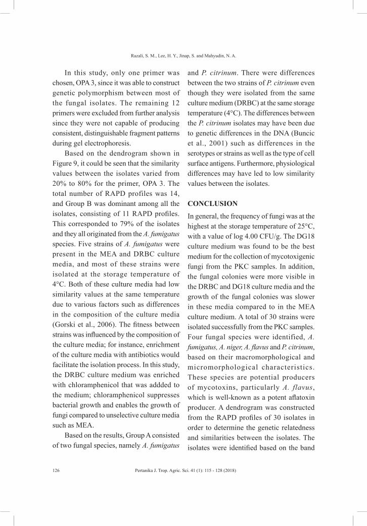
Micromorphology of A. flavus. Figure 6 shows an image of A. flavus as viewed under a
microscope with 40× magnification. It could be observed that the shape of the conidial
heads of A. flavus ranged from radiate to columnar. The length and width of the stipes for
A. flavus were within a range of 10-30 µm and 0.5-1.0 µm, respectively. The colour of
the stipes was clear brown, whereas the foot cells were colourless. The shape of the
vesicle varied from spherical to pyriform, with a diameter within the range of 1.0 to 1.5
µm. The seriation of the vesicles varied from radiate to columnar for most of the A.
flavus isolates, while some were biseriate and uniseriate, as shown in Figure 6.
Micromorphology of A. flavus. Figure 6 shows an image of A. flavus as viewed under a microscope with 40× magnification. It could be observed that the shape of the conidial heads of A. flavus ranged from radiate to columnar. The length and width of the stipes for A. flavus were within a range of 10-30 µm and 0.5-1.0 µm, respectively. The colour of the stipes was clear brown, whereas the foot cells were colourless. The shape of the vesicle varied from spherical to pyriform, with a diameter within the range of 1.0 to 1.5 µm. The seriation of the vesicles varied from radiate to columnar for most of the A. flavus isolates, while some were biseriate and uniseriate, as shown in Figure 6.
Penicillium citrinum
Macromorphology of P. citrinum. The top view and underside view of P. citrinum on PDA culture media at 25°C after five days of incubation is shown in Figure 7(a) and Figure 7(b), respectively. The diameter of
Figure 6. Image of A. flavus as viewed under a microscope with 40× magnification
19
Figure 6. Image of A. flavus as viewed under a microscope with 40× magnification.
Penicillium citrinum.
Macromorphology of P. citrinum. The top view and underside view of P. citrinum on
PDA culture media at 25 °C after five days of incubation is shown in Figure 7(a) and
Figure 7(b), respectively. The diameter of the fungal colonies was found to be within a
range of 12-15 mm. The fungal colonies had compact, dense conidiophores that were
bluish green in colour. From the underside, it could be seen that the colour of the fungal
colonies was yellowish orange, as shown in Figure 7(b).

Razali, S. M., Lee, H. Y., Jinap, S. and Mahyudin, N. A.
124 Pertanika J. Trop. Agric. Sci. 41 (1): 115 - 128 (2018)
the fungal colonies was found to be within a range of 12-15 mm. The fungal colonies had compact, dense conidiophores that were bluish green in colour. From the underside,
it could be seen that the colour of the fungal colonies was yellowish orange, as shown in Figure 7(b).
Figure 7. Image of P. citrinum on PDA: (a) top view, (b) underside view
20
(a) (b)
Figure 7. Image of P. citrinum on PDA: (a) top view, (b) underside view.
Micromorphology of P. citrinum. Figure 8 shows an image of P. citrinum as viewed
under a microscope with 40× magnification. Unlike Aspergillus spp., the conidia heads of
P. citrinum were present in columns and the shape of the conidia heads varied from
globose to subglobose, with a fine wall within a range of 2.5-3.0 µm. The growth of P.
citrinum was rather fast on PDA compared to its growth on the DG18 culture medium. In
addition, the fungal colonies appeared to be less dense.
Micromorphology of P. citrinum. Figure 8 shows an image of P. citrinum as viewed under a microscope with 40× magnification. Unlike Aspergillus spp., the conidia heads of P. citrinum were present in columns and the shape of the conidia heads varied from globose to subglobose, with a fine wall within a range of 2.5-3.0 µm. The growth of P. citrinum was rather fast on PDA compared to its growth on the DG18 culture medium. In addition, the fungal colonies appeared to be less dense.
Figure 8. Image of P. citrinum as viewed under a microscope with 40× magnification
21
Figure 8. Image of P. citrinum as viewed under a microscope with 40× magnification.
Molecular Characteristics
Randomly amplified polymorphic DNA cluster analysis. In this study, it was found
that three out of four species, A. fumigatus, A. niger and P. citrinum, were typable on the
RAPD assay when OPA 3 was used as the primer. Only A. flavus was not amplified by
the primer. Thirteen primers were preliminarily assessed for RAPD PCR and only one
primer was chosen based on its ability to produce consistent, distinguishable fragment
patterns. The bands produced from the OPA 3 primer had a band size within a range of
1500-200 bp. The gel obtained from gel electrophoresis was analysed using the
GelCompare 4.2 software to construct the dendrogram, as shown in Figure 9. The
dendrogram was constructed based on the RAPD profiles generated using the OPA 3
primer.
Molecular Characteristics
Randomly amplified polymorphic DNA cluster analysis. In this study, it was found that three out of four species, A. fumigatus, A. niger and P. citrinum, were typable on the RAPD assay when OPA 3 was used as the primer. Only A. flavus was not amplified by the primer. Thirteen primers were preliminarily assessed for RAPD PCR and only one primer was chosen based on its ability to produce consistent, distinguishable fragment patterns. The bands produced from the OPA 3 primer had a band size within a range of 1500-200 bp. The gel obtained from gel electrophoresis was analysed using the GelCompare 4.2 software to construct the dendrogram, as shown in Figure 9. The dendrogram was constructed based on the RAPD profiles generated using the OPA 3 primer.

Characterization of Fungi from Palm Kernel Cake (PKC)
125Pertanika J. Trop. Agric. Sci. 41 (1): 115 - 128 (2018)
The dendrogram in Figure 9 showed that the cluster was divided into two main groups, Group A and Group B, which consisted of 14 RAPD profiles. Only three strains were present in Group A: one strain of A. fumigatus and two strains of P. citrinum. In this segment, the strain of A. fumigatus was isolated from the MEA culture medium stored at the temperature of 25°C. The two strains of P. citrinum were isolated from the DRBC culture medium at the storage temperature of 4°C.
Group B consisted of two main types of fungal species, with a total of 11 RAPD profiles. The first type consisted of six strains of A. fumigatus, making this group the largest among all the groups. In general, the origins of the fungal isolates in this segment were different. Two strains of A. fumigatus were isolated from the MEA culture medium at a storage temperature of 4°C, whereas one strain of A. fumigatus was isolated from the same medium at a storage temperature of 25°C. In addition, two strains of A. fumigatus from this group were isolated from the DRBC culture medium at
25°C and 60°C. One strain of A. fumigatus was also isolated from the DG18 culture medium at 4°C.
The second type of fungal species from Group B consisted of four strains of A. fumigatus and one strain of A. niger. Three strains of A. fumigatus in this group were isolated at the same storage temperature of 4°C; however, two strains were isolated from the DRBC culture medium, whereas one strain was isolated from the MEA culture medium. One strain of A. fumigatus was also isolated from the MEA culture medium at a storage temperature of 25°C. Finally, one strain of A. niger was isolated from the DG18 culture medium, which was stored at the temperature of 4°C. It should be noted that only one fungal species, A. flavus, was not typable using the OPA 3 primer in addition to some strains of A. niger.
RAPD was slightly different from conventional PCR analysis as it was not necessary for the analyst to have specialist knowledge regarding DNA sequence of the target organism. RAPD has been proven to be one of the quickest and easiest techniques for the detection of DNA polymorphism (Williams et al., 1990). RAPD is an alternative technique that can be used to distinguish genotypic variants (Meyer et al., 1991). RAPD basically involves the application of short primers followed by PCR analysis using a large genomic DNA template. These short primers will or will not amplify the DNA template. The process is generally dependent on the positions that are complementary to the sequence of primers.
Figure 9. Dendrogram of typable fungi using OPA 3
22
Figure 9. Dendrogram of typable fungi using OPA 3.
The dendrogram in Figure 9 showed that the cluster was divided into two main
groups, Group A and Group B, which consisted of 14 RAPD profiles. Only three strains
were present in Group A: one strain of A. fumigatus and two strains of P. citrinum. In this
segment, the strain of A. fumigatus was isolated from the MEA culture medium stored at
the temperature of 25 °C. The two strains of P. citrinum were isolated from the DRBC
culture medium at the storage temperature of 4 °C.
Group B consisted of two main types of fungal species, with a total of 11 RAPD
profiles. The first type consisted of six strains of A. fumigatus, making this group the
largest among all the groups. In general, the origins of the fungal isolates in this segment
were different. Two strains of A. fumigatus were isolated from the MEA culture medium
A
B
Razali, S. M., Lee, H. Y., Jinap, S. and Mahyudin, N. A.
126 Pertanika J. Trop. Agric. Sci. 41 (1): 115 - 128 (2018)
In this study, only one primer was chosen, OPA 3, since it was able to construct genetic polymorphism between most of the fungal isolates. The remaining 12 primers were excluded from further analysis since they were not capable of producing consistent, distinguishable fragment patterns during gel electrophoresis.
Based on the dendrogram shown in Figure 9, it could be seen that the similarity values between the isolates varied from 20% to 80% for the primer, OPA 3. The total number of RAPD profiles was 14, and Group B was dominant among all the isolates, consisting of 11 RAPD profiles. This corresponded to 79% of the isolates and they all originated from the A. fumigatus species. Five strains of A. fumigatus were present in the MEA and DRBC culture media, and most of these strains were isolated at the storage temperature of 4°C. Both of these culture media had low similarity values at the same temperature due to various factors such as differences in the composition of the culture media (Gorski et al., 2006). The fitness between strains was influenced by the composition of the culture media; for instance, enrichment of the culture media with antibiotics would facilitate the isolation process. In this study, the DRBC culture medium was enriched with chloramphenicol that was addded to the medium; chloramphenicol suppresses bacterial growth and enables the growth of fungi compared to unselective culture media such as MEA.
Based on the results, Group A consisted of two fungal species, namely A. fumigatus
and P. citrinum. There were differences between the two strains of P. citrinum even though they were isolated from the same culture medium (DRBC) at the same storage temperature (4°C). The differences between the P. citrinum isolates may have been due to genetic differences in the DNA (Buncic et al., 2001) such as differences in the serotypes or strains as well as the type of cell surface antigens. Furthermore, physiological differences may have led to low similarity values between the isolates.
CONCLUSION
In general, the frequency of fungi was at the highest at the storage temperature of 25°C, with a value of log 4.00 CFU/g. The DG18 culture medium was found to be the best medium for the collection of mycotoxigenic fungi from the PKC samples. In addition, the fungal colonies were more visible in the DRBC and DG18 culture media and the growth of the fungal colonies was slower in these media compared to in the MEA culture medium. A total of 30 strains were isolated successfully from the PKC samples. Four fungal species were identified, A. fumigatus, A. niger, A. flavus and P. citrinum, based on their macromorphological and micromorphological characteristics. These species are potential producers of mycotoxins, particularly A. flavus, which is well-known as a potent aflatoxin producer. A dendrogram was constructed from the RAPD profiles of 30 isolates in order to determine the genetic relatedness and similarities between the isolates. The isolates were identified based on the band

Characterization of Fungi from Palm Kernel Cake (PKC)
127Pertanika J. Trop. Agric. Sci. 41 (1): 115 - 128 (2018)
patterns on the dendrogram. The similarity values between the typable isolates, A. fumigatus, A. niger and P. citrinum, were within a range of 20-80%. In general, the temperature at which the PKC samples were stored had a significant effect on the growth of mycotoxigenic fungi.
ACKNOWLEDGEMENT
This research was funded by the Institure Tropical and Agricultute (LRGS 5526001) and the Faculty Food Science and Technology, University Putra Malaysia.
REFERENCESAl-gabr, H. M., Zheng, T., & Yu, X. (2013). Occurrence
and quantification of fungi and detection of mycotoxigenic fungi in drinking water in Xiamen City, China. Science of The Total Environment, 1, 466–467.
Atasie, V. N., & Akinhanmi, T. F. (2009). Extraction, compositional studies and physico chemical characteristics of palm kernel oil. Pakistan Journal of Nutrition, 8(6), 1680-5194.
Buncic, S., Avery, S. M., Rocourt, J., & Dimitrijevic, M. (2001). Can food-related environmental factors induce different behaviour in two key serovars, 4b and 1/2a, of Listeria monocytogenes? International Journal of Food Microbiology, 65(3), 201–212.
Chelkowski, J. (1991). Mycological quality of mixed feeds and ingredients. In J. Chelkowski (Ed.), Cereal grain, mycotoxins, fungi and quality in drying and storage (pp. 217–227). Netherland: Elsevier.
Chilvers, K. F., Reed, R. H., & Perry, J. D. (1999). Phototoxicity of rose bengal in mycological media implications for laboratory practice. Letters in Applied Microbiology, 28(2), 103–107.
Dalcero, A., Magnoli, C., Chiacchiera, S., Palacios, G., & Reynoso, M. (1997). Mycoflora and incidence of aflatoxin B1, zearalenone and deoxinyvalenol in poultry feeds in Argentina. Mycopathologia, 137(3), 179–184.
Deak, T., Chen, J., Golden, D. A, Tapia, M. S., Tornai-Lehoczki, J., Viljoen, B. C., & Beuchat, L. R. (2001). Comparison of dichloran 18% glycerol (DG18) agar with general purpose mycological media for enumerating food spoilage yeasts. International Journal of Food Microbiology, 67(1), 49–53.
Diener, U. L., Cole, R. J., Sanders, T. H., Payne, G. A., Lee, L. S. & Klich, M. A. (1987). Epidemiology of aflatoxin formation by Aspergillus flavus. Annual Review of Phytopathology, 25(1), 249–270.
Gao, J., Liu, Z., & Yu, J. (2007). Identification of Aspergillus section Flavi in maize in northeastern China. Mycopathologia, 164(2), 91–95.
Gonzalez-Mendoza, D., Argumendo-Delira, R., Morales-Trejo, A., Pulido-Herrera, A., Cervantes-Diaz, L., Grimaldo-Juarez, O., & Alarcon, A. (2010). A rapid method for isolation of total DNA from pathogenic filamentous plant fungi. Genetics and Molecular Research, 9(1), 162–166.
Gorski, L., Flaherty, D., & Mandrell, R. E. (2006). Competitive fitness of Listeria monocytogenes serotype 1/2a and 4b strains in mixed cultures with and without food in the U. S. Food and Drug Administration enrichment protocol. Applied and Environmental Microbiology, 72(1), 776–783.
King, A. D., Hocking, A. D., & Pitt, J. I. (1979). Dichloran rose bengal medium for enumeration isolation of molds from foods. Applied and Environmental Microbiology, 37(5), 959–964.
MPOB. (2009). Economics & Industry Development Division. Malaysian Palm Oil Board. Retrieved from http://econ.mpob.gov.my/economy/ Overview_2009.pdf

Razali, S. M., Lee, H. Y., Jinap, S. and Mahyudin, N. A.
128 Pertanika J. Trop. Agric. Sci. 41 (1): 115 - 128 (2018)
Meyer, W., Koch, A., Niemann, C., Beyermann, B., Epplen, J. T., & Borner, T. (1991). Differentiation of species and strains among filamentous fungi by DNA fingerprinting. Current Genetics, 19(3), 239–242.
Ong, L. G. A., Abd-Aziz, S., Noraini, S., & Karim, M. I. A., (2004). Enzyme production and profile by Aspergillus niger during solid state fermentation using palm kernel cake as substrate. Applied Biochemistry and Biotechnology, 118(1-3), 73–79.
Pitt, J. I., & Hocking, A. D. (Eds). (1997). Fungi and food spoilage (2nd Ed.). London: Blackie Academic Press.
Williams, J. G. K., Kubelik, A. R., Livak, K. J., Rafalski, J. A., & Tingey, S. V. (1990). DNA polymorphisms amplified by arbitrary primers are useful as genetic markers. Nucleic Acids Research, 18(22), 6531–6535.