characterization of bacillus spore membrane … their roles in the spore germination process ... in...
TRANSCRIPT
Characterization of Bacillus Spore Membrane Proteomes and Investigation
of Their Roles in the Spore Germination Process
Yan Chen
Dissertation submitted to the faculty of the Virginia Polytechnic Institute and
State University in partial fulfillment of the requirements for the degree of
Doctor of Philosophy
In
Biological Sciences
David L. Popham, Chair
Stephen B. Melville
Richard F. Helm
Birgit E. Scharf
Sep 5th, 2014
Blacksburg, VA
Keywords: Bacillus, subtilis, anthracis, germination, membrane proteins,
proteome, spore, SleB, YpeB, HtrC, protease
Copyright 2014, Yan Chen
Characterization of Bacillus Spore Membrane Proteomes and Investigation
of Their Roles in the Spore Germination Process
Yan Chen
ABSTRACT
Components of the bacterial spore germination apparatus are crucial for survival and for
initiation of infection by some pathogens. While some components of the germination
apparatus are well conserved in spore-forming species, such as the spoVA operon, each
species may possess a different and possibly unique germinant recognition mechanism. The
significance of several individual proteins in the germination process has been characterized.
However, the mechanisms of how these proteins perform their functions and the network
connecting these proteins in the complete germination process are still a mystery.
In this study, we characterized a Bacillus subtilis superdormant spore population and
investigated the abundance of 11 germination-related proteins. The relative quantities of these
proteins in dormant, germinating and superdormant spores suggested that variation in the
levels of proteins, other than germinant receptor proteins may result in superdormancy.
Specifically, variation in the abundance of the GerD lipoprotein may contribute to
heterogeneity of spore germination rates.
Spore membrane proteomes of Bacillus anthracis and B. subtilis were characterized to
generate a candidate protein list that can be further investigated. Proteins that were not

iii
previously known to be spore-associated were identified, and many of these proteins shared
great similarity in both Bacillus species. A significant number of these proteins are implicated
in functions that play major roles in spore formation and germination, such as amino acid or
inorganic ion transport and protein fate determination.
By analyzing the in vivo and in vitro activity of HtrC, we proved that the protease is
responsible for YpeB proteolytic processing at specific sites during germination. However,
without HtrC present in the spore, other proteases appear to degrade YpeB at a reduced rate.
The activity of purified HtrC in vitro was stimulated by a relatively high concentration of
Mn2+ or Ca2+ ions, but the mechanism behind the stimulation is not clear. We also
demonstrated that YpeB and SleB, in the absence of their partner protein, were degraded by
unknown proteases other than HtrC during spore formation. Identification and
characterization of these unknown proteases would be a future direction for revealing the
roles of proteases in spore germination.

iv
DEDICATION
This dissertation is dedicated to my lovely wife Hualan for encouraging me to start this
journey, also to each member of my family who have supported me and loved me
unconditionally through my life.

v
ACKNOWLEDGEMENTS
First of all, I want express my deepest gratitude to my advisor, Dr. David Popham, for
his enthusiastic mentorship and his invaluable role model during my PhD study at his lab. I
could not have gotten this far without his excellent guidance, remarkable patience, constant
encouragement and support. I am thankful to him for shaping up my academic interests,
offering great career advices, and spending countless time on editing my conference
abstracts, posters, presentation slides, and manuscripts etc. He is a brilliant, extraordinary and
genuine scientist whom I respect and admire as both a scientist and a person.
I would also like to thank my committee members, Dr. Stephen Melville, Dr. Richard
Helm, and Dr. Birgit Scharf for guiding my research for the past several years. Their tough
questions and incisive insights constantly motivated me to maintain high standards for my
work. I am grateful for their advice, support, and encouragements when my research faced
challenges.
I always feel so fortunate that I could finish my PhD in the Popham lab and the
microgroup in VT. I thank Jared and Emily for recommending me into the Popham lab and
providing great examples of successful graduate students. Casey and Sean, two lab members
and also life friends I have pleasure to know and work with for the majority of my time in the
Popham lab. I will never forget the many wonderful lab dinners and fun talks and activities
we have done together. Their generous supports not only on my research but also on my
English improvement are deeply appreciated. They are a key reason that makes the Popham
lab “sporific”. I also like to thank Cameron, the newest lab member, for taking over the future
directions of this work. I look forward to hearing his journey in the Popham lab. I also must
thank Hannah for her valuable support and diligent work as my undergraduate research
assistant.

vi
I also thank the rest of Micro Group faculty, staff, and graduate students for creating such
a remarkable friendly and collaborative environment. My thanks also goes to staff in the
teaching labs over the years, Katie, Katrina, Carla and Ranee for their help and support
during my GTA semesters. My special thanks to Andrea, Tim, Ben, and Craig for their
friendship during the hardest time when we first arrived in the United States.
vii
ATTRIBUTION
Several colleagues aided in research projects that presented as part of this dissertation. A
brief description of their contributions is included here.
Chapter 2: Levels of Germination Proteins in Bacillus subtilis Dormant, Superdormant,
and Germinating Spores.
W. Keith Ray, PhD (Department of Biochemistry) is currently a research scientist in
Mass Spectrometry Incubator at Virginia Tech. Dr. Ray contributed on performing the LC-
MS/MS analysis, and editorial comments of manuscript.
Richard F. Helm, PhD (Department of Biochemistry) is currently a professor in
biochemistry at Virginia Tech. Dr Helm contributed on conceiving and designing the
experiments, and editorial comments of manuscript.
Stephen B. Melville, PhD (Department of Biological Sciences) is currently a professor in
microbiology at Virginia Tech. Dr. Melville contributed on conceiving and designinig the
experiments, and editorial comments of manuscript.
David L. Popham, PhD (Department of Biological Sciences) is currently a professor in
microbiology at Virginia Tech. Dr. Popham was the principle investigator of this project.
Chapter 3: Membrane Proteomes in Bacillus anthracis and Bacillus subtilis Dormant
and Germinating Spores.
W. Keith Ray, PhD (Department of Biochemistry) is currently a research scientist in
Mass Spectrometry Incubator at Virginia Tech. Dr. Ray contributed on performing the LC-
MS/MS analysis, and editorial comments of manuscript.
viii
Richard F. Helm, PhD (Department of Biochemistry) is currently a professor in
biochemistry at Virginia Tech. Dr Helm contributed on conceiving and designing the
experiments, and editorial comments of manuscript.
Stephen B. Melville, PhD (Department of Biological Sciences) is currently a professor in
microbiology at Virginia Tech. Dr. Melville contributed on conceiving and designinig the
experiments, and editorial comments of manuscript.
David L. Popham, PhD (Department of Biological Sciences) is currently a professor in
microbiology at Virginia Tech. Dr. Popham was the principle investigator of this project.
Chapter 4: HtrC is involved in proteolysis of YpeB during germination of Bacillus
anthracis and Bacillus subtilis spores.
Casey B. Bernhards, PhD (Department of Biological Sciences) was a graduate student in
Dr. Popham’s lab. Dr. Bernhards is responsible for the work presented in Figures 4.1, 4.2,
4.3, 4.4, and 4.9.
Hannah K. Toutkoushian is an undergraduate student in Virginia Tech. Hannah
performed germination rate assays involving htrC mutants and provided laboratory
assistance.
David L. Popham, PhD (Department of Biological Sciences) is currently a professor in
microbiology at Virginia Tech. Dr. Popham was the principle investigator of this project.
ix
TABLE OF CONTENT
ABSTRACT .............................................................................................................................. ii
DEDICATION......................................................................................................................... iv
ACKNOWLEDGEMENTS .................................................................................................... v
LIST OF FIGURES ................................................................................................................ xi
LIST OF TABLES ................................................................................................................. xii
CHAPTER 1 Introduction and Review of Literature .......................................................... 1 Bacterial endospore structure. ................................................................................................ 2 Bacterial endospore formation. .............................................................................................. 3
Spore germination. ................................................................................................................. 4 Superdormant spore. ............................................................................................................... 5
Spore germination apparatus. ................................................................................................. 5 Germinant receptors and other Ger proteins. ..................................................................... 6 DPA and ion channels. ....................................................................................................... 9 Germination specific lytic enzymes (GSLEs). ................................................................. 10
Previous proteomic studies on Bacillus species. .................................................................. 12 Objectives of this work. ....................................................................................................... 12
CHAPTER 2 Levels of Germination Proteins in Bacillus subtilis Dormant, Superdormant, and Germinating Spores ..................................................... 18
ABSTRACT ............................................................................................................................. 20 INTRODUCTION ................................................................................................................... 21
MATERIALS AND METHODS ............................................................................................. 24 Spore sample preparation. .................................................................................................... 24
Superdormant spore characterization. .................................................................................. 25 Preparation of spore membrane fractions. ............................................................................ 25 Protein digestion. .................................................................................................................. 26 Liquid chromatography and mass spectrometry. ................................................................. 27
Data collection and refining. ................................................................................................ 28 RESULTS ................................................................................................................................ 30
Isolation and characterization of spore populations. ............................................................ 30 Quantification of spore membrane proteins by MRM assays. ............................................. 31
DISCUSSION .......................................................................................................................... 34
ACKNOWLEDGEMENTS ..................................................................................................... 38
CHAPTER 3 Membrane Proteomes in Bacillus anthracis and Bacillus subtilis Dormant
andGerminating Spores.................................................................................. 45 ABSTRACT ............................................................................................................................. 47 INTRODUCTION ................................................................................................................... 48 MATERIALS AND METHODS ............................................................................................. 50
Spore sample preparation. .................................................................................................... 50 Preparation of spore membrane fractions. ............................................................................ 50 SDS-PAGE, Trypsin digestion, and peptide fractionation. .................................................. 51
Mass spectrometry and protein identification. ..................................................................... 52

x
RESULTS ................................................................................................................................ 56
Proteins identified in dormant spore membrane preparations. ............................................. 56 Proteins identified in germinated spore membrane preparations. ........................................ 56 Novel membrane proteins identified in spore membrane fractions. .................................... 57 Membrane proteins under control of sporulation-specific sigma factors. ............................ 58
Similarities between the spore membrane proteomes in the two Bacillus species. ............. 58 Membrane protein changes during spore germination. ........................................................ 59
DISCUSSION .......................................................................................................................... 61 ACKNOWLEDGEMENTS ..................................................................................................... 66
CHAPTER 4 HtrC is involved in proteolysis of YpeB during germination of Bacillus
anthracis and Bacillus subtilis spores .......................................................... 84 ABSTRACT ............................................................................................................................. 86 INTRODUCTION ................................................................................................................... 87 MATERIALS AND METHODS ............................................................................................. 89
Strains, culture conditions, and spore preparation. .............................................................. 89 Mutant strain construction. ................................................................................................... 89
Spore germination assays. .................................................................................................... 91 Preparation and analysis of spore fractions. ......................................................................... 91
Western blot analysis. .......................................................................................................... 94 HtrC and YpeB purification and assay. ................................................................................ 94
RESULTS ................................................................................................................................ 96 B. anthracis YpeB is proteolytically processed during spore germination. ......................... 96 Identification of candidate B. anthracis proteases that might cleave YpeB. ....................... 97
Strains lacking HtrC are altered in YpeB proteolysis. ......................................................... 98 In vitro cleavage of YpeB by HtrC. ..................................................................................... 98
HtrC is not an important factor in SleB or YpeB degradation during spore formation. ...... 99 DISCUSSION ........................................................................................................................ 101 ACKNOWLEDGEMENTS ................................................................................................... 105
CHAPTER 5 Final Discussion ............................................................................................ 119
REFERENCES ..................................................................................................................... 125
xi
LIST OF FIGURES
CHAPTER 1 Figure 1.1 Bacterial endospore structure and components of spore germination apparatus ... 15
Figure 1.2. Spore germination ................................................................................................. 16
CHAPTER 2
Figure 2.1. Example determinations of MRM Limit of Quantitation (LOQ) using synthetic
peptides. ................................................................................................................ 39
Figure 2.2. Germination of dormant and superdormant spores with nutrient and non-nutrient
germinants. ............................................................................................................ 40
Figure 2.3. Gel electrophoresis of membrane-associated spore proteins................................. 41
Figure 2.4. Relative quantities of germination proteins in dormant, superdormant, and
germinated spores. ................................................................................................. 42
CHAPTER 3
Figure 3.1. SDS-PAGE separation of B. anthracis and B. subtilis spore membrane
preparations.
................................................................................................................................ 82
Figure 3.2. Predicted membrane-spanning domains of B. anthracis and B. subtilis spore
membrane proteins. ................................................................................................ 83
CHAPTER 4
Figure 4.1. YpeB is cleaved during germination of B. anthracis spores. .............................. 110
Figure 4.2. Locations of major B. anthracis YpeB cleavage sites. ........................................ 111
Figure 4.3. YpeB cleavage during germination of B. anthracis mutant spores. .................... 112
Figure 4.4. Altered YpeB proteolysis does not slow spore germination and outgrowth. ...... 113
Figure 4.5. HtrC cleaves YpeB during germination of B. anthracis and B. subtilis spores. . 114
Figure 4.6. Loss of HtrC does not alter the germination rates of B. anthracis and B. subtilis
spores. .................................................................................................................. 115
Figure 4.7. Purification of B. anthracis HtrC. ....................................................................... 116
Figure 4.8. In vitro cleavage of YpeB by HtrC. ..................................................................... 117
Figure 4.9. SleB and YpeB production during sporulation. .................................................. 118
xii
LIST OF TABLES
CHAPTER 1
Table 1.1. Germinant apparatus reported in spores of Bacillus species. ................................ 17
CHAPTER 2
Table 2.1. Peptide Details for MRM Analysis of the B. subtilis germination proteins. .......... 43
CHAPTER 3
Table 3.1. B. subtilis and B. anthracis spore membrane-associated proteins identified by mass
spectrometry. ........................................................................................................... 67
Table 3.2. Known B. anthracis and B. subtilis spore germination proteins that were
identified. ................................................................................................................................. 73
Table 3.3. Proteins detected in both Bacillus species spore membrane proteomes. ................ 74
Table 3.4. Validation of membrane protein quantification. ..................................................... 77
Table 3.5. Spore membrane proteins that were no longer detected following germination. ... 78
Table 3.6. Changes in Bacillus spore membrane protein detection following germination. ... 80
CHAPTER 4
Table 4.1. Bacterial strains and plasmid. ............................................................................... 106
Table 4.2. Primer sequences. ................................................................................................. 108
Table 4.3. Germination efficiencies of ∆htrC B. anthracis and B. subtilis spores. ............... 109

1
CHAPTER 1
Introduction and Review of Literature

2
Bacteria of several genera are capable of transitioning between two cellular
morphologies: vegetative cells and endospores. When they detect environmental conditions
are becoming unfavorable for their survival, they form endospores. Dormant spores are
resistant to most agents that would normally kill the vegetative cells, such as heat, oxidizing
agents and even ultraviolet and ionizing radiation (1). Due to their high resistance, spores
cause much trouble in food sterilization, hospital decontamination, and medical treatment,
and some are potent bioweapons (2). These resistance characteristics enable spores to persist
for a long period of time until the environment is changed to a favorable condition. The
spores then germinate into vegetative living cells. Once a dormant spore begins germination,
the integral spore structures which maintain the intrinsic resistance, dormancy and
germination properties are degraded, and the spore simultaneously loses its heat resistance
and becomes much more susceptible to most antimicrobial treatments (3).
Bacterial endospore structure. A full copy of the bacterial chromosome DNA and
essential metabolic enzymes such as ribosomes are embedded in the spore core, also known
as protoplast. A high concentration of calcium dipicolinic acid (Ca2+-DPA) and relatively
dehydrated conditions protect proteins and nucleic acids from denaturation (4). There is also
a large family of small, acid-soluble proteins (SASPs) that package the chromosome DNA
into a toroid-like structure, therefore protecting it against many types of DNA damage (5).
The spore dehydration is maintained by several surrounding layers, which also isolate and
protect the core from other damaging agents, such as acid, organic chemicals and digestive
enzymes. The significantly compressed and immobile inner forespore membrane (IFM)
provides the major permeable barrier that restricts passage of small molecules into the spore
core (6). The low permeability is critical for spore resistance to heat and chemicals. In
contrast, the outer forespore membrane (OFM) is not an effective permeability barrier, but it
plays important role in spore coat assembly and spore cortex formation (7). Between the two
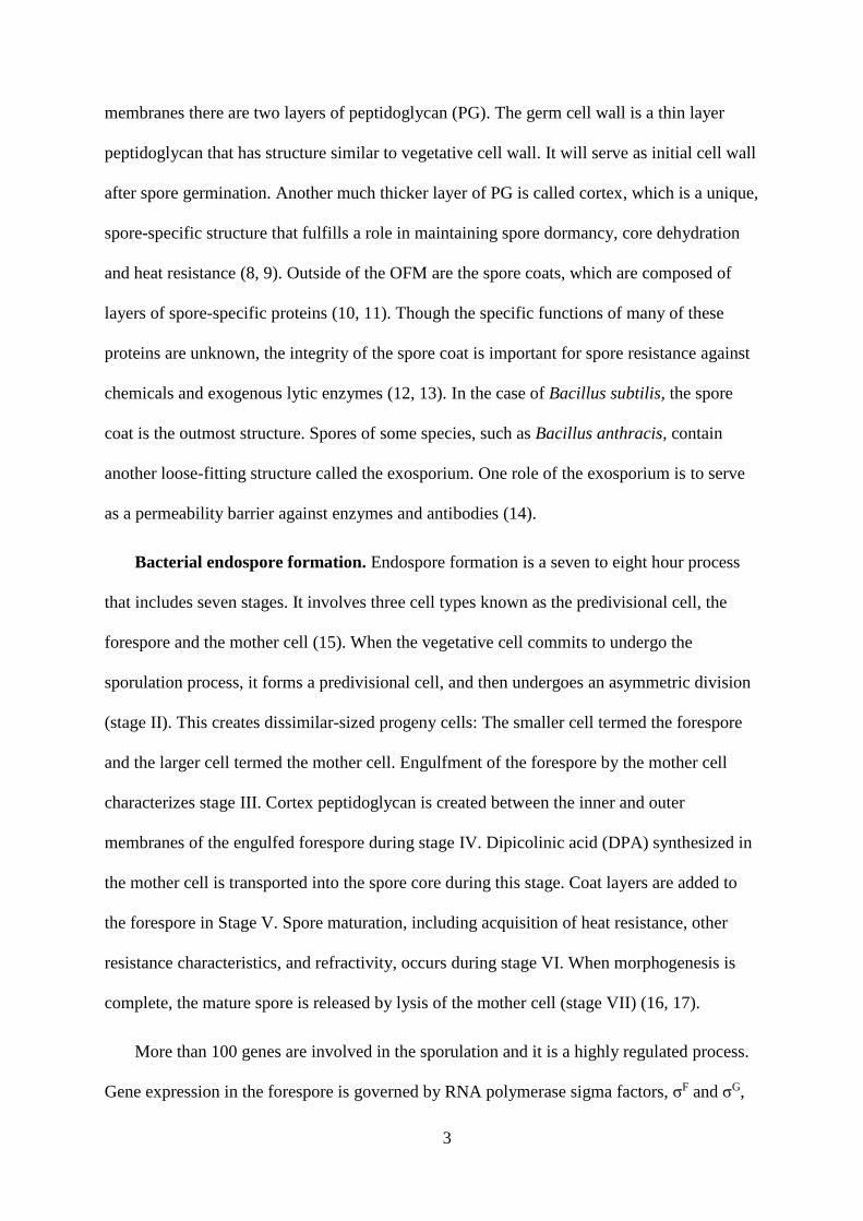
3
membranes there are two layers of peptidoglycan (PG). The germ cell wall is a thin layer
peptidoglycan that has structure similar to vegetative cell wall. It will serve as initial cell wall
after spore germination. Another much thicker layer of PG is called cortex, which is a unique,
spore-specific structure that fulfills a role in maintaining spore dormancy, core dehydration
and heat resistance (8, 9). Outside of the OFM are the spore coats, which are composed of
layers of spore-specific proteins (10, 11). Though the specific functions of many of these
proteins are unknown, the integrity of the spore coat is important for spore resistance against
chemicals and exogenous lytic enzymes (12, 13). In the case of Bacillus subtilis, the spore
coat is the outmost structure. Spores of some species, such as Bacillus anthracis, contain
another loose-fitting structure called the exosporium. One role of the exosporium is to serve
as a permeability barrier against enzymes and antibodies (14).
Bacterial endospore formation. Endospore formation is a seven to eight hour process
that includes seven stages. It involves three cell types known as the predivisional cell, the
forespore and the mother cell (15). When the vegetative cell commits to undergo the
sporulation process, it forms a predivisional cell, and then undergoes an asymmetric division
(stage II). This creates dissimilar-sized progeny cells: The smaller cell termed the forespore
and the larger cell termed the mother cell. Engulfment of the forespore by the mother cell
characterizes stage III. Cortex peptidoglycan is created between the inner and outer
membranes of the engulfed forespore during stage IV. Dipicolinic acid (DPA) synthesized in
the mother cell is transported into the spore core during this stage. Coat layers are added to
the forespore in Stage V. Spore maturation, including acquisition of heat resistance, other
resistance characteristics, and refractivity, occurs during stage VI. When morphogenesis is
complete, the mature spore is released by lysis of the mother cell (stage VII) (16, 17).
More than 100 genes are involved in the sporulation and it is a highly regulated process.
Gene expression in the forespore is governed by RNA polymerase sigma factors, σF and σG,
4
and the DNA-binding proteins RsfA and SpoVT (18). Comparative genomic studies reveal a
core of genes under the σF and σG regulons that are widely conserved among endospore-
forming species but are absent from closely related, non-spore-forming species (15). Most
known germination related genes that localized in the IFM are under σF and σG regulons, for
instance, all known germinant receptor genes, most of spore germination specific lytic
enzyme genes and DPA transport genes. Notably, the functions of more than one third of σF
and σG regulated genes have not been characterized (15). Characterizing these genes will help
us better understand bacteria endospore structure and functional apparatus, therefore helping
in the development of improved decontamination methods.
Spore germination. Germination can be stimulated by nutrient or nonnutrient
germinants. Nutrient germinants are normally amino acids and sugars, such as L-alanine and
glucose, and purine nucleosides, such as inosine. The interaction of these germinants with
their respective germinant receptors triggers germination initiation. However, the molecular
mechanism following the receptor interactions with germinants is not clear (19). Nonnutrient
germinants include dipicolinic acid (DPA), lysozyme, salts, cationic surfactants and high
pressure.
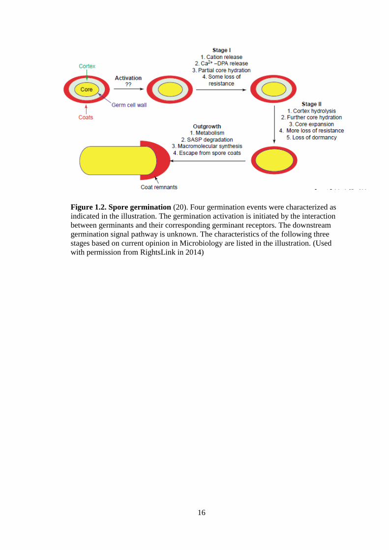
The current model of the spore germination process includes germination activation,
stage I, stage II and outgrowth as shown in Figure 1.2 (20). Briefly, nutrient germinants travel
through the outer layers of dormant spore via an undefined pathway until they reach the inner
membrane and interact with their specific germinant receptors. Once the interaction happens,
germination activation is accomplished, and the spore becomes committed to proceed through
germination, even if the germinant is subsequently removed (20). During germination stage I,
the activated spore releases ions and calcium DPA rapidly from the core, coupled with uptake
of water. The core is partially rehydrated and the spore loses some resistance properties. In
germination stage II, rehydration and the large amount of released Ca2+-DPA triggers
5
activation of germination-specific lytic enzymes, followed by degradation of the cortex and
release of cortical fragments into the surrounding environment. At this point, the spore is
fully rehydrated and loses its dormancy and all resistance characteristics. Metabolism and
protein synthesis resume in the spore core, followed by a period of outgrowth (21).
Superdormant spore. A subpopulation of dormant spores, termed superdormant spores
has been recently investigated in B. subtilis (22). The definition of spore superdormancy is
dependent on the germinant used for isolation of these spores. Superdormant spores are
germination defective to the germinant used for isolation, and more interestingly, in some
cases they also show a poor response to germinants that are recognized by other germinant
receptors (22). This subpopulation also has been reported in other Bacillus species (23) such
as Bacillus cereus, which is closely related to B. anthracis. Investigating the presence of
superdormant spores in B. anthracis will be a requirement for developing further methods to
control its spore contamination. Since superdormant spores germinate as well as other
dormant spores with a nonnutrient germinant, such as Ca2+-DPA, and vegetative cells that
grow out from the superdormant spores can sporulate naturally, a genetic defect is not
considered as the reason causing the germination defect (22). A later publication from the
same research group showed that low level of germinant receptor proteins could be the reason
for spore superdormancy (24). Previous evidences showed that altering other germination-
related proteins, such as SpoVA proteins, could also affect the spore germination rate (25,
26). Perhaps there may be other mechanisms that could result in the production of
superdormant spores.
Spore germination apparatus. As discussed above, components of the germination
apparatus, such as germinant receptors, play an important role in the process of triggering
spore germination, and therefore are crucial for initiation of infection by some pathogens.
Some of the germination apparatus is well conserved across species, whereas each species
6
may possess unique class of germinant recognition mechanisms. The previously characterized
germination apparatus in B. subtilis and B. anthracis are shown in Table 1.1, including
germinant receptors, germination specific lytic enzymes, and DPA transporters. The
localization of these germinant proteins has been investigated either by using specific
antibodies to detect their presence in various spore fractions or by using fluorescent tags to
demonstrate their locations during sporulation (27-29).
To sum up, Figure 1.1 illustrates the possible locations of these proteins (20). The fact
that most germination proteins are located in the IFM focuses attention on research into this
particular membrane and its associated proteins.
Germinant receptors and other Ger proteins. B. subtilis spore germinant receptors
(GRs) have been widely studied. Three germinant receptors have been identified by isolating
germination defective mutants for particular germinants. GerA, GerB, and GerK receptors are
composed of three proteins encoded by homologous tricistronic operons, termed as gerA
operon homologs (30-33). All receptor genes are expressed in the developing forespore and
belong to the σG regulons (20). The GerA receptor recognizes L-alanine, whereas GerB and
GerK cooperate together for germination with nutrient mixture of asparagine, glucose,
fructose and potassium ions (AGFK) (34). Two other gerA operon homologs have been
identified by genome sequencing: yndDEF and yfkQRT (35). A previous study suggested that
the expression of these two gene clusters is low and their products had no observable
contribution on nutrient-triggered spore germination (19).
GerD is a lipoprotein expressed in the forespore under control of the forespore-specific
ϭG (36). There are conflicting reports about the GerD location in spores, but the commonly
accepted model is that GerD is located at the inner membrane of dormant spores (37, 38). B.
subtilis gerD mutation spores have germination defects in response to both types of
7
germinant, which suggested that GerD may be involved in downstream activation of nutrient-
mediated germination events (26).
The gerF gene was first characterized in B. subtilis, and it is the only gene in its operon
that affects spore germination (39). Mutations in gerF result in failure of germination in
respose to both amino acids and sugars (39, 40). GerF has high similarity to Lgt proteins,
which are prolipoprotein diacylglyceryl-transferases, enzymes catalyzing the transfer of a
diacylglyceride group to the N-terminal cysteine of bacterial membrane lipoproteins (41).
GerF may contribute in correct localization or assembly of a number of Ger proteins by
catalyzing lipid attachment (39). Blast analysis of Lgt homologues indicated that GerF is a
unique candidate to perform the enzymic function in B. subtilis.
The categorization of germinant receptors and their response germinants in B. anthracis
is more complicated than in B. subtilis due to the complex combination of germinants. Five
distinct germination pathways have been recognized (42-44). Briefly, the alanine germination
pathway is the only pathway that triggers spore germination with a sole amino acid.
However, the concentration of L-alanine needs to be above 30 mM, which is much higher
than the physiological level in the host (43) . The alanine and proline (AP) response requires
only physiologically relevant concentration of L-alanine in combination with L-proline. The
physiologically relevant concentration of L-alanine can also cooperate with L-histidine, L-
tyrosine or L-tryptophan to active the aromatic amino acid-enhanced alanine (AEA) pathway
(43). Purine ribonucleosides, in cooperation with a cogerminant, have been demonstrated as
effective germinants for B. anthracis spore (43). Binary combination of inosine with L-
alanine, L-serine, L-valine, L-methionine, or L-proline can active amino acid and inosine-
dependent (AAID) 1 pathway (44). A combination of inosine with L-histidine, L-tyrosine, L-
tryptophan, or L-phenylalanine is required to active AAID-2 pathway (44). Similar to B.
subtilis, specific germinant receptors are required to response to these germinants
8
combinations. Seven gerA operon homologs have been identified in B. anthracis: gerA, gerH,
gerK, gerL, gerS, gerY and gerX (45). Only gerX is found within a pathogenicity island on
the pX01 virulence plasmid, while other operons are located on the chromosome (44, 46).
Various single germinant receptor locus mutants were constructed to study the roles of each
germinant receptor in the five signal pathways (42). The alanine and AP pathway showed a
requirement for both the GerK and GerL receptors. GerK apparently could sense both L-
alanine and L-pronine, whereas GerL can only sense L-alanine. The AEA pathway required
GerL, GerS and GerH. GerS and GerH seem to cooperate to recognize the aromatic amino
acid. GerH is involved in the AAID-1 pathway for its recognition of an aromatic amino acid.
Within the AAID-1 pathway, GerL is required when using L-serine or L-valine as
cogerminant, whereas GerK is required when using L-proline or L-methionine as
cogerminant. Finally, the AAID-2 pathway requires only the GerH and GerS receptors. gerA
and gerY may not encode functional receptors due to multiple frameshift mutations within
their coding regions and nonfunctional promoter (42). Previous studies suggested that gerX
encoded proteins may involve in macrophage-associated germination, and therefore are
important for the establishment of anthrax infection and disease progression (46, 47).
However, it is still not clear what specific ligand this receptor recognizes.
A completely different class of germinant receptor, the PrkC protein, has been reported
to be functional in B. anthracis and B. subtilis (48). PrkC has a Ser/Thr kinase domain inside
of the membrane and an extracytoplasmic domain that responds to soluble peptidoglycan
fragments. PrkC function in triggering downstream steps in germination is not clear.
The Ger proteins are not universal in all endospore-forming species. For example,
Clostridium bacteria do not encode GerD protein homologues, either clostridia do not need
them, or the GerD function is met by an alternative, undefined protein. Clostridium difficile is
the only spore-forming species that does not have any known classic germinant receptor gene
9
in its genome. This result is consistent with the fact that C. difficile spores do not respond to
classic nutrient germinants, such as L-alanine, AGFK, or rich undefined medium. C. difficile
spores do respond to glycine and bile salts (49), which are not common germinants for other
species. Therefore, there must be some unknown germinant recognition mechanism that is
unique for C. difficile.
DPA and ion channels. Several features of the spore core play major roles in spore
resistance. The high DPA content in the spore core is one such feature. DPA, predominantly
chelated with Ca2+, comprises nearly 25% of the dry weight of the core (50). DPA less spores
are more sensitive to wet heat, hydrogen peroxide and dry heat (51). Besides its role in spore
resistance, the large amount of DPA released during early germination is essential for
triggering the activation of a germination specific lytic enzyme. Thus, the DPA channel and
its releasing pattern are another activity need to be unveiled for a complete understanding of
the overall process of spore germination.
DPA synthesis starts with an intermediate in the lysine biosynthetic pathway in a
sporulating mother cell. DPA synthetase, which is coded by spoVF, catalyzes the sole DPA-
specific synthetic step (52). DPA is then transported through both the OFM and IFM into the
spore core (53). It has been demonstrated that the proteins encoded by the spoVA operon are
involved in DPA uptake into the developing spore (54). In addition, the involvement of
SpoVA proteins in DPA release during during nutrient-triggered spore germination has been
reported (55). The localization of SpoVAD in the spore inner membrane is consistent with
the role of SpoVA proteins in transporting DPA into and out of the spore core (27). Recent
genome sequence data have shown that at least SpoVAC, SpoVAD and SpoVAE are well
conserved in Bacillus and Clostridium species.
Examining the germination of individual spores of a number of Bacillus species by using
integrated phase contrast microscopy, Raman spectroscopy, and optical tweezers have

10
revealed a specific DPA release pattern during spore germination (56). Spores experienced
different time for completion of Ca2+-DPA release during germination, termed as Trelease. The
difference is due to a various initial time of slower rate of Ca2+-DPA release in advance,
termed as Tlag. Within a germinating spore population, although spores have different Tlag
times, they have consistent △T, which equals Trelease-Tlag (56). This finding indicates that no
matter how long the spore prepares for DPA release, once it starts, it will finish rapidly in a
consistent amount of time. The diversity of spore Tlag may explain the germination
heterogeneity of individual spores. Germinant receptor level, germinant concentration and
heat activation are factors affecting variability in time of initiation of rapid DPA release (56-
59).
Besides DPA channels, some other ion channels are also considered to be important in
the germination process. The first event of germination is an efflux of monovalent ions, such
as Na+, K+ and H+. The release of hydrogen ions is essential for allowing the internal pH
increase from 6.6 to 7, and therefore making the internal environment suitable for metabolic
resumption (20, 34). These ions cannot diffuse freely through the IFM, so how germination
triggers specific ion release is important to better understanding the signal transduction
pathway during germination. The GerN protein has been reported as a Na+/H+-K+ antiporter
in B. cereus spores, which may be one example of an ion transporter that participates in
cation movement during spore germination (60).
Germination specific lytic enzymes (GSLEs). Studies suggest that B. subtilis GSLEs
have at least two types of hydrolytic activity: lytic transglycosylase and N-
acetylglucosaminidase (61, 62). These GSLEs can be subclassified as either spore cortex lytic
enzymes (SCLEs) or cortical fragment lytic enzymes (CFLEs). SCLEs are thought to be
responsible for the initial disruption of the cortex, then CFLEs act on SCLE products to
11
further dissolve the peptidoglycan (63) . Although these lytic enzymes have different
substrates and functions they cooperate in the overall hydrolysis of the cortex (64, 65).
SleB and its homologs are SCLEs found in their mature form in the dormant spores of B.
cereus (66), B. subtilis (64), and B. anthracis (67). Its function is characterized as lytic
transglycosylase in B. anthracis (61). Expression of sleB is controlled by ϭG in the forespore
(68). Immunoelectron microscopic localization of SleB just inside the spore coat layer
suggested that SleB is translocated across the IFM by a secretion signal peptide and is
translocated into the intermembrane space of the developing spore (68). However, further
investigations should be conducted because SleB could be detected both at the outer edge of
the spore cortex and inner spore membrane (64). ypeB is the downstream gene in the
bicistronic operon with sleB. The fact that SleB could not be detected immunochemically in
the spores of a ypeB mutant suggested that YpeB might be required for the localization and/or
stabilization of SleB (64, 69).
When either the sleB or ypeB mutant spores are plated on nutrient medium, the spores are
able to germinate indicating that there are other components involved in germination to
bypass sleB/ypeB pathway (70). The discovery of CwlJ, another SCLE which is localized at
the inner surface of the coat layers revealed the alternative pathway (71, 72). Unlike sleB,
which is regulated by ϭG in the forespore, cwlJ is regulated by ϭE in the mother cell. Mutants
lacking cwlJ germinate more slowly than wild-type cells, like sleB mutants (72). A previous
study also suggested that high concentration of Ca2+-DPA can trigger spore germination as
nutrient germinants do by first activating CwlJ-dependent cortex hydrolysis (73). When both
SleB and CwlJ are nonfunctional, the spore’s peptidoglycan is not depolymerized and the
spore cannot complete germination (74).
The 48 kDa CFLE SleL was firstly reported in B. cereus. It is localized at the outer
periphery of the spore cortex with a function characterized as N-acetylglucosaminidase (75).

12
It plays the major role in hydrolyzing the large products of SCLEs into small, rapidly released
muropeptides (63). The enzyme homologues, which are coded by yaaH, was reported in B.
subtilis and B. anthracis (62, 64). The SleL enzymatic activity in B. anthracis is the same as
that of B. cereus (62), while it was characterized to have epimerase activity in B. subtilis (64).
Previous proteomic studies on Bacillus species. Gel-based and gel-free mass
spectrometry has been a powerful tool for identification and even quantification of entire
proteomes of Bacillus vegetative cells (76-78). In 2002, Kuwanna et al. performed the first
comprehensive analysis of the protein composition of B. subtilis spores using a combination
of SDS-PAGE and LC-MS/MS (79). A total of 69 novel proteins were identified, and 26 of
these were expressed under the control of sporulation-specific sigma factors. Taking
advantage of the two-dimensional polyacrylamide gel electrophoresis and better separation of
proteins, the protein profile of B. subtilis spores was further expanded by Mao et al (80).
Similar proteomic analyses were carried out on B. anthracis, B. cereus and B. thuringiensis in
2004 and 2006 (81-83). Subproteomes of Bacillus spore coat protein fractions were carried
out due to the requirement for solubilization of tightly associated coat proteins (84, 85). To
identify potential protein targets for rapid detection of Bacillus and Clostridium spores, a
subproteome of the coat and the exosporium layers of Bacillus and Clostridium species
spores was carried out by Abhyankar et al in 2013 (86). These proteomic studies contributed
toward a comprehensive understanding of how species differentially express the genome
sequence under nutrient depletion stress, what proteins are specifically produced during the
sporulation process, and what the fates of these spore-specific proteins are.
Objectives of this work. Traditional spore decontamination methods require the use of
harsh chemicals. Research has been focused on developing methods and reagents that could
prematurely trigger spore germination, potentially permitting decontamination using common
antimicrobial disinfectants. The presence of extremely germination resistant but viable

13
superdormant spores make such method development problematic. In Chapter 2, we
investigated the quantity of germination-related proteins in dormant, germinating and
superdormant spores. A Multiple Reaction Monitoring Mass Spectrometry (MRM-MS)
approach was established, and the relative abundance of 11 germination-related proteins was
determined between spore samples. It was discovered that a deficiency in the GerD
lipoprotein, besides low levels of germinant receptor proteins (24), may result in
superdormancy. Specifically, variation in the abundance of the GerD lipoprotein may
contribute to heterogeneity of spore germination rates.
In chapter 3 we investigate the identities of Bacillus spore membrane proteins by
carrying out shot-gun proteomic studies on B. subtilis and B. anthracis spore membrane
fractions. A total of 104 and 87 membrane-associated proteins were identified in B. subtilis
and B. anthracis, respectively. These proteins were further characterized with regard to
membrane association, cellular function, and conservation across species. Proteins that were
not previously known to be spore associated were identified, and many of these proteins
shared great similarity in both Bacillus species. A significant number of these proteins are
implicated in functions that play major roles in spore formation and germination. This study
generated a candidate protein list that can be further investigated in future studies.
HtrC, a membrane serine protease identified in our spore membrane proteome, is the
major focus in Chapter 4. This study revealed that YpeB was proteolytically processed at
specific sites during germination, and that HtrC is the protease responsible for specific
cleavage events using both in vivo and in vitro methods. The proteolytic processing of YpeB
during spore germination is proposed to be the signal that terminates the relationship between
SleB and YpeB, which have been demonstrated to be co-dependent and co-localized in the
inner forespore membrane.

14
The results of these studies provide a better understanding of spore membrane protein
content and their roles in the spore germination process. This body of work begins to uncover
the framework of the mysterious and complex network in the early steps of spore
germination.

15
Figure 1.1. Bacterial endospore structure and components of spore germination
apparatus (20). General structures of bacterial endospore are indicated in the illustration.
Exosporium, an outmost additional spore structure of some spore forming species, is not
included in the illustration. The localization of some major germination apparatus proteins
are indicated as well. (Used with permission from RightsLink in 2014)
16
Figure 1.2. Spore germination (20). Four germination events were characterized as
indicated in the illustration. The germination activation is initiated by the interaction
between germinants and their corresponding germinant receptors. The downstream
germination signal pathway is unknown. The characteristics of the following three
stages based on current opinion in Microbiology are listed in the illustration. (Used
with permission from RightsLink in 2014)
17
Table 1.1. Germinant apparatus reported in spores of Bacillus species
Species Germinant apparatus Functions
B.subtilis
GerA Response to L-alanine
GerB &GerK Response to AGFK (L-asparagine, glucose, fructose and
K+)
YndDEF & YfkQRT Unknown germinant receptors
GerF Add diacylglycerol to membrane proteins
GerC Probably an enzyme of menaquinone biosynthesis
GerD Unknown function in nutrient germination
CwlJ lytic Lytic enzyme, degrade Degrades cortex
peptidoglycan
GerQ Essential for the presence of CwlJ
SleB Lytic transglycosylase, degrade Degrades cortex
peptidoglycan
YpeB Essential for the presence of SleB
SpoVA Proteins involved in DPA transport
B.anthracis
GerK&GerL Alanine and AP*pathway
GerL, GerS & GerH AEA*pathway
GerH, GerL &GerK AAID-1* pathway
GerH&GerS AAID-2* pathway
GerA &GerY Unknown or non-functional germinant receptors
GerX Possible role in amino acid and inosine-dependent
responses and macrophage- associated germination
CwlJ1&CwlJ2 lytic Lytic enzyme, degrade Degrades cortex
peptidoglycan
SleB Lytic transglycosylase, degrade Degrades cortex
peptidoglycan
SleL Further degrade cortex fragments
YpeB Essential for the presence of SleB
AP: Alanine and proline response;
AEA: aromatic amino acid-enhanced alanine response;
AAID: amino acid and inosine dependent response;
AAID-1: Binary combination of inosine and either L-alanine, L-serine, L-valine, L-methionine
or L-proline;
AAID-2: Binary combination of inosine and either L-histidine, L-tyrosine, L-tryptophan or L-
phenylalanine.

18
CHAPTER 2
Levels of Germination Proteins in Bacillus subtilis Dormant,
Superdormant, and Germinating Spores
Yan Chen, W. Keith Ray, Richard F. Helm, Stephen B. Melville, and David L. Popham
PLoS ONE 9(4): e95781. doi:10.1371/journal.pone.0095781

19
AUTHOR CONTRIBUTIONS
Conceived and designed the experiments: Yan Chen, W. Keith Ray, Richard F. Helm,
Stephen B. Melville, and David L. Popham. Performed the experiments: Yan Chen, W. Keith
Ray, and David L. Popham. Analyzed the data: Yan Chen, W. Keith Ray, Richard F. Helm,
Stephen B. Melville, and David L. Popham. Contributed reagents/materials/analysis tools:
Yan Chen, W. Keith Ray, Richard F. Helm, and David L. Popham. Wrote the paper: Yan
Chen, W. Keith Ray, Richard F. Helm, Stephen B. Melville, and David L. Popham.
20
ABSTRACT
Bacterial endospores exhibit extreme resistance to most conditions that rapidly kill other
life forms, remaining viable in this dormant state for centuries or longer. While the majority of
Bacillus subtilis dormant spores germinate rapidly in response to nutrient germinants, a small
subpopulation termed superdormant spores are resistant to germination, potentially evading
antibiotic and/or decontamination strategies. In an effort to better understand the underlying
mechanisms of superdormancy, membrane-associated proteins were isolated from populations
of B. subtilis dormant, superdormant, and germinated spores, and the relative abundance of 11
germination-related proteins was determined using multiple-reaction-monitoring liquid
chromatography-mass spectrometry assays. GerAC, GerKC, and GerD were significantly less
abundant in the membrane fractions obtained from superdormant spores than those derived
from dormant spores. The amounts of YpeB, GerD, PrkC, GerAC, and GerKC recovered in
membrane fractions decreased significantly during germination. Lipoproteins, as a protein
class, decreased during spore germination, while YpeB appeared to be specifically degraded.
Some protein abundance differences between membrane fractions of dormant and
superdormant spores resemble protein changes that take place during germination, suggesting
that the superdormant spore isolation procedure may have resulted in early, non-committal
germination-associated changes. In addition to low levels of germinant receptor proteins, a
deficiency in the GerD lipoprotein may contribute to heterogeneity of spore germination rates.
Understanding the reasons for superdormancy may allow for better spore decontamination
procedures.
21
INTRODUCTION
Bacterial endospores are metabolically dormant and resistant to a variety of anti-
microbial treatments due to their protective structures and dehydrated spore core (87, 88).
These spores can survive for decades in the absence of nutrients. However, they are able to
return to a metabolically active state through a series of events termed spore germination. Once
spores lose many of their resistance properties during germination, they can then be easily
eliminated by routine decontamination methods (20). Since the spores of Bacillus and
Clostridium species cause food spoilage and are infectious agents in several human diseases
(89), the development of methods or reagents that stimulate highly efficient germination across
a spore population could greatly simplify decontamination efforts and reduce morbidity and
mortality.
Procedures used for triggering spore germination do not achieve 100% efficiency due
to heterogeneity in germination rate within spore populations. Studies of single germinating B.
cereus and Clostridium spores indicated that the spore germination heterogeneity results from
the variation in time of initiation of rapid Ca2+-dipicolinic acid (DPA) release (Tlag) (56, 90).
Subpopulations of B. subtilis spores termed superdormant spores can be isolated following
multiple rounds of germination with saturating nutrient germinant levels (22). These
superdormant spores exhibit extremely poor germination response to the germinant used for
isolation, but will germinate to varying degrees when triggered with germinants that utilize
other germinant receptors (22, 91). Individual germinating superdormant spores exhibit longer
times for initiation of rapid Ca2+-DPA release relative to initial dormant spore populations (91).
However, once rapid Ca2+-DPA release is initiated, the rate of release is similar for all spores.
Thus one may hypothesize that the state of superdormancy is related to processes occurring
prior to Ca2+-DPA release.
22
Four groups of proteins have been implicated to be involved in the early steps of
germination: 1) germinant receptors; 2) DPA channel proteins; 3) germination-specific lytic
enzymes (GSLEs) and their partner proteins; and 4) lipoproteins potentially involved in
transducing germinant-binding signals (20, 21). In B. subtilis, three major germinant receptors
(GRs) have been characterized: GerA, GerB, and GerK (32, 92, 93). Each GR is comprised of
at least A, B, and C subunits (some receptors have D subunits encoded within or associated
with the receptor operon (94)) and is localized to the spore inner membrane. The A and B
subunits are believed to be integral membrane proteins with multiple transmembrane domains.
The C subunits are putative lipoproteins based on their N-terminal signal peptides and on the
effect of a gerF mutation, which eliminates the only protein diacylglycerol transferase in this
species, on their function (40).
Previous genetic studies of these GRs illustrated their germinant specificity. GerA
alone responds to L-alanine or L-valine, while GerB and GerK are required for germination
with a mixture of L-asparagine, D-glucose, D-fructose, and potassium ions (AGFK) (93, 95).
The binding of nutrient germinants to their cognate GR or GRs initiates irreversible
germination activation (20), and via an unclear pathway, results in the opening of DPA
channels and the rapid release of this abundant spore solute. Proteins encoded by the spoVA
operon are involved in DPA uptake during sporulation as well as release during spore
germination. Since SpoVA proteins are transcribed exclusively in the developing forespore and
some appear to be integral membrane proteins, they are most likely localized to the inner spore
membrane (54, 96, 97). The PrkC protein has been identified as an alternate class of germinant
receptor that recognized the presence of peptidoglycan fragments in the medium (48).
Complete germination requires that the thick layer of spore cortex peptidoglycan be
degraded by GSLEs (70, 71, 98). SleB is a key GSLE (70), and some evidence indicates that
it and a co-expressed protein involved in SleB stabilization, YpeB, are localized to the inner
23
spore membrane in the dormant spore (64). Spores with a gerD deletion mutation had a
dramatically slower response to nutrient germinants utilizing any of the Ger receptors (26).
Lipoproteins involved in germination, including GerAC, GerBC, GerKC and GerD, are
believed to be anchored in the spore inner membrane by a covalently attached lipid (40).
Spores of a B. subtilis gerF null mutant also lacked both the GerAC and GerD proteins (99).
This mutant exhibited a significant defect in germination with a greater effect on germination
triggered through the GerA receptor relative to responses via the GerB and GerK receptors (40,
99).
Several studies have indicated that the abundance of germination-associated proteins
can impact the rate of spore germination. Overexpression of the GerA receptor significantly
increased the germination rate triggered by its corresponded germinants but did not affect GerB
and GerK abundance or germination function (100). In contrast, overexpression of SpoVA
proteins increased germination rates triggered through any germinant receptor (25). It is
hypothesized, based upon quantitative Western blot analyses, that a significant reduction in the
amount of a Ger receptor could be the reason for spore superdormancy (24).
In an effort to provide additional insight into the mechanisms of germination, we
developed a multiple-reaction monitoring (MRM) mass spectrometry assay (101) to quantify
11 germination proteins believed to be associated with the spore inner membrane. MRM assays
are based upon the analyses of peptides specific to the target protein (proteotypic peptides),
which become surrogates for protein abundance. The method has high specificity and
sensitivity for target protein quantification, and permits reproducible analyses of multiple
samples. MRM analyses were performed on membrane preparations obtained from dormant,
rapidly germinating, and superdormant spore samples. The results of these analyses indicate
that the GerD lipoprotein level can contribute to the heterogeneity of spore germination rate
and superdormancy.
24
MATERIALS AND METHODS
Spore sample preparation. The B. subtilis strain used was PS832, a prototrophic
laboratory derivative of strain 168. Spores were prepared on 2xSG (102) agar plates without
antibiotics. Spores were harvested after 72 h incubation at 37°C and purified by water
washing and centrifugation through a 50% sodium diatrizoate (Sigma) layer as described
(103). All spores used in this work were 99% free of vegetative cells and were stored in
deionized water at 4°C until analysis.
A 10-ml suspension of dormant spores at an optical density at 600 nm (OD600) of 20 in
water were heat-activated at 75°C for 30 min and cooled on ice for at least 10 min. The
spores were then germinated at 37°C and at an OD600 of 2 with 10 mM L-valine in 25 mM
Tris-HCl buffer (pH 7.4). The germination of spores was terminated after the OD600 dropped
to 50% of the initial value. Germinated spores were collected by centrifugation at 12,000 x g
for 5 min at 4°C, quickly washed with cold deionized water, centrifuged again, and frozen at
-80°C. Examination by phase-contrast microscopy indicated that >95% of the spores in these
preparations had germinated.
Superdormant spores were isolated and characterized as described previously (22).
Briefly, dormant spores at OD600 of 1 were germinated as described above for 2 h and
collected by centrifugation. The pellet was washed with deionized water, suspended in 20 %
w/v sodium diatrizoate, and centrifuged through a 50 % w/v sodium diatrizoate solution
(13,000 x g for 45 min) to separate dormant spores from germinated spores. The dormant
spore pellets were collected and washed thoroughly with deionized water. These dormant
spores were subjected to another 2 h round of germination and were separated by density
gradient centrifugation again. The final superdormant spore pellet was washed thoroughly
with deionized water and stored at 4°C.
25
Superdormant spore characterization. For phenotypic studies, isolated superdormant
spores as well as initial dormant spores were germinated with nutrient germinants: 10 mM L-
Valine or AGFK (13 mM L-asparagine, 13 mM D-glucose, 13 mM D-fructose, 13 mM KPO4
[pH 7.4]); or the non-nutrient germinant 60 mM Ca2+-DPA [pH 7.4]. Prior to nutrient-
triggered germination, spores were heat-activated in water at 75°C for 30 min and then
briefly cooled on ice. Germination was initiated by diluting spores to an OD600 of 0.2 in
germination solutions and incubating at 37°C. Germination was monitored as the change in
OD600 over time. Spores used for Ca2+-DPA germination were not heat-activated and the
germination was at 30°C. To assess Ca2+-DPA germination, 100 spores were examined by
phase-contrast microscopy at several incubation time points.
Preparation of spore membrane fractions. Spore membrane fractions were prepared
by a modification of previously described methods (28, 37, 104). Dormant, germinated, and
superdormant spores prepared as described above were lyophilized. The dry spores (~19 mg
for germinated spores and ~24 mg for dormant and superdormant spores) were pulverized
with 100 mg of glass beads in a dental amalgamator (Wig-L-Bug) at 4,600 rpm for pulses of
30 s each, with 30 s pauses on ice between pulses. Spore disruption was monitored by
suspending a small sample of spore material in H2O and observing under phase-contrast
microscopy. Once >80% of spores were disrupted, the dry powder was suspended in 0.5 ml
of 4°C extraction buffer (10 mM Tris-HCl [pH 7.4], 1 mM EDTA, 2 mg/ml RNase A, 2
mg/ml DNase I, 1 mM phenylmethylsulfonyl fluoride (PMSF)). The suspension was
centrifuged (6,000 X g, 10 min, 4°C) and the resultant supernatant was centrifuged again
(13,000 X g, 10 min, 4°C) to remove insoluble material. The remaining supernatant was
subjected to ultracentrifugation (100,000 X g, 60 min, 4°C). The resulting supernatant was
considered the spore core soluble fraction and was stored at -80°C. The resulting pellet,
designated the crude spore membrane fraction, was homogenized in 1 ml high salt buffer (20
26
mM Tris-HCl [pH 7.5], 10 mM EDTA, 1 M NaCl, and 1 mM PMSF) and was gently shaken
for 30 min at 4°C. The homogenate was subjected to ultracentrifugation again as described
above. The remaining pellet was homogenized in 1 ml alkaline buffer (100 mM Na2CO3-HCl
[pH 11], 10 mM EDTA, 100 mM NaCl, and 1 mM PMSF) and was again subjected to
ultracentrifugation. After a final wash with 1 ml TE buffer (10 mM Tris-HCl [pH 7.4], 1 mM
EDTA, 1 mM PMSF), the resulting pellet was homogenized in 200 µl TE buffer, flash
frozen, and stored at -80°C until analysis. The protein concentration was determined by acid
hydrolysis and amino acid analysis (105) with comparison to a standard set of amino acids
(Sigma).
Protein digestion. Proteins in spore membrane fractions (70 µg) were precipitated with
1 mL of acetone -20°C overnight and collected by centrifugation for 20 min at 12,000 g.
Protein was resuspended in 250 l of freshly-prepared 8 M urea, 20 mM Tris-HCl, pH 8.0 to
give a final protein concentration of 1 mg/ml. Proteins were denatured by the addition of
27.8 µl of freshly-prepared 45 mM dithiothreitol, 20 mM Tris-HCl, pH 8.0, and incubation
for 1 h at 37°C. Free cysteines were alkylated by the addition of 30.9 µl of freshly-prepared
100 mM iodoacetamide, 20 mM Tris-HCl, pH 8.0, incubation at room temperature in the dark
for 30 min. Unreacted iodoacetamide was inactivated by the addition of 102.9 µl of freshly-
prepared 45 mM dithiothreitol, 20 mM Tris-HCl, pH 8.0. Proteins were digested by the
addition of 1.03 ml of 20 mM Tris-HCl, pH 8.0, and 5 µg trypsin in 10 µl 50 mM acetic acid
followed by incubation overnight at 37°C with shaking. Trifluoroacetic acid was added to a
final concentration of 0.25% and formic acid was added to a final concentration of 1%. The
pH was measured and additional formic acid was added until the pH was at or below 3.
Conditioning of 0.1 ml OMIX C18 solid phase extraction cartridges used 0.2 ml methanol,
followed by 0.2 ml 50% acetonitrile, 0.1% TFA and finally 0.2 ml 2% acetonitrile, 0.1%
TFA. A protein sample was applied to the cartridge, which was then washed three times with
27
0.2 ml 2% acetonitrile, 0.1% TFA. Peptides were eluted with 0.2 ml 75% acetonitrile, dried,
and resuspended in 0.02 ml solvent A (2:98 acetonitrile:water containing 0.1% formic acid).
Liquid chromatography and mass spectrometry. Thirteen germination-related
membrane proteins (Table 2.1) were initially targeted for MRM method development, with a
list of potential proteotypic tryptic peptides generated using the Enhanced Signature Peptide
Prediction tool (106) using a cutoff value of 0.6. Peptides were synthesized by JPT Peptide
Technologies GmbH Inc., and were directly infused into the mass spectrometer for
determination of target fragment ions and ionization conditions. For each synthesized
peptide, elution times were identified, the dominant precursor ion of predicted m/z (Q1 ion)
was identified and fragmented, and dominant fragment ions of expected m/z (Q3 ions) were
identified and quantified. Limits of quantification (LOQ) (Table 2.1) were determined using
the established MRM methods and dilution series from 10-1500 fmol of each synthetic
peptide (Fig. 2.1).
Proteins in spore membrane fractions were solubilized with 20 mM Tris-HCl [pH 8.0], 8
M urea, 45 mM dithiothreitol at a final protein concentration of 1 mg/ml, followed by a 37°C
overnight trypsin digestion at 20:1 (w/w) protein:Trypsin ratio. The tryptic peptides were
desalted and concentrated using OMIX C18 microextraction pipette tips (Varian) following
the manufacturer’s protocol. Peptides was separated using an Eksigent Nano 2-D liquid
chromatography system connected to a 100 x 0.075 mm Magic C18AQ (200Å, 3µm, Bruker)
column packed in-house using an eFRIT fused silica capillary (Phoenix S&T). Ten
microliters of each sample was first loaded onto a C18 trap cartridge at 10 µl/min for 15
minutes using solvent A (2:98 acetonitrile:water containing 0.1% formic acid). The trap
cartridge was switched in-line with the analytical column and the trap and column were
flushed with 95% solvent A, 5% solvent B (98:2 acetonitrile:water containing 0.1% formic
acid) for 5 minutes at 300 nl/min. This was followed by a linear gradient to 86% solvent A

28
over 5 minutes then a linear gradient to 71% solvent A over 45 minutes and finally a linear
gradient to 35% solvent A over 5 minutes. The column was flushed for 2 minutes with 35%
solvent A and reequilibrated at the starting conditions for 13 minutes prior to the next sample
injection. The eluent was introduced into an AB Sciex 4000 QTrap mass spectrometer
controlled by Analyst 1.4.2 software (AB Sciex) via a nano-electrospray source (Phoenix
S&T). The mass spectrometer was operated in positive ion mode utilizing an MRM method
containing precursor/product ion transitions corresponding to peptides described below.
Dwell time for each transition was 40 ms and the total cycle time was 6.6 seconds. The first
quadrupole was operated at low resolution while the third quadrupole was set to unit
resolution. Ion spray voltage was 2400V, curtain and sheath gases were 12 (arbitrary units),
interface heater temperature was 120˚C and the entrance potential was 10V for all transitions.
CAD gas was set to medium corresponding to a vacuum of 3.1 x 10⁻⁵ Torr.
Data collection and refining. When determining which of the identified Q3 ion peak
areas were suitable for quantitative comparisons across all samples, we applied the following
raw data refining criteria. 1) The retention time of a Q3 ion in all samples should be the same
as that determined for the corresponding synthetic peptide. Q3 ions that did not have
consistent retention times were excluded from further analysis. 2) If a quantified peptide had
less than two quantifiable Q3 ions, the peptide was excluded from further analysis. 3) If the
peak area of a Q3 ion was below established limited of quantification, the Q3 ion was
excluded from further analysis. 4) Among all nine samples, if the Q3 ion peaks in more than
three samples had S/N ratio values less than 10, then the Q3 ion was excluded from further
analysis. (The end section of each Q3 ion spectrum was considered as base line (noise) when
collecting the S/N ratio for limit of quantification evaluation.)
Within each biological replicate set, there were three membrane fraction samples:
dormant, germinated, and superdormant. Three biological replicates were derived from three
29
independent spore preparations. For each quantified Q3 ion, peak area ratios between two
membrane fractions were calculated only within a biological replicate set. Ratios were then
compared across biological replicates. Theoretically, if a protein’s abundance was the same in
two different samples, the peak area ratios for the Q3 ions of its peptides would be 1. Among
all Q3 ion peak area ratios calculated, those of proteins GerAA, GerBA, and GerKA were
always close to 1.0. We took these proteins to represent unchanged proteins within the
samples, and pooled their Q3 ion peak area ratios to represent the level of physiological
variance. For each comparison group, we then evaluated the significance of a protein change
by comparing peak area ratios of the protein to this unchanged protein peak area ratio pool
using a two samples student t-test. In addition, for each protein, we evaluated the
significance of two comparison groups using the Student’s t-test. Both tests used two-tailed,
unequal variance p values, and statistical significance for both t-tests was set at p < 0.05.
30
RESULTS
Isolation and characterization of spore populations. Three independent preparations
of B. subtilis dormant spores were germinated using L-valine, with downstream processing
producing rapidly germinating and superdormant spore populations. The yield of
superdormant spores was 1.09 ± 0.16 % (n=3); somewhat less than the 3.8 % yield in a
previous publication (22). Two reported characteristics of superdormant spore populations
isolated using L-valine are that the superdormant spores germinate poorly with L-valine as
well as with germinants that use a different germinant receptor, with the superdormant spores
being as viable as the initial dormant spores when germinated with non-nutrient germinants
(22). Our superdormant spores also germinated slowly with L-valine, in comparison with the
rapid germination of the initial dormant spores (Fig. 2.2A). However, when using AGFK as
germinant, which acts through different germinant receptors than does L-valine (93), the
superdormant spores germinated more rapidly than the initial dormant spores (Fig. 2.2B). In
addition, the superdormant spores also reached a higher efficiency of germination based on a
greater OD600 decrease than the initial dormant spores. While our results are different from
those of the original description of superdormant spores (22), similar observations were
reported for superdormant spores isolated in a more recent study (91). The effect of a non-
nutrient germinant on the superdormant spores was tested using Ca2+-DPA, which causes
activation of the GSLE CwlJ (73), bypassing part of the germination apparatus that may be
deficient in superdormant spores. The superdormant spores completed Ca2+-DPA-triggered
germination as efficiently as the initial dormant spores after an initial lag period (Fig. 2.2C),
similar to a previous report (91). In summary, the results of the phenotypic analyses support
the claim that spores isolated after extensive L-valine germination can be classified as
superdormant. To verify that these spores were not superdormant due to a genetic alteration,
they were germinated and spread on plates, and 10 randomly selected colonies were selected,
31
cultured, sporulated, and tested for germination rate. Similar to a previous report (22), spore
populations produced by these strains germinated equivalently to those of the wild type
strain.
Quantification of spore membrane proteins by MRM assays. Membrane samples
were prepared from dormant, germinated, and superdormant spores and were used to quantify
the targeted germination-related proteins relative to the total protein concentration. The total
protein in each sample was determined by amino acid analysis. SDS-PAGE analysis of total
proteins was consistent with this quantification and revealed essentially identical protein band
patterns across biological replicates (Fig. 2.3).
The MRM assays centered on the detection of 11 of the 13 proteins expected to be
membrane associated and involved in spore germination (Table 2.1). As peptides of varying
compositions exhibit different ionization efficiencies, we determined LOQs for each peptide.
We were not able to identify any proteotypic peptides for GerAB that were even predicted to
function well in an MRM assay, and we were not able to obtain quantifiable MRM data for
GerBB and GerKB due to the fact that the signal for the proteotypic peptides designed for
these integral membrane proteins were below the limit of detection. Nonetheless, we were
able to quantify the A and C subunits of the germinant receptors. The ratios of GerAA,
GerBA, and GerKA between dormant and superdormant spores were very close to 1.0. In
contrast, the amounts of GerAC and GerKC in superdormant spores were 3.4 and 1.9-fold
lower than the amounts in dormant spores (Fig. 2.4A). These decreases of GerAC (p=0.002)
and GerKC (p=0.023) were statistically significant. GerBC, however, showed no significant
difference in amount between superdormant and dormant spores (Fig. 2.4A).
GerD is a lipoprotein that is localized predominantly to the spore inner membrane (37)
and functions in both GerA and GerB/K-mediated germination responses (26). GerD was 1.8-
fold less abundant in membranes isolated from superdormant spores in comparison to those

32
from dormant spores (Fig. 2.4A). PrkC, SpoVAC, SpoVAD, and YpeB exhibited no
significant difference in abundance between superdormant and dormant spore samples (Fig.
2.4A).
The relative amounts of GerAA, GerBA, and GerKA in germinated spore samples were
similar to the amounts of these proteins in those from dormant spores. In contrast, the
amounts of GerAC, GerBC, and GerKC in germinated spore membranes decreased 1.9, 1.6,
and 2.4-fold respectively in comparison to dormant spores (Fig. 2.4B). Previous western blot
results showed that membrane-associated GerD decreased during spore germination (37).
This was confirmed in the MRM assays, which showed that membrane-associated GerD
decreased 3.5-fold during spore germination (Fig. 2.4B). The decreases of GerAC (p=0.009),
GerKC (p=0.032), and GerD (p<0.0001) were statistically significant.
To date, there is no report regarding PrkC function in a nutrient germinant receptor-
mediated pathway. The MRM assays indicated that membrane-associated PrkC was
significantly decreased 3.8-fold in amount after spore germination (Fig. 2.4B). Similarly,
three proteotypic YpeB peptides decreased 6.8-fold in amount during spore germination (Fig.
2.4B). This result is consistent with a previous observation of YpeB degradation during
germination (64). A fourth YpeB peptide was clearly quantifiable in dormant spore samples,
but was undetectable in germinated spore membrane fractions. This peptide is near the YpeB
N-terminus (residues 57-67), whereas the three detectable YpeB peptides were closer to the C
terminus.
SpoVAC and SpoVAD proteins were chosen as representatives of the spoVA-encoded
proteins. SpoVAC is predicted to be an integral membrane protein (97, 107), and SpoVAD is
more likely to be a peripheral membrane protein based on its crystal structure (108). While
SpoVAD could be detected in both dormant and germinated membrane fraction samples by
Western-blot (data not shown), the MRM assays indicated that SpoVAC and SpoVAD
33
significantly increased 1.3 and 1.7-fold respectively in germinated spore membrane samples
in comparison to those of dormant spores (Fig. 2.4B).
The germinant receptor A subunit amounts detected were similar in superdormant and
germinated spore samples. While membranes of both superdormant and germinated spores
had significantly less GerAC and GerKC proteins relative to dormant spores, the decrease in
GerAC was significantly greater than that of GerKC and was 1.6-fold lower in superdormant
spores in comparison to germinated spores (Fig. 2.4C). Similarly, while GerD was also less
abundant in membranes from both superdormant and germinated spores than in dormant
spores, the difference was greater in the germinated spores, such that GerD was 2-fold more
abundant in superdormant spores than in germinated spores (Fig. 2.4C). This difference was
statistically significant (p<0.0001). If germination-induced protein changes initiated but did
not progress past GerD in superdormant spores, the difference in abundance for proteins
involved in later germination events in comparison to germinated spores should be similar to
a dormant/germinated spores comparison. Indeed, our results showed that YpeB and PrkC
were 4.9 and 2.9-fold more abundant in samples from superdormant spores than in those from
germinated spores, and SpoVAC and SpoVAD were 1.6 and 1.5-fold less abundant in
superdormant spore samples than in germinated spore samples (Fig. 2.4C).

34
DISCUSSION
Spore germination starts at the spore inner membrane with the interaction of germinant
with Ger receptor proteins and progresses through core rehydration and cortex breakdown.
Deficiencies in Ger receptors and associated proteins, Ca2+-DPA channels, and lytic enzymes
can potentially inhibit the germination process, leading to the production of superdormant
spores. In this work, L-valine superdormant spores responded poorly to valine but
germinated well with AGFK; a result that is different from the initial report of superdormant
spores (22) but consistent with a later report (91). In contrast to a high yield (12%) of
superdormant spores isolated with AGFK in previous work (22), we were unable to isolate
any superdormant spores using AGFK. The reasons for these differences are not clear, but
raise the possibility that there may be multiple pathways to superdormancy, and slight
differences in the method of preparation and isolation may result in significantly different
superdormant spore populations.
Our quantitative MRM assays were performed on membrane samples derived from
broken spores. Results published while this work was in progress indicate that these samples
may represent a subset of the spore membrane fraction, as extensive chemical extraction is
required to recover the full amount of several spore membrane proteins (109). While the
sample analyzed here may not represent the entire spore membrane fraction, two lines of
evidence indicate that the samples recovered from each spore type are similar fractions. The
relative abundance of the majority of the proteins analyzed was the same in dormant and
superdormant spores, and the amounts of the integral membrane A subunits of the Ger
receptors were not altered in germinated spores, indicating that the membrane fractions
obtained from the different spore types were comparable in their protein complements.
35
We interpret the similarity in Ger receptor A protein abundance in dormant and
superdormant spore fractions to imply that the isolated superdormant population is not
delayed in germination due to a low level of Ger receptors. These results differ from a
previous publication that reported GerAA and GerAC protein levels were 7-fold lower and
other GR subunits 3-fold lower in superdormant spores (24). We did find that the C subunits
of the GerA and GerK receptors were decreased in samples from our superdormant spores.
Germination-induced decreases in C subunit abundance, relative to dormant spores, were in
one case less (GerAC) and in other cases slightly greater (GerBC and GerKC) than those
observed in superdormant spores. As the C subunits are believed to be present in a
stoichiometric association with the A subunits in the membranes of dormant spores (109), a
decrease in the C subunits may indicate a change that takes place during early germination or
may indicate a change in the maintenance of subunit association with the membrane during
membrane fraction washing and isolation. Published data indicate that GerD, a lipoprotein
like the C subunits, can be partially extracted by a high salt wash similar to that used in this
study and is released from the membrane during germination (37). The decreased abundance
of C subunits observed in superdormant spore samples could be either a cause of
superdormancy or a result of the superdormant spore isolation process. Protein associations
could decrease during Ger receptor activation upon germinant binding, and the membrane-
association may be less stable as the spore membrane regains fluidity during germination (6).
Prolonged exposure to germinants during the superdormant spore isolation may result in the
loss of C subunits from the membrane, with a blockage in the germination pathway at a point
downstream of the Ger receptors.
An absence of GerD was previously shown to result in a germination initiation defect
(25, 26). Previous studies also indicated that the germination receptors and GerD co-localize
to a discrete cluster on the membrane (110) and that SpoVA proteins can associate with Ger

36
receptors (111), and thus we expect that the comparable membrane factions we derived from
different spore types would contain similar amounts of GerD. Superdormant spore
membrane samples had 2-fold less GerD than those from dormant spores, and germinated
spore samples contained even less GerD. Similar to the case for the Ger receptor C subunits,
the decreased level of GerD in superdormant spores could be either a cause of superdormancy
or a result of a partial germination response that is blocked at a subsequent point.
Other germination-active proteins, PrkC, SpoVAC, SpoVAD, and YpeB were present in
dormant and superdormant spore membrane preparations in similar quantities, but did show
changes in abundance during germination. This suggests that any early germination-related
events that have taken place within the superdormant spores did not progress to later events
such as SpoVA-assisted DPA release (96) or YpeB-related cortex degradation (64).
Interestingly, the abundance of membrane-associated SpoVAC and SpoVAD proteins
increased during germination. As this increase is taking place well before any new protein
synthesis, it must represent an increased association of the proteins with the membrane or
with other membrane-bound proteins, most likely other products of the SpoVA DPA-
transport complex. Several studies indicate that SpoVAD localizes to the spore inner
membrane (27, 112), and recent structural analyses of this protein show that it is likely to be a
peripheral membrane protein (108, 113). In our western-blot analysis of SpoVAD, it was
detected in both soluble and membrane fractions, suggesting a weak membrane association
(data not shown). Another novel finding is that PrkC abundance decreased significantly
during spore germination triggered by L-Val. Although this protein was previously
demonstrated to be a germinant receptor that responds to muropeptides (48), there is no
report of its activity in other germinant receptor-mediated pathways.
The most dramatic change in protein abundance during germination was that of YpeB,
which is required for incorporation of SleB in spore and thus for normal cortex degradation
37
during germination (64). This result is consistent with a previous observation of YpeB
degradation during germination. The 52 kDa YpeB is processed to a ~30 kDa product during
germination (64). The 6.8-fold decrease in YpeB abundance we observed was calculated
using those peptides we could detect in both dormant and germinated spore samples, all of
which were in the C-terminal half of the protein. One peptide nearer the N-terminus of YpeB
was detected in dormant spore samples but was undetectable in germinated spore samples,
indicating a decrease of >14-fold. This differential loss of peptides indicates that the more
stable 30-kDa portion of YpeB represents a C-terminal portion. Ongoing studies in our lab
are consistent with this (data not shown). It is not clear if the 6.8-fold decrease in the
observed YpeB peptides is due to protein degradation, a decrease in membrane association,
or both. Because the N-terminus of YpeB apparently contains an uncleaved signal peptide,
proteolytic removal of this domain would be expected to decrease YpeB-membrane
association.
Studies of spore germination heterogeneity have shown that the major variable in
kinetics of nutrient-triggered spore germination is the germination initiation time, termed Tlag,
with the range of superdormant spores’ Tlag times being significantly greater than that of
dormant spores (91). Previous studies also showed that gerD spores had significantly longer
Tlag times than wild-type spores (25). The results from our work in relation to those previous
efforts lead us to propose that decreased abundance of GerD can be a contributing factor in
superdormancy.

38
ACKNOWLEDGEMENTS
This work was supported by the National Institute of Allergy and Infectious Disease of
the National Institutes of Health under award number R21AI088298. The mass spectrometry
resources used in this work are maintained in part through funding by the Fralin Life Science
Institute at Virginia Tech and the Agricultural Experiment Station Hatch Program at Virginia
Tech (CRIS Project Number: VA-135981).
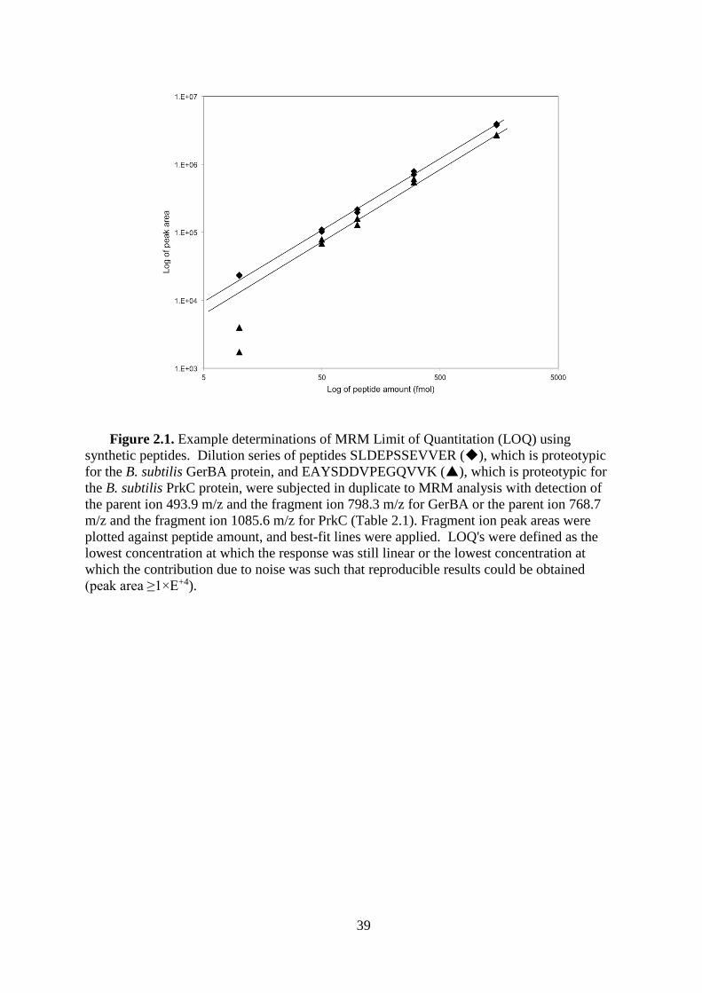
39
Figure 2.1. Example determinations of MRM Limit of Quantitation (LOQ) using
synthetic peptides. Dilution series of peptides SLDEPSSEVVER (), which is proteotypic
for the B. subtilis GerBA protein, and EAYSDDVPEGQVVK (), which is proteotypic for
the B. subtilis PrkC protein, were subjected in duplicate to MRM analysis with detection of
the parent ion 493.9 m/z and the fragment ion 798.3 m/z for GerBA or the parent ion 768.7
m/z and the fragment ion 1085.6 m/z for PrkC (Table 2.1). Fragment ion peak areas were
plotted against peptide amount, and best-fit lines were applied. LOQ's were defined as the
lowest concentration at which the response was still linear or the lowest concentration at
which the contribution due to noise was such that reproducible results could be obtained
(peak area ≥1×E+4).
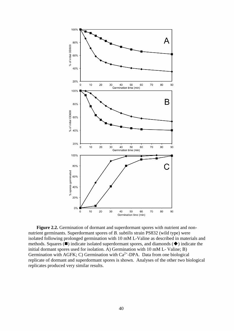
40
Figure 2.2. Germination of dormant and superdormant spores with nutrient and non-
nutrient germinants. Superdormant spores of B. subtilis strain PS832 (wild type) were
isolated following prolonged germination with 10 mM L-Valine as described in materials and
methods. Squares () indicate isolated superdormant spores, and diamonds () indicate the
initial dormant spores used for isolation. A) Germination with 10 mM L- Valine; B)
Germination with AGFK; C) Germination with Ca2+-DPA. Data from one biological
replicate of dormant and superdormant spores is shown. Analyses of the other two biological
replicates produced very similar results.

41
Figure 2.3. Gel electrophoresis of membrane-associated spore proteins. Membrane
preparations were obtained from dormant (D) and germinated (G) spores produced from three
independent spore preparations (1, 2, and 3). Protein concentrations in membrane
preparations were determined by quantitative amino acid analyses, and identical protein
amounts were loaded onto a 9% polyacrylamide gel. Sizes of protein standard markers (M)
are indicated on the left. Proteins were stained using Coomassie blue.
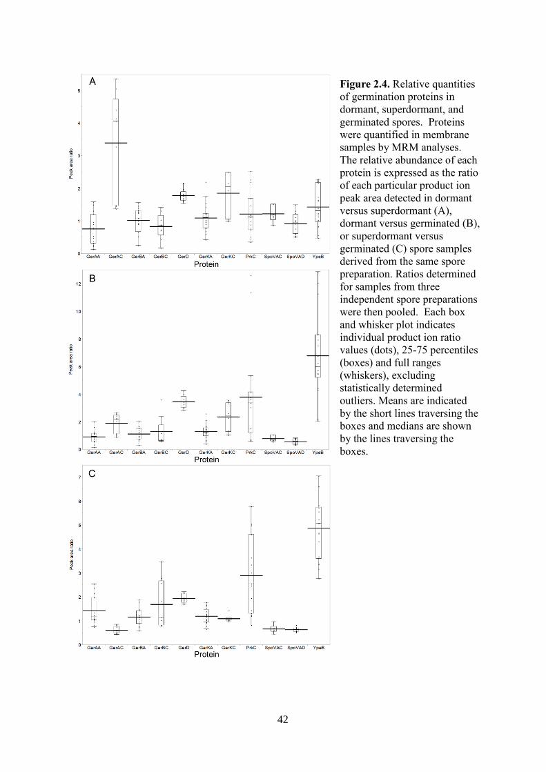
42
Figure 2.4. Relative quantities
of germination proteins in
dormant, superdormant, and
germinated spores. Proteins
were quantified in membrane
samples by MRM analyses.
The relative abundance of each
protein is expressed as the ratio
of each particular product ion
peak area detected in dormant
versus superdormant (A),
dormant versus germinated (B),
or superdormant versus
germinated (C) spore samples
derived from the same spore
preparation. Ratios determined
for samples from three
independent spore preparations
were then pooled. Each box
and whisker plot indicates
individual product ion ratio
values (dots), 25-75 percentiles
(boxes) and full ranges
(whiskers), excluding
statistically determined
outliers. Means are indicated
by the short lines traversing the
boxes and medians are shown
by the lines traversing the
boxes.

43
Table 2.1. Peptide Details for MRM Analysis of the B. subtilis germination proteins.
Protein Proteotypic peptide sequence Parent ion Fragment ion LOQ
(position in protein) m/z (charge
state) m/z (ion)
fmoles (peak
area) c
GerAA
LDQLDARPVETAK (77-89) 486.2 (+3) 672.0 (y12
+2) 10 (3×E+5)
486.2 (+3) 614.5 (y11+2) 10 (3×E+5)
DEETLTLDQVK (64-74) 646.1 (+2) 703.4 (y6+1) 10 (8×E+4)
VSSALFNGR (233-241) 476.3 (+2) 765.3 (y7
+1) 10 (1×E+4)
476.3 (+2) 374.1 (y7-NH3 +2) 10 (1×E+4)
GerAC ADVTGLGNEVR (321-331)
565.8 (+2) 744.3 (y7+1) 50 (1×E+4)
565.8 (+2) 845.5 (y8+1) 100 (1×E+4)
565.8 (+2) 687.4 (y6+1) 50 (1×E+4)
GerBA
TSDPNLVIK (156-164) 493.9 (+2) 683.2 (y6
+1) 10 (2×E+4)
493.9 (+2) 798.3 (y7+1) 10 (2×E+4)
SLDEPSSEVVER (124-135) 674.1 (+2) 902.3 (y8
+1) 10 (4×E+4)
674.1 (+2) 316.2 (y5+2) 10 (3×E+4)
VESSLLEGR (235-243) 495.4 (+2) 761.3 (y7+1) 10 (1×E+4)
GerBC
GILTEDQNPNENSFSK (279-294) 897.3 (+2) 922.4 (y8
+1) 50 (1×E+4)
897.3 (+2) 1036.6 (y9+1) 50 (1×E+4)
GNAADVFTK (135-143) 461.8 (+2) 609.4 (y5
+1) 10 (1×E+4)
461.8 (+2) 680.3 (y6+1) 50 (1×E+4)
GerKA
ERPVLISPSLAK (31-42)
437.4 (+3) 595.3 (b5+1) 10 (2×E+4)
437.4 (+3) 515.2 (y5+1) 10 (2×E+4)
437.4 (+3) 602.1 (y6+1) 50 (1×E+4)
SIQEPSTQVSFR (159-170)
690.2 (+2) 921.7 (y8+1) 10 (1×E+4)
690.2 (+2) 329.0 (b3+1) 50 (1×E+4)
690.2 (+2) 1050.5 (y9+1) 50 (1×E+4)
EVGSSSDVIIR (50-60) 581.5 (+2) 789.5 (y7+1) 10 (1×E+4)
GerKC TLDFTEAQYGR (166-176) 651.0 (+2) 330.2 (b3
+1) 10 (1×E+4)
651.0 (+2) 723.5 (y6+1) 50 (1×E+4)

44
GerD NIFEDTDFAEGFAK (90-103)
802.5 (+2) 1376.6 (y12+1) 100 (1×E+4)
802.5 (+2) 422.1 (y4+1) 300 (1×E+4)
802.5 (+2) 1229.6 (y11+1) 300 (7×E+5)
SpoVAC
SEGLVLGVATNM(ox)FK (109-122) 741.5 (+2) 883.2 (y8
+1) 50 (1×E+4)
741.5 (+2) 996.3 (y9+1) 50 (1×E+4)
SEGLVLGVATNMFK (109-122) 733.6 (+2) 867.3 (y8
+1) 300 (1×E+4)
733.6 (+2) 980.7 (y9+1) 300 (1×E+4)
SpoVAD
ETIPTIAHGVVFER (320-333) 523.9 (+3) 409.3 (y11
+3) 50 (1×E+4)
523.9 (+3) 613.6 (y11+2) 50 (1×E+4)
QLMEDAVNVALQK (57-69) 730.2 (+2) 459.3 (y4
+1) 100 (1×E+4)
730.2 (+2) 672.3 (y6+1) 100 (1×E+4)
YpeB
IGVFSYVPVENK (326-337) 676.7 (+2) 586.1 (y5
+1) 50 (1×E+4)
676.7 (+2) 935.3 (y8+1) 50 (1×E+4)
TIPKPAITEAEAK (372-384) 457.2 (+3) 577.9 (y11
+2) 10 (2×E+5)
457.2 (+3) 929.5 (y9+1) 10 (5×E+4)
VALDDGEVVGFSAR (349-362) 718.2 (+2) 636.3 (y6
+1) 100 (1×E+5)
718.2 (+2) 537.4 (y5+1) 100 (3×E+5)
PrkC
EAASGYLEDNGLK (508-520) 684.3 (+2) 789.3 (y7
+1) 10 (1×E+4)
684.3 (+2) 1096.5 (y10+1) 10 (1×E+4)
EAYSDDVPEGQVVK (525-538) 768.7 (+2) 756.3 (y7
+1) 50 (1×E+4)
768.7 (+2) 1085.6 (y10+1) 50 (1×E+4)
TEIGDVTGQTVDQAK(429-443) 781.8 (+2) 947.6 (y9
+1) 50 (1×E+4)
781.8 (+2) 1218.8 (y12+1) 10 (1×E+4)
a LOQ is the limit of quantitation determined for this fragment ion, as described in Materials and Methods.

45
CHAPTER 3
Membrane Proteomes in Bacillus anthracis and Bacillus subtilis Dormant
and Germinating Spores
Yan Chen, W. Keith Ray, Richard F. Helm, Stephen B. Melville, and David L. Popham
Manuscript in preparation
46
ATTRIBUTIONS
Conceived and designed the experiments: Yan Chen, W. Keith Ray, Richard F. Helm,
Stephen B. Melville, and David L. Popham. Performed the experiments: Yan Chen, and
David L. Popham. Analyzed the data: Yan Chen, Richard F. Helm, and David L. Popham.
Contributed reagents/materials/analysis tools: Yan Chen, W. Keith Ray, Richard F. Helm,
and David L. Popham. Wrote the paper: Yan Chen, W. Keith Ray, Richard F. Helm, Stephen
B. Melville, and David L. Popham.
47
ABSTRACT
Bacterial endospores produced by Bacillus and Clostridium species can remain dormant
and highly resistant to environmental insults for long periods, but can also rapidly germinate
in response to a nutrient rich environment. Multiple proteins involved in sensing and
responding to nutrient germinants, initiating solute and water transport, and accomplishing
spore wall degradation are associated with the membrane surrounding the spore core. To
identify protein changes taking place during germination, as well as to identify unknown
proteins that might be involved in spore germination, a global proteomics approach was
applied to membrane preparations isolated from dormant and germinated spores of Bacillus
anthracis and Bacillus subtilis. Membrane-associated proteins were fractionated by SDS-
PAGE, gel slices were trypsin-digested, and isolated peptides were fractionated by liquid
chromatography and MALDI-TOF-TOF mass spectrometry. Over 500 proteins were
identified from each preparation. Bioinformatic methods were used to characterize proteins
with regard to membrane association, cellular function, and conservation across species.
Numerous proteins not previously known to be spore associated, 35 in B. subtilis and 59 in B.
anthracis, were identified. A significant number of these proteins are implicated in the
transport of metal ions, a process that plays a major role in spore formation and germination.
Spectral counting methods indicated that the majority of spore membrane proteins decrease in
abundance during the first 20 min of germination.
48
INTRODUCTION
Bacterial endospores produced by Bacillus, Clostridium, and related genera can remain
dormant for extended periods of time. In addition, these spores are highly resistant to most
chemical and physical treatments commonly used to reduce bacterial contamination (13).
Upon exposure to conditions conducive to resumption of vegetative growth, generally a
nutrient rich environment, these spores can rapidly germinate and resume metabolism (20,
21). These factors allow certain spore-forming species to act as significant human pathogens
including potential biological weapons (89), as agents of food poisoning and spoilage (114),
and, on a positive note, as effective vehicles for delivery of antigens or metabolic and
enzymatic activities of industrial, consumer, or patient benefit (115-117).
Major factors in spore dormancy and resistance properties are the relative dehydration of
the spore core (cytoplasm) and high core concentrations of solutes such as calcium
dipicolinate (Ca2+-DPA) (13). The dehydrated and metabolically inactive state of the core is
maintained by the spore membrane, which exists in a novel non-fluid state (6), and the
surrounding spore cortex peptidoglycan cell wall (118). Spore germination proceeds through
the rapid release of the core Ca2+-DPA pool, a concomitant uptake of water (20), and the
subsequent return of the spore membrane fluidity (6). This is immediately followed by
degradation of most of the cortex wall (63), allowing full core rehydration, resumption of
metabolic activity, and spore outgrowth. Knowledge of the mechanisms driving spore
germination will allow targeting of this process for the improvement of decontamination
regimens as well as regulating germination during antigen or activity delivery (119).
Genome sequencing and transcriptome profiling have produced predictions of proteins
present within spores (15, 67, 120-122). A number of proteome studies have examined spore
fractions in several species (67, 79-85). Most of these studies were biased towards soluble
49
proteins, as opposed to membrane-associated proteins, although many of the proteins
currently known to function in the spore germination process are associated with the spore
membrane. These include germinant receptor complexes (28, 112), the SpoVA proteins
involved in Ca2+DPA transport (27, 54, 96, 97), the GerD lipoprotein (37, 40), and the YpeB-
SleB proteins involved in cortex degradation (64). Some of these germination-associated
proteins are known to decrease in amount or in their membrane-associations during
germination (37, 64).
The goals of the current study were to catalog the spore membrane proteomes of Bacillus
subtilis and Bacillus anthracis and to examine changes in these proteomes during
germination. Over 500 proteins were identified in each proteome, and approximately 100 of
these were found to contain amino acid sequences predicted to result in integral or peripheral
membrane association. The majority of previously recognized membrane-associated
germination proteins were identified, and over 50 proteins were identified for the first time in
the membrane proteomes of both species. Spectral counting methods for determining protein
abundance changes during germination revealed that the level of a majority of spore
membrane proteins decreased significantly during germination.

50
MATERIALS AND METHODS
Spore sample preparation. Spores of B. anthracis Sterne strain 34F2, an attenuated
vaccine strain, were prepared in Modified G broth (123). Spores of B. subtilis strain PS832, a
prototrophic laboratory derivative of strain 168 were prepared in 2xSG broth (102). Spores
were harvested after 3-4 days incubation at 37°C, washed in water for several days, and
purified by centrifugation through a 50% sodium diatrizoate (Sigma) layer as described
(103). All spores used in this work were 99% free of vegetative cells and were stored in
deionized water at 4°C until analysis.
To prepare germinated spores, a 10-ml suspension of dormant spores at an optical
density at 600 nm (OD600) of 20 in water was heat-activated at 70°C for 30 min (B. anthracis)
or 75°C for 30 min (B. subtilis) and cooled on ice for 10 min. The spores were then
germinated at 37°C and at an OD600 of 2 with 50 mM L-alanine plus 1 mM inosine (B.
anthracis) or 10 mM L-valine (B. subtilis) in 25 mM Tris-HCl buffer (pH 7.4). The
germination of spores was terminated after the OD600 dropped to 50% of the initial value
(within 10 min and 35 min after germinant addition for B. anthracis and B. subtilis,
respectively). Germinated spores were collected by centrifugation at 12,000 g for 5 min at
4°C, quickly washed with cold deionized water, centrifuged again, and frozen at -80°C.
Examination by phase-contrast microscopy indicated that >95% of the spores in each
preparation had germinated.
Preparation of spore membrane fractions. Spore membrane fractions were prepared
by a modification of a previously described method (124). Dormant and germinated spores
were lyophilized, and the dry spores (~19 mg for germinated spores and ~24 mg for dormant
spores) were pulverized with 100 mg of glass beads in a dental amalgamator (Wig-L-Bug) at
4,600 rpm for pulses of 30 s each, with 30 s pauses on ice between pulses. Spore disruption
was monitored by suspending an aliquot of spore material in H2O and observing under phase-
51
contrast microscopy. After >80% of spores were disrupted, the dry powder was suspended in
0.5 ml of 4°C extraction buffer (10 mM Tris-HCl [pH 7.4], 1 mM EDTA, 2 mg/ml RNase A,
2 mg/ml DNase I, 1 mM phenylmethylsulfonyl fluoride (PMSF)). The suspension was
centrifuged (6,000 X g, 10 min, 4°C) and the resultant supernatant was centrifuged again
(13,000 X g, 10 min, 4°C) to remove insoluble material. The remaining supernatant was
centrifuged at 100,000 X g for 60 min at 4°C, and the resulting pellet was designated as the
crude spore membrane fraction. This membrane fraction was homogenized in 1 ml alkaline
buffer (100 mM Na2CO3-HCl [pH 11], 10 mM EDTA, 100 mM NaCl, 1 mM PMSF) and was
gently shaken for 60 min at 4°C. The homogenate was subjected to ultracentrifugation as
described above. The resulting pellet was homogenized in 1 ml high salt buffer (20 mM Tris-
HCl [pH 7.5], 10 mM EDTA, 1 M NaCl, 1 mM PMSF) and was again subjected to
ultracentrifugation. After a final wash with 1 ml TE buffer (10 mM Tris-HCl [pH 7.4], 1 mM
EDTA, 1 mM PMSF), the resulting pellet was homogenized in 200 µl TE buffer, flash frozen,
and stored at -80°C until analysis. Protein concentrations were determined by acid hydrolysis
and amino acid analysis (105) with comparison to a standard set of amino acids (Sigma).
SDS-PAGE, Trypsin digestion, and peptide fractionation. Membrane fractions were
dried and re-suspended in SDS-PAGE sample loading buffer (62.5 mM Tris-HCl pH 6.8, 2%
SDS, 10% glycerol, 5% -mercaptoethanol, 0.05% bromophenol blue) to a final protein
concentration of 2 µg/µl. SDS-PAGE was used to separate 30 µg protein of each spore
membrane sample in both 10% polyacrylamide and 12.5-14% gradient polyacrylamide gels
for B. subtilis samples, and in a 12.5-14% gradient polyacrylamide gel for B. anthracis
samples. Gels were stained with ProtoBlue Safe (National Diagnostics). Each gel lane was
cut into 10-12 slices, consistently for all samples within each species, in an effort to isolate
regions containing many lower abundance proteins away from higher abundance proteins that
could dominate MS profiles. Gel slices were ground and de-stained with 50% LC/MS-grade
52
acetonitrile supplemented with 25 mM NH4HCO3. The gel slices were then dehydrated with
100% acetonitrile and vacuum dried. Gel slices were soaked in 25 mM NH4HCO3 containing
10 µg/ml trypsin (Sigma), and digestion was carried out at 37°C for at least 16 hours. Tryptic
peptides were extracted from gel slices into 50% LC/MS-grade acetonitrile, 0.1%
trifluoroacetic acid (TFA) using a sonication bath. Peptides were vacuum dried and re-
suspended in 40 µl 2% LC/MS-grade acetonitrile, 0.1% TFA.
An Eksigent nano2D-LC unit, flowing at 0.7 µL/min, was used to inject 10 µl of each
peptide sample through a Captrap cartridge (Michrom Bioresources) and a self-packed New
Objective Integrafrit 50 X 0.1 mm column, both packed with Magic C18 AQ (200Å, 3
µm)(Michrom Bioresources). Elution was with 5% acetonitrile for 25 minutes, a 5 min linear
increase to 14% acetonitrile, and a 95 min linear increase to 34% acetonitrile. An Ekspot
plate spotter (Eksigent) was used to spot the eluate onto MALDI target plates at a rate of 10-
15 seconds per spot, dependent on the band intensity and the size of a gel slice, and the spots
were air-dried. Samples from matching gel slices of biological replicates were spotted at the
same rate.
Mass spectrometry and protein identification. Matrix was prepared by suspending
approximately 200 mg αCHCA (Aldrich) in 1 ml 100 mM acetic acid and mixing vigorously.
Following centrifugation for 2 min at 1000 x g, the supernatant was removed, and the wash
procedure was repeated. Two additional washes were performed with 100% acetonitrile, and
the αCHCA was vacuum-dried and stored at 4°C. Matrix solution was prepared by
dissolving 4 mg of washed αCHCA in 1 ml 1:1:0.001(v/v/v) water:acetonitrile:TFA, to which
2 M NH4Cl was then added to a final concentration of 10 mM. Each dried sample spot on the
MALDI plates were overlaid with 1 µl of matrix solution and air-dried.
For calibrating and tuning the MALDI-TOF/TOF 4800 analyzer (AB SCIEX), 200 µl of
the matrix solution was added to an aliquot of a mix of six standard peptides (Anaspec,

53
#60882). This mix was spotted onto all calibration spots when analyzing a sample MALDI
plate. For each peptide sample spot, a scan for the m/z range of 800-4,000 was acquired with
averaged data from 1,000 laser shots in reflector positive ion mode. The 10 most abundant
ions for each spot, above a minimum signal-to-noise ratio (>50), were then automatically
selected for subsequent MS/MS analysis. Parent ions chosen for one spot were excluded from
analyses in subsequent spots. MS/MS scans were the averages of 3,000 laser shots (1 kV,
positive ion mode).
The MS and MS/MS data were analyzed using ProteinPilotTM software version 4.0 (AB
SCIEX) and Scaffold version 3.0 software (Proteome Software, Inc.). The parameters used in
ProteinPilotTM were: identification as sample type, no cysteine alkylation, trypsin as digestion
enzyme, gel-based identification as special factors, biological modifications and amino acid
substitutions as ID focus, thorough ID as search effort, and detected protein threshold
(unused protein score) > 0.05 (10%) as result quality. The B. subtilis 168 protein database
(2/10/2012) and B. anthracis (BACAN) protein database (12/18/2012) were downloaded
from Universal Protein Resources (http://www.uniprot.org/). Based on the ProteinPilotTM
false discovery rate for analysis of acquired protein list, 5% false discovery rate (FDR) and a
minimum of one peptide at 95% identification confidence were applied to each biological
sample protein profile. The raw data is available from the PRIDE database
(http://www.ebi.ac.uk/pride/archive/) (In process). Proteins identified in at least two
biological replicates were considered a valid identification. For membrane proteins that only
produced one peptide with these parameters, confirmation of the identification was performed
by manual interpretation of their MS/MS spectra. All proteins in dormant and germinated
protein profiles were then annotated according to their accession number using two
databases: the National Center for Biological Information (http://www.ncbi.nlm.nih.gov/) and
Universal Protein Resources. Other online resources that were used for predicting membrane

54
associations are: Brinkman Laboratory at Simon Fraser University
(http://www.psort.org/psortb/index.html) and PRED-LIPO server at University of Athens
(http://bioinformatics.biol.uoa.gr/PRED-LIPO/input.jsp). Protein NCBI Cluster of
Orthologous Groups (COG) were predicted using the COMBREX database
(http://combrex.bu.edu/).
To acquire preliminary relative protein quantification, the Mascot data files of three
biological replicates were merged and onsite Mascot searches were performed for each
peptide fragmentation mass spectrum against the B. subtilis and B. anthracis protein
databases. Mascot parameters were: Trypsin specificity with one missed cleavage,
carbamidomethylation was a fixed modification, and deamidation, pyro-cmC, and oxidations
were considered variable modifications. The peptide mass tolerance and fragment mass
tolerance were set to ±500 ppm and ±0.2 Da, respectively. Search results were analyzed using
ScaffoldTM version 3.4.9. The ScaffoldTM parameters were 95% protein threshold and 1
minimum identified peptide at 95% of threshold, and normalization was selected for
quantitative analysis. The unique spectrum count of a protein was used to relatively quantify
the protein changes between two samples (125).
All identified B. anthracis membrane proteins were searched against the whole B.
subtilis protein database and the proteins identified in B. subtilis spore membrane profile
using the NCBI protein BLAST tool (126). Only sequences with alignment coverage ≥60%
were considered possible orthologs in the two species. If the protein in the full B. subtilis
protein database that had the highest identity percentage against a B. anthracis membrane
query protein was also identified in the B. subtilis spore membrane profile, these two proteins
were considered to be the most likely orthologs. If a protein in B. subtilis spore membrane
proteome had high similarity to a B. anthracis membrane query protein, but it was not the

55
highest identity match in the full B. subtilis protein database, then the two proteins were
considered to be likely paralogs.
56
RESULTS
Proteins identified in dormant spore membrane preparations. Membranes were
isolated from three independent preparations of B. anthracis and B. subtilis dormant spores.
Proteins were separated by SDS-PAGE (Figure 3.1) followed by gel slice preparation and
processing to provide peptides that were submitted to LC-MALDI separation and analysis. A
total of 603 and 592 proteins were identified in B. subtilis and in B. anthracis samples,
respectively. Bioinformatic predictions suggested that 104 (17%) and 87 (15%) of these
proteins were membrane-associated proteins in B. subtilis and B. anthracis, respectively
(Table 3.1). Predicted membrane proteins were further categorized based on their mechanism
of membrane association and number of predicted membrane-spanning helices (Figure 3.2).
The presence of a wide variety of lipoproteins, peripheral (monotopic), and integral polytopic
membrane proteins indicates that the method used was successful in recovering a broad
membrane proteome. A total of 11 known germination-related membrane proteins were
identified in B. subtilis samples (Table 3.2). Similarly, a total of 5 known germination related
proteins were identified in B. anthracis samples (Table 3.2). The remaining identified
membrane proteins have predicted functions in 14 COG function categories.
Proteins identified in germinated spore membrane preparations. A portion of each
spore preparation was exposed to nutrient germinants until the optical density of the
suspension had decreased 50%, which resulted in germination of ≥95% of the spores. The
membrane fractions were then isolated, and the proteins were separated by SDS-PAGE (Fig.
3.1). Collection and processing of gel slices provided samples for LC-MALDI separation
and analysis. A total of 497 and 508 proteins were identified in B. subtilis and B. anthracis
germinated spore membrane profiles, respectively. In each species, some proteins (82 and 88
proteins in B. subtilis and B. anthracis, respectively) were identified only in the germinated
samples and not in the dormant spore samples, and these were predominantly predicted to be
57
cytoplasmic proteins. Of the 52 (10.5%) and 38 (7.5%) proteins that were predicted to be
membrane-associated in B. subtilis and B. anthracis, respectively, all but two in B. subtilis
(P40780 and Q01464) were also present in the dormant spore samples.
Novel membrane proteins identified in spore membrane fractions. The spore
membranes are derived from the sporangium cytoplasmic membranes during the engulfment
stage of sporulation, so, it was no surprise to find that membrane proteins expected to be in
vegetative cells were present in spore membrane fractions. Among the predicted membrane
proteins we detected in dormant or germinated spores, 57 out of 104 B. subtilis proteins had
been identified in previous vegetative proteome studies (Table 3.1). An additional three
proteins, YhcM, YodI, and YlaJ were reported in previous proteome studies of B. subtilis
spores (79), but were not included in vegetative cell proteome studies. The genes that encode
these three proteins are transcribed under control of sporulation sigma factors ϭF, ϭK, and ϭG,
respectively (79, 121), and are therefore likely spore-specific proteins. GerAC, GerBC,
GerKC, GerD, SpoVAD, and YpeB are known germination-active proteins that were
previously detected in spores (64, 99). An additional five known germination-active proteins:
PrkC, SpoVAC, SpoVAF, YfkR, and YhcN were assumed to be present in spores, but had
not been previously detected or had been detected only in a previous vegetative cell proteome
study (77). Among the remaining 35 spore membrane proteins identified here, 13 have
unknown functions and others have predicted COG functions in inorganic ion transport and
metabolism (6); amino acid transport and metabolism (4); posttranslational modification,
protein turnover, chaperones (2); lipid biogenesis and metabolism (2); intracellular
trafficking, secretion, and vesicular transport (2); cell wall biogenesis (2); general transport
(1); signal transduction (1); cell cycle control (1); and energy production and conversion (1).
A similar analysis of the B. anthracis samples revealed that 15 predicted membrane
proteins were reported previously in a vegetative cell proteome study (78), and 13 proteins
58
were reported previously in spore proteome studies (82, 83). Among the remaining 59 spore
membrane proteins identified here, 16 are presently listed with unknown functions, 5 are
known germination-active proteins (Table 3.2), and the rest sort into 12 different COG
categories.
Membrane proteins under control of sporulation-specific sigma factors. Predicted
membrane-associated proteins identified in B. subtilis samples were searched against the
relatively well-characterized sporulation transcriptomes of that species (15, 79, 122, 127). Of
the 104 predicted membrane-associated proteins, 29 have been shown to be controlled at the
transcriptional level by the sporulation-specific sigma factors ϭE, ϭF, ϭG, and ϭK (Table 3.1).
Only a small number of membrane proteins were identified from the mother cell-specific σE
and ϭK regulons, 4 and 2 proteins, respectively. The remaining membrane proteins for which
transcriptome data is available were in the forespore-specific ϭF (4 proteins) and ϭG (19
proteins) regulons, though the similarity between these two sigma factors results in some
overlap of their regulons (15). The genes of 10 known germination-active proteins identified
are all within the ϭG regulon. Eight ϭG-dependent proteins had not been previously detected
in spore proteomes. Among these, YutC, YhcC, YrbG, and YqfX have unknown functions,
while YveA, YwjE, YitG, and YthA were categorized into amino acid transport and
metabolism, lipid biogenesis and metabolism, general transporter, and energy production and
conversion, respectively.
Similarities between the spore membrane proteomes in the two Bacillus species.
Searches using the BLAST program were done to compare the predicted membrane proteins
identified in samples from each species. Fifty-one B. anthracis spore membrane proteins
showed high similarity to 48 spore membrane proteins identified in B. subtilis (Table 3.3).
Forty-five of these proteins were considered to be orthologous, based on A) a high level of
amino acid sequence identity (>23%) and similarity (>42%) across an alignment of >62% of
59
the protein sequences and B) synteny. Among these, GerD, MisCA, SpoVAC, SpoVAD,
SpoVAF, YetF, YpeB, YthA, and YutC have been previously identified in spores under more
defined studies and/or were expressed in the forespore-specific ϭF and ϭG regulons. Several of
these are known germination-active proteins. MisCA (SpoIIIJ) is required for activation of ϭG
after engulfment of the forespore is completed (128). YthA was suggested to compensate for
the loss of cytochrome aa3, which is critical for sporulation as one of two heme copper
terminal oxidases in B. subtilis (129). There is no known function reported for YutC and
YetF. This leaves eight proteins that were detected in both species and had not been
previously identified in spore proteomes or sporulation-specific regulons. ArtP, MetQ, and
AdcA are components of ABC transporters involved in amino acid or inorganic ion transport.
YpmQ is involved in assembly of a copper center in cytochrome c oxidase, which has an
important function in energy conversion and metabolism (130). YyxA was predicted to be a
serine protease due to its sequence similarity to the S1B peptidase family. YndM, YugS and
YkrK are proteins of unknown function.
Three B. subtilis membrane proteins, YhcC, YveA, and YitG, that are expressed in the ϭG
regulon (15, 127) and do not have similar proteins in the B. anthracis protein database, were
detected. Three other ϭG regulon proteins, YhbJ, YrbG, and YwjE (127), have highly similar
proteins in B. anthracis (BAS4465, BAS4309, and BAS5195, respectively) but were only
detected in B. subtilis.
Membrane protein changes during spore germination. Spectral counting methods
(125, 131) were applied to determine protein abundance information before and after spore
germination. To test the validity of this method, we compared the ratio changes of proteins in
this data to more precise, previously published quantification data for several germination
proteins (124) (Table 3.4). The trends in terms of proteins increasing, decreasing, or not
changing in membrane association during germination were generally consistent in the two
60
data sets with only one protein, SpoVAC, exhibiting an inverse trend in the two studies. The
decreased abundance of B. anthracis proteins BAS2560, BAS1302, and BAS4323 after spore
germination was also consistent with a previous whole spore quantitative proteomic study
(83). On the other hand, our results showed that BAS0812 and BAS3922 decreased 5.9- and
3.7-fold respectively, and BAS0405 increased 2-fold after spore germination, but these
proteins showed no change in quantity in the previous study (83). Preliminary protein
quantity changes during germination were determined for 99 B. subtilis and 83 B. anthracis
spore membrane proteins. Strikingly, most of the membrane proteins (>70%) in both species
were either not detected (Table 3.5) or greatly reduced in quantity after spore germination (73
B. subtilis proteins and 65 B. anthracis proteins) (Table 3.6). In B. subtilis samples, two
membrane proteins, YtxH and MinD, were present in germinated spore samples that were not
detected in the dormant spore samples.
61
DISCUSSION
Proteins associated with the membranes separating the mother cell and forespore play
crucial roles in communication between the cells during sporulation, allowing the two cells to
coordinate the timing of gene expression changes and morphological development.
Membrane proteins localized at the inner spore membrane have been demonstrated to play
key roles in multiple stages of spore germination. In addition, germination processes such as
ion efflux and water influx are likely to involve membrane proteins, yet few proteins
involved in these processes have been identified. The goal of this study was to compare and
contrast spore membrane-associated proteins present in two Bacillus species in an effort to
further our understanding of cell differentiation processes at the molecular level and to
identify new germination-active proteins. A gel-based approach was used to provide a set of
integral membrane proteins, lipoproteins, and peripheral membrane proteins. This study
identified 65 membrane-associated spore proteins that had not been previously reported in
any B. subtilis or B. anthracis vegetative or spore proteomic study. The percentages of
proteins identified that are predicted to be membrane associated were high, 15-17% of all
proteins identified, which is a significant improvement over previous spore proteomic studies
(<5%) (79, 82, 83). Identification of these proteins provides potential novel pathways to
better understand the role of membrane proteins during sporulation and germination.
The functions of identified spore membrane proteins in both Bacillus species spread into
almost all membrane protein functional categories. Aside from those proteins known to
function in sporulation or germination, the top three functional categories for identified spore
membrane proteins were transport (carbohydrate, amino acid, and inorganic ions), protein
fate determination (posttranslational modification, trafficking, and turnover) and energy
production and conversion.
62
Cations such as Ca2+, Mn2+, and Zn2+ accumulate in spores the during the sporulation of
Bacillus species (132). Divalent metal ions, predominantly Ca2+, conjugated with DPA
contribute to spore resistance to heat (50, 133) and ionizing radiation (134). The rapid release
of cations is a distinct early germination step (135). Nine newly-identified spore membrane
proteins have predicted functions involved in inorganic ion transport and metabolism. These
proteins may play roles in accumulation of ions during sporulation and/or the rapid release of
ions during germination. Zn2+ ions are accumulated during sporulation and are released very
early during germination (136). AdcA was suggested to be important for zinc transport in B.
subtilis vegetative cells (137), and this protein and its apparent B. anthracis ortholog were
present in the spore membranes. YfkE was previously demonstrated to be a Ca2+/H+
antiporter expressed under the control of forespore-specific ϭG (138) and was detected in the
spore membrane. This protein could be involved in Ca2+ accumulation during sporulation
and/or Ca2+ release during germination. YcnL, YflS, and YjbQ were present in the B. subtilis
spore membrane, and may be involved in copper, malate, potassium, and sodium transport
(139, 140). BAS4953, BAS0564 and BAS0368 are three novel B. anthracis spore membrane
proteins that are predicted to be involved in iron and other heavy metal cation transport. B.
subtilis YugS and the homologous B. anthracis protein BAS0575 belong to the
uncharacterized protein family 0053 (UPF0053). Several proteins from other bacterial species
in this family have been suggested to function in the transport of magnesium and cobalt ions
(141). Further characterization of these proteins is needed to determine their functions and
potential roles during sporulation and germination.
Several proteins involved in amino acid transport were identified in the spore
membranes. YveA was previously characterized to be the primary L-aspartate transporter in
B. subtilis vegetative cells (142), and transcriptome analysis showed that this gene was
expressed under control of the forespore-specific ϭG (15). MetQ and ArtP were previously
63
characterized in B. subtilis vegetative cells as involved in methionine and arginine transport,
respectively (143, 144). These proteins, as well as their apparent B. anthracis orthologs, were
present in the spore membranes. The roles of these amino acid transporters in either
sporulation or germination processes are unknown at present.
Several proteins detected in spore membranes are involved in protein degradation. B.
subtilis HtrC (YyxA, YycK), a predicted Htr-type serine protease, was suggested to be
transcribed from its own ϭG promoter (145). Loss of HtrC produced no obvious phenotypic
change in B. subtilis vegetative cells (145, 146), however, the presence of HtrC, and its
apparent B. anthracis ortholog, which is also expressed during sporulation (67), in the spore
membrane suggests that it may play a role in spore formation or germination. B. subtilis
HtpX and B. anthracis RasP are two predicted zinc proteases detected in the spore
membranes. Their functions have not been characterized, but may be involved in membrane
protein quality control based on the function of the well-known homolog FtsH (147). Further
characterization of the roles these proteases play in the spore may shed light on the observed
decrease in membrane proteins during spore germination.
In both Bacillus species, one quarter of the identified spore membrane proteins have no
assigned COG functional category and most have no significant sequence similarity to known
genes or functional domains. More interestingly, most of these proteins were not identified in
vegetative cell membranes previously, and some were present in both Bacillus spores,
suggesting that these proteins are likely expressed for particular purposes in spores. Further
characterization of these proteins may reveal roles in the sporulation, germination, or
outgrowth processes. For example, B. subtilis YetF and YutC were previously characterized
to be expressed under control of spore-specific ϭF and ϭG, respectively (127), and their
apparent B. anthracis orthologs were also identified in this study. Five other B. subtilis
proteins together with YetF are grouped in an uncharacterized protein family (UPF0702), in
64
which a ϭG-regulated protein YrbG (127) was also identified in B. subtilis spore membrane.
Homologs of these proteins with no known function are not found in other bacteria,
indicating that they might be very specific for Bacillus species. Considering their expression
is under the control of forespore sigma factors, these proteins may play key roles in either
spore formation, stabilization, and/or germination processes.
To provide preliminary information about the fate of spore membrane proteins during
germination, relative quantities were derived using label-free spectral counting methods. This
strategy is based on the empirical observation that the amount of unique MS/MS spectrum
counts of a protein has strong linear correlation with relative protein abundance (125, 131).
Although this method is not as accurate as targeted MRM-based methods, it has higher
dynamic range than other mass spectrometry quantification methods (148) and therefore is a
popular method to detect global protein changes between experiments groups. Note that this
relative quantification method was established using data generated by electrospray
ionization mass spectrometry. The limitations of applying this method on data generated by
MALDI mass spectrometry have not been previously examined. However, the relative
quantity values for multiple proteins acquired in this work were consistent to those acquired
by our previous MRM study (124) and a previous stable isotope labeled quantitative study
(83), suggesting that this method is applicable to MALDI-generated data. Results published
while this work was in progress indicate that membrane fractions produced by methods such
as those used here recover only a portion of the spore membrane proteins, and extensive
chemical extraction is required to recover the full amount of several spore membrane proteins
(109). However, such a chemical extraction precludes the separation of membrane and
soluble proteins. In the absence of a spore membrane extraction process that provides high
recovery, protein quantitation will have to be done using whole spore extracts or will require
evidence that the partial membrane extract is representative of the entire membrane.

65
A notable trend in our study is that the number of identified membrane proteins is greatly
reduced after spore germination. Several membrane proteins, mostly lipoproteins, have been
previously suggested to dissociate from the membrane during spore germination (37, 83,
124). Spectral counting showed similar results with most lipoproteins diminished or
undetectable following germination. The decreased recovery of lipoproteins may be due to
proteolysis or may indicate a change in ability to maintain membrane association during
fractionation, which in turn could be associated with the membrane reorganization processes.
Surprisingly, over half of the membrane proteins that decreased to below detectable levels
during germination of both Bacillus species spores were integral membrane proteins.
Although previous efforts to quantify spore integral membrane proteins are rare, decreased
levels of BAS1302 after spore germination was consistent with previous work (83). Since the
membrane proteins identified that were observed to decrease are involved in multiple COG
function categories, this decrease appears to be a general spore germination phenomenon.
The shift from a relatively nonfluid impermeable membrane to a fluid membrane with solute
transfer may require a rather complex rearrangement of the membrane and membrane-
associated proteome. Targeted knockout studies involving these proteins and their associated
germination phenotypes may lend insight into this rearrangement process.

66
ACKNOWLEDGEMENTS
Research reported in this publication was supported by the National Institute of Allergy
and Infectious Disease of the National Institutes of Health under award number
R21AI088298. The mass spectrometry resources used in this work are maintained in part
through funding by the Fralin Life Science Institute at Virginia Tech and the Agricultural
Experiment Station Hatch Program at Virginia Tech (CRIS Project Number: VA-135981)
67
Table 3.1. B. subtilis and B. anthracis spore membrane-associated proteins identified by mass spectrometry.
Genea COG Functional category
B. subtilis
Uniprot
number
B. anthracis
Uniprot
number
Membrane
predictionb
Previous
IDc Regulond
adcA/BAS188
9 Ion transport & metab. O34966 Q81RK9 Lipoprotein
ahpF Posttranslational
modification P42974 Q81ZC5 Peripheral Y2
artP/BAS0353 Amino acid transport &
metab. P54535 Q6I447 Lipoprotein
atpD Energy production P37809 Q81JZ5 Peripheral Y1/Y4
atpF Energy production P37814 Q81JZ1 Integral Y1
atpG Energy production P37810 Q81JZ4 Peripheral Y1
bdbD Posttranslational
modification O32218 Q81YT8 Integral Y1 ϭE
era Unknown P42182 Q81LT7 Peripheral Y6
fhuD/BAS0336 Ion transport & metab. P37580 Q81ZB8 Integral Y1/Y5
ftsH Posttranslational
modification P37476 Q81VX5 Integral Y1/Y5
gerD Germination P16450 Q81VP4 Lipoprotein ϭG
lytR Transcription Q02115 Q81K33 Integral Y1
metQ/BAS485
3
Amino acid transport &
metab. O32167 Q81XL5 Lipoprotein
misCA/yidC2 Intracellular trafficking Q01625 Q81JH1 Integral
msmX/PotA Amino acid transport &
metab. P94360 Q81TH8 Peripheral Y2/Y5
oppA/BAS062
3
Amino acid transport &
metab. P24141 Q81V45 Lipoprotein Y1/Y5
oppB/BAS110
2 General Transporter P24138 Q81TS3 Integral Y2/Y5
oppC/BAS110
3 General Transporter P24139 Q81TS2 Integral Y2
oppD/BAS110
4 General Transporter P24136 Q81TS1 Peripheral Y2/Y5
oppF/BAS110
5
Amino acid transport &
metab. P24137 Q81TS0 Peripheral Y2
pbpF/BAS136
3 Cell wall biogenesis P38050 Q81T17 Integral Y2/Y5
plsY/plsY3 Lipid metabolism Q45064 Q81Y92 Integral
ponA/BAS218
5 Cell wall biogenesis P39793 Q6HYZ2 Integral Y1
prsA/prsA1 Posttranslational
modification P24327 Q81U45 Lipoprotein Y1
68
ptsG/ptsG Carbohydrate transp. &
metab. P20166 Q81MH9 Integral Y1
qcrA Energy production P46911 Q81SV1 Integral Y2
qcrB Energy production P46912 Q81SV0 Integral Y2
qoxA/ctaC Energy production P34957 Q81MT9 Integral Y1
resA Posttranslational
modification P35160 Q81SZ9 Integral Y2
secDF Intracellular trafficking O32047 Q81LH8 Integral Y1/Y5
secY/secY1 Intracellular trafficking P16336 Q81VR0 Integral Y1
spoVAC DPA transport P40868 Q81X68 Integral ϭG
spoVAD DPA transport P40869 Q81X67 Peripheral ϭG
spoVAF DPA transport P31845 Q81MG2 Integral ϭG
tcyA/
BAS0812
Amino acid transport &
metab. P42199 Q81UL3 Lipoprotein Y6
yetF/BAS4999 Unknown O31533 Q81X64 Integral ϭF
yfmC/BAS442
4 Ion transport & metab. O34348 Q81L65 Lipoprotein Y1/Y5
ykrK/BAS2651 Unknown O31656 Q81PG5 Peripheral
ylaJ/BAS2560 Unknown O07634 Q81PQ5 Lipoprotein Y3/Y6 ϭG
yndM/BAS290
9 Unknown O31816 Q6HWX1* Integral *Same
protein as
Q81NQ1.
Maybe
ϭG?
ypeB Germination P38490 Q81PQ4 Integral ϭG
ypmQ/BAS209
3 Energy production P54178 Q81R11 Lipoprotein
yqfX/BAS1302 Unknown P54481 Q81T77 Integral Y6 ϭG
ythA/cydA3 Energy production C0SP90 Q81WZ0 Integral ϭG
yugS/BAS0575 Unknown O05241 Q81V91 Integral
yutC/BAS4834 Unknown O32128 Q81XN4 Lipoprotein ϭG
yyxA/BAS5314 Posttranslational
modification P39668 Q81JJ5 Integral
artQ Amino acid transport &
metab. P54536 Integral
atpB Energy production P37813 Integral Y1
dacA Cell wall biogenesis P08750 Integral Y1
dltD Cell wall biogenesis P39578 Integral Y1
ezrA Cell cycle control O34894 Integral Y2
fruA Carbohydrate transp. &
metab. P71012 Integral Y1
69
ftsY Intracellular trafficking P51835 Peripheral Y2 ϭK
gerAC Germinations P07870 Lipoprotein ϭG
gerBC Germination P39571 Lipoprotein ϭG
gerKC Germination P49941 Lipoprotein ϭG
htpX Posttranslational
modification O31657 Integral
lgt Posttranslational
modification O34752 Integral Y2
murG Cell wall biogenesis P37585 Peripheral ϭE
opuAB Amino acid transport &
metab. P46921 Integral Y2
opuAC Amino acid transport &
metab. P46922 Lipoprotein Y1
pbuG General transporter O34987 Integral Y1
phoR Signal transduction P23545 Integral ϭE
prkC Germination O34507 Integral Y2
rbsA Carbohydrate transp. &
metab. P36947 Peripheral Y1
sipS Intracellular trafficking P28628 Integral Y2 ϭE
sipT Intracellular trafficking P71013 Integral Y2
sppA Intracellular trafficking O34525 Integral Y2
tagG Cell wall biogenesis P42953 Integral Y2
yacD Posttranslational
modification P37566 Integral Y1
yckB Amino acid transport &
metab. P42400 Lipoprotein Y1
ycnL Ion transport & metab. P94434 Integral
yerB Unknown O34968 Lipoprotein
yerH Unknown O34629 Lipoprotein Y1
yfkE Ion transport & metab. O34840 Integral
yfkR Germination O35028 Lipoprotein ϭG
yflS Ion transport & metab. O34726 Integral
yhbJ Defense mechanisms O31593 Integral Y2 ϭG
yhcC Unknown P54587 Integral ϭG
yhcM Unknown P54597 Integral Y3 ϭF
yhcN Germination P54598 Lipoprotein Y2 ϭF & ϭG
70
yheB Unknown O07543 Integral Y1
yhfQ Ion transport & metab. C0SP94 Lipoprotein Y1
yitG General transporter Q796Q1 Integral ϭG
yjbQ Ion transport & metab. O31615 Integral
ylbC Unknown O34586 Integral Y2 ϭF
ylbL Signal transduction O34470 Integral Y1
yodI Unknown O34654 Integral Y3 ϭK
yqaR Unknown P45914 Integral
yqfA Unknown P54466 Integral Y1
yqgF Cell wall biogenesis P54488 Integral
yrbF Intracellular trafficking O32052 Integral
yrbG Unknown O32050 Integral ϭG
yrvD Unknown O32045 Integral Y2
ysdB Transcription P94520 Integral Y2
ytxG Unknown P40779 Integral Y2
ytxH Unknown P40780 Integral Y2
yuaB Colony structure P71014 Integral
yuaG Unknown O32076 Integral Y2
yugP Posttranslational
modification O05248 Integral Y2
yvcA Colony structure O06965 Lipoprotein Y2
yveA Amino acid transport &
metab. O07002 Integral ϭG
ywjE Lipid metabolism P45865 Integral ϭG
BAS0191 Unknown Q81VK7 Lipoprotein Y6
BAS0350 Energy production Q81ZA6 Peripheral
BAS0360 Unknown Q81Z98 Integral
BAS0368 Ion transport & metab. Q81Z90 Lipoprotein
BAS0405 Unknown Q81Z55 Peripheral Y6
BAS0564 Ion transport & metab. Q81VA3 Integral
BAS0569 Unknown Q6I3I4 Lipoprotein
71
BAS0581 Ion transport & metab. Q81V85 Lipoprotein Y5
BAS1412 Unknown Q81SX1 Integral
BAS2689 Unknown Q81PC7 Lipoprotein
BAS2733 Unknown Q81P74 Lipoprotein Y6
BAS2751 Unknown Q81P56 Integral
BAS2834 Defense mechanisms Q81NX4 Peripheral Y5
BAS2910 Unknown Q81NQ0 Peripheral Y4
BAS3134 Energy production Q81N37 Peripheral
BAS3468 Unknown Q81Y17 Integral
BAS3643 Unknown Q81WP4 Lipoprotein
BAS3922 Carbohydrate transp. &
metab. Q81ML8 Lipoprotein Y6
BAS3998 Unknown Q81MD9 Lipoprotein
BAS4323 Unknown Q6HSW8 Lipoprotein Y6
BAS4406 Unknown Q81L82 Lipoprotein
BAS4700 Unknown Q81KD9 Peripheral Y4
BAS4796 Energy production Q81XS1 Peripheral
BAS4797 Posttranslational
modification Q81XS0 Peripheral
BAS4808 Energy production Q81XR0 Peripheral
BAS4865 Unknown Q81XK0 Lipoprotein
BAS4935 Energy production Q81XC7 Peripheral
BAS4953 Ion transport & metab. Q6HR45 Lipoprotein
BAS4994 Unknown Q6HR05 Lipoprotein
BAS5287 Cell wall biogenesis Q81JM0 Integral
cccA Energy production Q81LU6 Integral
ecsA Defense mechanisms Q81U40 Peripheral
fabF Lipid metabolism Q81JF9 Peripheral
ffh Intracellular trafficking Q81WJ2 Peripheral
ftsY Intracellular trafficking Q81WJ0 Peripheral
potD Amino acid transport &
metab. Q81TH5 Lipoprotein
72
proV2 Amino acid transport &
metab. Q81PL6 Peripheral
psd Lipid metabolism Q81LP7 Peripheral
rasP Cell wall/membrane
biogenesis Q81WL5 Integral
typA Signal transduction Q81MS8 Peripheral
a If a protein doesn’t have an annotated gene name, the ordered locus name is listed.
b Mechanism of membrane association predicted as described in Materials and Methods.
c Previous identification of the protein in a proteomic analysis: Y1, B. subtilis vegetative cell proteome (76); Y2: B. subtilis vegetative cell
proteome (77); Y3: B. subtilis spore proteome (79); Y4: B. anthracis spore proteome (82); Y5: B. anthracis vegetative cell proteome (78); Y6: B.
anthracis spore proteome (83).
d Sigma factor dependence was drawn from references: (15, 79, 122, 127).

73
Table 3.2. Known B. anthracis and B. subtilis spore germination proteins that were
identified.
Gene Function
B. subtilis
Uniprot
number
B. anthracis
Uniprot
number
Membrane
predictiona
gerAC Germinant receptor P07870 Lipoprotein
gerBC Germinant receptor P39571 Lipoprotein
gerD Germinant response P16450 Q81VP4 Lipoprotein
gerKC Germinant receptor P49941 Lipoprotein
prkC Peptidoglycan
receptor O34507 Integral
spoVAC DPA transport P40868 Q81X68 Integral
spoVAD DPA transport P40869 Q81X67 Peripheral
spoVAF DPA transport P31845 Q81MG2 Integral
yfkR Germinant receptor O35028 Lipoprotein
yhcN Outgrowth P54598 Lipoprotein
ypeB Cortex degradation P38490 Q81PQ4 Integral
a Mechanism of membrane association predicted as described in Materials and Methods.

74
Table 3.3. Proteins detected in both Bacillus species spore membrane proteomes
B. subtilis
Uniprot
number
B. anthracis
Uniprot
number
Protein function
Sequence
identity1
(%)
Sequence
similarity1 (%)
Alignment
length2 (%)
C0SP90 Q81WZ0 Cytochrome bd menaquinol
oxidase 53 71 98
O05241 Q81V91 Unknown 59 75 100
O07634 Q81PQ5 Unknown , lipoprotein 47 67 100
O31533 Q81X64 Unknown 35 59 63
O31656 Q81PG5 Unknown 37 59 94
O31816 Q81P56 Unknown 27 47 92
O31816 Q6HWX1 Unknown 35 50 88
O32047 Q81LH8 Protein translocase, SecDF 60 76 97
O32128 Q81XN4 Unknown, lipoprotein 35 58 72
O32167 Q81XL5 Methionine-binding lipoprotein,
MetQ 57 76 100
O32218 Q81YT8 Disulfide bond formation 42 64 85
O34348 Q6HR453 Unknown 25 46 93
O34348 Q81V853 Unknown 34 49 99
O34348 Q81L65 Fe-citrate-binding, YfmC 33 53 96
O34966 Q81RK9 Zinc uptake, ZnuA 37 55 97
P16336 Q81VR0 Protein translocase, SecY 72 82 100
P16450 Q81VP4 Germination, GerD 43 69 89
P20166 Q81MH9 PTS system 61 79 100
P24136 Q81TS1 Oligopeptide transport, OppD 73 85 97
P24137 Q81TS0 Oligopeptide transport, OppF 80 89 97
P24138 Q81TS3 Oligopeptide transport, OppB 50 70 100
P24139 Q81TS2 Oligopeptide transport, OppC 46 66 98

75
P24141 Q81V45 Oligopeptide transport, OppA 28 45 92
P24327 Q81U45 Foldase, PrsA 47 66 96
P31845 Q81MG2 DPA transport SpoVAF 63 80 96
P34957 Q81MT93 Quinol oxidase 36 56 62
P35160 Q81SZ9 Thiol-disulfide oxidoreductase,
ResA 52 69 99
P36947 Q81U403 Ribose import, RbsA 28 49 84
P37476 Q81VX5 Zinc metalloprotease, FtsH 80 89 100
P37580 Q81ZB8 Iron-hydroxamate-binding, FhuD 38 61 93
P37809 Q81JZ5 ATP synthase 86 92 99
P37810 Q81JZ4 ATP synthase 68 84 100
P37814 Q81JZ1 ATP synthase 57 76 98
P38050 Q81T17 Penicillin-binding protein 1F 42 61 89
P38490 Q81PQ4 Cortex degradation, YpeB 57 77 99
P39668 Q81JJ5 Serine protease, YyxA 50 68 97
P39793 Q6HYZ2 Penicillin-binding protein 1 46 65 89
P40868 Q81X68 DPA transport, SpoVAC 60 78 89
P40869 Q81X67 DPA transport, SpoVAD 50 65 98
P42182 Q81LT7 GTPase Era 75 89 100
P42199 Q81UL3 L-cystine-binding protein TcyA 53 69 98
P42974 Q81ZC5 NADH dehydrogenase 84 91 100
P46911 Q81SV1 Menaquinol-cytochrome c
reductase 70 80 95
P46912 Q81SV0 Menaquinol-cytochrome c
reductase 95 97 100
P54178 Q81R11 SCO1 protein homolog 49 70 96
P54481 Q81T77 Unknown 33 48 92
P54535 Q6I447 Arginine-transport, ArtP 37 55 95
P94360 Q81TH83 Maltodextrin import, MsmX 51 71 76
76
Q01625 Q81JH1 Membrane protein insertase,
MisCA 68 81 100
Q02115 Q81K33 Transcriptional regulator, LytR 50 72 93
Q45064 Q81Y92 Glycerol-3-PO4 acyltransferase 63 75 94
1 Sequences were aligned using BlastP (126).
2 The percent of the B. subtilis protein sequence that was aligned with the B. anthracis sequence.
3 A more similar protein than that detected in the spore proteome was present in the full B. subtilis genome: (B. anthracis gene:Most similar B.
subtilis gene) (Q6HR45:P94421) (Q81V85:O31567) (Q81MT9:P24011) (Q81U40:P55339) (Q81TH8:O32151)
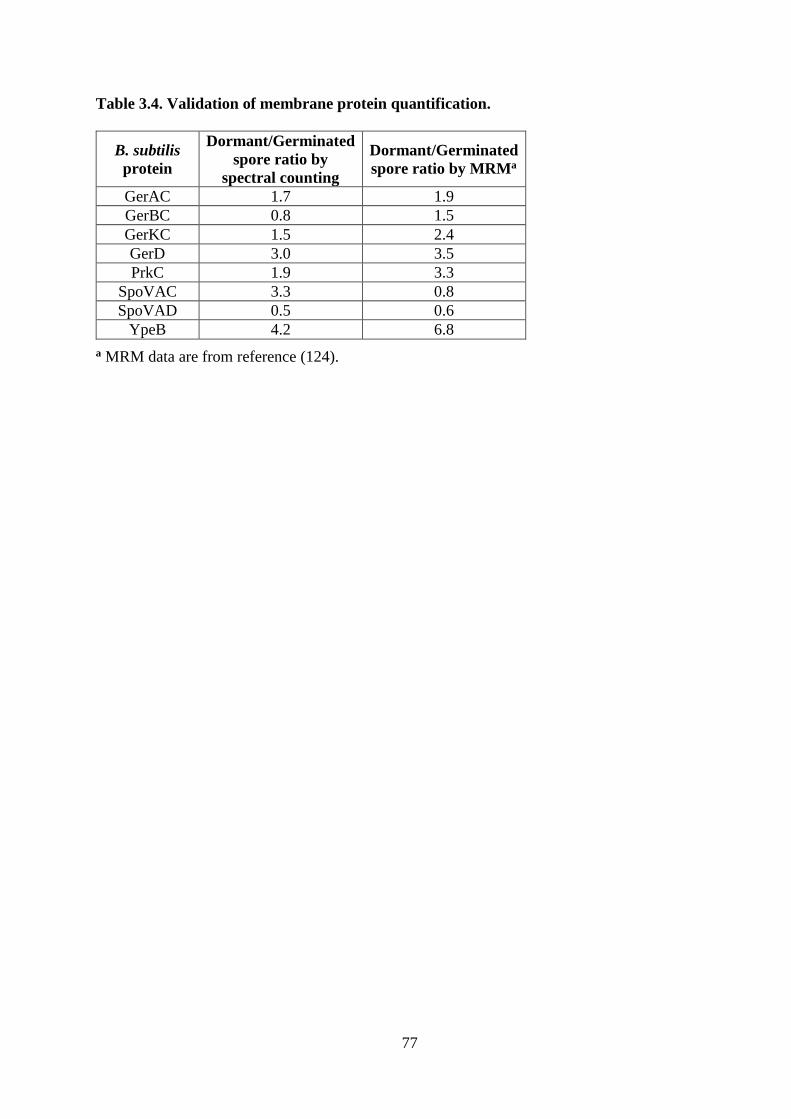
77
Table 3.4. Validation of membrane protein quantification.
B. subtilis
protein
Dormant/Germinated
spore ratio by
spectral counting
Dormant/Germinated
spore ratio by MRMa
GerAC 1.7 1.9
GerBC 0.8 1.5
GerKC 1.5 2.4
GerD 3.0 3.5
PrkC 1.9 3.3
SpoVAC 3.3 0.8
SpoVAD 0.5 0.6
YpeB 4.2 6.8
a MRM data are from reference (124).
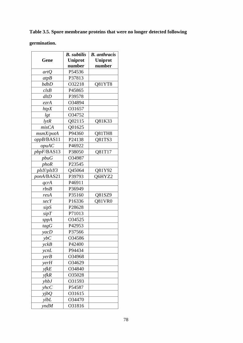
78
Table 3.5. Spore membrane proteins that were no longer detected following
germination.
Gene
B. subtilis
Uniprot
number
B. anthracis
Uniprot
number
artQ P54536
atpB P37813
bdbD O32218 Q81YT8
clsB P45865
dltD P39578
ezrA O34894
htpX O31657
lgt O34752
lytR Q02115 Q81K33
misCA Q01625
msmX/potA P94360 Q81TH8
oppB/BAS11
02 P24138 Q81TS3
opuAC P46922
pbpF/BAS13
63 P38050 Q81T17
pbuG O34987
phoR P23545
plsY/plsY3 Q45064 Q81Y92
ponA/BAS21
85 P39793 Q6HYZ2
qcrA P46911
rbsB P36949
resA P35160 Q81SZ9
secY P16336 Q81VR0
sipS P28628
sipT P71013
sppA O34525
tagG P42953
yacD P37566
ybC O34586
yckB P42400
ycnL P94434
yerB O34968
yerH O34629
yfkE O34840
yfkR O35028
yhbJ O31593
yhcC P54587
yjbQ O31615
ylbL O34470
yndM O31816
79
yqaR P45914
yqgF P54488
yrbG O32050
ysdB P94520
ytxG P40779
yuaB P71014
yuaG O32076
yutC O32128
yvcA O06965
BAS0336 Q81ZB8
BAS0353 Q6I447
BAS0360 Q81Z98
BAS0368 Q81Z90
BAS0564 Q81VA3
BAS1103 Q81TS2
BAS1104 Q81TS1
BAS1889 Q81RK9
BAS2093 Q81R11
BAS2560 Q81PQ5
BAS2651 Q81PG5
BAS2689 Q81PC7
BAS2733 Q81P74
BAS2909 Q6HWX1
BAS3998 Q81MD9
BAS4406 Q81L82
BAS4424 Q81L65
BAS4700 Q81KD9
BAS4808 Q81XR0
BAS4834 Q81XN4
BAS4853 Q81XL5
BAS4865 Q81XK0
BAS4953 Q6HR45
BAS4994 Q6HR05
BAS5314 Q81JJ5
ctaC Q81MT9
ecsA Q81U40
ffH Q81WJ2
gerD Q81VP4
ptsG Q81MH9
qcrB Q81SV0
rasP Q81WL5
secDF Q81LH8
spoVAC Q81X68
spoVAF Q81MG2
ypeB Q81PQ4
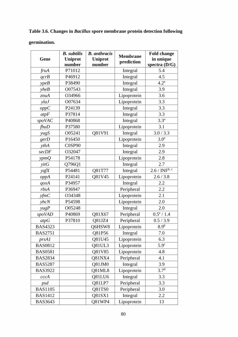
80
Table 3.6. Changes in Bacillus spore membrane protein detection following
germination.
Gene
B. subtilis
Uniprot
number
B. anthracis
Uniprot
number
Membrane
prediction
Fold change
in unique
spectra (D/G)
fruA P71012 Integral 5.4
qcrB P46912 Integral 4.5
ypeB P38490 Integral 4.2e
yheB O07543 Integral 3.9
znuA O34966 Lipoprotein 3.6
ylaJ O07634 Lipoprotein 3.3
oppC P24139 Integral 3.3
atpF P37814 Integral 3.3
spoVAC P40868 Integral 3.3a
fhuD P37580 Lipoprotein 3.1
yugS O05241 Q81V91 Integral 3.0 / 3.3
gerD P16450 Lipoprotein 3.0e
ythA C0SP90 Integral 2.9
secDF O32047 Integral 2.9
ypmQ P54178 Lipoprotein 2.8
yitG Q796Q1 Integral 2.7
yqfX P54481 Q81T77 Integral 2.6 / INFb, c
oppA P24141 Q81V45 Lipoprotein 2.6 / 3.8
qoxA P34957 Integral 2.2
rbsA P36947 Peripheral 2.2
yfmC O34348 Lipoprotein 2.1
yhcN P54598 Lipoprotein 2.0
yugP O05248 Integral 2.0
spoVAD P40869 Q81X67 Peripheral 0.5e / 1.4
atpG P37810 Q81JZ4 Peripheral 0.5 / 3.9
BAS4323 Q6HSW8 Lipoprotein 8.9b
BAS2751 Q81P56 Integral 7.0
prsA1 Q81U45 Lipoprotein 6.3
BAS0812 Q81UL3 Lipoprotein 5.9c
BAS0581 Q81V85 Lipoprotein 4.8
BAS2834 Q81NX4 Peripheral 4.1
BAS5287 Q81JM0 Integral 3.9
BAS3922 Q81ML8 Lipoprotein 3.7d
cccA Q81LU6 Integral 3.3
psd Q81LP7 Peripheral 3.3
BAS1105 Q81TS0 Peripheral 3.0
BAS1412 Q81SX1 Integral 2.2
BAS3643 Q81WP4 Lipoprotein 13

81
BAS0405 Q81Z55 Peripheral 0.5d
a Result is not consistent that of (124).
b Result is consistent with that of (83).
c This protein was detected in B. anthracis dormant spores but not in germinated spores.
d Result is not consistent with that of (83).
e Result is consistent that of (124).
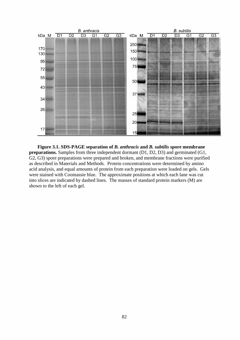
82
Figure 3.1. SDS-PAGE separation of B. anthracis and B. subtilis spore membrane
preparations. Samples from three independent dormant (D1, D2, D3) and germinated (G1,
G2, G3) spore preparations were prepared and broken, and membrane fractions were purified
as described in Materials and Methods. Protein concentrations were determined by amino
acid analysis, and equal amounts of protein from each preparation were loaded on gels. Gels
were stained with Coomassie blue. The approximate positions at which each lane was cut
into slices are indicated by dashed lines. The masses of standard protein markers (M) are
shown to the left of each gel.
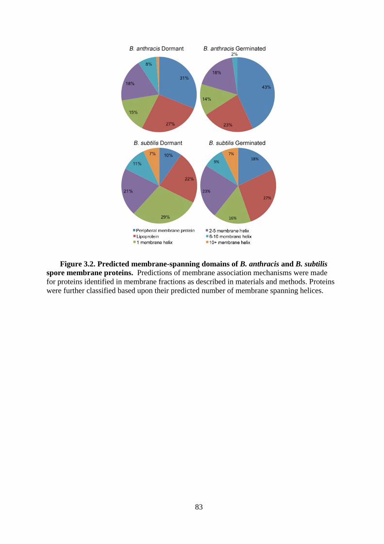
83
Figure 3.2. Predicted membrane-spanning domains of B. anthracis and B. subtilis
spore membrane proteins. Predictions of membrane association mechanisms were made
for proteins identified in membrane fractions as described in materials and methods. Proteins
were further classified based upon their predicted number of membrane spanning helices.

84
CHAPTER 4
HtrC is involved in proteolysis of YpeB during germination of Bacillus
anthracis and Bacillus subtilis spores
Casey B. Bernhards*, Yan Chen*, Hannah Toutkoushian, and David L. Popham
Manuscript in preparation

85
Attributions
Yan Chen and Casey B. Bernhards contributed equally to the research, experimentation,
and data analysis of the material presented herein. Yan is responsible for the work presented
in Table 4.3 and Figures 4.5, 4.6, 4.7, 4.8. Casey is responsible for the work presented in
Figures 4.1, 4.2, 4.3, 4.4, and 4.9. Hannah K. Toutkoushian performed germination rate
assays involving htrC mutants and provided laboratory assistance. David L. Popham was the
principle investigator. Casey, Yan, and David all contributed to the writing of the
manuscript.
86
ABSTRACT
Bacterial endospores can remain dormant for decades, yet can respond to nutrients,
germinate, and resume growth within minutes. An essential step in the germination process is
degradation of the spore cortex peptidoglycan wall, and the SleB protein in Bacillus species
plays a key role in this process. Stable incorporation of SleB into the spore requires the YpeB
protein, and some evidence suggests that these two proteins interact within the dormant
spore. Early during germination, YpeB is proteolytically processed to a stable fragment. In
this work, the primary sites of YpeB cleavage were identified in B. anthracis, and it was
shown that the stable products are comprised of the C-terminal domain of YpeB.
Modification of the predominant YpeB cleavage sites reduced proteolysis, but cleavage at
other sites still resulted in loss of full-length YpeB. A B. anthracis strain lacking the HtrC
protease did not generate the same stable YpeB products. In B. anthracis and B. subtilis htrC
mutants, YpeB was partially stabilized during germination but was still degraded at a reduced
rate by other unidentified proteases. Purified HtrC cleaved YpeB to a fragment similar to that
observed in vivo, and this cleavage was stimulated by Mn2+ or Ca2+ ions. A lack of HtrC did
not stabilize YpeB or SleB during spore formation in the absence of the partner protein,
indicating other proteases are involved in their degradation during sporulation.
87
INTRODUCTION
Endospores produced by Gram-positive genera such as Bacillus and Clostridium possess
extreme resistance properties and can remain in a fully dormant state for years. The dormant
state and resistance properties are dependent on the maintenance of the spore core
(cytoplasm) in a relatively dehydrated state, and this in turn depends on the intact state of the
inner spore membrane and the cortex peptidoglycan (PG) wall surrounding that membrane
(13). Upon exposure to nutrient germinants, spores begin to release low molecular weight
solutes, including a large depot of Ca2+-dipicolinic acid (Ca2+-DPA), and take up water (149).
Degradation of the cortex PG by germination-specific lytic enzymes (GSLEs) is required for
full expansion of the membrane, full hydration of the core, and resumption of metabolism
(98, 150-152). As GSLEs hydrolyze the cortex PG before new protein synthesis can occur,
they must be produced during spore formation and held stable and inactive in the dormant
spore until germination is triggered (63).
Bacillus species possess two major, partially redundant GSLEs: CwlJ and SleB (63).
CwlJ is produced in the mother cell of the developing sporangium (71), is associated with the
spore coats on the outer surface of the cortex (64, 72, 73, 153), and becomes active when
exposed to a high concentration of Ca2+-DPA normally when that solute is released from the
germinating spore (65, 73, 154). SleB is produced within the developing forespore (68, 70)
and is located interior to the cortex in the dormant spore, most likely in close association with
the inner spore membrane (64, 112). The mechanisms by which SleB is held inactive during
spore dormancy and released to become active during germination are unclear.
A potential factor in regulation of SleB activity is YpeB, which is encoded in an operon
with sleB and possesses a transmembrane anchor sequence that should also localize it to the
outer surface of the inner spore membrane (61, 69, 70). SleB and YpeB exhibit a co-
88
dependence for their stable incorporation into the dormant spore (64, 69, 155). In the absence
of their partner protein, both SleB and YpeB are produced and rapidly degraded during spore
formation (69). It has also been observed that YpeB is proteolytically processed during spore
germination (64), and it has been suggested that this processing could be involved in the
initiation of SleB activity during germination (155).
The current study examined the cleavage sites generating the stable YpeB products
during germination and identified HtrC as the protease responsible for these cleavage events.
The resulting cleavage products were shown to contain the C-terminus and PepSY domains
of YpeB. Strains lacking HtrC or with mutations at the YpeB cleavage sites were constructed
to evaluate the role of YpeB processing in SleB activation. In the absence of HtrC, YpeB was
degraded in a more non-specific manner during germination, and activity of SleB was not
significantly affected.
89
MATERIALS AND METHODS
Strains, culture conditions, and spore preparation. Escherichia coli strains used for
plasmid propagation were grown at 37°C in LB medium with 500 μg/ml erythromycin or 20
g/ml kanamycin. Strains used for protein overexpression were grown in LB with 30 μg/ml
chloramphenicol and 50 μg/ml ampicillin. Bacillus anthracis strains were derived from the
Sterne strain 34F2 and were grown on brain heart infusion (BHI; Difco) agar plates with 5
g/ml erythromycin or 20 g/ml kanamycin, where appropriate. B. anthracis strains
maintaining pBKJ236-derivatives extrachromosomally were grown at 25°C, while those in
which the plasmid was integrated into the chromosome were grown at 37°C. Bacillus subtilis
strains were derived from PS832, a prototrophic laboratory derivative of strain 168, and were
grown on 2xSG medium (102) with 0.5 µg/ml erythromycin and 12.5 µg/ml lincomycin
(MLS resistance), and/or 10 μg/ml tetracycline, when appropriate. Spores were prepared in
Modified G (123) broth for B. anthracis, or 2xSG (102) broth for B. subtilis with appropriate
antibiotics. After 72 h incubation at 37°C, spores were harvested, washed in deionized water
for several days, and any remaining vegetative cells were heat killed at 65C for 25 min. B.
anthracis spores were further purified by centrifugation through a 50% sodium diatrizoate
(Sigma) layer as described (103). Where indicated, B. anthracis spores were decoated as
previously detailed (69, 98). All spores used in this work were 99% free of vegetative cells
and were stored in deionized water at 4°C until analysis.
Mutant strain construction. All strains and plasmids used are listed in Table 4.1, and
the primer sequences used for plasmid construction are listed in Table 4.2. Construction of
DPBa127, a B. anthracis strain expressing YpeB-His6, was published previously (69). In this
strain, the plasmid pDPV424 encoding YpeB-His6 was integrated into the ∆ypeB
chromosome. Strains in which the YpeB T202-S203 or A168-S169 cleavage sites were
90
changed to E202-L203 or E168-L169 were created by site-directed mutagenesis of ypeB
using overlap extension PCR (156). The PCR products were subsequently cloned into
pDPV424 through restriction-free cloning (157), generating plasmids encoding
YpeBT202E/S203L-His6 (pDPV447) and YpeBA168E/S169L-His6 (pDPV454). To construct a strain
in which both cleavage sites had been mutated, pDPV447 was used as template for overlap
extension PCR, and the PCR product was cloned into pDPV447, producing pDPV455.
Plasmids pDPV447, pDPV454, and pDPV455 were screened for the gain or loss of a
restriction site in ypeB that was designed as part of the mutagenic primers, and were also
verified by DNA sequencing. Plasmids were introduced into the ∆ypeB strain of B. anthracis
(69) by conjugation, as in the initial steps of the markerless gene replacement procedure
(158), and chromosomal integrants were selected by shifting the temperature to 42C. PCR
amplification and sequencing verified plasmid integration within the 500-bp homologous
region downstream of ∆ypeB in the chromosome and confirmed the presence of the desired
cleavage site mutations.
To create a B. anthracis ∆htrC strain, htrC and approximately 500-bp flanking each side
of the gene were PCR amplified from chromosomal DNA. The PCR product was inserted
into the vector pBKJ236 (158) by digesting with the restriction enzymes BamHI and PstI and
ligating the DNA to create pDPV459. Inverse PCR of the plasmid using primers with BglII
restriction sites at the 3 ends resulted in a linear PCR product with the majority of htrC
deleted, leaving only the first five and last two codons of the gene. Subsequent BglII
digestion and ligation of the PCR product produced pDPV460. This plasmid containing the
htrC deletion was introduced into B. anthracis using the markerless gene replacement
strategy as previously described (158), except plasmid pSS4332 (159) was used for I-SceI
expression in place of pBKJ223. Gene deletion was verified by PCR amplification and
sequencing. Complementation of the ∆htrC mutation was achieved by introduction and
91
chromosomal integration of pDPV459. To create ∆sleB ∆htrC and ∆ypeB ∆htrC double
deletion strains, pDPV460 was used to introduce the htrC deletion into ∆sleB (DPBa38, (61))
and ∆ypeB (DPBa89, (69)) strains of B. anthracis using the markerless gene replacement
strategy (158) with pSS4332 (159). Gene deletions were verified by PCR amplification and
sequencing.
To create a B. subtilis ∆htrC strain, approximately 1000-bp flanking each side of the
gene were PCR amplified from chromosomal DNA and linked to the ermC Macrolide-
Lincosamide-Streptogramin (MLS) antibiotic resistance gene cassette by long-flanking
homology PCR (160). The PCR product was transformed into B. subtilis PS832 with
selection for MLS resistance to produce DPVB668, in which all but the first 7 and last 8
codons of the htrC coding sequence were deleted and replaced by the MLS cassette.
Chromosomal DNA from FB111 (73) was transformed into B. subtilis DPVB668 with
selection for tetracycline resistance to produce DPVB669.
Spore germination assays. The rate of germination and outgrowth of spores in liquid
BHI (B. anthracis) or 2xYT (B. subtilis) was assessed by monitoring the change in optical
density at 600 nm (OD) as described previously (69). For germination efficiency assays,
spores at an OD of 0.2 were heat activated at 70C for 20 min and quenched on ice. Heat
activated spores were serially diluted in deionized water, plated on BHI or 2xSG medium
without antibiotics, and incubated at 37C overnight. Colonies were counted to determine
CFU/OD unit values. Statistical analyses of spore germination rates were performed using
unpaired, two-tailed Student’s t-tests with unequal variance.
Preparation and analysis of spore fractions. Sporangia samples were collected at
hours 2 through 6 of sporulation from strains grown in modified G broth, as described
previously (69). To prepare germinated spores for western blot analysis, a suspension of
dormant spores at a concentration of 5 OD units per ml in 10 mM Tris-HCl pH 7.0 were heat
92
activated at 70°C for 30 min (B. anthracis) or 75°C for 30 min (B. subtilis) and cooled on ice
for 10 min. Chloramphenicol was added to a final concentration of 10 g/ml to inhibit
protein synthesis and 7.5 OD units of spores were removed for the dormant spore sample.
The spore suspension was briefly prewarmed before spores were germinated at 37°C with
shaking at 250 rpm by the addition of 10 or 100 mM L-alanine with 1 mM inosine (B.
anthracis) or 10 mM L-valine (B. subtilis). Samples equivalent to 7.5 OD units based on the
starting OD were collected at various times after the addition of germinants and centrifuged
at 15,800 x g for 2 min. The resulting pellets were flash frozen in liquid N2 and lyophilized.
Dried spores or sporangia were pulverized with 100 mg of 0.1 mm glass beads in a dental
amalgamator (Wig-L-Bug) at 4,200 rpm for 20 pulses of 30 s each. Samples were put on ice
for at least 30 s between pulses. Proteins were extracted from the mixture with 75 l
(dormant and germinated spores) or 100 l (sporangia) of 1X sample loading buffer (62.5
mM Tris-HCl pH 6.8, 2% SDS, 10% glycerol, 5% -mercaptoethanol, 0.05% bromophenol
blue). Following heating at 100C for 5 min, extracts were centrifuged at 15,800 x g for 1
min, and supernatants were analyzed by western blotting. In some cases, proteins were
extracted in sample loading buffer without bromophenol blue and quantified using amino
acid analyses (105).
To extract and purify His6-tagged YpeB cleavage products for N-terminal sequencing,
250 OD units of DPBa127 or DPBa157 spores in a final volume of 100 ml 10 mM Tris-HCl
pH 7.0 were heat activated and germinated with 100 mM L-alanine and 1 mM inosine as
described above. Spores were germinated for approximately 1 h, or until 90% of spores
had germinated, as determined by phase contrast microscopy. The germinated spores were
collected by centrifugation at 10,000 x g for 10 min at 4C and washed with 5 ml Buffer A
(50 mM NaCl, 50 mM Tris-HCl pH 7.5, 5% glycerol, 30 mM imidazole). Following another
93
centrifugation, the pellet was resuspended in 5 ml Buffer A, split between 4 microcentrifuge
tubes, and centrifuged at 15,800 x g for 2 min. The pellets were frozen at -80C, lyophilized,
and broken with glass beads as described above. Proteins were extracted by resuspending
and combining samples in a total of 5 ml Buffer B (500 mM NaCl, 50 mM Tris-HCl pH 7.5,
30 mM imidazole, 8M urea) and incubating at 4C with slow rocking for at least 2 h. The
sample was centrifuged at 6,800 x g for 10 min at 4C, and the supernatant containing soluble
extracted proteins was filtered and loaded on a 1-ml Ni-Sepharose HisTrap HP affinity
column (GE Healthcare) equilibrated with Buffer B. YpeB cleavage products were eluted
with Buffer C (500 mM NaCl, 50 mM Tris-HCl pH 7.5, 500 mM imidazole, 8M urea). To
precipitate the affinity-purified cleavage products, deoxycholate and TCA (0.05% and 20%
final concentrations, respectively) were added to the pooled elution fractions, which were
then incubated on ice overnight. The mixture was centrifuged at 15,000 x g for 10 min at
4C, and the pellet was washed 3 times with 1 ml ice-cold 100% acetone. The pellet was
dried and mixed with 25 l 1X sample loading buffer, and additional 0.5 M Tris-HCl pH 6.8
was added until the sample was blue again.
The entire precipitated sample was run on a SDS-PAGE gel that had first been incubated
overnight in 1X Tris-Glycine SDS Running Buffer with 0.1 mM sodium thioglycolate.
Sodium thioglycolate scavenges free radicals, thereby protecting the N-termini of proteins
from modification. The proteins were transferred to Immobilon-PSQ PVDF membrane
(Millipore) in CAPS transfer buffer (10 mM CAPS pH 11.0, 10% methanol), and the
membrane was briefly washed with deionized water before being stained with 0.1%
Coomassie Brilliant Blue R-250 (Jersey Lab Supply) for 30 s. The membrane was destained
repeatedly with 50% methanol until bands were visible. Bands corresponding to YpeB
cleavage products were cut from the membrane, washed with deionized water, dried, and
stored at -80°C. N-terminal sequencing via automated Edman degradation was performed on
94
extracted bands using an ABI 494 Protein Sequencer (Tufts University Analytical Core
Facility).
Western blot analysis. Polyclonal antibodies raised in rabbits against B. anthracis SleB
and YpeB (69), and monoclonal mouse anti-His (C-term) antibodies (Invitrogen) were used
for detection of SleB, YpeB, and derivatives in western blots. Anti-SleB and anti-YpeB
antibodies were used at 1:1,000 and 1:3,000 dilutions, respectively, while anti-His antibodies
were used at a 1:5,000 dilution. Secondary goat anti-rabbit-HRP antibodies (Bio-Rad) were
used at a 1:5,000 (cat. #166-2408) or 1:200,000 (cat. #170-6515) dilution, while goat-anti-
mouse-HRP antibodies (PerkinElmer) were used at a 1:5,000 dilution. Western blots utilized
Amersham Hybond-P (PVDF) membranes (GE Healthcare), and antibody detection was
carried out using colorimetric (BM Blue POD Substrate, Precipitating; Roche) or
chemiluminescent (Amersham ECL Prime Western Blotting Detection Reagent; GE
Healthcare) substrates.
HtrC and YpeB purification and assay. The B. anthracis htrC coding sequence,
lacking the first 44 codons containing the predicted transmembrane anchor, was PCR-
amplified and introduced into the entry vector pDonR201 (Invitrogen) and then the
destination vector pDEST-HisMBP-T (a modified version of pDEST-HisMBP (161)
containing a tobacco etch virus (TEV) cleavage site). The resulting plasmid, pDPV458,
encoding an N-terminal His6-tagged maltose binding protein (MBP) fused to a TEV cleavage
site and HtrC lacking its signal sequence/membrane anchor, was verified by DNA
sequencing. Overexpression strain DPVE501 was cultured at 37°C until the OD reached
~0.8, at which point isopropyl--D-thiogalactopyranoside was added to a final concentration
of 0.7 mM, the culture temperature was reduced to 12°C, and incubation was continued for
16 h.
95
Cells were harvested by centrifugation, resuspended in Buffer D (50 mM NaCl, 50 mM
Tris-HCl pH 7.5, 5% glycerol, 25 mM imidazole) and lysed using sonication. Soluble and
insoluble protein fractions were separated by centrifugation at 117,000 x g for 1 h. Soluble
His6-MBP-HtrC45-391 was purified using a Ni-Sepharose HisTrap HP affinity column
equilibrated with Buffer D. Protein was eluted with a step gradient of Buffer D containing
100 mM increasing concentrations of imidazole and fractions were dialyzed in Buffer D.
His6-MBP-HtrC45-391 at a concentration of ~1.5 mg/ml was incubated with 0.5 mg/ml His6-
tagged TEV (S219V) protease (162) at 15°C for 16 h. Cleavage was verified by SDS-PAGE
analysis. HtrC45-391 was separated from His6-TEV, His6-MBP, and undigested protein using a
Ni-Sepharose HisTrap HP affinity column. Fractions containing HtrC45-391 were dialyzed in
Buffer D, flash frozen, and stored at -80°C. YpeB21-446 overexpression and purification was
performed as described previously (69).
Protease activity of HtrC was assayed in 50 mM Tris-HCl pH 8.0 with 1 µM HtrC45-391
and 6 M YpeB21-446 at 37°C for 4 h, unless indicated otherwise. The reaction was
terminated by adding 2X SDS-PAGE sample loading buffer to a final concentration of 1X
and incubating at 100°C for 5 min. Proteins were separated using SDS-PAGE and were
stained with Coomassie Brilliant Blue.
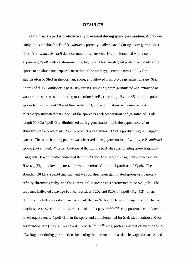
96
RESULTS
B. anthracis YpeB is proteolytically processed during spore germination. A previous
study indicated that YpeB of B. subtilis is proteolytically cleaved during spore germination
(64). A B. anthracis ypeB deletion mutant was previously complemented with a gene
expressing YpeB with a C-terminal His6 tag (69). This His6-tagged protein accumulated in
spores to an abundance equivalent to that of the wild-type, complemented fully for
stabilization of SleB in the dormant spore, and allowed a wild-type germination rate (69).
Spores of this B. anthracis YpeB-His6 strain (DPBa127) were germinated and extracted at
various times for western blotting to examine YpeB processing. By the 45 min time point,
spores had lost at least 50% of their initial OD, and examination by phase-contrast
microscopy indicated that > 95% of the spores in each preparation had germinated. Full-
length 51 kDa YpeB-His6 diminished during germination, with the appearance of an
abundant stable product at ~28 kDa product and a minor ~32 kDa product (Fig. 4.1, upper
panel). The same banding pattern was observed during germination of wild-type B. anthracis
spores (not shown). Western blotting of the same YpeB-His6 germinating spore fragments
using anti-His6 antibodies indicated that the 28 and 32 kDa YpeB fragments possessed the
His6 tag (Fig. 4.1, lower panel), and were therefore C-terminal portions of YpeB. The
abundant 28 kDa YpeB-His6 fragment was purified from germinated spores using metal-
affinity chromatography, and the N-terminal sequence was determined to be SAQKN. The
sequence indicated cleavage between residues T202 and S203 of YpeB (Fig. 4.2). In an
effort to block this specific cleavage event, the ypeB-His6 allele was mutagenized to change
residues T202-S203 to E202-L203. The altered YpeB-T202E/S203L-His6 protein accumulated to
levels equivalent to YpeB-His6 in the spore and complemented for SleB stabilization and for
germination rate (Figs. 4.3A and 4.4). YpeB-T202E/S203L-His6 protein was not cleaved to the 28
kDa fragment during germination, indicating that the mutation at the cleavage site succeeded
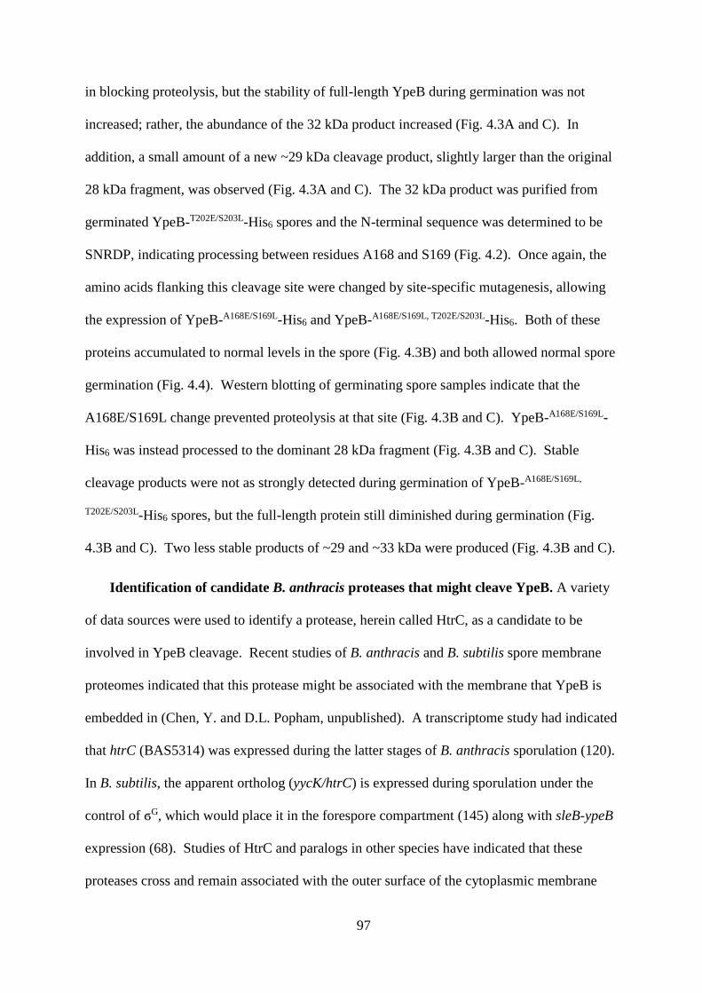
97
in blocking proteolysis, but the stability of full-length YpeB during germination was not
increased; rather, the abundance of the 32 kDa product increased (Fig. 4.3A and C). In
addition, a small amount of a new ~29 kDa cleavage product, slightly larger than the original
28 kDa fragment, was observed (Fig. 4.3A and C). The 32 kDa product was purified from
germinated YpeB-T202E/S203L-His6 spores and the N-terminal sequence was determined to be
SNRDP, indicating processing between residues A168 and S169 (Fig. 4.2). Once again, the
amino acids flanking this cleavage site were changed by site-specific mutagenesis, allowing
the expression of YpeB-A168E/S169L-His6 and YpeB-A168E/S169L, T202E/S203L-His6. Both of these
proteins accumulated to normal levels in the spore (Fig. 4.3B) and both allowed normal spore
germination (Fig. 4.4). Western blotting of germinating spore samples indicate that the
A168E/S169L change prevented proteolysis at that site (Fig. 4.3B and C). YpeB-A168E/S169L-
His6 was instead processed to the dominant 28 kDa fragment (Fig. 4.3B and C). Stable
cleavage products were not as strongly detected during germination of YpeB-A168E/S169L,
T202E/S203L-His6 spores, but the full-length protein still diminished during germination (Fig.
4.3B and C). Two less stable products of ~29 and ~33 kDa were produced (Fig. 4.3B and C).
Identification of candidate B. anthracis proteases that might cleave YpeB. A variety
of data sources were used to identify a protease, herein called HtrC, as a candidate to be
involved in YpeB cleavage. Recent studies of B. anthracis and B. subtilis spore membrane
proteomes indicated that this protease might be associated with the membrane that YpeB is
embedded in (Chen, Y. and D.L. Popham, unpublished). A transcriptome study had indicated
that htrC (BAS5314) was expressed during the latter stages of B. anthracis sporulation (120).
In B. subtilis, the apparent ortholog (yycK/htrC) is expressed during sporulation under the
control of ϭG, which would place it in the forespore compartment (145) along with sleB-ypeB
expression (68). Studies of HtrC and paralogs in other species have indicated that these
proteases cross and remain associated with the outer surface of the cytoplasmic membrane
98
due to an uncleaved signal sequence/membrane anchor (147). HtrC expressed in the
developing spore would therefore be expected to remain on the outer surface of the inner
spore membrane, in the same location as YpeB. While multiple proteases in each species
possess some properties consistent with a role in YpeB processing, the HtrC proteins of B.
anthracis and B. subtilis were considered the strongest candidates.
Strains lacking HtrC are altered in YpeB proteolysis. B. anthracis and B. subtilis
strains carrying null mutations in htrC were constructed, and YpeB cleavage was examined
by western blotting using antisera against B. anthracis YpeB (69). The B. anthracis ∆htrC
mutant lost full-length YpeB during germination more slowly than the wild-type and did not
accumulate the specific 27 and 31 kDa cleavage products (Fig. 4.5A) (These cleavage
products are slightly smaller than those in Fig. 4.1 because they lack the His6 tag). In some
cases, western blots exhibited a heavier background in the 30-50 kDa range for germinating
∆htrC spores (Fig. 4.5A), suggesting that YpeB might be degraded to non-specific products,
but this has not been further confirmed. While the antibodies did not recognize the cleavage
products of B. subtilis YpeB, the htrC mutant maintained increased amounts of full-length
YpeB during germination relative to the wild-type strain (Fig. 4.5B).
The germination of htrC mutant spores was examined to determine if slowed degradation
of YpeB resulted in decreased SleB activity. This was done in B. anthracis decoated spores
and in a B. subtilis ∆cwlJ background in order to render the function of SleB more easily
observed (65, 163). In both Bacillus species, the germination rates of ∆htrC spores were not
significantly different from those of the wild-type spores (Fig. 4.6A and B). In both species,
the colony-forming efficiency of ∆htrC spores lacking CwlJ activity was also not different
from the corresponding wild-type spores (Table 4.3).
In vitro cleavage of YpeB by HtrC. B. anthracis HtrC, lacking its signal
sequence/membrane anchor, was expressed in E. coli as a His6-MBP fusion protein and was

99
purified by metal-affinity chromatography. Following purification of His6-MBP-HtrC45-391,
the His6-MBP domain was removed using TEV protease, and HtrC45-391 was purified (Fig.
4.7). The pure protease exhibited little activity against a generic substrate (fluorescently-
labeled casein, data not shown) and, initially, very little activity against purified B. anthracis
YpeB (Fig. 4.8, lanes 8, 10, and 15-16). Assay attempts using a wide variety of conditions
revealed that HtrC-catalyzed cleavage of YpeB to produce an ~27 kDa fragment, as observed
in vivo, was greatly stimulated by the presence of Mn2+ (Fig. 4.8, lanes 10-13) and, to a lesser
degree, by Ca2+ (Fig. 4.8, lanes 16-19), but not by Mg2+, Zn2+, Na+, or K+ (Fig. 4.8, lanes 1-
10). Relatively high concentrations of Mn2+ or Ca2+ were required to achieve maximum HtrC
activity (Fig. 4.8, lanes 10-13 and 16-19). Metal-stimulated HtrC activity was inhibited by
EDTA, a chelator of divalent cations (Fig. 4.8, lanes 16, 18, and 20). The presence of an
equimolar concentration of DPA, another strong chelator, blocked the positive effect of Ca2+
on HtrC (Fig. 4.8, lanes 22-26). HtrC action on YpeB was found to be maximal at pH 7-9
(data not shown).
HtrC is not an important factor in SleB or YpeB degradation during spore
formation. To determine if HtrC is involved in degradation of SleB and/or YpeB in the
absence of the partner protein during spore formation, ∆sleB ∆htrC and ∆ypeB ∆htrC double
mutants were constructed in B. anthracis. As demonstrated previously (69), in a ∆sleB
mutant, YpeB failed to accumulate during spore formation, and in a ∆ypeB mutant, SleB did
not accumulate (Fig. 4.9A and B). The additional deletion of htrC from these strains still
resulted in the degradation of YpeB or SleB, respectively, during sporulation (Fig. 4.9),
indicating that other proteases are likely active in their degradation during spore formation.
Additionally, the production of YpeB during ∆sleB ∆htrC spore formation coincided with the
appearance of YpeB-specific degradation products (Fig. 4.9C), similar to those seen during
sporulation of the ∆sleB strain (69) and to specific cleavage products seen during spore

100
germination. This finding indicates that, at least during sporulation, other proteases are
capable of producing similar cleavage products.
101
DISCUSSION
B. anthracis YpeB is processed to a relatively stable fragment during germination, as in
B. subtilis (64). The ultimate N-termini of the stable YpeB fragments were determined to be
in a region between an undefined N-terminal YpeB domain and the C-terminal region
containing three putative PepSY domains. This C-terminal fragment persists well into the
germination process. The N-terminal cleavage product was observed neither in western blots
of germinating spore extracts nor in in vitro reactions using purified proteins. While this
might be due to poor recognition of this domain by the antiserum and/or poor staining of the
domain, it raises the possibility that this domain is subject to more rapid degradation to
smaller fragments. Interestingly, the YpeB N-terminal domain was recently shown to exert
an inhibitory effect on SleB activity in vitro (155). Processing of YpeB could play a role in
the activation of SleB during germination.
Deletion of htrC was found to result in the disappearance of specific YpeB degradation
products in both species, though the amount of full-length YpeB still decreased during
germination. This indicates that multiple proteases are active in this region of the spore
during germination. The degradation of YpeB during germination may be a multistep
process, carried out by multiple proteases, with the ultimate stable products being produced
by HtrC. However, initial cleavage of YpeB by another protease is apparently not a
requirement for HtrC action, as the action of HtrC was reproducible in vitro using purified
proteins.
The inability to fully block YpeB degradation during germination precludes
demonstration of a role of YpeB instability in SleB activation during germination. Even when
YpeB degradation was slowed by the loss of HtrC, the spore germination rate was not
significantly affected. Under the experimental conditions tested, germination was dependent
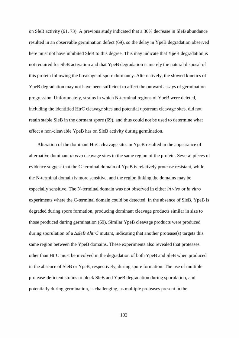
102
on SleB activity (61, 73). A previous study indicated that a 30% decrease in SleB abundance
resulted in an observable germination defect (69), so the delay in YpeB degradation observed
here must not have inhibited SleB to this degree. This may indicate that YpeB degradation is
not required for SleB activation and that YpeB degradation is merely the natural disposal of
this protein following the breakage of spore dormancy. Alternatively, the slowed kinetics of
YpeB degradation may not have been sufficient to affect the outward assays of germination
progression. Unfortunately, strains in which N-terminal regions of YpeB were deleted,
including the identified HtrC cleavage sites and potential upstream cleavage sites, did not
retain stable SleB in the dormant spore (69), and thus could not be used to determine what
effect a non-cleavable YpeB has on SleB activity during germination.
Alteration of the dominant HtrC cleavage sites in YpeB resulted in the appearance of
alternative dominant in vivo cleavage sites in the same region of the protein. Several pieces of
evidence suggest that the C-terminal domain of YpeB is relatively protease resistant, while
the N-terminal domain is more sensitive, and the region linking the domains may be
especially sensitive. The N-terminal domain was not observed in either in vivo or in vitro
experiments where the C-terminal domain could be detected. In the absence of SleB, YpeB is
degraded during spore formation, producing dominant cleavage products similar in size to
those produced during germination (69). Similar YpeB cleavage products were produced
during sporulation of a ∆sleB ∆htrC mutant, indicating that another protease(s) targets this
same region between the YpeB domains. These experiments also revealed that proteases
other than HtrC must be involved in the degradation of both YpeB and SleB when produced
in the absence of SleB or YpeB, respectively, during spore formation. The use of multiple
protease-deficient strains to block SleB and YpeB degradation during sporulation, and
potentially during germination, is challenging, as multiple proteases present in the
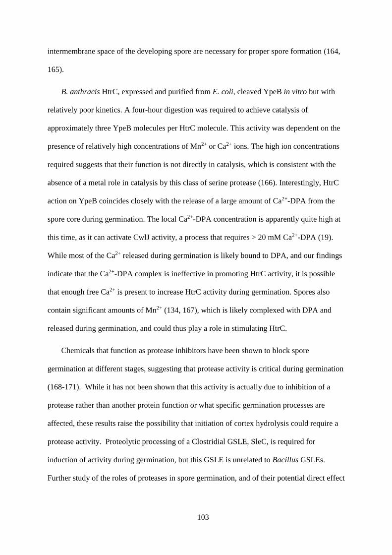
103
intermembrane space of the developing spore are necessary for proper spore formation (164,
165).
B. anthracis HtrC, expressed and purified from E. coli, cleaved YpeB in vitro but with
relatively poor kinetics. A four-hour digestion was required to achieve catalysis of
approximately three YpeB molecules per HtrC molecule. This activity was dependent on the
presence of relatively high concentrations of Mn2+ or Ca2+ ions. The high ion concentrations
required suggests that their function is not directly in catalysis, which is consistent with the
absence of a metal role in catalysis by this class of serine protease (166). Interestingly, HtrC
action on YpeB coincides closely with the release of a large amount of Ca2+-DPA from the
spore core during germination. The local Ca2+-DPA concentration is apparently quite high at
this time, as it can activate CwlJ activity, a process that requires > 20 mM Ca2+-DPA (19).
While most of the Ca2+ released during germination is likely bound to DPA, and our findings
indicate that the Ca2+-DPA complex is ineffective in promoting HtrC activity, it is possible
that enough free Ca2+ is present to increase HtrC activity during germination. Spores also
contain significant amounts of Mn2+ (134, 167), which is likely complexed with DPA and
released during germination, and could thus play a role in stimulating HtrC.
Chemicals that function as protease inhibitors have been shown to block spore
germination at different stages, suggesting that protease activity is critical during germination
(168-171). While it has not been shown that this activity is actually due to inhibition of a
protease rather than another protein function or what specific germination processes are
affected, these results raise the possibility that initiation of cortex hydrolysis could require a
protease activity. Proteolytic processing of a Clostridial GSLE, SleC, is required for
induction of activity during germination, but this GSLE is unrelated to Bacillus GSLEs.
Further study of the roles of proteases in spore germination, and of their potential direct effect

104
on activation of cortex hydrolysis, may allow the improvement of spore decontamination or
application methods.

105
ACKNOWLEDGEMENTS
Research reported in this publication was supported by the National Institute of Allergy
and Infectious Disease of the National Institutes of Health under award number
R21AI088298. The content is solely the responsibility of the authors and does not
necessarily represent the official views of the National Institutes of Health. We thank Roger
Plaut and Scott Stibitz for the gift of plasmid pSS4332. Dr. Florian Schubot contributed
reagents and Jessica Blackwood provided laboratory assistance.
106
Table 4.1. Bacterial strains and plasmid.
Strain or
plasmid Relevant genotype/phenotype a Construction b Source
E. coli
DPVE13 BL21 (DE3) pLysS (Cmr) Novagen
DPVE501 pDPV458 (His6-MBP-HtrC45-391 Ampr)
Cmr pDPV458DPVE13 This study
B. anthracis
Sterne 34F2 pXO1+ pXO2- P. Hanna
DPBa38 ∆sleB (61)
DPBa89 ∆ypeB (69)
DPBa113 ∆ypeB::pDPV416 (ypeB+ Err) (69)
DPBa127 ∆ypeB::pDPV424 (YpeB-His6 Err) (69)
DPBa157 ∆ypeB::pDPV447 (YpeBT202E/S203L-His6
Err) pDPV447DPBa89 This study
DPBa167 ∆ypeB::pDPV454 (YpeBA168E/S169L-His6
Err) pDPV454DPBa89 This study
DPBa168 ∆ypeB::pDPV455 (YpeBA168E/S169L,
T202E/S203L-His6 Err) pDPV455DPBa89 This study
DPBa178 ∆htrC pDPV46034F2 This study
DPBa182 ∆htrC::pDPV459 (htrC+ Err) pDPV459DPBa178 This study
DPBa187 ∆sleB ∆htrC pDPV460DPBa38 This study
DPBa188 ∆ypeB ∆htrC pDPV460DPBa89 This study
B. subtilis
PS832 Prototrophic derivative of strain 168 P. Setlow
FB111 ΔcwlJ::Tetr (73)
DPVB668 ∆htrC::MLSr PCRPS832 This study
DPVB669 ΔcwlJ::Tetr ∆htrC::MLSr FB111DPVB668 This study

107
Plasmids
pBKJ236 Err ori(Ts) (158)
pSS4332 Kanr, I-SceI, AmCyan (159)
pDEST-
HisMBP-T His6-MBP, Ampr Cmr F. Schubot
pDPV416 ypeB+ (69)
pDPV424 YpeB-His6 (69)
pDPV447 YpeBT202E/S203L-His6 pBKJ236::∆sleB
ypeBT202E/S203L-His6 This study
pDPV454 YpeBA168E/S169L-His6 pBKJ236::∆sleB
ypeBA168E/S169L-His6 This study
pDPV455 YpeBA168E/S169L, T202E/S203L-His6
pBKJ236::∆sleB
ypeBA168E/S169L, T202E/S203L-
His6
This study
pDPV458 His6-MBP-HtrC45-391 pDEST-HisMBP-T::htrC45-
391 This study
pDPV459 htrC+ pBKJ236::htrC This study
pDPV460 ∆htrC pBKJ236::∆htrC This study
a Cmr, chloramphenicol resistance; Ampr, ampicillin resistance; Err, erythromycin resistance; Tetr, tetracycline resistance; MLSr, macrolide-
lincosamide-streptogramin B resistance; ori(Ts), temperature-sensitive origin of replication; Kanr, kanamycin resistance.
b Strains were constructed by conjugation or electroporation for B. anthracis, or transformation for B. subtilis. The designation preceding the arrow
is the plasmid or source of donor DNA, while the designation following the arrow is the recipient strain
108
Table 4.2. Primer sequences.
Plasmid/Strain
constructed Primer name Sequence 5 to 3 a
pDPV447 593 CTTCGGACCTACCTTTGAATTAGCACAAAAAAATAAA
AAAGGTGG
594 CCACCTTTTTTATTTTTTTGTGCTAATTCAAAGGTAGG
TCCGAAG
603 GTCTTCTTTACATAAAAGCGAGCCTTTTACAAAACAT
AACC
604 GCTTTACGTTCTTCCATAACTTTCACATCTGGATTG
pDPV454 605 GTTGAGATGGCACTCGAGTTAAATCGTGATCCTGCCG
606 CGGCAGGATCACGATTTAACTCGAGTGCCATCTCAAC
603, 604
pDPV455 603, 604, 605,
606
pDPV458
612
613
GTGGAGAACCTGTACTTCCAGGGTTCAAATGATACGG
GCGC
CCACTTTGTACAAGAAAGTTGCATTTTAATACTTTTGAA
TGCCTAGTTTAAC
pDPV459 614
615
CGCGGATCCGATATTGAGGTCGAGTCATTTG
CGCCTGCAGCAATGACATGCGTATCATCAG
pDPV460 616
617
CGCAGATCTAAAGGACATATTTGTTCACCTATC
CGCAGATCTAAGTATTAAAAACAGGAGGGCCTAC
DPVB668
607
608
609
610
ACAAGTACGCAGGTGCTGGC
CGATTATGTCTTTTGCGCAGTCGGCCCTCACGTTCGT
AATCCACC
GAGGGTTGCCAGAGTTAAAGGATCCCTCCGCAGACC
AATTAGGC
GATACACCGATTGACGTACG
a Restriction sites are underlined, TEV cleavage site regions of pDEST-HisMBP-T are
italicized and in bold, and attR2.1 regions of pDEST-HisMBP-T are italicized and underlined.

109
Table 4.3. Germination efficiencies of ∆htrC B. anthracis and B. subtilis spores.
Species Strain Genotype CFU/OD/ml a
B. anthracis
DPBa2 WT 6.7 x 10⁷
DPBa178 ∆htrC 6.0 x 10⁷
DPBa182 htrC+ 6.3 x 10⁷
B. subtilis
PS832 WT 1.7 x 10⁸
FB111 ∆cwlJ 3.0 x 10⁸
DPVB668 ∆htrC 1.9 x 10⁸
DPVB669 ∆cwlJ ∆htrC 2.0 x 10⁸
a Decoated spores were used for B. anthracis studies. Values are averages of three
independent spore preparations. Among the strains for each species, no statistical difference
in colony forming efficiency was present.
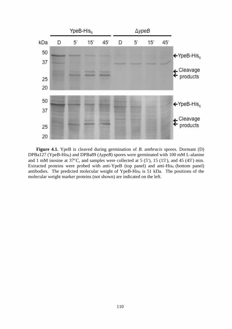
110
Figure 4.1. YpeB is cleaved during germination of B. anthracis spores. Dormant (D)
DPBa127 (YpeB-His6) and DPBa89 (∆ypeB) spores were germinated with 100 mM L-alanine
and 1 mM inosine at 37C, and samples were collected at 5 (5), 15 (15), and 45 (45) min.
Extracted proteins were probed with anti-YpeB (top panel) and anti-His6 (bottom panel)
antibodies. The predicted molecular weight of YpeB-His6 is 51 kDa. The positions of the
molecular weight marker proteins (not shown) are indicated on the left.
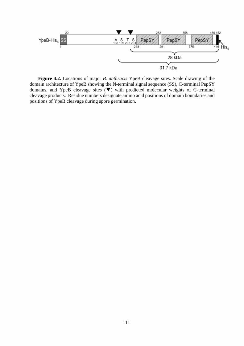
111
Figure 4.2. Locations of major B. anthracis YpeB cleavage sites. Scale drawing of the
domain architecture of YpeB showing the N-terminal signal sequence (SS), C-terminal PepSY
domains, and YpeB cleavage sites () with predicted molecular weights of C-terminal
cleavage products. Residue numbers designate amino acid positions of domain boundaries and
positions of YpeB cleavage during spore germination.
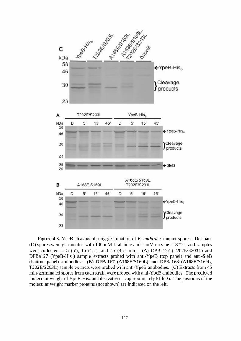
112
Figure 4.3. YpeB cleavage during germination of B. anthracis mutant spores. Dormant
(D) spores were germinated with 100 mM L-alanine and 1 mM inosine at 37C, and samples
were collected at 5 (5), 15 (15), and 45 (45) min. (A) DPBa157 (T202E/S203L) and
DPBa127 (YpeB-His6) sample extracts probed with anti-YpeB (top panel) and anti-SleB
(bottom panel) antibodies. (B) DPBa167 (A168E/S169L) and DPBa168 (A168E/S169L,
T202E/S203L) sample extracts were probed with anti-YpeB antibodies. (C) Extracts from 45
min-germinated spores from each strain were probed with anti-YpeB antibodies. The predicted
molecular weight of YpeB-His6 and derivatives is approximately 51 kDa. The positions of the
molecular weight marker proteins (not shown) are indicated on the left.

113
Figure 4.4. Altered YpeB proteolysis does not slow spore germination and outgrowth.
Decoated ∆ypeB (), YpeBT202E/S203L-His6 (), YpeBA168E/S169L-His6 (), YpeBA168E/S169L,
T202E/S203L-His6 (), and YpeB-His6 () B. anthracis spores were heat activated and
germinated in BHI medium at 37C; germination and outgrowth was tracked as changes in OD.
Data shown are averages of results from three independent spore preparations; error bars are
omitted for clarity. Germination of YpeBT202E/S203L-His6, YpeBA168E/S169L-His6, and
YpeBA168E/S169L, T202E/S203L-His6 spores is not significantly different (P > 0.06) at any time point.
Likewise, germination of YpeB-His6 and YpeBT202E/S203L-His6 spores is not significantly
different (P > 0.07) at any time point. Focusing on stage two of germination, from 45 to 95
min (69), YpeBA168E/S169L-His6 and YpeB-His6 spores do not significantly differ (P > 0.07),
whereas YpeBA168E/S169L, T202E/S203L-His6 and YpeB-His6 spores are not significantly different
(P > 0.05), except from 75 to 80 min (P < 0.05).
30
40
50
60
70
80
90
100
0 50 100 150 200
% o
f In
itia
l O
D6
00
nm
Time (min)

114
Figure 4.5. HtrC cleaves YpeB during germination of B. anthracis and B. subtilis spores.
Spores were prepared, germinated, and analyzed by western blot with B. anthracis anti-YpeB
antibodies as described in materials and methods. (A) B. anthracis spores were dormant (D)
or collected 5 (5), 15 (15), and 45 (45) min after addition of germinants. Strains were
DPBa2 (WT), DPBa178 (∆htrC), DPBa182 (htrC+), and DPBa89 (∆ypeB). (B) B. anthracis
and B. subtilis spores were dormant (D) or collected 10 (10) and 30 (30) min after addition
of germinants. Strains were DPBa2 (B. anthracis WT), PS832 (B. subtilis WT), and
DPVB668 (B. subtilis ∆htrC). The predicted molecular weights of YpeB from B. anthracis
and B. subtilis are 50 and 51 kDa, respectively. The positions of the molecular weight
marker proteins (not shown) are indicated on the left.
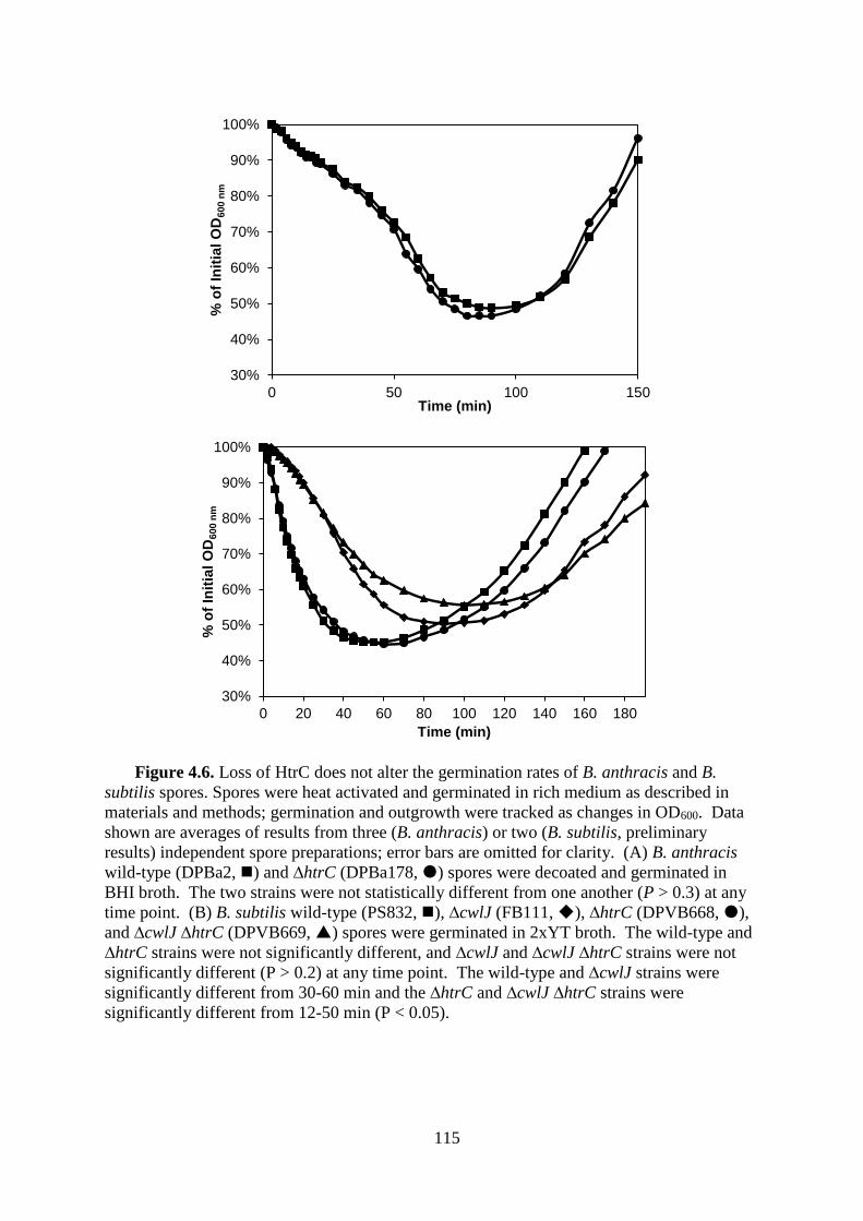
115
Figure 4.6. Loss of HtrC does not alter the germination rates of B. anthracis and B.
subtilis spores. Spores were heat activated and germinated in rich medium as described in
materials and methods; germination and outgrowth were tracked as changes in OD600. Data
shown are averages of results from three (B. anthracis) or two (B. subtilis, preliminary
results) independent spore preparations; error bars are omitted for clarity. (A) B. anthracis
wild-type (DPBa2, ) and ∆htrC (DPBa178, ) spores were decoated and germinated in
BHI broth. The two strains were not statistically different from one another (P > 0.3) at any
time point. (B) B. subtilis wild-type (PS832, ), ∆cwlJ (FB111, ), ∆htrC (DPVB668, ),
and ∆cwlJ ∆htrC (DPVB669, ) spores were germinated in 2xYT broth. The wild-type and
∆htrC strains were not significantly different, and ∆cwlJ and ∆cwlJ ∆htrC strains were not
significantly different (P > 0.2) at any time point. The wild-type and ∆cwlJ strains were
significantly different from 30-60 min and the ∆htrC and ∆cwlJ ∆htrC strains were
significantly different from 12-50 min (P < 0.05).
30%
40%
50%
60%
70%
80%
90%
100%
0 50 100 150
% o
f In
itia
l O
D6
00
nm
Time (min)
30%
40%
50%
60%
70%
80%
90%
100%
0 20 40 60 80 100 120 140 160 180
% o
f In
itia
l O
D6
00
nm
Time (min)
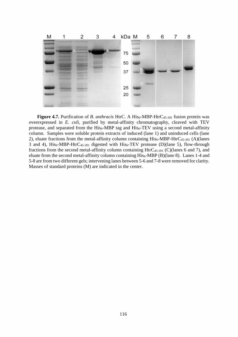
116
Figure 4.7. Purification of B. anthracis HtrC. A His6-MBP-HtrC45-391 fusion protein was
overexpressed in E. coli, purified by metal-affinity chromatography, cleaved with TEV
protease, and separated from the His6-MBP tag and His6-TEV using a second metal-affinity
column. Samples were soluble protein extracts of induced (lane 1) and uninduced cells (lane
2), eluate fractions from the metal-affinity column containing His6-MBP-HtrC45-391 (A)(lanes
3 and 4), His6-MBP-HtrC45-391 digested with His6-TEV protease (D)(lane 5), flow-through
fractions from the second metal-affinity column containing HtrC45-391 (C)(lanes 6 and 7), and
eluate from the second metal-affinity column containing His6-MBP (B)(lane 8). Lanes 1-4 and
5-8 are from two different gels; intervening lanes between 5-6 and 7-8 were removed for clarity.
Masses of standard proteins (M) are indicated in the center.

117
Figure 4.8. In vitro cleavage of YpeB by HtrC. Purified YpeB21-446 (A) (6 µM) was
incubated for 4 hours with purified HtrC45-391 (B) (1 µM), unless otherwise indicated, with
the indicated small molecule additions. Reactions were terminated using SDS and heat.
Samples were separated by SDS-PAGE and proteins, including the YpeB cleavage product
(C), were detected by staining with Coomassie Brilliant Blue. Protein concentrations were 12
µM YpeB21-446 and 2 µM HtrC45-391 in lanes 22-26. Lanes 1-7, 8-13, 14-21, and 22-26 are
from four different gels. Masses of standard proteins (M) are indicated on the left.
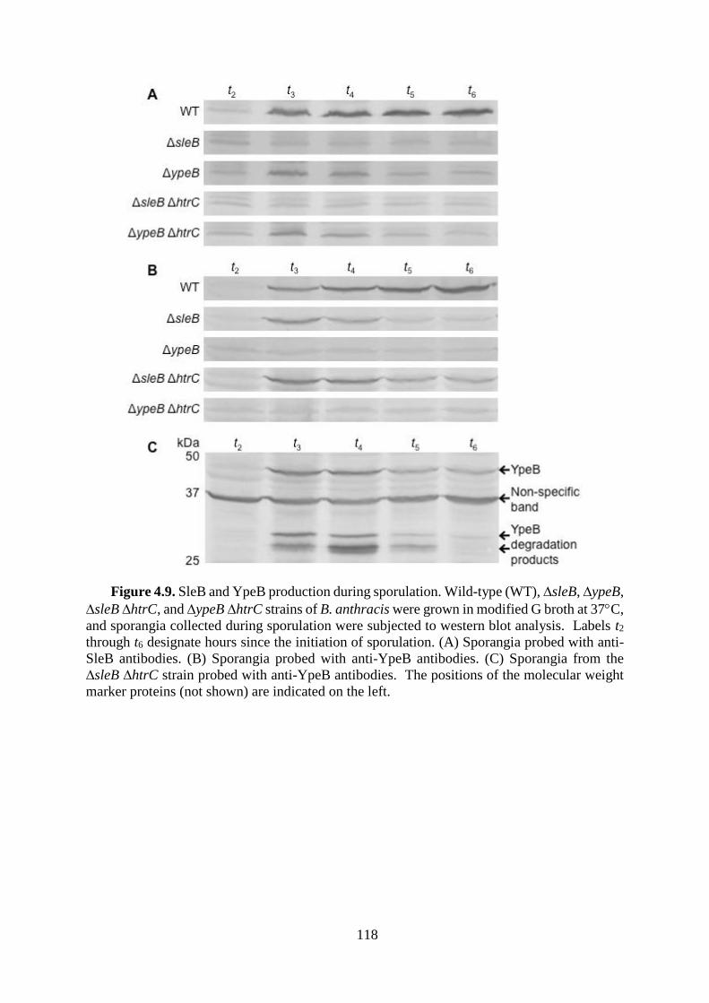
118
Figure 4.9. SleB and YpeB production during sporulation. Wild-type (WT), ∆sleB, ∆ypeB,
∆sleB ∆htrC, and ∆ypeB ∆htrC strains of B. anthracis were grown in modified G broth at 37C,
and sporangia collected during sporulation were subjected to western blot analysis. Labels t2
through t6 designate hours since the initiation of sporulation. (A) Sporangia probed with anti-
SleB antibodies. (B) Sporangia probed with anti-YpeB antibodies. (C) Sporangia from the
∆sleB ∆htrC strain probed with anti-YpeB antibodies. The positions of the molecular weight
marker proteins (not shown) are indicated on the left.

119
CHAPTER 5
Final Discussion
120
The germination of B. subtilis spores has been intensively studied for decades. Many
germination-related proteins have been shown to be involved in four distinct germination
stages. While the significance of several individual proteins in the germination process has
been characterized, the mechanisms of how these proteins perform their functions and the
network connecting these proteins in the complete germination process are still a mystery. A
substantial number of proteins that are expressed under the control of sporulation specific
sigma factors have no significant sequence similarity to known genes or functional domains.
Characterization of these unknown proteins may provide key insights into the germination
network. For example, when and how does a large quantity of water penetrate the spore inner
membrane in a short time in the conditions that the inner membrane lipids are immobile? If
peptidoglycan degradation is triggered by exogenous Ca2+-DPA, how is the timing of this
process related to spore core Ca2+-DPA release? How is the germination signal transmitted
from germinant receptors to DPA channel proteins? This work is focused on generating a
comprehensive protein identity list for the spore membrane fraction and investigating
candidate proteins for their roles in the spore germination process.
In Chapter 2, we characterized a superdormant spore population that drastically differs
from that described in a previous publication. A quantitative MRM-MS assay was performed
to analyze the relative abundance of 11 germination-related proteins in dormant, germinating,
and superdormant spores. Our findings showed that superdormancy was not due to a low
abundance of germinant receptor proteins. We found that germination of the superdormant
spores was delayed at a germination stage where the GerD lipoprotein is involved. This
evidence suggested that there is more than one pathway that can result in spore
superdormancy.
Previous studies have shown that germinant receptor proteins, SpoVA proteins, and
GerD interact with each other at the membrane (110, 111). The interaction is most likely
121
involved in germination signal transmission from recognition of the germinant to initiation of
Ca2+-DPA release. The mechanism of these interactions remains to be elucidated. Our results
showed that the abundance of germinant receptor C-subunit proteins significantly decreased
after incubating spores with nutrient germinant. A recent study also showed that altering the
level of germinant receptor C-subunit proteins or influencing their lipidation had negative
effects on both spore formation and germination (172). This evidence suggests that the
decreased level of germinant receptor C-subunit proteins might be a critical step in the
germination process. However, it is difficult to elucidate the function of each germinant
receptor subunit in vivo because the function of germinant receptors relies on the complete
heterocomplex of three subunits (27). Expression and purification of individual subunits or
intact receptor complexes could be the future direction to a comprehensive biochemical
analysis of Ger receptor proteins.
The interaction between germinant receptor proteins and the GerD lipoprotein is another
key element to be elucidated in the future. Previous evidence suggested that the germination
signal passed from multiple germinant receptors converge at GerD lipoprotein (26). The co-
localization of GerD with germinant receptor proteins might facilitate the signal transduction.
Previous studies also showed that the level of GerD is greatly reduced in germinating spores
(38, 124). However, whether the loss of GerD has direct or indirect connection to the loss of
germinant receptor C-subunit proteins is unclear. A recent crystallographic study
demonstrated that GerD protein (PDB ID: 4O8W) forms a triple-stranded rope structure that
is essential for its role on nutrient germination (173). It is likely, as suggested in the
publication, that GerD trimers self-associate to form a large mechanically rigid structure to
facilitate the interaction of various germination related proteins. If this is the case, then what
proteins receive the signal from GerD and how do they transmit it to SpoVA proteins?
Considering a significant loss of GerD from the membrane after germination, it is also
122
possible that this structure is disrupted following the release of germinant receptor C-subunit
proteins. The disruption of the GerD protein complex might be the signal to release Ca2+-
DPA through SpoVA proteins. Although the nature GerD signal transduction is unknown, its
resolved crystal structure will speed up the process of understanding its role in germinant
receptor dependent germination.
Another question that needs to be addressed in the future is the function of PrkC. Our
study showed that the quantity of this protein decreased significantly after spore germination.
PrkC was previously characterized as a germinant receptor that responds to vegetative
peptidoglycan fragments (48). What function, if any, it plays during nutrient germination
remains to be determined.
To further explore the full map of protein content on the spore membrane, we carried out
the shot-gun proteomic study in chapter 3. We chose SDS-PAGE-based MS approach
because it gave the best coverage of membrane proteins in a previous study (104). A total of
104 and 87 proteins were identified as membrane-associated in B. subtilis and B. anthracis,
respectively. The percentages of membrane proteins among the total proteins identified
achieved 15-17%. This study identified 65 membrane-associated spore proteins that had not
been previously reported in any B. subtilis or B. anthracis vegetative or spore proteomic
studies. The functions of identified spore membrane proteins spread into almost all
membrane protein function categories, including transport, protein fate determination, and
energy production and conversion. Novel membrane-associated spore proteins that were
expressed under the control of forespore-specific ϭG are highly interesting targets to be
further investigated. Some of these proteins have amino acid sequence homologies to known
inorganic ion transporters, amino acid transporters, and proteases. Proteins with these
functions may play major roles in spore formation and germination, therefore further
characterization of these proteins is needed. A quarter of these membrane-associated spore

123
proteins have no known function or significant sequence similarities to known proteins.
Further characterization of these proteins may reveal missing pieces in understanding the
sporulation, germination, and/or outgrowth processes.
Quantitative analysis of proteins in both Bacillus species revealed a notable trend that the
number of identified membrane proteins is greatly reduced in germinating spores. These
reduced membrane proteins are in multiple COG function categories, which suggested that
the protein abundance decrease is a general spore germination phenomenon. The reason
behind this phenomenon is unclear. Evidence showed that the area of the IFM experienced
significant decrease late in sporulation and dramatic increase after spore cortex is degraded
(6). Perhaps the change of membrane-associated proteome is due to the rearrangement of
membrane during spore germination.
HtrC, a membrane-associated serine protease homolog identified in both Bacillus spore
membrane proteomes, was characterized in Chapter 4 to examine its role in determining
YpeB protein fate during spore germination. We demonstrated that YpeB was proteolytically
processed during germination and a stable C-terminal fragment was produced. The primary
cleavage site was identified in B. anthracis, but other cleavage sites are accessible when the
primary site is blocked. The in vivo and in vitro evidence accumulated in this study proved
that HtrC is responsible for proteolytically cleaving YpeB and producing the stable fragment
during germination. However, without HtrC presenting in the spore, other proteases appear to
degrade YpeB at a reduced rate. This may explain why there was no significant germination
defect in htrC deletion spores. We also demonstrated that YpeB and SleB, in the absence of
their partner protein, were degraded by unknown proteases other than HtrC during spore
formation. The unknown proteases cleave YpeB at a site close to the HtrC cleavage site, and
therefore they may not be the proteases that cleave YpeB during germination when HtrC is
absent. Identification and characterization of these unknown proteases would be a future

124
direction for revealing the roles of proteases in spore germination. Further study of the
proteases identified in spore membrane proteomes could be a good start point.
In vitro characterization of HtrC showed a relatively low protease activity with a
requirement for a relatively high concentration of Mn2+ or Ca2+ ions. We suspect that the ions
may not be directly involved in HtrC catalytic function for the following reasons: 1) HtrC
belongs to a class of serine protease that requires no metal ion in catalysis (166); 2) Most
Mn2+ and Ca2+ released during germination is likely bound to DPA, and we have
demonstrated that at least Ca2+-DPA is ineffective in promoting HtrC activity; 3) The
concentration requirement of metal ions as cofactor for a protease activity is at least 1,000-
fold less than that is required for HtrC activation (174, 175). The reason why a high
concentration of Mn2+ or Ca2+ ions promotes HtrC activity in vitro is still unclear. Perhaps
like other HtrA family proteases, the PDZ domain of HtrC is critical for fully activating its
protease activity (176, 177), and the high concentration of metal ions may drive the PDZ
domain towards the active configuration. It is also possible that other proteins associated with
HtrC or other native materials released from spore core could be the native cofactors for HtrC
protease activity. Further characterization of this protease, and possibly study of its crystal
structure, would help achieve a full understand its role in the YpeB degradation process.
We hope that the studies presented here will promote an understainding of the nutrient-
mediated germination signal pathway in which many spore membrane proteins may be
involved. Our long-term purpose is to drive the development of more effective
decontamination methods utilizing knowledge of the spore germination pathway to germinate
spores prematurely and synchronously. These efforts will alleviate the public health burden
caused by spore contamination and will prevent disease transmission.
125
REFERENCES
1. Waites WM (1984) Antimicrobial Agents Acting on Bacterial-Spores. Journal of Applied Bacteriology
57(3):R6-R6.
2. Russell AD (1990) Bacterial spores and chemical sporicidal agents. Clin Microbiol Rev 3(2):99-119.
3. Foster SJ & Johnstone K (1990) Pulling the trigger: the mechanism of bacterial spore germination. Mol
Microbiol 4(1):137-141.
4. Setlow P (2007) I will survive: DNA protection in bacterial spores. Trends Microbiol 15(4):172-180.
5. Raju D, Waters M, Setlow P, & Sarker MR (2006) Investigating the role of small, acid-soluble spore
proteins (SASPs) in the resistance of Clostridium perfringens spores to heat. BMC Microbiol 6:50.
6. Cowan AE, et al. (2004) Lipids in the inner membrane of dormant spores of Bacillus species are
largely immobile. Proc Natl Acad Sci U S A 101(20):7733-7738.
7. Ramamurthi KS, Clapham KR, & Losick R (2006) Peptide anchoring spore coat assembly to the outer
forespore membrane in Bacillus subtilis. Mol Microbiol 62(6):1547-1557.
8. Atrih A, Zollner P, Allmaier G, Williamson MP, & Foster SJ (1998) Peptidoglycan structural dynamics
during germination of Bacillus subtilis 168 endospores. J Bacteriol 180(17):4603-4612.
9. Meador-Parton J & Popham DL (2000) Structural analysis of Bacillus subtilis spore peptidoglycan
during sporulation. J Bacteriol 182(16):4491-4499.
10. Driks A (1999) Bacillus subtilis spore coat. Microbiol Mol Biol Rev 63(1):1-20.
11. Driks A (2002) Maximum shields: the assembly and function of the bacterial spore coat. Trends
Microbiol 10(6):251-254.
12. Nicholson WL, Munakata N, Horneck G, Melosh HJ, & Setlow P (2000) Resistance of Bacillus
endospores to extreme terrestrial and extraterrestrial environments. Microbiol Mol Biol Rev 64(3):548-
572.
13. Setlow P (2006) Spores of Bacillus subtilis: their resistance to and killing by radiation, heat and
chemicals. J Appl Microbiol 101(3):514-525.
14. Henriques AO & Moran CP, Jr. (2007) Structure, assembly, and function of the spore surface layers.
Annu Rev Microbiol 61:555-588.
15. Wang ST, et al. (2006) The forespore line of gene expression in Bacillus subtilis. J Mol Biol 358(1):16-
37.
16. Stragier P & Losick R (1996) Molecular genetics of sporulation in Bacillus subtilis. Annu Rev Genet
30:297-241.
17. Errington J (2003) Regulation of endospore formation in Bacillus subtilis. Nat Rev Microbiol 1(2):117-
126.
18. Phillips ZE & Strauch MA (2002) Bacillus subtilis sporulation and stationary phase gene expression.
Cell Mol Life Sci 59(3):392-402.
19. Paidhungat M & Setlow P (2000) Role of ger proteins in nutrient and nonnutrient triggering of spore
germination in Bacillus subtilis. J Bacteriol 182(9):2513-2519.
20. Setlow P (2003) Spore germination. Curr Opin Microbiol 6(6):550-556.
21. Moir A (2006) How do spores germinate? Journal of Applied Microbiology 101(3):526-530.
22. Ghosh S & Setlow P (2009) Isolation and characterization of superdormant spores of Bacillus species.
J Bacteriol 191(6):1787-1797.
23. Ghosh S & Setlow P (2009) The preparation, germination properties and stability of superdormant
spores of Bacillus cereus. J Appl Microbiol.
24. Ghosh S, Scotland M, & Setlow P (2012) Levels of germination proteins in dormant and superdormant
spores of Bacillus subtilis. J Bacteriol 194(9):2221-2227.
25. Wang G, Yi X, Li YQ, & Setlow P (2011) Germination of individual Bacillus subtilis spores with
alterations in the GerD and SpoVA proteins, which are important in spore germination. J Bacteriol
193(9):2301-2311.
26. Pelczar PL, Igarashi T, Setlow B, & Setlow P (2007) Role of GerD in germination of Bacillus subtilis
spores. J Bacteriol 189(3):1090-1098.
27. Vepachedu VR & Setlow P (2005) Localization of SpoVAD to the inner membrane of spores of
Bacillus subtilis. J Bacteriol 187(16):5677-5682.
28. Paidhungat M & Setlow P (2001) Localization of a germinant receptor protein (GerBA) to the inner
membrane of Bacillus subtilis spores. J Bacteriol 183(13):3982-3990.
29. Masayama A, et al. (2006) Subcellular localization of a germiantion-specific cortex-lytic enzyme,
SleB, of Bacilli during sporulation. Genes Genet Syst 81(3):163-169.
126
30. Moir A, Lafferty E, & Smith DA (1979) Genetic-Analysis of Spore Germination Mutants of Bacillus-
Subtilis 168 - Correlation of Phenotype with Map Location. Journal of General Microbiology
111(Mar):165-180.
31. Sammons RL, Moir A, & Smith DA (1981) Isolation and Properties of Spore Germination Mutants of
Bacillus-Subtilis-168 Deficient in the Initiation of Germination. Journal of General Microbiology
124(Jun):229-241.
32. Corfe BM, Sammons RL, Smith DA, & Mauel C (1994) The gerB region of the Bacillus subtilis 168
chromosome encodes a homologue of the gerA spore germination operon. Microbiology 140 ( Pt
3):471-478.
33. Irie R, Fujita Y, & Kobayashi M (1996) Nucleotide sequence and gene organization of the gerK spore
germination locus of Bacillus subtilis 168. Journal of General and Applied Microbiology 42(2):141-
153.
34. Moir A, Corfe BM, & Behravan J (2002) Spore germination. Cell Mol Life Sci 59(3):403-409.
35. Kunst F, et al. (1997) The complete genome sequence of the gram-positive bacterium Bacillus subtilis.
Nature 390(6657):249-256.
36. Kemp EH, Sammons RL, Moir A, Sun D, & Setlow P (1991) Analysis of transcriptional control of the
gerD spore germination gene of Bacillus subtilis 168. J Bacteriol 173(15):4646-4652.
37. Pelczar PL & Setlow P (2008) Localization of the germination protein GerD to the inner membrane in
Bacillus subtilis spores. J Bacteriol 190(16):5635-5641.
38. Mongkolthanaruk W, Robinson C, & Moir A (2009) Localization of the GerD spore germination
protein in the Bacillus subtilis spore. Microbiology 155(Pt 4):1146-1151.
39. Robinson C, Rivolta C, Karamata D, & Moir A (1998) The product of the yvoC (gerF) gene of Bacillus
subtilis is required for spore germination. Microbiology 144 ( Pt 11):3105-3109.
40. Igarashi T, Setlow B, Paidhungat M, & Setlow P (2004) Effects of a gerF (lgt) mutation on the
germination of spores of Bacillus subtilis. J Bacteriol 186(10):2984-2991.
41. Sankaran K & Wu HC (1994) Lipid modification of bacterial prolipoprotein. Transfer of diacylglyceryl
moiety from phosphatidylglycerol. J Biol Chem 269(31):19701-19706.
42. Fisher N & Hanna P (2005) Characterization of Bacillus anthracis germinant receptors in vitro. J
Bacteriol 187(23):8055-8062.
43. Ireland JA & Hanna PC (2002) Amino acid- and purine ribonucleoside-induced germination of
Bacillus anthracis DeltaSterne endospores: gerS mediates responses to aromatic ring structures. J
Bacteriol 184(5):1296-1303.
44. Weiner MA, Read TD, & Hanna PC (2003) Identification and characterization of the gerH operon of
Bacillus anthracis endospores: a differential role for purine nucleosides in germination. J Bacteriol
185(4):1462-1464.
45. Read TD, et al. (2003) The genome sequence of Bacillus anthracis Ames and comparison to closely
related bacteria. Nature 423(6935):81-86.
46. Guidi-Rontani C, et al. (1999) Identification and characterization of a germination operon on the
virulence plasmid pXO1 of Bacillus anthracis. Mol Microbiol 33(2):407-414.
47. Sirard JC, Guidi-Rontani C, Fouet A, & Mock M (2000) Characterization of a plasmid region involved
in Bacillus anthracis toxin production and pathogenesis. Int J Med Microbiol 290(4-5):313-316.
48. Shah IM, Laaberki MH, Popham DL, & Dworkin J (2008) A eukaryotic-like Ser/Thr kinase signals
bacteria to exit dormancy in response to peptidoglycan fragments. Cell 135(3):486-496.
49. Sorg JA & Sonenshein AL (2008) Bile salts and glycine as cogerminants for Clostridium difficile
spores. J Bacteriol 190(7):2505-2512.
50. Paidhungat M, Setlow B, Driks A, & Setlow P (2000) Characterization of spores of Bacillus subtilis
which lack dipicolinic acid. J Bacteriol 182(19):5505-5512.
51. Magge A, et al. (2008) Role of dipicolinic acid in the germination, stability, and viability of spores of
Bacillus subtilis. J Bacteriol 190(14):4798-4807.
52. Daniel RA & Errington J (1993) Cloning, DNA sequence, functional analysis and transcriptional
regulation of the genes encoding dipicolinic acid synthetase required for sporulation in Bacillus
subtilis. J Mol Biol 232(2):468-483.
53. Driks A (2002) Overview: Development in bacteria: spore formation in Bacillus subtilis. Cell Mol Life
Sci 59(3):389-391.
54. Tovar-Rojo F, Chander M, Setlow B, & Setlow P (2002) The products of the spoVA operon are
involved in dipicolinic acid uptake into developing spores of Bacillus subtilis. J Bacteriol 184(2):584-
587.
55. Vepachedu VR & Setlow P (2004) Analysis of the germination of spores of Bacillus subtilis with
temperature sensitive spo mutations in the spoVA operon. FEMS Microbiol Lett 239(1):71-77.
127
56. Kong L, Zhang P, Setlow P, & Li YQ (2010) Characterization of Bacterial Spore Germination Using
Integrated Phase Contrast Microscopy, Raman Spectroscopy, and Optical Tweezers. Anal Chem.
57. Zhang P, Setlow P, & Li Y (2009) Characterization of single heat-activated Bacillus spores using laser
tweezers Raman spectroscopy. Opt Express 17(19):16480-16491.
58. Zhang P, et al. (2010) Factors affecting variability in time between addition of nutrient germinants and
rapid dipicolinic acid release during germination of spores of Bacillus species. J Bacteriol
192(14):3608-3619.
59. Zhang P, Kong L, Setlow P, & Li YQ (2010) Characterization of wet-heat inactivation of single spores
of bacillus species by dual-trap Raman spectroscopy and elastic light scattering. Appl Environ
Microbiol 76(6):1796-1805.
60. Thackray PD, Behravan J, Southworth TW, & Moir A (2001) GerN, an antiporter homologue
important in germination of Bacillus cereus endospores. J Bacteriol 183(2):476-482.
61. Heffron JD, Orsburn B, & Popham DL (2009) Roles of germination-specific lytic enzymes CwlJ and
SleB in Bacillus anthracis. J Bacteriol 191(7):2237-2247.
62. Lambert EA & Popham DL (2008) The Bacillus anthracis SleL (YaaH) protein is an N-
acetylglucosaminidase involved in spore cortex depolymerization. J Bacteriol 190(23):7601-7607.
63. Popham DL, Heffron JD, & Lambert EA (2012) Degradation of Spore Peptidoglycan During
Germination. Bacterial Spores: Current Research and Applications:121-142.
64. Chirakkal H, O'Rourke M, Atrih A, Foster SJ, & Moir A (2002) Analysis of spore cortex lytic enzymes
and related proteins in Bacillus subtilis endospore germination. Microbiology 148(Pt 8):2383-2392.
65. Heffron JD, Lambert EA, Sherry N, & Popham DL (2010) Contributions of four cortex lytic enzymes
to germination of Bacillus anthracis spores. J Bacteriol 192(3):763-770.
66. Moriyama R, et al. (1996) A germination-specific spore cortex-lytic enzyme from Bacillus cereus
spores: cloning and sequencing of the gene and molecular characterization of the enzyme. J Bacteriol
178(17):5330-5332.
67. Liu H, et al. (2004) Formation and composition of the Bacillus anthracis endospore. J Bacteriol
186(1):164-178.
68. Moriyama R, et al. (1999) Expression of a germination-specific amidase, SleB, of Bacilli in the
forespore compartment of sporulating cells and its localization on the exterior side of the cortex in
dormant spores. J Bacteriol 181(8):2373-2378.
69. Bernhards CB & Popham DL (2014) The role of YpeB in cortex hydrolysis during germination of
Bacillus anthracis spores. J Bacteriol.
70. Boland FM, Atrih A, Chirakkal H, Foster SJ, & Moir A (2000) Complete spore-cortex hydrolysis
during germination of Bacillus subtilis 168 requires SleB and YpeB. Microbiology 146 ( Pt 1):57-64.
71. Ishikawa S, Yamane K, & Sekiguchi J (1998) Regulation and characterization of a newly deduced cell
wall hydrolase gene (cwlJ) which affects germination of Bacillus subtilis spores. J Bacteriol
180(6):1375-1380.
72. Bagyan I & Setlow P (2002) Localization of the cortex lytic enzyme CwlJ in spores of Bacillus subtilis.
J Bacteriol 184(4):1219-1224.
73. Paidhungat M, Ragkousi K, & Setlow P (2001) Genetic requirements for induction of germination of
spores of Bacillus subtilis by Ca(2+)-dipicolinate. J Bacteriol 183(16):4886-4893.
74. Atrih A & Foster SJ (2001) In vivo roles of the germination-specific lytic enzymes of Bacillus subtilis
168. Microbiology 147(Pt 11):2925-2932.
75. Chen Y, Fukuoka S, & Makino S (2000) A novel spore peptidoglycan hydrolase of Bacillus cereus:
biochemical characterization and nucleotide sequence of the corresponding gene, sleL. J Bacteriol
182(6):1499-1506.
76. Eymann C, et al. (2004) A comprehensive proteome map of growing Bacillus subtilis cells. Proteomics
4(10):2849-2876.
77. Dreisbach A, et al. (2008) Monitoring of changes in the membrane proteome during stationary phase
adaptation of Bacillus subtilis using in vivo labeling techniques. Proteomics 8(10):2062-2076.
78. Francis AW, et al. (2005) Proteomic analysis of Bacillus anthracis Sterne vegetative cells. Biochim
Biophys Acta 1748(2):191-200.
79. Kuwana R, et al. (2002) Proteomics characterization of novel spore proteins of Bacillus subtilis.
Microbiology 148(Pt 12):3971-3982.
80. Mao L, et al. (2011) Protein profile of Bacillus subtilis spore. Curr Microbiol 63(2):198-205.
81. Huang CM, et al. (2004) Identification of Bacillus anthracis proteins associated with germination and
early outgrowth by proteomic profiling of anthrax spores. Proteomics 4(9):2653-2661.
82. Delvecchio VG, et al. (2006) Proteomic profiling and identification of immunodominant spore antigens
of Bacillus anthracis, Bacillus cereus, and Bacillus thuringiensis. Appl Environ Microbiol 72(9):6355-
6363.
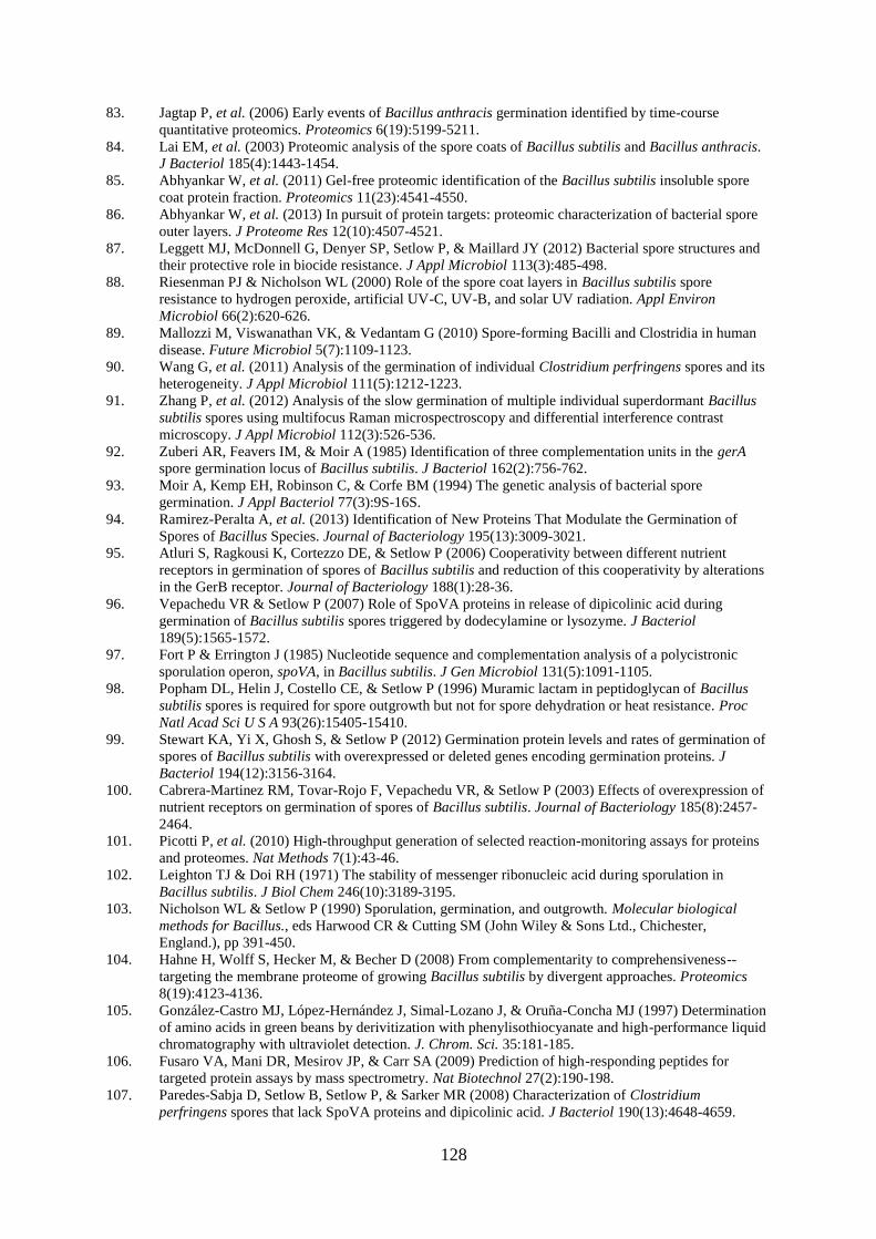
128
83. Jagtap P, et al. (2006) Early events of Bacillus anthracis germination identified by time-course
quantitative proteomics. Proteomics 6(19):5199-5211.
84. Lai EM, et al. (2003) Proteomic analysis of the spore coats of Bacillus subtilis and Bacillus anthracis.
J Bacteriol 185(4):1443-1454.
85. Abhyankar W, et al. (2011) Gel-free proteomic identification of the Bacillus subtilis insoluble spore
coat protein fraction. Proteomics 11(23):4541-4550.
86. Abhyankar W, et al. (2013) In pursuit of protein targets: proteomic characterization of bacterial spore
outer layers. J Proteome Res 12(10):4507-4521.
87. Leggett MJ, McDonnell G, Denyer SP, Setlow P, & Maillard JY (2012) Bacterial spore structures and
their protective role in biocide resistance. J Appl Microbiol 113(3):485-498.
88. Riesenman PJ & Nicholson WL (2000) Role of the spore coat layers in Bacillus subtilis spore
resistance to hydrogen peroxide, artificial UV-C, UV-B, and solar UV radiation. Appl Environ
Microbiol 66(2):620-626.
89. Mallozzi M, Viswanathan VK, & Vedantam G (2010) Spore-forming Bacilli and Clostridia in human
disease. Future Microbiol 5(7):1109-1123.
90. Wang G, et al. (2011) Analysis of the germination of individual Clostridium perfringens spores and its
heterogeneity. J Appl Microbiol 111(5):1212-1223.
91. Zhang P, et al. (2012) Analysis of the slow germination of multiple individual superdormant Bacillus
subtilis spores using multifocus Raman microspectroscopy and differential interference contrast
microscopy. J Appl Microbiol 112(3):526-536.
92. Zuberi AR, Feavers IM, & Moir A (1985) Identification of three complementation units in the gerA
spore germination locus of Bacillus subtilis. J Bacteriol 162(2):756-762.
93. Moir A, Kemp EH, Robinson C, & Corfe BM (1994) The genetic analysis of bacterial spore
germination. J Appl Bacteriol 77(3):9S-16S.
94. Ramirez-Peralta A, et al. (2013) Identification of New Proteins That Modulate the Germination of
Spores of Bacillus Species. Journal of Bacteriology 195(13):3009-3021.
95. Atluri S, Ragkousi K, Cortezzo DE, & Setlow P (2006) Cooperativity between different nutrient
receptors in germination of spores of Bacillus subtilis and reduction of this cooperativity by alterations
in the GerB receptor. Journal of Bacteriology 188(1):28-36.
96. Vepachedu VR & Setlow P (2007) Role of SpoVA proteins in release of dipicolinic acid during
germination of Bacillus subtilis spores triggered by dodecylamine or lysozyme. J Bacteriol
189(5):1565-1572.
97. Fort P & Errington J (1985) Nucleotide sequence and complementation analysis of a polycistronic
sporulation operon, spoVA, in Bacillus subtilis. J Gen Microbiol 131(5):1091-1105.
98. Popham DL, Helin J, Costello CE, & Setlow P (1996) Muramic lactam in peptidoglycan of Bacillus
subtilis spores is required for spore outgrowth but not for spore dehydration or heat resistance. Proc
Natl Acad Sci U S A 93(26):15405-15410.
99. Stewart KA, Yi X, Ghosh S, & Setlow P (2012) Germination protein levels and rates of germination of
spores of Bacillus subtilis with overexpressed or deleted genes encoding germination proteins. J
Bacteriol 194(12):3156-3164.
100. Cabrera-Martinez RM, Tovar-Rojo F, Vepachedu VR, & Setlow P (2003) Effects of overexpression of
nutrient receptors on germination of spores of Bacillus subtilis. Journal of Bacteriology 185(8):2457-
2464.
101. Picotti P, et al. (2010) High-throughput generation of selected reaction-monitoring assays for proteins
and proteomes. Nat Methods 7(1):43-46.
102. Leighton TJ & Doi RH (1971) The stability of messenger ribonucleic acid during sporulation in
Bacillus subtilis. J Biol Chem 246(10):3189-3195.
103. Nicholson WL & Setlow P (1990) Sporulation, germination, and outgrowth. Molecular biological
methods for Bacillus., eds Harwood CR & Cutting SM (John Wiley & Sons Ltd., Chichester,
England.), pp 391-450.
104. Hahne H, Wolff S, Hecker M, & Becher D (2008) From complementarity to comprehensiveness--
targeting the membrane proteome of growing Bacillus subtilis by divergent approaches. Proteomics
8(19):4123-4136.
105. González-Castro MJ, López-Hernández J, Simal-Lozano J, & Oruña-Concha MJ (1997) Determination
of amino acids in green beans by derivitization with phenylisothiocyanate and high-performance liquid
chromatography with ultraviolet detection. J. Chrom. Sci. 35:181-185.
106. Fusaro VA, Mani DR, Mesirov JP, & Carr SA (2009) Prediction of high-responding peptides for
targeted protein assays by mass spectrometry. Nat Biotechnol 27(2):190-198.
107. Paredes-Sabja D, Setlow B, Setlow P, & Sarker MR (2008) Characterization of Clostridium
perfringens spores that lack SpoVA proteins and dipicolinic acid. J Bacteriol 190(13):4648-4659.
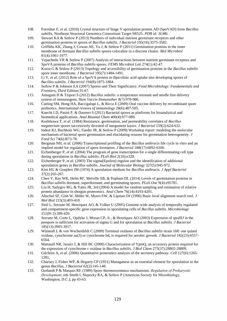
129
108. Forouhar F, et al. (2010) Crystal structure of Stage V sporulation protein AD (SpoVAD) from Bacillus
subtilis, Northeast Structural Genomics Consortium Target SR525. PDB id: 3LM6.
109. Stewart KA & Setlow P (2013) Numbers of individual nutrient germinant receptors and other
germination proteins in spores of Bacillus subtilis. J Bacteriol 195(16):3575-3582.
110. Griffiths KK, Zhang J, Cowan AE, Yu J, & Setlow P (2011) Germination proteins in the inner
membrane of dormant Bacillus subtilis spores colocalize in a discrete cluster. Mol Microbiol
81(4):1061-1077.
111. Vepachedu VR & Setlow P (2007) Analysis of interactions between nutrient germinant receptors and
SpoVA proteins of Bacillus subtilis spores. FEMS Microbiol Lett 274(1):42-47.
112. Korza G & Setlow P (2013) Topology and accessibility of germination proteins in the Bacillus subtilis
spore inner membrane. J Bacteriol 195(7):1484-1491.
113. Li Y, et al. (2012) Role of a SpoVA protein in dipicolinic acid uptake into developing spores of
Bacillus subtilis. J Bacteriol 194(8):1875-1884.
114. Setlow P & Johnson EA (2007) Spores and Their Significance. Food Microbiology: Fundamentals and
Frontiers, Third Edition:35-67.
115. Amuguni H & Tzipori S (2012) Bacillus subtilis: a temperature resistant and needle free delivery
system of immunogens. Hum Vaccin Immunother 8(7):979-986.
116. Cutting SM, Hong HA, Baccigalupi L, & Ricca E (2009) Oral vaccine delivery by recombinant spore
probiotics. International reviews of immunology 28(6):487-505.
117. Knecht LD, Pasini P, & Daunert S (2011) Bacterial spores as platforms for bioanalytical and
biomedical applications. Anal Bioanal Chem 400(4):977-989.
118. Koshikawa T, et al. (1984) Resistance, germination, and permeability correlates of Bacillus
megaterium spores successively divested of integument layers. J Bacteriol 159(2):624-632.
119. Indest KJ, Buchholz WG, Faeder JR, & Setlow P (2009) Workshop report: modeling the molecular
mechanism of bacterial spore germination and elucidating reasons for germination heterogeneity. J
Food Sci 74(6):R73-78.
120. Bergman NH, et al. (2006) Transcriptional profiling of the Bacillus anthracis life cycle in vitro and an
implied model for regulation of spore formation. J Bacteriol 188(17):6092-6100.
121. Eichenberger P, et al. (2004) The program of gene transcription for a single differentiating cell type
during sporulation in Bacillus subtilis. PLoS Biol 2(10):e328.
122. Eichenberger P, et al. (2003) The sigma(Epsilon) regulon and the identification of additional
sporulation genes in Bacillus subtilis. Journal of Molecular Biology 327(5):945-972.
123. Kim HU & Goepfert JM (1974) A sporulation medium for Bacillus anthracis. J Appl Bacteriol
37(2):265-267.
124. Chen Y, Ray WK, Helm RF, Melville SB, & Popham DL (2014) Levels of germination proteins in
Bacillus subtilis dormant, superdormant, and germinating spores. PLoS One 9(4):e95781.
125. Liu H, Sadygov RG, & Yates JR, 3rd (2004) A model for random sampling and estimation of relative
protein abundance in shotgun proteomics. Anal Chem 76(14):4193-4201.
126. Altschul SF, Gish W, Miller W, Myers EW, & Lipman DJ (1990) Basic local alignment search tool. J
Mol Biol 215(3):403-410.
127. Steil L, Serrano M, Henriques AO, & Volker U (2005) Genome-wide analysis of temporally regulated
and compartment-specific gene expression in sporulating cells of Bacillus subtilis. Microbiology
151(Pt 2):399-420.
128. Serrano M, Corte L, Opdyke J, Moran CP, Jr., & Henriques AO (2003) Expression of spoIIIJ in the
prespore is sufficient for activation of sigma G and for sporulation in Bacillus subtilis. J Bacteriol
185(13):3905-3917.
129. Winstedt L & von Wachenfeldt C (2000) Terminal oxidases of Bacillus subtilis strain 168: one quinol
oxidase, cytochrome aa(3) or cytochrome bd, is required for aerobic growth. J Bacteriol 182(23):6557-
6564.
130. Mattatall NR, Jazairi J, & Hill BC (2000) Characterization of YpmQ, an accessory protein required for
the expression of cytochrome c oxidase in Bacillus subtilis. J Biol Chem 275(37):28802-28809.
131. Gilchrist A, et al. (2006) Quantitative proteomics analysis of the secretory pathway. Cell 127(6):1265-
1281.
132. Charney J, Fisher WP, & Hegarty CP (1951) Managanese as an essential element for sporulation in the
genus Bacillus. J Bacteriol 62(2):145-148.
133. Gerhardt P & Marquis RE (1989) Spore thermoresistance mechanisms. Regulation of Prokaryotic
Development, eds Smith I, Slepecky RA, & Setlow P (American Society for Microbiology,
Washington, D.C.), pp 43-63.
130
134. Granger AC, Gaidamakova EK, Matrosova VY, Daly MJ, & Setlow P (2011) Effects of Mn and Fe
levels on Bacillus subtilis spore resistance and effects of Mn2+, other divalent cations, orthophosphate,
and dipicolinic acid on protein resistance to ionizing radiation. Appl Environ Microbiol 77(1):32-40.
135. Swerdlow BM, Setlow B, & Setlow P (1981) Levels of H+ and other monovalent cations in dormant
and germinating spores of Bacillus megaterium. J Bacteriol 148(1):20-29.
136. Foster SJ & Johnstone K (1986) The Use of Inhibitors to Identify Early Events during Bacillus-
Megaterium Km Spore Germination. Biochemical Journal 237(3):865-870.
137. Gaballa A & Helmann JD (1998) Identification of a zinc-specific metalloregulatory protein, Zur,
controlling zinc transport operons in Bacillus subtilis. J Bacteriol 180(22):5815-5821.
138. Fujisawa M, Wada Y, Tsuchiya T, & Ito M (2009) Characterization of Bacillus subtilis YfkE (ChaA): a
calcium-specific Ca2+/H+ antiporter of the CaCA family. Arch Microbiol 191(8):649-657.
139. Tanaka K, Kobayashi K, & Ogasawara N (2003) The Bacillus subtilis YufLM two-component system
regulates the expression of the malate transporters MaeN (YufR) and YflS, and is essential for
utilization of malate in minimal medium. Microbiology 149(Pt 9):2317-2329.
140. Chillappagari S, Miethke M, Trip H, Kuipers OP, & Marahiel MA (2009) Copper acquisition is
mediated by YcnJ and regulated by YcnK and CsoR in Bacillus subtilis. J Bacteriol 191(7):2362-2370.
141. Gibson MM, Bagga DA, Miller CG, & Maguire ME (1991) Magnesium transport in Salmonella
typhimurium: the influence of new mutations conferring Co2+ resistance on the CorA Mg2+ transport
system. Mol Microbiol 5(11):2753-2762.
142. Lorca G, Winnen B, & Saier MH, Jr. (2003) Identification of the L-aspartate transporter in Bacillus
subtilis. J Bacteriol 185(10):3218-3222.
143. Sekowska A, Robin S, Daudin JJ, Henaut A, & Danchin A (2001) Extracting biological information
from DNA arrays: an unexpected link between arginine and methionine metabolism in Bacillus
subtilis. Genome Biol 2(6):RESEARCH0019.
144. Hullo MF, Auger S, Dassa E, Danchin A, & Martin-Verstraete I (2004) The metNPQ operon of
Bacillus subtilis encodes an ABC permease transporting methionine sulfoxide, D- and L-methionine.
Res Microbiol 155(2):80-86.
145. Fabret C & Hoch JA (1998) A two-component signal transduction system essential for growth of
Bacillus subtilis: implications for anti-infective therapy. J Bacteriol 180(23):6375-6383.
146. Fukuchi K, et al. (2000) The essential two-component regulatory system encoded by yycF and yycG
modulates expression of the ftsAZ operon in Bacillus subtilis. Microbiology 146 ( Pt 7):1573-1583.
147. Dalbey RE, Wang P, & van Dijl JM (2012) Membrane proteases in the bacterial protein secretion and
quality control pathway. Microbiol Mol Biol Rev 76(2):311-330.
148. Patel VJ, et al. (2009) A comparison of labeling and label-free mass spectrometry-based proteomics
approaches. J Proteome Res 8(7):3752-3759.
149. Setlow P (2014) Germination of spores of Bacillus species: what we know and do not know. J
Bacteriol 196(7):1297-1305.
150. Atrih A, Zollner P, Allmaier G, & Foster SJ (1996) Structural analysis of Bacillus subtilis 168
endospore peptidoglycan and its role during differentiation. J Bacteriol 178(21):6173-6183.
151. Sekiguchi J, et al. (1995) Nucleotide sequence and regulation of a new putative cell wall hydrolase
gene, cwlD, which affects germination in Bacillus subtilis. J Bacteriol 177(19):5582-5589.
152. Setlow B, Melly E, & Setlow P (2001) Properties of spores of Bacillus subtilis blocked at an
intermediate stage in spore germination. J Bacteriol 183(16):4894-4899.
153. Ragkousi K, Eichenberger P, van Ooij C, & Setlow P (2003) Identification of a new gene essential for
germination of Bacillus subtilis spores with Ca2+-dipicolinate. J Bacteriol 185(7):2315-2329.
154. Setlow B, et al. (2009) Characterization of the germination of Bacillus megaterium spores lacking
enzymes that degrade the spore cortex. J Appl Microbiol 107(1):318-328.
155. Li Y, et al. (2013) Activity and regulation of various forms of CwlJ, SleB, and YpeB proteins in
degrading cortex peptidoglycan of spores of Bacillus species in vitro and during spore germination. J
Bacteriol 195(11):2530-2540.
156. Ho SN, Hunt HD, Horton RM, Pullen JK, & Pease LR (1989) Site-directed mutagenesis by overlap
extension using the polymerase chain reaction. Gene 77(1):51-59.
157. van den Ent F & Lowe J (2006) RF cloning: A restriction-free method for inserting target genes into
plasmids. J Biochem Bioph Meth 67(1):67-74.
158. Janes BK & Stibitz S (2006) Routine markerless gene replacement in Bacillus anthracis. Infect Immun
74(3):1949-1953.
159. Bishop-Lilly KA, et al. (2012) Whole genome sequencing of phage resistant Bacillus anthracis
mutants reveals an essential role for cell surface anchoring protein CsaB in phage AP50c adsorption.
Virol J 9:246.
131
160. Wach A (1996) PCR-synthesis of marker cassettes with long flanking homology regions for gene
disruptions in S. cerevisiae. Yeast 12(3):259-265.
161. Austin BP, Nallamsetty S, & Waugh DS (2009) Hexahistidine-tagged maltose-binding protein as a
fusion partner for the production of soluble recombinant proteins in Escherichia coli. Methods Mol Biol
498:157-172.
162. Kapust RB, et al. (2001) Tobacco etch virus protease: mechanism of autolysis and rational design of
stable mutants with wild-type catalytic proficiency. Protein Eng 14(12):993-1000.
163. Giebel JD, Carr KA, Anderson EC, & Hanna PC (2009) The germination-specific lytic enzymes SleB,
CwlJ1, and CwlJ2 each contribute to Bacillus anthracis spore germination and virulence. J Bacteriol
191(18):5569-5576.
164. Campo N & Rudner DZ (2007) SpoIVB and CtpB are both forespore signals in the activation of the
sporulation transcription factor sigma K in Bacillus subtilis. J Bacteriol 189(16):6021-6027.
165. Mastny M, et al. (2013) CtpB assembles a gated protease tunnel regulating cell-cell signaling during
spore formation in Bacillus subtilis. Cell 155(3):647-658.
166. Clausen T, Southan C, & Ehrmann M (2002) The HtrA family of proteases: implications for protein
composition and cell fate. Mol Cell 10(3):443-455.
167. Slepecky R & Foster JW (1959) Alterations in metal content of spores of Bacillus megaterium and the
effect on some spore properties. J Bacteriol 78(1):117-123.
168. Boschwitz H, Gofshtein-Gandman L, Halvorson HO, Keynan A, & Milner Y (1991) The possible
involvement of trypsin-like enzymes in germination of spores of Bacillus cereus T and Bacillus subtilis
168. J Gen Microbiol 137(5):1145-1153.
169. Boschwitz H, Halvorson HO, Keynan A, & Milner Y (1985) Trypsinlike enzymes from dormant and
germinated spores of Bacillus cereus T and their possible involvement in germination. J Bacteriol
164(1):302-309.
170. Boschwitz H, Milner Y, Keynan A, Halvorson HO, & Troll W (1983) Effect of inhibitors of trypsin-
like proteolytic enzymes Bacillus cereus T spore germination. J Bacteriol 153(2):700-708.
171. Cortezzo DE, Setlow B, & Setlow P (2004) Analysis of the action of compounds that inhibit the
germination of spores of Bacillus species. J Appl Microbiol 96(4):725-741.
172. Igarashi T & Setlow P (2005) Interaction between individual protein components of the GerA and
GerB nutrient receptors that trigger germination of Bacillus subtilis spores. Journal of Bacteriology
187(7):2513-2518.
173. Li Y, et al. (2014) Structural and functional analysis of the GerD spore germination protein of Bacillus
species. J Mol Biol 426(9):1995-2008.
174. Petersen LC, Olsen OH, Nielsen LS, Freskgard PO, & Persson E (2000) Binding of Zn2+ to a Ca2+ loop
allosterically attenuates the activity of factor VIIa and reduces its affinity for tissue factor. Protein Sci
9(5):859-866.
175. Wakabayashi H, Zhen Z, Schmidt KM, & Fay PJ (2003) Mn2+ binding to factor VIII subunits and its
effect on cofactor activity. Biochemistry 42(1):145-153.
176. Krojer T, et al. (2008) Interplay of PDZ and protease domain of DegP ensures efficient elimination of
misfolded proteins. Proc Natl Acad Sci U S A 105(22):7702-7707.
177. Murwantoko, et al. (2004) Binding of proteins to the PDZ domain regulates proteolytic activity of
HtrA1 serine protease. Biochem J 381(Pt 3):895-904.