characterization and comparative temperate bacillusmegaterium bacteriophages · 2018-12-10 ·...
TRANSCRIPT
Submitted 27 February 2018Accepted 3 September 2018Published 10 December 2018
Corresponding authorAbdoallah Sharaf,[email protected],[email protected]
Academic editorRamy Aziz
Additional Information andDeclarations can be found onpage 16
DOI 10.7717/peerj.5687
Copyright2018 Sharaf et al.
Distributed underCreative Commons CC-BY 4.0
OPEN ACCESS
Characterization and comparativegenomic analysis of virulent andtemperate Bacillus megateriumbacteriophagesAbdoallah Sharaf1,2, Miroslav Oborník2,3, Adel Hammad4, Sohair El-Afifi5 andEman Marei5
1Genetic Department, Faculty of Agriculture, Ain Shams University, Cairo, Egypt2 Institute of Parasitology, Biology Centre, Czech Academy of Sciences, České Budějovice, Czech Republic3 Faculty of Science, University of South Bohemia, České Budějovice, Czech Republic4Department of Microbiology, Faculty of Agriculture, Minia University, Minia, Egypt5Department of Agricultural Microbiology, Virology Laboratory, Ain Shams University, Cairo, Egypt
ABSTRACTNext-Generation Sequencing (NGS) technologies provide unique possibilities forthe comprehensive assessment of the environmental diversity of bacteriophages.Several Bacillus bacteriophages have been isolated, but very few Bacillus megateriumbacteriophages have been characterized. In this study, we describe the biologicalcharacteristics, whole genome sequences, and annotations for two new isolates of theB. megaterium bacteriophages (BM5 and BM10), which were isolated from Egyptiansoil samples. Growth analyses indicated that the phages BM5 and BM10 have a shorterlatent period (25 and 30 min, respectively) and a smaller burst size (103 and 117 PFU,respectively), in comparison to what is typical for Bacillus phages. The genome sizesof the phages BM5 and BM10 were 165,031 bp and 165,213 bp, respectively, withmodular organization. Bioinformatic analyses of these genomes enabled the assignmentof putative functions to 97 and 65 putative ORFs, respectively. Comparative analysisof the BM5 and BM10 genome structures, in conjunction with other B. megateriumbacteriophages, revealed relatively high levels of sequence and organizational identity.Both genomic comparisons and phylogenetic analyses support the conclusion thatthe sequenced phages (BM5 and BM10) belong to different sub-clusters (L5 and L7,respectively), within the L-cluster, and display different lifestyles (lysogenic and lytic,respectively). Moreover, sequenced phages encode proteins associated with Bacilluspathogenesis. In addition, BM5 does not contain any tRNA sequences, whereas BM10genome codes for 17 tRNAs.
Subjects Agricultural Science, Bioinformatics, Genomics, VirologyKeywords Bacillus megaterium, Bacteriophage, Phylogenetic analysis, Comparative genomics
INTRODUCTIONBacillus megaterium (Firmicutes) is ubiquitous in nature. It is recognized as an endophyteand a potential biocontrol agent for plant diseases (Kildea et al., 2008). Furthermore, itis known to produce penicillin, amidase, various baking amylases and is used for the
How to cite this article Sharaf et al. (2018), Characterization and comparative genomic analysis of virulent and temperate Bacillus mega-terium bacteriophages. PeerJ 6:e5687; DOI 10.7717/peerj.5687
biotechnological production of pyruvate, vitamin B12, and so on (Bunk et al., 2009).In alkaline soils, B. megaterium plays a significant role in making insoluble phosphorusaccessible to plants via the production of CO2 and organic acids, which increases soilacidity and the fraction of solubilized phosphates. Certain B. megaterium strains evenexhibit nitrogen fixation capability (Bergey, 2009). Bacteriophages (phages) are the mostabundant biological entities present on the planet (Hambly & Suttle, 2005). Their capacityto infect and kill their hosts makes them an important factor driving the bacterial evolutionand preservation of the ecological balance (Chan, Abedon & Loc-Carrillo, 2013; Singh,Poshtiban & Evoy, 2013). Phages can be utilized for various purposes, such as therapyfor bacterial infections, for biocontrol of pathogens as protein exposure systems, and forbacterial typing (Haq et al., 2012). A number of phages infecting the genus Bacillus havebeen identified so far (Zeigler, 1999; Lee et al., 2011). Bacillus-specific phages demonstrategreat diversity with regard to morphology, genome sequence length, gene content, and hostrange, exhibiting high variability among and within the species of this genus. Moreover,numerous bacteriophages that infect and lyse B. megaterium have been studied (Cooney,Jacob & Slepecky, 1975; Carvalho & Vary, 1977; Vary & Halsey, 1980; Van Elsas & Penido,1982; Serwer et al., 2007; Eppinger et al., 2011). These phages possess double-stranded,linear DNA genomes (Van Elsas, Linhares & Penido, 1982).
Furthermore, the expansion of next generation sequencing (NGS) technologies(Eisenstein, 2012; Klumpp, Fouts & Sozhamannan, 2012), in addition to the possibilityof sequencing entire genomes or transcriptomes more efficiently and economically,rather than using the Sanger sequencing strategy, has allowed for obtaining full genomicsequences for a wide range of species. Therefore, the advent of NGS technology providesnew opportunities for sequencing of a broad range of organisms, including bacteriophages,quickly, reliably, and considerably inexpensively (Hatfull, 2008).
Here, we shed light on the biological features, genome sequence and annotation oftemperate and virulent B. megaterium bacteriophages, representing two different groupsaccording to their host range and amplified fragment length polymorphism (AFLP) profile(Elmaghraby et al., 2015). Both phages display different thermal inactivation points (82and 78 ◦C) and pH tolerant range (5–9.2 and 5–8.4 pH) while having the same longevityin vitro (192 h) (Elmaghraby et al., 2015). Electron microscopy proved that both phagesbelonged to the Myoviridiae family (Elmaghraby et al., 2015). Furthermore, in this paper,we present an updated phylogenetic analysis of Bacillus bacteriophages based on the aminoacid sequence of terminases.
MATERIALS AND METHODBacteriophagesTwo isolates of B. megaterium phages, namely BM5 and BM10, were obtained from soil.Their biological and morphological properties were reported previously (Elmaghraby et al.,2015). The high titer phage suspension of each isolate was prepared using a liquid cultureenrichment technique (Marie, 2013). Fifteen ml of the same high titer phage suspensionfor each phage was ultra-centrifuged at 30,000 rpm for 90 min at 4 ◦C in a Beckman
Sharaf et al. (2018), PeerJ, DOI 10.7717/peerj.5687 2/24
L7-35 ultracentrifuge. The pellet was gently re-suspended in 0.5 ml of a 0.2 M potassiumphosphate buffer, with a pH level of 7.2 (Lanningan & Williams, 1982). A single-stepgrowth experiment was performed with minor modifications, as reported previously, todetermine the latent and rise period and the phage burst size (Pajunen, Kiljunen & Skurnik,2000; Sillankorva, Neubauer & Azeredo, 2008). Furthermore, 1 ml of the phage suspension(1010 pfu/ml) was mixed with 1 ml of exponential phase culture of B. megaterium (108
cfu/ml) and incubated at 30 ◦C for five minutes for phage adsorption. Subsequently, themixture was centrifuged at 10,000 rpm for 10 min to remove free phage particles. Thepellet was re-suspended in 60 ml of a nutrient broth, and the culture was continuouslyincubated at 30 ◦C. Phages were sampled at intervals of 5 to 60 min, and their titers weredetermined. The burst sizes of the phages were estimated by dividing the bacteriophagetiters at the plateau phase using the initial phage titers.
DNA isolation, library preparation, and whole genome sequencingGenomic DNA was extracted from the bacteriophages, as described by Irshad, Waqas &Saadia (2012), with minor adjustments. The purity and concentration of the DNA wasevaluated using a Nanodrop Bioanalyzer N1000 (Thermo Scientific, Waltham, MA, USA).In addition, sequencing libraries were prepared by shearing 1 µg of the phage DNA, togenerate blunt-ended fragments, after which the Ion adapters were linked using an IonXpressTM Fragment Library Kit (Life Technologies, Carlsbad, CA, USA), in accordance withthemanufacturer’s instructions. The produced fragments were amplified employing the IonOneTouch 200Template kit (Life Technologies, Carlsbad, CA,USA). Furthermore, librarieswere sequenced on an Ion Torrent PGM semiconductor sequencer using the Ion Torrent314 chip, by applying the standard protocol (Life Technologies, Carlsbad, CA, USA).
Sequence assembly and annotationRaw sequencing reads were trimmed and masked using the FASTQ Trimmer andthe FASTQ Masker Galaxy Tools Version 1.0.0 (Blankenberg, Gordon & Kuster, 2010),which were assembled by the 454 Newbler Assembler software (Roche Applied Science,IN). The collected contigs were visualized and validated by Consed (Gordon & Green,2013), resulting in the presence of contigs in each phage, which demonstrated a 60-foldsequence read coverage approximately. In addition, sequence homology analysis andassignment to the phage clusters were performed using BLASTn against the Bacillusphage database (http://bacillus.phagesdb.org/: 09/2016) (Altschul et al., 1990). The genomesequences of each phage were annotated using the NCBI Prokaryotic AnnotationPipeline. In addition, protein alignment was used for gene predictions to guaranteethe consistency of the annotation for closely related genomes. ProSplign (proteinaligner software) was utilized to align all the protein sequences. For further refinement,frameshifted alignments and partial alignments were processed using a gene predictionprogram (GeneMarkS+), and final annotations were established by searching againstbacterial and bacteriophage proteins in the SwissProt database (Besemer, Lomsadze &Borodovsky, 2001). The gene predictions were verified using the CPT (Center for PhageTechnology) Galaxy (https://cpt.tamu.edu/galaxy-pub/). Furthermore, for genome-wide
Sharaf et al. (2018), PeerJ, DOI 10.7717/peerj.5687 3/24
analyses and other searches, a Phamerator software was employed (Cresawn et al., 2011).Subsequently, conserved domains relationships within genes were determined. Proteinswere assorted into ‘‘Phamilies’’ (generally referred to as ‘‘Phams’’), which are groupsof proteins that are largely similar to one another. BLASTp and ClustalW were used todetermine the pairwise alignment scores (Cresawn et al., 2011). Phage RNA Polymeraseand RNP-recognized promoters were located using the extractupstreamDNA software(https://github.com/ajvilleg/extractUpStreamDNA), after which theMEMEprogram (http://meme-suite.org) was used for motifs elicitation (Bailey & Elkan, 1994; Bailey et al., 2009).
Genomic similarity and phylogenetic analysisProteome similarity and average nucleotide identity (ANI) values of 16 genome sequencesof Bacillus phages, which were retrieved from GenBank, were determined using JSpecies1.2.1 (http://imedea.uib-csic.es/jspecies/download.html) at the default BLASTp thresholdof score 75 (Richter & Rosselló-Móra, 2009). Gepard was used to create dot plots (Krumsiek,Arnold & Rattei, 2007). To simplify the dot plot analyses, certain phage genomes werereverse complemented, and new sequences were generated to re-orient on the basis of themajority of the phages. A maximum likelihood phylogenetic tree was computed by theRAxML program and by utilizing the lg model (Stamatakis, 2014). The Bayesian tree wascalculated by the PhyloBayes program, with both the lg and the C20 models (Lartillot,Lepage & Blanquart, 2009) based on the terminase protein sequence. The supportingvalues from both methods were merged into a one-rooted tree while the Alloherpesviridaesequences (Anguillid and Cyprinid herpesvirus) were used as the out-group.
Nucleotide sequence accession numbersThe complete genome sequences of phages BM5 and BM10 were deposited in the NCBIGenBank database under accession numbers KT995479 and KT995480, respectively.Moreover, the raw sequencing data were published in the Sequence Read Archive (SRA)database under the accession number SRP136802.
RESULTSSingle-step growth curveTo assess the ability of both phages (BM5 and BM10) to lyse B. megaterium, the latent andrise periods and the burst sizes of each phage were determined by a single-step growth curveanalysis (Fig. 1). The latent periods for BM5 and BM10 phages were estimated to be 25 minand 30 min, respectively. The rise period for both phages was 45 min. The calculated burstsize was 103 pfu/cell and 117 pfu/cell, for the BM5 and BM10 phages, respectively.
Genome organizationThe genome sequencing generated 267,273 reads for BM5 phage and 288,254 reads forBM10 phage, forming an approximately 60-fold coverage. Assemblies yielded 165 kbpand 165.2 kbp genomes for BM5 and BM10, respectively. A comparison of the translatedBLAST (tblastx) data (Altschul et al., 1990) from the two genome sequences revealed thatthey shared a high level of overall similarity; conserved regions reached 91% identity,
Sharaf et al. (2018), PeerJ, DOI 10.7717/peerj.5687 4/24

Figure 1 Single-step growth curve of BM5 and BM10 bacteriophages.Full-size DOI: 10.7717/peerj.5687/fig-1
distributed within the variable regions and with variability ranging from 23% to 88%. Bothphage genomes show the common bacteriophages genome organization, with genes of asimilar function clustering together (Fig. 2). The last 30 kb of DNA genome sequences ofBM5 encoding, for helicases and helicase related proteins, were highly similar to the DNAgenome sequences of BM10 (Fig. 2). Annotation of the BM5 genome revealed 225 ORFs(Open Reading Frames), while the BM10 genome encodes revealed 283 ORFs. A total of97 of the 225 ORFs (43%) in the BM5 genome and 65 of the 283 (23%) in the BM10genome were assigned functions in comparison with the identified conserved domains(Marchler-Bauer, Derbyshire & Gonzales, 2015) (Tables S1 and S2). In addition, that weidentified 17 genes’ coding for tRNAs in the BM10 genome (Table 1).
Phage structure and assembly genesGenes Bm5_9 and Bm5_62 were identified as encoding for the major capsid protein(TIGR04387) (Choi, McPartland & Kaganman, 2008) and the phage capsid protein(pfam05065) (Sutter, Boehringer & Gutmann, 2008), respectively. They were concurrentlythought to be involved in the stabilization of the condensed formof theDNA in phage heads.Genes Bm5_61 andBm5_184 encode for phage headmaturation proteases (Peptidase_U35)(pfam04586), which are also involved in phage head development (Duda, Martincic &Hendrix, 1995). Some genes that are essential for the tail development, such as Bm5_66(TIGR01725), Bm5_68 (cl21657), and Bm5_76 (pfam05709), were also found. Genesinvolved in head and tail development, such as Bm10_67 encoding for Caudovirus prohead
Sharaf et al. (2018), PeerJ, DOI 10.7717/peerj.5687 5/24
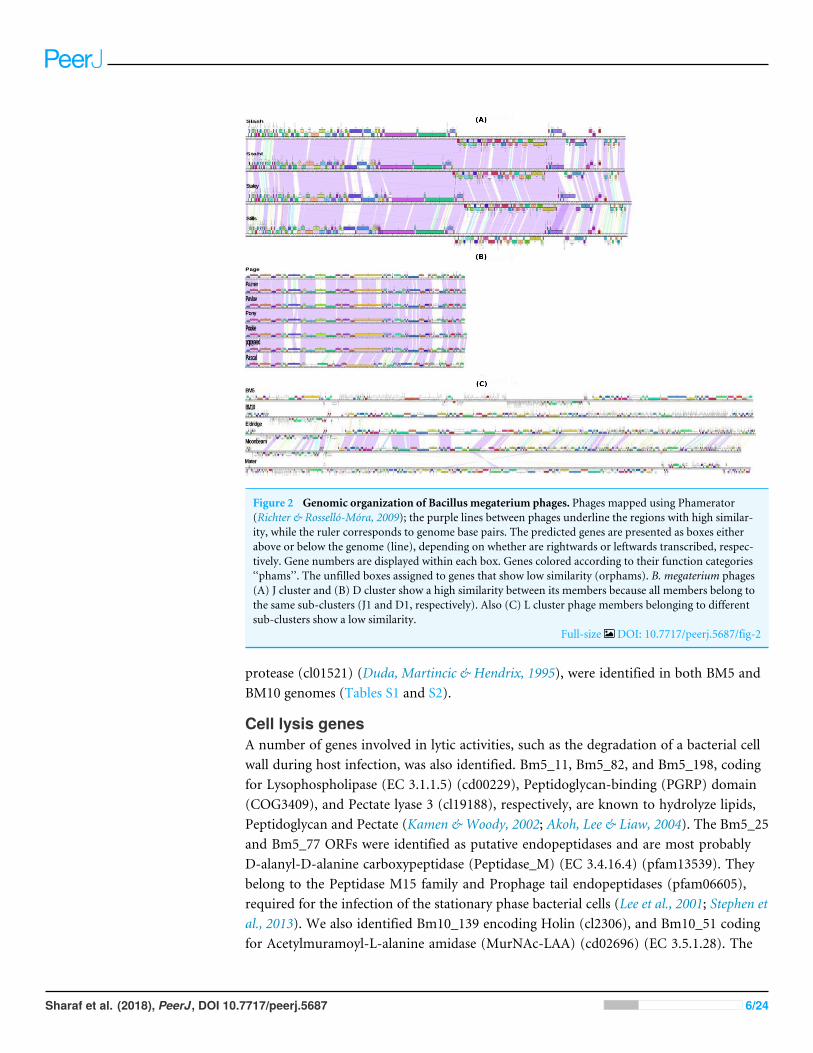
Figure 2 Genomic organization of Bacillus megaterium phages. Phages mapped using Phamerator(Richter & Rosselló-Móra, 2009); the purple lines between phages underline the regions with high similar-ity, while the ruler corresponds to genome base pairs. The predicted genes are presented as boxes eitherabove or below the genome (line), depending on whether are rightwards or leftwards transcribed, respec-tively. Gene numbers are displayed within each box. Genes colored according to their function categories‘‘phams’’. The unfilled boxes assigned to genes that show low similarity (orphams). B. megaterium phages(A) J cluster and (B) D cluster show a high similarity between its members because all members belong tothe same sub-clusters (J1 and D1, respectively). Also (C) L cluster phage members belonging to differentsub-clusters show a low similarity.
Full-size DOI: 10.7717/peerj.5687/fig-2
protease (cl01521) (Duda, Martincic & Hendrix, 1995), were identified in both BM5 andBM10 genomes (Tables S1 and S2).
Cell lysis genesA number of genes involved in lytic activities, such as the degradation of a bacterial cellwall during host infection, was also identified. Bm5_11, Bm5_82, and Bm5_198, codingfor Lysophospholipase (EC 3.1.1.5) (cd00229), Peptidoglycan-binding (PGRP) domain(COG3409), and Pectate lyase 3 (cl19188), respectively, are known to hydrolyze lipids,Peptidoglycan and Pectate (Kamen &Woody, 2002; Akoh, Lee & Liaw, 2004). The Bm5_25and Bm5_77 ORFs were identified as putative endopeptidases and are most probablyD-alanyl-D-alanine carboxypeptidase (Peptidase_M) (EC 3.4.16.4) (pfam13539). Theybelong to the Peptidase M15 family and Prophage tail endopeptidases (pfam06605),required for the infection of the stationary phase bacterial cells (Lee et al., 2001; Stephen etal., 2013). We also identified Bm10_139 encoding Holin (cl2306), and Bm10_51 codingfor Acetylmuramoyl-L-alanine amidase (MurNAc-LAA) (cd02696) (EC 3.5.1.28). The
Sharaf et al. (2018), PeerJ, DOI 10.7717/peerj.5687 6/24
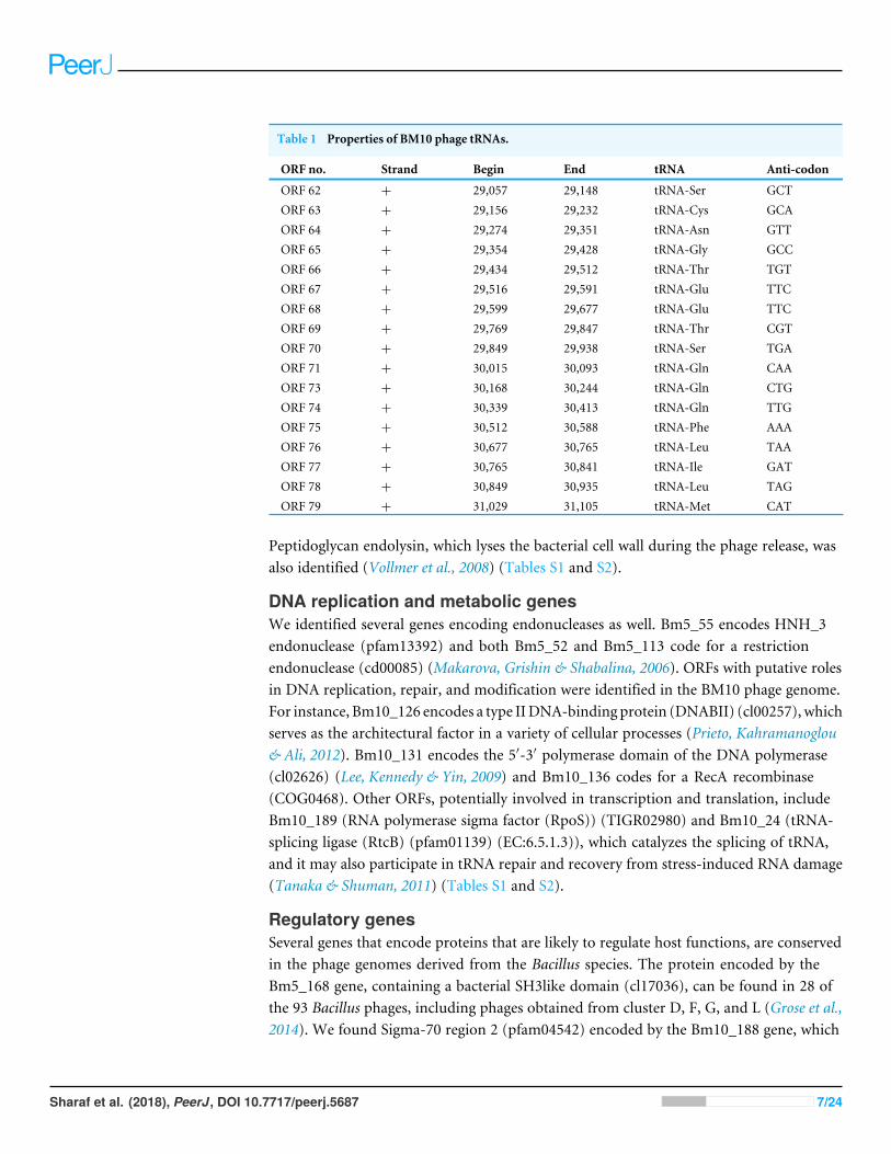
Table 1 Properties of BM10 phage tRNAs.
ORF no. Strand Begin End tRNA Anti-codon
ORF 62 + 29,057 29,148 tRNA-Ser GCTORF 63 + 29,156 29,232 tRNA-Cys GCAORF 64 + 29,274 29,351 tRNA-Asn GTTORF 65 + 29,354 29,428 tRNA-Gly GCCORF 66 + 29,434 29,512 tRNA-Thr TGTORF 67 + 29,516 29,591 tRNA-Glu TTCORF 68 + 29,599 29,677 tRNA-Glu TTCORF 69 + 29,769 29,847 tRNA-Thr CGTORF 70 + 29,849 29,938 tRNA-Ser TGAORF 71 + 30,015 30,093 tRNA-Gln CAAORF 73 + 30,168 30,244 tRNA-Gln CTGORF 74 + 30,339 30,413 tRNA-Gln TTGORF 75 + 30,512 30,588 tRNA-Phe AAAORF 76 + 30,677 30,765 tRNA-Leu TAAORF 77 + 30,765 30,841 tRNA-Ile GATORF 78 + 30,849 30,935 tRNA-Leu TAGORF 79 + 31,029 31,105 tRNA-Met CAT
Peptidoglycan endolysin, which lyses the bacterial cell wall during the phage release, wasalso identified (Vollmer et al., 2008) (Tables S1 and S2).
DNA replication and metabolic genesWe identified several genes encoding endonucleases as well. Bm5_55 encodes HNH_3endonuclease (pfam13392) and both Bm5_52 and Bm5_113 code for a restrictionendonuclease (cd00085) (Makarova, Grishin & Shabalina, 2006). ORFs with putative rolesin DNA replication, repair, and modification were identified in the BM10 phage genome.For instance, Bm10_126 encodes a type IIDNA-binding protein (DNABII) (cl00257), whichserves as the architectural factor in a variety of cellular processes (Prieto, Kahramanoglou& Ali, 2012). Bm10_131 encodes the 5′-3′ polymerase domain of the DNA polymerase(cl02626) (Lee, Kennedy & Yin, 2009) and Bm10_136 codes for a RecA recombinase(COG0468). Other ORFs, potentially involved in transcription and translation, includeBm10_189 (RNA polymerase sigma factor (RpoS)) (TIGR02980) and Bm10_24 (tRNA-splicing ligase (RtcB) (pfam01139) (EC:6.5.1.3)), which catalyzes the splicing of tRNA,and it may also participate in tRNA repair and recovery from stress-induced RNA damage(Tanaka & Shuman, 2011) (Tables S1 and S2).
Regulatory genesSeveral genes that encode proteins that are likely to regulate host functions, are conservedin the phage genomes derived from the Bacillus species. The protein encoded by theBm5_168 gene, containing a bacterial SH3like domain (cl17036), can be found in 28 ofthe 93 Bacillus phages, including phages obtained from cluster D, F, G, and L (Grose et al.,2014). We found Sigma-70 region 2 (pfam04542) encoded by the Bm10_188 gene, which
Sharaf et al. (2018), PeerJ, DOI 10.7717/peerj.5687 7/24
represents the most conserved region of the entire RNA polymerase sigma-G factor proteinand is known to interact with the clamp domain of the largest beta polymerase subunit(Jones et al., 2001) (Tables S1 and S2).
Genes involved in pathogenesisThe Trimeric dUTP diphosphatase (cl00493) and NTP-Ppase (cd11533) proteins thatmay show the host pathogenesis are encoded by the Bm10_110 and Bm5_108 genes,respectively. These proteins are commonly present in the bacteriophage genomes and havebeen made to function as G protein-like regulators, which are required for the transferof staphylococcal virulence factors (Tormo-Más, Mir & Shrestha, 2010; Tormo-Más et al.,2013). Bm5_70 encoding Sialidase (cd15482) plays a vital role in the phage pathogenesis(Telford et al., 2011) (Tables S1 and S2).
Furthermore, 224 and 266 phage-RNA polymerase (RNP) promoters were identified inthe BM5 and BM10 phages, respectively, which were all located between the genes (TablesS3 and S4). Motif analysis conducted in this regard reveals 213 conserved motifs (27nucleotides) for the BM5 phage, while a shorter conserved motif (21 nucleotides) and withthe same motif logo was identified in 254 of the BM10 phage-RNA polymerase promoters(Fig. S1). Moreover, additional eight conserved motifs (27 nucleotides) were identified inthe BM10 phage with specific DNA recognition sites for the host (Bacillus) transcription.So far, none of the downstreamORFs of this host promoter have been functionally assigned(Fig. S1).
Temperate lifestyle genesOnly the BM5 encodes genes that are suggestive of a temperate lifestyle, such asIntegrase_AP2 (pfam14657), were found in a variety of phage integrase proteins, includingthe ICEBs1 integrase from the Bacillus subtilis (Lee et al., 2007). Furthermore, the BM5encodes a putative site-specific tyrosine recombinase (cd01189) with a conserved C-terminal catalytic domain of other phage integrases, such as the P1 Cre and the lambda Int(Argos et al., 1986) (Tables S1 and S2).
tRNA genes and codon usagePhage genomes usually encode a few or no tRNA genes, because they use the hostmachineryfor the synthesis of proteins encoded in the phage genomes. Seventeen tRNA genes wereidentified in the BM10 genome, while no tRNA genes were identified in the BM5 genome.The 17 BM10 tRNA genes ranged from 77 to 92 nucleotides in size, with one copy eachfor tRNA-Cys, tRNA-Asn, tRNA-Gly, tRNA-Phe, tRNA-Ile, tRNA-Met, two copies oftRNA-Ser, tRNA-Thr, tRNA-Glu, and tRNA-leu, and three copies of tRNA-Gln (Table 1).All the tRNAs are located in the region between the HTH_XRE (HTH: Helix-turn-Helix)protein-encoding gene and a phage portal protein gene. The presence of tRNAs have beenreported in several other phage genomes and are likely to be supporting the phage proteintranslation efficiency (Grose et al., 2014) and virulence (Bailly-Bechet, Vergassola & Rocha,2007).
We compared the codon usage pattern of the BM10 tRNAs with that of its host (B.megaterium) using the tRNAscan-SE program (Chan & Lowe, 2016; Lowe & Eddy, 1997).
Sharaf et al. (2018), PeerJ, DOI 10.7717/peerj.5687 8/24
We saw that in 10 cases the phage-encoded tRNAs may significantly enhance translation inthe phage (Table S5), based on the amino acid usage and/or the codon usage. The productsof two genes, Bm10_123 and Bm10_154, appear to encode the nucleotidyltransferase(El-Arabi et al., 2013) and the tRNAHis guanylyltransferase (Hyde et al., 2010; El-Arabi etal., 2013), which may modify the phage or the host tRNAs. The BM10 phage also includesputative tRNAs (tRNA-ThrCGT, tRNA-GlnCTG and tRNA-PheAAA), which could be utilizedduring the infection of an alternative host (Delesalle et al., 2016).
The genomic relationship between BM5 and BM10Analysis of the conserved predicted proteins, encoded in the genomes of BM5 and BM10phages, revealed the huge diversity of these phages, with a total of 407 protein families(phams) of which only 52 (12.8%) were shared by both phages and 355 (87.2%) phamswere phage specific or orphams (phams containing a gene product from a single phage).Twenty-one (40.4%) of the 52 shared phams are associated with an identified function(Fig. 3), while the remaining are not.
Bm10_41 and Bm5_159 are members of the pham no.12, assigned to Thymidylatesynthase (TS), a well-conserved nucleotide metabolism enzyme identified in 28 Bacillusphages (Fig. 3). This enzyme catalyzes the alkylation of the C5 carbon of the pyrimidinenucleotides, known to induce protection against the host restriction system (Sotelo-Mundo et al., 2006). Bm10_121, Bm5_222, and Bm5_223 are members of pham no.58,composed of the Exonuclease subunit SbcD, one of the DNA replicator proteins. Genesencoding structural and assembly proteins were also conserved within the Bacillus phages(Fig. 3). These proteins show a high level of sequence conservation across the Myoviridae,Siphoviridae, Podoviridae, and Tectiviridae phage families. Identified phams include thephage tail sheath protein, encoded by pham n.35 (Bm10_94 and Bm5_193) and theBaseplate J-like protein, encoded by pham n.51 (Bm10_112 and Bm5_214). These geneshave homologs in 33 of the 93 recognized Bacillus phages (35%). In addition, Phageterminase, encoded by pham n.18 (Bm10_52 and Bm5_170), and Caudovirus proheadprotease, encoded by pham n.27 (Bm10_84 and Bm5_184), have homologs in 27 of the 93investigated Bacillus phages (Fig. 3).
Several proteins that regulate host metabolism (including pathogenesis) are alsoconserved in Bacillus phages (Fig. 3). The 235 kDa-fam (TIGR01612) protein, encodedby pham n.43 (Bm10_102, Bm10_104, and Bm5_206), represents a Reticulocyte-bindingprotein which is localized on the host cell surface and is required in the process of invasionof the parasite (Khan, Jarra & Preiser, 2001). The carbohydrate-binding domain (cl19911),encoded by pham n.52 (Bm10_113 and Bm5_215), is necessary for the carbohydrate-protein interaction (Johnson, Joshi & Tomme, 1996). Two protein phams involved inlysogenic pathways were identified (Fig. 3): Bm10_96 and Bm5_199, members of phamn.37, were identified in three conserved aspartate residues (3D) domain-containing proteins(cd14667), typically found in conjunction with a membrane-bound lytic transglycosylase.Bm10_100 and Bm5_203, members of pham n.41, were assigned to Lysozyme (EC 3.2.1.17)(Fig. 3).
Sharaf et al. (2018), PeerJ, DOI 10.7717/peerj.5687 9/24
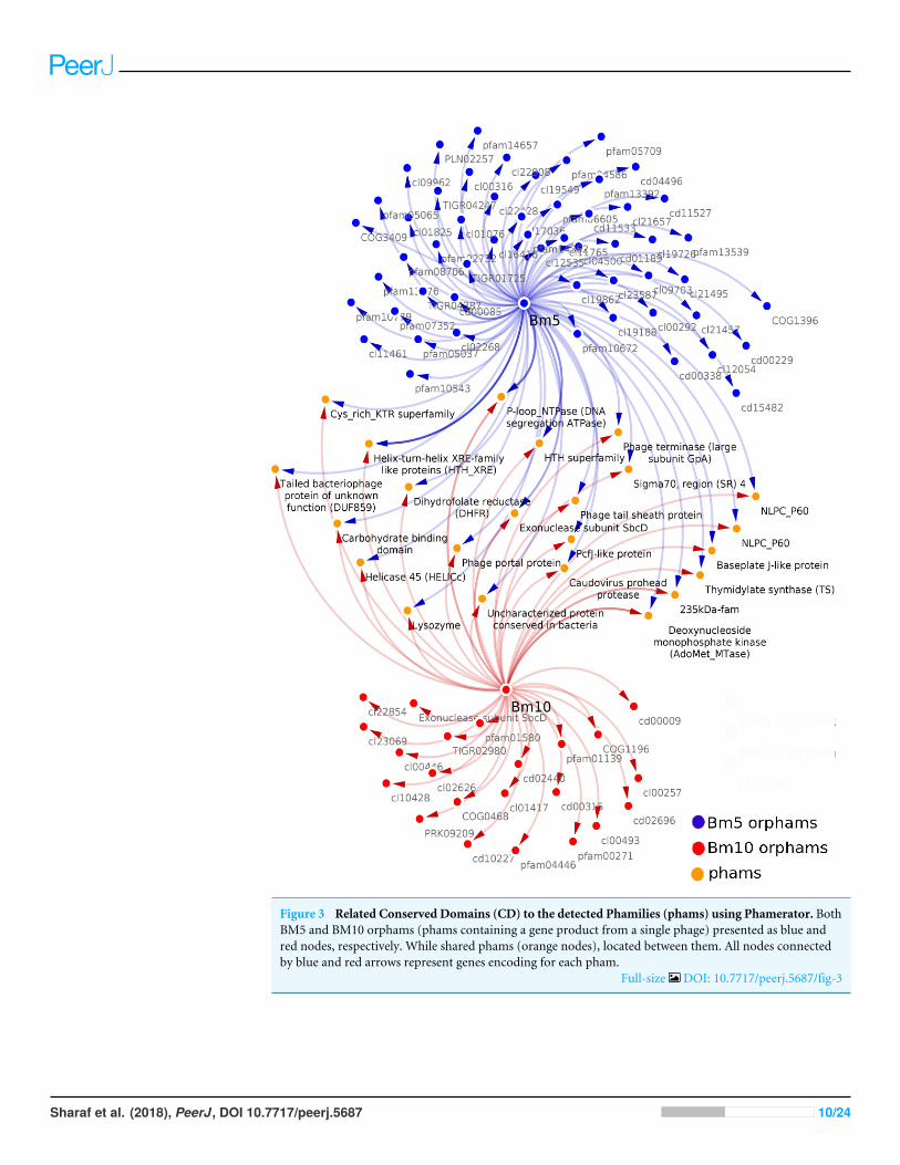
Figure 3 Related Conserved Domains (CD) to the detected Phamilies (phams) using Phamerator. BothBM5 and BM10 orphams (phams containing a gene product from a single phage) presented as blue andred nodes, respectively. While shared phams (orange nodes), located between them. All nodes connectedby blue and red arrows represent genes encoding for each pham.
Full-size DOI: 10.7717/peerj.5687/fig-3
Sharaf et al. (2018), PeerJ, DOI 10.7717/peerj.5687 10/24
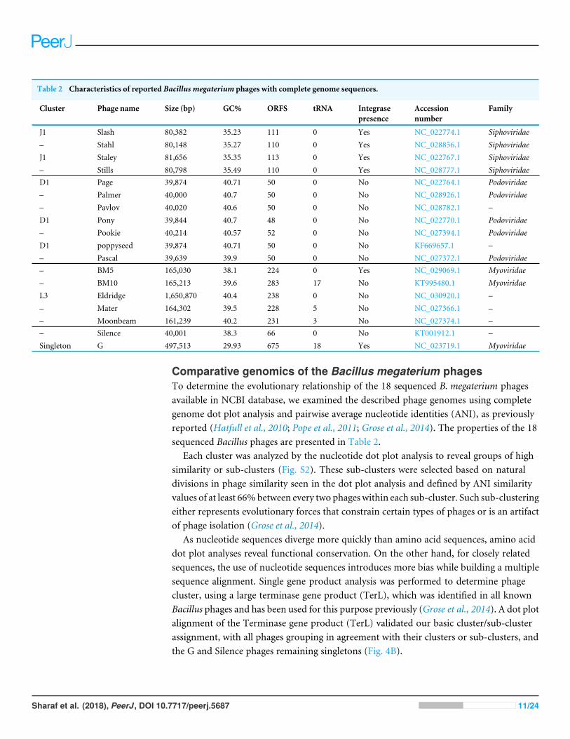
Table 2 Characteristics of reported Bacillus megaterium phages with complete genome sequences.
Cluster Phage name Size (bp) GC% ORFS tRNA Integrasepresence
Accessionnumber
Family
J1 Slash 80,382 35.23 111 0 Yes NC_022774.1 Siphoviridae– Stahl 80,148 35.27 110 0 Yes NC_028856.1 SiphoviridaeJ1 Staley 81,656 35.35 113 0 Yes NC_022767.1 Siphoviridae– Stills 80,798 35.49 110 0 Yes NC_028777.1 SiphoviridaeD1 Page 39,874 40.71 50 0 No NC_022764.1 Podoviridae– Palmer 40,000 40.7 50 0 No NC_028926.1 Podoviridae– Pavlov 40,020 40.6 50 0 No NC_028782.1 –D1 Pony 39,844 40.7 48 0 No NC_022770.1 Podoviridae– Pookie 40,214 40.57 52 0 No NC_027394.1 PodoviridaeD1 poppyseed 39,874 40.71 50 0 No KF669657.1 –– Pascal 39,639 39.9 50 0 No NC_027372.1 Podoviridae– BM5 165,030 38.1 224 0 Yes NC_029069.1 Myoviridae– BM10 165,213 39.6 283 17 No KT995480.1 MyoviridaeL3 Eldridge 1,650,870 40.4 238 0 No NC_030920.1 –– Mater 164,302 39.5 228 5 No NC_027366.1 –– Moonbeam 161,239 40.2 231 3 No NC_027374.1 –– Silence 40,001 38.3 66 0 No KT001912.1 –Singleton G 497,513 29.93 675 18 Yes NC_023719.1 Myoviridae
Comparative genomics of the Bacillus megaterium phagesTo determine the evolutionary relationship of the 18 sequenced B. megaterium phagesavailable in NCBI database, we examined the described phage genomes using completegenome dot plot analysis and pairwise average nucleotide identities (ANI), as previouslyreported (Hatfull et al., 2010; Pope et al., 2011; Grose et al., 2014). The properties of the 18sequenced Bacillus phages are presented in Table 2.
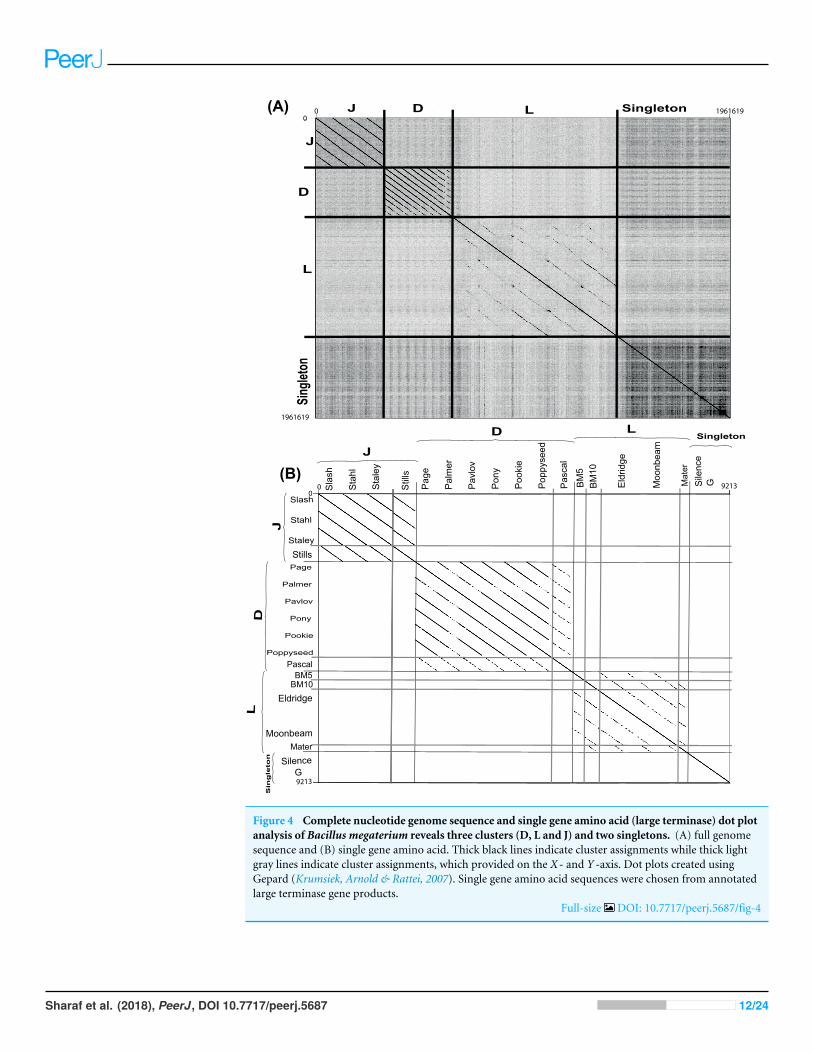
Each cluster was analyzed by the nucleotide dot plot analysis to reveal groups of highsimilarity or sub-clusters (Fig. S2). These sub-clusters were selected based on naturaldivisions in phage similarity seen in the dot plot analysis and defined by ANI similarityvalues of at least 66%between every two phageswithin each sub-cluster. Such sub-clusteringeither represents evolutionary forces that constrain certain types of phages or is an artifactof phage isolation (Grose et al., 2014).
As nucleotide sequences diverge more quickly than amino acid sequences, amino aciddot plot analyses reveal functional conservation. On the other hand, for closely relatedsequences, the use of nucleotide sequences introduces more bias while building a multiplesequence alignment. Single gene product analysis was performed to determine phagecluster, using a large terminase gene product (TerL), which was identified in all knownBacillus phages and has been used for this purpose previously (Grose et al., 2014). A dot plotalignment of the Terminase gene product (TerL) validated our basic cluster/sub-clusterassignment, with all phages grouping in agreement with their clusters or sub-clusters, andthe G and Silence phages remaining singletons (Fig. 4B).
Sharaf et al. (2018), PeerJ, DOI 10.7717/peerj.5687 11/24
Slas
h
Stah
l
Stal
ey
Stills
Stills
Slash
Stahl
Staley
Page
Palmer
Pavlov
Pony
Pookie
Poppyseed
Pascal
Pasc
al
BM5BM10
BM5
BM10
Mater
Mat
er
Sile
nce
G
Silence G
Eldridge
Moonbeam
(B)
Page
Palm
er
Pavl
ov
Pony
Pook
ie
Popp
ysee
d
Eldr
idge
Moo
nbea
m
J D L Singleton
J
D
L
Singleton
(A)
J
D LSingleton
JD
LSingleton
9213
9213
00
00
1961619
1961619
Figure 4 Complete nucleotide genome sequence and single gene amino acid (large terminase) dot plotanalysis of Bacillus megaterium reveals three clusters (D, L and J) and two singletons. (A) full genomesequence and (B) single gene amino acid. Thick black lines indicate cluster assignments while thick lightgray lines indicate cluster assignments, which provided on the X- and Y -axis. Dot plots created usingGepard (Krumsiek, Arnold & Rattei, 2007). Single gene amino acid sequences were chosen from annotatedlarge terminase gene products.
Full-size DOI: 10.7717/peerj.5687/fig-4
Sharaf et al. (2018), PeerJ, DOI 10.7717/peerj.5687 12/24
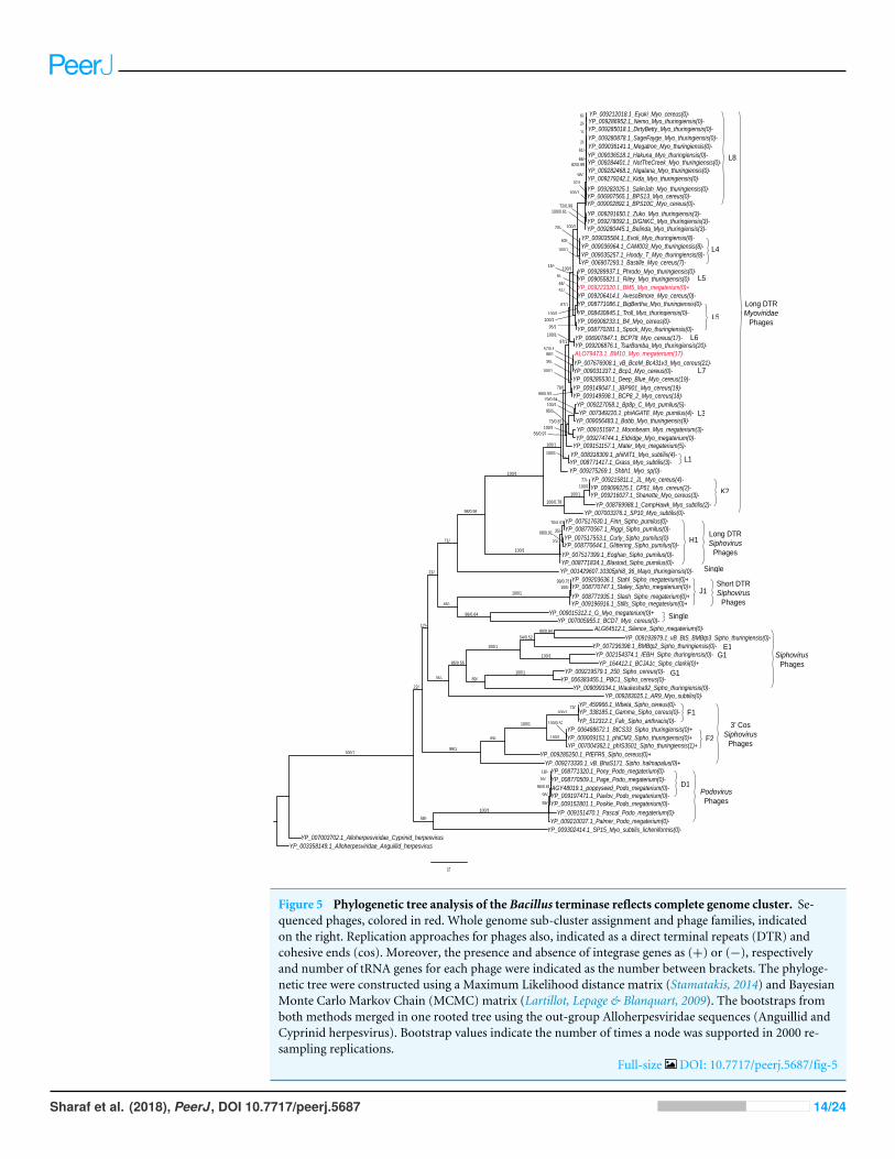
Phylogenetic analysisAnalysis of terminase (TerL) diversity in BM5, BM10 and the other 87 Bacillus phages(Fig. 5) provides a robust assignment of their phylogeny (Casjens, 2011) and can be usedto interpret the phage replication strategy, (Grose et al., 2014) and to investigate the hybridgenerations between phage genera (Canchaya, Fournous & Chibani-Chennoufi, 2003) orfamilies (Recktenwald & Schmidt, 2002). The 89 Bacillus phages were grouped into fivemain clades (Fig. 5).
On the phylogenetic tree, we have indicated the numbers of tRNA genes encoded ineach phage order to describe its distribution among the Bacillus phages. An associationbetween sub-cluster and number of tRNA genes was been identified. Phages without tRNAswere observed in all sub-clusters—L8, L5, H1, J1, F1, K2, and D1. Phages encoding similarnumbers of tRNAs were identified in sub-clusters L1 and L4; the same pattern was observedinMycobacteriophages (Delesalle et al., 2016). An exception was found in the F2 sub-cluster,where only one member (phiIS3501) contained one tRNA gene. It was described as anunusual pro-phage, different from the rest of its family, containing the tRNA-Met genelocated upstream of the lysogenic integrase gene (Moumen, Nguen-The & Sorokin, 2012).Also, for identification of the lytic and temperate clusters within the Bacillus phage, thepresence and absence of integrase genes in each phage was exhibited in the tree. Weidentified seven lytic sub-clusters (L8, L4, L5, K2, H1, f1 and D1), with all their memberslacking the integrase gene, and two temperate sub-clusters (J1 and F2). Interestingly, oursequenced BM5 phage was the only phage containing the lysogenic integrase gene withinthe L cluster and even within all long DTR Myoviridae Bacillus phages. Other temperatephages were previously observed within the lytic Mycobacteriophages cluster (Delesalle etal., 2016). To confirm clustering of the BM5 and BM10 phages, a whole genome pairwiseANI analysis was performed using JSpecies 1.2.1 between the BM5 and BM10 phages andother phages clustered in the same clade (Table S6). The ANI percentages between theBM5 phages and the L5 cluster phages (Phrodo, Riley, AvesoBmore, BigBertha, and Troll)ranged from 94% to 98%, while it ranged from 86% to 93% between the BM10 phage andthe L7 cluster phages (vB_BceM_Bc431v3 and Bcp1).
DISCUSSIONOur isolated phages (BM5 and BM10) show low latent period, which is lower than expectedforB. megaterium (45–60min) and otherBacillus phages (34–145min) (Brodetsky & Romig,1965; Colasito & Rogoff, 1969a; Colasito & Rogoff, 1969b; Cooney, Jacob & Slepecky, 1975;El-Arabi et al., 2013; Krasowska, Biegalska & Augustyniak, 2015). This is probably becauseof the strong activity of endolysins in the BM5 and BM10 phages, which is likely due tomutations in their genes encoding of holin (Stephen et al., 2013) and large burst size, which,in turn, might be caused due to different factors, such as host cell size, culture age, and/orthe host physiological conditions (Abedon, Hyman & Thomas, 2003; Choi, Kuatsjah & Wu,2010; Bolger-Munro, Cheung & Fang, 2013). Moreover, the larger burst size and shorterlatency period for both phages reflect their active propagation in the host (Delesalle et al.,2016;Wilson, 1973).
Sharaf et al. (2018), PeerJ, DOI 10.7717/peerj.5687 13/24
0.7
ALO79473.1_BM10_Myo_megaterium(17)-
YP_008430845.1_Troll_Myo_thuringiensis(0)-
YP_008770747.1_Staley_Sipho_megaterium(0)+
YP_009219579.1_250_Sipho_cereus(0)-
YP_009035584.1_Evoli_Myo_thuringiensis(8)-
YP_009036518.1_Hakuna_Myo_thuringiensis(0)-
YP_009282468.1_Nigalana_Myo_thuringiensis(0)-
YP_009151597.1_Moonbeam_Myo_megaterium(3)-
YP_008769988.1_CampHawk_Myo_subtilis(2)-
YP_007005955.1_BCD7_Myo_cereus(0)-
YP_006383455.1_PBC1_Sipho_cereus(0)-
YP_009274744.1_Eldridge_Myo_megaterium(0)-
YP_009206876.1_TsarBomba_Myo_thuringiensis(20)-
YP_338185.1_Gamma_Sipho_cereus(0)-
YP_009055821.1_Riley_Myo_thuringiensis(0)-
YP_009285530.1_Deep_Blue_Myo_cereus(19)-
YP_459966.1_Wbeta_Sipho_cereus(0)-
YP_009196916.1_Stills_Sipho_megaterium(0)+YP_009015312.1_G_Myo_megaterium(0)+
YP_008318309.1_phiNIT1_Myo_subtilis(4)-
YP_007349220.1_phiAGATE_Myo_pumilus(4)-
YP_009193979.1_vB_BtS_BMBtp3_Sipho_thuringiensis(0)-
YP_006907565.1_BPS13_Myo_cereus(0)-
YP_009036964.1_CAM003_Myo_thuringiensis(8)-
YP_009291650.1_Zuko_Myo_thuringiensis(3)-
YP_008770281.1_Spock_Myo_thuringiensis(0)-
ALG64512.1_Silence_Sipho_megaterium(0)-
YP_009283025.1_AR9_Myo_subtilis(0)-
YP_009203636.1_Stahl_Sipho_megaterium(0)+
YP_009286952.1_Nemo_Myo_thuringiensis(0)-
YP_009289937.1_Phrodo_Myo_thuringiensis(0)-
YP_006488672.1_BtCS33_Sipho_thuringiensis(0)+
YP_008770509.1_Page_Podo_megaterium(0)-
YP_009216027.1_Shanette_Myo_cereus(3)-
YP_009285250.1_PfEFR5_Sipho_cereus(0)+
YP_009215811.1_JL_Myo_cereus(4)-
YP_009197471.1_Pavlov_Podo_megaterium(0)-
YP_003358149.1_Alloherpesviridae_Anguillid_herpesvirus
AGY48019.1_poppyseed_Podo_megaterium(0)-
YP_007517553.1_Curly_Sipho_pumilus(0)-
YP_007236398.1_BMBtp2_Sipho_thuringiensis(0)-
YP_009285018.1_DirtyBetty_Myo_thuringiensis(0)-
YP_007517630.1_Finn_Sipho_pumilus(0)-
YP_006908233.1_B4_Myo_cereus(0)-
YP_002154374.1_IEBH_Sipho_thuringiensis(0)-
YP_009009151.1_phiCM3_Sipho_thuringiensis(0)+
YP_009284401.1_NotTheCreek_Myo_thuringiensis(0)-
YP_001429607.10305phi8_36_Mayo_thuringiensis(0)-
YP_009227058.1_Bp8p_C_Myo_pumilus(5)-
YP_009035257.1_Hoody_T_Myo_thuringiensis(8)-
YP_009149047.1_JBP901_Myo_cereus(19)-
YP_009279242.1_Kida_Myo_thuringiensis(0)-
YP_009212018.1_Eyuki_Myo_cereus(0)-
YP_009282025.1_SalinJah_Myo_thuringiensis(0)-
YP_008770644.1_Glittering_Sipho_pumilus(0)-YP_007517399.1_Eoghan_Sipho_pumilus(0)-
YP_008771935.1_Slash_Sipho_megaterium(0)+
YP_009273330.1_vB_BhaS171_Sipho_halmapalus(0)+
YP_009206414.1_AvesoBmore_Myo_cereus(0)-
YP_009002892.1_BPS10C_Myo_cereus(0)-
YP_009302414.1_SP15_Myo_subtilis_licheniformis(0)-YP_007003702.1_Alloherpesviridae_Cyprinid_herpesvirus
YP_009151157.1_Mater_Myo_megaterium(5)-
YP_009275269.1_Shbh1_Myo_sp(0)-
YP_008771320.1_Pony_Podo_megaterium(0)-
YP_006907293.1_Bastille_Myo_cereus(7)-
YP_007004362.1_phIS3501_Sipho_thuringiensis(1)+
YP_009036141.1_Megatron_Myo_thuringiensis(0)-
YP_009099225.1_CP51_Myo_cereus(2)-
YP_009280878.1_SageFayge_Myo_thuringiensis(0)-
YP_008770567.1_Riggi_Sipho_pumilus(0)-
YP_009280445.1_Belinda_Myo_thuringiensis(3)-
YP_009151470.1_Pascal_Podo_megaterium(0)-
YP_008771086.1_BigBertha_Myo_thuringiensis(0)-
YP_008771834.1_Blastoid_Sipho_pumilus(0)-
YP_009278092.1_DIGNKC_Myo_thuringiensis(3)-
YP_009031337.1_Bcp1_Myo_cereus(0)-
YP_009223320.1_BM5_Myo_megaterium(0)+
YP_008771417.1_Grass_Myo_subtilis(3)-
YP_009152801.1_Pookie_Podo_megaterium(0)-
YP_006907847.1_BCP78_Myo_cereus(17)-
YP_007003376.1_SP10_Myo_subtilis(0)-
YP_512312.1_Fah_Sipho_anthracis(0)-
YP_009099334.1_Waukesha92_Sipho_thuringiensis(0)-
YP_009210037.1_Palmer_Podo_megaterium(0)-
YP_009149598.1_BCP8_2_Myo_cereus(18)-
YP_009056483.1_Bobb_Myo_thuringiensis(9)-
YP_007676908.1_vB_BceM_Bc431v3_Myo_cereus(21)-
YP_164412.1_BCJA1c_Sipho_clarkii(0)+
79/0.94
98/-
66/-
16/-
71/-
69/-
97/1
100/1
98/0.64
82/0.99
100/1
79/1
21/-
72/0.99
100/1
100/1
99/0.93
100/1
76/0.63
100/1
97/1
100/1
73/-
44/-
35/-
35/-
100/1
2/-
100/1
36/-
98/-
100/1
100/1
96/0.61
22/-
100/1
95/1
100/1
100/1
59/-
100/1
20/-
68/0.91
63/-
56/0.97
54/0.52
100/1
89/-
18/-
73/0.87
95/1
100/-
87/1
100/1
100/1
77/-
100/1
100/1
98/1
85/0.55
100/1
100/1
99/0.76
61/-
17/-
9/-
1/-
100/0.78
51/-
100/0.61
99/1
57/0.8
20/-
58/-
100/0.52
15/-
100/1
6/-
100/1
2/-
98/0.98
100/1
90/0.84
46/-
L8
L4
L5
L5
L6
L7
L3
L1
K2
H1
J1
Single
Single
E1G1
G1
F1
F2
D1
Long DTRMyoviridae
Phages
Long DTRSiphovirus
Phages
Short DTRSiphovirus
Phages
SiphovirusPhages
3’ CosSiphovirus
Phages
PodovirusPhages
Figure 5 Phylogenetic tree analysis of the Bacillus terminase reflects complete genome cluster. Se-quenced phages, colored in red. Whole genome sub-cluster assignment and phage families, indicatedon the right. Replication approaches for phages also, indicated as a direct terminal repeats (DTR) andcohesive ends (cos). Moreover, the presence and absence of integrase genes as (+) or (−), respectivelyand number of tRNA genes for each phage were indicated as the number between brackets. The phyloge-netic tree were constructed using a Maximum Likelihood distance matrix (Stamatakis, 2014) and BayesianMonte Carlo Markov Chain (MCMC) matrix (Lartillot, Lepage & Blanquart, 2009). The bootstraps fromboth methods merged in one rooted tree using the out-group Alloherpesviridae sequences (Anguillid andCyprinid herpesvirus). Bootstrap values indicate the number of times a node was supported in 2000 re-sampling replications.
Full-size DOI: 10.7717/peerj.5687/fig-5
Sharaf et al. (2018), PeerJ, DOI 10.7717/peerj.5687 14/24
Bioinformatic analyses of the BM5 and BM10 genomes reveal that the genomeorganization and annotation are in general agreement with other studies of bacteriophages(Grande et al., 2014; Leon-Velarde et al., 2014). A similar mosaic genome structure wasobserved in most other phage genomes, indicating the extensive horizontal geneticexchange within the phage communities (Rohwer & Edwards, 2002;Van Dessel et al., 2005).Moreover, bacteriophage proteins represent the majority of the members of the identifiedprotein families. The obtained identified genes were involved in phage structuring, DNAreplication, nucleotide metabolism, lysis, and the repairing of proteins. The presence of the17th tRNAs in the BM10 phage genome, which has a larger burst size (117 pfu/cell) thanthe BM5 phage (103 pfu/cell), along with the absence of tRNAs support the hypothesis thatbacteriophages with tRNA genes have a larger burst size as a result of their fast propagationin their hosts (Delesalle et al., 2016;Wilson, 1973). This high number of tRNAs within phagegenomes is associated with phage virulence and higher codon usage bias (Bailly-Bechet,Vergassola & Rocha, 2007).
The analysis of the genome sequences of the BM5 and BM10 phages revealed highdiversity (66.5% ANI) in these phages. Some of the identified phams can be used inevolutionary and phylogenetic studies. As reported for Bacillus phages (Grose et al., 2014),single, ubiquitous, and semi-conserved genes can be utilized for the cluster prediction,particularly, when the whole genome sequence is unavailable. A close evolutionaryrelationship between the two isolated phages was emphasized by using the 21 identifiedphams, and it provided information thatmight prove useful for the comparing the evolutionof the gene complement in B. megaterium phages and the phylogenetically related Bacillusbacteriophages.
Whole genome comparisons of the B. megaterium phages generated three clusters (D,L, and J) with genome similarity values of over 50% (Fig. 4A). Two singletons (G andSilence phage) were identified. The ANI values were also estimated within each cluster anddetermined to have at least 63% similarity of phages within a single cluster. In addition toshowing strong evolutionary relationships, whole genome nucleotide dot plots also showedsmaller regions of homology (<50% span length) between phages of different clusters. Theselocal homology regions are supposed to be sites of recombination (Fig. 4A). Likewise, theanalysis of the proteins conserved between each cluster phage revealed that cluster J, whichcontained only temperate phages, contained the highest number of conserved proteins (78phams), whereas 41 and 13 phams were found in the cluster L and D phages, respectively.Interestingly, 20 phams were identified between the BM5 phage and the cluster D phages(Fig. 2). These results are comparable with those described for the 98 Bacillus phages byusing the same gene product (TerL) (Grose et al., 2014).
The TerL-based phylogenetic analysis supported the clustering and sub-clustering ofthe genome comparison analysis, and it can be used for conducting further studies of theevolutionary relationships between phage families and phage-packaging strategies. MostBacillus phages that belong to the Myoviridae family (specifically, those that belong toclusters H1, K2, L1, L3, L4, L5, L6, L7, and L8) pack their DNA in a similar concatemerstructure, thereby resulting in DNAs with long and direct terminal repeats (DTRs).Clusters F1 and F2 (Siphoviridae Bacillus phages) are packed with 3′ cos ends. Cluster
Sharaf et al. (2018), PeerJ, DOI 10.7717/peerj.5687 15/24

J of Bacillus phages, which belongs to the Siphoviridae family, has short DTRs. Thistype of prediction can be used as a guide to facilitate the experimental determination oftailed-phage chromosome’s end structure. Based on our phylogenetic analysis, we rejectedthe hypothesis that the lytic phage clusters are more likely to contain tRNA genes thantemperate phage clusters that are similar to Mycobacteriophages (Bailly-Bechet, Vergassola& Rocha, 2007).
CONCLUSIONThe present study reports the biological and genome properties of virulent and temperateB. megaterium phages. Both the BM5 and BM10 phages displayed high lytic activity to B.megaterium. Moreover, both phages lacked repressor determinants to maintain lysogeny bydown-regulating lytic promoters and confer superinfection immunity. This increases thepotential risk of using B. megaterium as a biocontrol and a biofertilizer agent. Moreover,putative tRNAs were identified, thereby revealing the ability of the BM10 phage to infectthe other hosts. The first comparative whole genome nucleotide sequences analysis and alarge terminase (TerL) protein phylogeny, which reveal the clustering and sub-clusteringof B. megaterium phages, are presented.
Finally, the study attempts to present the most comprehensive phylogenetic analysis ofthe Bacillus phages, based on terminase amino acids sequences, thereby revealing a robustrelationship between the phage families and packing strategies and supporting the factthat the distribution of tRNA genes in the Bacillus phage is specific to sub-clusters. On theother hand, our screening of the 87 Bacillus phages genomes for integrases and tRNA genesrevealed that 66.7% of the 66 lytic Bacillus phages lacked tRNA genes while 25% of the 20temperate Bacillus phages contained tRNA genes (Tables S3 and S4). Hence, we do notagree with the assumption that lytic bacteriophages are more likely to contain tRNA genesthan temperate bacteriophages.
ACKNOWLEDGEMENTSThe authors would like to thank the Virology Laboratory, Agricultural MicrobiologyDepartment, Faculty of Agriculture, Minia University, Minia, Egypt. for providing thestudied phages; Center for Research in Agricultural Genomics (CRAG) service laboratory,Barcelona, Spain for providing the sequencing instruments and reagents used in the study;MetaCentrum/CERIT-SC for providing the computing cloud to perform the bioinformaticanalyses and Prof. Don Cowan, Director of UP Genomics Research Institute, University ofPretoria for assistance with language editing and scientific reviewing.
ADDITIONAL INFORMATION AND DECLARATIONS
FundingThis work was supported by the project Centre for Research of Pathogenicity and Virulenceof Parasites (No. CZ.02.1.01/0.0/0.0/16_019/0000759) funded by the European RegionalDevelopment Fund (ERDF), and the Ministry of Education, Youth and Sport (MEYS). The
Sharaf et al. (2018), PeerJ, DOI 10.7717/peerj.5687 16/24
funders had no role in study design, data collection and analysis, decision to publish, orpreparation of the manuscript.
Grant DisclosuresThe following grant information was disclosed by the authors:Centre for Research of Pathogenicity and Virulence of Parasites:CZ.02.1.01/0.0/0.0/16_019/0000759.European Regional Development Fund (ERDF), Ministry of Education, Youth and Sport(MEYS).
Competing InterestsThe authors declare there are no competing interests.
Author Contributions• Abdoallah Sharaf conceived anddesigned the experiments, analyzed the data, contributedreagents/materials/analysis tools, prepared figures and/or tables, authored or revieweddrafts of the paper, approved the final draft.• Miroslav Oborník conceived and designed the experiments, contributed reagents/-materials/analysis tools, authored or reviewed drafts of the paper, approved the finaldraft.• Adel Hammad and Eman Marei conceived and designed the experiments, performedthe experiments, authored or reviewed drafts of the paper, approved the final draft.• Sohair El-Afifi conceived and designed the experiments, authored or reviewed drafts ofthe paper, approved the final draft.
Data AvailabilityThe following information was supplied regarding data availability:
The complete genome sequences of the phages BM5 and BM10 are available at NCBIGenBank: KT995479 and KT995480. The raw sequencing data are available at SequenceRead Archive (SRA): SRP136802.
Supplemental InformationSupplemental information for this article can be found online at http://dx.doi.org/10.7717/peerj.5687#supplemental-information.
REFERENCESAbedon ST, Hyman P, Thomas C. 2003. Experimental examination of bacteriophage
latent-period evaluation as response to bacterial availability. Applied and Environ-mental Microbiology 69:7499–7506 DOI 10.1128/AEM.69.12.7499-7506.2003.
Akoh CC, Lee GC, Liaw YC. 2004. GDSL family of serine esterases/lipases. Progress inLipid Research 43:534–552 DOI 10.1016/j.plipres.2004.09.002.
Altschul SF, GishW,MillerW,Myers EW, Lipman DJ. 1990. Basic local alignmentsearch tool. Biologia 215:403–410.
Sharaf et al. (2018), PeerJ, DOI 10.7717/peerj.5687 17/24
Argos P, Landy A, Abremski K, Egan JB, Haggard-Ljungquist E, Hoess RH, KahnML, Kalionis B, Narayana SV, Pierson LS. 1986. The integrase family of site-specific recombinases: regional similarities and global diversity. The EMBO Journal5:433–440 DOI 10.1002/j.1460-2075.1986.tb04229.x.
Bailey TL, BodenM, Buske FA, Frith M, Grant CE, Clementi L, Ren J, Li WW, NobleWS. 2009.MEME SUITE: tools for motif discovery and searching. Nucleic AcidsResearch 37:202–208 DOI 10.1093/nar/gkp335.
Bailey TL, Elkan C. 1994. Fitting a mixture model by expectation maximization to discovermotifs in biopolymers. Menlo Park: AAAI Press, 28–36.
Bailly-Bechet M, Vergassola M, Rocha E. 2007. Causes for the intriguing presence oftRNAs in phages. Genome Research 17:1486–1495 DOI 10.1101/gr.6649807.
Bergey DH. 2009. Bergey’s manual of systematic bacteriology—vol 3: the firmicutes. NewYork Inc.: Springer-Verlag DOI 10.1007/b92997.
Besemer J, Lomsadze A, BorodovskyM. 2001. GeneMarkS: a self-training method forprediction of gene starts in microbial genomes. Implications for finding sequencemotifs in regulatory regions. Nucleic Acids Research 29:2607–2618.
Blankenberg D, Gordon A, Kuster G. 2010. Von Galaxy Team.Manipulation of FASTQData with Galaxy Bioinformatics 26:1783–1785.
Bolger-MunroM, Cheung K, Fang A. 2013. T4 bacteriophage average burst size varieswith Escherichia coli B23 cell culture age. Journal of Experimental Microbiology andImmunology 17:115–119.
Brodetsky AM, RomigWR. 1965. Characterization of Bacillus subtilis bacteriophages.Journal of Bacteriology 90:1655–1663.
Bunk B, Biedendieck R, Jahn D, Vary PS, Flickinger MC. 2009. Encyclopedia of IndustrialBiotechnology: Bacillus Megaterium and other Bacilli: industrial applications. Hobo-ken: John Wiley & Sons, Inc. DOI 10.1002/9780470054581.eib063.
Canchaya C, Fournous G, Chibani-Chennoufi S. 2003. Phage as agents of lateral genetransfer. Current Opinion in Microbiology 6:417–424DOI 10.1016/S1369-5274(03)00086-9.
Carvalho PM, Vary JC. 1977. Isolation and characterization of a Bacillus mega-terium QMB1551 bacteriophage. The Journal of General Virology 36:547–550DOI 10.1099/0022-1317-36-3-547.
Casjens SR. 2011. The DNA-packaging nanomotor of tailed bacteriophages. NatureReviews Microbiology 9:647–657 DOI 10.1038/nrmicro2632.
Chan BK, Abedon ST, Loc-Carrillo C. 2013. Phage cocktails and the future of phagetherapy. Future Microbiology 8:769–783 DOI 10.2217/fmb.13.47.
Chan PP, Lowe TM. 2016. GtRNAdb 2.0: an expanded database of transfer RNA genesidentified in complete and draft genomes. Nucleic Acids Research 44:D184–D189DOI 10.1093/nar/gkv1309.
Choi C, Kuatsjah E,Wu E. 2010. The effect of cell size on the burst size of T4 bacterio-phage infections of Escherichia coli B23. Journal of Experimental Microbiology andImmunology 14:85–91.
Sharaf et al. (2018), PeerJ, DOI 10.7717/peerj.5687 18/24
Choi KH, McPartland J, Kaganman I. 2008. Insight into DNA and protein transportin double-stranded DNA viruses: the structure of bacteriophage N4. Journal ofMolecular Biology 378:726–736 DOI 10.1016/j.jmb.2008.02.059.
Colasito DJ, Rogoff MH. 1969a. Characterization of lytic bacteriophages of Bacillusthuringiensis. Journal of General Virology 5:267–274DOI 10.1099/0022-1317-5-2-267.
Colasito DJ, Rogoff MH. 1969b. Characterization of temperate bacteriophages of Bacillusthuringiensis. Journal of General Virology 5:275–281DOI 10.1099/0022-1317-5-2-275.
Cooney PH, Jacob RJ, Slepecky RA. 1975. Characteristics of a Bacillus megateriumbacteriophage. Journal of General Virology 26:131–134DOI 10.1099/0022-1317-26-1-131.
Cresawn SG, Bogel M, Day N, Jacobs-Sera D, Hendrix RW, Hatfull GF. 2011. Phamera-tor: a bioinformatic tool for comparative bacteriophage genomics. BMC Bioinformat-ics 12:395 DOI 10.1186/1471-2105-12-395.
Delesalle VA, Tanke NT, Vill AC, Krukonis GP. 2016. Testing hypotheses for thepresence of tRNA genes in mycobacteriophage genomes. Bacteriophage 6:e1219441DOI 10.1080/21597081.2016.1219441.
Duda RL, Martincic K, Hendrix RW. 1995. Genetic basis of bacteriophage HK97 pro-head assembly. Journal of Molecular Biology 247:636–647DOI 10.1016/S0022-2836(05)80144-5.
EisensteinM. 2012. Oxford nanopore announcement sets sequencing sector abuzz.Nature Biotechnology 30:295–296 DOI 10.1038/nbt0412-295.
El-Arabi TF, Griffiths MW, She YM, Villegas A, Lingohr EJ, Kropinski AM. 2013.Genome sequence and analysis of a broad-host range lytic bacteriophage that infectsthe Bacillus cereus group. Virology Journal 10:48 DOI 10.1186/1743-422X-10-48.
Elmaghraby I, Carimi F, Sharaf A, Marei EM, Hammad AMM. 2015. Isolation andidentification of Bacillus megaterium Bacteriophages via AFLP technique. CurrentResearch in Bacteriology 8:77–89 DOI 10.3923/crb.2015.77.89.
Eppinger M, Bunk B, JohnsMA, Edirisinghe JN, Kutumbaka KK, Koenig SSK, CreasyHH, Rosovitz MJ, Riley DR, Daugherty S, Martin M, Elbourne LDH, Paulsen I,Biedendieck R, Braun C, Grayburn S, Dhingra S, Lukyanchuk V, Ball B, Ul-QamarR, Seibe J, Bremer E, Jahn D, Ravel J, Vary PS. 2011. Genome sequences of thebiotechnologically important Bacillus megaterium strains QM B1551 and DSM319.Journal of Bacteriology 193:4199–4213 DOI 10.1128/JB.00449-11.
Gordon D, Green P. 2013. Consed: a graphical editor for next-generation sequencing.Bioinformatics 29(22):2936–2937 DOI 10.1093/bioinformatics/btt515.
Grande L, Michelacci V, Tozzoli R, Ranieri P, Maugliani A, Caprioli A, MorabitoS. 2014.Whole genome sequence comparison of vtx2-converting phages fromEnteroaggregative Haemorrhagic Escherichia coli strains. BMC Genomics 15:574DOI 10.1186/1471-2164-15-574.
Sharaf et al. (2018), PeerJ, DOI 10.7717/peerj.5687 19/24
Grose JH, Jensen GL, Burnett SH, Breakwell DP. 2014. Correction: genomic comparisonof 93 Bacillus phages reveals 12 clusters, 14 singletons and remarkable diversity. BMCGenomics 15:1184 DOI 10.1186/1471-2164-15-1184.
Hambly E, Suttle CA. 2005. The viriosphere, diversity, and genetic exchangewithin phage communities. Current Opinion in Microbiology 8:444–450DOI 10.1016/j.mib.2005.06.005.
Haq IU, ChaudhryWN, Akhtar MN, Andleeb S, Qadri I. 2012. Bacteriophagesand their implications on future biotechnology: a review. Virology Journal 9:9DOI 10.1186/1743-422X-9-9.
Hatfull GF. 2008. Bacteriophage genomics. Current Opinion in Microbiology 11:447–453DOI 10.1016/j.mib.2008.09.004.
Hatfull GF, Jacobs-Sera D, Lawrence JG, PopeWH, Russell DA, Ko CC,Weber RJ,Patel MC, Germane KL, Edgar RH, Hoyte NN, Bowman CA, Tantoco AT, PaladinEC, Myers MS, Smith AL, Grace MS, Pham TT, O’BrienMB, Vogelsberger AM,Hryckowian AJ, Wynalek JL, Donis-Keller H, Bogel MW, Peebles CL, CresawnSG, Hendrix RW. 2010. Comparative genomic analysis of 60 Mycobacteriophagegenomes: genome clustering, gene acquisition, and gene size. Journal of MolecularBiology 397:119–143.
Hyde SJ, Eckenroth BE, Smith BA, EberleyWA, Heintz NH, Jackman JE, Doublié S.2010. tRNA(His) guanylyltransferase (THG1), a unique 3′-5′ nucleotidyl transferase,shares unexpected structural homology with canonical 5′-3′ DNA polymerases.Proceedings of the National Academy of Sciences of the United States of America107:20305–20310 DOI 10.1073/pnas.1010436107.
Irshad LH,Waqas NC, Saadia A. 2012. Isolation and partial characterization of a virulentBacteriophage IHQ1 specific for Aeromonas punctata from stream water.MicrobialEcology 63:954–963 DOI 10.1007/s00248-011-9944-2.
Johnson PE, Joshi MD, Tomme P. 1996. Structure of the N-terminal cellulose-bindingdomain of Cellulomonas fimi CenC determined by nuclear magnetic resonancespectroscopy. Biochemistry 35:14381–14394 DOI 10.1021/bi961612s.
Jones CE, Mueser TC, Dudas KC, Kreuzer KN, Nossal NG. 2001. Bacteriophage T4gene 41 helicase and gene 59 helicase-loading protein: a versatile couple with rolesin replication and recombination. Proceedings of the National Academy of Sciences ofthe United States of America 98:8312–8318 DOI 10.1073/pnas.121009398.
Kamen DE,Woody RW. 2002. Folding kinetics of the protein pectate lyase C reveal fast-forming intermediates and slow proline isomerization. Biochemistry 41:4713–4723DOI 10.1021/bi0115129.
Khan SM, JarraW, Preiser PR. 2001. The 235 kDa rhoptry protein of Plasmodium(yoelii) yoelii: function at the junction.Molecular and Biochemical Parasitology117:1–10 DOI 10.1016/S0166-6851(01)00333-4.
Kildea S, Ransbotyn V, KhanMR, Fagan B, Leonard G, Mullins E, Doohan FM. 2008.Bacillus megaterium shows potential for the biocontrol of septoria tritici blotch ofwheat. Biological Control 47:37–45 DOI 10.1016/j.biocontrol.2008.07.001.
Sharaf et al. (2018), PeerJ, DOI 10.7717/peerj.5687 20/24
Klumpp J, Fouts DE, Sozhamannan S. 2012. Next generation sequencing technolo-gies and the changing landscape of phage genomics. Bacteriophage 2:190–199DOI 10.4161/bact.22111.
Krasowska A, Biegalska A, Augustyniak D, Łoś M, Richert M, Łukaszewicz M. 2015.Isolation and characterization of phages infecting Bacillus subtilis. BioMed ResearchInternational 2015:1–10 DOI 10.1155/2015/179597.
Krumsiek J, Arnold R, Rattei T. 2007. Gepard: a rapid and sensitive tool for creatingdotplots on genome scale. Bioinformatics 23:1026–1028DOI 10.1093/bioinformatics/btm039.
Lanningan S,Williams DST. 1982.Methods for the direct ssolation and enumer-ation of Actinophages in soil. Journal of General Microbiology 128:2063–2071DOI 10.1099/00221287-128-9-2063.
Lartillot N, Lepage T, Blanquart S. 2009. PhyloBayes a Bayesian software package forphylogenetic reconstruction and molecular dating. Bioinformatics 25:2286–2288DOI 10.1093/bioinformatics/btp368.
Lee CA, Auchtung JM, Monson RE, Grossman AD. 2007. Identification and charac-terization of int (integrase), xis (excisionase) and chromosomal attachment sitesof the integrative and conjugative element ICEBs1 of Bacillus subtilis.MolecularMicrobiology 66:1356–1369 DOI 10.1111/j.1365-2958.2007.06000.x.
LeeWJ, Billington C, Hudson JA, Heinemann JA. 2011. Isolation and characterizationof phages infecting Bacillus cereus. Letters in Applied Microbiology 52:456–464DOI 10.1111/j.1472-765X.2011.03023.x.
Lee YS, KennedyWD, Yin YW. 2009. Structural insight into processive humanmitochondrial DNA synthesis and disease-related polymerase mutations. Cell139:312–324 DOI 10.1016/j.cell.2009.07.050.
LeeW,McDonoughMA, Kotra L, Li Z-H, Silvaggi NR, Takeda Y, Kelly JA, MobasheryS. 2001. A snapshot of the final step of bacterial cell wall biosynthesis. Proceedingsof the National Academy of Sciences of the United States of America 98:1427–1431DOI 10.1073/pnas.98.4.1427.
Leon-Velarde CG, Kropinski AM, Chen S, Abbasifar A, Griffiths MW, Odumeru JA.2014. Complete genome sequence of bacteriophage vB_YenP_AP5 which infectsYersinia enterocolitica of serotype O:3. Virology Journal 11:188DOI 10.1186/1743-422X-11-188.
Lowe TM, Eddy SR. 1997. tRNAscan-SE: a program for improved detection oftransferRNA genes in genomic sequence. Nucleic Acids Research 25:955–964DOI 10.1093/nar/25.5.0955.
Makarova KS, Grishin NV, Shabalina SA. 2006. A putative RNA-interference-basedimmune system in prokaryotes: computational analysis of the predicted enzymaticmachinery, functional analogies with eukaryotic RNAi, and hypothetical mecha-nisms of action. Biology Direct 1:7 DOI 10.1186/1745-6150-1-7.
Marchler-Bauer A, Derbyshire MK, Gonzales NR. 2015. CDD: NCBI’s conserveddomain database. Nucleic Acids Research 43:222–226 DOI 10.1093/nar/gku1221.
Sharaf et al. (2018), PeerJ, DOI 10.7717/peerj.5687 21/24

Marie EM. 2013. Isolation and characterization of Bacillus subtilis phage from soilcultivated with Liquorices root.Microbiological Research 4:43–49DOI 10.5829/idosi.ijmr.2013.4.1.7231.
Moumen B, Nguen-The C, Sorokin A. 2012. Sequence analysis of inducible prophagephIS3501 integrated into the Haemolysin II gene of Bacillus thuringiensisvar israelensis ATCC35646. Genetics Research International 2012:e543286DOI 10.1155/2012/543286.
PajunenM, Kiljunen S, SkurnikM. 2000. Bacteriophage 8 Ye, specific for Yersiniaenterocolitica serotype 0: 3; is related to coliphages T3 and T7. Journal of Bacteriology182:5114–5120 DOI 10.1128/JB.182.18.5114-5120.2000.
PopeWH, Jacobs-Sera D, Russell DA, Peebles CL, Al-Atrache Z, Alcoser TA, AlexanderLM, AlfanoMB, Alford ST, Amy NE, AndersonMD, Anderson AG, Ang AA, AresJr M, Barber AJ, Barker LP, Barrett JM, BarshopWD, Bauerle CM, Bayles IM,Belfield KL, Best AA, Borjon Jr A, Bowman CA, Boyer CA, Bradley KW, BradleyVA, Broadway LN, Budwal K, Busby KN, Campbell IW, Campbell AM, Carey A,Caruso SM, Chew RD, Cockburn CL, Cohen LB, Corajod JM, Cresawn SG, DavisKR, Deng L, Denver DR, Dixon BR, Ekram S, Elgin SC, Engelsen AE, English BE,ErbML, Estrada C, Filliger LZ, Findley AM, Forbes L, ForsythMH, Fox TM, FritzMJ, Garcia R, George ZD, Georges AE, Gissendanner CR, Goff S, Goldstein R,Gordon KC, Green RD, Guerra SL, Guiney-Olsen KR, Guiza BG, Haghighat L,Hagopian GV, Harmon CJ, Harmson JS, Hartzog GA, Harvey SE, He S, He KJ,Healy KE, Higinbotham ER, Hildebrandt EN, Ho JH, Hogan GM, HohensteinVG, Holz NA, Huang VJ, Hufford EL, Hynes PM, Jackson AS, Jansen EC, Jarvik J,Jasinto PG, Jordan TC, Kasza T, KatelynMA, Kelsey JS, Kerrigan LA, KhawD, KimJ, Knutter JZ, Ko CC, Larkin GV, Laroche JR, Latif A, Leuba KD, Leuba SI, LewisLO, Loesser-Casey KE, Long CA, Lopez AJ, Lowery N, Lu TQ, Mac V, Masters IR,McCloud JJ, McDonoughMJ, Medenbach AJ, Menon A, Miller R, Morgan BK, NgPC, Nguyen E, Nguyen KT, Nguyen ET, Nicholson KM, Parnell LA, Peirce CE,Perz AM, Peterson LJ, Pferdehirt RE, Philip SV, Pogliano K, Pogliano J, PolleyT, Puopolo EJ, Rabinowitz HS, Resiss MJ, Rhyan CN, Robinson YM, RodriguezLL, Rose AC, Rubin JD, Ruby JA, SahaMS, Sandoz JW, Savitskaya J, SchipperDJ, Schnitzler CE, Schott AR, Segal JB, Shaffer CD, Sheldon KE, Shepard EM,Shepardson JW, Shroff MK, Simmons JM, Simms EF, Simpson BM, SinclairKM, Sjoholm RL, Slette IJ, Spaulding BC, Straub CL, Stukey J, Sughrue T, TangTY, Tatyana LM, Taylor SB, Taylor BJ, Temple LM, Thompson JV, Tokarz MP,Trapani SE, Troum AP, Tsay J, Tubbs AT,Walton JM,Wang DH,Wang H,WarnerJR,Weisser EG,Wendler SC,Weston-Hafer KA,Whelan HM,Williamson KE,Willis AN,Wirtshafter HS,Wong TW,Wu P, Yang Yj, Yee BC, Zaidins DA,Zhang B, Züniga MY, Hendrix RW, Hatfull GF. 2011. Expanding the diversity ofmycobacteriophages: insights into genome architecture and evolution. PLOS ONE6:e16329 DOI 10.1371/journal.pone.0016329.
Sharaf et al. (2018), PeerJ, DOI 10.7717/peerj.5687 22/24
Prieto AI, Kahramanoglou C, Ali RM. 2012. Genomic analysis of DNA binding and generegulation by homologous nucleoid-associated proteins IHF and HU in Escherichiacoli K12. Nucleic Acids Research 40:3524–3537 DOI 10.1093/nar/gkr1236.
Recktenwald J, Schmidt H. 2002. The nucleotide sequence of Shiga toxin (Stx)2e-encoding phage phiP27 is not related to other Stx phage genomes, but themodular genetic structure is conserved. Infection and Immunity 70:1896–1908DOI 10.1128/IAI.70.4.1896-1908.2002.
Richter M, Rosselló-Móra R. 2009. Shifting the genomic gold standard for the prokary-otic species definition. Proceedings of the National Academy of Sciences of the UnitedStates of America 106:19126–19131 DOI 10.1073/pnas.0906412106.
Rohwer F, Edwards R. 2002. The phage proteomic tree: a genome-based taxonomy forphage. Journal of Bacteriology 184:4529–4535 DOI 10.1128/JB.184.16.4529-4535.2002.
Serwer P, Hayes SJ, Thomas JA, Hardies SC. 2007. Propagating the missing bacterio-phages: a large bacteriophage in a new class. Virology journal 4:21DOI 10.1186/1743-422X-4-21.
Sillankorva S, Neubauer P, Azeredo J. 2008. Isolation and characterization of a T7-likelytic phage for Pseudomonas fluorescens. BioMed Central Biotechnology 8:1–11.
Singh A, Poshtiban S, Evoy S. 2013. Recent advances in bacteriophage based biosensorsfor food-borne pathogen detection. Sensors 13:1763–1786 DOI 10.3390/s130201763.
Sotelo-Mundo RR, Changchien L, Maley F, MontfortWR. 2006. Crystal structuresof thymidylate synthase mutant R166Q: structural basis for the nearly completeloss of catalytic activity. Journal of Biochemical and Molecular Toxicology 20:88–92DOI 10.1002/jbt.20122.
Stamatakis A. 2014. RAxML version 8: a tool for phylogenetic analysis and post-analysisof large phylogenies. Bioinformatics 30:1312–1313DOI 10.1093/bioinformatics/btu033.
Stephen RS, Mahony J, Courtin P, Chapot-Chartier MP, Van Pijkeren JP, BrittonRA, Neve H, Heller KJ, Aideh B, Vogensen FK, Van Sinderen D. 2013. Thelactococcal phages Tuc2009 and TP901-1 incorporate two alternate forms of theirtail fiber into their virions for infection specialization. Journal of Biological Chemistry288:5581–5590 DOI 10.1074/jbc.M112.444901.
Sutter M, Boehringer D, Gutmann S. 2008. Structural basis of enzyme encapsula-tion into a bacterial nanocompartment. Nature Structural & Molecular Biology15:939–947 DOI 10.1038/nsmb.1473.
Tanaka N, Shuman S. 2011. RtcB is the RNA ligase component of an Escherichia coliRNA repair operon. Chemistry 286:7727–7731.
Telford JC, Yeung JH, Xu G, Kiefel MJ, Watts AG, Hader S, Chan J, Bennet AJ, MooreMM, Taylor GL. 2011. The Aspergillus fumigatus sialidase is a 3-deoxy-D-glycero-D-galacto-2-nonulosonic acid hydrolase (KDNase): structural and mechanistic insights.Chemistry 286:10783–10792.
Tormo-MásMA, Donderis J, García-Caballer M, Alt A, Mir-Sanchis I, MarinaA, Penadés JR. 2013. Phage dUTPases control transfer of virulence genes by
Sharaf et al. (2018), PeerJ, DOI 10.7717/peerj.5687 23/24
a proto-oncogenic G protein-like mechanism.Molecular Cell 49:947–958DOI 10.1016/j.molcel.2012.12.013.
Tormo-MásMA, Mir I, Shrestha A. 2010.Moonlighting bacteriophage pro-teins derepress staphylococcal pathogenicity islands. NATURE 465:779–782DOI 10.1038/nature09065.
Van Dessel W, VanMellaert L, Liesegang H, Raasch C, De Keersmaeker S, GeukensN, Lammertyn E, Streit W, Ann J. 2005. Complete genomic nucleotide se-quence and analysis of the temperate bacteriophage VWB. Virology 331:325–337DOI 10.1016/j.virol.2004.10.028.
Van Elsas JD, Linhares MM, Penido EGC. 1982. Properties of a new Bacillus megateriumbacteriophage with elongated head [isolated from the soil]. Revista de Microbiologia13.
Van Elsas JD, Penido EG. 1982. Characterization of a new Bacillus megateriumbacteriophage, MJ-1, from tropical soil. Antonie van Leeuwenhoek 48:365–371DOI 10.1007/BF00418289.
Vary PS, HalseyWF. 1980.Host-range and partial characterization of several newbacteriophages for Bacillus megaterium QM b1551. Journal of General Virology51:137–146 DOI 10.1099/0022-1317-51-1-137.
VollmerW, Joris B, Charlier P, Foster S. 2008. Bacterial peptidoglycan (murein)hydrolases. FEMS Microbiology Reviews 32:259–286DOI 10.1111/j.1574-6976.2007.00099.x.
Wilson JH. 1973. Function of the bacteriophage T4 transfer RNA’s. Journal of MolecularBiology 74:753–757 DOI 10.1016/0022-2836(73)90065-X.
Zeigler DR. 1999. Bacillus Genetic Stock Center catalog of strains. Part 2: Bacillusthuringiensis and Bacillus cereus. Columbus: The Bacillus Genetic Stock Center, TheOhio State University.
Sharaf et al. (2018), PeerJ, DOI 10.7717/peerj.5687 24/24