characteristics of corynebacterium … · doc url type bulletin file information kj00002369740.pdf...
TRANSCRIPT
Instructions for use
Title CHARACTERISTICS OF CORYNEBACTERIUM RENALE PHAGE
Author(s) YANAGAWA, Ryo; SHINAGAWA, Morikazu
Citation Japanese Journal of Veterinary Research, 16(4): 137-143
Issue Date 1968-12
DOI 10.14943/jjvr.16.4.137
Doc URL http://hdl.handle.net/2115/1915
Type bulletin
File Information KJ00002369740.pdf
Hokkaido University Collection of Scholarly and Academic Papers : HUSCAP

CHARACTERISTICS OF
CO.RYNERACTERIU1JI RJiJ..LVALJiJ PHAGE
Ryo Y ANAGA W A and Morikazu SHINAGA W A
Department of Hygiene and lidicrobiology Faculty of Veterinary Medicine
Hokkaido University, Sapporo, Japan
(Received for publication, September 1, 1 968}
INTRODUCTION
In a previous report YANAGAWA et al. (1968) described the lysogeny of
Corynebacterium renale. Lysogenic strains were found only in C. renale type I,
about two-thirds of the type I strains were lysogenic, and it was found that
different phage-types existed in Japan and Scotland.
Since phages were thus known in the strains of C. renale an effort was made
by the authors to clarify the characteristics of the C. renale phage. This paper
deals with the results obtained from our studies of the C. renale phage.
MATERIALS AND METHODS
Phages were isolated from C. renale type I strains by ultraviolet irradiation as previously
reported (YANAGAWA et aI., 1968). These phages were designated by prefixing the word
"RP" to the name of C. renale strains which produced the phages. In this study we used 10 phages obtained from 10 strains of C. renale isolated in Hokkaido, Japan. These were
RP 1, 2, 3, 6, 9, 16, 27, 28, 33 and 79. Phage RP 6, obtained from C. renale No.6, was
investigated most thoroughly because this phage could be propagated easier than the others,
and incidentally, as described under Results the phages used were very similar to each other
and phage RP 6 could be regarded as a typical representative of the C. renale phages used.
In addition phage RP 50 was used only for morphological examination because it could not
be propagated easily enough.
Phages were induced by ultraviolet irradiation and propagated by the soft agar method
(ADAMS, 1959), then chloroformed and stored as described in the previous report. C. renale
No. 71, was very susceptible to phage RP 6 as well as other phages, and therefore was used
as a propagating strain. The phage titers were expressed as plaque-forming units (PFU).
After several propagations, using the soft agar method increasing the volume of the
propagating bacteria, the titer of the phages reached 109 -1010 PFUjml. It was difficult to
obtain phage titers of more than 101O PFU/ml.
The rate of phage adsorption by the host cell, C. renale No. 71, was calculated by
determining the number of unadsorbed phages at varied time, left in the mixture of phage
and host cells. The input ratio of phage to bacteria was nearly 1: 1000. Adsorption was
performed at 37°C in a shake -flask and 0.1 ml samples were removed at timed intervals and
JAP. 1. VET. REs., VOL. 16, No.4, 1968

138 YANAGAWA, R. & SHINAGAWA, M.
mixed with 9.9 ml of chilled broth, and then diluted 10 times and immediately centrifuged
for 5 min at 3,500 rpm. This supernate was used to determine the unadsorbed phage titer.
A single-step growth experiment was performed principally by the technique described
by ADAMS (1959). After 10 min adsorption of phage to the bacteria, anti-phage RP 6 serum
(1 : 20) was added to inactivate the remaining free phage. The first and second growth tubes,
representing 10- 4 and 2 X 10⁻⁵ dilutions of the adsorption mixture, were held at 37°C for
several hours, and samples were removed for phage assay at frequent intervals. The latent
period and average burst size were then determined.
The anti-phage RP 6 serum used was the same one prepared and used in the previously
reported experiment (Y ANAGA W A et al., 1968). The neutralization constant (K value) of the
immune serum for phage RP 6 was 243.8.
For the determination of the phage nucleic acid, it was necessary to obtain a purified
phage preparation. The procedure used for this preparation was as follows. Fully propa
gated phage RP 6, as described in the previous report, was centrifuged at 50,000 X Y for 30 min and the resulting sediment was resuspended in a phosphate buffer saline (pH 7.2) containing
0.05% gelatin. This phage concentrate was then purified by differential centrifugation,
first, low speed of 5,000 X g for 20 min and then high speed of 80,000 X g for 30 min. The high speed centrifugation was conducted by placing the phage material on a cushion of 1/5 volume
of 15 % sucrose solution. This differential centrifugation was repeated twice, and the resulting
phage concentrate was treated with DNase (5 pg/ml with Mg* and Catt) at 37°C for 1 hr,
and then with RNase (40 pg/ml in 0.01 M EDT A) at 37°C for 1 hr. It was then washed
twice again by the high speed centrifugation on the sucrose cushion. The purified phage
preparation thus obtained was used for extracting the phage nucleic acid.
The phage nucleic acid was extracted from the purified phage preparation by phenol as
originally described by MANDELL & HERSHEY (1960). Characterization of the nucleic acid
was done by the indol reaction and orcinol test (W A TANABE & MIURA, 1957). Acridine
orange staining (MAYOR & HILL., 1961) was also applied.
For electron microscopy, the purified phage preparations were mounted on carbon coated
collodion membrane grids, and after reduction of the material by adsorption with filter paper, stained with either 4 % silicotungstic acid or 2 % phosphotungstic acid which was adjusted
with 1 N potassium hydroxide to pH 7.2. The specimens were examined with a JEM-7
electron microscope (Japan Electrical Optics Laboratory Co.) at instrumental magnification
up to 100,000 x.
RESULTS
1 Plaque morphology
Turbid, circular plaques were visible within 24 hr, these plaques reached their maximal
size after 48 hr. The size of the plaques of phage RP 6 was not uniform, they ranged from
0.4 to 1.2 mm in diameter (fig. 1). Phages obtained from both the small and large plaques
again formed plaques of various sizes, as shown in figure 1, respectively. When the adsorp-
tion time of the phage in a 12 hr broth culture of the host cell, C. renale No. 71, was limited to 10 min and unadsorbed phage was eliminated with antiserum, plaques of uniform size of
1.2 mm in diameter were observed (fig. 2). The plaque size of the other phages was essentially
Characteristics of C. renale phage 139
the same as those of phage RP 6. All 10 phages showed the same host range, by lysing
C. renale Nos. 71, 11, 8, 31, 74, 29 and 49.
2 Phage morphology
Figures 3-7 are the electron micrographs of phage RP 6. No morphological difference
was found between RP 6 and other phages (fig. 8). The phage has polygonal head 50 mp in
diameter and tail 260 mp. in length and 10 mp. wide. The tail has fine cross-striations. The
empty virion with its transparent head has a hollow tail exhibiting the capsomere structure.
In the full phages the tail was also full. The tail tips have a sixfold star-shaped assembly
(figs. 6 & 7). A number of very fine fibers could be seen developing from the tail tip
holding the cell debris to which the tail tip is attached (fig. 7).
The bacterial site of phage attachment found by electron microscopy was cell wall and
not the pili.
3 Stability of phage
The phage RP 6 was examined for stability. At 45°C 2070 of the phage were inactivated
after 2-20 min, at 50°C 90 % were inactivated after 2 min and 99 % were inactivated after
30 min, and at 55°C complete inactivation occurred after 30 min. Thus, the phage was heat
labile. Mixing of phage with 3 % chloroform resulted in 90 % inactivation. Treatment of
the phage with 0.125 % trypsin and 0.15 % pronase in a phosphate buffer saline at 37°C for
4 hrs resulted in complete inactivation. The original phage titer of 4 X 10⁹ PFU Iml was kept
nearly 10 months stored at -20°C.
4 Adsorption of the phage to the host cell
The rate of adsorption of phage RP 6 to the cells of C. renale No. 71 is shown in table
1. The adsorption curve consisted of two phases, rapid until 10 min but slowing afterwards.
The K value was calculated from the former. The adsorption rate of RP 6 to C. renale
No. 71 was expressed as the K value=5.5xlO- 11 ml/min. Therefore, the adsorption rate
is slow.
TABLE 1 Rate of adsorption of phage RP 6
to C. renale No. 71
TIME UNADSORBED PHAGE
mm % 0 100
5 74
10 46
20 36
30 25
40 21
Rate of adsorption (K value) = 5.5 X 10 11 ml/min Input ratio: 2 X 10⁶ phage particles to 1.4 X 10⁹ bacteria
140 Y ANAGA W A, R. & SHINAGA W A, M.
FIGURE 9 Single-step growth experiment using a 4 hr broth culture of C. renale No. 71
10 20 30 40 50 60 70 80 90 100 120
Latent period: 50 min Rise period; 50 min
Incubation time in min
Input ratio: 4.8 X 10⁷ phage particles to 6 X 10⁶ bacteria
Burst size: 16 phage particles per cell
FIGURE 10 Neutralization of 10 phage strains after
exposure to anti-phage RP 6 serum
Pt/Po
0.5
0.1 RP 33 79,27,28,2,6
0.0 1 O!i'-__ -=-___ ~1.~16::.2.'-=3..L. 9'---_ 5 10
Time in min Po Phage particles at 0 time Pt Phage particles at t min after exposure
to the anti-phage serum
Characteristics of C. renale phage 141
5 Single-step growth experiment
A typical growth experiment of phage RP 6 on C. renale No. 71 is shown in figure 9.
A latent period of about 50 min and average burst size of 16 particles were obtained for
the 4 hr broth cultures. The burst size is characteristically small.
6 Serological relation of phages
Ten phages, RP 1, 2, 3, 6, 9, 16, 27, 28, 33, and 79, were tested against anti-phage RP
6 serum. The reduction of the phage titer was determined 5 and 10 min after mixing with
anti-phage RP 6 serum. As shown in figure 10, all these phages were neutralized quite
similarly, indicating their intimate serological relationships.
7 Characterization of phage nucleic acid
The indol test was positive and the orcinol reaction was negative for the extracted phage
RP 6 nucleic acid. The acridine orange stain gave the color of yellow-green. These results
showed that the nucleic acid was a double-stranded deoxyribonucleic acid (DNA).
DISCUSSION
Investigations of phages from the genus Corynebacterium have been focused
on the conversion of a nontoxigenic strain of C. diphtheriae to toxigenic strain
by phage p. Phages from other species of Corynebacterium have not been
extensively investigated. In the previous report (Y ANAGA WA et aI., 1968) we
described the lysogeny of C. renale type I and the existence of different phage
types of C. renale in Japan and Scotland. We applied the methods of phage
characterization on the phages of C. renale, of Japanese origin.
It is interesting that the general morphological features of the C. renale
phage are similar to those of the phage f3 of C. diphtheriae recently reported by
MATHEWS et al. (1966). This unexpected similarity of phage morphology in both species of Corynebacterium is interesting. The morphological features of the C.
renale phage also somewhat resembled those of phages A and T 5 of E. coli, this
includes the capsomere structure of the tail (BRADLEY, 1967).
Our morphological observation also went into detail on the phage's tail. The tail tip had a sixfold star-shaped assembly which was similar to that of the
Staphylococcus phage (BRADLEY, 1967). The tail fibers which are usually hard to
detect, were obvious in a few pictures in that the fine fibers held the cell debris
to which the phage tail tip was attached. These tail fibers could not be studied
further but generally they resembled those of the T -even phages. The bacterial site of phage attachment -was found by electron microscopy to
be the cell wall. Some strains of C. renale type I were known to possess pili (YANAGAWA et aI., 1968). The cell wall was the site of attachment for the phage
of C. renate, and not the pili.
Ten phages were almost neutralized by anti-phage RP 6 serum. Thus, the

142 Y ANAGA W A, R. & SHINAGA W A, M.
findings of identical morphology and host range, suggest that these phages
belong to the same type.
C. renale phage is rather labile. They are inactivated considerably by heating
at 50°C and also by 3 % chloroform. Therefore, heating was not used to kill
contaminant bacteria. Chloroform treatment for the same purpose can be used only when the phage titer is high. However, the phage was, stable at -20°C.
The adsorption rate of the C. renale phage was slow (5.5 X 10⁻¹¹ ml/min). The
difference in plaque size could be attributed to this slow adsorption rate. A similar phenomenon was observed with Brucella phage, by BRINLy-MORGAN et al.
(1960) and McDUFF et al. (1962). Slowly adsorbing phage-bacterial systems produce
a great diversity in plaque size. The phage adsorbed early produced large plaques, and those adsorbed late produced small plaques.
The burst size of the phage RP 6 was only 16, which is very small compared
to other phages. This may be due to the fact that the phage was a temperate
one and produced turbid plaques. We considered the difficulty of obtaining
a high titer phage preparation as resulting from the small burst SIze.
SUMMARY
Methods of characterizing phage have been applied to the phages obtained
from 10 strains of Corynebacterium renale, isolated in Japan. These phages are
identical morphologically, serologically and in host range. They have a polygonal
head 50 mp in diameter and a tail 260 rnp in length and 10 mp. wide. The tail
is exhibiting a capsomere structure. The tail tips have a sixfold star-shaped
assembly and very fine fibers are developed from the tips. Phage titers of 109 ,....,
1010 plaque-forming units per ml were finally obtained after several propagations
with C. renale No. 71 by the soft agar method of increasing the volume of the
propagating bacteria. The following data were also obtained from a representative phage RP 6 and the propagating bacteria. Some loss in activity resulted from
heating at 50°C, and in 3 % chloroform. All activity was lost by heating at 55°C for 30 min. It had a slow adsorption rate (K =5.5 X 10⁻¹¹ ml/min) with a latent
period of 50 min and a burst size of 16 particles. Its nucleic acid was a double
stranded DNA.
ACKNOWLEDGEMENTS
Acknowledgements are expressed to Mr. Y. MIFUNE for electron microscopy and Miss
Y. FUKAGA W A for technical assistance and assistance in preparing the manuscript.

Characteristics of C. renale phage 143
REFERENCES
1) ADAMS, M. H. (1959): Bacteriophages, New York: Interscience Publishers
2) BRADLEY, D. E. (1967): Bact. Rev., 31, 230
3) BRINLEy-MORGAN, W. J., KAY, D. & BRANDLEY, D. E. (1960): Nature, London.
188, 74
4) MANDELL, J. D. & HERSHEY, A. D. (1960): .llnal. Biochem., 1, 66
[SAITO, H. (1966): Tanpakllshitsu-Kakusan-Koso, 11, 446]
5) MATHEWS, M. M., MILLER, P. A. & PAPPENHEIMER, Jr., A. M. (1966): Virology,
29, 402
6) MAYOR, H. D. & HILL, N. O. (1961): Ibid., 14, 264
7) McDuFF, C. R, JONES, L. M. & WILSON, J. B. (1962): J. Bact., 83, 324
8) W A TANABE, 1. & MIURA, K. (1957): (translated title) "Isolation and analysis of
nucleic acids and their components" Jikken Kagaku Koza, Ed. Nihon Kagakukai,
23, Tokyo: Maruzen (in Japanese)
9) Y ANAGA W A, R, OTSUKI, K. & TOKUI, T. (1968): Jap. J. vet. Res, 16, 31
10) Y ANAGA W A, R, SHINAGA W A, M. & NEROME, K. (1968): Ibid., 16, 121

EXPLANATION OF PLATES
PLATE I
Fig. 1 Phage RP 6 plaque morphology
Fig. 2 Phage RP 6 plaque morphology when adsorption time is limited
to 10 Illin
Fig. 3 Electron microscopy of phage RP 6
Fig. 4 Electron microscopy of phage RP 6
Scale indicates 100 mp. in figs. 3 & 4.
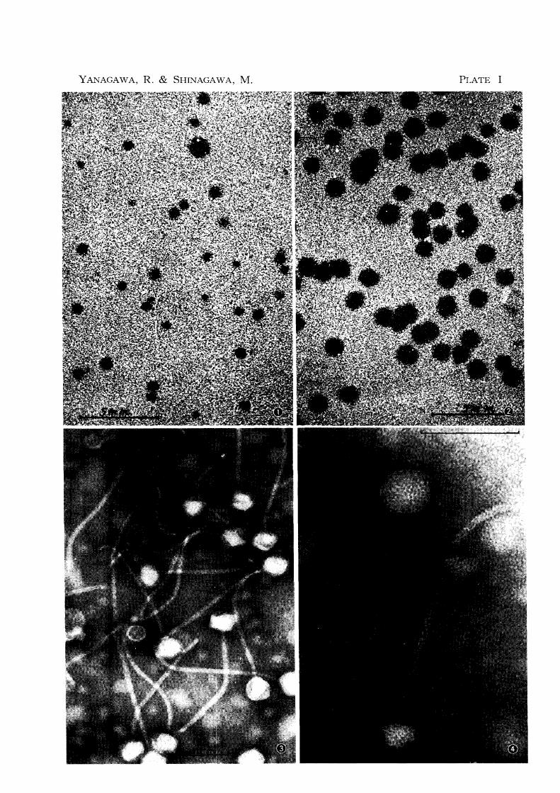
YANAGAWA, R. & SHINAGAWA, M. PLATE I

PLATE II
Fig. 5 Electron microscopy of phage RP 6 showing the striation of tail
Fig. 6 Electron microscopy of phage RP 6 showing the tail tip with
its sixfold star-shaped assembly (indicated with an arrow)
Fig. 7 Very fine fibers several in number (indicated with arrows) de
veloped from the star-shaped tail tip holding the cell debris to which the tail tip is attached. The phage is RP 6.
Fig. 8 Electron microscopy of phage RP 2
The scales indicate 100 mp.
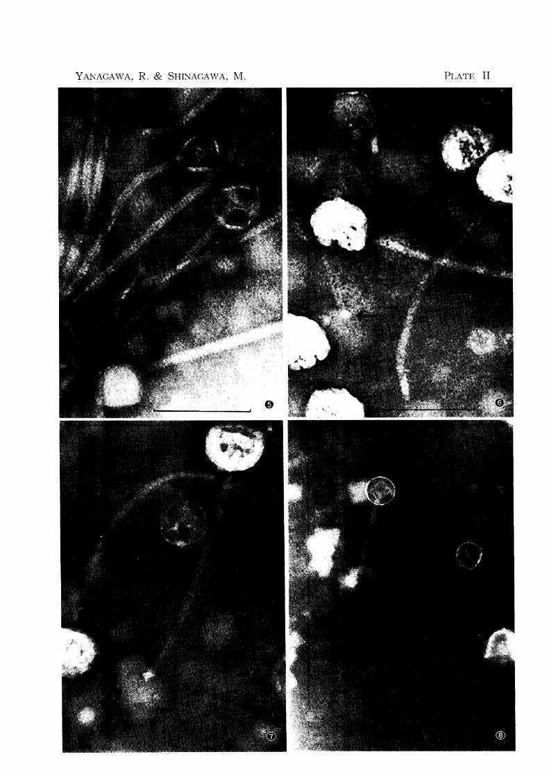
YANAGA\VA, R. & SHINAGAWA, M. PLATE II