chapter v - shodhgangashodhganga.inflibnet.ac.in/bitstream/10603/1911/12/12_chapter5.pdf · both in...
TRANSCRIPT
CHAPTER V
Topoisomerase II poisoning by the structuralanalogues of cobalt salicylaldoxime:
Elucidation of molecular mechanism ofaction using DNA interaction and computer
simulation studies.

Introduction
During the last three decades, the complexes of many transition metals have been tested
both in cell culture and animal models for antitumor activity ( Kopf-maier, 1987; Lumme
et al., 1987, Kopf-Maier et al, 1989). Only the complexes of platinum however, are
currently in routine clinical use. These platinum complexes are highly toxic, especially
causing nephrotoxicity (Lumme et al, 1987). It would be useful to search for complexes
with metal centers, which could minimize the toxicity-associated with the therapeutic use
of such complexes. Also, the development of metal complexes which specifically
antagonize a molecular target involved in the progression of cancer would go a long way
in reducing in vivo cytotoxicity.
The identification of such intracellular targets through which active drugs exert their
selective anticancer action would facilitate the design of more effective antineoplastic
drugs. In the past decade, the molecular basis of the antitumor activity of many DNA
binding agents has been recognized to rest on the interference of topoisomerase II
mediated DNA breakage-reunion catalytic cycle (Beck, 1989; Liu. L. F, 1989; Zwelling,
L. A, 1989). Topoisomerase II (topoisomerase II) is required for cell division and also
plays important roles in transcription (Osheroff, 1990), DNA replication (Brill et al,
1987), condensation and segregation of chromosomes (Newport et al, 1987). It has been
shown that the topoisomerase II levels and activity is highly regulated in the cell cycle of
normal cells. In normal dividing cells the enzyme levels are in the order of G1<S<M<G2
and activity is in the order G1<S<G2<M at different phases of cell cycle respectively. But
in cancerous cells the levels and activity are maintained constantly high irrespective of the
62
phase of cell cycle (Schneider et al., 1990). The importance of topoisomerase II in the cell
cycle of fast growing neoplastic cells stirred the development of numerous molecules
which antagonize this enzyme. The biochemical actions of these drugs on topoisomerase II
are of two types (a) inhibition of enzyme catalysed DNA double-strand passage and (b)
stabilization of the DNA-protein intermediate in the strand passage action known as the
"cleavage complex", that is these drugs trap the transient cleaved DNA-topoisomerase II
complexes in which the cleaved DNA strands are covalently-linked to the topoisomerase
II subunits. This latter action of the drugs can be rapidly quantified in cellular and
biochemical systems (Osheroff, 1989; Robinson et al., 1990). Numerous evidences
suggest that the production of such stabilized configurations (cleavage complexes) initiate
a cascade of events which lead to cell death (reviewed by Zwelling, L. A. 1989 and
D'Arpa et al., 1989). Hence, the development of cleavage complex forming drugs is very
important in specific targeting and effective anticancer action.
In the present study, five derivatives of cobalt (III) salicylaldoxime (Structures VI -X)
were designed which are Cobalt (III) N-aminosalicylaldimine, cobalt (II)
salicylalthiosemicarbazone, cobalt (III) iV-phenylsalicylaldimine, cobalt (III) N-
phenylaminosalicylaldimine and cobalt (III) (2-4, dinitro) 7V-phenylaminosalicylaldimine in
order to poison the activity of topoisomerase II with a similar mechanism of action but
with a higher potency of cleavage complex formation.
63
Materials and methods:
Biochemicals, Enzyme and DNA: Salicylaldehyde, Hydrazine hydrochloride,
Phenylamine, Phenylhydrazine hydrochloride, Thiosemicarbazone and 2,4-di-
nitrophenylhydrazine were from Aldrich. y 7?P ATP and 3H thymidine were supplied by
BARC, India. Polyethyleneimine (PEI) Cellulose-F sheets were from Merck. Other
chemicals and biochemicals used were of standard grade.
Synthesis of the cobalt Compounds:
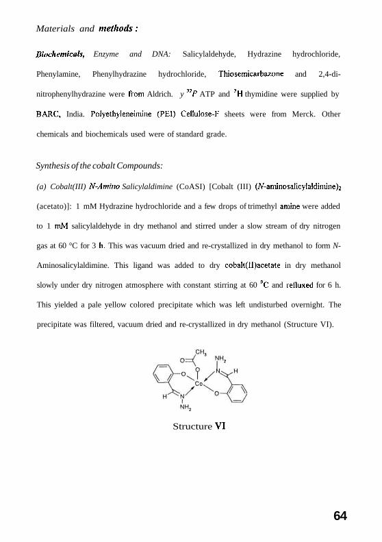
(a) Cobalt(III) N-Amino Salicylaldimine (CoASI) [Cobalt (III) (Af-aminosalicylaldimine^
(acetato)]: 1 mM Hydrazine hydrochloride and a few drops of trimethyl amine were added
to 1 mM salicylaldehyde in dry methanol and stirred under a slow stream of dry nitrogen
gas at 60 °C for 3 h. This was vacuum dried and re-crystallized in dry methanol to form N-
Aminosalicylaldimine. This ligand was added to dry cobalt(ll)acetate in dry methanol
slowly under dry nitrogen atmosphere with constant stirring at 60 °C and refluxed for 6 h.
This yielded a pale yellow colored precipitate which was left undisturbed overnight. The
precipitate was filtered, vacuum dried and re-crystallized in dry methanol (Structure VI).
Structure VI
64
ir spectra
The coordination of the amine nitrogen atom to the metal (III) ion is indicated by the shift
of the v (C=N) stretching vibrations. The spectra of the complex exhibits downward shift
of v (C=N) from ca. 1624 cm"1 for the ligand to approximately 1618 cm"1, a stretching
band appears at 1471 cm"1 corresponding to v (M-O) and a stretching band appears at
3294 cm"1 corresponding to free NH2 group, which confirms the complex formation
(Spectrum VI).
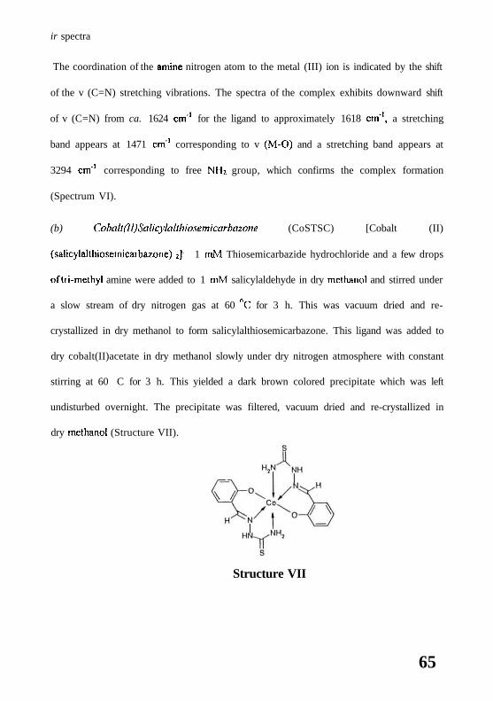
(b) Coball(ll)Salicylalthiosemicarbazone (CoSTSC) [Cobalt (II)
(salicylalthiosemicarbazone) {\. 1 mM Thiosemicarbazide hydrochloride and a few drops
of tri-methyl amine were added to 1 mM salicylaldehyde in dry methanol and stirred under
a slow stream of dry nitrogen gas at 60 nC for 3 h. This was vacuum dried and re-
crystallized in dry methanol to form salicylalthiosemicarbazone. This ligand was added to
dry cobalt(II)acetate in dry methanol slowly under dry nitrogen atmosphere with constant
stirring at 60 C for 3 h. This yielded a dark brown colored precipitate which was left
undisturbed overnight. The precipitate was filtered, vacuum dried and re-crystallized in
dry methanol (Structure VII).
Structure VII
65
ir spectra
The coordination of the imine nitrogen atom to the metal (II) ion is indicated by the
displacement of the v (C=N) stretching vibrations. The spectra of the complex exhibits
downward shift of v (C=N) from ca. 1616 cm'1 for the ligand to approximately 1612 cm"1.
A stretching band appears at 1427 cm'1 corresponding to v (M-O), and a stretching band
appears at 3011 cm"1 corresponding to NH2 in bound form, which confirms the complex
formation (Spectrum VII).
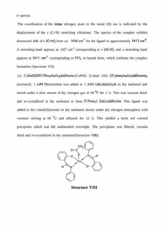
(c) Coball(II)N-Phenylsalicylaldimme (CoPSI) [Cobalt (III) (A^-phenylsalicylaldimine)2
(acetato)]: 1 mM Phenylamine was added to 1 mM salicylaldehyde in dry methanol and
stirred under a slow stream of dry nitrogen gas at 60 nC for 5 h. This was vacuum dried
and re-crystallized in dry methanol to form N-Phenyl Salicylaldimine. This ligand was
added to dry cobalt(II)acetate in dry methanol slowly under dry nitrogen atmosphere with
constant stirring at 60 °C and refluxed for 12 h. This yielded a brick red colored
precipitate which was left undisturbed overnight. The precipitate was filtered, vacuum
dried and re-crystallized in dry methanol(Structure VIII).
Structure VIII
ir spectra
The coordination of the amine nitrogen atom to the metal (111) ion is indicated by the shift
of the v (C=N) stretching vibrations. The spectra of the complex exhibits downward shift
of v (C=N) from ca. 1618 cm'1 for the ligand to approximately 1612 cm"1, stretching band
appears at 1572 cm"1 corresponding to v (M-O) and stretching bands appear at 1427, 1344
and 1292 cm"1 corresponds to acetate group, which confirms the complex formation
(Spectrum V11I).
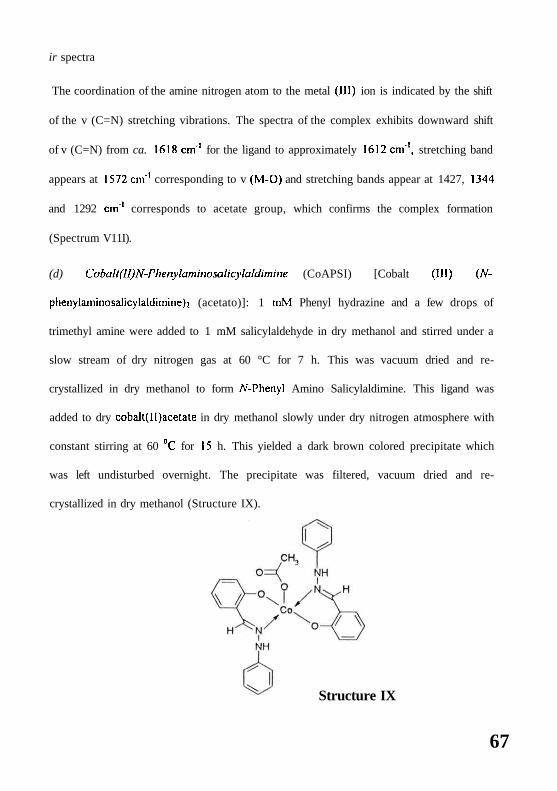
(d) Cobalt(II)N-Phenylammosalicylaldimme (CoAPSI) [Cobalt (111) {N-
phenylaminosalicylaldimine)} (acetato)]: 1 mM Phenyl hydrazine and a few drops of
trimethyl amine were added to 1 mM salicylaldehyde in dry methanol and stirred under a
slow stream of dry nitrogen gas at 60 °C for 7 h. This was vacuum dried and re-
crystallized in dry methanol to form N-Phenyl Amino Salicylaldimine. This ligand was
added to dry cobalt(ll)acetate in dry methanol slowly under dry nitrogen atmosphere with
constant stirring at 60 °C for 15 h. This yielded a dark brown colored precipitate which
was left undisturbed overnight. The precipitate was filtered, vacuum dried and re-
crystallized in dry methanol (Structure IX).
Structure IX
67
ir spectra
The coordination of the amine nitrogen atom to the metal (III) ion is indicated by the shift
of the v (C=N) stretching vibrations. The spectra of the complex exhibits downward shift
of v (C=N) from ca. 1618 cm'1 for the ligand to approximately 1602 cm1, stretching band
appears at 1566 cm"1 corresponding to v (M-O) and stretching bands appear at 1486, 1358
and 1302 cm'1 corresponds to acetate group, which confirms the complex formation
(Spectrum IX).
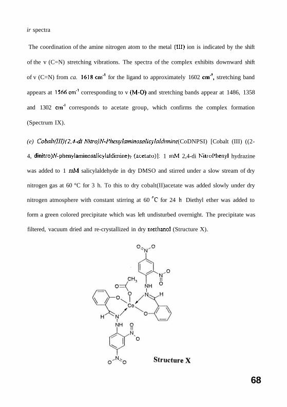
(e) Coball(III)( 2,4-di Nitro)N-Phenylammosalicytoldimme (CoDNPSI) [Cobalt (III) ((2-
4, dinitro)N-phenylaminosalicylaldimine)2 (acetato)]: 1 mM 2,4-di Nitrol'henyl hydrazine
was added to 1 mM salicylaldehyde in dry DMSO and stirred under a slow stream of dry
nitrogen gas at 60 °C for 3 h. To this to dry cobalt(II)acetate was added slowly under dry
nitrogen atmosphere with constant stirring at 60 nC for 24 h Diethyl ether was added to
form a green colored precipitate which was left undisturbed overnight. The precipitate was
filtered, vacuum dried and re-crystallized in dry methanol (Structure X).
68

ir spectra
The coordination of the amine nitrogen atom to the metal (III) ion is indicated by the shift
of the v (C=N) stretching vibrations. The spectra of the complex exhibits a stretching band
at 1651 cm'1 corresponding to NO2 groups, a stretching band appears at 1496 cm'1
corresponding to v (M-O) and stretching bands appear at 1439, 1386 and 1331 cm"
corresponds to acetate group, , which confirms the complex formation (Spectrum X)
In vitro anti-proliferation assay:
3H-Thymidine incorporation assays were carried out to examine the effect of the cobalt
complexes on the proliferative response of IIL-60 ( Human T-cell leukemia) cancer cells.
The cells were grown in RPM1-1640 medium supplemented with 20% fetal calf serum. 0.2
million cells in 200 /A were distributed in a 96 well microtitre tissue culture plate.
Increasing concentrations of the two cobalt drugs were added to the cells. The cells were
incubated for 48 h in a CO2 incubator at 37 °C maintaining 5% CO2. The cultures were
then pulsed with 0.5 juCi of H-thymidine. Incubation was continued for 6 h to allow
thymidine incorporation by cells. The cells were harvested on glass microfibre strips using
a Skatron automated cell harvester. Radioactivity was measured in a Wallac liquid
scintillation counter.
DNA- Drug Binding Studies:
Absorption titration experiments were performed in a buffer containing 5 mM tris pH
7.0, 50 mM NaCl, with 25 juM of metal complex to which 10 //M increments of calf
thymus DNA solution were added. The concentration of the metal complex used was 25
69

/iM and that of DNA ranged between 0-100 micromolar equivalents (base pairs). After the
addition of DNA to the metal complex, the resulting solution was allowed to equilibrate
for 5-10 min. at 25 °C. The absorption readings (corresponding to the changes in
absorption maxima of the drug used) were noted. The data were then fit into the following
equation to obtain the intrinsic binding constant Kb (Wolfe et al., 1987).
[DNA]/(e. - ef) - [DNA]/(eb- er) + I/Kb (Eb - er)
Where Ea, Ef, and Eb are the apparent, free and bound metal complex extinction coefficients
respectively. A plot of [DNA]/(E» - Er) versus [DNA] gave a slope of l/(Eb - Ef) and a Y
intercept equal to Kb/(Eb - Ef); Kb is the ratio of slope to Y intercept.
Oxidation state of Cobalt in the DNA bound drugs:
The Cyclic Staircase Voltametry (CV) spectra of complexes was recorded in absence
and presence of DNA using the system of DMSO/TB AP/glass carbon working electrode/
Ag+-AgCl. The concentration of drug to DNA was maintained two drug molecules per
five nucleotide bases.
Molecular modeling analysis:
Drug conformations of the cobalt salicylaldoxime and its analogues were examined by
computer-aided molecular modeling techniques. The software package SPARTAN
(version 4.1, Wave Functions Inc., California, USA) was utilized.
For each complex the following tasks were performed.
l)To build and optimize 3-dimensional model structures of the molecules, SPARTAN
graphic interface free-valence geometry force-field and semi-empirical I IF program was
used for energy minimization. The partial atomic charges needed for the coulombic
70

electrostatic potentials were obtained by RHF PM3 (tm) calculations. PM3 (tm) is a semi-
empirical method when the parameters of different systems are calibrated with a large set
of experimental results. This kind of method is known as a parameterized method.
2) Frequency vibration calculations were carried out to analyze the nature of the optimized
structures to show all the structures to be at a stable minima.
3) From the 3-D structures the distance of the active groups from the central metal ion
was calculated.
All molecular modeling calculations were performed on an EBMRS/6000 model 530
UNIX workstation.
Results
Anti-Proliferation Activity:
The *H thymidine incorporation experiments were conducted with HL60 T-cell leukemia
cells with increasing concentrations of the cobalt drugs. The results show that 35, 30 and
20 yM of CoPSl, CoPASl/and CoDNPSl could inhibit 50% of the cancer cell
proliferation, while CoASI and CoTSSC shows negligible anti-proliferation activity
(Figure. 24).
Action of Cobalt drugs on the DNA Relaxation Activity of Topoisomerase 11:
CoPSl, CoAPSl and CoDNPSl inhibits the topoisomerase 11 catalysed relaxation activity
in a dose dependent manner and shows complete inhibition at 200, 175 and 150 /zM
concentration respectively (Figure. 25 panel D lane 7, panel E lane 6 and panel F lane 6)
while CoASI and CoTSSC does not affect the DNA relaxation activity of topoisomerase
II (Figure. 25B & C).
Formation of cleavage complex:
The cleavage assay was conducted to see if cobalt drugs could form drug-induced ternary
complex of DNA-drug-topoisomerase II, called the cleavage complex. The formation of
topoisomerase II-drug-DNA cleavage complex can be visualised by the appearance of
linear DNA upon treatment of the cleavage complex with SDS and proteinase K. The
results show that phenyl derivatives induced the cleavage complex formation (Figure. 26
11

Figure 24
Figure 24: HL 60 Human leukemic cells were incubated with increasing concentrations
of CoASI, CoTSSC, CoPSI, CoAPSI & CoDNPSI. 3H thymidine incorporation during the
last 6 h of incubation was measured as described in methods. Values are presented as
mean of three independent experiments. Data is graphically expressed as percentage
increase in inhibition versus concentration of CoASI (•), CoTSSC (A), CoPSI (T),
CoAPSI ( • ) & CoDNPSI ( • ) in /zM. mAMSA (•) and Etoposide (0) (positive controls)
are also shown.
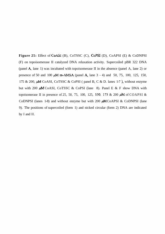
Figure 25: Effect of CoASI (B), CoTSSC (C), CoPSI (D), CoAPSI (E) & CoDNPSI
(F) on topoisomerase II catalyzed DNA relaxation activity. Supercoiled pBR 322 DNA
(panel A, lane 1) was incubated with topoisomerase II in the absence (panel A, lane 2) or
presence of 50 and 100 //M m-AMSA (panel A, lane 3 - 4) and 50, 75, 100, 125, 150,
175 & 200, //M CoASI, CoTSSC & CoPSI ( panel B, C & D. lanes 1-7 ), without enzyme
but with 200 [M CoASI, CoTSSC & CoPSI (lane 8). Panel E & F show DNA with
topoisomerase II in presence of 25, 50, 75, 100, 125, 150, 175 & 200 /JM of COAPSI &
CoDNPSI (lanes 1-8) and without enzyme but with 200 /M CoAPSI & CoDNPSI (lane
9). The positions of supercoiled (form 1) and nicked circular (form 2) DNA are indicated
by I and II.
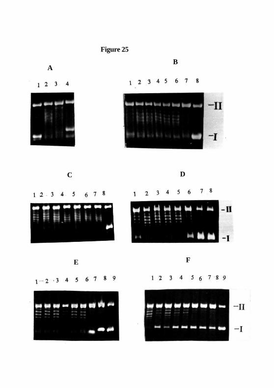
Figure 25
B
FE
C D
A
Figure 26: (A) Cleavage reaction was conducted by incubating pBR322 DNA (lane 1)
with topoisomerase II (lane 2) in presence of 60 /zM m-AMSA (lane 3) and 25, 50, 75,
100, 125, 150, 175 & 200 / M of CoPSI, CoAPSI & CoDNPSI ( panel B, C & D lanes 1-
8), and without enzyme but with 200 /iM of CoPSI, CoAPSI & CoDNPSI (lane 9). The
positions of supercoiled, nicked circular and linear (form 3) DNA are indicated by I, II
and HI. (E) The plot shows the percentage of linear DNA formed with increasing
concentration of CoPSI, CoAPSI & CoDNPSI
Figure 26
B
D
E
A
C
B, C and D) while CoASI and CoTSSC do not. Density analysis of DNA bands in the
agarose gels shows the increase in intensity of linear DNA formation with increasing drug
concentration in a dose dependent manner (Figure. 26E)
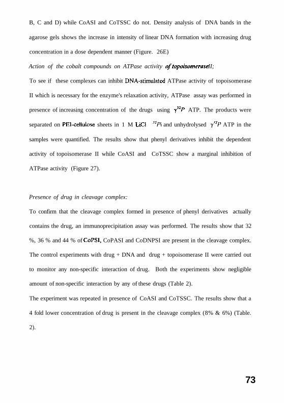
Action of the cobalt compounds on ATPase activity oftopoisomerase II;
To see if these complexes can inhibit DNA-stimulated ATPase activity of topoisomerase
II which is necessary for the enzyme's relaxation activity, ATPase assay was performed in
presence of increasing concentration of the drugs using y32/-* ATP. The products were
separated on PEl-cellulose sheets in 1 M LiCl. "Pi and unhydrolysed y"P ATP in the
samples were quantified. The results show that phenyl derivatives inhibit the dependent
activity of topoisomerase II while CoASI and CoTSSC show a marginal inhibition of
ATPase activity (Figure 27).
Presence of drug in cleavage complex:
To confirm that the cleavage complex formed in presence of phenyl derivatives actually
contains the drug, an immunoprecipitation assay was performed. The results show that 32
%, 36 % and 44 % of CoPSI, CoPASI and CoDNPSI are present in the cleavage complex.
The control experiments with drug + DNA and drug + topoisomerase II were carried out
to monitor any non-specific interaction of drug. Both the experiments show negligible
amount of non-specific interaction by any of these drugs (Table 2).
The experiment was repeated in presence of CoASI and CoTSSC. The results show that a
4 fold lower concentration of drug is present in the cleavage complex (8% & 6%) (Table.
2).
73
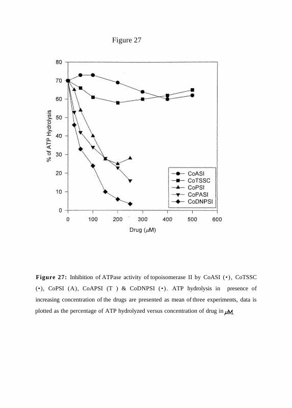
Figure 27
Figure 27: Inhibition of ATPase activity of topoisomerase II by CoASI (•) , CoTSSC
(•), CoPSI (A), CoAPSI (T ) & CoDNPSI (•) . ATP hydrolysis in presence of
increasing concentration of the drugs are presented as mean of three experiments, data is
plotted as the percentage of ATP hydrolyzed versus concentration of drug in jM.
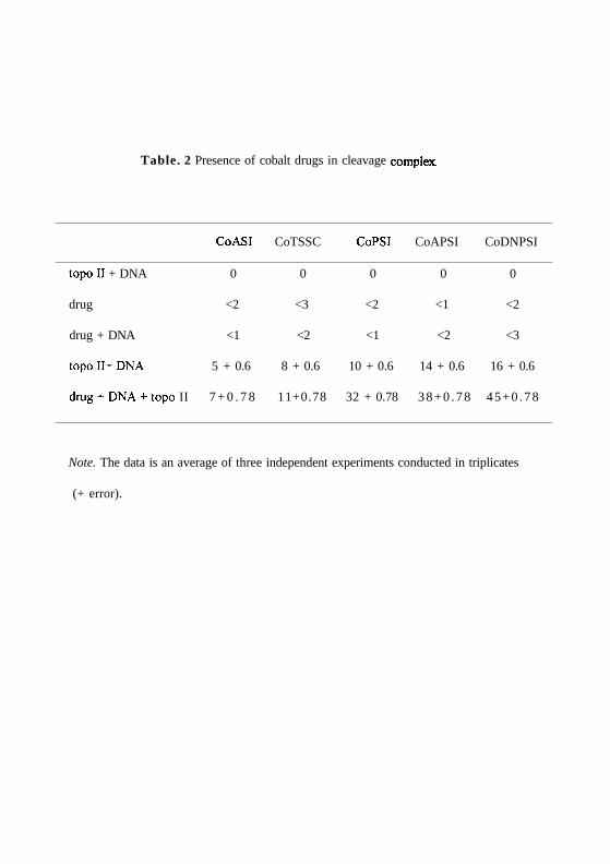
Table. 2 Presence of cobalt drugs in cleavage complex.
CoASI CoTSSC CoPSI CoAPSI CoDNPSI
topoII + DNA 0 0 0 0 0
drug <2 <3 <2 <1 <2
drug + DNA <1 <2 <1 <2 <3
topoII+DNA 5 + 0.6 8 + 0.6 10 + 0.6 14 + 0.6 16 + 0.6
drug + DNA + topo II 7 + 0 . 7 8 11+0.78 32 + 0.78 3 8 + 0 . 7 8 45+0 .78
Note. The data is an average of three independent experiments conducted in triplicates
(+ error).

DNA - Drug Binding Studies:
(a) DNA melting temperature experiments were carried out by incubating calf thymus
DNA with increasing concentrations of Cobalt complexes. The absorbance of nucleotide
bases at 260 nm was monitored by increasing temperatures from 50 °C to 90 nC. The
results show that both Cobalt complexes protect melting of calf thymus DNA (Figure 28
a, b, c, d & e ). The phenyl derivatives (CoPSI, CoPASI and CoDNPSI) increase the TM of
DNA from 56 °C to 78, 79 and 80 °C respectively, where as amine and thiosemicarbazone
derivatives (CoASI and CoTSSC) increase the Tm up to 74 and 72 °C.
To understand the mode of binding to DNA, curve width of the Tm curves were measured
at different drug to nucleotide ratios according to the procedure of Kelly et al (1985) and
were plotted (Figure. 29a) and Figure 29b shows the TM verses D/N. The results show that
these complexes bind DNA in a manner similar to major groove binding molecules eg.
polypyridyl liganded metal complexes.
The degree of binding was established using a scatchard plot analysis of
spectrophotometric data obtained by the method described by Peacocke et al (1956). A
typical scatchard plot analysis of binding of cobalt complexes to DNA is shown in Figure
30 as r / m versus r. The letter r represents the number of moles of cobalt complexes
bound per mole of DNA base pairs, and m is the concentration of unbound complex. A
value of ~0.6 was determined for n, which is the number of moles of cobalt complexes
bound per mole of DNA base pairs (Table 3).
74

Figure 28: Drug-DNA binding studies, (a) CoASI increases the Tm of calf thymus DNA
from 57 °C for DNA control (—) to70, 72, 73, 72 and 74 °C , (b) CoTSSC increases the
7mof calf thymus DNAfrom 57 °C for DNA control (—) to68, 70.76, 73.87, 75 and 75.5
°C , (c) CoPSI increases the Tm of calf thymus DNA from 57 °C for DNA control(—)
to70.15, 73.01, 76.13, 77.19 and 78 °C , (d) CoAPSI increases the Tm of calf thymus
DNA from 57 °C for DNA control(—) to67, 69, 73, 76 and 79 °C , and (e) CoDNPSI
increases the Tm of calf thymus DNAfrom 57 °C for DNA control(—) to 65, 66, 71, 75
and 80 °C . In all cases, DNA nucleotide to drug ratios of 20:1 ( ), 10:l(—), 5:1( )
2:1(—) and 1:1(—) respectively were used.
Figure 28
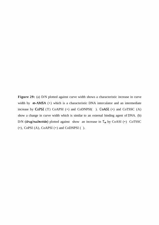
Figure 29: (a) D/N plotted against curve width shows a characteristic increase in curve
width by m-AMSA (• ) which is a characteristic DNA intercalator and an intermediate
increase by CoPSI (T) CoAPSI ( • ) and CoDNPSI( ). CoASI (•) and CoTSSC (A)
show a change in curve width which is similar to an external binding agent of DNA. (b)
D/N (drug/nucleotide) plotted against show an increase in Tm by CoASI (•) CoTSSC
(•), CoPSI (A), CoAPSI ( • ) and CoDNPSI ( ).

Figure 29
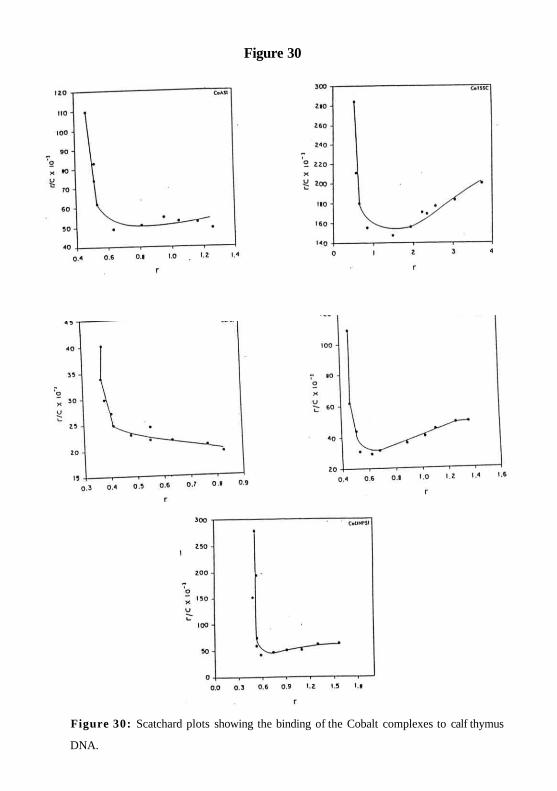
Figure 30
Figure 30: Scatchard plots showing the binding of the Cobalt complexes to calf thymus
DNA.
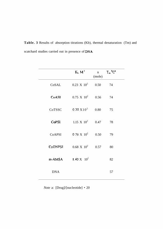
Table. 3 Results of absorption titrations (Kb), thermal denaturation (Tm) and
scatchard studies carried out in presence of DNA.
Note a: [Drug]/[nucleotide] • 20
Kb.MT1 n Tm°Ca
(mole)
CoSAL 0.23 X 10² 0.50 74
CoASI 0.75 X 10² 0.56 74
CoTSSC 0.30 X10² 0.80 75
CoPSI 1.15 X 10² 0.47 78
CoAPSI 0.76 X 10² 0.50 79
CoDNPSI 0.68 X 10² 0.57 80
m-AMSA 1.40 X 10² 82
DNA 57
(b) The binding constant (Kb) was determined from the spectroscopic titration
experiments. In the presence of increasing amounts of calf thymus DNA. the phenyl
derivatives CoPSI, CoAPSI and CoDNPSI show a bathochromic shift (7+1 nm, 3+1 nm
& 5+lnm) and a hypsochromism of 74+2%, 85+4% and 77+3% respectively in the UV-
visible spectra (Fig. 31). CoASl and CoTSSC do not show any change in UV-visible
spectra (Figure. 31). The binding constant values for the cobalt drugs, CoASI, CoTSSC,
CoPSI, CoAPSI and CoDNPSI were determined and/the jare given in Table 3. CoPSI
shows a very high binding constant followed by CoDNPSI, CoAPSI, CoASI and
CoTSSC in the decreasing order. The DNA intercalating drug m-AMSA shows the
highest binding constant.
(c) The circular dichroic spectra of the phenyl derivatives CoPSI, CoPASI and CoDNPSI
show that these complexes induce a hyperchromic shift in the positive [0]M of DNA and a
hypochromic shift in negative [0]M suggesting that the binding of these drugs may induce
unwinding, possibly by intercalation. CoASI does not change CD spectra but CoTSSC
shows a small negative shift in CD spectra (Figure. 32). These observations show that all
these complexes bind DNA with similar affinity with small differences in their mode of
interaction.
Analysis of oxidation state of cobalt in DNA bound Complexes:
The CV spectra of cobalt complexes CoASI, CoPSI, CoAPSI and CoDNPSI (Figure.
33a, b, c & d) shows that cobalt is present in +3 oxidation state. In higher oxidation state,
cobalt may undergo reduction while oxidizing DNA, thus resulting in nonspecific cleavage
and unwinding of supercoiled DNA. To confirm this, the CV spectra of these complexes
in presence of linear calf thymus DNA was recorded ( Figure 33e, f, g & h). The re:
75
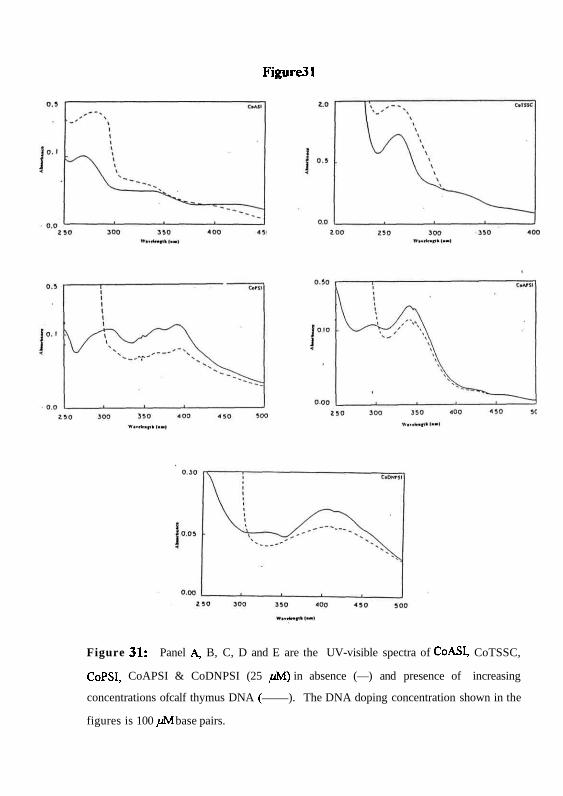
Figure31
Figure 31: Panel A, B, C, D and E are the UV-visible spectra of CoASI, CoTSSC,
CoPSI, CoAPSI & CoDNPSI (25 /M) in absence (—) and presence of increasing
concentrations ofcalf thymus DNA ( ). The DNA doping concentration shown in the
figures is 100 / JM base pairs.
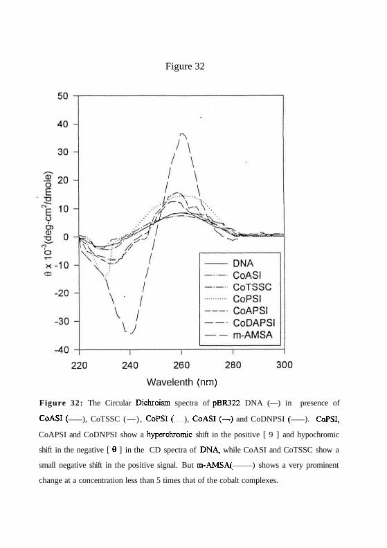
Figure 32
Figure 32: The Circular Dichroism spectra of pBR322 DNA (—) in presence of
CoASI ( ), CoTSSC (—) , CoPSI ( ), CoASI (—) and CoDNPSI ( ). CoPSI,
CoAPSI and CoDNPSI show a hyperchromic shift in the positive [ 9 ] and hypochromic
shift in the negative [ 0 ] in the CD spectra of DNA, while CoASI and CoTSSC show a
small negative shift in the positive signal. But m-AMSA( ) shows a very prominent
change at a concentration less than 5 times that of the cobalt complexes.
Wavelenth (nm)
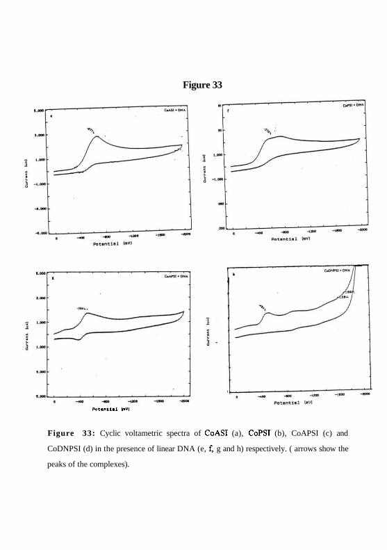
Figure 33
Potential ImVI
Figure 33: Cyclic voltametric spectra of CoASI (a), CoPSI (b), CoAPSI (c) and
CoDNPSI (d) in the presence of linear DNA (e, f, g and h) respectively. ( arrows show the
peaks of the complexes).

Figure 33

show that the +3 oxidation state of cobalt in the complexes remains unaltered even when
bound to DNA and so, it does not oxidise DNA. The CV spectra of CoTSSC suggests
that it is in +2 state in presence and absence of DNA (Figure. 34a & b).
Molecular modeling analysis.
To identify the structural elements in the drug that play a role in the DNA interaction and
topoisomerase II poisoning, molecular modeling studies using the transition metal
modeling software SPRAT AN (version 4.1) were conducted. Minimized Energy and
charge around the central metal ion are given in Table 4. The frequency vibration
calculations (Table 5) of the complexes show that these conformations impart stability to
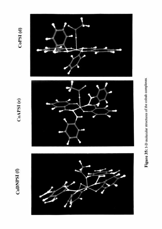
the structures. The 3 D structures (Figure. 35) show that the salicilyl groups of the
ligands form two identical domains around the central metal atom in a single plane and the
substitutions on the imine nitrogen (amine, phenyl, aminophenyl and dinitro aminophenyl)
are present perpendicular to the salicylal groups in different planes. While in CoTSSC, the
thiosemicarbazone forms a closed pentacyclic ring structure by bonding to the metal atom
with the free NII2 group.
76
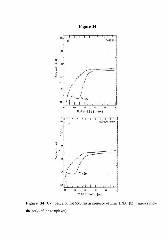
Figure 34
Figure 34: CV spectra of CoTSSC (a) in presence of linear DNA (b) ( arrows show
the peaks of the complexes).

Table. 4
CoSAL CoASI CoTSSC CoPSI CoAPSI CoDNPSI
Charge -0.11 -0.16 +0.034 +0.065 +0.049 +0.032
Minimumenergy -1701.13 -1705.1 -1408.26 -1669.79 -1525.05 -1634.08
(kcal/mol)
Distancebetween
cobalt and 2.9 2.0 1.9 2.9 4.5 -9.0possible
topoisomeraseII interactinggroup (A0)
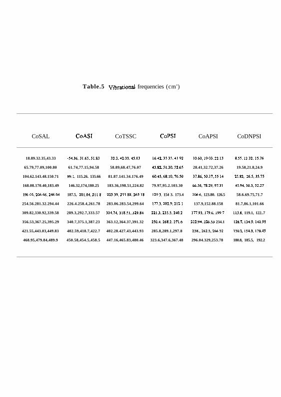
Table.5 Vibrational frequencies (cm')
CoSAL CoASI CoTSSC CoPSI CoAPSI CoDNPSI
18.89.32.35,43.33 -34.36.31.65.51.83 32.3,42.00.45.03 16.42,35.35.41.92 10.60.19.03,22.13 8.55,13.32,15.76
65.79,77.09,100.80 61.74,77.15,94.58 58.89,68.47,76.07 43.82.51.30.52.65 28.41,32.72,37.26 19.58,21.8,24.9
104.62.143.48.150.71 99.1. 115.26. 135.66 81.87.141.34.176.49 60.45,68.10.76.50 37.86,50.17,55.14 25.82.26.5,35.75
168.08.170.40,183.49 146.32,174,180.25 183.36,198.51,224.82 79.97,95.2.103.30 66.56.78.29,97.31 45.94.50.3.52.27
196 03.206.66,246.04 187.5, 201.04.211.8 225.39.255.88,265.18 129.3. 154 3. 173.4 106.6, 123.80. 126.5 58.6.69.75,71.7
254.56.281.32.294.44 226.4.258.4,261.78 283.06.283.54,299.64 177.3.202.9,212.1 137.9,152.88.158 81.7,86.1,101.66
309.82,330.92,339.58 289.3,292.7,333.57 304.74,318.51,329.84 221.3.235.5,240.2 177.93,179.6.199.7 113.8. 119.1, 122..7
356.53,367.25,395.29 340.7,375.1,387.23 363.12,364.37,391.32 250.4,268.2.271.6 212.94,226.53 234.1 128.7,134.9.141.91
421.55,443.03,449.83 402.59,418.7,422.7 402.28.427.43,443.93 285.8,289.1,297.8 258., 262.5,266.52 150.2.154.3,178.45
468.95,479.84,489.9 450.58,454.5,458.5 447.16,465.83,480.46 323.6,347.6,367.48 296.04.329,253.78 180.8, 185.5, 192.2


Discussion
A wide variety of complexes of numerous metals have undergone pre-clinical testing as
anticancer agents (kopf-maier et al, 1989). The anticancer metal complexes are broadly
classified in to two groups. The first class consists of the metal complexes, like the
classical inorganic anticancer agent cisplatin which potentially forms intra-strand and inter-
strand DNA adducts and the other class consists of metal complexes which cleave DNA
and are known as antitumor chelating agents. Some of the DNA binding antitumor
complexes bind site-specifically to DNA through the metal center and its ligands are
involved in interaction with cellular targets. The development of such metal complexes
with a higher potency of action has received considerable attention in the development of
anticancer agents which have a specific cellular target.
During the last few years, studies have indicated that the antitumor activity of DNA
binding drugs and antibiotics depends in most cases on their capacity to interfere with the
catalytic activity of topoisomerases rather than on their ability to bind DNA sensu stricto
(Wang, 1985; Chen et al., 1994 & Ralph et al., 1994). This information has been exploited
by medical chemists to create new categories of antitumor drugs. On the one hand,
hundreds of sequence selective ligands derived from anti-viral antibiotics netropsin and
distamysin have been synthesized (Kopka et al., 1992 & Lown et al., 1994). On the other
hand, a growing variety of topoisomerase II inhibitors have been developed (Chen et al.,
77
1994 & Su et al., 1992). With a view to develop novel antitumor agents which bind to
DNA through the metal atom and interact with topoisomerase II through the ligands
attached to the metal, cobalt was selected as the central metal atom due to the following
reasons.
(1) Cobalt (III) complexes bind specifically to N7 and Nl nitrogen atoms of adenine
bases through covalent bond formation (Theophilus et al., 1976).
(2) Cobalt has the ability to alter the configuration of ligands attached to it.
(3) Cobalt has the capacity to form 2, 4, 5 and 6 coordination bonds based on the reaction
environment it is present in (Parsons et al., 1997).
(4) The complexes of Co(lII) are kinetically inert octahedral coordination complexes. This
inertness is due to the d6 lowspin electronic configuration of trivalent cobalt (Cotton
et al., 1972). Kinetically inert transition metal complexes undergo the water exchange
reaction relatively slowly with a half-life of about 24 h. (Kettle et al, 1969). The
biological consequences of kinetic inertness is that Co(III) complexes will remain
intact when added to a culture medium or injected to animals and should arrive at
their cellular target with their ligand configuration intact.
A strategy was devised based on both topoisomerase II inhibition and DNA binding to
design highly potent anticancer derivatives of cobalt salicylaldoxime. To improve
topoisomerase II antagonism by the salicylaldoxime complex of cobalt, the approach was
to substitute the hydroxyl group on the imine nitrogen of the salicylal groups (which was
shown to be important for topoisomerase II poisoning by CoSAL) with amino,
thiosemicarbazone, phenyl, amino phenyl and dinitro substituted amino phenyl groups.
This was done to retain the salicylal backbone which was found to be necessary for DNA
78
interaction; the imine nitrogen substitutions would effectively change topoisomerase II
interaction by the new cobalt complexes which should result in increase or decrease of
topoisomerase 11 poisoning and the corresponding anticancer activity.
The results from in vitro antiproliferation studies of these cobalt complexes show that the
phenyl derivatives of CoSAL inhibit 50% of cell poliferation at 35, 30 and 20 / M
concentrations of CoPSI, CoAPSI and CoDNPSI respectively. But the other two
derivatives CoASI and CoTSSC require/ >75 /Al. This suggests that the imine nitrogen
substitutions play an important role in the activity of these complexes. This point was
strengthened when the ability of these complexes to inhibit the DNA relaxation activity of
topoisomerase II was tested. The phenyl derivatives CoPSI, CoAPSI and CoDNPSI
inhibited the DNA relaxation action of topoisomerase II at 200, 175 and 150 juM
respectively while CoASI and CoTSSC could not inhibit the same at any concentration
tested. The ATPase assay also showed a similar inhibition profile of the ATPase activity of
topoisomerase II in presence of these drugs. The three phenyl derivatives seem to inhibit
this activity through the formation of the enzyme-drug-DN A cleavage complex as shown
by the cleavage assay. This was further confirmed by the immunopricipitation assay which
showed 35, 40 and 48% of CoPSI, CoAPSI and CoDNPSI in the drug induced
topoisomerase II-DNA cleavage complex.
To find out the possible molecular mechanism of action of these analogues on
topoisomerase II, the DNA binding and computer-aided molecular modeling analysis were
carried out. The DNA binding studies of these complexes show different interaction
modes for the two sets of complexes. The curve width analysis of the DNA Tm curves in
presence of these complexes show that CoASI and CoTSSC possibly bind to DNA
79
externally, ie. they interact outside the DNA helix without affecting the conformation of
DNA. This was verified by the CD spectral analysis which showed a negligible change in
the CD spectra of DNA in presence of these two complexes. The absorbance spectra of
the two complexes in the visible region in presence of DNA show no change in the
absorbance maxima. The phenyl derivatives however bind more strongly to DNA and
show a curve width much higher than CoASI and CoTSSC. They also induce a significant
hyperchromic shift in the CD spectra of DNA. The absorbance spectra of these complexes
doped with increasing concentrations of DNA shows a decrease in the absorbance
maxima. The scatchard plot analysis of the phenyl derivatives shows a two mode
interaction, one seems to be an intercalative mode at low /• value and the other seems to be
an electrostatic interaction at the higher r value. This along with the CD spectral data and
the curve width analysis suggest a weak intercalative DNA interaction by these complexes.
This difference of DNA interaction by the phenyl derivatives compared to the amine and
the thiosemicarbazone derivatives definitely seems to be induced by the phenyl groups.
The differential inhibition of topoisomerase II by the complexes could be explained in
terms of the three-dimensional structures of these complexes by.
• charge around the central metal atom.
• size of their ligand
• distance between the metal center and the possible enzyme interacting groups of the
metal complex.
Molecular modeling analysis of the cobalt complexes showed that the two salicylal groups
in CoTSSC and CoASI are oriented in a single plane opposite to each other around the
central cobalt atom. In CoTSSC, two co-ordinations are occupied by thiosemicarbazone
80
groups which form pentacyclic rings oriented at 120 degrees to the salicylal backbone. In
CoASI, CoPSI, CoAPSI and CoDNPSI there is a single acetate co-ordination, a possible
leaving group. In CoASI, CoPSI and CoAPSI, the nitrogen on each salicylal group is
covalently bonded to amine, phenyl and amino phenyl groups which are oriented in
opposite directions of the salicylal backbone in different planes. In case of CoDNPSI, the
nitrogens on the salicylal groups are covalently linked to 2,4-dinitro amino phenyl groups.
The introduction of the phenyl rings in CoPSI and CoAPSI imparts a conformational
change in the salicylal groups, orienting them in two slightly different planes. In
CoDNPSI, due the presence of the nitro groups, there is an enormous change in the
salicylal conformations which orient themselves in two distinct planes.
In CoASI, the electronegative NH2 groups render a partial negative charge on the cobalt
atom. This could prevents a strong interaction by the molecule to the negatively charged
DNA. In case of CoTSSC, the cobalt atom is essentially uncharged. The rings formed by
the thiosemicarbazone groups may stesfrically hinder a strong interaction of the cobalt
atom to DNA. The planar salicylal groups in both the complexes may not involve in any
interaction with DNA. But in the phenyl derivatives, the presence of the phenyl rings
decreases the negative charge on the cobalt atom compared to CoASI and CoSAL
rendering a partial positive charge on the metal atom. This could result in an ionic or
covalent association of the complex with DNA through the cobalt atom. The proximity
resulting due to this interaction may enable a stacking interaction of one of the phenyl
rings of the complex with DNA bases which could lead to an intercalative mode of DNA
binding by these complexes.
81
CoASI does not poison topoisomerase II activity probably because the amino groups may
not interact with the enzyme as strongly as the hydroxyl groups of CoSAL (Jayaraju et al.,
1999). Also, the distance of the hydroxyl oxygen from the cobalt atom in CoSAL is 2.9 A0
while in CoASI, the distance of the amino nitrogen from the metal atom is only 2.0 A0.
Due to this, the DNA bound drug may not be able to interact with the enzyme with the
amino group. In CoTSSC, the amino groups of the thiosemicarbazones co-ordinate to the
cobalt atom and are hence unavailable for enzyme interaction. In both CoASI and
CoTSSC, the salicylal groups do not seem to exert any influence either on DNA binding
or enzyme interaction due to their orientation in a single plane. The phenyl derivatives
poison topoisomerase II in the order of CoPSKCoAPSKCoDNPSI. This could be
definitely due to the presence of the phenyl ring. On the one hand, the distance of the
phenyl groups from the metal atom is clearly more compared to CoASI and CoSAL which
could account for a greater interaction with the enzyme. On the other hand, the phenyl
groups (especially the DNP-phenyl) change the ligand environment significantly, which
could favor a stronger interaction with DNA and enzyme, thus increasing the potency of
cleavage complex formation compared to CoSAL.
The above data suggests that the phenyl derivatives of cobalt salicylaldoxime show a
bidirectional interaction with DNA and topoisomerase II by sandwiching between the two.
The cobalt atom and one of the phenyl rings may bind to DNA electrostatically and
through intercalation respectively while the other phenyl ring and the salicylal groups may
interact with topoisomerase II. This could effectively result in a stable cleavage complex
comprised of the enzyme, drug and DNA.
82
CONCLUSIONS
1. The work on the known anticancer metal complexes, cisplatin and copper
salicylaldoxime (CuSAL) suggests that topoisomerase II antagonism may
be one of the possible mechanism for their anticancer activity.
2. The metal center plays a very important role in topoisomerase II poisoning
as seen by the mechanism of topoisomerase II inhibition by CuSAL and
cobalt salicylaldoxime (CoSAL). Though both drugs have the same ligand
(salicylaldoxime), CuSAL forms a non-covalent cleavage complex while
CoSAL forms covalent cleavage complex by poisoning enzyme activity.
3. In CoSAL, the -OH group of oxime is involved in topoisomerase II
inhibition. This was revealed by the amine, semicarbazone and thio
semicarbazone derivatives of the complex, where the -OH group is
replaced. These drugs could not inhibit the enzyme activity
4. When the -OH was substituted with strong interacting groups (phenyl,
phenyl amine and dinitro phenyl amine), the enzyme inhibition was
increased. This suggests that large interacting domains increase the
topoisomerase II inhibition by the cobalt complexes.
83

5. The studies reveal that two domains are required for topoisomerase
poisoning by the cobalt complexes - A DNA binding domain and enzyme
interacting domain.
6. The studies on the cobalt metal complexes as potential topoisomerase II
poisons open a new avenue for designing potent anticancer drugs
possessing a metal center.
84