chapter 7 - protein flexibility and enzymatic catalysis - utopia
TRANSCRIPT

PROTEIN FLEXIBILITY AND ENZYMATIC CATALYSIS
By M. KOKKINIDIS,*,† N.M. GLYKOS,‡ AND V.E. FADOULOGLOU*,‡
*Department of Biology, University of Crete, Heraklion, Crete, Greece†Institute of Molecular Biology and Biotechnology, Heraklion, Crete, Greece
‡Department of Molecular Biology and Genetics, Democritus University of Thrace,
Alexandroupolis, Greece
I. In
ADVANSTRUCDOI: 1
troduction . . . . . . . . . . . . . . . . . . . . . . . . . . . . . . . . . . . . . . . . . . . . . . . . . . . . . . . . . . . . . . . . . . . . . . .
CES IN PROTEIN CHEMISTRY AND 181 Copyright 2012, ETURAL BIOLOGY, Vol. 87 All righ0.1016/B978-0-12-398312-1.00007-X
1
lseviets rese
82
II. S elected Methods Used for Studying Protein Flexibility. . . . . . . . . . . . . . . . . . . . . . . . . . 1 85A
. C rystallography and Time-Resolved X-ray Methods . . . . . . . . . . . . . . . . . . . . . . . . . . 1 86 B . X -ray Free Electron Lasers. . . . . . . . . . . . . . . . . . . . . . . . . . . . . . . . . . . . . . . . . . . . . . . . . . . . . 1 88 C . S pectroscopies, NMR, NSE, and Hydrogen–Deuterium Exchange . . . . . . . . . . 1 88 D . C omputational Methods. . . . . . . . . . . . . . . . . . . . . . . . . . . . . . . . . . . . . . . . . . . . . . . . . . . . . . . 1 90III. D
eterminants of Active Site Flexibility . . . . . . . . . . . . . . . . . . . . . . . . . . . . . . . . . . . . . . . . . . . . 1 91 A . F lexible Active Site Loops . . . . . . . . . . . . . . . . . . . . . . . . . . . . . . . . . . . . . . . . . . . . . . . . . . . . . 1 91 B . A ctive Sites Located at Domain Interfaces. . . . . . . . . . . . . . . . . . . . . . . . . . . . . . . . . . . . 1 96 C . F lexibility of Active Site Residues . . . . . . . . . . . . . . . . . . . . . . . . . . . . . . . . . . . . . . . . . . . . . 1 98 D . A Specific Example: Flexibility of the BcZBP Deacetylase . . . . . . . . . . . . . . . . . . . . 2 00IV. F
lexibility and Special Aspects of Enzyme Properties. . . . . . . . . . . . . . . . . . . . . . . . . . . . . 2 02 A . F lexibility and Thermal Enzymatic Adaptation. . . . . . . . . . . . . . . . . . . . . . . . . . . . . . . 2 02 B . F lexibility as an Essential Component of Enzymatic Allostery . . . . . . . . . . . . . . . 2 05 C . F lexibility and Ligand Specificity. . . . . . . . . . . . . . . . . . . . . . . . . . . . . . . . . . . . . . . . . . . . . . 2 09 R eferences. . . . . . . . . . . . . . . . . . . . . . . . . . . . . . . . . . . . . . . . . . . . . . . . . . . . . . . . . . . . . . . . . . . . . . . . . . 2 12Abbreviations
1-CPI 1
-(4-chlorophenyl)imidazole 3D t hree dimensional 4-BP 4 -benzylpyridine 4-CPI 4 -(4-chlorophenyl)imidazole 4-NBP 4 -(4-nitrobenzyl)pyridine (S/R)-GOP ( S/R)-glycidol phosphate TIM T riosephosphate Isomerase UDX U DP-a-d-xylose NMR N uclear Magnetic Reasonance NSE N eutron Spin Echo CYP C ytochrome P450r Inc.rved.

182 KOKKINIDIS ET AL.
Abstract
The dynamic nature of protein structures has been recognized, estab-lished, and accepted as an intrinsic fundamental property with majorconsequences to their function. Nowadays, proteins are considered asnetworks of continuous motions, which reflect local flexibility and a pro-pensity for global structural plasticity. Protein–protein and protein–smallligand interactions, signal transduction and assembly of macromolecularmachines, allosteric regulation and thermal enzymatic adaptation areprocesses which require structural flexibility. In general, enzymes repre-sent an attractive class among proteins in the study of protein flexibilityand they can be used as model systems for understanding the implicationsof protein fluctuations to biological function. Flexibility of the active site isconsidered as a requirement for reduction of free energy barrier andacceleration of the enzymatic reaction while there is growing evidencewhich concerns the connection between flexibility and substrate turnoverrate. Moreover, the role of conformational flexibility has been well estab-lished in connection with the accessibility of the active site, the binding ofsubstrates and ligands, and release of products, stabilization and trappingof intermediates, orientation of the substrate into the binding cleft, adjust-ment of the reaction environment, etc.
I. Introduction
Long before the appearance of any experimental evidence supporting thedynamic character of protein structures, their view as static and rigid objectswas proving increasingly insufficient to explain a large body of biochemicaldata, which were gathered mainly in the field of enzymes. The ‘‘key-lock’’hypothesis, proposed at the end of the nineteenth century by Fisher (1894)and used for the first half of the twentieth century to explain enzymaticactivity was based on the concept of fixed structures, that is, the substrate hadto fit into a structurally well-defined active site of fixed shape which was alsocomplementary to the substrate’s shape. However, the predictions of the‘‘key-lock’’ theorywerenot always sufficient to explain the experimental data.Thus, the need for revision led Koshland in 1958 to propose the ‘‘inducedfit’’ theory which introduced the concept of an enzyme active site whichundergoes structural changes induced by the binding of the substrate. Thistheory is based on the idea that protein structures or at least their active sitespossess a flexibility which enables them to adopt more than one

PROTEIN FLEXIBILITY AND ENZYMATIC CATALYSIS 183
conformation. This was proposed even before the determination of the firstprotein three-dimensional (3D) structure (myoglobin; Kendrew et al., 1958).One year later, Linderstrom-Lang and Schellmann (1959) proposed abreath-like continuous movement of the protein structures, an idea whichwas experimentally probed during the following decades. Evidence camefrom the analysis of crystallographic temperature factors which had lowervalues for buried/semi-buried areas and systematically higher for solvent-exposed residues. In some cases, loops and side chains exposed to the solventhave very high values indicating high mobility. Moreover, relatively hightemperature factors also characterize regions of the active site of enzymes.The same conclusions were drawn by comparing the apo- and holo-states of aprotein complexed with different compounds or by comparing homologousproteins from different organisms. Beginning with the analysis of crystallo-graphic structures of lysozyme, the importance of conformational flexibilityfor catalytic efficiency was widely accepted. In the 1970s, Karplus and cow-orkers studied the molecular dynamics of folded proteins by solving theequations of motion for the atoms, applying empirical potential energyfunctions, and the mobile character of protein structures was confirmedfor oncemore. In the 1980s, the development of nuclearmagnetic resonance(NMR) provided an additional experimental tool to explore the dynamicalaspects of protein structures. Nowadays, the dynamic nature of structures iswell established and proteins are considered as a network of continuousmotions, which reflect local flexibility and a propensity for global structuralplasticity; an implication of the latter is a mechanism for the establishment ofnew folds as proposed byGlykos et al. (1999). Internalmotions have differentamplitudes and their frequencies range over different time scales. Themotions which affect the protein structure include bond vibrations at thetime scale of femto- to pico-seconds, side-chain rotations at the time scale ofnanoseconds, and more complicated motions of larger time scales such asmotion of flexible termini and loops, large concerted domain motions, andconformational adjustment upon substrate binding.A growing variety of methods was used and/or developed to study the
protein mobility at time scales ranging from femtoseconds to seconds.Next to classical macromolecular crystallography, NMR and moleculardynamics simulations, the protein motions can be studied by a varietyof time-resolved techniques and a wide range of spectroscopies such astime-resolved Laue diffraction and intermediate trapping, single-moleculefluorescence resonance energy, neutron spin echo (NSE), etc.
184 KOKKINIDIS ET AL.
Some types of protein functions can be easily related to structuralflexibility. For example, almost any interaction between a protein andanother molecule is associated with conformational changes. Ligand rec-ognition and binding, protein–protein interactions, and processes ascomplicated as signal transduction and assembly of multiprotein machinesare based on protein flexibility (Gazi et al., 2009; Teilum et al., 2011).Allosteric regulation is achieved through conformationally flexible regu-latory proteins. Even the evolutionary process of thermophilic, mesophilic,or psychrophilic adaptations of protein structures, affect proteinsequences such that structural flexibility is directly affected with implica-tions for protein stability and enzymatic catalysis. In general, enzymesrepresent an attractive class among proteins in the study of proteinflexibility and they can be used as model systems for understanding theimplications of protein fluctuations to biological function.
Enzymes catalyze a variety of biological processes and may acceleratethem by as many as 20 orders of magnitude in comparison with thecorresponding uncatalyzed reaction (Wolfenden and Snider, 2001). Oneof the most intriguing questions in biochemistry is how the enzymesachieve this high rate enhancement. Numerous studies have beenreported aiming to elucidate dynamic effects associated with catalysis.These studies demonstrate that among other factors, the ability ofenzymes to adopt different conformations is of high importance for theefficiency of the catalytic processes (Kamerlin and Warshel, 2010). Flexi-bility of the active site is considered as a requirement for reduction of freeenergy barrier and acceleration of the enzymatic reaction (Hammes-Schiffer and Benkovic, 2006; Henzler-Wildman and Kern, 2007; Nashineet al., 2010). During the catalytic cycle, the enzyme molecule passesthrough different states and each state is associated with a different activesite conformation. Rapid transition between the different conformationalstates is therefore mandatory for the maximum enzyme activity. Someaspects of the role of this conformational flexibility have been establishedin connection with the accessibility of the active site, the bindingof substrates and ligands and release of products, stabilization andtrapping of intermediates, orientation of the substrate into the bindingcleft, adjustment of the reaction environment, etc. Furthermore,growing evidence concerns the connection between flexibility and thesubstrate turnover rate (reviewed in Yon et al., 1998; Hammes, 2002;Agarwal, 2006).

PROTEIN FLEXIBILITY AND ENZYMATIC CATALYSIS 185
Extreme cases of protein flexibility are the molten globule state and theintrinsically disordered proteins. These exhibit significant conformationalflexibility which is frequently necessary for the establishment of intermo-lecular interaction networks (Ohgushi and Wada, 1983; Gazi et al., 2009;Schlessinger et al., 2011). In the partially folded molten globule state, thepolypeptide chain adopts a nearly native-like secondary structure, and adynamic tertiary structure due to the partial absence of native intramolec-ular packing interactions.During the last decade, many proteins have been described that fail to
adopt a stable tertiary structure under physiological conditions and yetdisplay biological activity (Dunker et al., 2008; Uversky and Dunker, 2010).This state of the proteins, defined as intrinsic disorder, has been found tobe rather widespread; disordered regions lacking stable secondary andtertiary structure are often a prerequisite for biological activity, suggestingthat structure–function relationships can be frequently only understood ina dynamic context in which function arises from conformational freedom.Fully or partly nonstructured proteins are described as intrinsically disor-dered or intrinsically unstructured proteins. The term natively unfoldedproteins indicates that protein function is associated with a dynamicensemble of different conformations (Gazi et al., 2008, 2009).Here, we will focus on the flexibility of natively folded proteins and, in
particular, of enzymes. We will review the different ways in which flexibilityis linked to enzymatic catalysis through conformational flexibility of loopssurrounding the active site, and of domain and active site residue dynam-ics. Specific examples from the literature will be reviewed. In addition,an overview of widely used specific methodologies for studying proteinflexibility will be presented, as well as some more recently developed ones.The issues of allostericity, thermal adaptation, and ligand specificity inrelation to flexibility will be reviewed in detail. The topic is presented fromthe structural point of view with an emphasis on X-ray crystallographyresults.
II. Selected Methods Used for Studying Protein Flexibility
A summary of selected methods used today to investigate and analyzeprotein flexibility is presented below. A detailed application of X-raycrystallography, mutagenesis, and molecular dynamics simulations to aspecific example is described in a following section.

186 KOKKINIDIS ET AL.
A. Crystallography and Time-Resolved X-ray Methods
X-ray crystallography is the method of choice for obtaining the molecu-lar structure of proteins at atomic resolution. The X-ray diffraction patternof the crystalline specimen is recorded and the electron density map of themolecule under study is calculated. The molecular model is built into theelectron density map and refined and thus the position of the atoms isdetermined with high accuracy. In fact, crystallography provides a staticmodel of a dynamic molecule; therefore this model represents an average,in space and time, of 3D structure of the molecule. However, even thestatic crystal structure contains useful information for the dynamic natureof the molecule, as it is explained below.
The refined atomic model contains information for the degree ofthermal motion of the atoms. The mean-squared atomic displacement iscommonly expressed as the temperature factor (B-factor). Temperaturefactors express not only the static disorder, which is an ensemble ofsubstates present in solution and trapped in the crystal but also thedynamic disorder, which represents fluctuations in the crystal or otherwisecrystal defects. Thus, B-factors cannot be interpreted simply as the ampli-tude of atomic fluctuations because both true intramolecular motion andlattice disorder contribute to them. Moreover, crystal contacts affectB-factors. In the context of protein structures, the B-factors can be takenas indicating the relative vibrational motion of different parts of thestructure and comparing them along the structure may allow one todraw conclusions about which are the most flexible parts. Atoms withlow B-factors belong to a part of the structure that is well ordered whileatoms with large B-factors generally belong to a part that is very flexible.
It is quite common that someprotein segments, that is, amino and carboxyltermini, loops or even other protein segments yield weak or nondetectableelectron density. A common reason (apart from crystal lattice defects) formissing electron density is that the unobserved region fails to scatter X-rayscoherently due to variation inpositionof the atomsof this particular segment,that is, the unobserved atoms are disordered or highly flexible.
Sometimes there are residues in a structure which present clear electrondensity for two positions, or they present what we call multiple distinctconformations. This is a direct evidence that the side chain spend time inmore than one conformations or otherwise that present a relative flexibility.In other cases however, the movement from one conformation to the

PROTEIN FLEXIBILITY AND ENZYMATIC CATALYSIS 187
other occurs so quickly that there is no definite electron density for someof the atoms of the side chain.Several time-resolved X-ray structural methods (for a review see
Westenhoff et al., 2010) have been developed in the recent years aimingto follow in real time the conformational changes of a protein whileperforming its action. Time-resolved Laue diffraction, intermediatetrapping, time-resolved wide-angle X-ray scattering, and time-resolvedX-ray absorption are the most common of them. Time-resolved Lauediffraction (Schottea et al., 2004) provides conformational snapshots ofthe whole protein in high resolution and allow us to visualize in real timeand with atomic detail the conformational evolution of a protein. Detec-tion of structural changes as small as 0.2–0.3 A with a time resolution of100 ps is possible. The time-resolved Laue diffraction experiments are of apump-probe type. The reaction is triggered within the protein crystal byphotolysis caused by ultra-short laser pulses which play the role of thepump for the initiation of the reaction. Diffraction patterns are collectedat specific time delays after triggering. This cycle must be repeated manytimes for each spatial rotation of the crystal and many times for the sametime delay. The applicability of the method depends on whether themolecule retains its biological activity in the crystalline state, whetherthe molecule is inherently photosensitive or if it could be engineered asso, whether the system is reversible, whether the concentration of theproduced intermediate is sufficiently high to be detectable because at anygiven time during reaction several intermediates are likely to be presentunless they are well separated in time. The method has been successfullyapplied to heme-containing proteins (Srajer and Royer, 2008).As an alternative, the intermediate trapping method could be used.
In this method, the lifetime of intermediates is sufficiently extended sothat they can be studied by static crystallography. Freezing, changes in pH,chemical modifications, or solvent modifications are some ways which areused to trap the intermediate. The successful application of an alternativeapproach of this method has been used to reveal the dynamics of theactive site of the small guanosine nucleotide-binding protein H-Ras-p21(Klink et al., 2006; Klink and Scheidig, 2010).In time-resolved wide-angle X-ray scattering diffraction, data are recorded
as a function of time from the molecule in solution (Fischetti et al., 2003).Because the structural information obtained is averaged over all orienta-tions of the randomly oriented molecules in the solution, the atomic detail

188 KOKKINIDIS ET AL.
is poor. This technique is used to characterize large-scale global conforma-tional changes. On the other hand, the time-resolved X-ray absorptionspectroscopy is used to characterize the geometry and the changes of thegeometry of the coordination structure in the active sites of metalloproteins.
B. X-ray Free Electron Lasers
The X-ray free electron laser (XFEL) is a novel radiation source, whichcombines a particle accelerator with laser physics. Bunches of electrons arefirst brought to high energies in a superconducting accelerator. Then theyfly through a special arrangement of magnets, called undulator, in whichthey emit laser-like flashes of radiation. By this way, X-ray flashes of highenergy are generated and they can be directed to the sample whose diffrac-tion pattern is recorded just before its explosion. The special characteristicsof the generated flashes, that is, coherent radiation, wavelengths at the levelof Angstroms, and short pulses at the level of hundreds of femtosecondsallows for atomic resolution snapshots of the molecule under study. XFELmay allow imaging of single particles/molecules and a large variety ofdifferent applications such as investigation of protein folding and dynam-ics, the actions of catalysts, and the splitting of chemical bonds can beafforded (Neutze et al., 2004). Applicability of the method with tiny crystalsof photosystem I has been demonstrated (Chapman et al., 2009).
C. Spectroscopies, NMR, NSE, and Hydrogen–Deuterium Exchange
Spectroscopy is the study of the interaction of electromagnetic radiationwith matter. An advantage of the spectroscopic methods is that they cangive a dynamic picture of the selected part of the molecule, for example,time-resolved intrinsic fluorescence of Trp reflects internal mobility of thisamino acid. Real-time information on structural changes for macromole-cules containing a chromophore, such as heme proteins, is provided bytime-resolved spectroscopic studies including absorption, resonanceRaman, and infrared spectroscopy. In those cases, we observe structuralchanges limited to the chromophore environment.
The single-molecule fluorescence resonance energy transfer (FRET)reports proximity of moieties within a molecule for distances in therange of 1–10 nm. FRET is measured between two dyes, donor and accep-tor. If the dyes are separated by a large distance (larger than 10 nm), then
PROTEIN FLEXIBILITY AND ENZYMATIC CATALYSIS 189
there is little interaction between them and even if the donor emitsphotons upon its excitation by laser, acceptor will not interact with thisenergy. However, if the two dyes are brought closer, the acceptor takes theenergy from the donor and emits photons of different colors. So, we canuse FRET to measure distance changes in the nanometer scale. A recentapplication of the single-molecule FRET is described by Kahra et al. (2011)for the investigation of conformational plasticity and dynamics of thepeptidyl-prolyl cis–trans isomerase SlyD.NMR spectroscopy is used to obtain detailed structural information for
the whole system and, in addition, can yield information on the dynamics ofspecific parts of the structure. The spin of protons is the property whichcauses the nucleus to produce NMR signal. Specific nuclei, called NMRactive, that is, 15N and 13C, when they are found under a magnetic fieldabsorb radiation at a characteristic frequency and migrate to a higher spinenergy level. When spin returns to the basic level, energy is emitted and thissignal can be recorded and processed to generate the (NMR) spectrum.Protein NMR spectroscopy is the use of NMR phenomenon to extractstructural information. The protein structure determination by thismethodis a process which results in a convergent ensemble of structures.Molecular motions generate local fluctuating magnetic fields which
have, as a consequence, NMR relaxation. Relaxation times can bemeasured and used to determine parameters as correlation times andchemical exchange rates which yield information on the dynamics ofprotein parts as the backbone or side chains. Motions which can bedetected occur on the time scale of 10 ps to 10 ns but even slower motionsbetween 10 ms to 100 ms can be studied.NSE spectroscopy is an inelastic neutron-scattering technique with re-
cent applications to the study of protein dynamics (Mezei, 1980). It is atechnique of high effective energy resolution which determines the time–space correlation function at the temporal scales from nanoseconds tomicroseconds and at the spatial scale from several Angstroms to somehundreds. NSE is used to determine relaxation processes in a macromole-cule, that is, internal dynamic modes and it has the potential to determinethe global shape fluctuations and domain motions. NSE has been appliedto the NHERF1 multidomain protein to distinguish and characterizecouple domain motions which are involved in the dynamic propagationof allosteric signals at the nanoseconds timescale (Farago et al., 2010). It isshown that NSE can be used to determine the domain mobility tensor

190 KOKKINIDIS ET AL.
which determines the degree of dynamical coupling between domains.Bu et al. (2005) have used the method to determine internal coupleddomain motions within DNA polymerase I from Thermus aquatius.
The hydrogen–deuterium (H/D) exchange is a chemical reaction.A covalently bonded hydrogen atom is replaced by a deuterium atom fromthe solution.Usually the examinedprotons are the amides in the backboneofa protein. The method gives information about the solvent accessibility ofvarious parts of the molecule and detects global or local unfolding on time-scales of milliseconds and longer. It is monitored by NMR and/or massspectrometry. The conformational changes that occur in factor XIII due tomonovalent and divalent ion binding has been recently reported by WoofterandMaurer (2011). TheH/D exchange effects observed in the presence of awide range of ions in different conditions and the deuterium incorporationwas analyzed by MALDI-TOF MS. Moreover, Oyeyemi et al. (2011) haveapplied the technique to elucidate the relationship between flexibility andthermal adaptation in the case of dihydrofolate reductases.
D. Computational Methods
Biomolecular simulations are an important technique for characterizingprotein conformational changes (Klepeis et al., 2009). The advantage of thecomputational methods is that one can follow the details of the proteindynamics in the pico-second timescale and examine the structural featuresin the atomic level. The disadvantage is that without experimental validationof the potentials used in the simulations, the predictions are on the risk ofquestioning. The starting point is a high-resolution structure determined byX-rays orNMR.Aprerequisite is also the availability of computational power.Then, an empirical force field, which is a set of parameters describing thepotential energy of the system together with the equations of motion, isapplied to the system and the successive positions of all the atoms of thesystem, and thus the progressive motion of the molecule, can be watched.The computational demands prevent the method from reaching timescalesgreater than milliseconds. To achieve longer timescales, simplified modelscan be used as the implicit solvent or the coarse-graining models.
Molecular dynamics simulations apply empirical molecular mechanicspotential energy functions which are suitable for studying conformationalchanges and dynamics as the conformational changes during the catalyticcycle, associated with substrate binding and product release as well as

PROTEIN FLEXIBILITY AND ENZYMATIC CATALYSIS 191
fluctuations around an average structure. However, they cannot be ap-plied to model chemical reactions, so cannot investigate enzyme reactionmechanisms directly. To study the connection between conformationaland chemical changes as well as to investigate the hypothesis that proteinmotions accelerate the reaction rates of catalysis, the quantum mechanics/molecular mechanics (QM/MM) methods are used. An analytic overviewof how the computational methods have been used to increase ourunderstanding of the dynamical aspects of enzymatic catalysis has beenpresented by McGeagh et al. (2011).
III. Determinants of Active Site Flexibility
Enzymes are generally characterized by conservation of their functionalgroups through evolution. On the other hand, the flexibility requirementsof many active sites frequently impose a relaxation of strict conservation sothat different functional residues are not conserved during evolution whileconverging toward the same mechanistic role. The resulting plasticity ofactive sites has been reviewed by Todd et al. (2002). In principle, uponcatalysis, residues in and around the active site must undergo conforma-tional changes associated with the binding and release of substrates andcofactors, the protection of the reaction space from the aqueous environ-ment, stabilization of reaction intermediates, and interactions betweencatalytic residues and binding subsites with various groups of reactantsupon completion of catalysis. In some cases, the structural requirementsfor conformational flexibility are limited to individual amino acids espe-cially to side chains capable of adopting different rotamers. In other cases,the interactions of the macromolecule with substrate and cofactors requireconformational changes mainly involving the external loops located in theperiphery of the active site. On the other hand, several times the requiredstructural reorganization associated with catalysis involves large globalmovements and rearrangements of whole protein domains.
A. Flexible Active Site Loops
Loop regions belong to the most flexible parts of protein structures.In crystal structures, they are characterized byhigher than the average temper-ature factors and display the highest conformational variability among equiva-lent regions ofhomologousprotein structures. In some cases, they correspond

192 KOKKINIDIS ET AL.
to segments of weak or missing density in electron density maps because theyfail to scatter X-rays coherently due to their pronounced disorder. The confor-mational variations of loop regions become evident in NMR structural ensem-bles, which show a multitude of different conformations for the same loop.The high mobility of loops has been frequently confirmed by trajectories ofmolecular dynamics simulations.
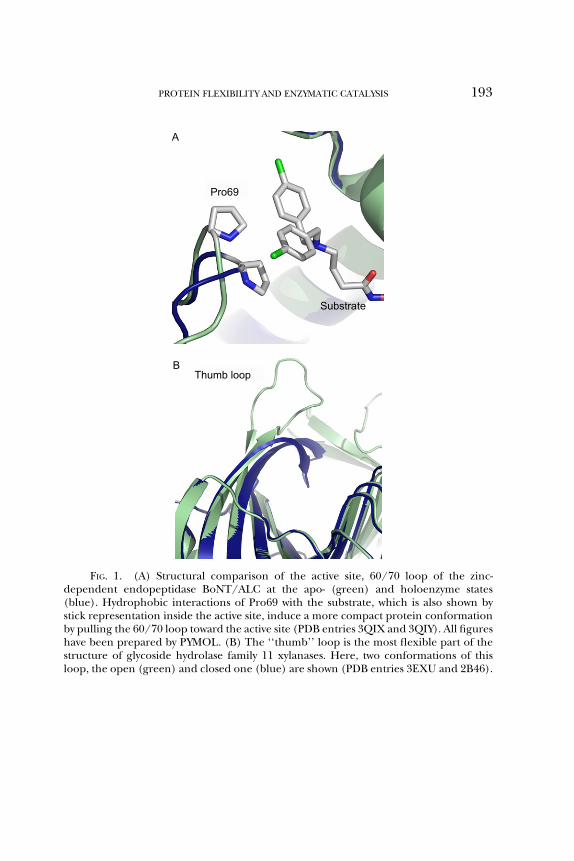
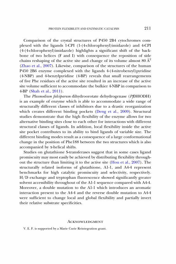
The extreme flexibility of loops which reflects the absence of conforma-tional constraints is consistent with their low sequence conservation, anindicator of low evolutionary pressure. A pronounced exception to thisconservation rule, are the loops participating in the active sites of enzymes.The critical role of loops for the enzymatic function was recognized earlyand their conformational transitions are now accepted as key events incatalytic processes (Malaban et al., 2010). Such loops, commonly called‘‘lids’’ or ‘‘flaps’’, have been the subject of investigation by many researchgroups. Their location at the entry of active site plays a major role insubstrate selectivity and recognition and facilitation of substrate bindinginto the binding cleft. The structural comparison of apo- and holo-states ofvarious enzymes highlights that the main structural difference between thetwo states is the different conformations of active/binding site loops.At the unbound state, the flaps adopt an open conformation which leavesthe binding cleft and active site accessible to solvent. In the bound state,the flaps adopt a closed conformation which blocks the entry to the activesite. A characteristic example is the 60/70 loop of the zinc-dependentendopeptidase BoNT/A LC (Thompson et al., 2011). Binding of threedifferent inhibitors induces a consistent change in the conformation ofthis loop as a result of the hydrophobic interaction of a loop residue(Pro69) with an aromatic moiety of the inhibitor (Fig. 1A). Compared tothe uncomplexed enzyme, ligand binding induces a more compact con-formation because the loop is pulled into the active site enclosing moretightly specific subsites of the inhibitor and inducing a more effectiveinhibition. Thus a disordered, solvent-exposed loop adopts upon substratebinding a more compact and ordered conformation-making interactionswith subsites of the ligand and/or other residues of the protein. Theclosing of the flaps traps the ligand into the cleft and shields fully orpartially the ligand molecule from the aqueous environment, therebystabilizing the bound state. Moreover, the access of the active site toother molecules is prevented and the reaction intermediates are protectedand stabilized (Kember, 1993). This mechanism provides an effective way
Pro69
A
Substrate
Thumb loopB
FIG. 1. (A) Structural comparison of the active site, 60/70 loop of the zinc-dependent endopeptidase BoNT/ALC at the apo- (green) and holoenzyme states(blue). Hydrophobic interactions of Pro69 with the substrate, which is also shown bystick representation inside the active site, induce a more compact protein conformationby pulling the 60/70 loop toward the active site (PDB entries 3QIX and 3QIY). All figureshave been prepared by PYMOL. (B) The ‘‘thumb’’ loop is the most flexible part of thestructure of glycoside hydrolase family 11 xylanases. Here, two conformations of thisloop, the open (green) and closed one (blue) are shown (PDB entries 3EXU and 2B46).
PROTEIN FLEXIBILITY AND ENZYMATIC CATALYSIS 193
194 KOKKINIDIS ET AL.
to control the accessibility to the active site. After the end of the reaction,the opening of the loops permits release of the product and the beginningof a new catalytic cycle. For example, in the glycoside hydrolase family 11xylanases, a highly conserved ‘‘thumb loop’’ in the proximity of the activesite has been found in three conformational states, a closed, a loose, andan extended open one (Pollet et al., 2009; Fig. 1B). Molecular dynamicssimulations suggest that the thumb is the most flexible part of the xylanasestructure: a dynamic catalytic cycle has been proposed based on thesethree different conformations of the thumb. These conformationalchanges are directly associated with the binding of the substrate and therelease of the product. Moreover, a mutation which hinders the thumbmovement results in a fourfold decrease of turnover number thus suggest-ing a direct relation between thumb movement and catalytic properties.
Flaps are not the only flexible structural elements around active sites.Quite often, the rearrangement of the active site geometry is accompaniedor facilitated by movements and conformational changes of secondarystructural elements directly adjacent to the active site, for example, heliceswhich frequently precede or follow the flaps. Examples are presented atthe following paragraphs for the cases of EryK P450 cytochrome andBcZBP deacetylase.
At least two models are used to describe and mechanistically explain thetransition from the open to the closed conformation. The first of them,the well-known ‘‘induced fit’’ model, postulates that large conformationalchanges at a local level, and the transition to the closed conformation areinduced by substrate binding into the active site cleft. In a number ofexamples, the unbound enzyme state has as a favored conformation theopen one and the bound enzyme state, the closed one. The extensivelystudied loop 6 of triosephosphate isomerase (TIM) is a typical case inter-preted through the ‘‘induced fit’’ model (Lolis and Petsko, 1990). TIMis a homodimer in which the subunits catalyze the interconversion ofD-glyceraldehyde 3-phosphate and dihydroxyacetone phosphate. Eachsubunit is a classical (ba)8 barrel (Branden and Tooze, 1999) with theactive site being formed by the loops located at the C-termini of the eightb-strands (loops 1–8) of the barrel. A detailed structural comparison of theunbound and bound forms revealed that the only significant difference isthe movement of the 10-residue loop 6 resulting in the blockage of theactive site entry at the bound state. Some residues of the active site loopare moved up to 7 A from their original position.

PROTEIN FLEXIBILITY AND ENZYMATIC CATALYSIS 195
However, in some cases, experimental evidence supports as an alterna-tive a ‘‘conformational selection’’ scenario in which the enzyme preexistsin two alternative and stable conformations which differ in their ability tobind ligands. This concept has been reported for the monomeric cyto-chrome P450 (CYP) from Saccharopolyspora erythraea, commonly called EryK(Savino et al., 2009). X-ray crystallography studies revealed two differentstructures, an open and a closed one, which were determined for theunbound state of EryK in different ionic strength conditions. Characteri-zation of binding kinetics of the ErD substrate to EryK could be sufficientlyexplained by the ‘‘conformational selection’’ model and allowed for theexclusion of the ‘‘induced fit’’ model. Comparison of the two structuresrevealed conformational shift involving among others a movement of anactive site loop of about 11 A and a shift of the N-terminus of an a-helix ofabout 10 A toward this active site loop. This reorganization closes theaccess channel for the active site.The rigid body movement observed for the TIM active site loop 6 is the
most common type of loop motion in general, and is termed ‘‘hinge-bending motion’’ because the loop is linked to the protein through tworegions known as hinge regions and moves around them as a rigid body.For a-helices, a common movement is rotation around one of their endswhich moves the other end away from its original position, occasionally upto several Angstroms. The high flexibility of the loops is often associatedwith the presence of glycine residues while in hinges alanines and prolinesare also quite common. Mutagenesis has been frequently used to probethe significance of loop flexibility for catalysis with hinge residues beingquite often the targets of such experiments. In the case of glutathionesynthetase (Tanaka et al., 1993), replacement of Gly and Pro in the hingeby Val significantly impaired the enzymatic activity. Structural studies showthat the mutations caused no structural alterations to the rest of thestructure. Absence of electron density indicated that a high degree offlexibility has been retained for the mutant loop as well as for the nativeone. The observed loss of catalytic activity could reflect problems inconformational adaption of the mutant loops in the closed state whichresults in disruptive interactions with the bound substrate. A study byKursula and coworkers (2004) demonstrated by mutagenesis the impor-tance of small residues at position 3 of the C-terminal three-residue hinge(LysThrAla) of TIM loop 6. When the small Ala residue at position 3 wasreplaced by bulky residues (Gln, Leu, or Lys), the unliganded form of the

196 KOKKINIDIS ET AL.
mutants resembled the closed conformation. The results suggest thatthese mutations shift the equilibrium of the oscillation motion of loopin favor of the closed conformation. Kinetic data implies that in thesemutants substrate binding is the rate-limiting step.
Except from hinge residues, the importance of residues in the middle ofloops for active site flexibility has also been investigated. When Gly76 ofthe active site loop (residues 71–80) of pepsin was replaced by Ala, Val,and Ser, a lower catalytic efficiency was measured and interpreted as theresult of lower flap flexibility. A lower flexibility could alter the pattern ofinteractions (hydrogen bonds) which are responsible for substrate align-ment in the active site (Okoniewska et al., 2000).
In conclusion, mobile loops in the proximity of active sites frequentlyplay various roles in catalysis and it is widely accepted that their pro-nounced conformational flexibility may contribute to their functions.Mutagenesis experiments support the concept of this flexibility beingessential for catalytic efficiency and activation processes.
B. Active Sites Located at Domain Interfaces
A significant portion of oligomeric enzymes form their catalytic andbinding sites at the interface between subunits while, in the case ofmonomeric enzymes, active and binding sites often lie between domains(Ali and Imperiali, 2005). Among the most well-studied examples ofenzymes with their active/binding sites formed between domains are thealcohol and glutamate dehydrogenases, citrate synthase and hexokinase,glutathione transferases and ribonuclease A. An obvious consequencefrom the location of active sites in the interdomain or intersubunit inter-face is that even subtle local conformational changes between thedomains, which are commonly called lobes, could affect catalysis andenzyme interaction with substrate and products. A model consistent withmany experiments and with the Koshland’s (1958) ‘‘induced fit’’ modelimplies that the ligand is first bound to one lobe; subsequent movement ofthe other lobe brings it near to the ligand permitting additional interac-tions which stabilize the structure (Stillman et al., 1993; Hayward, 2004).
Lesk and Chothia (1984) classified domain motions as hinge or sheardomain motions. In the former case, the two domains are connectedthrough a short extended and flexible peptide fragment, the hinge re-gion. Hinge permits rotations while the lobes move as rigid bodies with

PROTEIN FLEXIBILITY AND ENZYMATIC CATALYSIS 197
their internal structure being preserved and all the deformations restrictedto the hinge region. As a result, the binding cleft may exist in manydifferent intermediate states between the open and the closed ones. Onthe other hand, the shear motion is generated by small rearrangementmotions, that is, small changes in torsion angles and side-chain motionscausing small shifts of secondary structure elements with respect to theoriginal position. The shear domain closure is thus the cumulative effectof relatively small shifts of loosely packed secondary structures. This type ofmotion is more likely to occur when there is an extended surface betweendomains. Domain motions around the active sites of citrate synthase andhexokinase have been grouped with this type of motions.Large-scale domain motions that can be classified as hinge motions were
observed for the homodimeric restriction endonuclease PvuII in connec-tion with the binding of cognate oligonucleotides. The U-shaped dimerexists in an open (Athanasiadis et al., 1994) and a closed, DNA-boundconformation (Cheng et al., 1994). Each PvuII monomer consists of aDNA-binding subdomain which also carries the catalytic site and a helicaldimerization subdomain. Although the structures of the individual sub-domains are highly conserved in the open and the closed conformation,their relative orientation changes upon DNA binding, with the opening ofthe DNA-binding cleft being reduced by nearly 28 A upon transition fromthe open to the closed conformation. This large conformational changeresults mainly from small changes (<10�) in the ’,c backbone conforma-tional angles of two Gly residues located in a loop region at the interfacebetween the dimerization and DNA-binding subdomains.Alcohol dehydrogenases are present in many different organisms and
catalyze the conversion of alcohols to aldehydes functioning as dimers ortetramers (Lesk and Chothia, 1984). Each subunit consists of a catalyticand a coenzyme-binding domain which are linked together through twoa-helices (termed a2 and a3). Comparing the crystal structures of the apo-and holo-liver alcohol dehydrogenase, Eklund et al. (1981) described theconformational changes between these states as a rigid body rotation ofone domain relative to the other one. The two hinge regions were deter-mined at the a2, a3 helices and was found to consist of three and fourresidues, respectively. Colonna-Cesari and co-workers (1986) applied em-pirical energy functions to simulate the domain rotation. The analysisshowed that most of the hinge residues undergo small changes at theirmain chain, ’/c angles (<15�) while motion of the side chains with

198 KOKKINIDIS ET AL.
variations in w torsion angles of about 50� was observed. The internaldynamics of the enzyme was investigated by small angle neutron scatteringand NSE spectroscopy and confirmed a large-scale correlated domainmotion (Biehl et al., 2008). By normal mode analysis, this motion canbe understood as the result of the movement of the outside catalyticdomain with respect to the rigid core. Moreover, this motion results in acleft opening which is much larger than the relatively narrow pocketobserved in the crystal structures.
RNase A is a model system which has been used in numerous experi-ments for the study of dynamical properties of proteins and it is repre-sented in PDB with more than a hundred deposited structures (e.g., seeWlodawer et al., 1988; Santoro et al., 1993; Toiron et al., 1996; Leonidas etal., 1997). It is a stable monomeric enzyme which catalyzes the cleavage ofthe P��O5 bond in single-stranded RNA. Its structure consists of two all-beta domains which form a V shape and are linked by a hinge. Thebinding and active sites are formed at the interface between the domainswith the active site located at the bottom of the deep cleft. Petsko andcolleagues have shown that the enzyme is unable to bind the inhibitorcytidine 20-monophosphate (20-CMP) at temperatures lower than �50 �C(Rasmussen et al., 1992). Since no structural differences could be noticedcomparing the crystal structures at cryo temperatures and room tempera-ture, they suggested that flexibility is a required property for binding andcatalysis (Tilton et al., 1992). Vitagliano and co-workers (2002) couldobserve in the crystal a significant, reversible, hinge-bending domainmotion upon ligand binding which leads to a more compact structure.Upon substrate binding, the hinge region is moved, the angle between thedomains decreases, and residues belonging to different domains andinvolved in substrate binding present a significant reduction of theirbetween distances.
C. Flexibility of Active Site Residues
Active site residues play many different roles. They may be involved incatalysis and substrate binding, stabilize the intermediates of the reactionor the structure of the binding cleft. They provide the suitable for thecatalysis microenvironments and enable substrates to form enough contactpoints for strong binding. Some degree of flexibility is inevitable for theactive site residues to achieve their functions and accommodate the

PROTEIN FLEXIBILITY AND ENZYMATIC CATALYSIS 199
conformational changes which are necessary during the catalytic cycle.This flexibility facilitates active site rearrangements during the numerousintermediate steps of the reaction. The flexibility is achieved at a cost,usually in form of local strain or instability. A systematic mutationalanalysis of active site residues of barnase protein was one of the firststudies to demonstrate that there is an inverse relationship betweenstability of active site and activity (Meiering et al., 1992).The side chains of catalytic residues usually adopt more than one confor-
mation although only some of them may be catalytically competent.In addition, their conformational flexibility has been related to specificsteps of the catalytic cycle as, for example, for the proton shuttling. Acharacteristic example comes from the hydrolytic aldehyde dehydro-genases (ALDHs). Analysis of the crystal structures of several differentALDHs from more than nine different organisms determined in the ab-sence or presence of substrates, cofactors, and products showed that al-though these proteins do not undergomajor conformational changes uponbinding of cofactors, they exhibit a high degree of flexibility for the catalyticresidues Cys302 and Glu268 (Gonzalez-Segura et al., 2009; Munoz-Clareset al., 2011; Fig. 2). In particular, Cys302 has been found in two conforma-tions called ‘‘resting’’ and ‘‘attacking’’ conformation, which are far andclose to the carbonyl carbon of the bound aldehyde, respectively. Thecatalytic Glu268 adopts three conformations: (i) the ‘‘inside’’ conforma-tion which activates Cys302 for nucleophilic attack, (ii) the ‘‘intermediate,’’which activates the hydrolytic water molecule, and (iii) the ‘‘outside’’conformation which releases the proton taken from Cys or water througha proton relay mechanism. Examination of the various residue conforma-tions with respect to the binding cleft architecture in the presence andabsence of the cofactors reveals the sterically compatible combinations andhelps rationalize the details of the mechanism.Another recent example comes from the crystal structures of TIM
protein complexed with suicide inhibitors (Venkatesan et al., 2011). It isshown that two residues of the active site, the catalytic Glu167 and Glu97,are flexible and can adopt two different conformations when the enzymeexists in the closed liganded state. The differences are mainly associatedwith changes in the side-chain dihedrals which give rise to two active sitegeometries. Thus, when the (S)-glycidol phosphate ((S)-GOP) is bound inthe active site, Glu167 adopts its well-known competent conformation andGlu97 is salt bridged. When (R)-glycidol phosphate ((R)-GOP) is bound

Glu268
‘‘resting’’ ‘‘inside’’
‘‘attacking’’
‘‘outside’’
‘‘intermediate’’
Cys302
FIG. 2. Local flexibility of the active site residues of aldehyde dehydrogenases isdemonstrated by the multiple conformations of Cys302 and Glu268. As it is shown,Cys302 can adopt two conformations called ‘‘resting’’ and ‘‘attacking’’ while Glu268adopts three, the ‘‘inside’’ ‘‘intermediate’’ and ‘‘outside’’ conformations. Combinationsof these conformations determine different stages of the catalytic cycle (PDB entries1UZB and 1O02).
200 KOKKINIDIS ET AL.
both residues adopt unusual conformations. The first geometry enablesGlu167 to attack the terminal carbon of (S)-GOP in a stereochemicallyfavored (linear) arrangement, but this is not possible for (R)-GOP at thesecond geometry. These could explain the higher chemical reactivity of(S)-GOP compared with (R)-GOP.
D. A Specific Example: Flexibility of the BcZBP Deacetylase
BcZBP is a zinc-dependent deacetylase from Bacillus cereus whose crystalstructure has been reported at the resolution of 1.8 A (Fadouloglou et al.,2006, 2007). The biological pathway and the function of the protein wereunknown until recently when the ortholog from Bacillus anthracis BaBshB(with 97% sequence identity) was identified as a deacetylase involved in thebacillithiol biosynthesis (Newton et al., 2009; Parsonage et al., 2010). BcZBPis a hexamer and possesses six structurally equivalent active sites which areformed by the association of two monomers. X-ray crystallography,

PROTEIN FLEXIBILITY AND ENZYMATIC CATALYSIS 201
mutagenesis, and molecular dynamics simulations have been used to eluci-date aspects related with function and flexibility of the protein.B-factor analysis of the crystal structure and structural superposition of
different states of the enzyme demonstrates a high mobility for the threeactive site loops (42–51, 129–140, and 180–192) and highlights changes ofthe active site cavity between states. Structural comparison of BcZBP withthe homologous TT1542, which shares 38% sequence identity and hasbeen used as the search model for the molecular replacement whichleaded to the BcZBP’s structure, locates the greater differences betweenthe enzymes in the immediate environment of the active sites. In the caseof BcZBP, the N-terminus of a2-helix has been moved 4 A closer to theactive site together with the preceding loop (42–51) which has beenshifted toward the top of the active site. Because BcZBP represents thestructure of apoenzyme and TT1542, the structure without the zinc boundin the active site, we suggest that the observed differences highlight a zinc-induced organization of the active site environment by promoting a moreclosely packed conformation. On the other hand, the temperature factorsof crystal structures demonstrate as the most flexible part of both enzymesthe active site loop 180–192, joining helices a5 and a6.A 50 ns molecular dynamics simulation study (Fadouloglou et al., 2009)
supports the X-ray crystallography findings. The analysis resulted in anagreement between the simulation-derived atomic fluctuation and thecrystallographically determined atomic temperature factors. Thus, thethree loops which frame the active site, have atomic fluctuations whichare significantly greater than the average for the rest of the structuredemonstrating their high flexibility. The trajectory also reveals a concertedloop motion, which generates the effect of a breathing-like motion aroundthe active site with successive opening and closing events.The functional significance of the active site loops flexibility was further
investigated by mutagenesis of the hinge residues Arg140 and Ala42 (Deliet al., 2010). Arginine 140 is located at the rim of the substrate-bindingcleft. In the crystal structure, its side chain adopts two distinct conforma-tions. The one conformer blocks the active site’s entry while the other one,which is stabilized by electrostatic interactions, keeps the active site acces-sible. Arg was replaced by the small, hydrophobic Ala residue and by theoppositely charged Glu residue. In both variants, the ability for distinctconformations at position 140 has been disrupted. Both variants showed adecrease in their catalytic efficiency compared with the wild type. Ala42,

202 KOKKINIDIS ET AL.
on the other hand, which is located in the proximity of the substrate wasreplaced by a Ser residue. Ser is able to form putative hydrogen bondseither with the substrate or with adjacent amino acids. With either of theseways, the mutation is predicted to increase the rigidity or even to trap theloop to a closed-like conformation. Indeed, the produced variant exhib-ited a dramatic reduction of efficiency.
IV. Flexibility and Special Aspects of Enzyme Properties
A. Flexibility and Thermal Enzymatic Adaptation
Given the environmental variations at the different areas of earth, manyorganisms have evolved adaptation mechanisms for very low or very hightemperatures. As a consequence, there are enzymes isolated from suchorganisms whose optimal temperature for function is below 10 �C (psy-chrophilic) or above 45 �C (thermophilic). Catalytic residues are generallyconserved in homologous psychrophilic and thermophilic enzymes whichimply that cold adaptation resides on other parts of the structure. In theeffort to elucidate the molecular basis of enzymatic temperature, adapta-tion was observed that the cold adaptation is related with a reducedthermal stability (cold adaptation is reviewed by Siddiqui andCavicchioli, 2006). This led to the hypothesis that optimization of thecatalytic activity at low temperatures may be associated with an increasedstructural flexibility (Feller and Gerday, 1997). Since psychrophilicenzymes must function in low temperatures, the evolutionary pressureto retain structural features responsible for conformational rigidity (e.g.,disulfide and salt bridges) is relaxed. In the absence of conformationalconstraints for stability, they present an increase in flexibility which possi-bly leads to a reduction of the activation energy (D’ Amico et al., 2002).In other words, the increased active site flexibility results in a highernumber of conformational states of the enzyme/substrate complex. Theenergy of activation is used many times as a criterion for the evaluation ofpsychrophilicity as it is usually lower in the cold-adapted enzymes than totheir mesophilic counterparts. Sequence comparison among homologuesfrom thermophilic, mesophilic, and psychrophilic organisms shows thatgenerally in the cold-adapted enzymes (Siddiqui and Cavicchioli, 2006):(i) buried residues tend to be smaller and less hydrophobic, (ii) surface

PROTEIN FLEXIBILITY AND ENZYMATIC CATALYSIS 203
residues tend to be less polar, probably because polar residues conferstability by forming additional intramolecular H-bonds. (iii) Less Ile resi-dues are found, probably due to their good packing properties inside theprotein cores, (iv) Lys residues usually replace Arg which facilitate agreater number of electrostatic interactions and H-bonds over Lys, (v) aless number of salt bridges are observed. Radestock and Gohlke (2011)probed by computational means the corresponding states hypothesiswhich claims that homologues from different thermal-adapted organismsare in corresponding states of similar rigidity and flexibility at theirrespective optimal temperatures. Comparing a sample of 19 pairs ofhomologues from meso- and thermophilic enzymes, they showed thatadaptive mutations of thermophilic enzymes maintain the balance be-tween overall rigidity important for thermostability and local flexibilityimportant for activity at the respective temperature at which the proteinsfunction.In several studies, the psychrophilicity has been related to an overall
flexibility throughout the protein structure. Studies on the a-amylase fromPseudoalteromonas haloplanktis have shown that the cold adaptation strategyfor this enzyme leads to a uniformly unstable protein (Feller et al., 1999).Site-directed mutagenesis and comparison with mesophilic and thermo-stable a-amylases demonstrated a weakening of intramolecular interac-tions which lead to an overall decrease of the thermostability of thepsychrophilic protein. This provides the appropriate plasticity aroundthe active site, necessary to adapt the catalytic efficiency to low tempera-tures. In the case of Zn-metalloproteases of thermolysin family, an optimi-zation of the overall protein flexibility is achieved via the reduction of thehydrogen bonds stability in the dynamic structure due to a decrease ofamino acids which form hydrogen bonds (Xie et al., 2009).However, the need for a global elevated flexibility was questioned and
another cold adaptation model was proposed which implies that therequirement for increased structural flexibility can be limited only to asmall, crucial region of the protein structure and, in particular, aroundand inside the active site (Fields and Somero, 1998). Many studies presentevidence which supports this model. Comparing A4-lactase dehydro-genases from Antarctic (optimum temperature of function �2 to 1 �C)and South American (4–10 �C) notothenioid species was found that theactive site residues are fully conserved. Combination of kinetic, sequence,and structural data suggested that cold adaptation is based on increasing

204 KOKKINIDIS ET AL.
flexibility in small areas of the molecule, outside the active site, that affectthe mobility of adjacent to active site structures (Fields and Somero, 1998).The work of Watanabe and Takada (2004) demonstrates the importanceof amino acid substitutions around the active site to the thermal adapta-tion of isocitrate lyases. Gln207 and Gln217 of the psychrophilic lyase fromColwellia maris were replaced by a His and a Lys, respectively, which are theresidues occupying the equivalent positions at the lyase from Escherichiacoli and other organisms and are essential for catalysis. The catalytic activityat low temperatures is decreased or diminished respectively while theenzyme remains active at moderate temperatures. It is believed thateach of the two residues affects the thermal adaptation through a differentmechanism since the Gln217Lys replacement is associated with structuralchanges expressed as an increase in the thermostability and changes in theCD spectra (enhancement of a-helical components) while the Gln207Hisreplacement resulted in slight conformational changes. Comparative mo-lecular dynamics studies show that the strategy of members of elastasefamily for being adapted at cold also concerns increase of localizedflexibility (Papaleo et al., 2006; Riccardi and Papaleo, 2010). The mostobvious difference between mesophilic and psychrophilic elastases con-cerns the amino acid composition and flexibility of loops which areclustered around the active site and specificity pocket.
The importance of local flexibility into the active site for cold adapta-tion has been demonstrated in several cases. The psychrophilic alkalinephosphatase from the Antarctic strain TAB5 has been used as a modelsystem, and the possibility of modifying its psychrophilic properties byintroducing—via mutagenesis—predictable flexibility changes to key ac-tive site residues (Tsigos et al., 2001) or residues to the direct vicinity ofactive site (Mavromatis et al., 2002) has explored. Tsigos et al. (2001) havemodified the side-chain flexibility of the catalytic residues Trp260 andAla219 and that of His135 from the Mg2þ binding site. Trp260Lys is lessactive than the wild type at low temperatures, while the double mutantTrp260Lys/Ala219Gln has lost its psychrophilic character, although itsactivity at elevated temperatures exceeds that of the wild type. Finally,substitution of His135 by Asp resulted in stabilization of the enzyme and inthe case of the triple mutant restored a low energy of activation. Thus, thepsychrophilic character of an enzyme can be strongly affected by veryslight variations of its amino acid sequence which however is expectedto drastically change the local flexibility. As it was mentioned above,

PROTEIN FLEXIBILITY AND ENZYMATIC CATALYSIS 205
psychrophilicity has also been related with increased levels of Gly residueswhich ensure local flexibility. Psychrophilic alkaline phosphatase positions261 and 262, in the vicinity of active site are occupied by Gly residues.Mavromatis et al. (2002) have investigated their importance in the estab-lishment of the psychrophilic character of the enzyme. To constrain theconformational flexibility of the main chain with the minimum perturba-tion of the local structure, it was chosen to mutate Gly to Ala. TheGly262Ala mutant is completely inactive while the Gly261Ala has lost thepsychrophilic character although it is active at elevated temperatures.Thus, Gly clusters in the vicinity of active sites in combination with theirstructural environment are frequently essential determinants of the psy-chrophilic character.
B. Flexibility as an Essential Component of Enzymatic Allostery
Allostery is the process by which conformational changes at one site of aprotein (called regulatory or allosteric site) are coupled with changes to adifferent and usually distant functional site (active site). The most com-mon way for triggering the allosteric response of an enzyme is the bindingof a specific allosteric effector molecule which could be an activator or aninhibitor of the enzymatic function. Studies mainly by X-ray crystallogra-phy showed that the functional states of an allosteric enzyme are wellrepresented by two, structurally different forms: (i) the ‘‘R’’ form(relaxed), which has an optimal affinity for the substrate and (ii) the‘‘T’’ form (tensed), which has a minimal affinity. Attempts to explainthe nature of the allosteric transition have led to two models, the so-calledconcerted and sequential models. Both of them have been developedunder the condition that allosteric enzymes are symmetric oligomerswith identical protomers. Each subunit in the oligomer can adopt oneof the ‘‘R’’ or ‘‘T’’ conformations. According to the concerted model(Monod et al., 1965), the conformational changes induced by the bindingof an allosteric effector to one subunit is transmitted to all other subunits,thus switching them to the same conformation, corresponding to one ofthe two possible states, either ‘‘T’’ or ‘‘R’’. On the other hand, thesequential model (Koshland et al., 1966) permits a number of differentglobal states, that is, subunits can change conformations independentlyand alterations to one of them are not necessarily transmitted to theothers. Thus, the oligomer could be found in a number of hybrid ‘‘TR’’

206 KOKKINIDIS ET AL.
combinations. A relatively new idea for rationalizing the allosteric regula-tion is based on the concept that allosteric enzymes, as all proteins, exist asa dynamic ensemble of conformational states. According to the ensemblemodel of allostery, ligand binding to the allosteric site leads to redistribu-tion within the ensemble enabling an altered conformation at the regionaround the active site (Gunasekaran et al., 2004; Wrabl et al., 2011). Theshift in the relative populations of the protein may affect protein function,that is, as the distribution substrate affinities are altered within the confor-mational ensembles (Goodey and Benkovic, 2008). The dependence ofprotein function on conformational flexibility favors an expanded view ofallostery as an intrinsic property of all dynamic proteins which are poten-tially allosteric. Based on this idea and in contrast to what was believed inthe past, allostery is recognized now as a property of monomeric proteinsas well (Gunasekaran et al., 2004). The binding of the effector favors thetransition from the relatively rigid inactive T-form to a more flexible activeensemble and the increased flexibility facilitates the conformational tran-sitions during enzyme turnover. Hilser and Thompson (2011) have usedthe ensemble concept of allostery to discuss the behavior of steroidhormone receptors which are much more dynamic systems than is repre-sented by traditional models. Thus, a dynamic ensemble of structures andthe presence of intrinsically disordered segments which are stabilizedupon ligand binding can explain the broad variety of ligands whichdrive remote allosteric responses. The importance of this model to theexplanation of cooperativity of the bacterial flagellar switch was recentlydiscussed by Bai et al. (2010). Novinec et al. (2010) showed by intrinsicfluorescence spectroscopy that Cathepsin K, a human cysteine peptidase,has a flexible structure which converts slowly among distinct conforma-tional states. Addition in a protein sample of glucosaminoglycans, whichare allosteric regulators of Cathepsin K, causes a change in the enzymeconformation resulting in a rapid binding of the inhibitor stefin A. So, it isproposed that glucosaminoglycans affect the distribution of the preexist-ing conformational equilibrium.
An intuitively very appealing mechanism for the coupling of the effectorbinding to one site with the conformational changes to another site is theexistence of amino acid networks (Goodey and Benkovic, 2008). Accord-ing to this model, propagation of the signal through a protein structure isbased on networks of physically interconnected and thermodynamicallylinked residues. Amaro et al. (2007) combining data from crystallography,

PROTEIN FLEXIBILITY AND ENZYMATIC CATALYSIS 207
biochemical kinetic assay, and molecular dynamics simulations haverevealed a network of interactions directly correlated to the transmissionof the allosteric signal of the imidazole glycerol phosphate synthase. Thisnetwork is formed by a set of highly conserved amino acids which leadfrom the allosteric site of the enzyme to its active site, more than 25 Aapart. A correlated motion analysis confirms the involvement of theseresidues to a stream of coupled motions and their role in the allostericresponse.Two examples which clearly demonstrate that structural flexibility is an
essential component of allosteric activation are those of glucosamine-6-phos-phate deaminase (Bustos-Jaimes et al., 2002; Rudino-Pinera et al., 2002) andof the human hexameric UDP-glucose dehydrogenase (Kadirvelraj et al.,2011; Sennett et al., 2011). Glucosamine-6-phosphate deaminase catalyzesthe reversible isomerization–deamination of glucosamine 6-phosphate andit is allosterically activated by the N-acetyl glucosamine-6-phosphate. Com-paring a variety of structures Rudino-Pinera et al. (2002) demonstrated thatthe main structural differences between the T- and R-states are located atthe active site lid. This enzyme possesses a complex active site lid (residues158–187) formed by a helix–loop segment and a b-strand (Fig. 3A). The lidis directly connected with both, the active and allosteric sites, that is, Arg172is an active site residue which participates at the substrate binding whileArg158, Lys160, and Thr161 belong to the allosteric site. Experimentssupport the correlation of the lid flexibility with the allosteric transitionand the substrate-binding properties of the active site. It was shown that theallosteric transition from the ‘‘T’’ to ‘‘R’’ state is not associated with definedgeometrical changes of the lid but with pronounced changes to its confor-mational flexibility. Especially, the atoms in the central segment of the lidshow a marked decrease in their crystallographic B-factor from a B averageof 80 at the T-state to a B average of 40 A2 at the R-state. Moreover, when theenzyme is at the R-state, the active site lid has been found in three distinctconformations (Rudino-Pinera et al., 2002). Substrate binding stabilizes thelid in one of the three conformers and produces a general reduction in theatomic vibration of the whole protein.The connection of the conformational flexibility of the lid and the
function of the deaminase was investigated by mutating Phe174 to Ala.This mutation had, as a result, such an increase of the conformationalflexibility of the T-form that no electron density was visible for part of thelid (Bustos-Jaimes et al., 2002). The mutant was inactive in the absence of

Thr161
A
B
Lys160
Arg172
FIG. 3. (A) Cartoon representation of the glucosamine-6-phosphate deaminase.The enzyme possesses a complex active site lid (shown in green) which is directlyconnected with both, active and allosteric sites. To illustrate the location of these sites,a glucosamine 6-phosphate (space-filling model) is shown bound to each of them (PDBentry 1FS5). (B) Structural comparison of the P450 cytochromes CYP101 (P450cam,PDB entry 2L8M) in gray from Pseudomonas putida and CYP108 (P450terp, PDB entry1CPT) in red, a bacterial enzyme from Pseudomonas. The enzymes adopt a conserved foldwith a substantial variability around the substrate-binding region. The molecule of hemeis shown as stick representation. Small displacements of structural elements which abutsthe active/binding toward the heme together with the segmentation of one of thehelices (shown on the left of the figure) decreases the size of the active/binding site ofCYP101 relative to that of the CYP108.
208 KOKKINIDIS ET AL.

PROTEIN FLEXIBILITY AND ENZYMATIC CATALYSIS 209
the allosteric activator, a proof that, conformational flexibility of the activesite lid alters the binding properties of the active site.Except from the active site lid, the 144–154 loop has been shown to be
directly associated with the allosteric transition through interactions ofGlu148 with residues of the active site lid, which stabilize the R-form or theallosteric site, that is, a salt bridge with Lys 160, which stabilize the T-form.Structural superposition of R-form complexed with either both sites occu-pied or only the allosteric site shows that there is a correlation between themovement of 144–146 part and the active site lid. When the active site is free,the 144–146 part moves to open the lid and when the active site is occupiedmoves to close the lid.The human hexameric UDP-glucose dehydrogenase is an example of an
allosteric enzyme whose active site is bifunctional and can bind eithersubstrate or allosteric effector with distinct ‘‘induced fit’’ conformations(Kadirvelraj et al., 2011). In this case, it was shown that packing defects in theprotein core in combination with structural flexibility constitute a mecha-nism for the evolution of allostery. Binding of the UDP-a-d-xylose (UDX)allosteric inhibitor activates a distinct ‘‘induced fit’’ allosteric response: Theburied Thr131 loop directly connects the hexamer building interface to theactive site andplays the role of an allosteric switch.Upon substrate binding ispositioned in a way which supports the formation of a functional bindingsite. On the other hand, when the allosteric inhibitor binds, this loopmovesabout 4 A and rotates about 180�. This movement changes the packinginteractions of the protein core and rotates a neighboring helix, resultingin the remarkable repacking of the core and the conversion of the enzymeinto an inactive oligomer. Deletion of the buried Val132 traps the enzyme toan open intermediate conformation of the allosteric response (Sennettet al., 2011). Comparison of the closed wild type and the open D132structure identified a hinge-bending axis between two residues of thedimerization domain. A concerted hinge-bendingmotion between adjacentsubunits was proposed to be the basis of the allosteric transition.
C. Flexibility and Ligand Specificity
The broad substrate specificity which characterizes some enzymes hasbeen explained under the view of a flexible active site cavity capable ofaccommodating stereochemically diverse substrates. The different sub-strates may either induce conformational rearrangements to the

210 KOKKINIDIS ET AL.
broader area of the flexible active site or, if the protein structure isseen as a dynamic ensemble of conformations, each substrate is boundto different conformations with different affinity. The impact of plastic-ity and flexibility on ligand binding has been illustrated in the litera-ture, among others, with examples from the family of endonucleases V(Feng et al., 2005), glutathione S-transferases (Hou et al., 2007), dehy-drogenases (Deng et al., 2009), and CYP (Pochapsky et al., 2010). Insome cases, the local flexibility around the active site and the possiblereposition and repack of only few individual side chains could causesufficient space and shape alterations inside the active site to switchfrom one substrate to another without substantial structural changes.However, in other cases, global flexibility seems to be a requirement forpromiscuity.
CYPs are a superfamily of enzymes which catalyze the addition of anoxygen to an C��C or C��H bond, that is, they are oxidoreductasescatalyzing the oxidation of structurally dissimilar organic substrates.CYPs adopt a similar global fold which is combined with a remarkableadaptivity for substrate recognition. This is possibly due to the modularityof secondary structure features which surround the active site (Fig. 3B).These features vary even between different states of the same enzyme(Pochapsky et al., 2010). Some CYPs show broad substrate and hydroxyl-ation specificity and it has been proposed that their high flexibility andplasticity around the active site is responsible for the wide substrateselectivity and specificity they present. The typical topology of CYPs con-tains a four alpha helical bundle which also accommodates the hemegroup which is ligated by a Cys at the beginning of one of the bundlehelices. In total, the structure is composed by 14 helices and 5 beta sheets.In an early spectroscopic study of five individual CYPs (Anzenbacher andHudecek, 2001), it was demonstrated that their active sites exhibit signifi-cant differences in their flexibility and stability although in these enzymesthese properties cannot be related by a simple relationship since in somecases, a low stability does not necessarily reflect a high flexibility. Amongthe five CYPs which were tested, one demonstrates a significantly higherpromiscuity than the others by being able to accommodate and modify avariety of structurally different substrates. Comparison with the other fourcytochromes of higher specificity showed that what differentiates it fromthem is a more flexible active site together with a less conformationallyrestricted heme group.
PROTEIN FLEXIBILITY AND ENZYMATIC CATALYSIS 211
Comparison of the crystal structures of P450 2B4 cytochromes com-plexed with the ligands 1-CPI (1-(4-chlorophenyl)imidazole) and 4-CPI(4-(4-chlorophenyl)imidazole) highlights a significant shift of the back-bone of two helices (F and I) with consequence the reposition of sidechains reshaping of the active site and change of its volume almost 80 A2
(Zhao et al., 2007). Likewise, comparison of the structures of the humanP450 2B6 enzyme complexed with the ligands 4-(4-nitrobenzyl)pyridine(4-NBP) and 4-benzylpyridine (4-BP) reveals that small rearrangementsof five Phe residues of the active site resulted in an increase of the activesite volume sufficient to accommodate the bulkier 4-NBP in comparison to4-BP (Shah et al., 2011).The Plasmodium falciparum dihydroorotate dehydrogenase (PfDHODH)
is an example of enzyme which is able to accommodate a wide range ofstructurally different classes of inhibitors due to a drastic reorganizationwhich creates different binding pockets (Deng et al., 2009). Structuralstudies demonstrate that the high flexibility of the enzyme allows for twoalternative binding sites close to each other for interactions with differentstructural classes of ligands. In addition, local flexibility inside the activesite pocket contributes to its ability to bind ligands of variable size. Thedifferent binding modes result as a consequence of a large conformationalchange in the position of Phe188 between the two structures which is alsoaccompanied by a-helical shifts.Studies on glutathione S-transferases suggest that in some cases ligand
promiscuity may most easily be achieved by distributing flexibility through-out the structure than limiting it to the active site (Hou et al., 2007). Thestructurally related isoforms of glutathione, A1-1, and A4-4 representbenchmarks for high catalytic promiscuity and selectivity, respectively.H/D exchange and tryptophan fluorescence showed significantly greatersolvent accessibility throughout of the A1-1 sequence compared with A4-4.Moreover, a double mutation to the A1-1 which introduces an aromaticinteraction present to the A4-4 and the reverse double mutation to A4-4were sufficient to change local and global flexibility and partially inverttheir relative substrate specificities.
Acknowledgment
V. E. F. is supported by a Marie Curie Reintegration grant.
212 KOKKINIDIS ET AL.
References
Agarwal, P. K. (2006). Enzymes: an integrated view of structure, dynamics and function.Microb. Cell Fact. 5, 2.
Ali, M. H., Imperiali, B. (2005). Protein oligomerization: how and why. Bioorg. Med.Chem. 13, 5013–5020.
Amaro, R. E., Sethi, A., Myers, R. S., Davisson, V. J., Luthey-Schulten, Z. A. (2007).A Network of conserved interactions regulates the allosteric signal in a glutamineamidotransferase. Biochemistry 46, 2156–2173.
Anzenbacher, P., Hudecek, J. (2001). Differences in flexibility of active sites of cyto-chromes P450 probed by resonance Raman and UV–Vis absorption spectroscopy.J. Inorg. Biochem. 85, 209–213.
Athanasiadis, A., Vlassi, M., Kotsifaki, D., Tucker, P., Wilson, K. S., Kokkinidis, M.(1994). The crystal structure of PvuII endonuclease reveals extensive structuralhomologies to EcoRV. Nat. Struct. Biol. 1, 469–475.
Bai, F., Branch, R. W., Nicolau, D. V., Jr., Pilizota, T., Steel, B. C., Maini, F. K.,Berry, R. M. (2010). Conformational spread as a mechanism for cooperativity inthe bacterial flagellar switch. Science 327, 685–689.
Biehl, R., Hoffmann, B., Monkenbusch, M., Falus, P., Preost, S., Merkel, R., Richter, D.(2008). Direct observation of correlated interdomain motion in alcohol dehydro-genase. Phys. Rev. Lett. 101, 138102.
Branden, C., Tooze, J. (1999). Introduction to Protein Structure. 2nd edn. pp. 47–50.Garland Publishing, New York, NY.
Bu, Z., Biehl, R., Monkenbusch, M., Richter, D., Callaway, D. J. E. (2005). Coupledprotein domain motion in Taq polymerase revealed by neutron spin-echo spectros-copy. Proc. Natl. Acad. Sci. U.S.A. 102, 17646–17651.
Bustos-Jaimes, I., Sosa-Peinado, A., Rudino-Pinera, E., Horjales, E., Calcagno, M. L.(2002). On the role of the conformational flexibility of the active-site lid on theallosteric kinetics of glucosamine-6-phosphate deaminase. J. Mol. Biol. 319,183–189.
Chapman, H. N., et al. (2009). Femtosecond X-ray protein nanocrystallography. Nature470, 73–78.
Cheng, X., Balendiran, K., Schildkraut, I., Anderson, J. E. (1994). Structure of PvuIIendonuclease with cognate DNA. EMBO J. 13, 3927–3935.
Colonna-Cesari, F., Perahia, D., Karplus, M., Eklund, H., Branden, C. I., Tapia, O.(1986). Interdomain motion in liver alcohol dehydrogenase. J. Biol. Chem. 261,15273–15280.
D’ Amico, S., Claverie, P., Collins, T., Georlette, D., Gratia, E., Hoyoux, A., Meuwis, M.-A., Feller, G., Gerday, C. (2002). Molecular basis of cold adaptation. Philos. Trans.R. Soc. Lond. B 357, 917–925.
Deli, A., Koutsioulis, D., Fadouloglou, V. E., Spiliotopoulou, P., Balomenou, S.,Arnaouteli, S., Tzanodaskalaki, M., Mavromatis, K., Kokkinidis, M., Bouriotis, V.(2010). LmbE proteins from Bacillus cereus are de-N-acetylases with broad substratespecificity and are highly similar to proteins in Bacillus anthracis. FEBS J. 277,2740–2753.
PROTEIN FLEXIBILITY AND ENZYMATIC CATALYSIS 213
Deng, X., Gujjar, R., Mazouni, F. E., Kaminsky, W., Malmquist, N. A., Goldsmith, E. J.,Rathod, P. K., Phillips, M. A. (2009). Structural plasticity of malaria dihydroorotatedehydrogenase allows selective binding of diverse chemical scaffolds. J. Biol. Chem.284, 26999–27009.
Dunker, A. K., Oldfield, C. J., Meng, J., Romero, P., Yang, J. Y., Chen, J. W., Vacic, V.,Obradovic, Z., Uversky, V. N. (2008). The unfoldomics decade: an update onintrinsically disordered proteins. BMC Genomics 9(Suppl. 2), article S1.
Eklund, H., Samma, J. P., Wallen, L., Branden, C. I., Akeson, A., Jones, T. A. (1981).Structure of a triclinic ternary complex of horse liver alcohol dehydrogenase at 2.9A resolution. J. Mol. Biol. 146, 561–587.
Fadouloglou, V. E., Kotsifaki, D., Gazi, A. D., Fellas, G., Meramveliotaki, C., Deli, A.,Psylinakis, E., Bouriotis, V., Kokkinidis, M. (2006). Purification, crystallization andpreliminary characterization of a putative LmbE-like deacetylase from Bacilluscereus. Acta Crystallogr. F62, 261–264.
Fadouloglou, V. E., Deli, A., Glykos, N. M., Psylinakis, E., Bouriotis, V., Kokkinidis, M.(2007). Crystal structure of the BcZBP, a zinc-binding protein from Bacillus cereus.FEBS J. 274, 3044–3054.
Fadouloglou, V. E., Stavrakoudis, A., Bouriotis, V., Kokkinidis, M., Glykos, N. M. (2009).Molecular dynamics simulations of BcZBP, a deacetylase from Bacillus cereus: activesite loops determine substrate accessibility and specificity. J. Chem. Theory Comput. 5,3299–3311.
Farago, B., Li, J., Cornilescu, G., Callaway, D. J. E., Bu, J. (2010). Activation of nanoscaleallosteric protein domain motion revealed by neutron spin echo spectroscopy.Biophys. J. 99, 3473–3482.
Feller, G., Gerday, C. (1997). Psychrophilic enzymes: molecular basis of cold adapta-tion. Cell. Mol. Life Sci. 53, 830–841.
Feller, G., D’Amico, D., Gerday, C. (1999). Thermodynamic stability of a cold-activea-amylase from the Antarctic bacterium Alteromonas haloplanctis. Biochemistry 38,4613–4619.
Feng, H., Klutz, A. M., Cao, W. (2005). Active site plasticity of endonuclease V fromSalmonella typhimurium. Biochemistry 44, 675–683.
Fields, P. A., Somero, G. N. (1998). Hot spots in cold adaptation: localized increases inconformational flexibility in lactate dehydrogenase A4 orthologs of Antarcticnotothenioid fishes. Proc. Natl Acad. Sci. U.S.A. 95, 11476–11481.
Fischetti, R. F., Rodi, D. J., Mirza, A., Irving, T. C., Kondrashkina, E., Makowski, L.(2003). High resolution wide angle X-ray scattering of protein solutions. J. Synch.Res. 10, 398–404.
Fisher, E. (1894). Einfluss der configuration auf die wirkung der enzyme. Ber. Dtsch.Chem. Ges. 27, 2985–2993.
Gazi, A. D., Bastaki, M., Charova, S. N., Gkougkoulia, E. A., Kapellios, E. A.,Panopoulos, N. J., Kokkinidis, M. (2008). Evidence for a widespread interactionmode of disordered proteins in bacterial type III secretion systems. J. Biol. Chem.283, 34062–34068.
Gazi, A. D., Charova, S. N., Panopoulos, N. J., Kokkinidis, M. (2009). Coiled-coils intype III secretion systems: structural flexibility, disorder and biological implica-tions. Cell. Microbiol. 11, 719–729.
214 KOKKINIDIS ET AL.
Glykos, N. M., Cesareni, G., Kokkinidis, M. (1999). Protein plasticity to the extreme:changing the topology of a 4-a-helix bundle with a single amino-acid substitution.Structure 7, 597–603.
Gonzalez-Segura, L., Rudino-Pinera, E., Munoz-Clares, R. A., Horjales, E. (2009). Thecrystal structure of a ternary complex of betaine aldehyde dehydrogenase fromPseudomonas aeruginosa provides new insight into the reaction mechanism andshows a novel binding mode of the 20-phosphate of NADPþ and a novel cationbinding site. J. Mol. Biol. 385, 542–557.
Goodey, N. M., Benkovic, S. J. (2008). Allosteric regulation and catalysis emerge via acommon route. Nat. Chem. Biol. 4, 474–482.
Gunasekaran, K., Ma, B., Nussinov, R. (2004). Is allostery an intrinsic property of alldynamic proteins? Proteins 57, 433–443.
Hammes, G. G. (2002). Multiple conformational changes in enzyme catalysis. Biochem-istry 41, 8221–8228.
Hammes-Schiffer, S., Benkovic, S. J. (2006). Relating protein motion to catalysis. Annu.Rev. Biochem. 75, 519–541.
Hayward, S. (2004). Identification of specific interactions that drive ligand-inducedclosure in five enzymes with classic domain movements. J. Mol. Biol. 339,1001–1021.
Henzler-Wildman, K., Kern, D. (2007). Dynamic personalities of proteins. Nature 450,964–972.
Hilser, V. J., Thompson, E. B. (2011). Structural dynamics, intrinsic disorder, andallostery in nuclear receptors as transcription factors. J. Biol. Chem. 286,39675–39682.
Hou, L., Honaker, M. T., Shireman, L. M., Balogh, L. M., Roberts, A. G., Ng, K.-C.,Nath, A., Atkins, W. M. (2007). Functional promiscuity correlates with conforma-tional heterogeneity in A-class glutathione S-transferases. J. Biol. Chem. 282,23264–23274.
Kadirvelraj, R., Sennett, N. C., Polizzi, S. J., Weitzel, S., Wood, Z. A. (2011). Role ofpacking defects in the evolution of allostery and induced fit in human UDP-glucose dehydrogenase. Biochemistry 50, 5780–5789.
Kahra, D., Kovermann, M., Low, C., Hirschfeld, V., Haupt, C., Balbach, J.,Hubner, C. G. (2011). Conformational plasticity and dynamics in the genericprotein folding catalyst SlyD unraveled by single-molecule FRET. J. Mol. Biol. 411,781–790.
Kamerlin, S. C. L., Warshel, A. (2010). At the dawn of the 21st century: is dynamics themissing link for understanding enzyme catalysis? Proteins 78, 1339–1375.
Kember, E. S. (1993). Movable lobes and flexible loops in proteins. Structural deforma-tions that control biochemical activity. FEBS 326, 4–10.
Kendrew, J., Bodo, G., Dintzis, H., Parrish, R., Wyckoff, H., Phillips, D. (1958). A three-dimensional model of the myoglobin molecule obtained by X-ray analysis. Nature181(4610), 662–666.
Klepeis, J. L., Lindorff-Larsen, K., Dror, R. O., Shaw, D. E. (2009). Long-timescalemolecular dynamics simulations of protein structure and function. Curr. Opin.Struct. Biol. 19, 120–127.

PROTEIN FLEXIBILITY AND ENZYMATIC CATALYSIS 215
Klink, B. U., Scheidig, A. J. (2010). New insights into the dynamic properties and theactive site architecture of H-Ras p21 revealed by X-ray crystallography at very highresolution. BMC Struct. Biol. 10, 38.
Klink, B. U., Goody, R. S., Scheidig, A. J. (2006). A newly designed microspectrofluo-rometer for kinetic studies on protein crystals in combination with X-ray diffrac-tion. Biophys. J. 91, 981–992.
Koshland, D. E. (1958). Application of the theory of enzyme specificity to proteinsynthesis. Proc. Natl. Acad. Sci. U.S.A. 44, 98–104.
Koshland, D. E., Nemethy, G., Filmer, D. (1966). Comparison of experimental bindingdata and theoretical models in proteins containing subunits. Biochemistry 5,365–385.
Kursula, I., Salin, M., Sun, J., Norledge, B. V., Haapalainen, A. M., Sampson, N. S.,Wierenga, R. K. (2004). Understanding protein lids: structural analysis of activehinge mutants in triosephosphate isomerase. Protein Eng. Des. Sel. 17, 375–382.
Leonidas, D. D., Shapiro, R., Irons, L. I., Russo, N., Acharya, K. R. (1997). Crystalstructures of ribonuclease A complexes with 50-diphosphoadenosine 30-phosphateand 50-diphosphoadenosine 20-phosphate at 1.7 A resolution. Biochemistry 36,5578–5588.
Lesk, A. M., Chothia, C. (1984). Mechanisms of domain closure in proteins. J. Mol. Biol.174, 175–191.
Linderstrom-Lang, K. U., Schellmann, J. A. (1959). Protein structure and enzymeactivity. In: The Enzymes, Boyer, P. D. (Ed.), vol. 1. 2nd edn. pp. 443–510. Aca-demic Press, New York.
Lolis, E., Petsko, G. A. (1990). Crystallographic analysis of the complex betweentriosephosphate isomerase and 2-phosphoglycolate at 2.5 A resolution: implica-tions for catalysis. Biochemistry 29, 6619–6625.
Malaban, M. M., Amyes, T. L., Richard, J. P. (2010). A role for flexible loops in enzymecatalysis. Curr. Opin. Struct. Biol. 20, 702–710.
Mavromatis, K., Tsigos, I., Tzanodaskalaki, M., Kokkinidis, M., Bouriotis, V. (2002).Exploring the role of a glycine cluster in cold adaptation of an alkaline phospha-tase. Eur. J. Biochem. 269, 2330–2335.
McGeagh, J. D., Ranaghan, K. E., Mulholland, A. J. (2011). Protein dynamics andenzyme catalysis: insights from simulations. Biochim. Biophys. Acta 1814, 1077–1092.
Meiering, E. M., Serrano, L., Fersht, A. R. (1992). Effect of active site residues inbarnase on activity and stability. J. Mol. Biol. 225, 585–589.
Mezei, F. (1980). The principles of neutron spin echo. Neutron Spin Echo: Proceed-ings of a Laue-Langevin Institut Workshop. Springer, Heidelberg, Germany,pp. 3–26.
Monod, J., Wyman, J., Changeux, P. (1965). On the nature of allosteric transitions:a plausible model. J. Mol. Biol. 12, 88–118.
Munoz-Clares, R., Gonzalez-Segura, L., Diaz-Sanchez, A. G. (2011). Crystallographic evi-dence for active-site dynamics in thehydrolytic aldehydedehydrogenases. Implicationsfor the deacylation step of the catalyzed reaction. Chem. Biol. Interact. 191, 137–146.
Nashine, V. C., Hammes-Schiffer, S., Benkovic, S. J. (2010). Coupled motions inenzyme catalysis. Curr. Opin. Chem. Biol. 14, 644–651.

216 KOKKINIDIS ET AL.
Neutze, R., Huldt, G., Hajdu, J., Spoel, D. (2004). Potential impact of an X-ray freeelectron laser on structural biology. Rad. Phys. Chem. 71, 905–916.
Newton, G. L., Rawat, M., La Clair, J. J., Jothivasan, V. K., Budiarto, T., Hamilton, C. J.,Claiborne, A., Helmann, J. D., Fahey, R. C. (2009). Bacillithiol is an antioxidantthiol produced in Bacilli. Nat. Chem. Biol. 5, 625–627.
Novinec, M., Kovacic, L., Lenarcic, B., Baici, A. (2010). Conformational flexibility andallosteric regulation of cathepsin K. Biochem. J. 429, 379–389.
Ohgushi, M., Wada, A. (1983). ‘Molten-globule state’: a compact form of globularproteins with mobile side-chains. FEBS 164, 21–24.
Okoniewska, M., Tanaka, T., Yada, R. Y. (2000). The pepsin residue glycine-76 con-tributes to active-site loop flexibility and participates in catalysis. Biochem. J. 349,169–177.
Oyeyemi, O. A., Sours, K. M., Lee, T., Kohen, A., Resing, K. A., Ahn, N. G.,Klinman, J. P. (2011). Comparative hydrogen�deuterium exchange for a meso-philic vs thermophilic dihydrofolate reductase at 25 �C: identification of a singleactive site region with enhanced flexibility in the mesophilic protein. Biochemistry50, 8251–8260.
Papaleo, E., Riccardi, L., Villa, C., Fantucci, P., Gioia, L. (2006). Flexibility and enzy-matic cold-adaptation: a comparative molecular dynamics investigation of theelastase family. Biochim. Biophys. Acta 1764, 1397–1406.
Parsonage, D., Newton, G. L., Holder, R. C., Wallace, B. D., Paige, C., Hamilton, C. J.,Dos Santos, P. C., Redinbo, M. R., Reid, S. D., Claiborne, A. (2010). Characteriza-tion of the N-acetyl-a-D-glucosaminyl l-malate synthase and deacetylase functionsfor bacillithiol biosynthesis in Bacillus anthracis. Biochemistry 49, 8398–8414.
Pochapsky, T. C., Kazanis, S., Dang, M. (2010). Conformational plasticity and struc-ture/function relationship in cytochromes P450. Antioxid. Redox Signal. 13,1273–1290.
Pollet, A., Vandermarliere, E., Lammertyn, J., Strelkov, S. V., Delcour, J. A., Courtin, C. M.(2009). Crystallographic and activity-based evidence for thumb flexibility and itsrelevance in glycoside hydrolase family 11 xylanases. Proteins 77, 395–403.
Radestock, S., Gohlke, H. (2011). Protein rigidity and thermophilic adaptation. Proteins79, 1089–1108.
Rasmussen, B. F., Stock, A. M., Ringe, D., Petsko, G. A. (1992). Crystalline ribonucleaseA loses function below the dynamical transition at 220 K. Nature 357, 423–424.
Riccardi, L., Papaleo, E. (2010). Unfolding simulations of cold- and warm-adaptedelastases. IIOABJ 1, 11–17.
Rudino-Pinera, E., Morales-Arrieta, S., Rojas-Trejo, S. P., Horjales, E. (2002). Structuralflexibility, an essential component of the allosteric activation in Escherichia coliglucosamine-6-phosphate deaminase. Acta Crystallogr. D Biol. Crystallogr 58, 10–20.
Santoro, J., Gonzalez, C., Bruix, M., Neira, J. L., Nieto, J. L., Rico, M. (1993). Highresolution three dimensional structure of ribonuclease A in solution by nuclearmagnetic resonance spectroscopy. J. Mol. Biol. 229, 722–734.
Savino, C., Montemiglio, L. C., Sciara, G., Miele, A. E., Kendrew, S. G., Jemth, P.,Gianni, S., Vallone, B. (2009). Investigating the structural plasticity of a cyto-chrome P450. J. Biol. Chem. 284, 29170–29179.

PROTEIN FLEXIBILITY AND ENZYMATIC CATALYSIS 217
Schlessinger, A., Schaefer, C., Vicedo, E., Schmidberger, M., Punta, M., Rost, B. (2011).Protein disorder—a breakthrough invention of evolution? Curr. Opin. Struct. Biol.21, 412–418.
Schottea, F., Somanb, J., Olsonb, J. S., Wulff, M., Anfinruda, P. A. (2004). Picosecondtime-resolved X-ray crystallography: probing protein function in real time. J. Struct.Biol. 147, 235–246.
Sennett, N. C., Kadirvelraj, R., Wood, Z. A. (2011). Conformational flexibility in theallosteric regulation of human UDP-a-D-glucose 6-dehydrogenase. Biochemistry 50,9651–9663.
Shah, M. B., Pascual, J., Zhang, Q., Stout, C. D., Halpert, J. R. (2011). Structures ofcytochrome P450 2B6 bound to 4-benzylpyridine and 4-(4-nitrobenzyl) pyridine:insight into inhibitor binding and rearrangement of active site side chains. Mol.Pharmacol. 80, 1047–1055.
Siddiqui, K. S., Cavicchioli, R. (2006). Cold-adapted enzymes. Annu. Rev. Biochem. 75,403–433.
Srajer, V., Royer, W. E., Jr. (2008). Time-resolved X-ray crystallography of heme pro-teins. Methods Enzymol. 437, 379–395.
Stillman, T. J., Baker, P. J., Britton, K. L., Rice, D. W. (1993). Conformational flexibilityin glutamate dehydrogenase. Role of water in substrate recognition and catalysis.J. Mol. Biol. 234, 1131–1139.
Tanaka, T., Yamaguchi, H., Kato, H., Nishioka, T., Katsube, Y., Oda, J. (1993). Flexibili-ty impaired by mutations revealed the multifunctional roles of the loop in glutathi-one synthetase. Biochemistry 32, 12398–12404.
Teilum, K., Olsen, J. G., Kragelund, B. B. (2011). Protein stability, flexibility andfunction. Biochim. Biophys. Acta 1814, 969–976.
Thompson, A. A., Jiao, G.-S., Kim, S., Thai, A., Cregar-Hernandez, L., Margosiak, S. A.,Johnson, A. T., Han, G. W., O’Malley, S., Stevens, R. C. (2011). Structural charac-terization of three novel hydroxamate-based zinc chelating inhibitors of the clos-tridium botulinum serotype A neurotoxin light chain metalloprotease reveals acompact binding site resulting from 60/70 loop flexibility. Biochemistry 50,4019–4028.
Tilton, R. F. J., Dewan, J. C., Petsko, G. A. (1992). Effects of temperature on proteinstructure and dynamics: X-ray crystallographic studies of the protein ribonuclease-A at nine different temperatures from 98 to 320 K. Biochemistry 31, 2469–2481.
Todd, A. E., Orengo, C. A., Thornton, J. M. (2002). Plasticity of enzyme active sites.Trends Biochem. Sci. 27, 419–426.
Toiron, C., Gonzalez, C., Bruix, M., Rico, M. (1996). Three-dimensional structure ofthe complexes of ribonuclease A with 20,50-CpA and 30,50-d(CpA) in aqueoussolution, as obtained by NMR and restrained molecular dynamics. Protein Sci. 5,1633–1647.
Tsigos, I., Mavromatis, K., Tzanodaskalaki, M., Pozidis, C., Kokkinidis, M., Bouriotis, V.(2001). Engineering the properties of a cold active enzyme through rationalredesign of the active site. Eur. J. Biochem. 268, 5074–5080.
Uversky, V. N., Dunker, A. K. (2010). Understanding protein non-folding. Biochim.Biophys. Acta 1804, 1231–1264.

218 KOKKINIDIS ET AL.
Venkatesan, R., Alahuhta, M., Pihko, P. M., Wierenga, R. K. (2011). High resolutioncrystal structures of triosephosphate isomerase complexed with its suicide inhibi-tors: the conformational flexibility of the catalytic glutamate in its closed, ligandedactive site. Protein Sci. 20, 1387–1397.
Vitagliano, L., Merlino, A., Zagari, A., Mazzarella, L. (2002). Reversible substrate-induced domain motions in ribonuclease A. Proteins 46, 97–104.
Watanabe, S., Takada, Y. (2004). Amino acid residues involved in cold adaptation ofisocitrate lyase from a psychrophilic bacterium, Colwellia maris. Microbiology 150,3393–3403.
Westenhoff, S., Nazarenko, E., Malmerberg, E., Davidsson, J., Katona, G., Neutze, R.(2010). Time-resolved structural studies of protein reaction dynamics: a smorgas-bord of X-ray approaches. Acta Crystallogr. A A66, 207–219.
Wlodawer, A., Svensson, L. A., Sjolin, L., Gilliland, G. L. (1988). Structure ofphosphate-free ribonuclease A refined at 1.26 A. Biochemistry 27, 2705–2717.
Wolfenden, R., Snider, M. J. (2001). The depth of chemical time and the power ofenzymes as catalysts. Acc. Chem. Res. 34, 938–945.
Woofter, R. T., Maurer, M. C. (2011). Role of calcium in the conformational dynamicsof factor XIII activation examined by hydrogen-deuterium exchange coupled withMALDI-TOF MS. Arch. Biochem. Biophys. 512, 87–95.
Wrabl, J. O., Gu, J., Liu, T., Schrank, T. P., Whitten, S. T., Hilser, V. J. (2011). The roleof protein conformational fluctuations in allostery, function, and evolution. Bio-phys. Chem. 159, 129–141.
Xie, B.-B., Bian, F., Chen, X.-L., He, H.-L., Guo, J., Gao, X., Zeng, Y.-X., Chen, B.,Zhou, B.-C., Zhang, Y.-Z. (2009). Cold adaptation of Zinc metalloproteases in thethermolysin family from deep sea and arctic sea ice bacteria revealed by catalyticand structural properties and molecular dynamics. J. Biol. Chem. 284, 9257–9269.
Yon, J. M., Perahia, D., Ghelis, C. (1998). Conformational dynamics and enzymeactivity. Biochimie 80, 33–42.
Zhao, Y., Sun, L., Muralidhara, B. K., Kumar, S., White, M. A., Stout, C. D., Halpert, J. R.(2007). Structural and thermodynamic consequences of 1-(4-chlorophenyl)imidaz-ole binding to cytochrome P450 2B4. Biochemistry 46, 11559–11567.