chapter 67: tuberculosis - stjoesemresidency.com
TRANSCRIPT

9/18/2018
1/25
Tintinalli’s Emergency Medicine: A Comprehensive Study Guide, 8e >
Chapter 67: TuberculosisVu D. Phan; Janet M. Poponick
INTRODUCTION AND EPIDEMIOLOGYTuberculosis remains an important worldwide infection, with more than one third of the overall population harboring the bacterium. It isthe second leading cause of death among infectious diseases and a major cause of death among those with human immunodeficiency
disease (HIV), especially in countries with limited resources.1,2 Despite therapeutic progress over the past 20 years, drug resistance and
HIV coinfection continue to challenge the global control of tuberculosis.2
Tuberculosis has been on the decline in the United States, with an average 3.8% decrease each year from 2000 to 2010.3 Thisreduction is primarily due to tuberculosis control programs targeting high-risk individuals. In addition, improved infection control policies,increased vigilance among physicians, implementation of directly observed therapy, and standardized drug regimens all contributed tothe decline of tuberculosis rates. Although overall national cases have decreased, the incidence in foreign-born patients remains 12
times that of U.S.-born persons.3 In foreign-born patients, clinical tuberculosis is usually from reactivation of latent disease. Overall,
reactivation of latent tuberculosis is responsible for 70% of active tuberculosis cases.4
Continued improvement in tuberculosis control and prevention requires recognition and treatment of high-risk populations (Table 67-1).
Screening and treatment of latent infection in high-risk individuals are key to reducing tuberculosis in the United States.4
Loading [Contrib]/a11y/accessibility-menu.js

9/18/2018
2/25
TABLE 67-1Patients with a High Prevalence of Tuberculosis (Highest to Lowest Risk)
Immigrants from high-prevalence countriesPatients with the human immunodeficiency virusResidents and staff of prisons or shelters for the homelessAlcoholics and illicit drug usersElderly and nursing home patients
PATHOPHYSIOLOGYMycobacterium tuberculosis is a slow-growing aerobic rod that has a multilayered cell wall containing lipids that account for its acid-faststaining property. Because the organism is an obligate aerobe, it settles in areas of high oxygen content and blood flow. Transmissionfrom person to person occurs through inhalation of droplet nuclei into the lungs. Persons with active tuberculosis who excrete
mycobacteria in saliva or sputum are the most infectious.5 Only 30% of patients actually become infected after a droplet exposure.6
PRIMARY AND LATENT INFECTION
Once the organisms reach the lungs, host defenses are activated. Some organisms survive and are transported to the regional lymphnodes, where the host cell-mediated immunity is activated to contain the infection. Granulomas, known as tubercles, form as a result ofthis process, which involves activated macrophages and T lymphocytes in addition to active bacteria in most cases. Tubercles are a signof primary infection and may progress to caseation necrosis and calcification. In the lung, the Ghon complex (Figure 67-1) is amanifestation, appearing as calcified hilar lymph nodes.
FIGURE 67-1.Primary Ghon complex (arrow).
Loading [Contrib]/a11y/accessibility-menu.js

9/18/2018
3/25
If the tubercle fails to contain the infection, the mycobacteria may spread by hematogenous, lymphatic, or direct mechanical routes. Thetendency is for survival in areas of high oxygen content or blood flow, such as the apical and posterior segments of the upper lobe andthe superior segment of the lower lobe of the lung, the renal cortex, the meninges, the epiphyses of long bones, and the vertebrae. Inthese areas, the mycobacteria can remain dormant for years. During dormancy (or latent infection), the disease is detected by a positivetuberculin skin test. The skin test generally becomes positive 1 to 2 months after initial exposure. Only 1% to 13% of otherwisehealthy patients will go on to develop active postprimary disease. However, children and HIV patients have a higher risk, approaching a
20% frequency of postprimary infection.5,7
REACTIVATION TUBERCULOSIS
Whether latent infection progresses to recurrently active (or "reactivation") tuberculosis is dependent on the immune status of the
host.5,6 As the host defense system weakens, it is no longer capable of containing the foci of previous hematogenous spread, and active
tuberculosis may develop. The risk for reactivation is higher among HIV-infected persons and those more than 50 years old.6 In 5% ofpersons, latent infection may progress to active disease within 2 years after initial exposure, with another 5% developing disease later inLoading [Contrib]/a11y/accessibility-menu.js
9/18/2018
4/25
life.5,7 In immunocompromised hosts, spread often occurs rapidly, and progression of early active disease is more frequent. HIV-infected
patients are at particularly high risk, with progression reported at 7% to 10% per year.5 Other groups at risk for developing tuberculosisactivation include those immunocompromised from carcinoma of solid organs, leukemia, transplantation, or medications such asantagonists of tumor necrosis factor-α (etanercept or infliximab) or corticosteroids. Those with select chronic diseases such as diabetes,
chronic renal failure requiring hemodialysis, psoriasis, and silicosis are also at increased risk for tuberculosis activation.5,8
CLINICAL FEATURESPRIMARY TUBERCULOSIS
The initial infection is usually asymptomatic, often detected only by a positive screening tuberculin skin test or by abnormalities on chest
radiograph. When the infection is primary and active, common symptoms include fever, malaise, weight loss, and chest pain.5
Infrequently, a pneumonitis that is similar to a viral or bacterial infection appears. Hilar adenopathy is present but rarely massive. Insome cases, especially in immunocompromised patients, the primary infection may be rapidly progressive and fatal.
REACTIVATION TUBERCULOSIS
When latent infection progresses to tuberculosis reactivation, symptoms may be systemic or pulmonary. The most common reactivationsymptoms are similar to those of primary tuberculosis and include fever, night sweats, malaise, fatigue, and weight loss. Productivecough, hemoptysis, dyspnea, and pleuritic chest pain develop as the infection spreads within the lungs. Physical examination isgenerally unremarkable, although rales may be noted in areas of pulmonary infection.
Although most cases of active tuberculosis involve the lungs, up to 20% of cases will have extrapulmonary manifestations.5 The mostcommon extrapulmonary site of tuberculosis is the lymphatic system—painless lymphadenopathy (i.e., scrofula, cervical lymphadenitis).Other extrapulmonary manifestations include abdominal pain due to hepatosplenomegaly, peritoneal tubercles, prostatitis, epididymitis,or orchitis; adrenal insufficiency; bone pain with arthritis, osteomyelitis, or Pott's disease (bony destruction, often in the spine); hematuriaand sterile puree; and meningitis. Tuberculosis can also cause pericarditis, which can lead to tamponade and constrictive symptoms.One key extrapulmonary tuberculosis axiom is that it can mimic many other common diseases, especially in the elderly and HIVpatients.
DIAGNOSIS AND DIFFERENTIAL DIAGNOSISLoading [Contrib]/a11y/accessibility-menu.js

9/18/2018
5/25
With the aging population, consider tuberculosis in all patients over 50 years old with a pneumonia-like presentation or prominent
respiratory complaint.5,6 Similarly, consider the disease in those with HIV or on immunosuppressive medications (notably aftertransplantation or with a connective tissue disease). The variable clinical presentation and the time required to culture the organismmake diagnosis in the ED challenging. The goal is to have considered and begun testing for tuberculosis and to start respiratoryprecautions while awaiting results.
The ED is the point of entry into the healthcare system for many patients.9 Prehospital and ED personnel should think of potentialtuberculosis in higher-risk patients with lung symptoms, institute appropriate respiratory precautions, and notify healthcare providersabout the possibility of tuberculosis. Place patients with suspected tuberculosis in separate waiting areas, provide them with surgicalmasks, and instruct them to cover the mouth and nose when coughing. Immunocompromised patients with respiratory symptoms should
be evaluated promptly and isolated until tuberculosis can be excluded based on a chest radiograph.10 A negative-pressure room is idealfor isolation when available.
During triage or initial assessment, consider the diagnosis of tuberculosis in any patient with a persistent cough (weeks or months) thathas not improved despite appropriate treatment. Tuberculosis can mimic community-acquired pneumonia; clues that suggesttuberculosis include hemoptysis, night sweats, and weight loss. On chest radiograph, look carefully for upper lung field involvement,
fibrocalcific changes, pleural capping, or a calcified Ghon complex.5
Once tuberculosis is suspected, give empiric antibiotics for pneumonia, admit the patient to an isolation bed, and institute airborneprecautions. The evaluation should include sputum culture and tuberculosis skin testing and should also include HIV testing if thepatient's HIV status is unknown.
MANTOUX OR TUBERCULIN SKIN TEST
The most common method for screening for exposure to M. tuberculosis is a skin test. The Mantoux test uses intracutaneous injection of0.1 mL of purified protein derivative into the forearm. The test relies on a delayed-type hypersensitivity reaction that is triggered inthose with past infection or those with a significant recent exposure to tuberculosis. The test is read between 48 and 72 hours afteradministration by measuring the extent of skin induration at the test site; erythema or other skin changes are not assessed (Table 67-
2).10 All persons with a new positive skin test or recent conversion should be referred for treatment of latent tuberculosis.
Loading [Contrib]/a11y/accessibility-menu.js

9/18/2018
6/25
*A positive reaction does not necessarily indicate disease.
TABLE 67-2Interpretation of a Purified Protein Derivative Skin Test*
≥5-mm induration is positive in:Patients with the human immunodeficiency virusPatients with close contact with a tuberculosis-infected individualPatients with abnormal chest radiograph suggestive of healed tuberculosisPatients with organ transplants and other immunosuppressed patients receiving the equivalent of prednisone >15 milligrams per day for >1month
≥10-mm induration is positive in patients not meeting the above criteria but who have other risks:Injection drug usersHigh-prevalence groups (immigrants, long-term care facility residents, persons in local high-risk areas)Patients with conditions that increase the risk of progression to active disease (silicosis, diabetes, carcinoma of the head, neck, or lung)Children <4 y of age
≥15-mm induration is positive in all others
Detection of newly infected persons in a screening program:≥10-mm induration increase within any 2-y period is positive if <35 y≥15-mm induration increase within any 2-y period is positive if >35 yIf the patient is anergic, other epidemiologic factors must be considered
In a few situations, a positive skin test is not diagnostic of tuberculosis. Those who received Bacillus Calmette-Guérin (BCG)immunization for tuberculosis prevention will often have a positive skin test response in absence of infection. Exposure tonontuberculosis mycobacteria also can result in a false-positive test. False-negative skin test results occur with improper administration
or reading, very early in the disease, or with profound underlying immunosuppression (notably in HIV).7,8,11Loading [Contrib]/a11y/accessibility-menu.js

9/18/2018
7/25
BLOOD TESTS
Interferon-gamma release assays (IGRA) are blood tests that indirectly assess for tuberculosis. The test seeks the response to
peptides present in all M. tuberculosis proteins, which trigger the release of interferon-gamma by the infected host.11 These proteins are
absent in the BCG vaccine and in most nontuberculous mycobacteria, making IGRA more specific than skin testing.8,11 IGRA is used in
conjunction with a history, chest radiograph, and culture in those with suspected active tuberculosis.11,12 Currently used IGRA tests giveresults in 16 to 24 hours. These tests are especially helpful when follow-up care compliance is a concern, notably in the homeless ordrug-abusing patient, and can aid in those patients in whom skin testing is not helpful for the previously mentioned reasons or with
known previous exposure (e.g., a healthcare worker).8,11
CHEST RADIOGRAPH
The chest radiograph is used to identify disease in those with pulmonary symptoms or after a positive skin test. No singular findings are
pathognomonic for primary tuberculosis,13 and the most common finding is a normal chest radiograph.5 In primary infection,parenchymal infiltrates in any area of the lung may be found (Figure 67-2). Isolated ipsilateral hilar or mediastinal adenopathy issometimes the only finding. Pleural effusions are usually unilateral and occur alone or in association with parenchymal disease. Duringprimary infection, younger patients are more likely to have enlarged hilar lymph nodes, whereas adults more frequently haveparenchymal abnormalities and effusions. The enlarged lymph nodes commonly encountered in children may cause externalcompression, leading to bronchial obstruction, atelectasis, and postobstructive hyperinflation. Because tuberculosis has a wide variety ofappearances on chest radiographs, comparison with previous films is extremely helpful in determining the significance of an abnormal or
unusual finding.5,13
FIGURE 67-2.Reactivation tuberculosis. A. This elderly patient was treated with antibiotics for community-acquired pneumonia. B. When the patientdid not respond, a past history of asymptomatic exposure to tuberculosis was elicited. Infiltrates were noted to be worse on hospital day5 when tuberculosis skin test turned positive, diagnosing reactivation pulmonary tuberculosis.
Loading [Contrib]/a11y/accessibility-menu.js

9/18/2018
8/25
Loading [Contrib]/a11y/accessibility-menu.js

9/18/2018
9/25
In latent tuberculosis, nonspecific findings include upper lobe or hilar nodules and fibrotic lesions, which may be calcified. Otherfindings are bronchiectasis, volume loss, and pleural scarring. Healed primary areas of infection appear as Ghon foci, areas of scarring,and calcification (see Figure 67-1).
Reactivation infections often have the classic findings of tuberculosis: cavitary or noncavitary lesions in the upper lobe or superiorsegment of the lower lobe of the lungs (Figures 67-3 and 67-4).
FIGURE 67-3.Cavitary tuberculosis of the right upper lobe.
Loading [Contrib]/a11y/accessibility-menu.js

9/18/2018
10/25
FIGURE 67-4.Advanced pulmonary tuberculosis involving apex and upper lobe.
Loading [Contrib]/a11y/accessibility-menu.js

9/18/2018
11/25
The radiographic appearance of tuberculosis is often dependent on the integrity of the immune system rather than the stage oftuberculous disease, with classic findings seen in the immunocompetent patient. Thus, immunocompromised patients are more likely to
have radiographic findings traditionally considered findings of primary disease.13,14 The frequency of classic findings on chestradiographs is directly related to the degree of immunosuppression; those HIV patients with low CD4 counts have more atypical findings
on radiographs. Normal radiographs occur in up to 22% of tuberculosis patients with advanced HIV.14,15
MICROSCOPY/CULTURES
Sputum is normally collected to detect the presence of M. tuberculosis. In the absence of a satisfactory sputum sample, gastricaspirates, pleural and other body fluids, or tissue samples may be used for culture and other diagnostic tests. The samples are typicallystained with either a Ziehl-Neelsen stain or a fluorochrome procedure followed by exposure to an acidic agent. Mycobacteria will not losethe stain despite being rinsed with an acidic chemical. Hence, the term "acid-fast bacilli" is used to describe the appearance on
Loading [Contrib]/a11y/accessibility-menu.js

9/18/2018
12/25
microscopic smears. Unfortunately, the staining procedure is not sufficiently sensitive or specific to confirm or exclude the diagnosis of
tuberculosis.2 Negative smears are found in approximately 60% of culture-positive cases of tuberculosis. This number may be even
higher in children and HIV patients.13,15
Cultures for M. tuberculosis are the best method of confirming diagnosis. Cultures also can aid detecting resistance to treatmentregimens. However, culture results are not available for weeks, thus creating the need for newer adjunctive tests to expedite diagnosisand treatment. The nucleic acid amplification test (NAAT) can yield results within 1 day. In patients with positive smears, it has areported sensitivity of greater than 95%. Patients with positive smears and positive NAAT should receive treatment pending culture
results.2 However, a negative NAAT result cannot rule out tuberculosis. For these cases, treatment may depend on further testing and
should be guided by clinical suspicion and culture results.13
TREATMENTTREATMENT OF ACTIVE TUBERCULOSIS
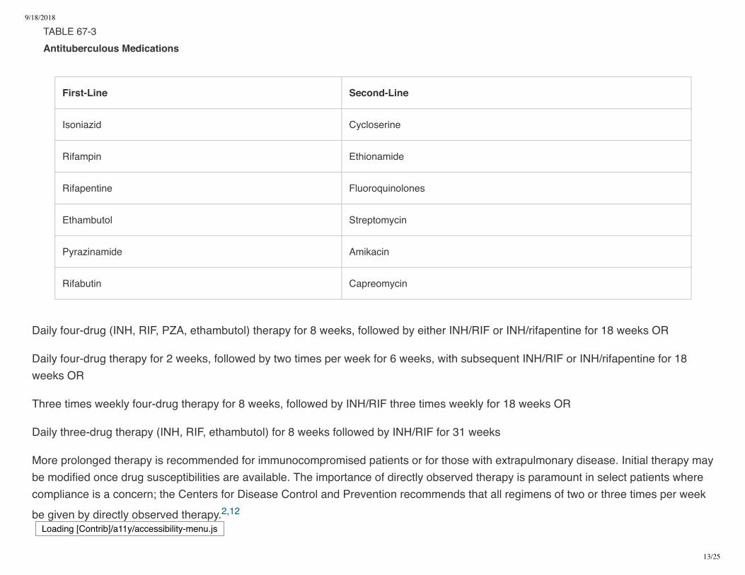
Active tuberculosis treatment requires the use of a combination of antituberculous medications to overcome resistance (Table 67-3).12
Initial therapy includes first-line medications (isoniazid [INH], rifampin [RIF], pyrazinamide [PZA], ethambutol) for 8 weeks, followed bytwo-drug continuation treatment for 18 to 31 weeks based on culture results. Second-line medications are used for drug-resistant casesor when side effects from initial therapy are not tolerable. In the ED, patients are either admitted to the hospital to determine the need forantituberculous medications or referred to specialists in the community for follow-up if compliance is likely. In most cases,antituberculous medications will not be started in the ED unless done in consultation and for classic cases. The recommended Centers
for Disease Control and Prevention regimens are as follows12:
Loading [Contrib]/a11y/accessibility-menu.js
9/18/2018
13/25
TABLE 67-3Antituberculous Medications
First-Line Second-Line
Isoniazid Cycloserine
Rifampin Ethionamide
Rifapentine Fluoroquinolones
Ethambutol Streptomycin
Pyrazinamide Amikacin
Rifabutin Capreomycin
Daily four-drug (INH, RIF, PZA, ethambutol) therapy for 8 weeks, followed by either INH/RIF or INH/rifapentine for 18 weeks OR
Daily four-drug therapy for 2 weeks, followed by two times per week for 6 weeks, with subsequent INH/RIF or INH/rifapentine for 18weeks OR
Three times weekly four-drug therapy for 8 weeks, followed by INH/RIF three times weekly for 18 weeks OR
Daily three-drug therapy (INH, RIF, ethambutol) for 8 weeks followed by INH/RIF for 31 weeks
More prolonged therapy is recommended for immunocompromised patients or for those with extrapulmonary disease. Initial therapy maybe modified once drug susceptibilities are available. The importance of directly observed therapy is paramount in select patients wherecompliance is a concern; the Centers for Disease Control and Prevention recommends that all regimens of two or three times per week
be given by directly observed therapy.2,12
Loading [Contrib]/a11y/accessibility-menu.js

9/18/2018
14/25
Although the standard medications used to treat tuberculosis are generally effective and safe, side effects or drug interactions may occur(Table 67-4). Hepatotoxicity, or hepatitis, is the major adverse effect of INH. Preexisting liver disease, pregnancy, ethanol use, HIV, andhepatitis C infection have been associated with an increased risk for hepatotoxicity from INH. Those with preexisting medical conditions
requiring multiple medications may be at higher risk for drug interactions with antituberculous agents.2,12,15
Loading [Contrib]/a11y/accessibility-menu.js

9/18/2018
15/25
TABLE 67-4Treatment of Tuberculosis (Adults)*
DrugDaily(maximum)
ThreeTimesWeekly DOT(maximum)
Two Times WeeklyDOT (maximum)
Potential Side Effects and Comments
Isoniazid 5milligrams/kgPO* (300milligrams)
15milligrams/kgPO (900milligrams)
15 milligrams/kg PO(900 milligrams)
Hepatitis, peripheral neuropathy, drug interactions.
Rifampin(RIF)
10milligrams/kgPO* (600milligrams)
10milligrams/kgPO (600milligrams)
10 milligrams/kg PO(600 milligrams)
Hepatitis, thrombocytopenia, GI disturbances, drug interactions.
Rifapentine Not givendaily
Not given 3times weekly
600 milligrams POtwice weekly in adults;not approved inchildren <12 y old
Hepatitis, thrombocytopenia, exacerbation of porphyria.Recommended by Centers for Disease Control and Preventionfor continuation therapy only for human immunodeficiency virus–negative patients.
Rifabutin 5milligrams/kgPO (300milligrams)
5milligrams/kgPO (300milligrams)
5 milligrams/kg PO(300 milligrams)
Similar to RIF; used for patients who cannot tolerate RIF.
Loading [Contrib]/a11y/accessibility-menu.js

9/18/2018
16/25
Abbreviation: DOT = directly observed therapy.
*See http://www.cdc.gov.une.idm.oclc.org/tb for more accurate weight-based protocols and dosages for children.
DrugDaily(maximum)
ThreeTimesWeekly DOT(maximum)
Two Times WeeklyDOT (maximum)
Potential Side Effects and Comments
Ethambutol 15–20milligrams/kgPO (1.6grams)
25–30milligrams/kgPO (2.5grams)
50 milligrams/kg PO(2.5 grams)
Retrobulbar neuritis, peripheral neuropathy.
Pyrazinamide 15–30milligrams/kgPO (2grams)
50milligrams/kgPO (3grams)
50 milligrams/kg PO (2grams)
Hepatitis, arthralgia, hyperuricemia.
A portion of patients being treated for tuberculosis will clinically worsen after the initiation of antituberculous medications.10,15,16 Thiseffect is called a paradoxical reaction or immune reconstitution syndrome and can be seen in any patient receiving treatment fortuberculosis, although it is more commonly seen in those with HIV infection. Signs and symptoms include fever, worsening respiratorystatus, lymphadenopathy, hepatosplenomegaly, ascites, meningitis, and new or worsening CNS lesions. Hypercalcemia is a uniquefinding in paradoxical reactions. Because treatment of tuberculosis and HIV both improve immune function, the paradoxical reaction isthought to be a result of improvement in the body's ability to mount an inflammatory response as mycobacteria are cleared. Thedilemma is differentiating this from treatment failures, drug resistance, and medication noncompliance. (See the Special Populationssection.)
TREATMENT OF LATENT TUBERCULOSISLoading [Contrib]/a11y/accessibility-menu.js

9/18/2018
17/25
Treatment of latent infection with INH alone is started in those with recent asymptomatic skin test conversion (see Table 67-2), any
person in close contact with an actively infected patient, and anergic patients with known tuberculosis contact.11,12 Unlesscontraindicated, therapy is for a minimum of 9 months for adults, children, and HIV-infected persons. Monitor those at risk for INHhepatotoxicity by serial laboratory assessment. For those exposed to INH-resistant strains or those who are intolerant, use RIF and PZA
for 2 months with hepatotoxicity monitoring.12
DISPOSITION AND FOLLOW-UPOUTPATIENT CARE
Most patients with tuberculosis can be treated initially as outpatients. If planning discharge or transfer for other nonmedical care, start ormaintain therapy while awaiting smear and culture results on all with suspicious findings of active tuberculosis, notably cavitary lesionsor known previous infection with new weakness or fevers.
Contact primary care physicians or public health services and arrange for long-term care before patient discharge. Dischargeinstructions include home isolation procedures and follow-up at the appropriate clinic to receive medication and ongoing care.Antituberculosis medications should not be instituted in the ED unless there is joint agreement with the consultant and follow-up providers.
HOSPITAL ADMISSION
Hospital admission should be done for the following cases: ill-appearing, hypoxemic, or dyspneic patients; if the diagnosis is uncertain; ifnoncompliance is likely or if the social situation makes it difficult to complete evaluation and start care; or patients with active drug-resistant tuberculosis. Hospitalized patients with suspected tuberculosis require respiratory isolation in a negative-pressure room (Table67-5). An alternative to admission for therapy compliance alone is a court-ordered drug observation program (if available), where
scheduled outpatient contacts to ensure medication use occur for the course of therapy.2
Loading [Contrib]/a11y/accessibility-menu.js

9/18/2018
18/25
TABLE 67-5Engineering Controls to Reduce the Transmission of Tuberculosis
High airflow (at least 6 room air changes per hour) with external exhaustHigh-efficiency particulate filters on ventilation systemUltraviolet germicidal irradiationNegative-pressure isolation roomsPersonal respiratory protection: high-efficiency particulate filter masks or respirators
SPECIAL POPULATIONSPATIENTS WITH TUBERCULOSIS AND HUMAN IMMUNODEFICIENCY VIRUS
HIV infection is the strongest known risk factor for tuberculosis, and the incidence of tuberculosis in HIV-positive patients is much higherthan in the general population. Patients with a new diagnosis of tuberculosis are almost 20 times more likely to have HIV, and HIVpatients are 20 to 30 times more likely to develop tuberculosis. Tuberculosis (pulmonary and extrapulmonary) often can be the initialclinical manifestation of immunodeficiency and is a defining event in the acquired immunodeficiency syndrome. Once active tuberculosishas developed, the risk of rapid progression and drug resistance is much higher in the HIV patient. Successful treatment with
antiretroviral therapy lowers the rate of tuberculosis and reduces the incidence of extrapulmonary involvement.17 For these reasons,physicians considering a diagnosis of tuberculosis should obtain HIV testing to provide early diagnosis and therapy.
Treatment of tuberculosis in HIV-positive patients is generally effective and not markedly different from others with the infection.However, due to the number of medications taken by patients with HIV, potential drug interactions are common, and compliance maybecome an issue. There is no consensus statement on the timing of antiretroviral therapy when treating both diseases. Recent studiessupport early antiretroviral therapy in combination with antituberculous medications, especially in those who are severely
immunocompromised.1,18
Immune reconstitution inflammatory syndrome or paradoxical reaction (see Treatment of Active Tuberculosis) is a condition in which HIVpatients clinically worsen as the immune system recovers after the initiation of antiretroviral therapy or antituberculous medications. TheLoading [Contrib]/a11y/accessibility-menu.js

9/18/2018
19/25
ideal timing of antiretroviral therapy in those with active tuberculosis is uncertain.1,18 See chapter 154, Human Immunodeficiency VirusInfection.
MULTIDRUG-RESISTANT TUBERCULOSIS
Multidrug-resistant tuberculosis is defined as tuberculosis with isolates that demonstrate resistance to at least INH and RIF. M.tuberculosis becomes resistant by spontaneous genetic mutation, often as a result of inadequate drug therapy or noncompliance withinitial treatment. While resistance is usually not confirmed until culture and sensitivity data are available, certain historical and clinicalfeatures raise the level of suspicion for multidrug-resistant tuberculosis. These include a history of tuberculosis treatment in the past,exposure to multidrug-resistant tuberculosis, known INH resistance in the community above 4%, and persistent symptoms or persistently
positive sputum cultures despite 4 months of standard treatment.19
Extensive drug-resistant tuberculosis is a more intense worldwide threat to public health and tuberculosis control. Extensive drug-resistant tuberculosis is defined as disease resistant to INH and RIF, plus resistance to any fluoroquinolone, and resistance to at leastone injectable second-line medication. It is associated with poor outcomes and higher mortality.
Treatment of multidrug-resistant tuberculosis depends on sensitivity patterns from culture. Some countries may use standardizedregimens based on known local resistance patterns. Usually a combination therapy with four to six medications, including the more toxic
and less potent second-line medications, is administered for up to 2 years. Success rates rarely exceed 75%.20 In refractory cases,
resectional surgery may be necessary in addition to ongoing medical therapy.19
The "Global Plan to Stop Tuberculosis" calls for new medications to fight against the problem of multidrug-resistant tuberculosis. In
addition to testing and better current therapy compliance, new medications show promise, especially delamanid.20
CHILDREN
Tuberculosis in children occurs in the same risk groups as in the adult population (see Table 67-1). The clinical course and diseasemanifestations in children have several unique aspects. Although children are at greater risk for developing rapidly progressive anddisseminated disease, their presenting signs and symptoms can be subtle. Primary tuberculosis in children is often asymptomatic and
only identified through screening programs or contact tracing.21 Children may be asymptomatic even with abnormal radiographs. Or,children may present with fever, cough, wheezing, poor feeding, and fatigue. The classic symptoms of fever, night sweats, and weightLoading [Contrib]/a11y/accessibility-menu.js

9/18/2018
20/25
loss may be seen in older children; however, in those younger than 5 years, presentation may be that of miliary tuberculosis (see below),meningitis, or a pneumonia that does not respond to therapy. The most common extrapulmonary presentation is cervicallymphadenitis, but other regions may be involved including the meninges, pericardium, abdomen, bone, joints, kidneys, skin, and eyes.
The yield of sputum smears and cultures is lower in children because of difficulty in obtaining adequate samples in addition to the lower
incidence of cavitary disease.22,23 Traditionally, obtaining three early morning consecutive gastric lavage or gastric aspirate samples hasbeen standard procedure. However, this is invasive, unpleasant, and often requires an overnight admission and trained staff. Sputum
induction using bronchodilators, followed by nebulized saline and expectoration of mucus, can improve sampling.23
The diagnosis of tuberculosis in children is confirmed by culture in only 30% to 40% of cases.13 The newer tests, IGRA and NAAT, are
not recommended for children less than 5 years old. The immune response differs in this age group, making the tests less reliable.11
Often, treatment is initiated based on a skin test or on clinical grounds (symptoms, a history of exposure, or abnormal radiographs), and
the diagnosis is presumed based on response to treatment.22 Multidrug therapy is currently recommended for all children considered tohave active disease, whereas monotherapy is used for latent infections.
MILIARY TUBERCULOSIS
Miliary tuberculosis is a historic term used in reference to the gross appearance of the lung during disseminated tuberculosis. In suchcases, the lung is often covered with multiple small lesions resembling millet seeds. Classic miliary tuberculosis shows diffuse noduleson radiographs (1 to 3 mm) in a patient with positive laboratory testing or by demonstration of mycobacteria in multiple organs. Theclassic radiographic findings may not appear on films until the disease has progressed over time. A miliary pattern on radiographs can
be found in conditions other than tuberculosis including histoplasmosis, malignancy, siderosis, and sarcoidosis.24
Today, miliary tuberculosis refers to wide hematogenous spread during the primary or reactivation disease, and it is associated withhigher mortality. Children, the elderly, and immunocompromised patients are all at increased risk of developing miliary disease.
Miliary disease during primary tuberculosis is generally more rapid and severe, often presenting with multiorgan failure, shock, and acuterespiratory distress syndrome. Conversely, miliary reactivation often manifests with a chronic, nonspecific clinical course affecting anynumber of organ systems. Fever, anorexia, night sweats, cough, weight loss, splenomegaly, lymphadenopathy, and signs of multisystemillness should cause one to suspect miliary disease. Cutaneous involvement, seen more often in HIV patients, manifests as papules or
Loading [Contrib]/a11y/accessibility-menu.js

9/18/2018
21/25
1.
2.
3.
4.
vesiculopapules (tuberculosis cutis miliaris disseminata or tuberculosis cutis acuta generalisata). Choroidal tubercles found on ocularexam are pathognomonic for miliary tuberculosis.
TUBERCULOUS MENINGITIS
Tuberculous meningitis is often seen in children, although those with HIV or others who are immunocompromised may also be afflicted.The challenge is the subtle and subacute presentation over days to weeks, with gradual fever, headache, and cognition or sensoriumchanges that often are not accompanied by neck stiffness or irritation, in contrast with those seen in other forms of bacterial meningitis.Focal neurologic deficits or cranial nerve palsies may also be evident. Suspecting the infection and requesting tuberculosis cultures and
smear are key to making a diagnosis, because other diagnostics are not helpful.25 Long-term neurologic dysfunction is common, withventriculoperitoneal shunting needed in 25% of patients for hydrocephalus. Tuberculous meningitis often seeds after a miliary infection.Treatment parallels other forms of tuberculosis.
PRACTICE GUIDELINESThe American Thoracic Society, Infectious Disease Society (http://www.idsociety.org), and Centers for Disease Control and Prevention(http://www.cdc.gov.une.idm.oclc.org) have issued recent joint guidelines for the management of tuberculosis.
REFERENCES
Blanc FX, Sok T, Laureillard D et al.: Earlier versus later start of antiretroviral therapy in HIV-infected adults with tuberculosis. NEngl J Med 365: 1471, 2011.
[PubMed: 22010913]
World Health Organization: Global tuberculosis control. WHO report 2011. Geneva, Switzerland: World Health Organization, 2011.
Centers for Disease Control and Prevention: Trends in tuberculosis—United States, 2011. MMWR 61: 181, 2012. [PubMed: 22437911]
Linas BP, Wong AY, Freedberg KA et al.: Priorities for screening and treatment of latent tuberculosis infection in the United States.Am J Respir Crit Care Med 184: 590, 2011.
Loading [Contrib]/a11y/accessibility-menu.js

9/18/2018
22/25
5.
6.
7.
8.
9.
10.
11.
12.
13.
14.
[PubMed: 21562129]
Schlossberg D: Acute tuberculosis. Infect Dis Clin N Am 24: 139, 2010.
Horsburgh CR, O'Donnell M, Chamblee S et al.: Revisiting rates of reactivation tuberculosis: a population-based approach. Am JRespir Crit Care Med 182: 420, 2010.
[PubMed: 20395560]
Horsburgh CR, Rubin EJ: Latent tuberculosis infection in the United States. N Engl J Med 364: 1441, 2011. [PubMed: 21488766]
Kardos M, Kinball AB: Time for a change? Updated guidelines using interferon gamma release assays for detection of latenttuberculosis in the office setting. J Am Acad Dermatol 66: 148, 2012.
[PubMed: 22177633]
Cruz A, Ong L, Starke J: Emergency department presentation of children with tuberculosis. Acad Emerg Med 18: 726, 2011. [PubMed: 21762235]
http://www.cdc.gov.une.idm.oclc.org/tb/ (Centers for Disease Control and Prevention: Tuberculosis.) Accessed October 10, 2013.
Centers for Disease Control and Prevention: Updated guidelines for using interferon gamma release assays to detectMycobacterium tuberculosis infection—United States, 2010. MMWR 59: 1, 2010.
[PubMed: 20577159]
Centers for Disease Control and Prevention: Treatment of tuberculosis: practice guidelines for treatment of tuberculosis. MMWR 52:No. RR-11, 2003.
Lange C, Mori T: Advances in the diagnosis of tuberculosis. Respirology 15: 220, 2011. [PubMed: 20199641]
Yoo S, Cattamanchi A, Den Boon S et al.: Clinical significance of normal radiographs among HIV-seropositive patients withsuspected tuberculosis in Uganda. Respirology 16: 836, 2011.
Loading [Contrib]/a11y/accessibility-menu.js

9/18/2018
23/25
15.
16.
17.
18.
19.
20.
21.
22.
23.
[PubMed: 21518124]
Sterling T, Pham P, Chaisson R: HIV infection-related tuberculosis: clinical manifestations and treatment. Clin Infect Dis 50 (Suppl3): S223, 2010.
[PubMed: 20397952]
Swaminathan S, Padmapriyadarsini C, Narendran G: HIV-associated tuberculosis: clinical update. Clin Infect Dis 50: 1377, 2010. [PubMed: 20388036]
Kwan CK, Ernst JD: HIV and tuberculosis: a deadly human syndemic. Clin Microbiol Rev 24: 351, 2011. [PubMed: 21482729]
Cohen K, Meintjes G: Management of individuals requiring antiretroviral therapy and tuberculosis treatment. Curr Opin HIV AIDS 5:61, 2010.
[PubMed: 20046149]
Kant S, Maurya A, Kushwaha RA et al.: Multi-drug resistant tuberculosis: an iatrogenic problem. Biosci Trends 4: 48, 2010. [PubMed: 20448341]
Gler MT, Skripconoka V, Sanchez-Garavito E et al.: Delamanid for multidrug-resistant pulmonary tuberculosis. N Engl J Med 366:2151, 2012.
[PubMed: 22670901]
Swaminathan S, Rekha B: Pediatric tuberculosis: global overview and challenges. Clin Infect Dis 50 (Suppl 3): S173, 2010. [PubMed: 20397947]
Connell TG, Zar HJ, Nicol MP: Advances in the diagnosis of pulmonary tuberculosis in HIV-infected and HIV-uninfected children. JInfect Dis 204 (Suppl 4): S1151, 2011.
[PubMed: 21996697]
Nicol MP, Zar HJ: New specimens and laboratory diagnostics for childhood pulmonary tuberculosis: progress and prospects.Paediatr Resp Rev 12: 16, 2011.
Loading [Contrib]/a11y/accessibility-menu.js

9/18/2018
24/25
24.
25.
1.
2.
3.
4.
5.
[PubMed: 21172670]
Furqan M, Butler J: Miliary pattern on chest radiography: tuberculosis or not tuberculosis? Mayo Clinic Proc 85: 108, 2010.
Thwaites GE, van Toorn R, Schoeman J: Tuberculous meningitis: more questions, still too few answers. Lancet Neurol 12: 999,2013.
[PubMed: 23972913]
USEFUL WEB RESOURCES
Centers for Disease Control and Prevention Interactive Core Curriculum on Tuberculosis Web-Based Course (2004)—http://www.cdc.gov.une.idm.oclc.org/tuberculosis/publications/Cont_Core_Curr_Course.htm
Centers for Disease Control and Prevention resources for tuberculosis including drug recommendations and dosages—http://www.cdc.gov.une.idm.oclc.org/tb
National Institute of Allergy and Infectious Diseases: New research and development at the National Institutes of Health—www.niaid.nih.gov
World Health Organization Tuberculosis: This Web site describes the work of the Stop Tuberculosis Department—http://www.who.int.une.idm.oclc.org/tuberculosis/en
WHO Tuberculosis Epidemiology and Surveillance Virtual Workshop—http://www.who.int.une.idm.oclc.org/tuberculosis/surveillanceworkshop
McGraw HillCopyright © McGraw-Hill Global Education Holdings, LLC.
All rights reserved. Your IP address is 132.174.255.223
Terms of Use • Privacy Policy • Notice • Accessibility Loading [Contrib]/a11y/accessibility-menu.js

9/18/2018
25/25
Access Provided by: University of New EnglandSilverchair
Loading [Contrib]/a11y/accessibility-menu.js