chapter 5 pediatric podiatry
TRANSCRIPT

5 Pediatric PodiatryRussell Volpe, DPM
Pediatric Podiatry 419
C o n t e n t s
5.1 Congenital Deformities . . . . . 420
5.2 Metatarsus Adductus . . . . . . 428
5.3 Serial Plaster Immobilization . 432
Of Congenital Foot Deformities
5.4 Podopediatric Exam. . . . . . . . 436
5.5 Collapsing Pes Plano Valgus . 440
5.6 Developmental Flatfoot . . . . . 444
5.7 Pediatric Orthoses. . . . . . . . . 445
5.8 Normal And Pathologic
Gait In Children . . . . . . . . . . . 449
5.9 Rotational And Angular Disorders
In Children. . . . . . . . . . . . . . . 456
5.10 The Pediatric Hip . . . . . . . . . 461
5.11 Pediatric Sports Medicine . . . 465
5.12 Knee. . . . . . . . . . . . . . . . . . . 468
5.13 ACL Injuries . . . . . . . . 471
5.14 Osteochondroses. . . . . 478
5.15 Toe Walking . . . . . . . . 481
5.16 Pediatric Neuromuscular
Disorders . . . . . . . . . . 482
5.17 Pediatric Pharmacology. . 486
5.18 Skeletal Syndromes And
Systemic Disorders In
Pediatric Orthopedics . . . 490
5.19 Other Syndromes Involving
Short Stature . . . . . . . 494
5.20 Malformations Of The Hand
And Foot. . . . . . . . . . . 503

5.1 Congenital Deformities
Calcaneovalgus
Definition• Congenital deformity
• Usually unilateral can be bilateral
• 1-3/1000 live births
• Extreme dorsiflexion of ankle
• Decreased plantarflexion
• Referred to as “up and out”
Figure 1.
Etiology• Abnormal intrauterine position
• Excessive internal limb rotation
• Rarely with congenital/neuromuscular/endocrine disease
• Associated findings
– Oligohydramnios
– Dislocated peroneal tendons
– Chromosomal abnormalities
• Thorough general, orthopedic, and neurologic physical recommended
• Uterine positions cause forefoot and calcaneus, to move on stable talus
• Open kinetic chain - talus in unit with leg, calcaneus, and foot move around it
Pathologic Anatomy• Navicular displaced laterally to talus
• Midfoot dorsiflexed and abducted on talus
• Distal calcaneus laterally displaced
• Prominent plantar-medial talar head
• Tight anterior and lateral musculature
• Pathologic anatomy
– Achilles tendon not contracted
– Talocalcaneal and plantar calcaneonavicular ligaments are relaxed
• Lack of rigidity (flexible congenital pes valgus)
Clinical Features• Excessive dorsiflexion
• Limited plantarflexion
• Forefoot touch anterior leg
420 The 2005 Podiatry Study Guide

• Off w.b. calcaneus in valgus
• Forefoot varus
• Talar head prominent medially
• MTJ abducted and dorsiflexed
• Subtalar joint ROM normal or increased
• Flexible foot type
• Achilles tendon not taut even in maximum dorsiflexion
• Redundant skin folds anterior and lateral ankle
• Sinus tarsi depression on plantarflexion
Figure 2.
Radiographic findings Lateral View• Plantarflexed talus- below cuboid if severe
• Forefoot dorsiflexed
• Talus overlaps anterior(distal) superior calcaneus
• Anterior break-cyma line
• Decreased calcaneal inclination
Radiographic findings Dorso-plantar View• Talar bisection falls medial to the 1st metatarsal
• Talo-calcaneal angle above 30-40°
• Altered cyma line
• Navicular not visible
• Radiographic differential diagnosis
– Stress lateral view taken with forefoot plantarflexed in radiograph – talar-first metatarsal relationships
will restore in calcaneovalgus; talar-first malalignment will persist in vertical talus.
Classification• Mild – plantarflexion of ankle to 90°(right angle) or beyond. Talar bisection central cuboid (lateral x-ray)
• Moderate - plantarflexion to less than 90°. Talar bisection lower 1/3 cuboid
• Severe - plantarflexion to 80°. Talar bisection below cuboid
Treatment Rationale
• Prevent deleterious osseous adaptation
• Prevent abnormal joint relationships
• “Support the superstructure” function of foot altered by congenital pes valgus - ambulation may be
delayed or awkward
• May produce muscle imbalance
Pediatric Podiatry 421

Result of Non-TreatmentThe untreated calcaneovalgus foot is predisposed to abnormal pronation after weightbearing and may lead
to a symptomatic flatfoot in childhood and beyond
Goal of Treatment in Infancy• Reduce forefoot deformity on the rearfoot
• Reduce calcaneovalgus (dorsiflexion + eversion)
• Reduce muscular and ligamentous adaptation
• Prevent osseous adaptation
Treatment - Mild Deformity in Infancy• Manipulation - begin in neonate. Heel vertical, forefoot plantarflexed, and adducted (at midtarsal joint).
Hold for 10 seconds
• 15 reps - diaper changes
• J-strap to hold
Treatment - Moderate Deformity in Infancy• Manipulation with strapping
• Tarsosupinator or straight last shoes
• D-B bar or Counter Rotation System - set straight or internal
• Serial plaster immobilization
Treatment - Severe Deformity in Infancy• Serial plaster immobilization
– Cast usually below-knee
• Maintenance with shoe, brace or splint
• Maintain for half as long as correction took
• Response is rapid
Serial Casting For Calcaneovalgus• Hold subtalar joint in neutral to slight varus
• Ankle in slight equinus
• Forefoot in neutral - reduce abducted/dorsiflexed MTJ
• Avoid using forefoot as lever arm to reduce “up and out” deformity at ankle and STJ
Figure 4. Figure 5.
Figure 3.
Treatment - Mild Deformity Ambulatory• High-top supportive shoes• Heel wedges• LA padding• Pre-fabricated orthoses < 3yr• Exercises / manipulation
• Custom orthoses > 3 yr. if concerns persist
422 The 2005 Podiatry Study Guide

Figure 6.
Treatment - Moderate Derfomity Ambulatory• Shoes, wedges, LA pads, pre-fab orthoses if under age 3
• Night-splint, tarso-supinator shoes
• Functional foot orthoses if > age 3, deep heel seat, medial flange recommended
Treatment - Severe Deformity Ambulatory• Night splints with tarso-supinator shoes
• High-top / supportive walking shoe
• Maximum-control pediatric foot orthoses - UCBL/DSIS
• Rehab intrinsic and extrinsic muscle groups
Vertical Talus Convex Pes Valgus• SYNONYMS
– Congenital rigid flatfoot
– Teratologic talo-navicular dislocation
• INCIDENCE
– First described by Henken in 1914
– Relatively rare
– 50% bilateral
– Males over females
– Right leg predominance
– High percentage of associated anomalies (congenital, syndromal, neurologic)
Components of Deformity
• Rigid rockerbottom (convex plantar aspect)
• Prominent talus medially and plantarly
• Navicular dislocates dorsally
• Equinovalgus deformity
• Forefoot abducted and dorsiflexed on rearfoot
• Rearfoot plantarflexed
• Tendoachilles tightness
• Heel perpendicular to slight valgus (not high valgus rearfoot as might be expected)
• Anterior and peroneal muscle tightness
Pediatric Podiatry 423

Figure 7.
Differential Diagnosis• Oblique talus – DDx on lateral plantarflexion (stress) x-ray
• Talipes calcaneovalgus
• Acquired flatfoot
• Spastic equinus flatfoot - cerebral palsy
• Iatrogenic rockerbottom (post TEV casting)
Primary Diagnosis (Associated Syndromes)• Arthrogryposis
• Myelodysplasia
• Epidermal Nevus Syndrome
• Sacral Agenesis
Pathomechanics• Growth disparity soft tissue/bone
• Dysplaisia of spring ligament leads to downward and medial rotation of the talar neck and head
• Absence of plantar intrinsics
• Aplasia of the anterior calcaneus
• Weakness of posterior tibial tendon associated with forward movement of the tendon along with the
flexor digitorum longus tendon. The PT tendon now acts anteriorly as a dorsiflexor of the fore part of
the foot
Radiology• AP/lateral/plantarflexion lateral (stress) views
• Increased talocalcaneal angle (>45 degrees)
• Calcaneus in equinus
• Navicular dislocation dorsally
• Plantarflexion lateral (stress) - No talar-first met congruity, i.e. the bisection of the talus will remain
plantar to the 1st ray. In calcaneovalgus, the plantarflexion lateral will demonstrate the talar bisection
approximately bisecting the first ray.
Muscle Imbalances• Posterior tibial and peroneals anteriorly displaced - ankle and forefoot dorsiflexor
• Medial column elongated
• Posterior tibial, peroneals, and long extensors shorten over time
Goals of Treatment• Restore Navicular/Talar/Calcaneal Relationship
• Plantigrade Foot
424 The 2005 Podiatry Study Guide

• Painfree Function
• Minimize Arthritic Changes
Non-Operative Treatment• SERIAL PLASTER CASTING (USUALLY UNSUCCESSFUL)
– Stretch the soft-tissue preoperatively
– Stretch triceps surae
– Pressure on anterior calcaneus
– Plantarfex, adduct and invert forefoot
– Attempt closed reduction?
• • Operative Correction
– Procedure to relocate midfoot plantarly and reduce the equinus of the rearfoot.
– Staged procedure
– Older children – extra-articular subtalar arthrodesis
Assessment Criteria (DeRosa, et al., Foot & Ankle, Vol. 5, No. 3)• Assess these criteria to determine correction:
– Frontal plane heel position
– Residual equinus
– Lateral border alignment
– Medial talar prominence
– Subtalar joint mobility
– Range of plantar flexion
Talipes Equino Varus• Incidence
– 1-2 per 1000 live births
– 1/35 with a sibling who has TEV
– Ethnic diversity
– Polynesians 6.81/1000
– 2 -2.5/1 male/female
– 30 - 50% bilateral
Etiology• Arrest of fetal development at ninth fetal week
• Combination of genetic and environmental factors
• Primary germ plasm defect
• Two types – one with positive family history the other without
Morphological Features• Dimple anterolateral
• Inversion, adduction of forefoot
• Varus of rearfoot
• Talocalcaneal equinus (little or no change on dorsiflexion lateral x-ray)
• External talar rotation (prominent laterally)
• Inverted and plantarflexed calcaneus
• Associated Internal tibial torsion
• Shorter/smaller calf
• Shorter/wider foot
Pediatric Podiatry 425

• Tendoachilles notch
• Small, bean-shaped heel
Figure 8. Figure 9.
Classification—Etiology BasedCongenital
• Idiopathic
– Intrinsic - type 1 – rigid(genetic/teratologic)
– More likely to have calcaneal anomalies (smaller, inverted, or plantarflexed)
– Extrinsic -type 2 – flexible
– Normal calcaneal anatomy
• Neurogenic
– Open or closed defects of the spina (spina bifida)
– Myogenic
– Osteogenic
– Collagenous/cartilagenous
Aquired
• Neurogenic – progressive neuromuscular disease, spinal cord tumors
• Vascular – Volkman’s ischemic contracture
• Traumatic – abuse, serious injury
Pathology
• Head and neck of talus medially and plantarly deviated on the body of the talus
• Superior surface of talus anteriorly displaced out of ankle mortise
• Calcaneus in equinovarus and internally rotated
Contractures - Posterior• Ankle capsule
• Subtalar capsule
• Calcaneofibular ligament
• Tendo achilles
Contractures – Medial• Spring ligament
• Deltoid ligament
• Tibiales posterior
• Flexor digitorum Longus
• Flexor hallucis Longus
Clinical Examination• History and physical
426 The 2005 Podiatry Study Guide

• Chief complaint
• Past medical history
• Prenatal
• Postnatal
• Growth and development
• Examination
• Musculoskeletal
• Neurophysiologic
• Special diagnostic
Radiographic EvaluationAP View
• Reduced talocalcaneal angle
• < 15 degrees to near parallel
• Superimposition of talar head on anterior surface of calcaneus
• Ossific center of navicular is medially displaced
Lateral View
• Decreased calcaneal inclination angle
Radiographic Angles• Talocalcaneal index (on lateral) <40 degrees = talonavicular dislocation. This is the sum of the AP and
lateral talocalcaneal angles.
• Simon’s rule of 15 (on AP). If talocalcaneal angle is less than 15 degrees and the talar-first metatarsal
angle is greater than 15 degrees than the talo-navicular joint may be said to be dislocated. This is useful
in an infant when the navicular is not yet ossified.
• Lateral T/C angle <35 on stress dorsiflexion
Non-Operative Treatment• Stretch soft-tissue
• Manipulate subluxed joints
• Maintain with casts
Duration of Casting• Weekly - neonates
• Bi-weekly - infants
• Maintain for half as long as to correct
Manipulation Position• Evert calcaneus
• Pressure over lateral talar head
• Abduct forefoot
• Reduce equinus at ankle/STJ – take care to reduce equinus at the ankle joint (as opposed to the
midtarsal joint). This prevents iatrogenic rockerbottom deformity from resulting.
Pediatric Podiatry 427

Figure 10. Figure 11.
Treatment Outcomes• Reduce equinovarus
• Reduce joint subluxations
• Create a plantigrade foot
• Functional ankle range of motion (minimum 5° ankle dorsiflexion)
Maintenance Options• Straight/abductory last shoes
• Night splints (as needed)
• Ankle foot orthoses
• UCBL/DSIS
Figure 12. Figure 13.
5.2 Metatarsus AdductusDefinition
A transverse plane adduction deformity of the metatarsals at the tarsal-metatarsal articulation (Lisfranc’s joint)
Incorrect Synonyms
• Metatarsus varus (frontal plane)
• Forefoot adductus (midtarsal joint)
Incidence
• 1/1,000 live births
• No sex predilection
• If one child affected risk of second affected 1:10
Determining Metatarus Adductus
Measure METATARSUS ADDUCTUS angle. Angle made by bisection of the lesser tarsus and the long axis of the
2nd metatarsal.
428 The 2005 Podiatry Study Guide

Radiographic Relationships
Bisect Lesser Tarsus• Lateral aspect base of 4,5 mets
• Medial aspect base of 1st met
• Ant lateral aspect calcaneus
• Ant medial aspect calcaneus
These four points create a box. Bisect this box. The metatarsus adductus angle is the angle made by this bisection
and the long axis of the second metatarsal.
Range of “Normal” Metatarus Adductus Angle• Birth
– 15-35 degrees
• Beginning Walker
– 20 degrees
• Four to six Year Old
– 5-15 degrees
Etiology• Retention of fetal alignment
• Abnormal in-utero position producing muscle imbalance
• Abnormal amniotic compression with resultant contractures of medial soft tissues
• Untoward sleeping and sitting positions may accentuate deformity
Clinical Features• “C”-shaped appearance
• Convex lateral border
• Concave medial border
• Prominent styloid
• May be increased interval between hallux and 2nd digit (met primus adductus)
• Rearfoot neutral - “normal” varus
Figure 14.
Classification• Flexible
• Rigid
• Dynamic (functional)
Differentiate From Primus Adductus• 95% excellent prognosis
• Proposed Etiology
– Demonstration of tight abductor hallucis
Pediatric Podiatry 429

Characteristic Radiographic Findings• MA angle increased
• Severity of adductus decreases laterally
• Superimposition of mets at their bases decreasing laterally
• May be associated met primus adductus
• May be hypoplasia of cuboid
Differential Diagnosis• Metatarsus varus
• Met primus adducutus
• Talar neck adductus
• Forefoot adductus
• Metatarsus adductus and metatarsus varus = metatarsus adductovarus
Talar Neck Adductus• The angle formed between the head and neck and trochlear surface in the transverse plane
• 9-12th fetal week TNA = 33 degrees
• Adult TNA = 22 degrees
Associated Fundings75% of patients with metatarsus adductus have lack of external tibial torsion (or internal tibial torsion)
Forefoot Adductus• An adduction deviation of the forefoot on the rearfoot in the transverse plane occurring at the midtarsal joint
• Forefoot adductus
– Adult norm 10°
• Metatarsus adductus is not one-third of clubfoot deformity
• Adductus in TEV occurs at the midtarsal joint
• Talipes equino varus (TEV)
– Rearfoot equinus
– Marked varus
• Metatarsus adductus
– Rearfoot Normal
– Valgus on weight
– Normal dorsiflexion
“Skew” or “Z” FootMet adductus with marked calcaneal eversion and talo-navicular subluxation
Implications• Cosmesis
• Shoe-fitting
• Early deformity and dysfunction
• The hallmark deformity associated with metatarsus adductus is:
– Juvenile hallux abductovalgus
Juvenile Digital Contractures
• Line of weight bearing normal foot
– L/M calcaneus, 5, 2, 1, hallux
• Line of weight bearing metatarsus adductus
– L/M calcaneus, 5,4,3-2, 3rd or 2nd digits
• Abnormal extensor alignment in met adductus results in digital contractures
430 The 2005 Podiatry Study Guide

Treatment Considerations• The more flexible the deformity the more favorable the prognosis
• The earlier treatment is instituted the more favorable the prognosis
• Benefits of early correction include bony remodeling to more normal alignment
CONSERVATIVE
• Manipulation
• Parental education
• Control of postural positions
• Shoe therapy
• Concomitant use of night splints
• Manipulation and serial plaster immobilization
SURGICAL
• Soft-tissue
• Osseous
Shoe Therapy• Outflare last or straight last• In shoe wedging (Tax)
– 1/8 inch felt• Shaft of 1st metatarsal
• Cuboid
Figure 15.
Night SplintsPurpose: To treat associated torsion and/or correct untoward sleeping positions
• Denis-Browne/Filauer bars
• Ganley splint
• Counter rotation splint (CRS)
Figure 16. Figure 17.
Serial Maniopulations and Holding Plaster Immobilization
Pediatric Podiatry 431

• Manipulate foot for five minutes or one wk prior to visit
• Protect cutisz
• Stockinette
• Minimal padding
• Apply two to three inch plaster against deformity encompassing foot and ankle
• Supinate rearfoot and brace cuboid with palm
• Abduct mets with free hand by grasping forefoot and exerting pressure along shaft of first in abduction
(not the hallux)
• Change cast q7 - 14 days
• Average Tx time 6 - 12 weeks
• To maintain correction utilize holding casts for one-half the time required to obtain correction
Figure 18. Figure 19.
Criteria for the Cessation of Manipulative Plaster Immobilization• Loss of “C” shape• Lack of palpable styloid• Decreased met adductus angle
Possible Surgical Intervention• Positive family history• Persistent deformity• Adequate trial of unsuccessful conservative therapy
SummaryThe oftentimes innocuous appearing deformity of metatarsus adductus if left unrecognized, inadequately or inap-
propriately treated, will produce dysfunction and further deformity at an early age
5.3 Serial Plaster Immobilization of Congenital Foot DeformitiesWhen to Cast
Based on age and severity of deformity
Age• Younger - greater joint mobility• Older - increase risk of iatrogenic joint dysfunction• Infants before the age of walking are the best candidates for casting
Severity
• More severe deformities need casting
Purpose of the Cast• Apply external forces to redirect growth• Stretch tight structures
• Avoid iatrogenic joint dysfunction
432 The 2005 Podiatry Study Guide

Manipulation
• Stretch soft tissue contractures
• Precede cast application
• Five to 10 minutes to abnormal segment
• Practice “hand positions” - create sound habits
Figure 20.
Other Concepts
Maintenance• Half as long as correction (cast or other device)
• Prevent recurrence
Serial Radiographs• Monitor progress
• Ensure correction is taking place at appropriate joint level
Preparation
• Warm water in bucket
• Draping materials
• Skin preparant
• Stockinette/tube gauze
• Webril/elastic cast padding
• Two, three or four inch extra-fast-setting plaster rolls
Figure 21.
Pediatric Podiatry 433

Advice to Parents
• Inform to reduce anxiety
• Child should be rested
• Bottle/pacifier
• Soak off casts before return
Cast Application
• Prepare skin
• Apply minimal padding two to three layers
• Extra pad prominences
• Avoid wrinkling
• Wet plaster and leave saturated to allow set time
• Apply distal to proximal
• Six layers of plaster
• Reinforce plantar and posterior heel
• Fold end of layers to create tag for ease of removal
• Slipper cast then leg cast or single unit casting
Figure 22. Figure 23.
434 The 2005 Podiatry Study Guide

Figure 24. Figure 25.
Cast Modifications
• Cast cutter removal not advised – fearful to child
• Long leg to effect posterior muscles or align knee
• Synthetic or other new materials, - may be used to reinforce over plaster for walking cast. Not ideal for
bottom layer as is less moldable
• Minimal or additional padding(to bony prominences)
Durations
• Four to eight casts
• Weekly or bi-weekly
Removal
• Soak at home just prior
• Add white vinegar to loosen bond
Metatarsus Adductus - Corrective Position
Left Foot (Patient) - Right Hand (Clinician)
• Stabilizes thenar eminence on cuboid
• Cup heel and rotate into slight varus
• Place cuboid plantar to navicular increasing MTJ stability
• Ankle joint at 90°
Left Foot - Left Hand
• Corrective force in transverse plane
• First web space at medial 1st metatarsal head
• IPJ’s of thumb and fingers fully extended
• Apply pressure parallel to heel to keep forefoot to rearfoot perpendicular
Internal Tibial Position - Corrective Position
• Diagnosed with excessive rotation in internal direction
• One hand stabilizes distal thigh
• Other hand rotates leg in appropriate direction
• Knee flexed at 30°
• Apply short-leg cast
• Correct foot deformities
Pediatric Podiatry 435

• Apply thigh cast - leave knee uncovered
• Flex knee to 30°
• Rotate leg to correction
• Join cast with three or four inch roll
Talipes Calcaneovalgus-Corrective Position
Right Foot (Patient) - Right Hand (Clinician)• Cup heel and invert and plantarflex
Right Hand - Left Hand• Thumb at plantar distal calcaneus
• Dorsal pressure to talus
• Plantarflex and adduct at MTJ
Talipes Equinovarus - Left Hand (Clinician)
Left Foot (Patient) - Left Hand (Clinician)• Over navicular-plantarflex, abduct and evert to pronate medial column
Left Foot - Right Hand• Evert and externally rotate heel
• Pull proximal posterior heel plantigrade to reduce equinus
• Long-leg cast - block g/s equinus
Talipes Equinus (Toe Walking)• Congenitally short g/s complex - soft tissue contracture• Lengthen to avoid compensation• Long-leg cast in pre-walker• Can short-leg in walking child as gait has knee extension
Talipes Equinus-Corrective Position• Short-leg cast• Invert heel to place cuboid below navicular to prevent CC joint pronation with dorsiflexion• Dorsiflex foot to resistance• Thigh cast• Join cast with three or four-inch roll• Keep knee gently extended
5.4 PodopediatricLigamentous Laxity
• Thumb to forearm - two points• Fifth digit extension - two points• Elbow hyperextends - two points• Knee hyperextends - two points• Toe touch ability - two points• Maximum - ten points (higher score – more laxity)
Spinal Exam• Curvatures - scoliosis• Rotations• Palpable defects - tumors, other masses• Hairy patches and dimples – myelomeningocoele, dysraphism, etc.
436 The 2005 Podiatry Study Guide

Neurologic Exam• Reflexes – deep tendon and superficial (pathologic)• Spontaneous movements• General tone• Overall integrity
Hip Exam• Femoral shortening – flexion (all is test) and extension• Skin fold irregularities• Loss of abduction in flexion• Ortolani clunk in neonate (reduce DDH)• Provoke located hip with Barlow maneuver
Exam Positioning of a Child• Sitting up• Lying back only when needed – defenseless position – scary for child• Use parents lap• Legs facing examiner
General Observation• Limb shortening• Limb angulation• Limb rotation• Limb - trunk proportions
Facial Exam• Parietal bossing• Blue sclarae• High-arched palate• Low-set ears• Ear irregularities• Tooth anomalies• Facial symmetry
These may suggest genetic syndromes with orthopedic or lower extremity implications.
Hand Exam• Square• Long tapering fingers• Nail anomalies• Thumb in palm• Single palmar crease
These may suggest genetic syndromes with orthopedic or lower extremity implications.
Gait ExamSee Gait section for details
• Short-legged• Antalgic• Trendelenburg• Neurogenic
• Rotational
• Angular
Pediatric Podiatry 437

Joint Exam
• Look - skin, shape, position
• Feel - skin, soft-tissue , bones
• Move - active, passive, power
• X-ray
Torsional Assessment
• Total foot rotation
• Femoral rotations
• Tibial rotations
• Ryder’s test
• Malleolar position
• Angular assessment
Figure 26. Figure 27.
Figure 28.
Genu Valgum• Measure intermalleolar distance, record patient height, as a given distance is more significant in a shorter
person.
• Knees will be touching in genu valgum
438 The 2005 Podiatry Study Guide

Figure 29.
Angular Assessment
• Genu varum - measure intercondylar distance
• Ankles will be touching in genu varum
Tips for Assessment
• Consider pseudo-bowing (displaced gastroc secondary to internal torsion)
• In-toe – Check patellar position
– Inward - femoral segment
– Outward - tibial/foot segment
• Gentle bow - physiologic
• Sharp, angular bow - true tibia vara (Blount’s)
Figure 30.
Foot Exam
Mobile Deformities - POSITIONAL
• In utero
• Attitudinal
SECONDARY
• Compensatory
• Neuromuscular
• Ligamentous laxity
Pediatric Podiatry 439

Rigid DeformitiesPRIMARY
• Talipes equino varus
• Vertical talus
SECONDARY
• Neuromuscular
• Tarsal coalition
• JRA
General Exam Tips
• Watch child remove clothing - check use of hands and limbs
• Observe trip to examination room from waiting area - early gait exam
• Examine painful area last
• Sit for exam - go to foot and leg level
• Toe and heel walk with limp to eliminate “fake limps” – stress gait
Torsional Screening• Perform total foot rotation• Perform thigh rotation• If total foot was abnormal and thigh rotation normal suggests tibial torsion• Perform total foot rotation• Perform thigh rotation• If total foot is abnormal and thigh rotation is abnormal, deformity likely in proximal segment
Prognosis• Consider natural history
• Metatarsus adductus - treat • Torsions - rapid growth - best chance for spontaneous correction and treatment under 18 months• 18 months - four years active growth - amenable to treatment• After five - static growth – treatment less successful
Lower Extremity Exam
A R M Method• Attitude-at-rest - include hopscotch technique• Relationship of parts• Measurement - quantify relationships
JIM GANLEY, DPM JAPMA, FEB. 1981
Conclusion• Child is unique• Focus exam• Master tips• Natural history to guide treatment
5.5 Collapsing Pes Plano ValgusConcerns
• Is flatfoot a pathological condition that effects foot function?
• If so, how do we determine which ones to treat and how aggressively to treat them?
440 The 2005 Podiatry Study Guide

Flatfoot As Pathology In The Literature• Oxygen consumption decreased with arch support - Otman, et.al.• A cause of spontaneous osteonecrosis of the navicular in otherwise healthy adults - Haller, et.al.• Susceptibility to stress fractures correlates to “pes planus” - Sullivan, et.al.• There is a relationship between pes valgus and hallux abducto valgus - Inman, Hohman, Kalen,
Brecher.
End-Stage Flatfoot“A painful tarsal enthesopathy which is the result of severe long-term pronation of the foot about the subtalar andother joints”
Signifigance“Those authors who dismiss the flatfoot as trivial, normal, or as something that is outgrown over time invariablyneglect to connect this condition with the many disabling conditions it creates”
Mahan, K.T., Comp Text - Foot Surgery – ed. McGlamry
Classification
Congenital• Calc. Valgus• Vertical talus• Peroneal spasm• Developmental • Accessory navicular• Tarsal coalition• Z-foot• Equinus
Acquired• Trauma• Infection• Arthritis• PT dysfunction• Neuromuscular• Biomechanical
Classification
Rigid Flatfoot• Convex pes plano valgus• Tarsal coalition• Trauma• Neuropathy
Flexible Flatfoot• Forefoot varus• Flexible forefoot valgus• Rearfoot equinus• Calcaneovalgus• Torsions• Muscle abnormalities• Neurotropic - early• Medial weight shift
Pediatric Podiatry 441

Clinical Features• Everted heel• Abduction of the forefoot on the rearfoot• Collapse of the medial column• Flexibility of the foot with reducibility of deformity• Pronated throughout gait, notably after heel lift• Posterior equinus as a primary force or secondary adaptation
Figure 31. Figure 32.
Why Treat The Pes Planovalgus Foot?
Pain• Reduction of activity• Reduced social interaction
Fatigue• Overuse
• Cramping
• Discomfort
Joint Degeneration• Slow, consider the effects over time
Associated Deformities• Hallux valgus
• Metatarsalgia
• Digital pathology
Planal Dominance
• Allows the treatment to address the pathology on the basis of clinical and radiographic findings.
• Understanding the primary plane of both deformity and compensation introduces a greater measure of
predictability and precision in the selection of treatment.
Planal Dominance As A Management Concept
• Determine clinically and radiographically
• Increased vertical subtalar axis increases transverse motion
• Increased horizontal subtalar axis increases frontal plane motion
442 The 2005 Podiatry Study Guide

Planal Dominance On X-ray
Transverse Plane Deformity• Inc. T-C angle - AP view
• Inc. Cuboid abductus angle -AP view
• Dec. Forefoot adductus angle -AP
• Dec. T-N congruency
Frontal Plane• Wide lesser tarsus - AP view
• Dec. first met declination - lateral
• Dec. height of sustentaculum
• Inc. superimposition of lesser tarsus on lateral view
SAGITTAL PLANE• Inc. Talar declination - lateral
• Navicular - cuneiform breach
• Inc. T-C angle - lateral
• Dec. calcaneal inclination - lateral
Clinical Evaluation
• General structural
• Muscle inventory
• Biomechanical
• Radiographic
• Hubscher maneuver
• Dynamic evaluation
• Other clinical exams and tests
Management
PEARLS FOR THOUGHT
Even in the absence of symptoms the child with a collapsing pes plano valgus will adapt their activities,
lifestyles, personality and vocation
“... The astute clinician will probe further...and unveil an entire lifestyle and personality adapted to the func-
tion of the deformed feet.”
MAHAN, K.T. COMP TEXT - FOOT SURGERY
Pre-Walkers
• Serial casting
• Rx shoes
• Splints - Ganley, CRS
“... Many of the changes labeled as compensatory, such as rearfoot pronation associated with forefoot varus,
are truly primary because they may be observed before the child ever takes a step...”
Toddler
• Orthoses
– Pre-fab
–Custom
• Night splints
• Rx shoes
• Evaluate equinus influence
Pediatric Podiatry 443

Figure 33.
Childhood And Adolescence
• Custom orthoses
• Stretching, strengthening programs
• Night splints
• Evaluate equinus influence
Figure 34.
Adolescence To Adults
• Custom orthoses
• Role changes to motion control and minimization of further deformity
• Surgery - failed conservative Tx
Surgical Indications
At least one of the following:
• Pain - in spite of control
• Progression - in spite of control
• Instability - abnormal joint stress
• Deformity - impossible to control
5.6 Development of Flat FootCommon Myths
• Since most children’s feet are flat, don’t hurt, and are not deformed they are assumed to be “normal”
• Children’s feet are flat due to an increased plantar fat pad
• Don’t worry they’ll grow out of it!
Facts
• 80% of the population will suffer from foot problems at one time or another in their lives
• The vast majority of these problems begin in childhood and are musculoskeletal in nature
• Often symptomatology and/or deformity is not apparent till the third or fourth decade
444 The 2005 Podiatry Study Guide

Differential - Pes Valgus
• Calcaneovalgus
• Convex pes valgus
• Os Tibiales externum
• Neurologic disorders
• Syndrome component
• Iatrogenic
• Mechanical
• Developmental
• Mechanical
• Equinus- forefoot, met, gastroc-soleus, hamstring, ileopsoas
• Genu valgum
• Transverse plane deficiencies
• Forefoot varus
• Morton’s syndrome
• Limb length discrepancy
5.7 Pediatric OrthosesCriteria For Selection
Objective - Motion Control• Pes plano valgus
• Ligamentous laxity
Objective - Accomodation• Talipes equino varus
• Convex pes plano valgus
• Peroneal spasm
• Juvenile arthritides
• Inflammatory conditions
Shell Selection
Motion Control• Thermoacrylics
• Thermoplastics
• Lamination of softer materials
Accomodation• Thermoplastics
• Toprelle
• Leather
• EVA
• Cork
• PPT
• Plastizote
Pediatric Podiatry 445

Specialty Devices
Dynamic Stabilizing Innersole System (DSIS)• 5 degree varus offset heel seat
• High medial and lateral flange
• Longitudinal split
• Individual control of medial and lateral column
Figure 35.
University of California Biomechanics Laboratory (UCBL)• Deep heel seat
• High medial and lateral flanges
• Brace-level control
Figure 36.
Blake Inverted Orthosis (BIO)• Pour and balance positive
• 15-45° inversion
• 20 mm heel seat
• Increase supination moment to STJ
• Severe pes valgus
• Medial axis deviation
• Ligamentous laxity
Gait Plate• Rigid to alter lever arm
• Extend past 4/5 MPJ’s
• Shoe with flexible ball
• Must have available hip motion (at least 30° external)
• More severe case – gait plate less effective
• Enhance with valgus FF post
446 The 2005 Podiatry Study Guide

Figure 37
Posting
Rearfoot• Stabilizes the device
• Resist rapid pronation at heel strike
Forefoot• Avoid under age six
• May be necessary in unstable foot/supinatus
• Consider with rigid valgus
Figure 38.
Shoe Selection
Properties Of The Shoe• Rigid counter
• Firm sole in sagittal plane
• Firm shank
• Flexible ball
• High top in severe pes valgus
Impression Casting
• Plaster - single splint in small child
• Hold STJ neutral from behind – helps to resist child’s movement
• Avoid biofoam, which makes a weak, wide cast in a moving child
• Dorsiflex hallux plantarflex first metatarsal
• Dorsiflex digits increases arch height
Pediatric Podiatry 447

Figure 39.
Cast Modifications
• Calcaneal pitch
• Plaster removed from positive to lock calcaneocuboid joint
• one-fourth to one-half inch
• Moderate to severe pes valgus
• Need range of motion to tolerate
• Do not add plaster except to smooth
• Increases control of device
• Narrow the positive
• Reduce a wide negative
• Alter the positive to reduce a metatarsus adductus
Figure 40. Figure 41.
Alternatives
Pre-Fabricated• Best under three
• Choose carefully
• Flanges, depth, etc.
• Better than nothing
Figure 42.
448 The 2005 Podiatry Study Guide

Supramalleolar Orthoses• Control the ankle
• Follow failed orthosis
• Superstructural deformity
• Neuromuscular disease
• Better tolerated than AFO
Figure 43.
5.8 NORMAL AND PATHOLOGIC GAIT IN CHILDRENOnset Of Ambulation
• Flat-foot strike
• Knee/ankle flexion
• Reciprocal arm swing absent
• Wide base
• Abrupt and choppy
• Low velocity (increase with cadence)
• Short step length (lack neural control to increase)
• Externally rotated limb with circumduction
Figure 44.
Two Year Old Gait
• Greater ankle DF at contact/lose foot drop
• Heel strike in large %
• Knee flexion after heel-strike
• Knee extension before toe-off
• Swing phase ankle DF
• 75% have arm swing
• Single support 35%
Pediatric Podiatry 449

Three Year Old Gait• Adult hip and knee motions• High stance ankle DF• Increasing single support• Greater velocity• Slower cadence• Awaiting control of ankle PF to increase step length
Maturation Of Gait
• Presence of heel strike
• Reciprocal arm swing
• Narrow base in double support
• Mature initial knee flexion wave
• Absence of swing phase equinus
Final Determinants Of Gait• Increasing single support 38% by age seven• Increasing velocity
– Age one - 60 cm/s– Age seven - 120 cm/s
• Decreasing cadence - greatest ages 1-3• Increasing step length with ankle PF control
• Ankle spread to pelvic width decreases
Age Parameters For Motor Maturity
• Six months - roll-over
• Six months - sitting
• Nine months - crawling
• 9-15 months - walking (average 12 months)
• Two factors needed to walk:
– Maturation of myelin
– Overcome inherent difficulties of learning a complex task
Gait Assessment
• Complete motor evaluation
• Muscle strength and tone
• Range of joint motion
• DTR’s - upper and lower
• Presence of involuntary movements
• Evaluation for joint pain
• Spinal exam
– Lordosis/kyphosis
– Scoliosis
• Derm markers?
– Hemangiomas
– Lipomas
450 The 2005 Podiatry Study Guide

– Café au lait spots
• Bony abnormalities
• Muscle mass
Examination Of Gait
• Standing Posture
– Wide base?
– Bring knees and toes together -cerebella ataxia
– Close eyes - sensory ataxia. Normal = 30 secs
• Check Gait
– Barefoot
– In shoes
– In shoes with device
Observation of Gait
• Arm swing
• Pelvic position
• Leg swing
• Hip and knee in sagittal plane
• Step symmetry
• Foot weight-bearing patterns
• Listen while you look
Techniques To Bring Out Pathlogy
• Run
• Heel walk
• Heel lift
• Away on toes/return on heels
• Is ground clearance equal?
• Arm weakness may emerge
Stressing Gait
• Tandem walking
–Tests cerebellar function
–Can perform by age 4-5 years
–Proficient by age 6-7
–Aptitude may vary
–If absence suspect mid-line cerebella disease
–Tumor?
–Confirm ask to execute quick turns - if base widens (+)
Stressing Gait
• Suspected proximal or gluteal weakness
–Stair climbing
–Rise from floor
–Gower’s sign
Pediatric Podiatry 451

Non-Neurologic Abnormal Gait
Antalgic Gait• Affected leg favored
• Limp results
• Caused by pain or inflammation
• Muscle strength and tone normal
• DTR’s may be reduced from guarding
• Numerous causes
• Thorough work-up indicated to find cause
Short Leg Gait• Sagittal plane head movement manifests as “up and down” head movements
• Step up in stance to long side
• Step down in swing to short side
• Minimal sideward head or trunk movement
• Pain and antalgia usually absent
• Common compensations include:
– Toe walking, flexion of the long limb, vaulting and circumduction of the long limb
Limp• Consider age and nature of limp
• 2 year old:
– DDH – short - leg or abductory twist not antalgic
– Septic hip - antalgic
– Toxic synovitis - antalgic
– Juvenile RA - antalgic
– Diskitis - antalgic
• School-aged child
– Decreased incidence of DDH and septic hip
– Calve-Legg Perthes incidence increases – antalgic
– Trauma
• Pre-teen and Teen
– Septic hip
– Calve-Legg Perthes
– Slipped Capital Femoral Epiphysis
Conversion Reacton Gait• “Hysterical gait” - most common ages 10-16
• Variable, normal walking interspersed
• May be simple or complex
• Strength, tone, reflexes and sensation normal
• May lurch but will rarely fall
• Complex tasks will confirm
• Circle walk
• Walk backwards
• Assume ballet positions
452 The 2005 Podiatry Study Guide

Neurologic Abnormal Gait
Hemplegia-Detection• Noted in swing
• Extended knee
• Hip hike
• Semicircular swing
• Lateral foot drop
• Reduced reciprocal arm swing
• Weak heel walking –reduced height of the foot
• Evaluate other limb for compensatory effects
Hemplegia-Causes• Cerebral palsy
• Corticospinal lesion above the medulla contralateral to affected limb
• CVA
• Porencephalic cyst
• Intracerebral mass
Spastic - Detection• Increased tone with out-of-phase muscle activity
• Scissoring due to increased adductor tone
• Toe-walking
• Partial crouch - hip and knee flexion
• Equinovarus or valgus
• Increased DTR’s
• Ankle clonus
Spastic - Causes• Bilateral corticospinal tract lesions
• Hypoxic ischemic injury
• Diffuse inflammatory disease - encephalitis
• Demyelinating disease - early MS
Cerebellar Ataxia - Detection• Divergence from the line of progression
• Erratic with lurching
• Wide base esp. in turns
• Stress gait may reveal instability
• Reduced single support
• Increased double support
• Acute onset
• Toxic ingestion or post-viral
• Increased tone
• Slurred speech
• Altered mentation
Pediatric Podiatry 453

Cerebellar Ataxia - Causes• Static
– Cerebella hypoxic/ischemic
• Progressive
– Posterior fossa cyst/tumor
• Spinocerebellar deterioration - Friedrich’s ataxia
• Acute onset
– Toxic ingestion or post-viral
– Increased tone
– Slurred speech
– Altered mentation
Sensory Ataxia - Detection• Disruption of proprioceptive input to cerebellum
• Cannot sense feet touching ground
• Raise leg unusually high
• Eyes fixed to ground
Sensory Ataxia - Causes• Posterior column disease
• Friedrich’s ataxia
• Demyelinating disease
• Polyneuritis
• Meningeal myelitis
• Subacute combined systemic degeneration
Neurologic Abnormal Gait Steppage - Detection• DF weakness of foot
• Swing phase foot drop
• Increased hip and knee flexion
• May resemble sensory ataxia if bilateral
• Test heel walking
• “Prancing horse”
Steppage - Causes• Anterior Horn Cell disease
• Peripheral nerve disorders
• Charcot Marie Tooth disease
• Guillain-Barre
• Progressive muscle atrophy
• Distal myopathy
• Polio
• Paralytic
• CVA
454 The 2005 Podiatry Study Guide

Extrapyramidal - Detection• Difficulty initiating gait
• Base normal
• Steps short and shuffling
• Arms close to body
• Elbow and wrist flexion
• Forward tilt
• Catch up to COG
• Decreased automatic movements
• Muscle rigidity
Extrapyramidal - Causes• Basal ganglia disease
• Hypoxic/ischemic injury
• Juvenile Parkinson’s
• Inherited metabolic disease
• Wilson’s disease
• Hallervorder-Spatz
• Post-streptococcal disease
• Sydenham’s chorea
• Toxic ingestion
• Phenytoin
• Carbemazapine
Apraxic - Detection• Glue-foot
• Difficulty initiating gait
• Lacks acceleration and shuffle of extra-pyramidal gait
• Difficulty initiating complex acts
• Dementia
• + primitive reflexes
Apraxic - Causes• severe frontal lobe disease
• hydrocephalus
• bifrontal tumor
Waddling - Detection• Labored/wide-base
• Side-to-side pelvic rotation
• Weak abductors and extensors
• Increased lumbar lordosis
• Equinovarus
• Gower’s sign
• Trendelenburg gait
Pediatric Podiatry 455

Waddling - Causes• Myopathies
• Duchenne’s muscular dystrophy
• Poliomyositis
• Anterior Horn Cell disease
• Spinal muscle atrophy
• Developmental dislocated hip
5.9 Rotational and Angular Disorders in ChildrenRotational Deformities - Overview
• “...rotational deformities of the lower limbs of infants are a frequent cause for orthopedic evaluation.
The most common deformities are medial tibial torsion and metatarsus adductus...”
• These conditions often present in older children as treatment options decline
• The greatest number of management options exist for the infant and toddler
Significance
Why does it matter?
• Abnormal gait
• Cosmetic
• Compensatory pes plano valgus
• Increased arthritic changes due to malalignment of joints
Etiology
• Intrauterine constraints
• Birth weight
• Sleeping positions
• Genetic
Intrauterine Constraints
• “... etiology of these deformities is related to extrinsic factors in the later weeks of gestation...”
• “... metatarsus adductus is rare in preterm infants with gestational age <30 weeks…”
• “...medial tibial torsion is rare in preterm infants with gestational age <30 weeks...”
Differential Diagnosis
• FOOT - Adductus
• TIBIA - Internal Tibial Torsion
• KNEE - Pseudotorsion
• FEMUR - Femoral antetorsion
• HIP - Internal Femoral Position
456 The 2005 Podiatry Study Guide

Figure 46.
Mixed Deformity
• “...it is not uncommon for these conditions to occur together, 75% of metatarsus adductus has internal
tibial torsion, etc...”
• “...rotational deformity at multiple levels can be additive leading to more severe in-toe...”
Assessment
• FOOT
– Bleck’s Test
– X-ray -base of 5th
– Photograph
• TIBIA - Malleolar position
• KNEE - Transverse plane rotation
• FEMUR - Ryder’s test
• HIP - Hip rotation -flexed/extended
Exam - Malleolar Position
• Place patella in frontal plane
• Take care not to move knee during exam
• Find apex of malleoli
• Transection of malleoli is angle
• Parallel is 0
• Lateral malleolus posterior is external
Figure 47.
Exam - Hip Range Of Motion
• Externally and internally rotate hip
• Hold leg above knee
• Use other hand on ankle only to assist
• Rotate only to resistance - do not over-rotate
• Use dot on patella to determine rotation
Pediatric Podiatry 457

Exam - Ryder’s Test
• Palpate greater trochanter laterally
• Rotate hip to place trochanter on frontal plane
• Find transcondylar axis of distal femur
• Measure transcondylar axis
• Parallel is 0
Figure 48.
Gait Assessment
• Where is the knee?
• Where is the foot?
• Are they both pointing in?
• Is the knee in with the foot straight or out?
• Is the knee straight or out with the foot straight or in?
Explaining The Diagnosis
• Failure to derotate or unwind
• Developmental lag
• Assist with development
Management Theory
• “... it seems prudent to use cast treatment in deformities classified as moderate to severe…”
• “... spontaneous correction of MA may occur by abduction at the MTJ and perpetuate an illusion of
successful observational management...”
• “... the evidence supports...that compensations in the distal segments of the closed kinetic chain often
account for perceived improvements in the angle of gait...”
• “... although 90% of children with femoral anteversion are said to have spontaneous resolution...in only
10% of cases did the gait become normal through anatomic normalization of the anteversion...in the
remaining cases straightening of the angle of gait occurred by compensations in the distal structures...”
Goals Of Management
Correct Malrotation• Treatment of underlying deformity
Improve Function• Modify the angle of gait
Treatment Of Underlying Deformity
Best used in infants and toddlers:
• Casts
• Denis-Browne Splint or Counter Rotation System (CRS)
• Ganley Splint
458 The 2005 Podiatry Study Guide

• Wheaton brace - leg extender
Figure 49. Figure 50 Figure 51.
Management Theory - Bars And Casts
• “... even extremely mild forces on growing bone can inhibit or modify epiphyseal growth...”
• “... there was a significant increase in the Static Foot Angle after even splinting...
...such changes associated with night splinting would be considered clinical improvement...”
Improve Function/Prevent Compensations
Gait plate/orthoses• Rigid shell
• Sneaker/flexible ball
• 20+ external hip rotation
• 0 rearfoot post
• Valgus forefoot post
Figure 52.
Shoe Wedging • These are often necessary when the child is older
Improve Function/Prevent Compensations• Foot orthoses to reduce compensatory pes valgus resulting from persistent transverse plane influences
• Control of pes valgus may accentuate in-toe
Modify the Angle of Gait• Pelvic band
– Ashley brace
– Severe in-toe
– Worn best under clothing
– Elastic tension produces external rotation
Pediatric Podiatry 459

Figure 53. Figure 54.
Angular Assessment• Gait - observe for level
• Genu varum - intercondylar distance
• Genu valgum - intermalleolar distance
• Measure leg length to monitor significance of change
Normal Values• < two years = genu varum
• Two to four years = straight
• Four to seven years = genu valgum
• Seven to twelve years = straight
• 13-18 years = genu valgum
• Adult = straight
• Geriatric = genu varum
Varum Differential• Physiologic genu varum
• Physiologic tibial varum
• True tibial varum - Blount’s disease
• Metabolic bowing -Rickets
• Pseudobowing - underlying ITT
Figure 55.
Valgum Differential• Physiologic genu valgum
• Lateral growth plate disruption
• Secondary to collapsing pes plano valgus
• Systemic ligamentous laxity
460 The 2005 Podiatry Study Guide

Figure 56.
Treatment - Varum• Tincture of time for physiologic
• KAFO for “true” vara
• Bars for pseudobowing
Treatment - Valgum• Tincture of time for physiologic
• KAFO for “true” valgum
• Foot orthoses
• Stabilization for ligamentous laxity
Conclusions“... these apparent improvements in gait are gained at the expense of undesirable compensatory changes of the knee,
tibia and foot. They are potentially damaging to the distal structures over time...”
5.10 The Pediatric HipDevelopmental Dislocated Hip
Contributing Factors• Family history
• Breech delivery
• C-section
• Abnormal position
• Congenital foot deformities
• Torticollis
• First born female
Clinical Exam• Limited abduction
• Thigh/perineal fold asymmetry
• Ortolani clunk (relocate dislocated hip)
• Barlow maneuver (provoke dislocatable hip)
• Galeazzi sign
• Leg length discrepancy
• Gait changes
Ortolani Test• Distract joint
• Abduct hip
• Pop femoral head into acetabulum
Pediatric Podiatry 461

Figure 57.
Barlow Maneuver• Adduct flexed limb
• Provoke down and out
• Ortolani to reduce
Figure 58.
Diagnostic Testing• Clinical not radiological
• X-rays variable < four to six months
– Bony nucleus forms then
• May be located on x-ray then dislocate
• X-rays not useful till after treatment window
• Useful to determine hip position in treatment
Diagnostic Ultrasound• Dynamic real-time
• Non- invasive
• Operator dependent
• Perform barlow’s during exam
• Follow-up in brace
X-ray Angles• Hilgenreiner’s line – horizontal line across the upper edge of the ishiopubic bone in the area of the
triradiate cartilage. Normally, the metaphysis is well below this line and the epiphysis should only just
reach to this line
• Perkins’ vertical line – a perpendicular to Hilgenreiner’s line dropped through the anterior inferior
ileac spine. The femoral epiphysis should be on the medial side of Perkin’s line
• Shentons’ line – along the upper margin of the obdurator foramen and continues outward and
downwards along the under surface of the femoral neck and the medial aspect of the shaft of the
femur. This should be unbroken in a normal hip
462 The 2005 Podiatry Study Guide

• Acetabular index – A line is drawn as a tangent to the acetabular roof. The angle this line makes to
Hilgenreiner’s line is the Acetabular Index. Normal is about 20° and in dislocated hips it is said to increase
to 30 to 40°.
Figure 59.
Treatment - Infancy
• Ortolani positive
• Concentric reduction in abduction and flexion
• Triple diapers
• Pavlik harness
• Freijka pillow
• Craig splint
• Von Rosen splint
Goals Of Treatment
• Maintain flexed/abducted
• Hip flexed > 90°
• Posterior strap prevents adduction - too tight avn
• Harnesses seldom effective after six mos.
• Document reduction - x-ray/sono
• Watch femoral nerve palsy
Treatment - Older Child• Closed reduction - general anesthesia• Skin traction• Adductor tenotomy• Spica cast• Failed or > two yrs - open reduce
Calve-Legg Perthes• Necrosis of the bony nucleus• May affect the physis• May lead to remodeling of regenerated bone • Loss of blood to physis• May need multiple episodes to get clinical picture
Incidence And Presentation• Age of onset 3 to 10• Boys: girls 5:1• Insidious onset limp• Pain-activity related - groin, thigh, or knee
• Limited hip ROM especially abduction and internal rotation
Pediatric Podiatry 463

Differential
• Toxic synovitis
• Infection
• Juvenile rheumatoid arthritis
• Rheumatic fever
• Tuberculosis
• Tumors
• Sickle cell/Gaucher - AVN
Radiology
• First seen with absorption and replacement of ossific nucleus - several months
• Revascularization with bony resorption leads to micro fractures
• Replaced by soft plastic bone - deformity may result
Phases
• Initial
• Fragmentation
• Reossification or reparative
• Healed
Classification (Catterall)
• Group 1 - small, central necrosis
• Group 2 - up to one half of head, lateral buttress intact
• Group 3 - greater that one half, lateral buttress lost
• Group 4 - whole head involved lateral buttress collapsed
Prognosis
• Best - Catterall one and two, younger child
• Worst - Catterall three and four, older child
Management Goals
• Containment - abduction
• “setting Jell-O in a bowl”
• Eliminate hip irritability
• Restore and maintain ROM
• Prevent femoral head extrusion/subluxation
• Goal is as spherical a head as possible after disease
Management Methods
• Complete bed rest
• Intermittent traction
• Ambulatory orthotics
• Toronto brace
• Scottish Rite brace
• If bracing a problem - osteotomy
464 The 2005 Podiatry Study Guide

Slipped Capital Femoral Epiphysis
Definition• Displacement of the femoral head to the femoral neck
Etiology• Hormonal
• Mechanical
• Vascular
Incidence• Most common hip disorder in adolescence
• Incidence 2:100,000
• Obese, skeletally immature
• More common in boys
• Propensity to left hip
• Bilateral in 40-80%
Types• Acute
• Acute on chronic
• Chronic
Management Goals• Stabilize to prevent further slip
• Stimulate early closure
• Prevent AVN and chondrolysis
Management Methods• Recumbency cast immobilization
• In situ fixation
• Osteotomy
5.11 Pediatric Sports MedicineInjury Rates
Boys• Football 81%
• Wrestling 75%
• Track and Field 33%
• Basketball 31%
• Soccer 30%
Girls• Softball 44%
• Gymnastics 40%
• Track and Field 35%
• Volleyball 10%
Pediatric Podiatry 465

Intrinsic Factors
• Strength
• Flexibility and bulk
• Associated disease states
• Potential for growth
Extrinsic Factors
• Environment
• Equipment
• Training program
The Child As Athlete
• Potential to heal and remodel quickly
• Open growth plates unique injuries
• Rapid growth rate
Injury Types
High-Energy Macrotrauma (Acute)• Fractures
• Dislocations
• Sprains
• Ligament tears
Repetitive Microtrauma (Overuse)• Stress fractures
• Apophysitis
• Factors
• Rigorous training
• Biomechanical imbalances
Injuries - Hip And Pelvic
• Acute fractures - rare from sports
Avulsion Fractures• Hamstring from ischial tuberosity *
– Most common - leads to sig. functional disability
• Ileopsoas from lesser trochanter
• Rectus femoris from ASIS
• sartorius from ASIS
• Abdominals from ileac crest
Incidence• Ages 13-17• Rapid growth• Inflexible• Secondary center has appeared - not yet fused• Males > females
High-Risk Sports• Kicking• Sprinting
• Jumping
466 The 2005 Podiatry Study Guide

Symptoms• Acute onset
• Local pain and swelling
• Guarding with decreased muscle use
Stages• Non-displaced apophyseal injury
• (Apophysiolysis)
• Acute avulsion
• Chronic non-union
Management• Non-operative
• Rest
• Partial WB
• Position extremity to reduce muscle strain
• If pain persists surgical excision
Rehabilitation5 STEP REHAB
• Patient can return to pre-injury level within 4 months—Metzmaker and Pappas
– Protected gait
– Protected gait and guided exercise
– Progressive resistance training
– Gradual return to athletics
– Gradual return to competition
Ileac Apophysitis
• Traction apophysitis - anterior and posterior
• Distance runners
• Gradual onset of local tenderness
• Pain on resisted hip abduction
• Oblique abdominals
• Treatment - three to four weeks rest - resume training
Anterior• Tensor fascia lata
• Gluteus medius
Posterior• Latisimus dorsi
• Gluteus maximus
Hip Dislocation
• Rare
• Mild to moderate trauma in athletics
• Average age seven to ten
• Most are posterior - shortened/flexed/adducted
• Football injury
• Fracture of posterior acetabulum
• Anterior - abducted/flexed/external rotated
Pediatric Podiatry 467

Complications• AVN
• Neuro exam - sciatic injury
• Treatment
• Closed or open reduction
• Protected weight-bearing four to six weeks
Muscle Contusion• 38% of pediatric athletic injuries
• Quadriceps - football/tackle
• Opponents knee to quads
• Pain, swelling, restricted passive knee flexion
Treatment• Phase I - RICE
• Phase II - active knee ROM with full extension >90° flexion
• Phase III -resistance training
• Return to athletics with full strength and ROM
Considerations• May develop myositis ossificans
• Severity
• Re-injury
Differential Diagnosis• Malignancy
• Ewings
• Osteosarcoma
• Heteroptopic new bone
Golden Rule• Prevention from reinjury
5.12 KneePatello Femoral Stress Syndrome
Etiology• Trauma
• Malalignment - biomechanics
• Combination
• Rapid growth
• Lateral force from TFL - VL
• Medial force - weaker VM
Symptoms• Anterior knee pain
• Pain on activity
• Difficulty with prolonged sitting
• Knee gives way
• May follow increase in training
468 The 2005 Podiatry Study Guide

Physical Exam• Lateral patellar tightness
• No bony malalignment
Treatment• Training modification
• Strengthen medial quadriceps
• Stretch TFL
• Hamstrings - SLR to 12 lbs.
• Orthotics to alter mechanics
• If persists lat pat retinacular release
• 76.7% good to excellent
Osgood-Schilatter
Definition• Overuse injury of the extensor mechanism of the knee
• Repetitive microtrauma to skeletally immature tib tubercle
• Avulsion of the developing ossification of the tibial tubercle
High Risk Sports• Kicking
• Jumping
• Squatting
• After growth spurt
Symptoms• Pain on palp over tt
• Swelling
• Exacerbation on resisted ext of knee
• X-ray/frag tibial tubercle
• Patella alta
Treatment• Pain relief
• NSAID
• Modality therapy
• Quadriceps stretch and strengthening
• Immobilization PRN
• 12% fail with ossicle
• May need to excise
Sinding Larsen Johansson Syndrome
• Similar to Osgood-Schlatter
• Patellar tendon traction tendonitis
• Proximal attachment of the patellar tendon at the inferior pole of the patella
• Type of Jumper’s knee
Symptoms• Tenderness at inferior pole of patella
• X-ray: calcification at the inferior pole
Pediatric Podiatry 469

Treatment• Non-operative care usually successful
Plica
• Folds of synovium
• DDx of anterior knee pain in adolescent
• Acute
– Follows blunt injury (fall)
• Chronic
– Gradual onset of anterio-medial pain
• At start of new season
• Increase in exercise
• Growth spurt
• Biomechanical imbalance - pronation
Symptoms• Pain
• Swelling
• Pseudo-lock
• Held in flexion
• Refusal to extend
Physcal Exam• Palpable shelf above joint line
• Audible snap
Treatment• Modify activity
• Treat symptoms
• If persists after three months
• Arthroscopic plica division
• 83% good to excellent results
Meniscal Tears• Rare < 10 - 12 years of age• May occur in adolescents• Cause is significant injury• “Memorable event”• Acute hemarthrosis (pediatric)• 45-50% had meniscal tear• ACL tear also possible• Arthroscope
Symptoms• Gait abnormality• Thigh atrophy• Knee effusion
Physical Exam• Palpate for tenderness at joint line• Range of motion
• Stability
470 The 2005 Podiatry Study Guide

Treatment• Preserve meniscus
• Non-operative
– Stable, vertical, longitudinal tears
• Surgical repair - 7 mm length
– In the vascular zone
– Within 3-5 mm of periphery
• Partial menisectomy
– Major damage
– Central lesion
• Post-op rehab before return to activity
5.13 ACL INJURIESTears
• Acute hemarthrosis - ages 7-12
• 47% had ACL tear
• Acute hemarthrosis - ages 13-18
• 65% had ACL tear
Figure 60.
Diagnosis• History
• Physical exam
• Imaging
Treatment ParadigmPast
• Activity modification• Bracing• Rehab• Only 44% returned to competition• Complaints included: effusion, pain, knee “giving way”
Present• Extra or intra-articular repair• 70% return to competition
Acl Avulsion• Most common ACL injury in the skeletally immature• Many result from athletics
• Falls from bicycles account for 50%
Pediatric Podiatry 471

Treatment• Grade 1 - minimally displaced
• Grade 2 - hinged anteriorly, attached posterior
– Immobilize, knee extended or 30° flexed
• Grade 3 - completely detached
– Arthroscopy or ORIF
Medial Collateral Ligament Injury
• Caused by valgus or external rotation force at knee
• Mostly from contact sports:
– Tackle football/rugby
Physical Exam• Valgus instability in 30° flexion
• Valgus stress radiographs
Treatment• Non-operative as good as surg
• Full WB ASAP, early ROM exercises
Ankle Injuries
• Anatomic considerations explain higher incidence of physeal ankle fractures as compared to sprains in
the skeletally immature.
• Ligaments insert below physis
• Physis weaker than:
• Bone
• Ligament
TypesSalter-Harris
• Types I and II - rapid healing, thick periosteal sleeve
• Types III and IV - high-risk
Juvenile Tillaux
• Intraarticular SH III
• Anterior tibio-fibular ligament avulses lateral distal tibial epiphysis
–
Figure 61.
Triplane Fracture
• Comminuted epiphyseal fracture
• Tillaux with SH II of posterior, distal tibial metaphysis
• High-risk Salter-Harris Injuries
– Tillaux
472 The 2005 Podiatry Study Guide

• Occur after medial growth plate has closed
• Would cause diastasis in adults
• External rotation force to supinated foot
• Treatment
– Reduce with internal rotation of tibia and pressure on fragment
– ORIF if gap > 3mm
High-Risk Salter InjuriesTriplane and Tillaux
• High-risk sports
• Skateboarding
• Baseball (sliding)
• Triplane treatment
• Similar to Tillaux
• ORIF
Ankle Sprains
• 16% of sports injuries
• Majority 15 - 19 years of age
• Under 15 with open plates assume SH injury
– Especially if injury is rotational
– Tenderness at distal tibial or fibular physis (15 mm proximal to tip of fibula)
Treatment• If severe - two weeks walking cast
• Prevent further injury
• Progressive motion
• Strengthening with proprioceptive training
Osteochondral Injuries
• Dome of talus - Bernt-Hardy classification
• DDx chronic ankle pain - CT/MRI useful
• Lateral lesion
– More often traumatic
– Persistent symptoms
– Caused by inversion/dorsiflexion
• Medial lesion
– Caused by inversion/plantarflexion with lateral rotation of the tibia
• Types I-III - treatment is immobilization
Pediatric Podiatry 473

Figure 62. Figure 63.
Sever’s Apophysitis
• Traction apophysitis
• Tight triceps surae and plantar fascia
• Open calcaneal apophysis
• Symptoms during growth spurt
• Study of 85 cases - onset 7 -16 years of age
High Risk Sports• Soccer
• Basketball
Treatment• Activity modification
• Triceps surae stretching
• Heel raises
• Heel cushioning
• Orthotics to accomplish raising and cushioning and cupping of heel
• Shoes with more counter support
Stress Fractures
• Overuse injury - excessive, repetitive force
• Common sites in children
• Proximal tibia (51%)
• Diaphyseal may go to fx.
• Distal fibula (20%)
• Navicular
• Metatarsal (2%)
• Sesamoids
High-Risk Sports• Running
• Football
• Gymnastics
• Ballet
• Ice-skating (fibula)
474 The 2005 Podiatry Study Guide
Differential Diagnosis• Osteomyelitis
• Osteogenic sarcoma
• Stress fractures
Symptoms• Gradual onset of local pain
• Increased by activity
• Relieved by rest (or dull ache)
• Local tenderness
• Swelling
Diagnosis• Bone scan - multiple foci of inc. uptake
• MRI
• Plain film- periosteal rxn
Treatment• Activity modification
• Non WB - rest 4-6 weeks
• Immobilize
• Foot orthoses
• Cushioning
• Weight-distribution
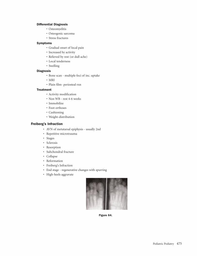
Freiberg’s Infraction
• AVN of metatarsal epiphysis - usually 2nd
• Repetitive microtrauma
• Stages
• Sclerosis
• Resorption
• Subchondral fracture
• Collapse
• Reformation
• Freiberg’s Infraction
• End stage - regenerative changes with spurring
• High-heels aggravate
Figure 64.
Pediatric Podiatry 475

Treatment• Short-leg cast for acute symptoms
• Activity modification
• Shoe modification
• Orthoses
• Surgery
Jones Fracture
• DDX - avulsion fracture of the tuberosity
• Avulsion by lateral band of plantar fascia
• May be apophysis
• 9-11 in females
• 11-14 in males
• Parallel to shaft
• Jones - transverse fracture of metaphysis 1.5 cm from tuberosity
Figure 65.
Incidence• Ages 15 -21
• 70% of Jones fractures
• 17% of tuberosity fractures
• Younger than 15
• Higher incidence of tuberosity fractures
Treatment Of Jones• Non-WB short-leg cast
• ORIF in competitive athlete
Tarsal Coalition
• May present as “injury” ages 8-12
• 8-12 year old, C-N bar most likely
• Adolescent, subtalar, middle facet
Figure 66.
Symptoms• Rigid pes valgus
• Pain in subtalar/midfoot
476 The 2005 Podiatry Study Guide

• Aggravated by activity
• Gait may be antalgic
• Higher incidence of ankle sprains
Types• Synostosis
• Synchondrosis
• Syndesmosis
Treatment• Peroneal block
• Cast
• Orthoses
• If persists surgical correction
Shin Splints
Symptoms• Anteromedial (PT) or anterolateral pain (AT)
• Local tenderness distal 2/3 of tibia
• Progressive during activity
• Persists after activity
Differential Diagnosis• Periostalgia/periostitis
• Stress fracture
• Chronic compartment syndrome
High-Risk Sports• Basketball
• Gymnastic dismounts
• Grand jette in ballet
• Running
• Sprinting
• Distance
Contributing Factors• Muscle weakness/tightness
• Poor shoes
• Training errors
• Surface
• Regimen change
• Biomechanical malalignment
• Anterior tibial
• Supinator
• Over-working to oppose pronation
• Pulled from tibia with pronation
Treatment• RICE
• NSAID prn
• PT - strengthen/stretch
Pediatric Podiatry 477

• Modify training
• Avoid increases in frequency or intensity
• Modify activity
• Cross-training
• With decreased pain return to activity
• Orthotics
• Shoe changes - after 500 miles - 40% loss
5.14 OsteochondrosesFrieberg’s Disease
• 2nd metatarsal head
• Vascular disruption & trauma
• Female – age 10-15
• Unilateral - 90%
Clinical Manifestations• Local pain, dull, aching
• Tenderness and swelling
X-ray Findings• Flattening of met. Head – distal
• Head – irreg. With increased density
• Fragmentation
• Widening of joint space
• Thickening of cortex – shaft
Treatment• Immobilization – BK cast 4-6weeks. Prevent further trauma and collapse
• Orthotic
• Surgical
Treve’s Disease
• Hallucial sesamoids
• Age 8 – 12
• Rare – confused with osteitis
Clinical Manifestations• Pain in region of sesamoids
• Pain ROM of hallux
X-ray Findings• Appearance of aseptic necrosis
• Pitting with fuzzy new bone formation
Treatment• Restriction of wt bearing
• Orthotic with dispersion
• Dancer’s pad
• Surgical
• Prognosis – favorable
478 The 2005 Podiatry Study Guide

Iselin’s Disease
• Apophysis – base of 5th met.
• Appears parallel to long axis of bone
• R/O fx. – transverse or oblique
• Peroneus brevis attaches at this site
• DDx - Os versaliarum
Clinical Manifestation• Pain and swelling
• Caused by increased tension (inversion sprain) or direct blow
• Up to 13 yrs of age of ossification
X-ray Finding’s• Fragmentation
Treatment• Rest
• Off to partial weight bearing
• Self limiting – relief of symptoms
Haglund’s Disease
• Accessory tarsal navicular
• Prominent fragmentation
• Pain over medial aspect of foot
• Male
• Trauma and tension
• Cast followed by orthotic
• Surgical – late
Thiemann’s Disease
• Proximal epiphysis of hallux
• Differentiate from traumatic conditions with good history
• Inc. incidence of other orthopedic problems
• Immobilization, protected weight bearing
Sever’s Disease
• Calcaneal apophysis
• May not exist as a disease entity
• 8 – 14 yrs. old males
• Closure from 15 –18 yrs
• R/O fracture infection, Still’s disease
Clinical Manifestations• Limping
• Pain and tenderness – inferior medial and lateral aspects of the calcaneus
• Males, may be obese
• Equinus present
• Soft tissue swelling
Pediatric Podiatry 479

X-ray Findings• Same as normal
• Sclerosis
• Fragmentation
• Irregularity
• Multiple ossification centers
Treatment• Severe cases – BK cast 4 – 6 wks. with slight PF of ankle
• Milder cases – restriction of activity with heel lifts/orthoses
• Orthoses with cushioning
Koehler’s Disease
• Navicular
• Coincides with ossification process
• Age 3 – 7 years
• Males – 75%
• Unilateral
Clinical Manifestations• Limping, local pain & tenderness over the navicular
• Trauma
• Biomechanically induced
• Mild swelling
X-ray Findings• Flattened, squashed, discoid
• Fuzzy fragmented, sclerotic
• Normal variant – multiple ossification centers
• X-ray changes may be normal variation in the sequence of navicular ossification
Treatment• Cast immobilization
• Reduced activities
• Orthotics
• Complete recovery is typical
Miscellaneous• Talus – Diaz or Mouchet’s
• Cuneiforms – Buschkes
480 The 2005 Podiatry Study Guide

5.15 Toe WalkingToe walking is a normal variant in the gait pattern of children for the first three to six months of independent
ambulation.
Differential Diagnosis
• Cerebral Palsy – abnormal muscle tone, hyperreflexia (lower greater than upper), clonus, extensor
plantar response, unstable, falls frequently
• Congenital contracture of heel cord –-The tendinous portion is shorter than normal with the muscle
bellies extending further down than usual. These children often stand on their toes before beginning to
ambulate and have limited ankle dorsiflexion with the knee flexed and extended.
• Limited range of motion of hamstrings or hip flexors.
• Muscular Dystrophy – late walker, frequent falling, (+) Gower’s sign, begins toe walking at 3 – 4 years of
age, waddling gait, lumbar lordosis
• Spina bifida
• Minimal brain dysfunction, attention deficit disorder, schizophrenia
• Diastematomyelia – toe walking begins at two to three years of age and worsens; spinal cord tethered in
spinal canal; cavus deformity common; foot ulceration, bladder and bowel problems.
• Charcot-Marie Tooth – difficulty in walking, paresthesias in legs, muscle cramps; toe walking begins
later; vibratory and position sense lost early; early weakness of intrinsics, ankle dorsiflexors, and
peroneals; progressive pes cavus and foot drop; check other family members
• Idiopathic – well coordinated, runs with minimal tripping and falling; can achieve a heel toe or foot flat
gait at lower walking speeds; neurologic exam normal, younger child has good ankle range of motion,
older child may have limited range (acquired shortening from functional equinus); began toe walking
at onset of walking
Evaluation Of Toe Walker
• History – pre, peri-, and postnatal history; decreased or absent fetal movements, low birth weight,
prematurity. When did child begin toe walking? Is toe walking intermittent or all the time? Unilateral or
bilateral? (Hemiparesis?) –developmental milestones -abnormal psychological behavior – unusual
walking pattern in other family members
• Physical Exam – range of motion at ankle, knee and hip; gait analysis
• Neurologic Exam– sensory, vibration, position sense, muscle power (Gower’s sign) deep tendon reflexes,
weakness of ankle dorsiflexors (if child can’t heel walk, have child lean against wall and dorsiflex feet
against resistance)
• Radiographs – if ankle range of motion is limited with knee flexed and extended, rule out bony block
• Special Diagnostic Tests – e.g. NCV, EMG, muscle biopsy reserved for children in which muscle
weakness cannot be ruled out clinically and when toe walking appears after a period of normal gait –
spinal MRI if a suspicion
Treatment
• Serial below knee stretching casts (three to six weeks) to increase ankle range of motion.
• Articulating ankle-foot orthoses
• Reverse gait plates or flat rigid shoe plate and high topped sneakers – need adequate ankle range of
motion
• Heel lifts
• Discourage jolly jumpers and walkers
• Surgical intervention
Pediatric Podiatry 481

5.16 Pediatric Neuromuscular DisordersCerebral Palsy
• Static encephalopathy 7.5 per 1,000 live births, UMN
• Clinical syndrome – characterized by a chronic, non-progressive disorder of motor control, resulting in
abnormality of posture, movement and tone
• Three criteria
– Fixed, non-progressive brain lesion or lesions
– Original lesion a result of pre-, peri-, or postnatal trauma or anoxia
– Lack of motor control results in abnormal coordination of movement and posture; may also see
mental retardation, convulsions, sensory disturbance, speech impediment, defects of hearing, language
or eyesight
• Risk factors
– Prematurity and low birth weight*
– Metabolic (hypoglycemia, hyperbilirubinemia)
– CVA
– Infections (toxoplasmosis, rubella)
– Cerebral malformation
• CP classification
– Spastic- most common group, includes diplegia, hemiplegia, and quadriplegia
– Extrapyramidal (includes choreoathetosis and dystonia)
– Hypotonic
– Ataxic
– Mixed
Figure .
• Clinical features
– In infants may see hypotonia, persistence of primitive neonatal reflexes, poor development of more
mature reflexes, toe walking, strong hand preference (hemi)
– Older children – hypertonicity, increased DTR’s, Babinsky, delayed gross motor milestones, abnormal
gait, drooling, strabismus, difficulty with feeding and speech, mental retardation, seizures, adductor
grab, clonus
Types• Spastic diplegic – more involvement of legs than arms, spastic crouch, equinovalgus foot deformity
• Spastic quadriplegic – involvement of all four limbs, usually more severely involved than diplegic, may
never walk, difficulty in feeding, speech and drooling, frequent pneumonia, mental retardation, seizures,
strabismus
• Hemiplegic – involvement of unilateral arm and leg, early hand preference, unilateral toe walking or
circumduction of limb, unilateral Babinsky
482 The 2005 Podiatry Study Guide

• Choreoathetoid – hypotonic until age two, slow, writhing, involuntary movements of flexion and
extension, pronation and supination of fingers and hands and sometimes toes and feet, abnormal
movements of face, tongue, and palate, facial grimaces, difficulties with speech and feeding, may be
very late in walking (8 or 10 years of age)
• Dystonia – sustained twisted posture involving trunk and proximal limb
• Ataxia – poor coordination, wide based gait
Spina Bifida
• Incomplete closure of the bony elements of spine with or without extrusion of contents of the spinal
canal through bony defect
• Spina bifida occulta
– Defect of fusion of posterior vertebral arch without disruption of underlying meninges and cord,
10% of pediatric population, asymptomatic, low mid-back hairy tuft, dimple or teratotoma
• Spina bifida aperta
– 1 in 1,000 live births, most common in lumbar and lumbosacral area
Types• Meningocoele – distention of meninges
• Myelomeningocoele – distention of meninges and cord
• Myelocoele – distention of meninges and cord with lack of closure of cord in defective area
Clinical• Paralysis and loss of sensation below lesion level (ulcerations, fractures)
• Lack of bladder and bowel control
• Hydrocephalus
• Hip dislocations, scoliosis and foot deformities (equinovarus—most common)
Diastemtomyella
• Congenital malformation – in which spinal cord is divided longitudinally into 2 hemicords by a bony,
fibrous, or cartilaginous spicule from the posterior aspect of a vertebral body; spicule transfixes spinal
cord or cauda equina (tethered cord syndrome), stopping its normal ascent during growth of the
vertebral column; neurologic deficit below level of lesion
• Clinical picture
– Various skin defects near midline at lesion level (usually lumbar) 30% of cases show no skin defect
– Late in walking or abnormal gait (limp)
– Chief complaint may be poor posture associated with leg length discrepancy or foot size difference
– Scoliosis common
– Cavovarus foot, ulcerations of feet
– Urinary incontinence
• Early diagnosis by MRI, followed by release of cord tethering; prevents further deformity but does not
reverse existing deformity
• 75 % of cases female
Muscular Dystrophies
• Duchenne MD
– Most common and most severe, x-linked recessive, high mutation rate
– Male to female, 9:1
– Onset usually apparent by age 3
– Weak fetal movements in last trimester
Pediatric Podiatry 483

– Delayed motor milestones, difficulty climbing stairs, use arms to rise from chair
– Difficulty or unable to run or hop, frequent falling
– Gower’s sign, waddling gait, toe walking
– DTR’s disappear early except ankle
– Cardiac involvement, 50% mental retardation
– Symmetrical involvement first of pelvis, then shoulders 3 to 5 years later pseudohypertrophy of
calves, unable to walk by 12, death teen to third decade
– Pes planus secondary to tight heel cord early stage, equinovarus later stage, lumbar lordosis
– Elevated serum enzyme levels, especially CPK 30-300 X normal
• Becker’s MD
– X-linked recessive
– Benign form, onset 5-20, slow progression, otherwise similar to Duchenne
• Limb Girdle MD
– Progressive weakness and atrophy of mm of shoulder and pelvic girdle and proximal limbs during
20’s or 30’s, weakness of neck flexors and extensors
– Autosomal recessive
• Fascioscapulohumeral MD
– Autosomal dominant, onset 20’s
– First facial weakness (whistling, sipping, keeping eyes closed) shoulder girdle weakness and scapular
winging (can’t raise arms above head), eventually anterolateral leg weakness (foot drop)
– Slow progression
Polymyositis
• Non-hereditary, inflammatory myopathy
• In presence of skin rash, dematomyositis
• Dermatomyositis – symmetrical weakness of proximal limb mm and neck flexors, rash (butterfly
distribution, heliotrope, erythematous, scaly extensor surfaces of joints especially ankles, (medial
malleolus), elbows, knees, knuckles and dorsal finger near nail bed; later stage- skin tight and glossy
with subcutaneous calcium deposits
• In children, no association with malignancy
Neurofibromatosis
• Von Recklinghausen’s disease – hereditary
• Most common of the neurocutaneous dysplasias
• Three classic signs in childhood: 1) café au lait spots 2) multiple soft tissue tumors (some malignant,
seizures associated with intracranial tumors) 3) skeletal changes e.g. kyphoscoliosis, vertebral body
defects, local gigantism
Peripheral Neuropathies
• Guillain Barre syndrome (post-infectious polyneuritis)
– Rapid onset following an illness
– Distal weakness that ascends
– 50% cranial nerve involvement, DTR’s absent, sensory deficit minimal
– Most children recover completely
• Charcot Marie Tooth type I HMSN
– 50% of all cases of childhood hereditary neuropathies
– Clinical picture
484 The 2005 Podiatry Study Guide

– Peroneals (foot slap), tib ant and intrinsics, calf, hand intrinsics
– “stork leg” and pes cavovarus
– Decrease vibration and position sense, DTR’s absent at ankles
– Nerve hypertrophy
– Decreased NCV – hallmark of disease*
• Peroneal muscular atrophy type II HMSN
– Zxonal degeneration of peripheral nerve, NCV normal
– Clinically similar to CMT
• Dejerine – Sottas disease type III HMSN
– Similar to CMT
– Scoliosis, pupillary abnormalities
• Refsum’s type IV HMSN
– Anorexia, gait abnormalities, icthyosis, eye problems, anosmia, hearing deficit, pes cavus
– Deficiency in fatty acid, metabolism, reduce dietary phytanic acid
• Riley Day syndrome HSN (familial dysautonomia)
– Onset in infancy, autonomic dysfunction, Jewish children of eastern European descent
– Failure to respond to pain
• Toxic Neuropathies
– Sciatic neuropathy
– Result of injection into buttocks, near sciatic nerve
– Paralysis within days
– Long term effects: limb growth retardation, pes cavus, trophic ulceration
• Antibiotic-induced, antimetabolite, heavy metal (e.g. lead exposure, seizures, headache and ataxia in
Children under five, dropfoot in children over five)
Poliomyositis (“infantile paralysis”)
• Etiology- one of three polioviruses attacks GI and respiratory tract and spreads to CNS (lower motor
neuron)
• Incidence- rarer because of immunizations
• Acute phase - 5-10 days, acute illness during which paralysis may occur, fever, headache, nuchal rigidity,
muscle tenderness, weakness asymmetric and scattered, bulbar poliomyelitis
• Convalescent phase- 16-month period following acute phase – varying degrees of spontaneous recovery
• Chronic phase- rest of patient’s life following convalescent period
• Steppage gait- weakness of peroneal and anterior tibial
• Lower extremity deformities often include flexion, abduction, and lateral rotation of hip, paralysis of
quadriceps, flexion and valgus deformity of knee, leg length discrepancy
• Feet - severe pes valgus on weight bearing, cavus foot, and claw toes off weight bearing
Pediatric Podiatry 485

5.17 Pediatric PharmacologyGeneral Considerations
• Children are not little adults
• Parents will be involved
– *Concise instructions
• Metabolic rate varies
• Weight, height, body morphology and body surface area are factors
• Larger proportion of surface area to body volume.
– *Surface area increases less rapidly
Drug Considerations
• Appropriate medication
– Established drugs over latest & greatest
• Dosage
– Toxicity
– Overdosage
• Dosage forms
• Strength
• Route of administration
Problems
• Contraindications
– Allergy
• Age parameters
• Drug interactions
• Food interactions
• Dosage
– Toxicity
– Overdosage
• Syrup of Ipecac if necessary to induce vomitting
Dosage
• Should be individualized
• Smallest amount to give the best therapeutic effect
• May be based upon weight, age, or body surface area
– Metabolic rate is a factor
– *Fever increases metabolic rate
– Obesity and edema are factors
Fried’s Rule
• Patient less than one year old
• Based on age
– × Adult Dose
Young’s Rule
• Patient two years or older
• Based upon age
Age in Months
150
486 The 2005 Podiatry Study Guide

– × Adult Dose
Clarke’s Rule
• Patient two years or older
• Based upon weight
– × Adult Dose
Body Surface Area
• Most accurate
• Calculate utilizing the West nomogram
• Correlates height and weight with body surface area
• Manufacturer suggests dose per square meter (m_)
• Can calculate based upon adult dosage
Manufacturer’s Recommended Dosage
• PDR (Physicians’ Desk Reference®)
• Utilize given formulation
Antibiotics
Oral Antibiotics• Penicillin
– Gram (+)
– Best for streptococcus
• B lactamase resistant PCNs
– Dicloxicillin
– Oxacillin
– Methicillin
– Cloxicillin
• Amoxicillin/Ampicilin
– Broader spectrum
– H. influenza
– P. mirabilis
– Klebsiella
– Enterococcus
– E. coli
– Not B lactamase resistant
• Augmentin
– Same spectrum as ampicillin
– Better at enterococcus
– Some anaerobes, especially B. Fragilis
– DOC for animal bites including human
– Better tasting for children
– Suspension
– Chewable tablets
• Cephalasporins
– Broad spectrum
Weight in Pounds
150
Age in Years
Age in Years × 12
Pediatric Podiatry 487

– Good gram (+) and adequate gram (-)
–Variable B lactamase resistant
– Duricef (Cefadroxil)
– Suspension
– Keflex (Cephalexin)
– Suspension and solution
• Erythromycin
– Good gram (+) including staphylococcus
– Good for penicillin allergy
– M. pneumonia, Legionella, Chlamydia
– GI upset common
– Suspension
• Tetracycline
– Some gram (+) and gram (-)
– Rickettsia m. pneumonia, chlamydia
– Avoid milk products
– Contraindicated under 8 years of age
• Ciprofloxacin
– A fluroquinolone
– Good gram (+) including staphylococcus-
– Occasional MRSA
– Very good gram (-) including pseudomonas
– Contra-indicated under 16 years of age
Topical Anitbiotics• Bacitracin
– Staphylococcus and streptococcus
– Cream and ointment
• Bactroban (mupirocin)
– Ointment 2%
– Staphylococcus and streptococcus
– Specifically indicated for impetigo
• Neosporin
– Polymyxin B and neomycin
– Cream and ointment
– Ointment also has Bacitracin
• Silvadene cream 1% (silver sulfadiazine)
– Excellent coverage
• Betadine (povidone iodine)
– Excellent coverage
– Solution, cream, and ointment
Analgesics
• Acetaminophen (Tylenol, Tempra)
– Most common
– Relatively safe and well-tolerated
– Chewable and solution
488 The 2005 Podiatry Study Guide

– Analgesic and anti-pyretic
– NOT anti-inflammatory
• Narcotics
Non-Steriodal Anti-Inflammatories
• Tolectin (Tolmetin sodium)
– NSAID indicated for pediatrics
• Ibuprofen (Motrin, Advil)
– Different strengths
– Children’s formulations
– Syrups and elixirs
• Naprosyn (Naproxen)
– Indicated for JRA
– Suspension
• Acetylsalicylic acid
– ASA
– Many forms
– Generally safe
Topical Steriods
• Utilized for dermatological inflammatory conditions—dermatitis, eczema, inflammatory tinea pedis
• Use the highest potency for the shortest time
• Generally, use only low or intermediate strength in pediatrics
• Different vehicles
• Avoid occlusion in pediatrics
• Avoid overuse or prolonged use
– Steroid atrophyÆ ulceration
– Potential for hypothalamic-pituitary-adrenal axis suppression
– Potential for Cushing’s syndrome
• Avoid intertriginous areas
• Contraindicated in varicella
Topical Steriods• Ultra high potency
– Diprolene (augmented betamethasone)– Ultravate– Temovate
• High potency– Betamethasone diproprionate– Betamethasone 0.05%– Lidex (fluocinonide)
• Intermediate potency– Betamethasone valerate– Betamethasone 0.025%– Elocon
• Low potency– Hydrocortisone
– Dexamethasone
Pediatric Podiatry 489

Anti-Fungus
• Orals – for severe cases only, check PDR for guidelines
• Most are broad spectrum
– Tineas, e.floccosum, candida
• Use the vehicle that is most effective
– Solution, lotion, creme
• Should use for four weeks
Topical Anti-Fungus
• Lotrimin
– Solution, lotion and crème
– Lotrisone crème formulation(combines steroid)
• Spectazole crème
– Once a day
• Loprox
– Crème and lotion
• Lamisil
– Two week treatment
5.18 Skeletal Syndromes and Systemic Disorders in Pediatric OrthopedicsSkeletal Dysplasias
Skeletal dysplasias involve basic and systemic abnormalities of bone and cartilage growth and development, usually
causing short stature.
Diagnosis of Skeletal Dysplasias• Height and proportion
– Birth length
– Current height and percentile
• Most skeletal dysplasias produce adult height under 60 inches.
• Mean adult height in achondroplasia for males is 52 in. (+2 SD = 56 in.) and for females is 50 in. (+2
SD = 52 in.).
• Mean adult height in diastrophic dwarfism is 42 in.
– Body proportion is determined by comparing limb and trunk lengths.
• Limb height ≈ lower segment = symphysis to soles of feet.
• Trunk height ≈ upper segment = total height minus lower segment.
• For a graph of normal values by age, see the Harriet Lane Handbook.
• Another approach to determining body proportion is to subtract total height from arm span (fingertip
to fingertip). Values for normal stature by this measure are –2 cm (–13/16 in.) (before age 8), +1 cm
(+3/8 in.) (adult females), and + 5 cm (+2 in.) (adult males).
– Limb segment ratios are best calculated from x-rays.
– Normal ratio of radius to humerus is 75%.
– Normal ratio of tibia to femur is 82%
– Types of disproportion include rhizomelic (short humerus and femur), mesomelic (short hands
and feet).
• Dysmorphism is morphologic variation of bone and soft tissue that may characterize a specific disorder
– Depressed nasal bridge often seen in achondroplasia
– Long philtrum suggest trichorhinophalangeal dysplasia
490 The 2005 Podiatry Study Guide

– Short broad thumb suggest multiple epiphyseal dysplasia
– V-shaped phalanges suggest trichorhinophalangeal dysplasia
– “Hitchhiker’s” thumb and abducted great toe indicates diastrophic dysplasia
• Radiographic evaluation should include these five key studies
– Lateral skull and cervical spine
– Lateral lumbar spine
– Anteroposterior pelvis
– Anteroposterior knees
– Anteroposterior hand and wrist
• Family evaluation
– Construct pedigree of at least all first-degree relatives.
– If family history is negative, consider the following:
– New mutation
– Gonadal mutation
– Recessive trait
– Nonpaternity
• Laboratory evaluation is usually not indicated but can be useful if prior work up is negative. It should
include the following studies:
– Chemistry profile
– Endocrine evaluation
– Urine work up for storage disorder
Achondroplasia
• Achondroplasia is autosomal dominant but usually due to frequent new mutation. It is the most com-
mon skeletal dysplasia.
• Major features
– Mid-face hypoplasia
– Rhizomelic dwarfism
– Genu varum (variable)
– Three- to three-month delay in motor milestones
– Thoracolumbar kyphosis, often resolving with growth
– Spinal stenosis. This is greatest in lumbar spine and distally but may affect entire spine, including fora-
men magnum, and cause severe developmental delay
• Height
– Final height for males is 46-56 in., and for females, 44-55 in.
Pseudoachondroplasia
• Although these patients are also rhizomelic, the epiphyseal involvement in this syndrome causes basic
differences from achondroplasia.
• Major features
– Rhizomelic shortening of extremities
– Variable knee deformities (varus on one side, valgus on the other)
– Mild platyspondyly, minimal scoliosis, no stenosis
– Odontoid hypoplasia, C1-C2 instability
– Epiphyseal deformation; eventual degeneration
– Ligamentous laxity
Pediatric Podiatry 491

Diastrophic Dysplasia
• Autosomal recessive
• Major features
– “Cauliflower ear” develops at approximately six months
– Rhizomelic shortening of extremities
– Contractures of major joints with later degenerative joint disease (DJD)
– Hands: hitchhiker’s thumb, symphalangism
– Dislocated hips (occasionally)
– Equinovarus foot deformities
– Cervical spina bifida with kyphosis; sometimes resolves
– Scoliosis of thoracic and lumbar spine
Spondyloepiphyseal Dysplasia Congenita
• Autosomal dominant with frequent new mutations
• Major features
– Odontoid hypoplasia or Os odontoideum; may have instability
– Platyspondyly, scoliosis
– Coxa vara, epiphyseal irregularity, DJD
Spondyloepiphyseal Dysplasia Tarda
• Variable transmission; diagnosis late in childhood
• Major features
– Irregular ossification, DJD
– Hips may resemble Perthes disease but are bilaterally synchronous.
– Scoliosis
– Occasional cervical instability
Multiple Epiphyseal Dysplasia
• Autosomal dominant
• Major features
– Variable, usually mild short stature due to short limbs
– Irregular epiphyseal ossification with deformity, pain, DJD
– Hips, knees, and ankles are most involved
– Usually presents in late childhood to adulthood
Metatropic Dysplasia
• Major features:
– Epiphyseal or metaphyseal enlargement: “knobby” joint with contractures
– Cervical stenosis, instability
– Scoliosis, kyphosis, later onset
– Coccygeal tail
– Thoracic hypoplasia; may cause respiratory compromise
– Initially short-limb dwarfism; becomes short-trunk type with onset of scoliosis
Chondrodysplasia Punctata (Conradi-Hünermann syndrome)
• Autosomal dominant
• Major features
492 The 2005 Podiatry Study Guide

• Multiple asymmetric epiphyseal calcifications
• Good prognosis
Multiple Osteocartilaginous Exostoses
• Autosomal dominant
• Clinical appearance features mild short stature. Categories of problems:
– Local impingement on tendons, nerves, spinal canal and ribs
– Asymmetric growth in two-bone segments leading to valgus knees, ankles, elbows, and wrists and
possi bly to radial head dislocation
– Leg length inequality (usually <4 cm)
– Risk of malignant degeneration (about 1% of patients)
• Radiographic features
– Osteochondromas in metaphysis, pointing away from joint
– Cortex of osteochondroma is confluent with that of host bone
– May be sessile or pedunculated
• Treatment
– Resect lesions only when symptomatic. Resection does not predictably increase forearm rotation
– Correct knee and ankle valgus when greater than 10°
– Monitor in adulthood every two years with bone scan
Dysplasia Epiphysealis Hemimelica (Trevor’s disease)
• Definition: epiphyseal osteochondroma; no genetic pattern
• Clinical features
– Presents in first decade
– Restricted joint motion
– Enlarged joint or locked joint
– Knee, foot, and ankle are most commonly involved
• Radiographic features include multiple opacity in exostotic cartilage; these eventually coalesce
• Treatment: resection, attempting to preserve normal cartilage
Multiple Enchondromas (Ollier’s disease)
• Clinical presentation
– Angular deformity
– Bony irregularity
– Limb length inequality
• Radiographic features
– Diffuse enchondromas in metaphysis; occasionally epiphyses. Usually asymmetric
– Treatment
– *Angular or length correction of limb
– *Monitor for malignancy, especially in Maffucci’s syndrome
Pediatric Podiatry 493

Cleidocranial Dysplasia
• Autosomal dominant
• Persistently open skull sutures with bulging calvarium
• Hypoplasia or aplasia of clavicles
• Wide symphysis pubis
• Hip abnormalities
• Short middle phalanx of fifth finger
• Syringomyelia may be present
Dyschondrosteosis
Major features:
• Madelung’s deformity
• Relative shortening of forearm and leg
• Females predominate
5.19 OTHER SYNDROMES INVOLVING SHORT STATURE Cornelia de Lange Syndrome
• Major features
– Synophrys (single confluent eyebrow)
– Down-turned mouth
– Mandibular spur, in infancy
– Hirsutism
– Gastroesophageal reflux
– Small for gestational age, with continued growth retardation
– Motor and intellectual delay
– Cardiac abnormalities
• Orthopedic involvement
– Upper-extremity anomalies in almost all patients
– Micromelia, phocomelia
– Decreased number of fingers
– Lobster claw hand
– Proximally placed thumb
– Elbow anomalies
• Lower extremities
– Miscellaneous foot deformities and contracture
– Avascular necrosis of femoral head in 10%
• Treatment
– Correct lower-extremity abnormalities if limiting ambulation. Individualized treatment for upper
extremities
494 The 2005 Podiatry Study Guide

Riley-Day Familial Dysautonomia• Ashkenazi Jews only; autosomal recessive
• Sympathetic over activity is key feature
• Major features
– Deficient sensation of pain and proprioception
– Gastroesophageal reflux, pneumonia
– Variable life expectancy
• Orthopedic abnormalities and implications
– Scoliosis or kyphosis before age eight; poor brace tolerance; fuse early
– Fractures due to osteopenia or dyscoordination
– Avascular necrosis of femoral head, distal femur, talus
– Hip dysplasia
Nail-patella Syndrome• Autosomal dominant, normal life expectancy
• Orthopedic features
– Nails grooved, small, or absent, especially on thumb
– Multiple knee anomalies: patella tripartite, small, or absent; lateral femoral condyle hypoplastic;
osteochondritis dissecans of lateral femur and talus
• Elbow: capitellar hypoplasia, cubitus valgus, flexion contracture
• Iliac horns
Sclerosing Bone Disorders
Fibrodysplasia Ossificans Progressiva• Progressive, disabling heteroptopic ossification or ankylosis
• Characteristic shortening or valgus of great toe
• Ossification starts as tender, hard nodule; progresses proximal to distal, posterior to anterior
• Do not biopsy - this may accelerate the process
• Genetics: usually a spontaneous mutation, but it may be transmitted as autosomal dominant
Progressive Diaphyseal Dysplasia• Clinical features: pain, fatigue, muscle atrophy
• Radiographic features: symmetrically widened, sclerotic diaphyses; epiphyses spared; tibia and femur
most commonly involved
• Treatment: osteotomies only if there is marked deformity
Melorheostosis• A syndrome involving asymmetric, extraosseous, longitudinal hyperostotic streaks; limb pain and soft-
tissue contracture.
• Treatment: analgesics, bracing, contracture releases, and bone shortening
Osteopathia Striata• Linear intraosseous metaphyseal striations
• Autosomal dominant
• Asymptomatic
• No treatment required
Osteopoikilosis• Multiple symmetric intraosseous epiphyseal or metaphyseal “spots”
• Autosomal dominant
• Asymptomatic
Pediatric Podiatry 495
Marfanoid Disorders
Marfan SyndromeThis disorder of fibrillin has multiple effects on the skeleton and connective tissue. Because some features may be
seen in the general population, the following diagnostic criteria have been developed.
• Diagnostic criteria. Positive family history or skeletal involvement.
• Involvement of two other systems - at least one must be a major manifestation (see * below)
• System involvement (*indicates major manifestation)
– Skeletal
• Anterior chest deformity
• Dolichostenomelia (long, narrow limbs)
• Arachnodactyly (long, narrow digits)
• Vertebral column deformity (kyphosis, scoliosis)
• Tall stature
• High, narrowly arched palate
• Increased appendicular joint mobility
• Protrusio acetabuli
– Ocular
• Ectopia lentis (superolateral lens dislocation)
• Flat cornea
• Retinal detachment
• Myopia
– Cardiovascular
• Ascending aortic aneurysm with dilation of valsalva sinuses
• Aortic dissection, usually ascending segment
• Aortic valve regurgitation
• Mitral valve regurgitation
• Abdominal aortic aneurysm
– Pulmonary
• Spontaneous pneumothorax
• Apical bleb
– Skin
• Striae atrophicae (stretch marks)
• Hernia
– Central nervous system
• Dural ectasia
• Learning disability
• Hyperactivity
• Implications
– Monitor aortic and cardiac status
– Beta blocker for aortic dilation
– Restrict from vigorous exertion
– Counsel regarding genetics
– Treat skeletal deformity if symptomatic
496 The 2005 Podiatry Study Guide

Homocystinuria
• Homocystinuria may be mistaken for Marfan syndrome, but it is most readily distinguished by mental
retardation. It has the following major features:
– Mental retardation
– Dislocated lens (inferomedial)
– Arachnodactyly
– Joint stiffness
– Cavus feet
– Scoliosis or kyphosis
• Diagnosis: urine amino acid screen
• Treatment: vitamin B6 administration, methionine restriction
Congenital Contractural Arachnodactyly
This is also a Marfan look-alike; so much so that Marfan’s original patient had this syndrome. Clinical features
include:
• Face: oval with recessed jaw, flattened ear
• Eyes: occasional intraocular coloboma
• Heart: congenital septal and valve defects
• Skeleton
– Flexion contracture that partially improves with time
– Hands: contracture of proximal interphalangeal (PIP) and distal interphalangeal (DIP) joints
– Scoliosis: appears by mid-childhood
Achard Syndrome
Clinical features include:
• Arachnodactyly
• Generalized ligamentous laxity
• Mandibular hypoplasia
Stickler Syndrome (Hereditary Arthro-Ophthalmopathy)
• Etiology. A mutation in type II collagen. Abraham Lincoln may have had hereditary arthro-
ophthalmopathy.
• Clinical features
– Progressive myopia beginning in first decade
– Retinal detachment
– Abnormal epiphyseal development, eventual DJD
– Mild joint hypermobility in some cases
– Marfanoid habitus in some cases
– Autosomal dominant
Pediatric Podiatry 497
Arthrogryposis Or Contracture
Arthrogryposis Multiplex CongenitalThis is the classic contractural syndrome. The etiology is unknown; it is probably multifactorial. Major features are
all skeletal: It may affect all four limbs, upper extremities only, or lower extremities only. Involvement is usually
greatest distally within each limb.
• Hips are frequently dislocated. Contractures are usually into abduction and external rotation
• Knees: flexion more common than extension
• Clubfoot and vertical talus are common, often resistant to cast treatment
• Upper extremities: extended at elbows, adducted, stiff fingers
• Spine: paralytic scoliosis, torticollis
• Implications for treatment
– Range of motion can be shifted but not increased
– Osteotomies most useful near end of growth
– Fractures common after manipulation or cast
Larsen Syndrome• Orthopedic features
– Dislocated hips, hyper-extended or dislocated knees
– Clubfeet
– Normal muscle mass
– Elbow dislocations
– Cervical kyphosis and instability
– Thoracic and lumbar scoliosis
• Non-orthopedic features
– Flattened face, depressed nasal bridge
– Cleft palate
Freeman-Sheldon (Whistling face) Syndrome• Small mouth and chin (may cause difficulty with intubation)
• Arthrogrypotic, flexed and ulnar-deviated fingers
• Clubfeet or vertical talus
• Scoliosis or kyphosis
Möbius Syndrome• Congenital facial diplegia
• Variable absence of shoulder girdle muscles
• Clubfeet and hand contractures or anomalies
Pterygium Syndromes• Multiple pterygium syndrome
– Static disorder with flexion contractures and webs at all flexion creases
– Short stature
– Congenital spinal anomalies
• Popliteal pterygium syndrome
– Webs only across perineum and knees
– Facial malformations
– Contractures are due to a single cord from ischium to calcaneus, with the nerve bow-strung across the
joint
498 The 2005 Podiatry Study Guide

Vascular Abnormalties
Kippel-Trenaunay-Weber Syndrome• Clinical features
– Cutaneous hemangioma (port-wine stain)
– Varicose veins
– Limb hemi-hypertrophy: often non-linear growth pattern
• Treatment
– Initially treated with compressive therapy
– May have specific indications for surgery
Maffucci SyndromeMajor features:
• Multiple enchondromas
• Cavernous hemangiomas
• Risk of sarcomatous transformation
Sturge-Weber SyndromeMajor features:
• Port-wine hemangioma in trigeminal distribution
• Neurologic sequelae due to meningeal hemangioma
Blue-Rubber Bleb Nevus Syndrome• Autosomal dominant
• Clinical features
– Bluish cavernous hemangiomas on trunk and upper arms; may bleed from GI tract locations;
may cause pain
– Regional hyperhidrosis
Kasabach-Merritt SyndromeClinical features:
• Solitary or multiple cavernous hemangiomas on trunk or extremities
• Consumptive coagulopathy secondary to hemangiomas
Ataxia-Telangiectasia (Louis-Bar Syndrome)Clinical features:
• Telangiectasia on conjunctivae, face, neck, and arms
• Progressive ataxia
• Genetics: autosomal dominant
Overgrowth Syndromes
Generalized bodily overgrowth• Prader-Willi syndrome
– Partial deletion of chromosome 15
– Major findings
– Infantile hypotonia
– Obesity beginning after age 1
– Mental retardation
– Cryptorchidism
– Short stature
– Eyes slant upward and lateral
Pediatric Podiatry 499

• Orthopedic findings
– Developmental dysplasia of the hip in 10%
– Scoliosis in 50%
– Small hands and feet
Bardet-Biedl Syndrome• Etiology unknown; genetics probably autosomal recessive
• Major findings
• Trunical obesity
• Mental retardation
• Hypogonadism
• Retinitis pigmentosa
• Renal abnormalities
– Orthopedic abnormalities: postaxial polydactyle (feet more than hands)
Beckwith-Wiedemann Syndrome• Autosomal dominant
• Major findings
• Large stature
• Omphalocele
• Macroglossia (may partially regress)
• Hypoglycemia
• Multiple organ enlargement; risk of Wilms’ tumor
– Orthopedic abnormalities
• Leg length inequality
• Neurologic damage if hypoglycemia not controlled
• Polydactyly, idiopathic scoliosis, radial head dislocation variable
Asymmetric Overgrowth• Idiopathic hemihypertrophy
– Skeletal findings
• One side of body larger in all dimensions
• Growth proportionate over time
• Lower extremities most often affected; trunk and upper extremities may be affected
– Genitourinary system
• Wilms’ tumor (nephroblastoma) in approximately 5%
• Medullary sponge kidney
• Renal malposition
– Vascular system: aortic, cerebral vascular, or congenital heart abnormalities
• Russell-Silver syndrome
– Short stature
– Sall, triangular face; may be asymmetric
– Genitourinary system and genital malformations
– Orthopedic features
• Hemihypertrophy
• Other miscellaneous skeletal findings
• Goldenhar syndrome
– Major features
• Epibulbar dermoids
500 The 2005 Podiatry Study Guide

• Preauricular skin tags, facial asymmetry
– Orthopedic abnormalities
• Congenital vertebral anomalies
• Other aspects of VATER syndrome association
– Implications: Monitor for scoliosis; be aware of difficult intubation.
• Klippel-feil syndrome
– Key feature: cervical spine fusions; may have stenosis, instability
– Possible skeletal associations
• Congenital or “idiopathic” scoliosis of lower spine (60% of patients)
• Sprengel’s deformity (30% of patients)
•Upper extremity or hand anomalies
– Non-skeletal association
• Genitorinary or renal malformations
•Hearing impairment
•Cardiac anomalies
•Facial asymmetry
– Implications
• Screen early for hearing impairment, genitourinary malformations (ultrasound)
– Counsel regarding activity and anesthesia if neck is unstable
• Proteus syndrome
• A hemartomatous disorder affecting all three germ layers
• Characterized by its variability; named for the Greek god able to change shape at will
• Macrodactyly and asymmetric tissue overgrowth are key feature
• Diagnosis can be established by adding up the points assigned to each criterion below for a total score
– Diagnostic criteria Points
• Definitive diagnosis is made if score is 13 points or higher; questionable diagnosis is 10-13 points;
diagnosis excluded is less than 10 points
• Macrodactyly, hemihypertrophy, or both—5
• Thickening of skin—4
• Lipomas and subcutaneous tumors—4
• Verrucous epidermal nevus—3
• Macrocephaly, irregular skull—2.5
• Other minor abnormalities—1
Neurofibromatosis• Neurofibromatosis is the most prevalent skeletal disorder to be caused by a single gene defect. It is a
hemartomatous disorder, probably of neural crest origin.
• Diagnosis is established when the patient meets at least two of the following NIH criteria.
– Cafe au lait spots (five or more spots>1.5 cm after puberty or >0.5 cm before puberty)
– Subcutaneous neurofibroma
– Positive biopsy
– Positive family history (first-degree relative affected)
– Skeletal manifestations
– *Long bone pseudarthrosis
– *Dystrophic curve of spine
– *Elephantiasis neuromatosa
– Optic glioma
Pediatric Podiatry 501

– Two or more Lisch nodules (hamartomas in iris)
– Axilliary or inguinal freckling
• Features of dystrophic spinal curve
– Severe apical rotation or wedging
– Paravetebral mass
– “Spindling” of transverse process
– Thinning of rib
– Enlargement of the foramen
– Vertebral scalloping
• Orthopedic implications
– Dystrophic curves require more aggressive treatment than non-dystrophic curves. (Fuse if>30 degrees;
anterior and posterior fusion if>70 degrees of if kyphosis>50 degrees. Explore fusion postoperatively.)
– Preoperative CT scan or myelogram on all dystrophic curves
– Rule out sarcoma if unexplained pain or localized growth occurs
Ehlers-Danlos Syndromes
This group of connective tissue abnormalities include at least 11 different subtypes. Many other patients
do not fit precisely into one of the groups.
Osteogenesis Imperfecta• A group of disorders of type II collagen causing bone fragility and in some cases blue sclerae, hearing
loss, and abnormal dentin. The osseous fragility tends to improve after puberty
• Silence types
Type I
• Variable osseous fragility (minimal through moderately severe)
• Distinctly blue sclerae (at all ages)
• Presenile hearing loss
• Autosomal dominant
Type II (lethal perinatal osteogenesis imperfecta)
• Extremely severe osseous fragility, with stillbirth or neonatal death
• Subgroup A: Radiographs show broad, crumpled long bones and broad ribs with continuous
beading. Autosomal dominant or new mutation
• Subgroup B: Radiographs show broad, crumpled long bones; ribs show discontinuous beading or
are not beaded. Autosomal recessive
• Subgroup C: Radiographs show thin, fractured long bones with thin, beaded ribs
Autosomal recessive
Type III
• Autosomal recessive
• Fractures at birth, then progressive deformity
• Normal sclerae and hearing
Type IV
• Moderate osseous fragility
• Normal sclerae (blue in infancy)
• Variable deformity of long bones and spine
• Autosomal dominant
Note: The value of opalescent dentin for subcategorization of osteogenesis imperfecta is uncertain
502 The 2005 Podiatry Study Guide

5.20 Malformations of the Hand and Foot• Terminology
– Extent—partial or complete
– Simple—skin only
– Complex—synostosis
– Polysyndactyly—hidden duplicated skeletal structures
• Isolated syndactyly
– Of the five types, long-ring syndactyly is the most common
– Look for duplicated phalanges, abnormalities of metacarpals or metatarsals
– Autosomal dominant
– Minimal risk of associated anomalies
• Poland syndrome
– Simple syndactyly of variable number of fingers
– Short fingers (absent or hypoplastic middle phalanges)
– Absent sternocostal head of pectoralis major
• Acrocephalosyndactylies
Apert syndrome
• Complete complex syndactyly D2-D4 with common nail, progressive interphalangeal (IP) synostosis
of hands and feet, medial deviation of great toe, and tarsal synostosis.
• Craniosynostosis
• Occasional cervical fusions, usually without deformity
Crouzon syndrome
• Craniosynostosis
• Calcaneocuboid coalition, cervical spine fusion
Many other syndromes, such as Saethre-Chotzen, Carpenter
• Congenital constriction bands (e.g., Streeter’s bands)
– A distal (aracral) syndactyly
– Thumb rarely involved
– Cutaneous rings or amputations
– May have distal paresis or deformity (e.g., clubfoot)
Polydactyly
• Ulnar: frequently isolated, especially in African-American children
• Radial: more frequently associated with syndromes, especially radial ray defects
Radial Clubhand
This is a spectrum ranging from hypoplasia to complete absence of preaxial parts. It may be isolated or associated
with the following conditions.
• Blood dyscrasias
– Fanconi’s syndrome: anemia to progressive pancytopenia, not present at birth; one-third of patients
have renal anomalies; often fatal
– Thrombocytopenia-absent radius (TAR) syndrome: neonatal thrombocytopenia; usually improves
with time; frequent knee anomalies
• Congenital heart defects. Holt-Oram syndrome: variable cardiac and preaxial deficiency, most
commonly atrial septal defect and hypoplastic thumb.
• Craniofacial anomalies (Nager syndrome)
Pediatric Podiatry 503

• Congenital scoliosis
– VATER syndrome (vertebral anomalies, anal atresia, tracheoesophageal fistual (TEF), esophageal atresia,
renal and rectal anomalies)
– Goldenhar syndrome (oculo-auriculo-vertebral dysplasia)
– Klippel-Feil syndrome
• Implications
– Examine previous chest and abdominal films for vertebral anomalies, or take new radiographs
– Evalute face, jaw, and palate
– CBC and platelet count
– Ask about feeding (esophageal abnormalities)
– Listen to heart, possibly by echocardiography
– Evaluate genitourinary systems: urinalysis, possibly echocardiography
– Perform chromosome analysis if multiple anomalies are found outside of the particular syndrome.
Ulnar Clubhand
Usually a mild dysgenesis; few frequent associations; may be seen with Cornelia de Lange syndrome
Amputated Limbs• Single: usually an isolated anomaly but may be associated with idiopathic scoliosis
• Congenital ring constriction syndrome: non-genetic, variable rings with two or more of the following
findings:
– Grooves in skin, occasionally with lymphatic or vascular impairment
– Transverse amputation with proximal limb normal
– Syndactyly (distal with proximal fenestrations)
– Clubfeet
– Craniofacial defects
Syndromes Due To Teratogens
Fetal Aalcohol Syndrome• Growth disturbance (of both length and weight) through childhood
• CNS dysfunction and decreased head size; learning deficit or attention deficit disorder or mental retar
dation
• Dysmorphic face (mild): (a) small eyes, (b) flat philtrum, (c) thin upper lip
• Orthopedic features
– Contractures (elbows, metacarpophalangeal [MP] and IP joints)
– Miscellaneous synostosis
– Hip dislocations, clubfeet
– Congenital cervical fusion (most often C2-C3)
Fetal Hydantoin Syndrome (Due To Maternal Use Of Phenytoin)• Growth retardation: mild
• Mental retardation: mild
• Face: hypertelorism, cleft lip
• Hands: hypoplasia or absence of phalanges, mostly distal
Chromosome Abnormalities
Down Syndrome• Trisomy, mosaicism, or translocation of chromosome 21
504 The 2005 Podiatry Study Guide

• Major findings
– Mental retardation (variable)
– Congenital heart defects: atrioventriculum communis, ventricular septal defect
• GI anomalies
• Short stature
• Leukemia (1%), seizures, diabetes, hypothyroidism (less frequent)
• Orthopedic findings
– Delayed walking (age 1_ to 5)
– Ligamentous laxity
– C1-C2 or occiput: C1 laxity (x-ray at about age four and yearly if atlanto-dens interval [ADI] >5 mm;
fuse if signs of myelopathy exist)
– Scoliosis, idiopathic type
– Hip dislocations: acute, subacute, or habitual
– Slipped capital femoral epiphysis (SCFE)
– Perthes disease
– Patellar subluxation or dislocation
– Metatarsus adductus or hallux valgus
Turner Syndrome• Monosomy X• Major findings
– Low birth weight and persistent growth retardation– Normal intelligence– Low hairline and webbed neck– Renal and cardiac anomalies, coarctation– Absent or hypoplastic gonads– Orthopedic
• Genu valgum, cubitus valgus• Scoliosis, idiopathic
Noonan Syndrome• Turner-like phenotype but normal chromosomes• Major findings
– Mental retardation– Hypertelorism, ptosis, downward-slanting eyes– Increased severity of scoliosis, end-plate changes
Klinefelter Syndrome• Genetics: 47 XXY• Clinical features
– Asthenic habitus, long legs– Failure of secondary sexual development– Scoliosis– Proximal radioulnar synostosis
Cri Du Chat Syndrome• Genetics: 5P• Clinical findings
– Profound mental retardation– Multiple hand and foot anomalies
– Scoliosis, congenital
Pediatric Podiatry 505

506 The 2005 Podiatry Study Guide