chapter 32 introduction to animal evolution what is an animal? how are animals classified….....
TRANSCRIPT

CHAPTER 32 INTRODUCTION TO ANIMAL
EVOLUTION
What is an animal?
How are Animals Classified….. IMPORTANT know the vocab thoroughly
Animal life began in Precambrian seas with the evolution of multicellular forms that lived by eating other organisms.
Early animals populated the seas, fresh waters, and eventually the land.
Introduction

(1) Animals are multicellular, heterotrophic eukaryotes.
– They must take in preformed organic molecules through ingestion, eating other organisms or organic material that is decomposing.
Criteria for categorizing as animal
(2) Animal cells lack cell walls (plants and fungi).–Animals - collagen, tight junctions, desmosomes, and gap junctions, that hold tissues together.
(3) Animals have two unique types of tissues: nervous tissue for impulse conduction and muscle tissue for movement.

(4) Most animals reproduce sexually, with the diploid stage usually dominating the life cycle.– In most species, a small flagellated sperm fertilizes a larger, nonmotile eggs.– The zygote undergoes cleavage, a succession mitotic cell divisions, leading
to the formation of a multicellular, hollow ball of cells called the blastula.
Fig. 32.1
– During gastrulation, part of the embryo folds inward, forming the blind pouch characteristic of the gastrula.• This produces two tissue layers: the endoderm as
the inner layer and the ectoderm as the outer layer.
(5) The transformation of a zygote to an animal of specific form depends on the controlled expression in the developing embryo of special regulatory genes called Hox genes.

This protist was probably related to choanoflagellates, a group that arose about a billion years ago.– Modern choanoflagellates
are tiny, stalked organisms inhabiting shallow ponds, lakes, and marine environments.
The animal kingdom probably evolved from a colonial, flagellated protist

One hypothesis for the origin of animals from a flagellated protist suggests that a colony of identical cells evolved into a hollow sphere.
The cells of this sphere then specialized, creating two or more layers of cells.
Fig. 32.3

The traditional view of relationships among animal phyla are based mainly on key characteristics of body plans and embryonic development.
Each major branch represents a grade, which is defined by certain body-plan features shared by the animals belonging to that branch.
The traditional phylogenetic tree of animals is based mainly on grades in body
“plans”

Fig. 32.4
KNOW THIS THOROUHLY
The major grades are distinguished by structural changes at four deep branches.
(1) The first branch point splits the Parazoa which lack true tissues from the Eumetazoa which have true tissues.– The parazoans, phylum Porifera or sponges,
represent an early branch of the animal kingdom.
– Sponges have unique development and a structural simplicity.

(2) The eumetazoans are divided into two major branches, partly based on body symmetry.– Members of the phylum Cnidaria (hydras,
jellies, sea anemones and their relatives) and phylum Ctenophora (comb jellies) have radial symmetry and are known collectively as the radiata.
– The other major branch, the bilateria, has bilateral symmetry with a dorsal and ventral side, an anterior and posterior end, and a left and right side.

Fig. 32.5

Linked with bilateral symmetry is cephalization, an evolutionary trend toward the concentration of sensory equipment on the anterior end.– Cephalization also includes the development of a
central nervous system concentrated in the head and extending toward the tail as a longitudinal nerve cord.
The symmetry of an animal generally fits its lifestyle.– Many radial animals are sessile or planktonic and need
to meet the environment equally well from all sides.– Animals that move actively are bilateral, such that the
head end is usually first to encounter food, danger, and other stimuli.

The basic organization of germ layers, concentric layers of embryonic tissue that form various tissues and organs, differs between radiata and bilateria.The radiata are said to be diploblastic because they have two germ layers. – The ectoderm, covering the surface of the
embryo, give rise to the outer covering and, in some phyla, the central nervous system.
– The endoderm, the innermost layer, lines the developing digestive tube, or archenteron, and gives rise to the lining of the digestive tract and the organs derived from it, such as the liver and lungs of vertebrates.

The bilateria are triploblastic.– The third germ layer, the mesoderm lies
between the endoderm and ectoderm.– The mesoderm develops into the muscles and
most other organs between the digestive tube and the outer covering of the animal.
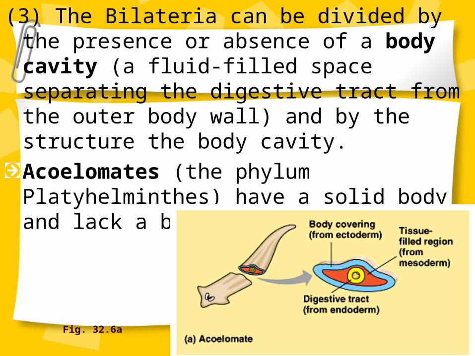
(3) The Bilateria can be divided by the presence or absence of a body cavity (a fluid-filled space separating the digestive tract from the outer body wall) and by the structure the body cavity.
Acoelomates (the phylum Platyhelminthes) have a solid body and lack a body cavity.
Fig. 32.6a

In some organisms, there is a body cavity, but it is not completely lined by mesoderm.– This is termed a pseudocoelom.– These pseudocoelomates include the rotifers
(phylum Rotifera) and the roundworms (phylum Nematoda).
Fig. 32.6b

Coelomates are organisms with a true coelom, a fluid-filled body cavity completely lined by mesoderm.– The inner and outer layers of tissue that
surround the cavity connect dorsally and ventrally to form mesenteries, which suspend the internal organs.
Fig. 32.6b

A body cavity has many functions.– Its fluid cushions the internal organs, helping to
prevent internal injury.– The noncompressible fluid of the body cavity
can function as a hydrostatic skeleton against which muscles can work.
– The present of the cavity enables the internal organs to grow and move independently of the outer body wall.

(4) The coelomate phyla are divided into two grades based on differences in their development.– The mollusks, annelids, arthropods, and
several other phyla belong to the protostomes, while echinoderms, chordates, and some other phyla belong to the deuterostomes.
– These differences center on cleavage pattern, coelom formation, and blastopore fate.

Fig. 32.7

Many protostomes undergo spiral cleavage, in which planes of cell division are diagonal to the vertical axis of the embryo.– Some protostomes also show determinate
cleavage where the fate of each embryonic cell is determined early in development.
The zygotes of many deuterostomes undergo radial cleavage in which the cleavage planes are parallel or perpendicular to the vertical egg axis.– Most deuterostomes show indeterminate
cleavage whereby each cell in the early embryo retains the capacity to develop into a complete embryo.

Coelom formation begins in the gastrula stage.– As the archenteron forms in a protostome,
solid masses of mesoderm split to form the coelomic cavities, called schizocoelous development.
– In deuterostomes, mesoderm buds off from the wall of the archenteron and hollows to become the coelomic cavities, called enterocoelous development.

The third difference centers on the fate of the blastopore, the opening of the archenteron.– In many protosomes, the blastopore develops
into the mouth and a second opening at the opposite end of the gastrula develops into the anus.
– In deuterostomes, the blastopore usually develops into the anus and the mouth is derived from the secondary opening.

STOP HERE, SKIP RESTThis phylogenetic tree is bases on nucleotide sequences from the small subunit ribosomal RNA. Fig. 32.8

The name Ecdysozoa (nematodes, arthropods, and other phyla) refers to animals that secrete external skeletons (exoskeleton).– As the animal grows, it molts the old exoskeleton
and secretes a new, larger one, a process called ecdysis.
– While named for this process, the clade is actually defined mainly by molecular evidence.
Fig. 32.10

In the traditional tree, the assignment of the three lophophorate phyla is problematic.– These animals have a lophophore, a
horseshoe-shaped crown of ciliated tentacles used for feeding.
– The lophophorate phyla share some characteristics with protostomes and other features with deuterostomes.
– The molecular data place the lophophorate phyla among the phyla with the trochophore larvae, hence the name lophotrochozoans.
Fig. 32.11

– In summary, the molecular evidence recognizes two distinct clades within the protostomes and distributes the acoelomates, pseudocoelomates, and lophophorate phyla among these two clades.
Copyright © 2002 Pearson Education, Inc., publishing as Benjamin Cummings Fig. 32.12

There are three main hypotheses for what caused the diversification of animals.
(1) Ecological Causes: The emergence of predator-prey relationships led to a diversity of evolutionary adaptations, such as various kinds of protective shells and diverse modes of locomotion.
(2) Geological Causes: Atmospheric oxygen may have finally reached high enough concentrations to support more active metabolism.
“Evo-devo” may clarify our understanding of the Cambrian diversification

(3) Genetic causes: Much of the diversity in body form among animal phyla is associated with variations in the spatial and temporal expression of Hox genes within the embryo.
A reasonable hypothesis is that the diversification of animals was associated with the evolution of the Hox regulatory genes, which led to variation in morphology during development.– Biologists investigating “evo-devo,” the new synthesis of
evolutionary biology and developmental biology, may provide insights into the Cambrian explosion.
These three hypotheses are not mutually exclusive.