chapter 3 - skin biothermomechanics: modeling and …bebc.xjtu.edu.cn/paper file/22.pdf ·...
TRANSCRIPT

Skin Biothermomechanics: Modeling
and Experimental Characterization
F. XUa and T. J. LUb
aDepartment of Engineering, Cambridge University, Cambridge CB2 1PZ,United Kingdom
bMOE Key Laboratory for Strength and Vibration, School of Aerospace, Xi’an Jiaotong University,Xi’an 710049, People’s Republic of China
Abstract. . . . . . . . . . . . . . . . . . . . . . . . . . . . . . . . . . . . . . . . . . . . . . . . . . . . . . . . 148
ADVAISSN:
NCES IN APPLIED MECHANICS, VOL. 43 147 Copyright # 2009 by Els0065-2156 DOI: 10.1016/S0065-2156(09)43003-5 All rights
er
1.
I ntroduction. . . . . . . . . . . . . . . . . . . . . . . . . . . . . . . . . . . . . . . . . . . . . . . . . . . . 1481
.1. S kin Biothermomechanics. . . . . . . . . . . . . . . . . . . . . . . . . . . . . 1491
.2. A ims and Structure. . . . . . . . . . . . . . . . . . . . . . . . . . . . . . . . . . 1502.
R eview of Related Studies. . . . . . . . . . . . . . . . . . . . . . . . . . . . . . . . . . . . . . . 1522
.1. S kin Structure. . . . . . . . . . . . . . . . . . . . . . . . . . . . . . . . . . . . . . 1522
.2. S kin Bioheat Transfer and Thermal Damage. . . . . . . . . . . . . . . 1552
.3. S kin Biomechanics. . . . . . . . . . . . . . . . . . . . . . . . . . . . . . . . . . 1582
.4. S kin Biothermomechanics. . . . . . . . . . . . . . . . . . . . . . . . . . . . . 1593.
M odeling of Skin Biothermomechanics and Thermal Pain. . . . . . . . . . . 1623
.1. M odeling of Skin Bioheat Transfer. . . . . . . . . . . . . . . . . . . . . . 1623
.2. M odeling of Skin Biothermomechanics. . . . . . . . . . . . . . . . . . . 1673
.3. M odeling of Skin Thermal Pain. . . . . . . . . . . . . . . . . . . . . . . . . 1764.
E xperimental Methodology. . . . . . . . . . . . . . . . . . . . . . . . . . . . . . . . . . . . . . . 1804
.1. E xperimental Characterization of SkinMechanical Properties. . . . . . . . . . . . . . . . . . . . . . . . . . . . . . . .
1814
.2. E xperimental Characterization of SkinThermal Denaturation. . . . . . . . . . . . . . . . . . . . . . . . . . . . . . . .
1824
.3. S ample Preparation Techniques. . . . . . . . . . . . . . . . . . . . . . . . . 1824
.4. D ifferential Scanning Calorimetry Tests. . . . . . . . . . . . . . . . . . . 1864
.5. H ydrothermal Tensile Tests. . . . . . . . . . . . . . . . . . . . . . . . . . . . 1874
.6. H ydrothermal Compressive Tests. . . . . . . . . . . . . . . . . . . . . . . . 1944
.7. D ynamic Mechanical Analysis. . . . . . . . . . . . . . . . . . . . . . . . . . 1955.
B iothermomechanical Behavior of Skin Tissue. . . . . . . . . . . . . . . . . . . . . 1965
.1. T hermal Denaturation of Collagen in Skin Tissue. . . . . . . . . . . 1975
.2. T ensile Behavior of Skin Tissue. . . . . . . . . . . . . . . . . . . . . . . . . 2005
.3. C ompressive Behavior of Skin Tissue. . . . . . . . . . . . . . . . . . . . 2045
.4. R elaxation Behavior of Skin Tissue. . . . . . . . . . . . . . . . . . . . . . 2145
.5. D ynamic Viscoelasticity of Skin Tissue. . . . . . . . . . . . . . . . . . . 227vier Inc.eserved.

148 F. Xu and T. J. Lu
6.
C onclusions. . . . . . . . . . . . . . . . . . . . . . . . . . . . . . . . . . . . . . . . . . . . . . . . . . . . 2296
.1. S ummary. . . . . . . . . . . . . . . . . . . . . . . . . . . . . . . . . . . . . . . . . . 2316
.2. L imitations. . . . . . . . . . . . . . . . . . . . . . . . . . . . . . . . . . . . . . . . 2316
.3. F uture Work. . . . . . . . . . . . . . . . . . . . . . . . . . . . . . . . . . . . . . . 233Ackn
owledgments. . . . . . . . . . . . . . . . . . . . . . . . . . . . . . . . . . . . . . . . . . . . . . . . . . 234Refer
ences. . . . . . . . . . . . . . . . . . . . . . . . . . . . . . . . . . . . . . . . . . . . . . . . . . . . . . . . . 234Abstract
The development and widespread use of thermal therapies for skin diseases and injuries
with advances in laser, microwave, and similar technologies have not been built upon the
detailed understanding of the biothermomechanical–neurophysiological behavior of skin
tissue. The emerging studies on skin biothermomechanics are therefore important for it
attempts to understand macroscale tissue response to heat-induced microstructural transfor-
mations.Skinbiothermomechanics ishighly interdisciplinary, involvingbioheat transfer, burn
damage, biomechanics, and physiology. This chapter presents firstly the various theoretical
approaches for determining the thermal, thermomechanical, and thermal pain response in skin
tissue inducedby transient heating.Given the complicatedmicrostructure of skin tissue and its
relatively lengthy thermal relaxation time, both Fourier and non-Fourier bioheat transfer
models are employed. While insightful, the predictive capability of the current theoretical
modeling is, nonetheless, limited by the comparatively few relative experimental studies on
temperature-dependent properties of the skin tissue. To better understand the variation in skin
propertieswith temperature and the corresponding collagen denaturation, focus is then placed
upon the experimental characterization of the thermomechanical behavior of skin tissue.
Uniaxial andbiaxialhydrothermal tensile testing systemsaswellashydrothermal compressive
testing systemare purposelydesigned and builtwhich, togetherwith a commercially available
dynamicmechanical analyzer, are employed toobtain suitabledata toquantify the influenceof
temperature and the corresponding thermal damage on the mechanical performance of skin
tissue, including tensile, compressive, and viscoelastic behaviors.
1. Introduction
As the protecting interface between the outside environment and the inside
body, skin plays a variety of important roles including thermoregulation, sensa-
tion, and host defense. Among these roles, thermoregulation is of particular
significance: acting as a generator, absorber, transmitter, radiator, conductor,
and vaporizer of heat, skin serves an important barrier for the human body to
various outside conditions. However, the skin fails to protect the body when the
temperature moves out of the normal physiological range, as uncomfortable
feeling, or even pain is often induced in extreme hot or cold environment.
On the other hand, advances in laser, microwave and similar technologies in

Skin Biothermomechanics 149
medicine have led to recent developments of thermal treatments for disease and
injury, involving skin tissue. In spite of the widespread use of thermal therapies in
dermatology, they do not draw upon the detailed understanding of the
biothermomechanical–neurophysiological behavior, for none exists to date,
even though each behavioral facet is somewhat established and understood.
In view of this dilemma, a new research area—skin biothermomechanics and
thermal pain—has recently emerged, which is the subject of this study.
1.1. Skin Biothermomechanics
1.1.1. Schematic of Skin Biothermomechanics and Thermal Pain
The study of skin biothermomechanics and thermal pain is a highly interdisci-
plinary area involving engineering (e.g., heat transfer and mechanics), biology, and
neurophysiology, as schematically shown in Fig. 1.1. The skin is characterized by its
biological structure and state, such as its constituent components, blood flow,
metabolism, and so on, and properties, such as thermal, mechanical, optical, and
dielectric properties. When thermal loading (either as contact heating, electromag-
netic energy or acoustic energy) and/or mechanical loading (either as force or
deformation) are applied to skin tissue, then there exist different skin states, includ-
ing temperature, thermal damage/inflammation, and stress/strain distributions.
These states then decide the level of pain sensation through the neural system. A
better understandingof skin properties, skin bioheat transfer and the thermal damage
kinetics, skin biomechanics, skin biothermomechanics and pain sensation, promise
to contribute to the continuing advancement of study of thermal pain in general.
1.1.2. Importance of Skin Thermomechanics
With advances in laser, microwave, radiofrequency, and similar technologies, a
variety of thermal methods have been developed and applied to the treatment of
disease/injury involving skin tissue, such as the removal of port-wine stains
(Asahina et al., 2006; Kono et al., 2006; Shafirstein et al., 2003), pigmented and
cutaneous lesions (Hamilton, 2004;Kauvar, Rosen,&Khrom, 2006; Pustovalov&
Jean, 2007) and tattoos (Diette, Bronstein, & Parrish, 1985). These thermal
treatment methods normally involve either raising or lowering the temperature
in a precise area of skin tissue, in order to kill or thermally denaturize the necrotic
cells; the precise monitoring of the spatial and temporal distribution of tempera-
ture, damage, and stress in the tissue is therefore required. Meanwhile, the remain-
ing healthy tissue is kept at a safe temperature level by adopting selective cooling
techniques on the skin surface during the treatment. In spite of these important and
widely used medical applications, an understanding of the responsible
thermomechanical–neurophysiological mechanism remains limited, which can

Nociceptor (ion channel)- Voltage gated- Heat gated- Proton gated- Mechanosensitive
Action potential
Pain level
Temperature fieldThermal denaturationThermal damageInflammation
Stress field- Thermal stress
- Thermal shrinkage- Mechanical strain
- Mechanical stressStrain field
- Thermal- Optical- Dielectric- Mechanical
Hea
t tr
ansf
erM
ech
anic
s
Normal heatElectromagnetic
- Laser- Microwave- Radiofrequency
ElectricalMagneticAcoustic
Thermal loading
ForceDeformation
Mechanical loading - Component- Blood flow- Metabolism- Sweat- Hair
Structure
PropertiesTransduction
Transmission
Modulationand perception
Loading Description State Pain sensation
Engineering Biology Neurophysiology
FIG. 1.1 Schematic of skin biothermomechanics and thermal pain.
150 F. Xu and T. J. Lu
restrict further refinement and innovation. In addition, a noxious thermal stimulus,
hot or cold, applied to human skin, is one of the three main causes of pain.1
Thermally induced damage plays an important role in causing thermal pain, for
example, burn damage has been found to induce severe pain that is difficult to
manage (Choiniere, Melzack, Girard, Rondeau, & Paquin, 1990; Perry, 1984) and
contribute to sensory problems (Gallagher, Rae, & Kinsella, 2000; van Loey &
van Son, 2003), all of which commonly reduce quality of life after burn damage.
Therefore, the research on skin biothermomechanics and related pain sensation is
important and contributes to medical applications for it attempts to understand the
macroscale tissue response to heat-induced microstructural transformations.
Besides biomedical applications, space and military missions can also benefit
from the proposed study. Extreme environments encountered in space travel and in
somemilitary activitiesmake it necessary to provide astronauts andmilitary person-
nel with sophisticated garments for thermal protection. Challenges are also posed by
the need to understand possible thermal effects on military personnel exposed to
irradiation.
1.2. Aims and Structure
The specific objectives of the study on skin biothermomechanics are to
theoretically model and to experimentally investigate the biothermomechanical
behavior of skin tissue under different thermomechanical loadings which are then
combined to provide a predictive framework for treating diseased tissue. In one
1 The other two are mechanically and chemically caused pain.

Skin Biothermomechanics 151
of our previous studies (Xu, Seffen, & Lu, 2008), we have described the devel-
opment of two types of skin bioheat transfer models, based on Fourier and non-
Fourier theories, which provide a comprehensive description of the thermal
behavior in skin tissue. A theoretical analysis is carried out and closed-form
solutions for a simple one-layer Fourier bioheat transfer model of skin tissue are
obtained. In view of the lengthy thermal relaxation time of skin tissue,
non-Fourier bioheat transfer models have been developed. In another study
(Xu, Wen, Seffen, & Lu, 2008), we have developed a scheme for characterizing
the skin biothermomechanical behavior to examine the heat transfer process,
thermal damage and the heat-induced mechanical response. A general closed-
form solution for thermal stress for a multilayer skin model is derived, while a
numerical method (finite element method) is employed for more complicated
cases. Two types of case studies are then performed with Fourier and non-Fourier
models: the first type of case studies is explored to understand the working
mechanism of the clinically applied thermal therapies and to quantify the differ-
ences among these thermal therapies; the second type of case studies is explored to
investigate the relationship between the thermal relaxation time and the thermo-
mechanical response of skin tissue. In a third study (Xu, Lu, & Seffen, 2008), we
have developed a holistic mathematical model for quantifying skin thermal pain to
build up a direct correlation between the parameters of thermal stimulation and the
level of the corresponding thermal pain sensation, by using the thermomechanical
models developed in Xu, Seffen, et al. (2008) and Xu, Wen, Seffen, et al. (2008)
and by considering the current biophysical and neurophysiological mechanisms of
pain sensation. In the first part of this review, the mathematical approaches
introduced in all the studies described above are presented.
While insightful, the present theoretical modeling has some limitations, where
the main deficiency is that the skin tissue is assumed to have constant properties
due to the comparatively few relative experimental studies. More experiments are
thus needed to better understand the variation in properties with temperature and
the corresponding collagen denaturation, so that these properties can be reliably
used in future, more sophisticated models. However, it is technically very difficult
to measure experimentally the biothermomechanical behavior of skin tissue in
physiological conditions. As a substitute, analytical and numerical simulations are
used, where the quantification of the temperature-dependent skin mechanical
properties is an essential step toward building reliable computer simulations. So
far, the mechanical properties alone of skin tissue have been studied experimen-
tally, both in vivo and in vitro. But, most of these studies are performed under
normal physiological temperature and little has been done on the characterization
of the effect of temperature and corresponding thermal damage. These are
addressed in the second part of this study, which specially aims to design and

152 F. Xu and T. J. Lu
build experimental systems and to obtain suitable data to quantify the influence of
the temperature and the corresponding thermal damage on the mechanical
behavior of skin tissue, including tensile, compressive, and viscoelastic behaviors.
This chapter is outlined as follows. Section 2 reviews the literature pertinent to
the current research, which is composed of four parts, including skin structure, skin
bioheat transfer and thermal damage, skin biomechanics, and skin biothermome-
chanics. This underpins the theoretical and experimental frameworks for investi-
gating the thermomechanical behavior of skin tissue in this review. Section 3
summarizes the modeling efforts on skin biothermomechanics and thermal pain
sensation. Section 4 provides detailed information on the preparation technique of
skin samples, the design of two novel experimental systems and measurement
techniques, and two commercial items of equipment employed in the current
experimental study: differential scanning calorimetry (DSC) and dynamic
mechanical analyzer (DMA). Section 5 presents results and corresponding discus-
sions from the experimental characterization of skin biothermomechanics, using
the experimental apparatus described in Section 4. The tensile, compressive, and
viscoelastic behaviors of skin tissue under different temperatures are examined
and the effects of temperature and corresponding collagen denaturation on the
mechanical properties of skin tissue are characterized. The main conclusions are
given in Section 6, where limitations of current theoretical and experimental
methodology are also discussed and future work is proposed.
2. Review of Related Studies
The study of skin biothermomechanics is an interdisciplinary problem, involv-
ing heat transfer, mechanics, and biology. Accordingly, this section briefly
reviews the literature pertinent to the current research, which is composed of
four parts: skin structure, skin bioheat transfer and thermal damage, skin biome-
chanics, and skin biothermomechanics.
2.1. Skin Structure
Skin tissue makes up approximately 14–16% of the human adult body weight
and plays a variety of important roles. It generally consists of three layers: the
epidermis, the dermis, and the subcutaneous tissue, as shown in Fig. 2.1A and B.
The thickness of these layers varies, depending on the bodily location. The
dermis makes up the bulk of the human skin tissue and its components are
introduced below.

Hair shaftA
Sweat gland Nerve Receptors
Oil gland
Hair follicle
Hair root
Artery
Vein
Smooth muscle
Adipose tissue
Free nerveendings
Small bloodvessels
Epidermis
Dermis
Hypodermis
B
Basementmembrane
Collagenfibril
Hyaluronan
GAG chains
Decorin ind and e bands
PG monomer
Link protein
Fibril-associatedcollagens(FACITS)
Coated withtype III collagen
Mixture oftypes I and V collagen
Epidermis
Dermis
Anchoring fibrils(collagen type VII)
FIG. 2.1 (A) Structure of human skin tissue, see Whitton and Everall (1973). (B) Macromolec-
ular components of skin tissue, see Silver, Siperko, and Seehra (2003).
Skin Biothermomechanics 153

MoleculeCrosslinkedend domain
Crosslinked
Fibril
Hydrogen-bonded helical domain285 nm × 1.5 nm
4–8 nm10–500 nm
1–500 mmdiameter
Tissue
Microfibril
Undulatedfiber
FIG. 2.2 Molecular/fibrillar configuration of type I collagen, see Wright and Humphrey (2002).
154 F. Xu and T. J. Lu
2.1.1. Collagen in Dermis
Collagen is the major dermal constituent and accounts for approximately 60–
80% of the dry weight of fat-free skin tissue and 18–30% of the volume of dermis
(Ebling, Eady, & Leigh, 1992; Reihsner, Balogh, & Menzel, 1995). The collagen
in human dermis is mainly the periodically banded, interstitial collagen (types I,
III, and IV),2 where about 80–90% is type I collagen and 8–12% is type III
collagen. Three polypeptide chains make up the type I collagen molecule and are
stabilized in a triple-helix arrangement by intramolecular crosslinks, as shown in
Fig. 2.2. These molecules are, in turn, aggregated into a parallel pattern to form
2 Collagen occurs in many places throughout the body and there are 28 types of collagen reported in
literature (Myllyharju & Kivirikko, 2004).

Skin Biothermomechanics 155
collagen fibrils, which are maintained by intermolecular crosslinks and provide
the tissue with its tensile properties. Type IV collagen codistributes and assem-
bles into fibrils with types I and III collagen in which it assists in regulating fibril
diameter. The wavy and unaligned collagen fiber bundles form an irregular planar
network, which allows considerable deformation in all directions without
requiring elongation of the individual fibers and provides both tensile strength
and elasticity (Finlay, 1969; Wegst & Ashbyy, 2004).
2.1.2. Elastin in Dermis
Elastin is a minor structural component of the dermis structure, accounting for
about 4% of the dermis dry weight and 1% of the volume of dermis (Ebling et al.,
1992; Hult & Goltz, 1965). Elastin fibers are considerably thinner and more
convoluted than collagen fibers. The base unit of elastin is a long protein chain
that is crosslinked by lysine molecules, and four elastin chains are joined at each
crosslink by the covalent bonding of a lysine molecule from each elastin chain.
Although direct connections between elastin and collagen fibers have not been
shown, collagen fibrils appear to wind around the elastin cores.
2.1.3. Ground Substance
The amorphous ground substance can be considered as a highly viscous,
thixotropic liquid whose fluid properties are determined by a low concentration
(0.05% wet weight of human dermis) of mucopolysaccharides, proteoglycans,
and glycoproteins (Tregear, 1966). The most important mucopolysaccharides in
ground substance is hyaluronic acid, which is analogous to the polymer mole-
cules found within rubbers. The hyaluronic acid chain has a proteoglycans side
chain, which in turn is crosslinked by glycoproteins to additional structures
within dermis, such as collagen or elastin fibrils. It is this crosslinking function
that is responsible for forming fibers from the collagen fibrils and providing the
dermis with its rubber-like behavior. Together they form a gel which does not
leak out of the dermis, even under high pressure.
2.2. Skin Bioheat Transfer and Thermal Damage
2.2.1. Skin Bioheat Transfer
The thermal behavior of skin tissue, or heat transfer in skin tissue, is a heat
conduction process coupled with complicated physiological processes, including
blood circulation, sweating, metabolic heat generation, and, sometimes, heat
dissipation via hair or fur above the skin surface. The thermal properties of
skin tissue vary among different layers; even within the same layer, there exists
large nonhomogeneity and anisotropy due to the presence of blood vessels and

156 F. Xu and T. J. Lu
structural anisotropy. Both the physiological processes and thermal properties of
skin tissue are influenced by a variety of factors such as temperature, damage,
pressure, age, and so on. To complicate matters, skin is an active, self-regulating
system: heat transfer through the skin tissue dramatically affects the state of skin
tissue, which can lead to the redistribution of skin blood flow over the cutaneous
vascular network, thereby influencing the thermal response of the tissue.
Since the appearance of Pennes bioheat equation3 in 1947 (Pennes, 1948), a
variety of models on the heat transfer in different tissues of human body have
been proposed, where the body tissue may be represented as a homogeneous
continuum material with an embedded hierarchical vascular network
(Lubashevsky & Gafiychuk, 2004). Based on how the influence of blood flow
in the vascular network is considered, these models can be classified into four
categories (Arkin, Xu, & Holmes, 1994; Charny, 1992; Crezee, Mooibroek,
Lagendijk, & van Leeuwen, 1994; Khaled & Vafai, 2003; Stanczyk & Telega,
2002): continuum models, vascular models, hybrid models, and models based on
porous media theory. Since the effect of blood vessels on heat transfer is strongly
related to their sizes (Abramson, 1967; Chato, 1980; Lemons, Chien, Crawshaw,
Weinbaum, & Jiji, 1987; Weinbaum, Jiji, & Lemons, 1984), a thermal equilibra-
tion length of blood vessels, Leq, is defined as the length at which the difference
between the blood and the tissue temperatures decreases to 1/e of the initial value
(e ¼ exp 1 ¼ 2.718). The ratio of Leq to the actual vessel length demonstrates the
distinction of thermal significance, e¼ Leq/ L, where if e� 1, that is, Leq is much
shorter than the characteristic length of the blood vessel length, L, the blood exits
the vessel at, essentially, the tissue temperature; if e �1, the blood temperature
does not change and leaves the tissue at the same inflow temperature (Chato,
1980; Chen & Holmes, 1980). It was found that, for skin tissue, Leq lies in the
range of 3� 10�5–2� 10�4 mm while L� 1.2 mm (Crezee & Lagendijk, 1992),
therefore, the blood in the skin tissue exits the vessel at essentially the tissue
temperature. Thus, many researchers use the Pennes equation to describe skin
heat transfer because it is simple and can be solved analytically and can be
programmed into finite difference and finite element models. The equation is
taken directly from Shih, Kou, Liauh, and Lin (2005) as
3 Pennes measured the radial temperature distribution in the forearm, from which he developed a
bioheat transfer model in basic terms of the local rate of tissue heat production and volume flow of
blood (Pennes, 1948). In the Pennes model, the microscopic thermal energy balance for perfused
tissue is linear, which enables analytical solutions of the heat conduction equation by various methods
commonly used. Consequently, it has been adopted by many authors to develop mathematical models
of heat transfer in different human tissues (Wissler, 1998).

Skin Biothermomechanics 157
rc@T
@t¼ kr2T þ$brbcbðTa � TÞ þ qmet þ qext; ð2:1Þ
where r, c, and k are the density, specific heat, and thermal conductivity of skin
tissue, respectively; rb and cb are the density and specific heat of blood;$b is the
blood perfusion rate; Ta and T are the temperatures of blood and skin tissue,
respectively; qmet is the metabolic heat generation in the skin tissue and qext is theheat generation due to external heating sources.
2.2.2. Skin Thermal Damage
When the skin temperature rises above a critical value (�43 �C), thermal
damage will be induced. Presently, the Arrhenius burn integration, proposed by
Henriques and Moritz (1947) and Moritz and Henriques (1947), is widely used
for quantifying thermal damage. They assert that skin damage can be represented
as a chemical rate process, which is calculated by using a first-order Arrhenius
rate equation, whereby damage is related to the rate of protein denaturation, k:
kðTÞ ¼ dO=dt ¼ A expð�Ea=RTÞ ð2:2Þ
or, equivalently:
O ¼ðt0
A expð�Ea=RTÞdt; ð2:3Þ
where A is a material parameter equivalent to a frequency factor,4 Ea is the
activation energy, and R ¼ 8.314 J/mol K is the universal gas constant. Equation
(2.2) indicates that a reaction proceeds faster with larger values of T or A for the
same Ea, or with smaller values of Ea for the sameA. The constants A and Ea are
obtained experimentally as will be discussed in some detail in Section 5.1. It should
be noted here that the Arrhenius equation is a simple empirical, but remarkably
accurate, formula for the temperature dependence of the rate of a chemical reaction
(Connors, 1990).
Many researchers have proposed other similar models. The main differences
between these models are the coefficients used in the burn damage integral,
arising from the different experimental databases used to define the models and
the different emphasis when analyzing the burn process. The available Arrhenius
parameters (A, Ea) used to calculate thermal damage for skin tissue from the
literature has been reviewed in our previous study (Xu, Wen, Seffen, et al., 2008)
4 The units of A are identical to those of the rate constant and vary depending on the order of the
reaction. If the reaction is first order, it has the unit of s�1 and for that reason it is often called the
frequency factor.

158 F. Xu and T. J. Lu
and is fitted with the method used by Wright (2003). The results clearly suggest a
linear relationship, after a least-squares fit, between the Arrhenius parameters for
skin tissue, given by:
Ea ¼ 21149:324þ 2688:367 ln A ð2:4Þ
2.3. Skin Biomechanics
The mechanical properties of the skin tissue have been experimentally studied
both in vivo and in vitro in relation to clinical and cosmetic applications, since the
first work of Langer in 1861. In vivo measurements are affected by both the skin
tissue itself and other structures to which it is attached, leading to a nonuniform
strain field in the sample; for better control of the experiment, in vitro tests are
often used. The mechanical behavior of skin tissue is found to be heterogeneous,
anisotropic, nonlinear, and viscoelastic in vivo because of its highly nonhomoge-
neous structure and composition. It is affected by many factors such as age,
gender, site, hydration, and so on. Furthermore, the classical constitutive models
for engineering materials are not suitable to describe the complicated mechanical
behavior of skin tissue.
2.3.1. Skin Behavior Under Stretch
The tensile behavior of skin tissue has been studied widely, as discussed by
several good reviews (Edwards & Marks, 1995; Larrabee, 1986; Pierard, 1999;
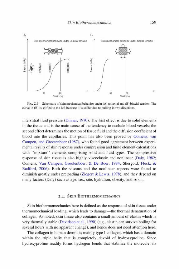
Vogel, 1994). Typical stress–strain relationships of skin tissue under uniaxial and
biaxial tension are shown in Fig. 2.3A and B, respectively. Three regimes of
behavior can be observed: at low modulus portion, there is a gradual
straightening of an increasing fraction of the wavy collagen fibers and stretching
of elastic fibers; a linear region at higher modulus where there is stretching and
slippage of collagen molecules within crosslinked collagen fibers and collagen
fibril slippage; the final softening region in which defibrillation of the collagen
fibrils results in a loss of fibrillar structure. Compared with uniaxial stretching,
the stress–strain curve for biaxial stretching is shifted to the left for there is a
bidirectional stretch of the collagen fibers, as shown in Fig. 2.3B.
2.3.2. Skin Behavior Under Compression
In vivo, skin tissue is regularly compressed by contact with chairs and shoes,
and even in the sockets of prosthetic limbs. Although there is abundant experi-
mental data for the tensile behavior of skin tissue, the compressive performance
has been rarely studied. Compression of skin tissue is resisted by solid tissue and
Skin mechanical behavior under uniaxial tension Skin mechanical behavior under biaxial tension
Str
ess
(MP
a)
Str
ess
(MP
a)
Strain(%)
30
A B
60
Strain(%)
30 60
FIG. 2.3 Schematic of skin mechanical behavior under (A) uniaxial and (B) biaxial tension. The
curve in (B) is shifted to the left because it is stiffer due to pulling in two directions.
Skin Biothermomechanics 159
interstitial fluid pressure (Dinnar, 1970). The first effect is due to solid elements
in the tissue and is the main cause of the tendency to occlude blood vessels; the
second effect determines the motion of tissue fluid and the diffusion coefficient of
blood into the capillaries. This point has also been proved by Oomens, van
Campen, and Grootenboer (1987), who found good agreement between experi-
mental results of skin response under compression and finite element calculations
with ‘‘mixture’’ elements comprising solid and fluid types. The compressive
response of skin tissue is also highly viscoelastic and nonlinear (Daly, 1982;
Oomens, Van Campen, Grootenboer, & De Boer, 1984; Shergold, Fleck, &
Radford, 2006). Both the viscous and the nonlinear aspects were found to
diminish greatly under preloading (Ziegert & Lewis, 1978), and they depend on
many factors (Daly) such as age, sex, site, hydration, obesity, and so on.
2.4. Skin Biothermomechanics
Skin biothermomechanics here is defined as the response of skin tissue under
thermomechanical loading, which leads to damage—the thermal denaturation of
collagen. As noted, skin tissue also contains a small amount of elastin which is
very thermally stable (Davidson et al., 1990) (e.g., elastin can survive boiling for
several hours with no apparent change), and hence does not need attention here.
The collagen in human dermis is mainly type I collagen, which has a domain
within the triple helix that is completely devoid of hydroxyproline. Since
hydroxyproline readily forms hydrogen bonds that stabilize the molecule, its

160 F. Xu and T. J. Lu
absence makes this domain particularly susceptible to thermal damage (Miles &
Bailey, 2001). There are two levels of organization where breakdown is thermo-
dynamically significant (Young, 1998): one is the collagen molecule itself, in
which three peptide chains are twisted around each other to form a helical,
rod-shaped molecule; the other is the semicrystalline fibril in which collagen
molecules are assembled side by side in a staggered manner with the long axis of
each molecule aligned with the axial orientation of the fibril. When collagen is
heated, the heat-labile intramolecular crosslinks are broken, as shown in Fig. 2.4,
and the collagen undergoes a transition from a highly organized crystalline
structure to a random, gel-like state, which is the denaturation process (Flory &
Garrett, 1958). Collagen shrinkage occurs through the cumulative effect of the
unwinding of the triple helix, due to the destruction of the heat-labile intramolec-
ular crosslinks, and the residual tension of the heat-stable intermolecular
crosslinks (Allain, Le Lous, Cohen-Solal, Bazin, & Maroteaux, 1980;
Arnoczky & Aksan, 2000; Flory & Garrett, 1958).
The effects of heating on collagen can be reversible or irreversible
(Hormann & Schlebusch, 1971; Stoop et al., 1999) and the precise heat-induced
behavior of collagenous tissue and shrinkage depend on several factors, including
the collagen content (Chvapil & Jensovsky, 1963), the maximum temperature
reached and exposure time (Allain et al., 1980), the mechanical stress applied to
the tissue during heating (Chen, Wright, & Humphrey, 1998a), and aging
(Chvapil & Jensovsky, 1963; Le Lous, Cohen-Solal, Allain, Bonaventure, &
Maroteaux, 1985).
Tropocollagenmolecule
Tropocollagenmolecule
a
a aa
aa a a
aaa
a
bb b b b
b bb b
b
bbb
b
Thermal shrinkage
a 1a 2
a 3a 1
a 2
a 3
a: Intramolecular crosslinksb: Intermolecular crosslinks
FIG. 2.4 Schematic of thermal denaturation of collagen, see Arnoczky and Aksan (2000).

Skin Biothermomechanics 161
Different metrics have been used to characterize the thermal denaturation and
heat-induced damage of collagen and collagenous tissues, including biological
metrics such as enzyme deactivation (Bhowmick & Bischof, 1998) and extrava-
sation of fluorescent-tagged plasma proteins (Green & Diller, 1978), thermal
metrics such as changes in enthalpy (Jacques, 2006; Miles, 1993), mechanical
metrics such as thermal shrinkage (Chen et al., 1998a,b; Kondo et al., 2005; Lin
et al., 2006), and optical metrics such as thermally induced loss of birefringence
(de Boer, Srinivas, Malekafzali, Chen, & Nelson, 1998; Pearce, Thomsen,
Vijverberg, & McMurray, 1993; Srinivas et al., 2004; Thomsen, 1991). Although
the shrinkage of collagen due to thermal denaturation has been widely used and
has been suggested as a convenient continuum metric of thermal damage (Diller
& Pearce, 1999; Fung, 1990; Wells, Harris, & Humphrey, 2004) pointed out that
shrinkage may not be a universal metric to measure thermal damage. Rather,
there is a need to identify an independent metric by which one can determine the
extent of thermal damage (Baek, Wells, Rajagopal, & Humphrey, 2005).
Diller and Pearce (1999) point out that O can be calculated as the logarithm of
the relative concentration of ‘‘reactants,’’ or undenatured collagen, in the
collagen denaturation process, where O can be considered as
OðtÞ ¼ lnCð0ÞCðtÞ� �
; ð2:5Þ
where C(0) and C(t) are the initial concentration and the concentration remaining
at time t of undenatured collagen, respectively. The degree of thermal denatur-
ation, defined as the fraction of denatured collagen, and denoted by Deg, can be
calculated by
DegðtÞ ¼ Cð0Þ � CðtÞCð0Þ ¼ 1� exp½�OðtÞ�: ð2:6Þ
Denaturation of collagen occurs as the temperature of the tissue increases. As
well as structural changes, the hydration level of collagen also changes, which
may involve an initial liberation and subsequent absorption of water (Humphrey,
2003). Not surprisingly, thermal denaturation of a collagenous tissue can result in
marked changes in its thermal (Davis, Doss, Humphrey, & Wright, 2000),
mechanical (Aksan & McGrath, 2003; Chae, Aguilar, Lavernia, & Wong,
2003; Chao, Burden, & Wong, 2001; Chen & Humphrey, 1998; Chen, Wright,
& Humphrey, 1997, 1998a,b; Diaz et al., 2001), and optical properties (Agah,
Gandjbakhche, Motamedi, Nossal, & Bonner, 1996; Bosman, 1993; Jun, Harris,
Humphrey, & Rastegar, 2003; Lin, Motamedi, & Welch, 1996). For example,
increased extensibility of soft tissues (e.g., pericardium and epicardium) due to

162 F. Xu and T. J. Lu
thermal treatment has been observed in both uniaxial (Chachra, Gratzer, Pereira,
& Lee, 1996; Chen & Humphrey; Lennox, 1949) and biaxial studies (Harris,
Wells, & Humphrey, 2003; Wells et al., 2004). However, none of these studies
are for skin tissue and there are only few studies focused on the thermal denatur-
ation process of the tissue (Le Lous, Flandin, Herbage, & Allain, 1982; Le Lous
et al., 1985; McHugh et al., 1997; Melling et al., 2000; Pierce, Sheridan, Park,
Cense, & De Boer, 2004; Reihsner, Melling, Pfeiler, & Menzel, 2000), despite
skin dermis being mainly composed of collagen.
3. Modeling of Skin Biothermomechanics and Thermal Pain
3.1. Modeling of Skin Bioheat Transfer
From a therapeutic viewpoint, a heating scheme with high-energy intensity
and short duration can efficiently produce an appropriate and precise dose of heat
during thermal therapies. In addition, reducing the overall treatment time is
important, especially when the treatment target volume is large. A rapid heating
scheme with a good strategy is therefore essential for an effective thermal
therapy. However, the possible non-Fourier nature of heat transfer5 in living
tissue may play an important role during rapid heating, such as thermal ablation
when a high-intensity thermal source (e.g., focused laser, ultrasound, or radio-
frequency) is used. For example, it has been shown that the non-Fourier behavior
of tissue will delay the appearance of peak temperature during thermal treat-
ments, leading to a lower thermal dose level (Shih et al., 2005). Furthermore,
damage to human tissue from thermal agitation is an exponential function of
temperature (see Eq. (2.3)), so even small improvements in the prediction
of temperature can strongly influence the prediction of damage. Knowledge of
temperature distribution in skin tissue is also essential for the understanding
of the corresponding thermomechanical behavior.
This section aims to provide a comprehensive description of the thermal
behavior in skin tissue, and is organized as follows. First, a skin bioheat transfer
model based on classical Fourier theory is presented. A theoretical analysis is
carried out and closed-form solutions for a simple one-layer skin model are
5 In classic Fourier’s law, it is assumed that the propagation speed of thermal disturbance is infinite.
However, in particular thermal conditions or heat conduction media, the heat conduction behavior
shows a non-Fourier feature such as thermal wave phenomenon or hyperbolic heat conduction.

Skin Biothermomechanics 163
obtained using the Green’s Function method6 (Ozisik, 1993). Afterward, differ-
ent non-Fourier bioheat transfer models including thermal wave model and dual-
phase-lag (DPL) model are presented.
3.1.1. Pennes Model
As is well known, the conduction term in the traditional Pennes bioheat
transfer equation (Pennes, 1948) is based on the classical Fourier’s law, which
has been known since the publication of French mathematical physicist Joseph
Fourier’s studies concerning heat conduction:
qð r!; tÞ ¼ �krTð r!; tÞ; ð3:1Þ
where q is the heat flux vector representing heat flow per unit time, per unit area;
k is the thermal conductivity which is a positive, scalar quantity; rT is the
temperature gradient; and r! stands for the position vector. The general bioheat
transfer equation is given as
rc@T
@t¼ �rqþ$brbcbðTa � TÞ þ qmet þ qext; ð3:2Þ
Note that Eq. (3.2) is different from Eq. (2.1) in that it has a term of ‘‘�rq’’
instead of ‘‘kr2T.’’ From Eqs. (3.1) and (3.2), the Pennes bioheat transfer
equation can be derived, and is duplicated here from Eq. (2.1) as
rc@T
@t¼ kr2T þ$brbcbðTa � TÞ þ qmet þ qext: ð3:3Þ
Using the Pennes equation (3.3), one can carry out theoretical analysis to
obtain closed-form solutions for a simple one-layer Fourier model: the skin tissue
is considered as a perfect, infinitely wide/long plate of thickness, H, according to
its anatomical structure, where the Cartesian coordinates are embedded at the
center of the plate, as shown in Fig. 3.1. This is a good approximation when heat
mainly propagates in the direction perpendicular to the skin surface (e.g., in the
case of laser heating). Closed-form analytical solutions of Eq. (3.3) have been
obtained for different boundary conditions. Since the metabolic heat generation
qmet is several orders less than that of external heat generation qext (Gordon,
Roemer, & Howath, 1976), it is neglected in the derivation of the solutions
without loss of accuracy. For example, for a one-layer one-dimensional skin
6 The method uses Greens’s function to solve inhomogeneous differential equations subject to
boundary conditions.

Geometricalmidplane
Skin surface
HX
Z
FIG. 3.1 Schematic of the one-layer skin model.
164 F. Xu and T. J. Lu
model under surface contact heating, the closed-form solution of temperature at
the location z (along the depth H of skin) is given by
Tðz; tÞ ¼ T0ðzÞ þ 2aH
"T1 � k
dT0ðzÞdz
�����x¼0
#
�X1m¼1
bm sin½bmðzþHÞ� 1
ab2m þ$brbcbrc
1� exp �ab2mt�$brbcb
rct
0@
1A
0@
1A
ð3:4Þ
where T0(Z) is the initial temperature field in the tissue and bm ¼ mp/ H,m ¼ 1, 2, 3,. . ..
In view of the lengthy thermal relaxation time of skin tissue, non-Fourier
bioheat transfer models have been developed, as described below.
3.1.2. Thermal Wave Model
In many situations, heat conduction has been treated according to the classical
Fourier’s law (Eq. (3.1)), which assumes that any thermal disturbance on a body
is instantaneously felt throughout the body or, equivalently, the propagation
speed of thermal disturbance is infinite. This assumption is reasonable in the
majority of practical applications. However, in particular thermal conditions or
heat conduction media, where the thermal behavior shows a non-Fourier feature
such as thermal wave phenomenon,7 or hyperbolic heat conduction as defined
mathematically, it fails. Since the experimental observation of a finite thermal
7 Temperature jumps are observed which can be regarded as the wave front.
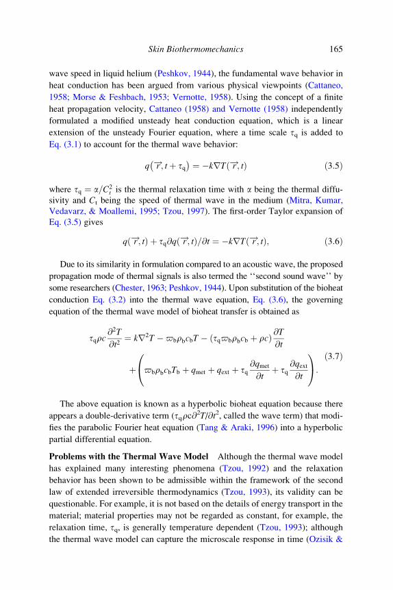
Skin Biothermomechanics 165
wave speed in liquid helium (Peshkov, 1944), the fundamental wave behavior in
heat conduction has been argued from various physical viewpoints (Cattaneo,
1958; Morse & Feshbach, 1953; Vernotte, 1958). Using the concept of a finite
heat propagation velocity, Cattaneo (1958) and Vernotte (1958) independently
formulated a modified unsteady heat conduction equation, which is a linear
extension of the unsteady Fourier equation, where a time scale tq is added to
Eq. (3.1) to account for the thermal wave behavior:
q !r ; tþ tq� � ¼ �krT !r ; tð Þ ð3:5Þ
where tq ¼ a=C2t is the thermal relaxation time with a being the thermal diffu-
sivity and Ct being the speed of thermal wave in the medium (Mitra, Kumar,
Vedavarz, & Moallemi, 1995; Tzou, 1997). The first-order Taylor expansion of
Eq. (3.5) gives
q !r ; tð Þ þ tq@q !r ; tð Þ=@t ¼ �krT !r ; tð Þ; ð3:6Þ
Due to its similarity in formulation compared to an acoustic wave, the proposed
propagation mode of thermal signals is also termed the ‘‘second sound wave’’ by
some researchers (Chester, 1963; Peshkov, 1944). Upon substitution of the bioheat
conduction Eq. (3.2) into the thermal wave equation, Eq. (3.6), the governing
equation of the thermal wave model of bioheat transfer is obtained as
tqrc@2T
@t2¼ kr2T �$brbcbT � ðtq$brbcb þ rcÞ @T
@t
þ $brbcbTb þ qmet þ qext þ tq@qmet
@tþ tq
@qext@t
0@
1A:
ð3:7Þ
The above equation is known as a hyperbolic bioheat equation because there
appears a double-derivative term (tqrc@2T/@t2, called the wave term) that modi-
fies the parabolic Fourier heat equation (Tang & Araki, 1996) into a hyperbolic
partial differential equation.
Problems with the Thermal Wave Model Although the thermal wave model
has explained many interesting phenomena (Tzou, 1992) and the relaxation
behavior has been shown to be admissible within the framework of the second
law of extended irreversible thermodynamics (Tzou, 1993), its validity can be
questionable. For example, it is not based on the details of energy transport in the
material; material properties may not be regarded as constant, for example, the
relaxation time, tq, is generally temperature dependent (Tzou, 1993); although
the thermal wave model can capture the microscale response in time (Ozisik &

166 F. Xu and T. J. Lu
Tzou, 1994; Tzou, 1997), the wave concept does not capture the microscale
response in space (Bayazitoglu & Peterson, 1992; Tzou, Ozisik, & Chiffelle,
1994) and the thermal wave model introduces some unusual physical solutions
(Godoy & Garcıa-Colın, 1997; Koerner & Bergmann, 1998; Taitel, 1972); due
to the assumption of a macroscopic behavior averaged over many grains,
the validity of the thermal wave model becomes debatable in view of the
fast-transient response with microstructural interaction effects (Tzou, 1995a).
To account for deviations from the classical approach involving Fourier conduc-
tion and to consider the effect of microstructural interactions in the fast-transient
process of heat transport—an effect absent in the thermal wave model—the DPL
model has been employed to study bioheat transfer in skin tissue, as described below.
3.1.3. Dual-Phase-Lag Model
In the DPL model, a phase lag for temperature gradient, tT, is introduced
(Ozisik & Tzou, 1994; Tzou, 1995a, 1997). Together with tq, the correspondinggoverning equation of heat transfer is called the DPL equation and is stated as
q !r ; tþ tq� � ¼ �krT !r ; tþ tTð Þ; ð3:8Þ
where tq and tT can be interpreted as periods arising from ‘‘thermal inertia’’ and
‘‘microstructural interaction,’’ respectively (Tzou, 1995b). Specifically, tq is thephase lag in establishing the heat flux and associated conduction through a
medium, while tT accounts for the diffusion of heat ahead of sharp wave front
that would be induced by tq, and is the phase lag in establishing the temperature
gradient across the medium during which conduction occurs through its small-
scale structures. Through the first and second-order Taylor expansions, the DPL
model can be developed into several pertinent models. For example, the simplest
example of the DPL model is its first-order expansions for both q and T, given as
q !r ; tð Þ þ tq@q !r ; tð Þ
@t¼ �k rT !r ; tð Þ þ tT
@rT !r ; tð Þ@t
� �: ð3:9Þ
Substituting the Eq. (3.2) into this equation, one obtains the so-called type 1
DPL model of bioheat transfer:
tqrc@T2
@t2¼ kr2T þ tTkr2 @T
@t�$brbcbT � tq$brbcb þ rc
� � @T@t
þ $brbcbTa þ qmet þ qext þ tq@qmet
@tþ tq
@qext@t
0@
1A:
ð3:10Þ

Skin Biothermomechanics 167
An in-depth description of the thermal behavior of skin tissue has thus far
been presented where different heat transfer models for skin tissue have
been developed based on both Fourier’ and non-Fourier’ theories. Associated
issues of thermal stresses and thermal damage are addressed in the following
section. The aim is to develop a computational approach to examine the heat
transfer process, thermal damage and the heat-induced mechanical response, so
that the underlying mechanism of clinically applied thermal therapies can be
understood and that the differences among these thermal therapies can be
quantified.
3.2. Modeling of Skin Biothermomechanics
Based on the above study on heat transfer, we present in this section the
scheme for characterizing the skin biothermomechanical behavior to examine the
heat transfer process, thermal damage, and the heat-induced mechanical
response. A general closed-form solution for thermal stress for a multilayer
skin model is derived, while numerical method (finite element method) is
employed for more complicated cases. Two types of case studies are then
performed with Fourier and non-Fourier models.
3.2.1. Model Development
As previously discussed, skin tissue has a complicated multilayer structure
(see Fig. 2.1). The thermal properties of different layers have the same order of
magnitude (Table 3.1), but the mechanical properties vary greatly by up to three
orders of magnitude from one layer to another. A one-layer continuum model for
heat transfer may be assumed; while, to obtain the distribution of heat-induced
stresses, the skin tissue must be treated as a laminated composite structure, with
each layer assumed to be uniform with linear, orthotropic thermoelastic proper-
ties, as shown in Fig. 3.2. The thermomechanical behavior of skin tissue is
simplified to be a ‘‘sequentially coupled’’ problem; in other words, the mechani-
cal behavior has no influence on the thermal behavior. The temperature field in
skin tissue obtained from solving the governing equations of bioheat transfer is
used as the input of the thermomechanical model, from which the corresponding
thermal stress field is obtained.
In multilayer models, skin is regarded as a multilayer structure, each
having a thickness, tk, and material properties of Young’s modulus, Ek, and
Poisson’s ratio, nk, where k ¼ 1, 2, . . . N with N being total number of layers.

TABLE 3.1Thermophysical properties of skin tissue for three-layer model.
Parameters Value References
Thermal expansion
coefficient (�10�4 K�1)
Epidermis 1 Assumptiona
Dermis 1 Assumptiona
Subcutaneous fat 1 Assumptiona
Poisson’s ratio (–) Epidermis 0.48 Delalleau et al. (2006)
Dermis 0.48 Delalleau et al. (2006)
Subcutaneous fat 0.48 Delalleau et al. (2006)
Young’s modulus (MPa) Epidermis 102 Hendriks et al. (2006)
Dermis 10.2 Hendriks et al. (2006)
Subcutaneous fat 0.0102 Hendriks et al. (2006)
Skin density (kg/m3) Epidermis 1190.0 Duck (1990)
Dermis 1116.0 Duck (1990)
Subcutaneous fat 971.0 Duck (1990)
Skin thermal
conductivity (W/mK)
Epidermis 0.235 Elkins and Thomson (1973)
Dermis 0.445 Elkins and Thomson (1973)
Subcutaneous fat 0.185 Elkins and Thomson (1973)
Skin specific heat
(J/kg K)
Epidermis 3600.0 Henriques and Moritz (1947)
Dermis 3300.0 Henriques and Moritz (1947)
Subcutaneous fat 2700.0 Henriques and Moritz (1947)
Metabolic heat
generation (W/m3)
Epidermis 368.1 Roetzel and Xuan (1998)
Dermis 368.1 Roetzel and Xuan (1998)
Subcutaneous fat 368.3 Roetzel and Xuan (1998)
Thickness (mm) Epidermis 0.1 Sejrsen (1972)
Dermis 1.5 Dahan, Lagarde, Turlier,
Courrech, and Mordon (2004)
Subcutaneous fat 4.4 Assumption
a There is no reported data on the thermal expansion coefficient of epidermis and dermis layer of skin
tissue, but available data for stratum corneum of neonatal rat (Humphries & Wildnauer, 1971) and
human fat (Fidanza, Keys, & Anderson, 1953) show that thermal expansion coefficient of skin tissue
is on the order of 10�4 K�1.
168 F. Xu and T. J. Lu
The in-plane extensional ([A]), coupling ([B]), and bending ([D]) stiffnesses of
the overall laminate of structure are given by, respectively (Kollar & Springer,
2003)
Aij ¼XNk¼1
�Qij
� �kzk � zk�1ð Þ; Bij ¼ 1
2
XNk¼1
�Qij
� �kz2k � z2k�1
� �;
Dij ¼ 1
3
XNk¼1
�Qij
� �kz3k � z3k�1
� �;
ð3:11Þ

Geometricalmidplane
Skin surface
Layer N
Layer 1
H
−H / 2
H / 2
X
Z
FIG. 3.2 Schematic of the multilayer skin tissue.
Skin Biothermomechanics 169
where zk is the coordinate of kth layer along depth; Aij, Bij, and Dij (i, j ¼ 1, 2, 6)8
are separately assembled into the elements of 3 � 3 stiffness matrices [A], [B],and [D]; �Q
� �is the stiffness matrix of the layered skin structure, defined by
�Q� � ¼
�Q11�Q12
�Q16
�Q12�Q22
�Q26
�Q16�Q26
�Q66
264
375;
�Q11
� �k¼ �Q22
� �k¼ Ek
1� n2k; �Q16
� �k¼ �Q26
� �k¼ 0;
�Q12
� �k¼ nkEk
1� n2k; �Q66
� �k¼ Ek
2 1þ nkð Þ :
8>>>><>>>>:
ð3:12Þ
Due to the combined thermal and mechanical loading, the vector of strains,
ef gk, at a point in each layer under plane-strain conditions are
ef gk ¼ �S½ �k sf gk þ lf gkDT; ð3:13Þ
where DT is the temperature difference at a point relative to a reference tempera-
ture, lf gk is the vector of the thermal expansion coefficient, and �S½ �k is the
compliance matrix. It should be noted here that the vectors of stresses, sf gk,and strains, ef gk, refer to in-plane strain and stress. By inverting the above
equation, the vector of stresses, sf gk, can be obtained as
sf gk ¼ �Q� �
kef gk � lf gkDT
� �: ð3:14Þ
8 ij means the directions of orthotropic material in plane stress state.

170 F. Xu and T. J. Lu
In a lamina, the total strains over the whole depth generally do not vanish, but
are given as
ef gk ¼ e0 þ zfkg: ð3:15Þ
The resulting stresses are � �0
� �
sf gk ¼ �Qke þ zfkg � lf gkDT ; ð3:16Þ
where e0
and fkg are the strain and curvature at the geometrical midplane. The
resultant in-plane laminate forces per unit length, fNg, are found by integrating
through the thickness of the laminate:
fNg ¼ Ð sf gkdz¼ Ð �Q
� �k
e0 þ zfkg � lf gkDT� �
dz
¼ A½ � e0 þ B½ �fkg � NTf g;
; ð3:17Þ
where
A½ � ¼A11 A12 A16
A12 A22 A26
A16 A26 A66
264
375; B½ � ¼
B11 B12 B16
B12 B22 B26
B16 B26 B66
264
375;
and NT ¼
ð�Q� �
klf gkDTdz:
Similarly, the resultant in-plane moments per unit length are found to be
fMg ¼ Ð sf gkzdz¼ Ð �Q
� �k
e0 þ zfkg � lf gkDT� �
zdz
¼ B½ � e0 þ D½ �fkg � MTf g;
; ð3:18Þ
where
D½ � ¼D11 D12 D16
D12 D22 D26
D16 D26 D66
24
35 and MT
¼ð
�Q� �
klf gkDTdz
From Eqs. (3.17) and (3.18) and by defining
NEf g ¼ fNg þ NTf g;MEf g ¼ fMg þ MTf g;
�
it is found
fNg þ NTf g ¼ A½ � e0 þ B½ �fkg
fMg þ MTf g ¼ B½ � e0 þ D½ �fkg ) e0
k
� �¼ A
0B
0
B0
D0
( )NE
ME
� �(
) e0 ¼ A0NE þ B
0ME;
k ¼ B0NE þ D
0ME;
( ð3:19Þ

Skin Biothermomechanics 171
where
A0
h i¼ A½ � � B½ � D½ ��1 C½ � ¼
a011 a
012 0
a012 a
011 0
0 0 a066
264
375;
C0
h i¼ B
0h i
¼c011 c
012 0
c012 c
011 0
0 0 c066
264
375;
B0½ � ¼ B½ � D½ ��1 ¼b
011 b
012 0
b012 b
011 0
0 0 b066
264
375;
D0
h i¼ D½ ��1 ¼
d011 d
012 0
d012 d
011 0
0 0 d066
264
375;
A½ � ¼ A½ ��1; B½ � ¼ � A½ ��1 B½ �; C½ � ¼ B½ � A½ ��1; and D½ � ¼ D½ � � B½ � A½ ��1 B½ �:
Thus, the in-plane stresses parallel to the skin surface, sxx, in each layer
(Fig. 3.2) can be obtained as
sxxf gk ¼ �Ek
��lkDT þ a011 þ a
012
� �1þ nkð Þ
XNi¼1
ðzizi�1
�Ei�liDTdz
!"
þ b011 þ b
012
� �1þ nkð Þ
XNi¼1
ðzizi�1
�Ei�liDTzdz
!#
þ z 1þ nkð Þ b011 þ b
012
� � XNi¼1
ðzizi�1
�Ei�liDTdz
!"
þ d011 þ d
012
� �1þ nkð Þ
XNi¼1
ðzizi�1
�Ei�liDTzdz
!#
0BBBBBBBBBBBBBB@
1CCCCCCCCCCCCCCA
;
ð3:20Þ
where �E ¼ E= 1� n2ð Þ, �l ¼ ð1þ nÞl, and N is the total number of skin layers.
3.2.2. Case Study
Description of the Problem The skin is initially cooled by natural convection
by environmental air (Te ¼ 25 �C, h ¼ 7 W / m2K). At t ¼ 0, the skin surface is
suddenly taken into contact with a hot source of constant temperature 100 �C

H
Epidermis
Dermis
Hypodermis
Heat source
Bloodperfusion
Stratum corneum
s
FIG. 3.3 Idealized skin model according to the real structure of skin as shown in Fig. 2.1.
172 F. Xu and T. J. Lu
such as boiling water; after contacting for 15 s, the hot source is removed and the
skin is cooled by use of water/ice mixture of 0 �C for 30 s (the heating/cooling is
uniform along the surface). A three-layer skin model is used, which is composed
of epidermis, dermis and fat layer, as shown in Fig. 3.3. The relevant parameters
used are given in Tables 3.1 and 3.2.
Results The above problem is solved by using different bioheat transfer mod-
els: Pennes model, thermal wave model and DPL model. For the multilayer skin
model, predictions from different models are shown in Fig. 3.4 for temperature,
in Fig. 3.5 for thermal damage, and in Fig. 3.6 for thermal stress. The tempera-
ture, thermal damage, and thermal stress distributions in the skin tissue at the end
of heating (t¼ 15 s) and cooling (t¼ 45 s) are shown separately in Figs. 3.4C–D,
3.5C–D, and 3.6C–D, while Figs. 3.4A – B and 3.5A – B plot the corresponding
temperature and burn damage at the epidermis–dermis (ED) interface and
dermis–fat (DF) interface of skin as functions of time, respectively. Since the
nociceptors9 emerge from superficial dermis running into epidermis
(Patapoutian, Peier, Story, & Viswanath, 2003) as low as 50 mm from the skin
surface (Kruger, Perl, & Sedivec, 1981), attention is focused on thermal stress at
the skin surface and ED interface, as shown in Fig. 3.6A and B.
Figure 3.4A–D demonstrates that the tissue temperature calculated from
different models deviates substantially under constant surface temperature heat-
ing. With the thermal wave model, the tissue temperature inside the body is
undisturbed during the initial stage of heating, which is attributed to the non-
thermally consumption related biological activities10 (Liu, Chen, & Xu, 1999);
9 Special receptor for pain sensation.10 Activities like driving a water particle or an ion, changing electrical potentials of cell membranes,
or triggering a biochemical process.

TABLE 3.2Thermophysical properties of skin tissue for one-layer model.
Parameters Value References
Thermal expansion
coefficient (�10�4 K�1)
1 Assumption
Poisson’s ratio (–) 0.48 Delalleau, Josse, Lagarde,
Zahouani, and Bergheau (2006)
Young’s modulus (MPa) 102 Hendriks, Brokken, Oomens,
Bader, and Baaijens (2006)
Skin density (kg/m3) 1190.0 Duck (1990)
Skin thermal conductivity (W/mK) 0.235 Elkins and Thomson (1973)
Skin specific heat (J/kg K) 3600.0 Henriques and Moritz (1947)
Metabolic heat generation (W/m3) 368.1 Roetzel and Xuan (1998)
Thickness (mm) 6.0 Sejrsen (1972)
0
20
40
60
80
100PBHTE
TWMBT
DPL
T (
�C)
T (
�C)
T (
�C)
T (
�C)
End of heating
0
10
20
30
40
50
60
70
PBHTE
TWMBT
DPL
End of heating
40
50
60
70
80
90
100PBHTE
TWMBT
DPL
Epidermis Dermis Fat
0
10
20
30
40
50
PBHTE
TWMBT
DPL
Epidermis Dermis Fat
ED interface, t q= 10 s, t T= 10 s
t (s)
0 5 10 15 20 25 30 35 40 45
t (s)
0 5 10 15 20 25 30 35 40 45
DF interface, t q= 10 s, t T= 10 s
t = 15 s, t q= 10 s, t T= 10 s
z/H
0.0 0.2 0.4 0.6 0.8 1.0
z/H
0.0 0.2 0.4 0.6 0.8 1.0
t = 45 s, t q= 10 s, t T= 10 s
A B
C D
FIG. 3.4 Comparison of predictions of temperature from different models: (A) variation with
time at ED interface, (B) variation with time at DF interface, (C) distribution along skin depth at
t ¼ 157s, and (D) distribution along skin depth at t ¼ 45s (PBHTE, Pennes model; TWMBT, thermal
wave model; DPL, dual-phase-lag model).
Skin Biothermomechanics 173

102
100
10−2
10−4
10−6
PBHTE
TWMBT
DPL
ED interface, tq= 10 s, tT= 10 s
t (s)
End of heatingPBHTE
TWMBT
DPL
DF interface, tq= 10 s, tT= 10 s
End of heating
PBHTE
TWMBT
DPL
t = 15 s, tq= 10 s, tT= 10 s
z/H
Epidermis Dermis Fat PBHTE
TWMBT
DPL
t = 45 s, tq= 10 s, t
T= 10 s
Epidermis Dermis Fat
0
0.0 0.2 0.4 0.6 0.8 1.0
z/H0.0 0.2 0.4 0.6 0.8 1.0
A B
C D
5
ΩΩ
10−6
10−2
102
106
1010
1014
10−6
10−2
102
106
1010
1014
10−6
10−2
102
106
1010
1014
ΩΩ
10 15 20 25 30 35 40 45
t (s)0 5 10 15 20 25 30 35 40 45
FIG. 3.5 Comparison of predictions of thermal damage from different models for the case of
surface contact heating in non-Fourier analysis: (A) variation with time at epidermis–dermis, (ED)
interface, (B) variation with time at DF interface, (C) distribution along skin depth at t ¼ 15s, and
(D) distribution along skin depth at t ¼ 45s (PBHTE, Pennes model; TWMBT, thermal wave model;
DPL, dual-phase-lag model). Note that z/H ¼ 0.0167 and 0.267 indicate the epidermis–dermis (ED)
and dermis–fat (DF) interfaces, respectively.
174 F. Xu and T. J. Lu
followed by a instantaneous jump, which may be viewed as the wave-front
emerging from the finite propagation of the thermal wave or the existence of
the relaxation time tq. Unlike the thermal wave model, no wave behavior is
observed in the DPL model as expected, but a non-Fourier diffusion-like behav-
ior exists due to the second thermal relaxation time, tT, whose effect is to weakenthe thermal wave effect, thereby to destroy the sharp wave front. Additionally, a
sudden temperature drop for heating or a step for cooling at the skin surface is
associated with the DPL model, as shown in Fig. 3.4C and D.
The large differences among the predicted temperatures of different models
cause significant deviations in thermal damage evaluations. For example, the
thermal damage predicted by the thermal wave model at the end of heating for the

−20
−10
0
10
20
30PBHTE
TWMBT
DPL
s (M
Pa)
s (M
Pa)
s (M
Pa)
s (M
Pa)
Skin surface, tq= 10 s, tT= 10 s
End of heating
0.0
−0.5
−1.0
−1.5
0.5
1.0
1.5PBHTE
TWMBT
DPL
ED interface, tq= 10 s, tT= 10 s
End of heating
−0.10
−0.05
0.00
0.05
0.10 PBHTE
TWMBT
DPL
t = 15 s, tq= 10 s, tT= 10 s
Epidermis Dermis Fat
−0.10
−0.05
0.00
0.05
0.10PBHTE
TWMBT
DPL
Epidermis Dermis Fat
t (s)0 5 10 15 20 25 30 35 40 45
t (s)0 5 10 15 20 25 30 35 40 45
z/H0.0 0.2 0.4 0.6 0.8 1.0
z/H0.0 0.2 0.4 0.6 0.8 1.0
t = 45 s, tq= 10 s, tT= 10 s
A B
C D
FIG. 3.6 Comparison of predictions of thermal stress from different models for the case of
surface contact heating in non-Fourier analysis: (A) variation with time at skin surface, (B) variation
with time at epidermis–dermis (ED) interface, (C) distribution along skin depth at t ¼ 15s, and (D)
distribution along skin depth at t¼ 45s (PBHTE, Pennes model, TWMBT, thermal wave model; DPL,
dual-phase-lag model). Note that z/H ¼ 0.0167 and 0.267 indicate the epidermis–dermis (ED) and
dermis–fat (DF) interfaces, respectively.
Skin Biothermomechanics 175
ED interface is the largest, about three orders larger in magnitude than that
from the Pennes model, and about seven orders larger than those from the DPL
model. A wave front for thermal damage is also observed for the thermal
wave model, which appears in Fig. 3.5B as a steep profile. As for the thermal
damage distribution along the skin depth, the Pennes and the thermal wave
models give similar results, which are much larger in magnitude than the DPL
model.
The thermal stresses developed due to nonuniform temperature distributions
are confined near the skin surface, see Fig. 3.6C and D and both the skin surface
and ED interface are subjected to large tensile stresses (>10 MPa), see Fig. 3.6A

176 F. Xu and T. J. Lu
and B, during both heating and cooling processes. This implies higher thermal
damage occurring in these locations, consistent with the results of Fig. 3.5A and
B. Furthermore, the magnitude of stress at the skin surface obtained with the DPL
model is much larger than that with other models, although the same boundary
condition is applied. This is caused by the sudden temperature drop at skin
surface, see Fig. 3.4C. It should be noted here that the mean mechanical threshold
of nociceptors in the skin tissue lies in the range of about 0–0.6 MPa and mainly
between 0.1 and 0.2 MPa (James & Richard, 1996), which suggests that, in
addition to heating, thermal stress may also contribute to thermal pain. Other
supporting evidence shows that, for the same level of nociceptor activity, a heat
stimulus evokes more pain than a mechanical stimulus and that tissue deforma-
tion due to heating and cooling may explain the origins of pain (Reuck & Knight,
1966; Van Hees & Gybels, 1981).
3.3. Modeling of Skin Thermal Pain
3.3.1. Model Development
When the skin temperature rises above a critical value, an uncomfortable
feeling or pain sensation is induced. In this section, the current understanding
of the mechanisms for thermal pain and its relation to skin biothermomechanics is
described.
Pain sensation has been studied extensively for a long time over a range of
scales, from the molecular level to the entire human neural system level. Thermal
stimulation is one of the three main causes of pain, the others being mechanical
and chemical stimulations, and has been widely used in the study of pain (Arendt-
Nielsen & Chen, 2003). However, the understanding of the underlying mechan-
isms of thermal pain sensation is still far from clear due to the influence of many
factors, both physiological and psychological.
Although the utilization of computational models in the field of pain has
been very limited and attempts at modeling pain have generally focused on
acute pain,11 there are strong arguments for the mathematical modeling of pain
(Britton & Skevington, 1996; Picton, Campbell, & Turner, 2001): it can handle
extremely complex processes; the model can be used to predict behavior which
has perhaps gone unnoticed; the method is noninvasive. So far several models
have been developed to capture effects at the molecular and cellular levels, up
11 Acute pain usually refers to the pain sensation caused by soft tissue damage, infection, and/or
inflammation among other causes (Fink, 2005).

Skin Biothermomechanics 177
to the level of network of neurons (Britton & Skevington; Haeri, Asemani, &
Gharibzadeh, 2003). However, none of these models have considered morpho-
logical plausibility (i.e., the structural reality) and the biothermomechanical
response of skin tissue, or correlated the external stimulus parameters directly
with the pain sensation level, and no transmission process has been considered.
We have therefore developed a holistic mathematical model for quantifying
skin thermal pain to build up a direct correlation between the parameters of
thermal stimulation and the level of the corresponding thermal pain sensation,
by using the thermomechanical models developed above and by considering
the current biophysical and neurophysiological mechanisms of pain sensation.
Both the transduction process within the skin tissue and the transmission
process along the neural fibers are addressed by the model.
For simplicity, the present holistic mathematical model only attempts to
model superficial nociceptive acute pain; neuropathic pain and chronic pain12
are not considered. Additionally, psychological factors that may influence pain
are not modeled, such as distraction, empathy, and so on. It is known that
classical descriptions of pain typically include four processes (as described
below): transduction, transmission, perception, and modulation. Due to the inter-
action between perception and modulation, the holistic model is thus composed
of three models: model of transduction, model of transmission, and model of
perception and modulation, as schematically shown in Fig. 3.7. Details of the
model can be found in our previous study (Xu, Lu, et al., 2008). Here we only
present results from a case study.
3.3.2. Case Study
Description of Problem The skin tissue is initially kept at constant tempera-
ture of 37 �C, when at t ¼ 0, its surface is suddenly taken into contact with a hot
source at a constant temperature, Ts for 5 s. The depth of nociceptor in the skin
tissue is assumed to vary in the range of 25 mm znoci 200 mm. Using the skin
thermomechanical model described in the previous section, the temperature
history of nociceptor is obtained first, which is then used as the input for the
neural model. The nociceptors are assumed to be C fibers13 with a conduction
12 Chronic pain was originally defined as pain that has lasted 6 months or longer. More recently it has
been defined as pain that persists longer than the temporal course of natural healing, associated with a
particular type of injury or disease process (Shipton & Tait, 2005).13 Peripheral nerves include myelinated afferent Ad, Aa, and Ab fibers, as well as unmyelinated
afferent C fibers. However, Ab fibers only respond to nonnoxious, low density mechanical stimuli.
Most nociceptors are either Ad or C fibers, while thermal pain sensations are mediated by both thin
myelinated Ad and unmyelinated C fibers (Meyer, Campbell, & Raja, 1994).

Stimulusparameters
Skin
Brain
Pain level
Spinal cord
Model of modulationand perception
Model oftransmission
Model oftransduction
FIG. 3.7 Schematic of the holistic skin thermal pain model.
TABLE 3.3Thermophysical properties of blood.
Parameters Value References
Blood density (kg/m3) 1060.0 Duck (1990)
Blood specific heat (J/kg K) 3770.0 Torvi and Dale (1994)
Arterial blood temperature (�C) 37
Core temperature (�C) 37
178 F. Xu and T. J. Lu
velocity of 1 m/s. The one-dimensional, three-layer skin model is used where the
effect of blood perfusion is only considered in the dermis layer. The relevant
parameters of skin tissue used are summarized in Tables 3.1 and 3.3.
Influence of Nociceptor Depth As described before (Xu, Wen, Lu, &
Seffen, 2008), thermal pain is decided by stimuli at the location of nociceptor,
which varies over different parts of the body, and from person to person.
For example, Tillman, Treede, Meyer, and Campbell (1995a) found that the
heat threshold for a C fiber mechanoheat nociceptor was determined by its
depth of location. In the present case study, nociceptors located at different
depths are considered with a stimulus at the skin surface of Ts ¼ 55 �C: fourdepths, znoci ¼ 25, 50, 100, 200 mm, are chosen which are in the range

Skin Biothermomechanics 179
reported in literature. The predicted temperature history at the location of
nociceptor is given in Fig. 3.8A, while the corresponding neural responses
and pain level are shown in Fig. 3.8B–D, respectively. From Fig. 3.8A, it can
be seen that the temperature at the location of the nociceptor increases
exponentially in the early stages of heating, before tending toward a constant
value with time. The temperature at the nociceptor located closer to skin
surface is higher, for example, at t ¼ 5 s, the temperature at znoci ¼ 25 mm is
almost 5 �C higher than at znoci ¼ 200 mm. The variation of membrane
voltage with time14 is plotted in Fig. 3.8B. The frequency of the action
potential impulses increases with time and, similar to that of temperature, it
tends to a constant value. The frequency also increases as the nociceptor
depth is reduced, which can be better seen from the frequency history shown
in Fig. 3.8C. Therefore, it can be seen from the results that the pain level, as
shown in Fig. 3.8D, is higher if the nociceptor is located closer to the surface
of skin tissue under the same stimulus intensity. This may be used to explain
why different pain thresholds were obtained by different studies for the same
stimulus (Tillman et al., 1995a,b).
It should be noted here that there is a clear latency of about 1 s between the
start of stimulation and the response of T cell as shown in Fig. 3.8D due to the
transmission process. Similar results have also been experimentally observed.
For example, Campbell and LaMotte (1983) found that, the time for detecting
temperature stimuli in the range of 39–51 �C was between 0.7 and 1.1 s for the
finger tip and between 0.4 and 1.1 s for the arm.
14 The essential function of nociceptors (pain receptor) depends on ion channels (Caterina & Julius,
1999; McCleskey & Gold, 1999), which mediate the selective passage of specific ions or molecules
across a cell membrane (Alberts et al., 1994). Ion channels belonging to nociceptors include heat
activated channels (Cesare & McNaughton, 1996; Dittert et al., 1998; Kirschstein, Busselberg, &
Treede, 1997; Nagy & Rang, 1999), capsaicin receptor (Caterina, Rosen, Tominaga, Brake, & Julius,
1999; Nagy & Rang, 1999), ATP-gated channels (Burnstock, 1996; Burnstock & Wood, 1996;
Hamilton, 2002; Hilliges et al., 2002), proton-gated channels or acid-sensing channels (Krishtal &
Pidoplichko, 1980, 1981; Waldmann, 2001), nociceptor-specific voltage-gated Naþ channels
(Waxman, Dib-Hajj, Cummins, & Black, 1999), and mechanosensitive channels, amongst others.
These channels are generally converted from closed to open states, or ‘‘gated,’’ by three types
of stimuli: thermal (hot or cold), mechanical, and chemical stimuli, with thresholds of 43 �Cand � 0.2 MPa for the first two. There is another important class of ion channels, which are gated
by changes in transmembrane voltage rather than the above-mentioned stimuli. These voltage-gated
channels respond to membrane depolarization or hyperpolarization, and are central to the generation
and transmission of electrical signals (action potential) along nerve fibers (Elmore, 2004).

350 1 2 3 4 5 1 2 3 4 5
40
45
50
55
AT
s= 55 �C
Tno
ci (�
C)
t (s) t (s)
znoci
(mm)
2550100200
Vm
em (m
v)
0
B
400
−40−80
400
−40−80
−80−40
400
−80−40
400
znoci
= 200 mm
znoci
= 100 mm
znoci
= 50 mm
znoci
= 25 mm
Ts= 55 �C
C D
30
25
20
15
10
5
0
f (H
z)
100
50
25
200
Ts= 55 �C
0 1 2 3 4 5
t (s)
znoci
(mm)20
0
−20
−40
−60
VT (
mV
)
Threshold ofpain (−55 mV)
0 1 2 3 4 5
t (s)
100
50
25
200
Ts= 55 �C
znoci
(mm)
FIG. 3.8 Influence of nociceptor location on transduction: (A) temperature, (B) membrane
voltage variation, (C) frequency response, and (D) pain level.
180 F. Xu and T. J. Lu
4. Experimental Methodology
In all the studies described above, a mathematical approach has been intro-
duced for determining the thermomechanical response in skin tissue induced by
heating and the corresponding skin thermal pain sensation, where the main
deficiency is that the skin tissue is assumed to have thermally independent
properties due to the comparatively few relative studies. These are addressed in
the following two sections, which specially aims to design and build experi-
mental systems and to obtain suitable data to quantify the influence of the
temperature and the corresponding thermal damage on the mechanical behavior
of skin tissue, including tensile, compressive, and viscoelastic behaviors. This
section presents the experimental methodology adopted to quantify the thermal
denaturation and mechanical behavior of skin tissue under thermal agitations. In
particular, it describes the methodology for obtaining and preparing the skin
samples; the methodology of thermal analysis with the DSC; the design and

Skin Biothermomechanics 181
manufacture of the hydrothermal tensile system and the hydrothermal compres-
sive system; and the thermodynamic analysis of skin viscoelasticity with
dynamic mechanical analyzer.
4.1. Experimental Characterization of Skin
Mechanical Properties
The only truly reliable method to determine skin mechanical properties is
through in vivo testing since the deformation of skin may be strongly dependent
on active biological processes while, with in vitro testing, the tissue has been
removed from collateral processes such as blood perfusion, lymphatic drainage,
metabolism, nervous, and hormonal controls. For example, during compression,
the deformation of skin tissue has been found to be largely a function of fluid
interchange with the surrounding unstressed skin tissue (Daly, 1982). The skin
tissue used for in vitro experiments is not perfused and thus, it lacks restorative
fluidic pressures, which can potentially reduce the visible compression of the
tissue after application of pressure (Bonilla, Massanet, & Almodovar, 2005).
However, in vivo measurements are affected by both the skin tissue itself and
other structures to which it is attached, which prevents a controllability of
loading.
Most of the in vitro experiments on skin mechanical properties employ a
tensile approach, where it is now accepted that biaxial testing can mimic the
sorts of deformation experienced in vivo. However, there are many difficulties in
performing such experiments on soft biological tissues (Sacks & Sun, 2003): for
example, the edges must be able to expand freely in the lateral direction, and in
the central target region, the stress and strain states should be uniform so that data
analysis can be performed simply. In view of these problems, an unconfined
compression15 test through the thickness of tissue has been suggested since it
results in an approximate in-plane biaxial behavior (Hepworth, Steven-fountain,
Bruce, & Vincent, 2001; Shergold et al., 2006). If the compressibility of skin
tissue is small, as has been reported in the literature, or in other words, skin tissue
deforms under constant volume (e.g., the Poisson ratio is 0.5), the compression
introduces equivalent biaxial extensions in the plane of the skin tissue. For
example, Shergold (2004) found that the Ogden model evaluated from his
compression tests of pig skin tissue provided a reasonable approximation to the
tensile orthotropic constitutive behavior measured by Ankersen, Birkbeck,
Thomson, and Vanezis (1999). However, the unconfined compression test is
15 Compression in which there is free expansion in the lateral direction of the sample.

182 F. Xu and T. J. Lu
very dependent on the friction characteristics between the skin sample and the
compression platens, which can restrict lateral expansion. Fortunately, this prob-
lem can be overcome in a number of ways: by covering the moving platen with a
polytetrafluoroethylene (PTFE) sheet (Miller & Chinzei, 1997); by using platens
of polished stainless steel (Miller-Young, Duncan, & Baroud, 2002); by remov-
ing the epidermis layer from skin sample (Hepworth et al.); by coating smooth
compression platens with lubricant or grease (margarine) (Hepworth et al.; Sher-
gold et al.); by using lubricated squeezing flow (Nasseri, Bilston, & Tanner,
2003), and so on.
4.2. Experimental Characterization of Skin
Thermal Denaturation
As for characterizing thermal denaturation of collagenous tissues, in spite of
the different metrics used (as described in Section 2), there are generally two
types of heating protocols used in most experiments (Wright & Humphrey,
2002), namely, the temperature-jump protocol and the controlled heating-rate
protocol. In the first case, the specimen is heated quickly to a specified hyperther-
mal temperature, where the time needed for the temperature rise is very short and
can be neglected, and the temperature is maintained isothermally during the test
period. In the case of controlled heating-rate protocol, the temperature of the
specimen is raised, usually at a constant rate, to a specified hyperthermal temper-
ature over time.
However, most of the studies using the above two heating protocols, such as
hydrothermal isometric tension (HIT) testing, are mainly aimed at the denatur-
ation process, while our study focuses on the temperature, thermal denaturation,
and their influence on the mechanical behavior of skin tissue. Therefore, the two
methodologies on mechanical response and thermal denaturation as described
above will be combined together below to characterize the thermomechanical
behavior of skin tissue.
4.3. Sample Preparation Techniques
4.3.1. Selection of Samples
The ethical and immunological issues associated with human skin tissue
testing require us to find a substitute tissue. Pig skin tissue is chosen due to its
structural and functional efficacy compared to human skin tissue (Douglas, 1972;
Johnson et al., 2001; Meyer, Schwarz, & Neurand, 1978; Shergold & Fleck,
2005; Shergold et al., 2006), including the histology, morphology, cell
Skin Biothermomechanics 183
kinetics, density of hair, etc. Furthermore, repetitive tests can be realized for the
same animal because of its large size, reducing the variation in results
(Middelkoop et al., 2004).
Pig skin tissue varies in thickness by site; however, there appears to be a good
concordance in thickness among age-matched donors. Therefore, skin samples
from the cheek, ear, back, belly, and flank areas are used. Specifically, pig ear
skin tissue taken from the center auricle is used for tensile tests, which has been
shown to be very similar to human skin tissue (Meyer, Kacza, Zschemisch,
Godynicki, & Seeger, 2007), while skin tissue from the back of pig is chosen
for compression tests in consideration of its large thickness and low anisotropy
(Ankersen et al., 1999). In the present experiments, skin samples from domestic
British breeds of pigs are used, which are obtained from a local slaughter house
near Cambridge, UK.
4.3.2. Sample Procurement
Samples of pig skin tissue to a depth of the subcutaneous fat at different
body sites are procured daily, 10-min postmortem by block dissection. The
skin sample is excised with sharp scissors and a single-edged razor blade,
taking care to minimize the mechanical strain applied to the tissue. They are
then fast-chilled following the standard tissue procurement protocol to 4 �C in
a pregassed Krebs–Henseleit Ringer (KHR, pH 7.4). Samples are transported
to the laboratory immediately afterward, where the samples are separated from
subcutaneous fat by wet/fast dissection in KHR at 4 �C and the epidermal
layers are not removed to prevent the death of cells caused by mechanical
trauma. The skin samples are tested within a few hours of slaughter, in order
to minimize degradation of the tissue structure. In all, efforts have been placed
on keeping the cellular component of the samples viable. Any storage of test
samples at room temperature has been avoided, and only samples that have
been stored at 4 �C are used. This routine has been shown to best preserve the
samples (Meyer, Zschemisch, & Godynicki, 2003; Meyer et al., 2007),
permitting fibroblasts to remain viable for long periods (Wright, Wiig,
Winlove, Bert, & Reed, 2003).
It should be pointed out that biological tissues, such as skin here, swell when in
contact with physiological solution, due to the osmotic pressure difference
induced by the fixed charges of the proteoglycans (Urban, Maroudas, Bayliss,
& Dillon, 1979). In our experimental study, all specimens were allowed to swell
more than 4 h before data was collected to eliminate the possibility of different
effects of swelling on the results, in view that most of the swelling of excised skin
tissue takes place within 3–4 h after immersion (Lanir & Fung, 1974).

184 F. Xu and T. J. Lu
4.3.3. Sample Preparation
Sample Cutting Before cutting, any hair on the skin tissue is closely clipped
with electric clippers. The outline geometry of the sample is marked on the skin
tissue with surgical skin marker prior to excision. Skin samples of different
shapes are prepared in KHR at 4 �C by use of tissue punches immediately
prior to mounting in the experimental apparatus. For tensile and relaxation
tests, a rectangular specimen, approximately 20 � 20 mm for biaxial tests
(20 � 2.5 mm for uniaxial tests), is cut from the excised skin tissue with a
press-knife constructed from disposable razor blades held in a block of perspex.
A dumbbell shape is not selected due to technical difficulty and potential tissue
damage, although it may have been preferable to use on strictly mechanical
grounds due to the uniform distribution of load in the central region of sample
and the avoidance of end effects (Wright et al., 2003). For compression tests,
cylindrical specimens16 are cut from pig skin tissue, using a 10-mm diameter
leather die punch mounted on a hand press in KHR at 4 �C. The punch is straightinternally and tapered externally, and has a sharp edge.
Sample Measurements For tensile tests, the thickness of each rectangular strip
is measured using a micrometer after stretching the samples to their original size
as measured before excision,17 accurate to 0.01 mm, and its width is measured
using digital slide calipers, accurate to 0.01 mm. Five sets of measurements are
performed at different locations and the average set is taken for the size of
sample. Any errors in sample size measurement due to penetration of the
micrometer probe into the skin tissue are observed to be less than 0.1 mm by
comparing with direct measurements using a microscope. Precise measurement
of the length of sample in tensile tests is not performed since an optical method is
used for the displacement/strain measurement, where the length can be optically
recorded instead. For compressive tests, both the thickness (height) and diameter
of each sample are measured using a micrometer, accurate to 0.01 mm, after
placing the sample between glass slides in the undeformed state.
Predamage of Samples To study the influence of thermal denaturation/
damage on skin properties, selected skin samples are thermally denatured
before testing by wrapping them in water-tight aluminum foil packets
and submerging in a KHR bath with prescribed heating histories (Essenpreis,
16 Compression of specimen during cutting produces a curved rather than straight edge to specimen.
The curved shape is induced due to tissue expansion resulted from compression during cutting.17 This is aimed to remove the effect of shrinkage induced during cutting.

Skin Biothermomechanics 185
1992; Lin, Motamedi, & Welch, 1996). Care is taken during this process to
minimize dehydration.
Mechanical Preconditioning of Samples Preconditioning18 is often applied to
mechanical testing of skin tissue both in vitro and in vivo. Compared to tensile
preconditioning, in which the samples are typically loaded/unloaded for more
than 20 cycles, compressive preconditioning requires fewer cycles (Wu, Cutlip,
Andrew, & Dong, 2007; Wu, Dong, Smutz, & Schoppe, 2003): Wu et al. (2003)
found that repeatable mechanical behavior is observed after 3–4 cycles of
compressive loading, irrespective of the loading rate and loading configurations.
Although preconditioning has been routinely applied in soft tissue tests, the exact
mechanism responsible for the improved replicability remains unclear (Wu et al.,
2003, 2007). From a microstructural point of view, uniaxial tensile precondition-
ing serves to orient the originally randomly distributed collagen fibers to a more
straight and uniform pattern (Lanir, 1987); in compression, preconditioning does
not significantly affect the orientation of the collagen fibers, rather, it improves
the contact conditions between the sample and the compression platens or
chamber walls (Wu et al., 2003).
In the present study, mechanical preconditioning of skin samples in KHR
solution at 37 �C is applied before each test, to obtain reproducible responses.
The skin sample is subjected to a displacement controlled sinusoidal excitation at
a specific frequency (0.05 Hz), and the loading/unloading curves are examined to
determine if repeatable stress–strain curves are achieved. The test data are
discarded if the loading/unloading curves fail to converge to a repeatable
stress–strain relationship. After preconditioning, the samples are preloaded to a
stress state of 10 kPa, which is a reasonable initial stress in vivo (Diridollou et al.,
2001), to mimic its in vivo condition. Enough time is allowed, prior to starting the
test, for the skin sample to get to the equilibrium state, which is chosen as the
reference configuration. It should be noted here that the mechanical precondition-
ing may have some effect on the following viscoelastic measurements. This
effect, however, can be neglected here since preconditioning recovery has a
much longer time constant (hours) compared with the duration of several minutes
test period.
18 Viscoelasticity is readily evident in many soft tissues, where there is energy dissipation, or
hysteresis, between the loading and unloading of the tissue during mechanical tests. Some soft tissues
can be preconditioned by repetitive cyclic loading to the extent where the stress–strain curves for the
loading and unloading portions of the tests nearly overlap.

A B
Skin sample
Mechanicalload
Skin sample
FIG. 4.1 Identification of material axis. A circular skin specimen has opposing pairs of small
clamps placed on its perimeter (A): each pair is separately preloaded, and two markers are made, and
aligned to the stretch axes. For several positions on the perimeter, the markers produce an ellipsoidal
pattern whose semi-axes are aligned to the material axes (B). For uniaxial tensile tests, the sample is
cut along the minor semi-axes, while for biaxial tests, the square sample is cut with adjacent sides
parallel to the semi-axes, respectively.
186 F. Xu and T. J. Lu
Identification of Material Axis The thermal and mechanical behaviors of skin
tissue are anisotropic; therefore, the material axis19 of test samples should be first
identified. Generally, identification of the material axis is based on observations
of the gross specimen shape or the fiber architecture. However, in skin tissue, the
fibers are too small to be observed visually. The equal-load method (Choi & Vito,
1990) is therefore used where the material axis is identified by determining which
direction, when loaded to the same stress, demonstrates the greatest and least
strain values, (Fig. 4.1).
4.4. Differential Scanning Calorimetry Tests
The thermal metric is used in this study to characterize the thermal denatur-
ation and heat-induced damage of skin tissue by using DSC, which has been used
extensively to characterize the thermal behavior of collagenous tissues including
skin tissue (Schiller, Funke, & Gunther, 2004; Young, 1998). DSC detects
thermodynamic changes by measuring the flow of heat between a sample and a
reference, whereby the variation of enthalpy or apparent specific heat20 of skin
samples with temperature is recorded during the heating and the parameters for
19 Direction of collagen fiber orientation in skin tissue.20 Representing the combined effects of specific and latent heats.

Skin Biothermomechanics 187
describing thermal denaturation process are then calculated based on the assumed
reaction model of first-order kinetics (Eq. (2.2) in Section 2.2).
The thermal stability of collagen in pig skin sample is assessed with a TA
Instruments DSC of type Q1000 Tzero, scanning from 20 to 100 �C at four
different heating rates (rh ¼ 2, 5, 10, and 20 �C/min). The skin sample, of
about 8 mg in weight, is put inside a hermetically sealed pan, which is kept
away from all contamination. The pan is then placed in a differential scanning
calorimetric cell and heating is applied at a constant rate, rh. The controlled
heating-rate protocol is chosen for it needs comparatively much fewer specimens
than temperature-jump tests (Wright & Humphrey, 2002).
4.5. Hydrothermal Tensile Tests
4.5.1. System Design
A hydrothermal tensile system21 has been designed and built, which can be
either force or displacement controlled, enabling a variety of uniaxial and biaxial
tensile tests under hydrothermal conditions, such as HIT tests and hydrothermal
isotonic tests. Biaxial mechanical testing is chosen in the present system for the
following reasons: (1) in vivo, skin tissue is loaded biaxially; (2) as the fibers in
skin tissue are multidirectional, the orientation of the fibers with respect to the
load axis must be taken into account; (3) for incompressible or nearly incom-
pressible materials, biaxial mechanical testing can be used to obtain the material
parameters needed for 3D constitutive models; (4) pursuit of a multiaxial analysis
is of clinical importance to surgeons for if the orientation of maximum extensi-
bility is known, the excision can be planned to optimize wound closure; and
(5) uniaxial test can also be performed with a biaxial testing system by loading in
only one direction.
To perform accurate tensile experiments on soft biological tissues, the follow-
ing should be considered. Small specimen sizes should be used for otherwise it
becomes difficult to precisely identify material axes. Correct gripping is needed
to avoid damage to the specimen and to assure a uniform distribution of forces
along the specimen edges. Biological degradation can be avoided by testing
quickly, and testing should aim for homogeneous deformation within the speci-
men. Compared to the uniaxial test, biaxial testing needs to address additional
requirements: the edges must be able to expand freely in the lateral direction; in
the central target region, the stress and strain states should be uniform so that data
analysis can be performed simply; and the target region must be small and located
21 A tensile system in a temperature controlled fluid environment.
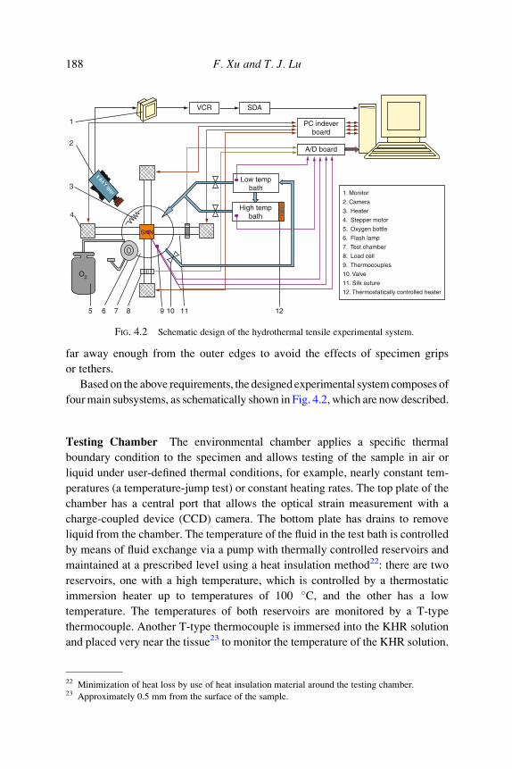
VCR SDA
1. Monitor
2. Camera
3. Heater
4. Stepper motor
5. Oxygen bottle
6. Flash lamp
7. Test chamber
8. Load cell
9. Thermocouples
10. Valve
11. Silk suture
12. Thermostatically controlled heater
PC indeverboard
A/D board
Low tempbath
High tempbath
1
2
3
4
5 6 7 8 9 10 11 12
O2
SHN
FIG. 4.2 Schematic design of the hydrothermal tensile experimental system.
188 F. Xu and T. J. Lu
far away enough from the outer edges to avoid the effects of specimen grips
or tethers.
Based on the above requirements, the designed experimental systemcomposes of
fourmain subsystems, as schematically shown in Fig. 4.2, which are now described.
Testing Chamber The environmental chamber applies a specific thermal
boundary condition to the specimen and allows testing of the sample in air or
liquid under user-defined thermal conditions, for example, nearly constant tem-
peratures (a temperature-jump test) or constant heating rates. The top plate of the
chamber has a central port that allows the optical strain measurement with a
charge-coupled device (CCD) camera. The bottom plate has drains to remove
liquid from the chamber. The temperature of the fluid in the test bath is controlled
by means of fluid exchange via a pump with thermally controlled reservoirs and
maintained at a prescribed level using a heat insulation method22: there are two
reservoirs, one with a high temperature, which is controlled by a thermostatic
immersion heater up to temperatures of 100 �C, and the other has a low
temperature. The temperatures of both reservoirs are monitored by a T-type
thermocouple. Another T-type thermocouple is immersed into the KHR solution
and placed very near the tissue23 to monitor the temperature of the KHR solution.
22 Minimization of heat loss by use of heat insulation material around the testing chamber.23 Approximately 0.5 mm from the surface of the sample.

Load cell
FIG. 4.3 Loading subsystem of the hydrothermal tensile testing system: user-defined loading,
such as isometric tension tests and isotonic tension tests, can be achieved through the pulley system.
Skin Biothermomechanics 189
Loading System The loading system applies either a uniaxial or a biaxial set of
forces or deformations to the specimen, Fig. 4.3. It is mounted on a square,
custom machined, aluminum plate. A circular watertight specimen chamber is
mounted centrally on the top plate and the loading assemblies are aligned at the
four ends of the chamber with two perpendicular loading axes.
Nearly uniformly distributed in-plane biaxial forces/deformations are applied
to the square specimen through four load carriages. The load can be applied in
each of the four carriage assemblies independently by use of a pulley system. Part
of the pulley system is connected to stepper motors, which impose deformations
on the skin being tested and allows computer-controlled loading in orthogonal
directions. The stepper motors are individually controlled using custom-designed
motor controller, which is modified from a four-axis indexer card. A pair of
motors is used to stretch the specimen in opposing directions to keep the
specimen located nearly at center of test chamber in order to improve video
strain resolution. The other part of the pulley/weight system is to apply isotonic
loads (equal loading): pairs of identical weights are placed at the ends of the
pulley system so that equal forces are applied along each axis.
One of the major challenges is the design of the holding system of connecting
the tissue to the load cell: the specially designed clamp for the current system has

FIG. 4.4 Clamp designed for the tensile tests.
190 F. Xu and T. J. Lu
a slot in each clamp, and the skin sample is fixed in the clamp by putting five pins
through the slot. In this way, the sample can be able to expand freely in the lateral
direction during tensile test, Fig. 4.4. The clamp is connected to the pulley system
by using inextensible string.
Another problem is that the density of skin tissue (�1200 kg/m3) is larger than
that of the liquid (�1000 kg/m3), which results in the sinking of tissue sample in
the liquid during the test. To solve this, small pieces of polystyrene foam are
placed on the underside of the tissue to render it nearly neutrally buoyant, as used
by other researchers (Wells, 2005).
Strain/Displacement and Stress/Force Measurement System Two force
transducers (load cells), which are watertight and temperature compensated, are
mounted outside of the specimen chamber on two orthogonal load carriages, as
shown in Fig. 4.3. In-plane finite strains and deformations in the tissue are
measured optically by tracking the position histories of contrasting markers on
the surface of the specimen, based on the method of Downs, Halperin,
Humphrey, and Yin (1990): the strain and deformation are traced between
different deformation states by performing a two-dimensional cross-correlation
on subsets of the images (Malcolm, Nielsen, Hunter, & Charette, 2002), which
are recorded with a CCD camera at 30 Hz. The image is captured to video file for
detailed off-line analysis, although in-time analysis can also be achieved via a
video frame grabber board. The accuracy of the cross-correlation relies on high-
contrast, high-frequency information within the images, which is provided by
staining the tissue sample with a surgical marker pen before each experiment and

Skin Biothermomechanics 191
by self-holding surface feature24 of skin tissue. The deformation field is obtained
by comparing the current marker positions, given by pixel coordinates, to the
reference positions. It should be noted here that the bidirectional tissue strain is
measured only in a central region so that the effects of local stress concentrations
of the gripping attachments are avoided. To keep the interested region within the
field of view of the CCD, the two motors are set to the same speed. Force
measurements are synchronized with CCD images by a LED in shot at the
beginning of each sequence.
Due to the large deformations, stretch ratio, instead of strain, is used in this
study, which is defined as l¼ l/l0 with l and l0 being the current and initial marker
distance on the sample as measured by the optical method. As for the stress, since
we were unable to precisely describe the sample thickness during testing to
determine true stress in a sample, engineering stress was calculated from the
applied force, F, divided by the initial cross-sectional area, A0, as s ¼ F/A0.
Data Acquisition System The current system uses two computers to acquire
the data: one for recording the video and the other for recording the force and
temperature. Analog signals from the load cells and thermocouples are transmit-
ted to a custom-made amplifier, and later to an A/D converter. The A/D conver-
sion board employed has eight signal channels, where two channels are reserved
for analog signals from the thermocouples and another two are occupied by
analog signals from the load cells, which are connected to a signal conditioner.
The data acquisition from the load cells and thermocouples and the motor control
are all performed using an integrated software system developed purposely with
LabView, which allows the user to operate the rig and perform experiments
through a single graphical user interface. This software supports motor control
in either position or force feedback modes, allowing deformation states to be
specified in terms of stress or stretch.
4.5.2. Validation of the System
Before being used to test skin samples, the system is calibrated and validated,
as described below.
Heat Transfer Analysis The system is aimed at testing the mechanical
behavior of skin samples under different heating conditions, as needed by the
theoretical modeling of skin biothermomechanics described in previous sections.
24 Some natural feature of skin surface can also be used as marker, such as wrinkle.

100
80
60
40
200 200 400
Time (s)
600 800 1000
Cen
terli
ne te
mpe
ratu
re (
�C)
Th (�C)
40 6080100
FIG. 4.5 Validation of the heating condition of the current system: centerline temperature of the
skin sample (with maximum thickness of 2.4 mm) reaches the target temperature Th very quickly as
compared to the duration of the tests.
192 F. Xu and T. J. Lu
It is thus important to analyze the heat transfer in the sample during the tests. The
Pennes equation (Eq. (2.1)) without blood perfusion term25 is used here for the
heat transfer analysis, where the skin sample is assumed to be thin and flat, and to
experience on its two surfaces a step change in temperature from a uniform room
temperature to an immersion fluid temperature up to 100 �C. The unloaded
thickness of skin sample is in the range of 1.6–2.4 mm and the maximum value
of 2.4 mm is used to estimate the maximum time required for the samples to reach
the target temperature. Thermal properties are obtained from published data in
the literature as given in Table 3.2. Using the closed-form solution of skin heat
transfer obtained in our previous study (Xu, Wen, Seffen, et al., 2008), the
centerline temperature history of the skin sample under heating of different
temperatures can be obtained, as shown in Fig. 4.5. For example, with heating
of 100 �C, the centerline temperature of the skin sample rises to 90 �C within
10 s, which is very short compared to the whole duration of the test (10 min).
Analyses for other temperatures reveal similar results (see Fig. 4.5).
Tissue-Like Phantom Experiment for System Validation Since the response
and stretch-rate sensitivity of silicone rubber is close to that of skin (Shergold,
2004), it is considered as an in vitro model for skin for the purpose of validating
25 In view that the samples are tested in vitro.

01.0 1.1
l1.2 1.3
1
2
3
4
5Current system
1%/s
10%/s
Model prediction1%/s10%/s
s (M
Pa)
Silicon rubber (Sil8800)
FIG. 4.6 Validation of the current system: comparison of biaxial tensile results with the predic-
tion of model from Shergold et al. (2006).
Skin Biothermomechanics 193
the system. Silicon rubber Sil8800 is used here since its response has been
obtained by the previous study in our lab (Shergold et al., 2006). The system
can thus be validated by comparing the measured results from the current system
with the predictions of the constitutive model. Square specimens of similar
dimensions as the skin samples to be tested (approximately 20 � 20 mm and
thickness 2.0 mm) are cut from silicone rubber sheets using a leather die punch
mounted onto a hand press. The biaxial tensile stress versus stretch responses are
measured at two stretch rates of 1%/s and 10%/s. Three tests are conducted for
each silicone rubber sample and the average response is calculated. The measured
tensile stress versus stretch responses of Sil8800 silicone rubber are shown in
Fig. 4.6 and a good agreement has been achieved between the measurement and
model prediction, which indicates the validity of the current test system.
4.5.3. Hydrothermal Tensile Test Procedure
In tensile testing, a square specimen (rectangular for uniaxial tests) is attached
to each of the four loading assemblies (two for uniaxial test) by a self-designed
holding system, across two perpendicular axes to allow physiological movement.
The applied thermal and mechanical loads are recorded, along with the time of
measurement, relative to the start of the experiment.

194 F. Xu and T. J. Lu
4.5.4. Hydrothermal Relaxation Test Procedure
The viscoelastic properties of skin tissue are usually characterized by two
classic experimental tests, either a stress relaxation test or a creep test. A uniaxial
stress–relaxation test is adopted in this study, where the stress relaxation test is
conducted with two protocols. First, the stress relaxation is performed for differ-
ent stretch levels (1.1, 1.3, and 1.5) with a high loading speed (g ¼ 50%/s) in
order to study the effect of stain level on relaxation: at low levels of stretch, the
mechanical behavior is mainly due to the role of elastin (Alexander & Cook,
1976; Silver, Kato, Ohno, & Wasserman, 1992) which has been shown to be
temperature-stable (Davidson et al., 1990); but the loads are too small relative to
the noise level (Baek et al., 2005; Eshel & Lanir, 2001), and high stretch levels
are adopted. In the second protocol, the tissue samples are stretched to 1.5 times
their initial length (a stretch of 1.5) at a low loading speed of 1%/s, and the
displacements of the samples are then kept constant while the time histories of the
force are recorded for a time period of 500 s. As before, each test is repeated three
times with three different samples and the averaged values are used.
4.6. Hydrothermal Compressive Tests
4.6.1. Hydrothermal Compressive System
A hydrothermal compressive system has been designed and built to study the
thermomechanical behavior of skin tissue under compression and is schemati-
cally shown in Fig. 4.7. The cylindrical skin sample is placed at the bottom of a
cylindrical test chamber filled with KHR solution. The chamber temperature is
1
Instron
High temp.bath
A/D board
Low temp.bath
2
3
4 5 6 7
1. Instron machine2. Compression rod3. Test chamber4. Skin sample5. Valve6. Thermocouples7. Thermostatically controlled heater
FIG. 4.7 Schematic of the hydrothermal compressive experimental system.

Skin Biothermomechanics 195
controlled by a circulation system similar to the hydrothermal tensile system. The
temperature of the KHR solution in the test chamber is measured by a T-type
thermocouple, which is placed approximately 0.5 mm away from the skin sample
surface. An axial compressive load is applied to the sample through a compres-
sion indentor that is connected to an InstronTM materials testing machine
(5500R), operating under the MerlinTM software. Longitudinal stretch measure-
ments are made with a linear variable displacement transducer (LVDT) and an
Electronic Instrument Research Ltd laser extensometer (LE-05) using two strips
of retroreflective material glued in the compression platens. All load cells,
thermocouples and laser extensometer signals are logged by a PC using an
ADC board, as before.
4.6.2. Hydrothermal Compressive Test Procedure
Only when friction is negligible, the sample expands uniformly during com-
pression so that the state of deformation within the sample is orthogonal. There-
fore, a lubricant (Vaseline) is applied to the faces of the specimen before loading
to reduce friction between the specimen and the compression platens. In the
unconfined compression tests, the cylindrically shaped samples, of diameter
10 mm and thickness 2–5 mm, are compressed between two impermeable,
smooth platens (with diameter of 20 mm), allowing the samples to expand
laterally during compression. The top platen is driven down until it just touches
the top surface of the specimen. Similar to tensile testing, the liquid in the high
temperature reservoir is heated to a chosen temperature, while the low tempera-
ture reservoir contained cold liquid. A valve in the test chamber is opened to
allow the hot or cold liquid to fill the test chamber until the sample is fully
immersed in the liquid, while a thermocouple near the sample is used to inform
the software of the exact time at which the experiment started. A force is applied
to the skin sample by the compression indentor and the applied displacement,
stretch, and loads are recorded. As before, each test is repeated three times with
three different samples and the averaged values are used.
4.7. Dynamic Mechanical Analysis
Various techniques have been used to characterize the viscoelastic behaviors
of engineering materials, including dynamic mechanical analysis (DMA), the
resonant method (Madigosky & Lee, 1983; Oyadiji & Tomlinson, 1985), the
nonresonant method (Ahn & Kim, 1994; Nielsen, Wismer, & Gade, 2000), and
the wave propagation method (Arbogast et al., 1997; Mossberg, Hillstrom, &
Soderstrom, 2001; Pereira, Mansour, & Davis, 1991). Among these, DMA is the
most commonly used due to its commercial availability and its popularity for

196 F. Xu and T. J. Lu
investigating the viscoelastic properties of polymers over a wide range of
temperatures and mechanical loading frequencies (Ferry, 1980; McCrum, Read,
& Williams, 1991). The dynamic viscoelasticity of the skin tissue is investigated
in this study by a dynamic mechanical analyzer (DMA Q800). During each DMA
test, the storage modulus, E00, loss modulus, E00, and loss factor, tan d, are all
measured as functions of temperature history. The storage modulus is analogous
to the stiffness measured from a monotonic test. The loss factor is defined as the
ratio between the amount of mechanical energy lost and stored during a cycle (tan
d ¼ E00/E0), representing the damping capability of skin tissue.
Themeasurements are carried outwith a single cantilever bending testingmode
at a fixed frequency of 1 Hz and different heating rates (rh¼ 2, 5, 10 �C/min), with
a temperature range of 25–80 �C. In this study, the measurements are performed
within the linear elastic region.26 Deformation of a cantilevered specimen pro-
duces a combination of both flexural and shear forces. In short, thick specimens,
shear forces are dominant while in long, thin specimens, flexural deformation is
the dominant mode. In the present tests, the length to thickness ratio of skin
samples is greater than 10 which leads to pure flexure (Chae et al., 2003).
The specimen is clamped between two clamps, one movable and the other
stationary, and then enclosed in a thermal chamber filled with air. The frequency,
amplitude, and a temperature range appropriate for the material being tested are
input into the analyzer, which applies an oscillation to the specimen while slowly
moving through the specified temperature range. All the tests are performed at a
fixed frequency of 1 Hz.
5. Biothermomechanical Behavior of Skin Tissue
The experimental characterization of the thermomechanical behavior of skin
tissue is presented in this section. The objective is to test the hypothesis that
collagen is a significant determinant of the thermally induced change in the
mechanical properties of skin tissue. For this purpose, DSC analysis is used to
detect the denaturation of collagen in skin tissue and to measure its thermal
stability. The integrity of the collagen network is analyzed using the thermal
damage integration (Eqs. (2.3) and (2.6) in Section 2). Hydrothermal tensile and
26 To accurately determine the mechanical properties, the deformation of skin sample must be based
on displacement amplitudes within the linear viscoelastic region to ensure that the material behavior is
independent of the magnitude of deformation (Chae et al., 2003). The nonlinear stress–strain curve
can be seen as being composed of several linear sections, for example, as a bilinear curve as shown in
Fig. 5.11.

3.030 40 50 60 70 80 90
3.2
3.4
3.6
3.8c p
(K
J/k
g�C
)
T (�C)
Pig flank skin; rh= 10 �C/min
Tmax= 66.83 �C
FIG. 5.1 Characteristic DSC thermogram, that is, variation of specific heat with temperature, of
a skin tissue sample: the temperature corresponding to the peak is 66.83 �C.
Skin Biothermomechanics 197
compressive experiments are then performed to assess the thermal dependency of
skin mechanical behavior. To characterize the viscoelastic properties of skin
tissue as a function of temperature and collagen denaturation, stress relaxation
tests under tension and DMA versus temperature are also performed.
5.1. Thermal Denaturation of Collagen in Skin Tissue
A typical DSC thermogram of a skin sample from pig flank is shown in
Fig. 5.1. The measured specific heat capacity falls in a range comparable to
published data in the literature (Henriques & Moritz, 1947; Young, 1998). The
result of Fig. 5.1 demonstrates that, for a stress-free skin sample subjected to
temperatures in the range of 30–90 �C, a broad endothermal27 peak appears:
denaturation is characterized by a sudden increase in the energy absorption,
which immediately precedes this peak, starting at about 60 �C. With further
heating, the endotherm reaches its maximum value at the denaturation tempera-
ture of 66.83 �C and then decreases. It is known that the temperature of thermal
denaturation strongly depends on the water content in collagen (e.g., it has been
found that there is an increase of the rate of shrinkage, and hence denaturation,
27 In thermodynamics, endothermal describes a process or reaction that absorbs energy in the form of
heat.

198 F. Xu and T. J. Lu
with increased hydration at a given temperature (Weir, 1949), and on the degree
of crosslinking between the chains (Pietrucha, 2005). The peak in Fig. 5.1 is
connected with the transition of collagen structure from the ordered triple helix
form to a randomly coiled conformation in the domains between crosslinks.
It has also been observed that the characteristics of denaturation are heating-
rate dependent (not shown here for brevity), where the maximum value of the
peak at higher heating rates moves toward higher temperatures. If a collagenous
tissue is heated from an initial value at a constant heating rate, rh, the rate of the
decline in the number of native molecules with time follows the first order
kinetics, given by Miles (1993), Miles, Burjanadze, and Bailey (1995), and
Ozawa (1970):
T ¼ Ti þ rht; ð5:1Þ
dN=dt ¼ �kðTÞN; ð5:2Þ
where Ti is the initial tissue temperature, N is the molecules of native, or
undenatured, collagen in the tissue, T is the current tissue temperature, and
k (T) is the usual reaction rate. Combining Eqs. (5.1) and (5.2) results in
1
N
dN
dT¼ � kðTÞ
rh: ð5:3Þ
This equation predicts that the maximum of ð�dN=dTÞ can be obtained when
d2N
dT2¼ 0 ) d
dT
kðTÞrh
N
� �¼ 0 ) N
dkðTÞdT
þ kðTÞ dNdT
¼ 0: ð5:4Þ
When combined with the Arrhenius equation (Eq. (2.2) from Section 2) �
kðTÞ ¼ A exp � EaRT; ð5:5Þ
the relation between Arrhenius parameters (Ea, A) and the peak temperature of
thermal denaturation (Tmax) is obtained as
rhEa
ART2max
¼ exp � Ea
RTmax
�: ð5:6Þ
Equation (5.6) can be rewritten as
lnrh
T2max
�¼ �Ea
R
1
Tmax� ln
Ea
R
�þ ln A: ð5:7Þ

2.92
−11
−10
−9
−8
ln(r
h / T
2 max
) (K
−1m
in−1
)
1/Tmax (× 10−3 K−1)
Pig flank skin
R 2= 0.9899
Y = 132.38–48259.16 X
2.93 2.94 2.95 2.96 2.97
FIG. 5.2 Characteristic plot of ln rh=T2max
� �versus 1/Tmax for pig flank skin tissue: dots (black)
are experimental data; line (red) is the best fit line by least squares; also indicated is the equation of
fitted line with X for 1/Tmax and Y for ln rh=T2max
� �. R2 value represents the scatter of data along the line
of best fit.
Skin Biothermomechanics 199
The activation energy Ea can be obtained from a log–log plot of lnðrh=T2maxÞ
versus 1/Tmax, while A can be derived through the intercept. With these results,
the thermal damage and the degree of thermal denaturation of skin collagen for a
given heating history can be quantified with Eqs. (2.3) and (2.6), respectively.
It should be noted here that the thermal denaturation of a collagenous tissue
depends not only on the temperature history but also on the stress state during
heating (Humphrey, 2003), which, however, is very difficult to quantify due to
the lack of corresponding research. Therefore, in the following calculation of
thermal damage, the parameters obtained from DSC measurements in the
absence of mechanical loading are used.
A characteristic plot of lnðrh=T2maxÞ versus 1/Tmax for a pig flank skin sample
is presented in Fig. 5.2. Tests with belly, back, ear, and face skin samples have
also been performed and the results of the derived Arrhenius parameters are
presented in Table 5.1. As shown in Section 2, there is a linear relationship
between the published experimental results of Arrhenius parameters ln A and Ea
for skin tissue, as given by Eq. (2.4). The comparison of the results here for
different pig skin samples with results calculated from Eq. (2.4) has been
presented in Fig. 5.3, where it can be seen that a very good agreement has been
achieved.

TABLE 5.1Experimental results of Arrhenius parameters (Ea, A).
Sample Ea (�105 J/mol) A (s�1)
Back skin 5.255 2.126�1081
Belly skin 3.935 1.151�1061
Ear skin 5.867 5.240�1091
Face skin 4.710 4.575�1072
Flank skin 4.012 1.501�1061
1003
4
5
6
7
8
Eq.(4)
Back
Belly
Ear
Face
Flank
Ea
( ×
105
J/m
ol)
ln(A) (ln(s−1))150 200 250 300
FIG. 5.3 Comparison of the results of Arrhenius parameters of this study with literature results.
200 F. Xu and T. J. Lu
5.2. Tensile Behavior of Skin Tissue
Hydrothermal tensile tests, which enable the variation of temperature during
mechanical tests, are performed to quantify the effects of temperature and
corresponding thermal denaturation on the tensile behavior of skin tissue.
5.2.1. Uniaxial Tensile Behavior
A typical uniaxial tensile behavior of pig ear skin sample measured at
T ¼ 37 �C is shown in Fig. 5.4 (black dots). Note that skin has a low stiffness
at low stretch but undergoes significant stretch hardening at high stretch level.
This J-shaped stress–stretch curve is typical for mammalian skin, although the
stretch hardening characteristic varies from specie to specie. In Fig. 5.4, the
uniaxial tensile stress–stretch responses of human chest skin (Dunn, Silver, &

1.00
0.5
1.0
1.5
2.0 Uniaxial tensile behavior
s (
MP
a) Pig ear skin (Exp.) Human chest skin (Dunn et al., 1985)
Cat back skin (Veronda and Westmann, 1970)
1.2 1.4 1.6 1.8l
FIG. 5.4 Comparison of the uniaxial tensile behavior of pig ear skin measured at T¼ 37 �C with
reported data on human chest skin (Dunn et al., 1985) and cat back skin (Veronda &Westmann, 1970).
Skin Biothermomechanics 201
Swann, 1985) and cat back skin (Veronda &Westmann, 1970) are also shown for
comparison. It is interesting to find that the uniaxial tensile behavior of pig ear
skin obtained in this study agrees well with that of human skin. This is, however,
not unexpected since pig ear skin is selected purposely because of its similarity
with human skin (Meyer et al., 2007) as described in Section 3. Compared with
pig ear skin and human chest skin, cat back skin stretch hardens faster.
Uniaxial tensile tests of pig ear skin samples under different temperatures have
been performed and the stress–stretch relationships are given in Fig. 5.5: a good
repeatability has been noted for three same tests and a single averaged response is
shown. As described above, there are two behavior regimes for all the curves.
First, when e < �50%, the curves almost overlap. This low modulus region is
thermally very stable due to the dominance of elastin in the skin behavior and the
gradual straightening of an increasing fraction of the wavy collagen fibers and
stretching of elastic fibers. When e > �50%, the stress increases almost linearly
with stretch; and the slopes under different temperatures are different and reduce
with increasing temperature. This effect is due to the stretching and slippage of
collagen molecules within crosslinked collagen fibers and to collagen fibril
slippage: with an increase in temperature, the highly organized crystalline struc-
ture of collagen changes to a random, gel-like state, which results in the
corresponding decrease in stiffness. Furthermore, the hydration change with
temperature may also make a contribution. Water plays a significant role in

1.00
0.25
0.50
0.75
1.00 T (ºC)
37 45 50 60 70 80
1.2 1.4 1.6 1.8
Pig ear skin, g = 6% / min
l
s (
MP
a)
FIG. 5.5 Stress–stretch relation of uniaxial hydrothermal tensile tests under different tempera-
tures: g is the stretch rate at which tests are performed.
202 F. Xu and T. J. Lu
governing the gross properties of skin tissue (Auriol, Vaillant, Machet,
Diridollou, & Lorette, 1993; Hoffman, Robichaud, & Grigg, 2003), which consist
primarily of water. However, due to heating, the level of hydration may vary, for
example, Luescher, Ruegg, and Schindler (1974) suggested that primary hydra-
tion water is set free during the process of thermal denaturation. It should be
noted that when T � 60 �C, there is relatively little change in the modulus with
temperature, which can be explained by the thermal damage process: when the
stretch rate and temperature are given, the relationship between the stretch and
thermal denaturation degree (Deg) calculated according to the heating history can
be obtained, as shown in Fig. 5.6. Figure 5.6 shows that when T � 60 �C, thecollagen is fully denaturized, while at T ¼ 45 and 50 �C, denaturation is much
slower.
5.2.2. Biaxial Tensile Behavior
The biaxial tensile behaviors of pig skin samples is tested at 37 and 60 �C. Thestress–stretch relationships measured for samples from different body sites are
given in Fig. 5.7. An interesting result can be observed from the figure: only the
skin tissue from porcine cheek appears to be abnormal in the fact that the curves
for the two different axes overlap each other. This means that it has an equal
distribution of collagen/elastin fibers across both axes of a square, independent of

1.00
100
10−2
10−4
10−6
10−8
10−10
T (ºC) 37 45 50 60 70 80
Deg
1.25 1.50 1.75 2.00l
Pig ear skin, g = 6%/min
FIG. 5.6 Stretch-thermal damage degree (Deg) relation of hydrothermal tensile tests under
different temperatures: Deg is obtained from Eq. (2.6) with given heating condition and stretch rate.
Skin Biothermomechanics 203
the cutting angle. This discovery may allow the series of experiments on
skin tissue to come to fruition since a uniform stress distribution in the
tissue can be easily achieved even in biaxial tests. However, this observation is
abnormal since it is well known that clear Langer’s lines28 have been observed
for face skin tissue (Rubin, 1948). Relative to that at body temperature, the
stiffness of skin tissue under hyperthermal temperature decreases considerably.
This feature is due to the similar reasons as those in uniaxial results described
above.
In Fig. 5.7, the uniaxial tensile behaviors of pig ear skin samples (dots)
obtained under the same stretch rate and heating conditions have also been
given for comparison. Consistent with the observation in the literature, as
described in Section 2.3, biaxial tensile loading, compared with uniaxial stretch,
results in lateral narrowed stress–stretch curve and the reduction in the stretch
before entry into the linear region occurs, which is due to the two-directional
stretch of collagen fibers.
28 ‘‘Langer’s lines,’’ sometimes also called cleavage lines, are a term used to define the direction
within the human skin along which the skin has the least flexibility. These lines correspond to the
alignment of collagen fibers within the dermis (Cox, 1942).

1.00
0.1
0.2
0.3A
B
Biaxial Axis 1 Axis 2
Uniaxial Axis 1
Face skin
Ear skin
Belly skin
0
25
50
75Biaxial
Axis 1
s (k
Pa)
s (M
Pa)
Axis 2
Uniaxial Axis 1
Face skin
Ear skin
Belly skin
1.1 1.2 1.3 1.4
1.0 1.1 1.2 1.3 1.4
g = 6%/min, T= 37 ºC
g = 6%/min, T= 60 ºC
l
l
FIG. 5.7 Stress–stretch relation of biaxial hydrothermal tensile tests for samples from different
sites at (A) T¼ 37 �C and (B) T¼ 60 �C: axis 1 (solid lines) and axis 2 (dashed lines) are material axes
identified with the equal-load method as described in Section 4.1; scattered points are experimental
results from uniaxial tests.
204 F. Xu and T. J. Lu
5.3. Compressive Behavior of Skin Tissue
Almost all of the previously published mechanical models of skin tissue are
based on experimental data under tension, although in vivo, skin tissue is often
subjected to compressive loading by different agents. The goal of the present

Skin Biothermomechanics 205
section is to study the relationship between temperature, thermal damage, and the
corresponding change in the compressive behavior of skin tissue.
5.3.1. Typical Example of Compressive Behavior
A representative compressive stress–stretch response obtained at a stretch rate
g ¼ 24%/min29 is presented in Fig. 5.8A. The relationship exhibits a consistent
three-stage hardening of a toe region with low stiffness at low stretch levels, a
transition region from low to high stiffness, and a high stiffness region at large
stretch levels. The results are comparable to the published compressive behavior
of pig back skin tissue obtained at similar stretch rate (g ¼ 25%/min) (Shergold
et al., 2006), as shown in Fig. 5.8A.
Compared with the mechanical characteristics of skin tissue in uniaxial ten-
sion, the compressive stress/stretch curves are similar in trend; however, the
transition from low to high stiffness in uniaxial tension occurs mostly at a stretch
level of 1.4–1.6 (Dunn et al., 1985; Lanir & Fung, 1974; Wan, 1994), which is
much larger than that in compression (about 1.2–1.3) as observed in the results
here (Fig. 5.8B). Similar results have also been reported elsewhere for pig back
skin tissue (Shergold et al., 2006; Wu et al., 2003, 2007). Wu et al. (2003, 2007)
observed a transition at an even lower stretch level of 1.04–1.05 for confined
compression and 6–7% for unconfined compression: the difference is because the
fat layer is removed in this study. However, compared with the biaxial tensile
behavior of pig back skin obtained by Ankersen et al. (1999), the compressive
behavior obtained in this study shows a slower rate of stretch hardening, as shown
in Fig. 5.8B. This is mainly due to the different testing environments adopted: in
the current study all the tests were performed in liquid (KHR), while the tests of
Ankersen et al. were performed in air environment. Testing in air will induce the
decrease of the hydration and humidity of the samples, which have been shown to
increase the stiffness of skin tissue (Bert & Reed, 1997; Jemec, Jemec, Jemec, &
Serup, 1990; Overgaard & Jemec, 1993).
5.3.2. Temperature-Dependent Compressive Behavior
The hydrothermal compressive tests of back skin tissue under different tem-
peratures have been performed and the results are given in Fig. 5.9. It is interest-
ing to see that, contrary to the tensile tests, the compressive stiffness increases
with increasing temperature although the thermal damage degree also increases,
as shown in Fig. 5.10. One key difference is that the compressive tests are
performed in a direction normal to the principal orientation of collagen and
29 This strain rate is chosen for a better comparison with data reported in the literature.

1.00
1
2
3
4
5A
01.0 1.2 1.4 1.6 1.8
1
2
3
4
5B
s (M
Pa)
s (M
Pa)
Exp. (control)
Compression result from Shergold et al. (2006)
l
l
g = 60%/min
g = 50%/min
g = 6%/min
Biaxial tension from Ankersen et al. (1999)
Uniaxial tension (Exp.)
Compression (Exp.)
g = 24%/min
g = 25%/min
1.1 1.2 1.3 1.4 1.5
FIG. 5.8 Representative stress–stretch responses from a compressive test (black solid dots) and
comparison between the compression results (A) with published compressive behavior (Shergold
et al., 2006) and (B) with uniaxial tensile behavior (red circle dots) obtained in this study and publi-
shed biaxial tensile behavior of pig back skin tissue (Ankersen et al., 1999).
206 F. Xu and T. J. Lu
elastin fibers. Even though there exists denaturation of collagen, which appears in
the tensile tests, the compressive behavior is governed by the mechanical proper-
ties of the gel-like ground substance, inside which the fibers are located (Dikstein
& Hartzshtark, 1983; Tregear & Dirnhuber, 1965). Very little is known about the

0
1
2
3
4
5
37
55
5867
T (°C)
Pig back skin, g = 6%/min
s (M
Pa
)
l1.11.0 1.2 1.3 1.4 1.5
FIG. 5.9 Stress–stretch relation of hydrothermal compressive tests under different temperatures.
Skin Biothermomechanics 207
mechanical properties of this substance; it can only be speculated that its stiffness
increases with increasing temperatures.
To characterize the temperature effect on skin mechanical behavior and
compare their characteristics under different testing methods, a procedure is
proposed here to obtain the modulus of skin sample at low (El) and high (Eh)
stretch levels, as shown in Fig. 5.11: the stretch–stress curve is treated as a
bilinear line, the tangents at low and high stretch levels are then obtained, and
El and Eh are defined as the slope of the two tangents. The variations of El and Eh
with temperatures for tensile and compressive behaviors are given in Fig. 5.12A
and B, respectively. While El for tensile behavior of skin tissue does not change
much with temperature, Eh decreases almost linearly with increasing temperature
until about 60 �C, after which it decreases little. Eh for tensile behavior
obtained here (0.3–4.5 MPa) also agrees well with the published in vitro
measurements of Young’s modulus for animal skin tissue, for example, cat skin
(0.5 MPa) (Veronda & Westmann, 1970) and rat skin (1.18–4.62 MPa) (Ranu,
Burlin, & Hutton, 1975; Vogel, 1971). Compared with that for tensile behavior,
Eh for compressive behavior increases with temperature and is much higher (28–54
MPa). This is due to (1) different mechanisms of tensile and compressive
behaviors as discussed above and (2) different skin samples used for tensile
(pig ear skin) and compressive (pig back skin) testing, and the regional difference
of skin mechanical behavior has been observed by many researchers (Elsner,
Wilhelm, & Maibach, 1990; Jansen & Rottier, 1958a,b; Pierard, Letawe,

Pig back skin, g = 6%/min
l
100
1.11.0 1.2 1.3 1.4 1.5
37555768
T (°C)
10−8
Deg
10−10
10−6
10−4
10−2
FIG. 5.10 Stretch-thermal damage degree relation of hydrothermal compressive tests under
different temperatures.
208 F. Xu and T. J. Lu
Dowlati, & Pierard-Franchimont, 1995; Pierard, Nikkels-Tassoudji, & Pierard-
Franchimont, 1995; Smalls, Wickett, & Visscher, 2006).
5.3.3. Thermal-Damage-Induced Changes in Compressive Behavior
To separate reversible changes from irreversible ones and to study the influ-
ence of thermal denaturation on skin properties, selected skin samples are
thermally denatured before testing (i.e., predamaged). Using the Arrhenius para-
meters (Ea, A) of pig skin tissue obtained from the DSC analysis, different heating
temperatures, Th, and durations, th, are chosen to induce damage in skin sample to
a specified degree. In all, 22 groups of samples are prepared, with groups 1–4
having no predamage and used as control groups. Details of each group are given
in Table 5.2.
Figure 5.13A–C presents the thermal-damage-induced changes in skin com-
pressive behavior at three different stretch rates (g ¼ 25, 50, 125%/min). The
results at all three stretch rates exhibit a clear trend of curve shifting toward the
right with increasing damage. When the damage degree (Deg) has a value near
unity, implying that the collagen is fully denaturized, there is little difference
between curves: the stiffness of skin tissue decreases with increasing degree of
thermal damage, which is reasonable; and increased extensibility of soft tissues
due to thermal treatment has been observed in both uniaxial (Chachra et al., 1996;

01.0 1.1 1.2 1.3 1.4 1.5
1
2
3
4
5
ElEh
σ (M
Pa)
l
FIG. 5.11 Method for calculating the moduli at low (El) and high (Eh) stretch levels.
Skin Biothermomechanics 209
Chen & Humphrey, 1998; Lennox, 1949) and biaxial tensile testing (Harris et al.,
2003; Wells et al., 2004) of skin tissue.
There are several possible mechanisms for explaining why the thermal dam-
age changes the compressive behavior of skin tissue. The first one is the
hyperthermal-temperature-induced thermal denaturation of dermal collagen.
It has been suggested that collagen may play an important role in determining
the overall mechanical properties of skin tissue (Lanir, 1987), and thus it seems
reasonable to assume that collagen would also play a major role in the compres-
sive behavior of skin tissue. However, this cannot be the principal reason since
the compressive tests are performed normal to the principal orientation of
collagen. Another possible mechanism is the dehydration change accompanying
thermal damage as well as rehydration during the thermal recovery process after
heating. It is known that there can be either an inward or outward flux of
interstitial fluid due to the heating of a soft tissue (Humphrey, 2003), and the
compressive behavior is governed by the mechanical properties of the gel-like
ground substance, inside which the fibers are located (Dikstein & Hartzshtark,
1983;Oomens et al., 1987; Tregear&Dirnhuber, 1965). Hormann and Schlebusch
(1971) and Luescher et al. (1974) suggested that renaturation, or reformation of
some of the triple-helix structures, occurs after heating, which involves the
slow rebounding of primary hydration water to the amorphous protein and the
formation of additional water bridges within the more disorganized molecule.
A third possible mechanism is the viability of cells in the skin tissue, where

0
30 40 50 60 70 80
30 40 50 60T (°C)
T (°C)
70 80
1
2
3
4
5
A
B
Mod
ulus
(M
Pa)
Eh
E l
Tensile behavior
0
10
20
30
40
50
60Compressive behavior
Mod
ulus
(M
Pa)
Eh
E l
FIG. 5.12 Variations of the moduli at low (El) and high (Eh) stretch levels with temperatures for
(A) tensile behavior and (B) compressive behavior.
210 F. Xu and T. J. Lu
specifically, Yip, Walker, Fernlund, and Pinder (2007) found that the in situ
fibroblast viability has great influence on the mechanical properties of rat back
skin tissue.
It is interesting to note that Fig. 5.13, compared to Fig. 5.9, exhibits contra-
dictory results: in Fig. 5.9, the compressive stiffness increases with increasing
temperature even though the thermal damage and the degree of thermal denatur-
ation also increase. It has been shown by others that the stiffness of skin tissue
increases progressively in dehydration and decreases in over hydration under
compression (Bert & Reed, 1997). In Fig. 5.9, all the tests are performed with
fresh samples at hyperthermal temperatures where only heat-induced dehydration

TABLE 5.2Details of predamaged samples.
Sample Group No. Th (�C) Th (s) O Deg g (%/min)
1 37 0 0 0 25
2 37 0 0 0 50
3 37 0 0 0 125
4 37 0 0 0 250
5 55 12 0.0579 0.0563 25
6 55 12 0.0579 0.0563 50
7 55 12 0.0579 0.0563 125
8 55 12 0.0579 0.0563 250
9 65 1 1.44 0.762 25
10 65 1 1.44 0.762 50
11 65 1 1.44 0.762 125
12 65 3 4.31 0.987 25
13 65 3 4.31 0.987 50
14 65 3 4.31 0.987 125
15 65 30 86.2 1 25
16 65 30 86.2 1 50
17 70 10 219 1 25
18 70 10 219 1 50
19 70 5 109 1 25
20 70 5 109 1 50
21 70 18.5 405 1 25
22 75 10 3081 1 25
Skin Biothermomechanics 211
occurs, while tests in Fig. 5.13 are performed with predamaged samples at body
temperature where the samples are rehydrated during the thermal recovery
process of predamage procedure.
5.3.4. Stretch-Rate Sensitivity at Different Thermal Damage Levels
The stretch-rate sensitivity of pig skin tissue at four different damage levels is
presented in Fig. 5.14A–D for uniaxial compressive loads. A clear sensitivity
emerges for all damage levels, where the skin tissue stiffens and strengthens with
increasing stretch rates. This feature mirrors results in the literature. For example,
it has been observed by others that, under stretch-controlled tests, the overall
stiffness of skin tissue decreases with decreasing stretch rates (Arumugam, Naresh,
& Sanjeevi, 1994; Giles, Black, & Bischoff, 2007; Jamison, Marangoni, & Glaser,
1968; Lanir & Fung, 1974; Shergold et al., 2006; Vogel & Hilgner, 1979a,b).

212 F. Xu and T. J. Lu
Since collagen fibers comprise bundles of collagen fibrils aligned parallel to a
hyaluronic acid chain, and linked to the hyaluronic acid by proteoglycan side
chains, it is likely that the stretch-rate sensitivity of skin tissue is attributable to
viscous losses due to the interaction of ground substance with collagen fibers at
1.0 1.1 1.2 1.3 1.4 1.5 1.60
1
2
3
4
5
A
B
Deg Group0 0 10.0579 0.0563 51.44 0.762 94.31 0.987 12
86.2 1.00 15219 1.00 17109 1.00 19405 1.00 21
1.0 1.1 1.2 1.3 1.4 1.5 1.60
1
2
3
4
5 Group
0 0 20.0579 0.0563 61.44 0.762 10
4.31 0.987 1386.2 1.00 16219 1.00 18
109 1.00 20
Tt= 37 °C, g = 25%/min
Tt= 37 °C, g = 50%/min
W
W
l
l
s (M
Pa)
s (M
Pa)
Deg
FIG. 5.13 (Continued)

1.0 1.1 1.2 1.3 1.4 1.5 1.60
1
2
3
4
5
6
C
Deg Group0 0 30.0579 0.0563 71.44 0.762 114.31 0.987 14
Tt= 37 °C, g = 125%/min
σ (M
Pa)
l
Ω
FIG. 5.13 Effect of thermal denaturation on skin compressive behavior at different stretch rates
of (A) g ¼ 25%/min, (B) g ¼ 50%/min, and (C) g ¼ 125%/min.
Skin Biothermomechanics 213
the macroscopic scale (Cohen, Hooley, & McCrum, 1976) as well as with
collagen fibers at the microscopic scale (Haut, 1983). For example, much of the
sensitivity to stretch rate has been attributed to the uncoiling and aligning
movement of collagen (Lanir, 1979), and Daly (1982) suggested that the
stretch-rate sensitivity of skin tissue occurred at all structure levels of collagen
and was due in part to bonds between fibrils and fibers. However, the uncoiling
and aligning movement of collagen should not be the main mechanism for
the case here, since the compressive tests are performed through the skin thick-
ness direction and a similar stretch sensitivity has been observed at all
damage levels. Thus the stretch-rate sensitivity may be attributed to the variation
of viscous interaction between collagen fibers and ground substance induced
by hydration change accompanying thermal damage. For example, the stretch-
rate sensitivity was well modeled by Shergold et al. (2006) by increasing
the shear modulus with increasing stretch rate, with no attendant change in the
stretch hardening exponent. Arumugam et al. (1994) also attributed the higher
stress to higher frictional force at higher stretch rates. The movement of fluid
in the hydrated matrix may also play an important role (Li, Armstrong, &
Mow, 1983).

214 F. Xu and T. J. Lu
5.4. Relaxation Behavior of Skin Tissue
Skin tissue is a viscoelastic material. However, to the authors’ best knowledge,
there is no published study on the temperature dependency of skin viscoelasticity,
although other collagenous tissues such as cartilage (Chae et al., 2003;
1.0 1.1 1.2 1.3 1.4 1.5 1.6
1.0 1.1 1.2 1.3 1.4 1.5 1.6
0
1
2
3
4
5
A
25
50
125
250
0
1
2
3
4
5
25
50
125
250
B
s (M
Pa)
s (M
Pa)
Th= 37 °C, t h= 0 s, W= 0.0
Th= 55 °C, t h= 1 2 s, W= 0.06
l
l
g (%/min)
g (%/min)
FIG. 5.14 (Continued)

0
1
2
3
4
5
C
D
0
1
s (M
Pa)
s (M
Pa)
2
3
4
5
Th= 65 °C, t h= 1 s, Ω = 1.44
Th= 65 °C, t h= 3 s, Ω = 4.31
1.0 1.1 1.2 1.3
l1.4 1.5 1.6
1.0 1.1 1.2 1.3l
1.4 1.5 1.6
25
g (%/min)
50
125
25
g (%/s)
50
125
FIG. 5.14 Stain-rate sensitivity at damage levels of (A)O ¼ 0, (B)O ¼ 0:06, (C)O ¼ 1:44, and
(D) O ¼ 4:31. Th and th are heating temperature and duration, respectively.
Skin Biothermomechanics 215
Chao et al., 2001) and bone (Mano, 2005; Yamashita et al., 2002) have been
studied in detail. The aim of the present section is therefore to characterize the
temperature-dependent relaxation behavior of skin tissue. Stress relaxation tests
under different temperatures are then performed, and results and discussion are
presented.

216 F. Xu and T. J. Lu
5.4.1. Modeling of Skin Viscoelastic Behavior
Quasilinear Viscoelastic Theory The time-dependent mechanical behavior of
soft tissues can be formulated using Boltzmann hereditary integrals (Fung, 1993;
Lakes, 1998), whereby the stress histories s(t) are related to the strain histories
E(t) according to
sðtÞ ¼ðt0
Gðt� t; eÞ_eðtÞdt; ð5:8Þ
where t is the time, t is the dummy variable for time, and G(t, e) is the relaxationmodulus.
For the quasilinear response of tissue, it is assumed that the relaxation function
G(t, e) can be further separated into a strain-dependent part and a time-dependent
part, as
GðtÞ ¼ gðtÞEðeÞ ð5:9Þ
where g(t) is the normalized relaxation function that only depend on time, and
E(e) is the elastic modulus. This form is also called the quasilinear viscoelastic
(QLV) theory, which was formulated by Fung (1971) and has been proved useful
for describing the viscoelastic behavior of different soft tissues such as skin tissue
(Decraemer, Maes, Vanhuyse, & Vanpeperstraete, 1980; Ledoux & Blevins,
2007; Wu, Cutlip, Welcome, & Dong, 2006), ligament (Defrate & Li, 2006;
Funk, Hall, Crandall, & Pilkey, 2000; Moon, Woo, Takakura, Gabriel, &
Abramowitch, 2006), cartilage (Simon, Coats, & Woo, 1984; Woo, Simon,
Kuei, & Akeson, 1980), and tendon (Defrate & Li; Johnson et al., 1994; Sarver
et al., 2003).
Insertion of Eq. (5.9) into Eq. (5.8) leads to the following nonlinear separable
integral formulations:
sðtÞ ¼ðt0
gðt� tÞ dse½eðtÞ�de
deðtÞdt
dt; ð5:10Þ
where se(t) is the elastic stress response.The QLV theory was developed on the basis that the reduced relaxation
function describes the normalized stress response of a sample in response to a
step increase in strain such that the tissue has no time to relax during this loading
process. This, however, is experimentally impossible to realize. To isolate the
nonlinear elastic properties of tissue from its viscous properties, relatively high
rates have been used. However, with a high stretch rate, it is technically difficult
to obtain reliable instantaneous material parameters due to inertial effects and,

Skin Biothermomechanics 217
thus, greater technical challenges must be overcome in the design of the testing
machine and data acquisition system, especially for small tissue samples (Wu
et al., 2007). Although several methods have been proposed to modify the
analysis where finite ramp times are considered in order to better approximate
the solutions (Carew, Talman, Boughner, & Vesely, 1999; Dortmans, Sauren, &
Rousseau, 1984; Kwan, Lin, & Woo, 1993; Lin, Kwan, & Woo, 1987; Myers,
McElhaney, Nightingale, & Doherty, 1991; Nigul & Nigul, 1987), these methods
are still dependent on fast ramp rates, and therefore, may be affected by the
associated errors. One of the main problems is overshoot, followed by a rebound
back to the target stretch under high rates due to limitations in the mechanical
testing equipment. This has been shown to have a considerable effect on the
measured forces (Ledoux & Blevins, 2007) and to introduce an unwanted bias
into the estimation of QLV parameters (Doehring, Carew, & Vesely, 2004;
Gimbel, Sarver, & Soslowsky, 2004). For example, during stress relaxation of
tendon, Gimbel et al. found that there was an average overshoot magnitude of
7.5% of the intended stretch, causing an error as large as 30% of the estimation
of QLV parameters when the overshoot is not accounted for in the data fitting
process.
It has been recommended that a slow stretch rate should be used in stress–
relaxation tests (Abramowitch & Woo, 2004; Gimbel et al., 2004), since at low
stretch rate the actual strain history (variation of strain with time) can be well
approximated by a linear ramp followed by holding at constant stretch. Thus, the
errors associated with fast stretch rates (e.g., overshoot, vibration, poorly
approximated strain histories) can be reduced or avoided (Abramowitch &
Woo; Funk et al., 2000; Gimbel et al.). For instance, recent studies have shown
that a slow ramp rate (0.15%/s) in conjunction with a direct fit for the ramp and
stress–relaxation portions can provide an accurate estimation of parameters,
avoiding thus the errors associated with fast stretch rates (Abramowitch &
Woo). In view of these, comparatively low stretch rates have been used in the
tests performed in this study.
Skin Model Development of Based on QLV Theory An exponential approx-
imation has often been chosen to describe the nonlinear elastic behavior of skin
tissue under tension. Here, the following expression is used to describe the
instantaneous elastic response:
seðeÞ ¼ AðeBe � 1Þ; ð5:11Þ
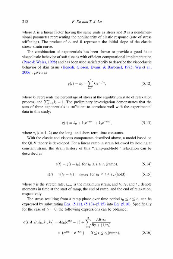
218 F. Xu and T. J. Lu
where A is a linear factor having the same units as stress and B is a nondimen-
sional parameter representing the nonlinearity of elastic response (rate of stress
stiffening). The product of A and B represents the initial slope of the elastic
stress–strain curve.
The combination of exponentials has been shown to provide a good fit to
viscoelastic behavior of soft tissues with efficient computational implementation
(Puso &Weiss, 1998) and has been used satisfactorily to describe the viscoelastic
behavior of skin tissue (Kenedi, Gibson, Evans, & Barbenel, 1975; Wu et al.,
2006), given as
gðtÞ ¼ k0 þXni¼1
kie�t=ti ; ð5:12Þ
where k0 represents the percentage of stress at the equilibrium state of relaxation
process, andPn
i¼ 0ki ¼ 1. The preliminary investigation demonstrates that the
sum of three exponentials is sufficient to correlate well with the experimental
data in this study:
gðtÞ ¼ k0 þ k1e�t=t1 þ k2e
�t=t2 ; ð5:13Þ
where ti (i ¼ 1, 2) are the long- and short-term time constants.
With the elastic and viscous components described above, a model based on
the QLV theory is developed. For a linear ramp in strain followed by holding at
constant strain, the strain history of this ‘‘ramp-and-hold’’ relaxation can be
described as
eðtÞ ¼ gðt� t0Þ; for t0 t tRðrampÞ; ð5:14Þ
eðtÞ ¼ gðtR � t0Þ ¼ emax; for tR t t1ðholdÞ; ð5:15Þ
where g is the stretch rate, emax is the maximum strain, and t0, tR, and t1 denote
moments in time at the start of ramp, the end of ramp, and the end of relaxation,
respectively.
The stress resulting from a ramp phase over time period t0 t tR can be
expressed by substituting Eqs. (5.11), (5.13)–(5.15) into Eq. (5.10). Specifically
for the case of t0 ¼ 0, the following expressions can be obtained:
s t;A;B; k0; k1; k2ð Þ ¼ Ak0ðeBgt � 1Þ þX2i¼1
ABgkiBgþ ð1=tiÞ
� eBgt � e�t=ti� �
; 0 t tRðrampÞ; ð5:16Þ

Skin Biothermomechanics 219
s t;A;B; k0; k1; k2ð Þ ¼Ak0 eBgtR � 1� �þX2
i¼1
ABgkiBgþ 1=tið Þ
� eBgtR � e�tR=ti� �
e�t=ti ; tR t t1ðholdÞ: ð5:17Þ
From Eq. (5.17) the stress during relaxation can be considered as the sum of
three components, an equilibrium stress at tR and the other two being long- and
short-term decay components, respectively.
5.4.2. Relaxation Behavior of Skin Tissue
Typical Example of Skin Stress–Relaxation Behavior A representative
uniaxial stress–time response from a relaxation test performed at body tempera-
ture (T ¼ 37 �C) is presented in Fig. 5.15, which shows the response of pig ear
skin tissue during both the ramp and relaxation phases of the test. In the ramp
region (0s t 50s), the response is concave due to the nonlinearity caused by
an increasing recruitment of collagen fibers with increasing stretch. In the
relaxation region, the stress–relaxation curve is characterized by a sharp decrease
in stress, followed by a gradual reduction up to 550 s. The uniaxial tensile
relaxation results of pig ear skin tissue obtained here are comparable with those
of human skin tissue (Barbenel & Evans, 1977) and rat skin tissue (Purslow,
Wess, & Hukins, 1998) under tension, and pig foot skin tissue under compression
(Wu et al., 2006) both qualitatively and quantitatively.
0
0.05
s (M
Pa)
0.10
0.15
0.20
End of ramping
0 100 200 300
t (s)
400 500
T = 37 �C, lmax= 1.5, g = 60%/min
FIG. 5.15 Representative uniaxial tensile stress–relaxation response of pig ear skin tissue.

220 F. Xu and T. J. Lu
Verification of the QLV Theory The model developed in Section 5.4.1 is
based on the QLV theory, which is built upon the assumption that the stress
relaxation function can be separated into one time-dependent portion and one
stretch-dependent portion. In other words, the normalized stress relaxation func-
tion, g(t), should be independent of the stretch/stress level. Different trends in
viscous-elastic separability have been observed in soft biological tissues. For
example, the validity of the assumption has been justified for relaxation behavior
of a variety of soft tissues including ligament, tendon and esophagi, both qualita-
tively (Haut & Little, 1972; Lanir, 1980) and quantitatively (Pioletti &
Rakotomanana, 2000; Yang, Fung, Chian, & Chong, 2006) and has been argued
to be reasonable for other soft tissues composed mainly of collagen (Pioletti &
Rakotomanana). But in some other studies, it has been also shown that the
relaxation behavior of collagenous tissues is dependent on stretch level
(Bonifasi-Lista, Lake, Ellis, Rosenberg, & Weiss, 2002; Haut & Little;
Hingorani, Provenzano, Lakes, Escarcega, & Vanderby, 2004; Komatsu,
Sanctuary, Shibata, Shimada, & Botsis, 2007; Lakes & Vanderby, 1999; Lanir,
1980; Oyen et al., 2005; Provenzano, Lakes, Keenan, & Vanderby, 2001;
Thornton, Oliynyk, Frank, & Shrive, 1997), which demonstrates the limits of
Fung’s QLV approach.
To verify the hypothesis of variables separation in QLV theory for skin tissue,
stress relaxation tests of pig skin samples at three different stretch levels (lmax ¼1.1, 1.3, 1.5 with rate g ¼ 50%/s) at T ¼ 37 �C are performed. The normalized
relaxation response, calculated as gðtÞ ¼ st=stR , from different values of emax is
plotted in Fig. 5.16. The results show that the normalized relaxation of skin
tissue is significant at all stretch levels. According to the QLV model, the
normalized responses, g(t), should have an identical shape, regardless of emax,
which is a direct result of the stretch–relaxation function separability (Eq. (5.10)).
However, the experimental results of Fig. 5.16 show that g(t) depends strongly
on the emax, which is inconsistent with the QLV modeling approach. This
inconsistency can also be shown by the strong dependence of the elastic (equi-
librium) stress fraction, FracE ¼ s1=stR , as proposed by Oyen et al. (2005), on
emax: FracE ¼ 0:69; 0:65; 0:57 for emax ¼ 50%; 30%; 10%, respectively. Similar
results with an increasing elastic fraction at larger stretch levels have also been
observed elsewhere for skin tissue (Dunn & Silver, 1983) and human amnion
(Oyen et al.).
The QLV approach is applicable only to tissues for which the separation
of viscous and elastic energy is independent of stretch level. All the
results here show that this model is not applicable to skin tissue tested in
this study. A different model is therefore needed, but this is beyond the scope
of this work.
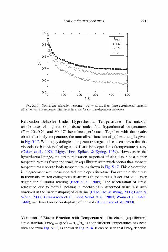
0.5
0.6
0.7
0.8
0.9
1.0
g (t
)
0 100 200 300t (s)
400 500
lmax
1.51.31.1
FIG. 5.16 Normalized relaxation responses, g tð Þ ¼ st=stR , from three experimental uniaxial
relaxation tests demonstrate differences in shape for the time-dependent responses.
Skin Biothermomechanics 221
Relaxation Behavior Under Hyperthermal Temperatures The uniaxial
tensile tests of pig ear skin tissue under four hyperthermal temperatures
(T ¼ 50,60,70, and 80 �C) have been performed. Together with the results
obtained at body temperature, the normalized function of gðtÞ ¼ st=stR is given
in Fig. 5.17. Within physiological temperature ranges, it has been shown that the
viscoelastic behavior of collagenous tissues is independent of temperature history
(Cohen et al., 1976; Rigby, Hirai, Spikes, & Eyring, 1959). However, in the
hyperthermal range, the stress–relaxation responses of skin tissue at a higher
temperature relax faster and reach an equilibrium state much sooner than those at
temperatures closer to body temperature, as shown in Fig. 5.17. This observation
is in agreement with those reported in the open literature. For example, the stress
in thermally treated collagenous tissue was found to relax faster and to a larger
degree for a similar loading (Baek et al., 2005). The acceleration of stress
relaxation due to thermal heating in mechanically deformed tissue was also
observed in the laser reshaping of cartilage (Chao, Ho, & Wong, 2003; Gaon &
Wong, 2000; Karamzadeh et al., 1999; Sobol et al., 2000; Wong et al., 1998,
1999), and laser thermokeratoplasty of corneal (Brinkmann et al., 2000).
Variation of Elastic Fraction with Temperature The elastic (equilibrium)
stress fraction, FracE ¼ gð1Þ ¼ s1=stR , under different temperatures has been
obtained from Fig. 5.17, as shown in Fig. 5.18. It can be seen that FracE depends

0.30 100 200 300
t (s)
400 500
0.4
0.5
0.6
g (t
) 0.7
0.8
0.9
1.0l max= 1.5
37
T (�C)
50
7080
60
FIG. 5.17 The normalized relaxation function, g tð Þ ¼ s tð Þ=stR , under different hyperthermal
temperatures.
222 F. Xu and T. J. Lu
strongly on the temperature: it decreases with increasing temperature over the
whole temperature range, but decreases much faster in the hyperthermal range
(T ¼ 50–80 �C). In other words, as the temperature decreases, the energy
dissipation decreases and the mechanical energy is increasingly elastic.
5.4.3. Discussion on Skin Relaxation Behavior
In this section, the possible roles played by different tissue components in skin
viscoelasticity and the thermally induced changes in these factors are discussed.
Effect of Different Components Histological examination reveals that the
viscoelastic response of skin tissue was mainly caused by the dermis, and not
the epidermis in nature (Tsubouchi et al., 2006): Silver, Freeman, and DeVore
(2001) also found the removal of epidermis does not change the viscoelastic
properties of the tissue. Therefore, the following discussion is relative to the roles
of different components of skin dermis.
Skin tissue is composed of cellular and extracellular compartments. The
extracellular compartment consists of dominant collagen fibers embedded in
ground substance. The mechanical response of skin tissue to applied loads
involves two components as noted previously: a viscous component associated
with energy dissipation by molecular and viscous sliding of collagen fibrils

0.4
0 20 40
T (�C)
60 80
0.5
0.6
0.7
0.8F
rac E
l max= 1.5
FIG. 5.18 The effect of temperature on the elastic fraction, FracE ¼ s1=stR , for skin tissue.
Skin Biothermomechanics 223
during alignment with the force direction (Silver et al., 1992); and an elastic
component associated with energy storage as a result of stretching of flexible
regions in the collagen triple helix (Silver, Christiansen, Snowhill, & Chen, 2000;
Silver, Horvath, & Foran, 2001; Silver et al., 2001). The viscous nature of
collagenous tissues arises from the inherent viscoelasticity of the solid phase as
well as fluid movement, resulting in viscous drag between the solid and fluid
phases during loading (Huang, Chang, Aggarwal, Lee, & Ehrlich, 1993).
Effect of Elastic Components The extent to which a tissue exhibits stress
relaxation has been shown to be directly related to the elastic content: elastin
and collagen (Fung, 1981). However, in the study here of pig ear skin tissue, the
elastin should not play an important role in the temperature-induced variation of
viscoelastic behavior, for a high stretch level (emax ¼ 50%) has been chosen. At
this level, the mechanical behavior of skin tissue is dominated by the response of
collagen fibers (Silver et al., 1992).
The collagen fiber component plays a major role in the stress–stretch response
of skin tissue, especially at high stretch levels (l � 1.3) (Potts & Breuer, 1981;
Silver et al., 2000; Silver, Horvath, et al., 2001). Collagen is intrinsically visco-
elastic (Haut & Little, 1972; Sanjeevi, Somanathan, & Ramaswamy, 1982),
implying that collagen viscoelasticity may be the source leading to the
viscoelastic behavior of skin tissue (Purslow et al., 1998). For example, a

224 F. Xu and T. J. Lu
correlation between collagen content and the viscoelastic behavior of rat skin has
been established (Vogel, 1988). An increase in collagen crosslinking was found
to increase both the viscoelastic storage and loss moduli of guinea pig skin tissue
under dynamic loading (Mansour, Davis, Srour, & Theberge, 1991). Further-
more, upon loading the initially crimped collagen fibers in dermis undergo
reorientation, suggesting that the time-dependent reorientation of collagen fibers
may be responsible for skin viscoelasticity. For example, the increase of elastic
fraction30 with increasing stretch in the skin stress–relaxation response was
attributed to the rearrangement of individual collagen fiber orientations, resulting
in an aligned collagen network at high stretch levels (Dunn & Silver, 1983).
Structural reorganization was also recognized to be important in tendon viscoe-
lasticity (Sasaki, Shukunami, Matsushima, & Izumi, 1999). However, Purslow
et al. found no involvement of collagen fiber rotation in skin viscoelastic stress
relaxation.
Role of Ground Substance The ground substance is a gel like substance
containing a class of chemicals including proteoglycans (PG) and glycoproteins.
The ground substance has been found to play an important role in the viscoelastic
behavior of dermis. According to Wilkes, Brown, and Wildnauer (1973), the
time-dependent behavior of dermis can be related to the viscous resistance that
fibers experience while moving through the ground substance. Minns, Soden, and
Jackson (1973) showed that the removal of ground substance in tendon, aorta and
ligamentum nuchae induced a decrease in relaxation hysteresis and other time-
dependent effects. Vogel (1976) found a correlation between the relaxation and
the content of glycosaminoglycans (GAG) in skin tissue, which constitute a part
of the PG. Purslow et al. (1998) also suggested that the viscoelastic effects of skin
tissue arise from molecular relaxations within the proteoglycan matrix surround-
ing the collagen fibers or within the collagen fibers themselves. Eshel and Lanir
(2001) found that the absence of proteoglycans influenced the low stretch relaxa-
tion patterns and deteriorated the viscoelastic mechanisms.
In summary, the skin viscoelasticity at high stretch level is decided by the
collagen and ground substance tissue while the component of elastin should not
play an important role. The temperature effect is discussed below.
30 Equivalent to the ratio of equilibrium force to initial force.

Skin Biothermomechanics 225
Effect of Temperature and Thermal Denaturation Results from thermome-
chanical tests on collagenous tissues can be difficult to interpret due to the
simultaneous occurrence of thermal damage, thermoelastic, and thermoviscoe-
lastic processes, the least understood being the altered viscoelastic behaviors
during and after heating (Baek et al., 2005).
As discussed in previous sections, three possible mechanisms exist for why
temperature effects change the viscoelastic behavior of skin tissue. The first one
is the thermal denaturation of dermis collagen and it seems reasonable to
assume that collagen plays a major role in the viscoelastic nature of skin tissue
subjected to thermal loading. For example, the acceleration of stress relaxation
in laser reshaping of cartilage was partly attributed to the temperature-
dependent, bound-free water transition in the cartilage matrix and selective
collagen denaturation (Chao et al., 2003; Gaon & Wong, 2000; Karamzadeh
et al., 1999; Sobol et al., 2000; Wong et al., 1998, 1999). Wells, Langille, Lee,
and Adamson (1999) found a correlation between the relaxation rate of ovine
aortic collagen and the concentration of thermally stable collagen crosslinks: if
more crosslinking is present, the slippage of adjacent chain fragments will be
inhibited, and the observed load relaxation will be slower. To check the role of
collagen denaturation, the heat-induced thermal damage degree (Deg) of pig
ear skin tissue under hyperthermal temperatures has been plotted as functions
of time, as shown in Fig. 5.19. These results clearly show that collagen in
skin tissue is damaged to a higher degree with increasing time and
temperature. This explains the abrupt change in the elastic fraction around
60 �C in Fig. 5.18.
Another possible mechanism is the temperature-induced change in hydration.
It has been shown that the viscoelastic behavior of soft tissues are related to the
interactions among collagen, proteoglycans, and water molecules (Nishimura
et al., 1998; Rochdi, Foucat, & Renou, 1999). There can be an inward or
outward flux of interstitial fluid due to heating a soft tissue (Humphrey, 2003)
and the viscoelastic properties of a tissue can be changed by altering its
water content (Chimich, Shrive, Frank, Marchuk, & Bray, 1992). Baek et al.
(2005) speculated that a loss of water causes the stress to relax, and the faster
relaxation in thermally treated tissues is caused by the faster and greater
water loss.
The third possible mechanism is the temperature-changed cell viability in skin
tissue. From Fig. 5.19, it can be seen that collagen in skin tissue is almost fully
damaged (Deg is near unity), indicating that fibroblast viability decreases with
heating duration and temperature. Using poison (2-deoxy-D-glucose), Yip et al.
(2007) studied the effect of in situ fibroblast viability on the mechanical
properties of rat back skin tissue. They found that there is greater stress relaxation

109A
B
106
103
100
10−3
10−6
0 100 200 300 400 500
T (°C)50607080
T (°C)50607080
t (s)
t (s)
0.0
10−2 10−1 10−0 101 102
0.2
0.4
0.6
0.8
1.0
Deg
W
FIG. 5.19 (A) Thermal damage and (B) damage degree due to temperature increase during the
relaxation tests.
226 F. Xu and T. J. Lu
in skin tissue stretched in normal Kreb’s solution than that in Kreb’s solution with
poison, and that there exists a correlation between the amount of stress relaxation
and the number of viable fibroblasts in the tissue: tissues with more viable
fibroblasts have lower stress relaxation.
In summary, temperature has significant effect on the viscoelastic behavior of
skin tissue; in the hyperthermal temperature range, the effect is caused by thermal
denaturation of collagen as well as heating-induced hydration and cell viability
changes.

Skin Biothermomechanics 227
5.5. Dynamic Viscoelasticity of Skin Tissue
Most studies on the viscoelasticity of skin tissue have been performed under
static experimental conditions. Although the understanding of mechanical
behavior under static loading is truly beneficial, it is also very important to
understand the mechanical behavior of skin tissue under dynamic loading. A
few researchers have described the viscoelastic properties of skin tissue under
dynamic loading (Dawes-Higgs, Swain, Higgs, Appleyard, & Kossard, 2004;
Jacquemoud, Bruyere-Garnier, & Coret, 2007; Pereira et al., 1991), but none has
considered temperature-dependent skin dynamic viscoelasticity. This is
addressed in this section.
Figure 5.20A–C plots, the measured storage modulus, loss modulus and loss
factor, respectively, of flank skin as functions of temperature. It is found that the
storage modulus has a value ranging from 0.5 to 10 MPa, consistent with the data
at room temperature reported in the literature (Pereira et al., 1991). The storage
modulus of pig skin tissue increases as the temperature increases, almost linearly,
implying that the skin tissue becomes stiffer with increasing temperature. The
loss factor, however, is practically independent of temperature, suggesting that
the viscous property of skin tissue is unaltered, that is, the damping capability of
pig skin tissue does not change with varying temperature. The loss factor has a
value of about 0.14, similar to that measured by the method of surface wave
propagation at room temperature (Kirkpatrick, Duncan, & Fang, 2004). The
results show no obvious variation in the slope of tan d as the DMA scan rate is
varied. However, the storage modulus appears to increase with increasing heating
rate.
To compare the dynamic results here with the previously obtained static
results, the storage modulus can be used as the corresponding Young’s modulus
in view of the comparably low testing frequency (1 Hz). By comparing the
modulus for the tensile behavior of pig ear skin sample under different tempera-
tures (Fig. 5.12A) with the storage modulus, as given in Fig. 5.21, it can found
that the magnitudes of the moduli agree well, but the storage modulus increases
with temperature, contrary to that of the tensile behavior. This is because the
dynamic testing was performed in a thermal chamber filled with air, which
resulted in the dehydration of the skin sample during heating, while the tensile
testing was performed in a chamber filled with liquid (KHR).
The storage modulus, loss modulus, and loss factor of pig skin tissue are
plotted as functions of thermal damage and thermal denaturation degree in
Fig. 5.22. These results show that the effects of thermal damage and thermal
denaturation degree on the viscoelastic properties of pig skin tissue are similar to
that of temperature. This means that the changes of collagen triple-helix structure

020 30 40 50 60 70 80
2
4
6
8A
B
C
E�
(MP
a)E
�� (
MP
a)
rh(°C/min)
1052
rh(°C/min)
1052
rh(°C/min)
1052
Pig flank skin
0
0.2
0.4
0.6
0.8
1.0Pig flank skin
T (°C)
20 30 40 50 60 70 80T (°C)
20 30 40 50 60 70 80T (°C)
0
0.1
0.2
0.3
0.4
tan
(d)
Pig flank skin
FIG. 5.20 Variations of (A) storage modulus, E0, (B) loss modulus, E00, and (C) loss factor, tan d,with temperature for flank skin sample.
228 F. Xu and T. J. Lu

030 40 50 60 70 80
2
4
6
8Pig ear skin
2
5
10
E�
(MP
a)
T (°C)
r h(°C/min)
FIG. 5.21 Variations of storage modulus, E0, with temperature for ear skin sample.
Skin Biothermomechanics 229
during thermal denaturation process may not be a major factor in deciding the
storage modulus and loss factor of tissue, at least at the test frequency used in this
study. It has been postulated that the mechanical changes observed using DMA
are the result of a combination of water loss and changes in the intrinsic
molecular structure of tissue with heating (Chae et al., 2003). In the present
investigation, the heat treatment causes collagen denaturation as well as dehy-
dration, and weight loss of the skin samples has also been observed during the
test. Thus, the variations in the tests here may be attributed to the release of bound
water during heating. This is supported by the fact that heat stabilization is
enhanced by the formation of strong interchains bonds as a result of condensation
reactions, making the collagen matrices stiffer with a higher resistance to defor-
mation (Pietrucha, 2005).
6. Conclusions
The widespread use of thermal therapies for skin disease treatments requires
the corresponding understanding of the biothermomechanical behavior of skin
tissue. In this chapter, a variety of mathematical approaches have been first
developed to examine the heat transfer process, thermal damage, and the heat-
induced mechanical response. Detailed analyses are then performed with both
Fourier and non-Fourier heat transfer models. For accurate modeling, the

10−6
A
B
10−4 10−2 100 102 104 106
10−6 10−5 10−4 10−3 10−2 10−1 100
0
0.1
0.2
0.3
0.4
tan
(d)
tan
(d)
r h(°C/min)2510
Pig flank skin
0
0.1
0.2
0.3
0.4Pig flank skin
Deg
W
rh(°C/min)
2510
FIG. 5.22 Variations of loss factor, tan d, with (A) thermal damage and (B) damage degree.
230 F. Xu and T. J. Lu
experimental characterization of the thermomechanical behavior of skin tissue
is necessary. Although the mechanical properties of skin tissue under normal
physiological conditions have been studied extensively, experimentally, both
in vivo and in vitro, there has been little characterization of the changes
in the mechanical properties of skin tissue due to temperature rising and the
corresponding thermal damage. These are thus addressed as the emphasis of this
study. The main summaries, limitations, and future work are described below.

Skin Biothermomechanics 231
6.1. Summary
The tensile, compressive, and viscoelastic behaviors of skin tissue under
different temperatures and thermal damage levels have been examined, to char-
acterize the effect of temperature variations and corresponding collagen denatur-
ation on the mechanical properties of skin tissue. The results have been discussed
together with the corresponding thermal damage process. Comparisons of the
experimental results with reported results in literature have been performed, and a
good agreement has been achieved. Internal comparisons of results obtained from
different methods have also been made. The following conclusions can be drawn.
DSC results show that the transition temperature of collagen in pig flank skin
samples is about 66.8 �C, and from the DSC results the Arrhenius parameters can
be obtained for skin samples from different locations. Under tensile loading, the
modulus at high stretch level of the tested skin samples decreases with increasing
temperature due to the thermal denaturation of skin collagen: compressive
loading, however, showed quite the opposite due to the different underlying
mechanisms between tensile and compressive behaviors of skin tissue. Biaxial
tests at 37 and 60 �C show that the skin tissue from porcine cheek appears to be
abnormal in the fact that it has an equal tensile response across both axes of a
square independent of cutting angle. This discovery has allowed the series of
experiments on skin tissue to come to fruition since a uniform stress distribution
in the tissue can be easily achieved even in biaxial tests. Compared with uniaxial
stretching, biaxial tensile loading results in the lateral narrowed stress–stretch
curve and the reduction in the stretch before entry into the linear region occurs
under all temperatures tested, which is due to the two-directional stretch of
collagen fibers. Under compression, the stiffness of skin tissue decreases with
increasing thermal damage degree. A stretch-rate sensitivity is observed at
different damage levels, and the stiffness of skin tissue decreases with thermal
damage. Temperature has a significant effect on the static viscoelasticity of skin
tissue. As for the dynamic skin viscoelasticity, DMA results showed that the
storage modulus is highly temperature dependent; however, the loss factor did
not show a significant dependency on either temperature or thermal denaturation.
6.2. Limitations
There are several limitations to the present study, as described below:
1. The thermal denaturation of a collagenous tissue depends on not only the
temperature history but also the stress state during heating (Humphrey,
2003), which is, however, very difficult to quantify due to the lack of

232 F. Xu and T. J. Lu
corresponding research. Therefore, in the calculation of thermal damage
the parameters obtained from DSC measurements in the absence of applied
tractions were used.
2. It is well known that the mechanical properties of skin tissue are in general
nonlinearly elastic and viscoelastic. In the tensile and compressive testing
of the present study, however, we concentrated only on the nonlinear
elasticity of the tissue. The data of compressive behavior were fit with
the Odgen model with the assumption of incompressivity. However, it was
found that pigskin under compression is compressible; even in unconfined
compression, the volume of specimens varies with increasing compressive
deformation (Wu et al., 2003).
3. Engineering stretch/strain and stress are used in this chapter. Most
mechanical models including Kelvin–Maxwell model and integrate
model use this kind of definitions and a good agreement with the experi-
ment results has been achieved. However, these are linear elastic defini-
tions, which have been pointed out to be inappropriate for studies of large
deformation and nonlinear solid mechanics should be used in the future
study.
4. The viscoelastic behavior of skin was only tested for uniaxial loading and,
thus, biaxial loading needs to be studied in the future for skin in vivo
experiences multiaxial natural tension. The stretch applied in the present
relaxation test was 1.5, but a previous study found that after a 1.2 stretch
the fibroblasts were apoptotic or necrotic (Martel, Walker, Reed, & Bert,
2001).
5. The dynamic testing in this studywas performed in a chamber filled with air,
and the mechanical changes observed were induced by a combination of
effects that include both water loss and changes in the intrinsic molecular
structure of tissue with heating. Further studies should focus on identifying
the relative contribution of water loss and thermal denaturation. Another
limitation of the dynamic testing is that all the tests were performed at a fixed
frequency of 1 Hz and it is expected that the loading frequency would also
play a role.
6. In our previous studies (Xu, Wen, Seffen, et al., 2008), a mathematical
approach has been developed for determining the thermomechanical
response in skin tissue, where temperature/damage independent linear
elastic mechanical properties of skin tissue have been used. The assump-
tions used are reasonable in view that (1) the stretch level considered in
that case is purely induced by thermal loads and is thus comparatively small
(< 1.01) and (2) the results in the present study have shown that temperature/
damage has little effect at this low stretch level (see Figs. 5.12 and 5.16).

Skin Biothermomechanics 233
However, in medical applications, deformation of skin at high stretch level
also occurs, for example, skin indentation during laser-skin treatment
(Basinger, Aguilar, & Nelson, 2004; Klavuhn, 2001). For these cases, it
can be expected that the incorporation of the temperature/damage depen-
dentmechanical properties in the thermomechanical model will have a great
effect on the predicted thermomechanical behavior of skin tissue.
6.3. Future Work
The approach developed in this study provides a platform to study the thermal
and mechanical behaviors, and corresponding pain sensation. This method intro-
duces engineering to the fields of biology and neurophysiology, opening the door
to future advancements at the interface of these fields.
First, the full suite of models and experimental results can be used for
development of a thermal therapy model with a user-friendly interface.
The therapy model can be validated by comparing it with the response from
the clinical patients. Once validated, the model can then be used for (1) charac-
terization of present strategies for delivering thermal therapies by considering
patient’s pain feeling as an additional factor; (2) optimization of thermal
treatments by maximizing the therapeutic effect while minimizing unwanted
side effects such as pain sensation; and (3) design of new and better treatment
strategies, to assert novel heat treatments in view of productive commercial
exploitation.
Second, it can be used for improving artificial skin, for example, robot skin.
Robot has wider applications in different environments, especially in space.
Robotic skin is needed to respond to work more efficiently in highly unstructured
environments and facilitate human–robot interactions for both work and enter-
tainment purposes. The approach developed in this study can help the design of
new robot skin which can detect pressure, temperature, moisture/humidity, hard-
ness of objects, and electrical conductivity of objects.
Third, the approach developed in this study can be helpful for the transdermal
drug delivery, for example, needle-free injection. Needle-free injection has been
used for more than 50 years with excellent bioavailability. However, pain and
bruising have limited its wider applications. The holistic skin pain model consid-
ering both skin mechanical and neural response can help to better understand the
physical mechanisms of jet penetration into skin and to improve the current
injectors by minimizing the pain sensation felt by patients.
234 F. Xu and T. J. Lu
Acknowledgments
This work was supported by the Overseas Research Studentship (ORS) and
Overseas Trust Scholarship of Cambridge University, the National Natural Sci-
ence Foundation of China (10572111, 10632060), the National Outstanding
Youth Foundation of China, the National 111 Project of China (B06024), and
the National Basic Research Program of China (2006CB601202).
References
Abramowitch, S. D., & Woo, S. L. (2004). An improved method to analyze the stress relaxation of
ligaments following a finite ramp time based on the quasi-linear viscoelastic theory. Journal ofbiomechanical engineering, 126(1), 92–97.
Abramson, D. I. (1967). Circulation in the extremiries. New York: Academic.
Agah, R., Gandjbakhche, A. H., Motamedi, M., Nossal, R., & Bonner, R. F. (1996). Dynamics of
temperature dependent optical properties of tissue: Dependence on thermally induced alteration.
IEEE Transactions on Bio-medical Engineering, 43(8), 839–846.
Ahn, T.-K., & Kim, K.-J. (1994). Sensitivity analysis for estimation of complex modulus of visco-
elastic materials by non-resonance method. Journal of Sound and Vibration, 176, 543–561.Aksan, A., &McGrath, J. J. (2003). Thermomechanical analysis of soft-tissue thermotherapy. Journal
of Biomechanical Engineering, 125(5), 700–708.
Alberts, B., Bray, D., Lewis, J., Raff, M., Roberts, K., & Watson, J. D. (1994). Molecular biology of
the cell. New York: Garland.
Alexander, H., & Cook, T. H. (1976). Variations with age in the mechanical properties of human skin
in vivo. In R. M. Kennedy (Ed.), Bedsore biomechanics (pp. 109–118). Bath: McMillan Press.
Allain, J. C., Le Lous, M., Cohen-Solal, L., Bazin, S., & Maroteaux, P. (1980). Isometric tensions
developed during the hydrothermal swelling of rat skin. Connect Tissue Research, 7, 127–133.Ankersen, J., Birkbeck, A. E., Thomson, R. D., & Vanezis, P. (1999). Puncture resistance and tensile
strength of skin simulants. Proceedings of the Institution of Mechanical Engineers. Part H,
213(6), 493–501.Arbogast, K. B., Thibault, K. L., Scott Pinheiro, B., Winey, K. I., & Margulies, S. S. (1997). A high-
frequency shear device for testing soft biological tissues. Journal of biomechanics, 30(7),
757–759.
Arendt-Nielsen, L., & Chen, A. C. N. (2003). Lasers and other thermal stimulators for activation of
skin nociceptors in humans. Neurophysiologie Clinique/Clinical Neurophysiology, 33(6),
259–268.
Arkin, H., Xu, L. X., & Holmes, K. R. (1994). Recent developments in modeling heat transfer in blood
perfused tissues. IEEE Transactions on Biomedical Engineering, 41(2), 97–107.Arnoczky, S. P., & Aksan, A. (2000). Thermal modification of connective tissues: Basic science
considerations and clinical implications. The Journal of the American Academy of Orthopaedic
Surgeons, 8(5), 305–313.
Arumugam, V., Naresh, M. D., & Sanjeevi, R. (1994). Effect of strain rate on the fracture behaviour of
skin. Journal of Bioscience, 19(3), 307–313.
Asahina, A., Watanabe, T., Kishi, A., Hattori, N., Shirai, A., Kagami, S., et al. (2006). Evaluation of
the treatment of port-wine stains with the 595-nm long pulsed dye laser: A large prospective study
in adult Japanese patients. Journal of the American Academy of Dermatology, 54(3), 487–493.
Auriol, F., Vaillant, L., Machet, L., Diridollou, S., & Lorette, G. (1993). Effects of short-time
hydration on skin extensibility. Acta Dermato-Venereologica, 73(5), 344–347.

Skin Biothermomechanics 235
Baek, S., Wells, P. B., Rajagopal, K. R., & Humphrey, J. D. (2005). Heat-induced changes in the finite
strain viscoelastic behavioir of a collaagenous tissue. Journal of Biomechanical Engineering,
127(4), 580–586.
Barbenel, J. C., & Evans, J. H. (1977). The time-dependent mechanical properties of skin. The Journalof Investigative Dermatology, 69(3), 318–320.
Basinger, B., Aguilar, G., & Nelson, J. S. (2004). Effect of skin indentation on heat transfer during
cryogen spray cooling. Lasers in Surgery and Medicine, 34(2), 155–163.
Bayazitoglu, Y., & Peterson, G. P. (1992). Fundamental issues in hyperbolic heat conduction. ASMEHTD, 227.
Bert, J. L., & Reed, R. K. (1997). Pressure-volume relationship for rat dermis: Compression studies.
Acta physiologica Scandinavica, 160(1), 89–94.
Bhowmick, S., & Bischof, J. C. (1998). Supraphysiological thermal injury in dunning AT-1 Prostate
tumor cells. Advances in Heat and Mass Transfer in Biotechnology, 77–78. ASME HTD-Vol. 362/
BED-40.
Bonifasi-Lista, C., Lake, S., Ellis, B., Rosenberg, T., &Weiss, J. A. (2002). Strain- and rate-dependent
viscoleastic properties of human MCL in tension. Transactions of the Orthopaedic Research
Society, 27, 609.
Bonilla, C. M. A., Massanet, A. R. F., & Almodovar, N. V. (2005). Mechanics of biomaterials: Skin
repair and grafts. In: Proceedings of applications of engineering mechanics in medicine.Mayaguez: University of Puerto Rico.
Bosman, S. (1993). Heat-induced structural alterations in myocardium in relation to changing optical
properties. Applied Optics, 32(4), 461–463.
Brinkmann, R., Radt, B., Flamm, C., Kampmeier, J., Koop, N., & Birngruber, R. (2000). Influence of
temperature and time on thermally induced forces in corneal collagen and the effect on laser
thermokeratoplasty. Journal of Cataract and Refractive Surgery, 26, 744–754.
Britton, N. F., & Skevington, S. M. (1996). On the mathematical modelling of pain. NeurochemicalResearch, 21(9), 1133–1140.
Burnstock, G. (1996). A unifying purinergic hypothesis for the initiation of pain. Lancet Neurol, 347,
1604–1605.
Burnstock, G., & Wood, J. N. (1996). Purinergic receptors: Their role in nociception and primary
afferent neurotransmission. Current Opinion in Neurobiology, 6, 526–532.
Campbell, J. N., & LaMotte, R. H. (1983). Latency to detection of first pain. Brain Research, 266,
203–208.
Carew, E. O., Talman, E. A., Boughner, D. R., & Vesely, I. (1999). Quasi-linear viscoelastic
theory applied to internal shearing of porcine aortic valve leaflets. Journal of Biomechanical
Engineering Transactions ASME, 121(4), 386–392.
Caterina, M. J., & Julius, D. (1999). Sense and specificity: A molecular identity for nociceptors.
Current opinion in neurobiology, 9, 525–530.
Caterina, M. J., Rosen, T. A., Tominaga, M., Brake, A. J., & Julius, D. (1999). Capsaicin-receptor
homologue with a high threshold for noxious heat. Nature, 398(6726), 436–441.
Cattaneo, C. (1958). A form of heat conduction equation which eliminates the paradox of instanta-
neous propagation. Compute Rendus, 247, 431–433.
Cesare, P., &McNaughton, P. A. (1996). A novel heat-activated current in nociceptive neurons and its
sensitization by bradykinin. Proceedings of the National Academy of Sciences of the USA, 93(26),
15435–15439.
Chachra, D., Gratzer, P. F., Pereira, C. A., & Lee, J. M. (1996). Effect of applied uniaxial stress on rate
and mechanical effects of cross-linking in tissue-derived biomaterials. Biomaterials, 17(19),
1865–1875.
Chae, Y., Aguilar, G., Lavernia, E. J., & Wong, B. J. (2003). Characterization of
temperature dependent mechanical behavior of cartilage. Lasers in Surgery and Medicine,
32(4), 271–278.

236 F. Xu and T. J. Lu
Chao, K. K. H., Burden, M. A., & Wong, B. J. F. (2001). Dynamic changes in the elastic modulus of
lagomorph nasal septal cartilage during Nd:YAG (&lambda ¼ 1.32 mm) laser irradiation
(pp. 247–254). San Jose, CA: Society of Photo-Optical Instrumentation Engineers.
Chao, K. K., Ho, K. H., & Wong, B. J. (2003). Measurement of the elastic modulus of rabbit nasal
septal cartilage during Nd:YAG (lambda ¼ 1.32 micron) laser irradiation. Lasers in Surgery and
Medicine, 32(5), 377–383.
Charny, C. K. (1992). Mathematical models of bioheat transfer. Advances in Heat Transfer, 22,
19–155.
Chato, J. C. (1980). Heat transfer to blood vessels. Journal of Biomechanical Engineering, Transac-
tions of the ASME, 102(2), 110–118.
Chen, M. M., & Holmes, K. R. (1980). Microvascular contributions in tissue heat transfer.
Annals New York Academy Science, 335, 137–150.Chen, S. S., & Humphrey, J. D. (1998). Heat-induced changes in the mechanics of a collagenous
tissue: Pseudoelastic behavior at 37 degrees C. Journal of Biomechanics, 31(3), 211–216.
Chen, S. S., Wright, N. T., & Humphrey, J. D. (1997). Heat-induced changes in the mechanics of a
collagenous tissue: Isothermal free shrinkage. Journal of Biomechanical Engineering, 119(4),
372–378.
Chen, S. S., Wright, N. T., & Humphrey, J. D. (1998a). Heat-induced changes in the mechanics of a
collagenous tissue: Isothermal, isotonic shrinkage. Journal of Biomechanical Engineering,120(3), 382–388.
Chen, S. S., Wright, N. T., & Humphrey, J. D. (1998b). Phenomenological evolution equations for
heat-induced shrinkage of a collagenous tissue. IEEE Transactions on Biomedical Engineering,
45(10), 1234–1240.Chester, M. (1963). Second sound in solid. Physical. Reviews, 131, 2013–2015.
Chimich, D., Shrive, N., Frank, C., Marchuk, L., & Bray, R. (1992). Water content alters viscoelastic
behavior of the normal adolescent rabbit medial collateral ligament. Journal of Biomechanics, 25,831–837.
Choi, H. S., & Vito, R. P. (1990). Two-dimensional stress-strain relationship for canine pericardium.
Journal of Biomechanical Engineering, Transactions of the ASME, 112, 153–159.
Choiniere, M., Melzack, R., Girard, N., Rondeau, J., & Paquin, M. J. (1990). Comparisons between
patients’ and nurses’ assessment of pain and medication efficacy in severe burn injuries. Pain, 40,
143–152.
Chvapil, M., & Jensovsky, L. (1963). The shrinkage temperature of collagen fibres isolated from the
tail tendons of rats of various ages and from different places of the same tendon. Gerontologia, 1,18–29.
Cohen, R. E., Hooley, C. J., & McCrum, N. G. (1976). Viscoelastic creep of collagenous tissue.
Journal of Biomechanics, 9(4), 175–184.Connors, K. A. (1990). Chemical kinetics. Wiley-VCH: The Study of Reaction Rates in Solution.
Cox, H. (1942). The cleavage lines of the skin. The British Journal of Surgery, 29, 234–240.
Crezee, J., & Lagendijk, J. J. (1992). Temperature uniformity during hyperthermia: The impact of
large vessels. Physics in Medicine and Biology, 37, 1321–1337.Crezee, J., Mooibroek, J., Lagendijk, J. J., & van Leeuwen, G. M. (1994). The theoretical and
experimental evaluation of the heat balance in perfused tissue. Physics in Medicine and Biology,
39(5), 813–832.
Dahan, S., Lagarde, J. M., Turlier, V., Courrech, L., &Mordon, S. (2004). Treatment of neck lines and
forehead rhytids with a nonablative 1540-nm Er:glass laser: A controlled clinical study combined
with the measurement of the thickness and the mechanical properties of the skin. Dermatologic
Surgery, 30(6), 872–879.
Daly, C. H. (1982). Biomechanical properties of dermis. The Journal of Investigative Dermatology,79(Suppl. 1), s17–s20.

Skin Biothermomechanics 237
Davidson, J. M., Giro, M., Sutclie, M., Zoia, O. D. Q., Liu, J. J. M., Perkett, E., et al. (1990).
Regulation of elastin synthesis. In A. M. Tamburro & J. M. Davidson (Eds.), Elastin: Chemical
and biological aspects. Galatina, Italy: Congedo Editore.
Davis, E. D., Doss, D. J., Humphrey, J. D., & Wright, N. T. (2000). Effects of heat-induced damage
on the radial component of thermal diffusivity of bovine aorta. Journal of Biomechanical
Engineering, 122(3), 283–286.
Dawes-Higgs, E. K., Swain, M. V., Higgs, R J E D, Appleyard, R. C., & Kossard, S. (2004). Accuracy
and reliability of a dynamic biomechanical skin measurement probe for the analysis of stiffness
and viscoelasticity. Physiological Measurement, 25(1), 97–105.
de Boer, J. F., Srinivas, S. M., Malekafzali, A., Chen, Z. P., & Nelson, J. S. (1998). Imaging thermally
damaged tissue by polarization sensitive optical coherence tomography. Optics Express, 3,
212–218.
Decraemer, W. F., Maes, M. A., Vanhuyse, V. J., & Vanpeperstraete, P. (1980). A non-linear
viscoelastic constitutive equation for soft biological tissues based upon a structural model.
Journal of Biomechanics, 13, 559–564.Defrate, L. E., & Li, G. (2006). The prediction of stress-relaxation of ligaments and tendons using the
quasi-linear viscoelastic model. Biomechanics and Modeling in Mechanobiology, 6, 245–251.
Delalleau, A., Josse, G., Lagarde, J. M., Zahouani, H., & Bergheau, J. M. (2006). Characterization of
the mechanical properties of skin by inverse analysis combined with the indentation test. Journalof Biomechanics, 39(9), 1603–1610.
Diaz, S., Lavemia, E., & Wong, B. J. F. (2001). Mechanical behavior of cartilage during laser
irradiation (pp. 192–197). San Jose, CA: Society of Photo-Optical Instrumentation Engineers.
Diette, K. M., Bronstein, B. R., & Parrish, J. A. (1985). Histologic comparison of argon and tunable
dye lasers in the treatment of tattoos. The Journal of Investigative Dermatology, 85(4), 368–373.
Dikstein, S., & Hartzshtark, A. (1983). What does low-pressure indentometry measure. Arztliche
Kosmetologie, 16, 327–328.Diller, K. R., & Pearce, J. A. (1999). Issues in modeling thermal alterations in tissues. Annals New
York Academy of Science, 888, 153–164.
Dinnar, U. (1970). A note on theory of deformation in compressed skin tissues. Mathematical
Bioscience, 8, 71–82.Diridollou, S., Vabre, V., Berson, M., Vaillant, L., Black, D., Lagarde, J. M., et al. (2001). Skin
ageing: Changes of physical properties of human skin in vivo. International Journal of Cosmetic
Science, 23, 353–362.
Dittert, I., Vlachova, V., Knotkova, H., Vitaskova, Z., Vyklicky, L., Kress, M., et al. (1998).
A technique for fast application of heated solutions of different composition to cultured neurones.
Journal of Neuroscience Methods, 82, 195–201.
Doehring, T. C., Carew, E. O., & Vesely, I. (2004). The effect of strain rate on the viscoelastic
response of aortic valve tissue: A direct-fit approach. Annals of Biomedical Engineering, 32(2),
223–232.
Dortmans, L. J., Sauren, A. A., & Rousseau, E. P. (1984). Parameter estimation using the quasi-linear
viscoelastic model proposed by fung. Journal of Biomechanical Engineering, Transactions of theASME, 106(3), 198–203.
Douglas, W. R. (1972). Of pigs and men and research: A review of applications and analogies of the
pig, Sus scrofa, in human medical research. Space and Life Sciences, 3, 226–234.
Downs, J., Halperin, H. R., Humphrey, J. D., & Yin, F. C. P. (1990). An improved video-based
computer tracking system for soft-biomaterials testing. IEEE Transactions on Bio-medical
Engineering, 37, 903–907.
Duck, F. A. (1990). Physical properties of tissue: A comprehensive reference book. New York:
Academic Press.
Dunn, M. G., & Silver, F. H. (1983). Viscoelastic behavior of human connective tissues: Relative
contribution of viscous and elastic components. Connect.Tissue Res., 12(1), 59–70.

238 F. Xu and T. J. Lu
Dunn, M. G., Silver, F. H., & Swann, D. A. (1985). Mechanical analysis of hypertrophic scar tissue:
Structural basis for apparent increased rigidity. The Journal of Investigative Dermatology, 84(1),
9–13.
Ebling, F., Eady, R., & Leigh, I. (1992). Anatomy and organization of human skin. In
R. H. Champion, J. L. Burrington & F. J. G. Ebling (Eds.), Textbook of dermatology.
New York: Blackwell Scientific Publications.
Edwards, C., & Marks, R. (1995). Evaluation of biomechanical properties of human skin. Clinics.
Dermatology, 13(4), 375–380.Elkins, W., & Thomson, J. G. (1973). Instrumented thermal manikin. Acurex Corporation, Aerotherm
Division Report AD-781, 176.
Elmore, D. E. (2004). Investigations of ion channel structure-function relationships using
molecular modeling and experimental biochemistry. California: California Institute of
Technology.
Elsner, P., Wilhelm, D., & Maibach, H. I. (1990). Mechanical properties of human forearm and vulvar
skin. British Journal of Dermatology, 122(5), 607–614.Eshel, H., & Lanir, Y. (2001). Effects of strain level and proteoglycan depletion on preconditioning
and viscoelastic responses of rat dorsal skin. Annals of Biomedical Engineering, 29, 164–172.
Essenpreis, M. (1992). Thermally induced changes in optical properties of biological tissues. London:
University College London.
Ferry, J. D. (1980). Viscoelastic properties of polymers. New York: Wiley.
Fidanza, F., Keys, A., & Anderson, J. T. (1953). Density of body fat in man and other mammals.
Journal of Applied Physiology, 6, 252–256.
Fink, W. A. Jr, (2005). The pathophysiology of acute pain. Emergency Medicine Clinics of NorthAmerica, 23(2), 277–284.
Finlay, B. (1969). Scanning electron microscopy of the human dermis under uni-axial strain.
Bio-medical Engineering, 4, 322–327.Flory, P. J., & Garrett, R. R. (1958). Phase transition in collagen and gelatin systems. Journal of
American Chemical Society, 80, 4836–4845.
Fung, Y. C. (1971). Stress-strain history relations of soft tissues in simple elongation. In Y. C. Fung,
N. Perrone & M. Anliker (Eds.), Biomechanics: Its foundations and objectives (pp. 181–208).Englewood Cliffs, NJ: Prentice-Hall, Inc.
Fung, Y. C. (1981). Biomechanics: Mechanical properties of living tissue. New York: Springer.
Fung, Y. C. (1990). Biomechanics: Motion, flow, stress, and growth. New York: Springer.
Fung, Y. C. (1993). Biomechanics: Mechanical properties of living tissues. New York: Springer.
Funk, J. R., Hall, G. W., Crandall, J. R., & Pilkey, W. D. (2000). Linear and quasi-linear viscoelastic
characterization of ankle ligaments. Journal of Biomechanical Engineering, Transactions of the
ASME, 122(1), 15–22.Gallagher, G., Rae, C. P., & Kinsella, J. (2000). Treatment of pain in severe burns. American Journal
of Clinical Dermatology, 1, 329–335.
Gaon, M. D., &Wong, B. J. F. (2000). Measurement of the elastic modulus of porcine septal cartilage
specimens following Nd:YAG laser treatment. Proceedings—Society of Photo-Optical Instru-mentation Engineers, 3907, 370–379.
Giles, J. M., Black, A. E., & Bischoff, J. E. (2007). Anomalous rate dependence of the preconditioned
response of soft tissue during load controlled deformation. Journal of Biomechanics, 40(4),
777–785.
Gimbel, J. A., Sarver, J. J., & Soslowsky, L. J. (2004). The effect of overshooting the target strain on
estimating viscoelastic properties from stress relaxation experiments. Journal of Biomechanical
Engineering, Transactions of the ASME, 126(6), 844–848.
Godoy, S., & Garcıa-Colın, L. S. (1997). Nonvalidity of the telegrapher’s diffusion equation in two
and three dimensionsfor crystalline solids. Physical Reviews E, 55, 2127–2131.

Skin Biothermomechanics 239
Gordon, R. G., Roemer, R. B., & Howath, S. M. (1976). A mathematical model of the
human temperature regulatory system—Transient cold exposure responses. IEEE Transactions
on Biomedical Engineering, 23(6), 434–444.
Green, D. M., & Diller, K. R. (1978). Measurement of burn induced leakage of macromolecules in
living tissue. Transactions of the ASME, Journal of Biomechanical, 100, 153–158.
Haeri, M., Asemani, D., & Gharibzadeh, S. (2003). Modeling of pain using artificial neural networks.
Journal of Theoretical Biology, 220(3), 277–284.
Hamilton, M. M. (2004). Laser treatment of pigmented and vascular lesions in the office. FacialPlastic Surgery, 20(1), 63–69.
Hamilton, S. G. (2002). ATP and pain. Pain Practice, 2(4), 289–294.
Harris, J. L., Wells, P. B., & Humphrey, J. D. (2003). Altered mechanical behavior of epicardium due
to isothermal heating under biaxial isotonic loads. Journal of Biomechanical Engineering, 125,381–388.
Haut, R. C. (1983). The sensitivity of skin to strain rate of loading. 7th Annual meeting of the american
society of biomechanics, Rochester, Minn.
Haut, R. C., & Little, R. W. (1972). A constitutive equation for collagen fibers. Journal of Biome-
chanics, 5(5), 423–430.
Hendriks, F. M., Brokken, D., Oomens, C. W., Bader, D. L., & Baaijens, F. P. (2006). The relative
contributions of different skin layers to the mechanical behavior of human skin in vivo using
suction experiments. Medical Engineering and Physics, 28(3), 259–266.
Henriques, F. C., & Moritz, A. R. (1947). Studies of thermal injury, 1. The conduction of heat to and
through skin and the temperatures attained therein. A theoretical and an experimental investiga-
tion. American Journal of Pathology, 23, 531–549.Hepworth, D. G., Steven-fountain, A., Bruce, D. M., & Vincent, J. F. (2001). Affine versus non-affine
deformation in soft biological tissues, measured by the reorientation and stretching of collagen
fibres through the thickness of compressed porcine skin. Journal of Biomechanics, 34(3),341–346.
Hilliges, M., Weidner, C., Schmelz, M., Schmidt, R., Orstavik, K., Torebjork, E., et al. (2002). ATP
responses in human C nociceptors. Pain, 98(1–2), 59–68.
Hingorani, R. V., Provenzano, P. P., Lakes, R. S., Escarcega, A., & Vanderby, R. J. (2004). Nonlinear
viscoelasticity in rabbit medial collateral ligament. Annals of Biomedical Engineering, 32,
306–312.
Hoffman, A. H., Robichaud, D. R., & Grigg, P. (2003). The effect of hydration upon the compliance
properties of rat skin (pp. 633–634). Proceedings of the 2003 summer bioengineering conference,Key Biscayne, FL.
Hormann, H., & Schlebusch, H. (1971). Reversible and irreversible denaturation of collagen fibers.
Biochemistry, 10(6), 932–937.Huang, D., Chang, T. R., Aggarwal, A., Lee, R. C., & Ehrlich, H. P. (1993). Mechanisms and
dynamics of mechanical strengthening in ligament-equivalent fibroblast-populated collagen
matrices. Annals of Biomedical Engineering, 21, 289.
Hult, A. M., & Goltz, R. W. (1965). The measurement of elastin in human skin and its quantity in
relation to age. Journal of Investigative Dermatology, 44, 408–412.
Humphrey, J. D. (2003). Continuum thermomechanics and the clinical treatment of disease and injury.
Applied Mechanics Reviews, 56(2), 231–260.
Humphries, W. T., & Wildnauer, R. H. (1971). Thermomechanical analysis of stratum corneum. I.
Technique. The Journal of Investigative Dermatology, 57(1), 32–37.
Jacquemoud, C., Bruyere-Garnier, K., & Coret, M. (2007). Methodology to determine failure char-
acteristics of planar soft tissues using a dynamic tensile test. Journal of Biomechanics, 40(2),
468–475.
Jacques, S. L. (2006). Ratio of entropy to enthalpy in thermal transitions in biological tissues. Journal
of Biomedical Optics, 11(4), 041108.

240 F. Xu and T. J. Lu
James, N. C., & Richard, A. M. (1996). Neurobiology of nociceptors. Oxford: Oxford University
Press.
Jamison, C. E., Marangoni, R. D., & Glaser, A. A. (1968). Viscoelastic properties of soft tissue by
discrete model characterization. Journal of Biomechanics, 1(1), 33–36.Jansen, L. H., & Rottier, P. B. (1958a). Comparison of the mechanical properties of strips of human
abdominal skin excised from below and from above the umbilic. Dermatologica, 117(4),
252–258.
Jansen, L. H., & Rottier, P. B. (1958b). Some mechanical properties of human abdominal skin
measured on excised strips: A study of their dependence on age and how they are influenced by
the presence of striae. Dermatologica, 117(2), 65–83.
Jemec, G. B., Jemec, B., Jemec, B. I., & Serup, J. (1990). The effect of superficial hydration on the
mechanical properties of human skin in vivo: Implications for plastic surgery. Plastic andReconstructive Surgery, 85(1), 100–103.
Johnson, T. E., Mitchell, M. A., Rico, P. J., Fletcher, D. J., Eurell, T. E., & Roach, W. P. (2001).
Corneal and skin laser exposures from 1540 nm laser pulses, laser-tissue interactions XI:
Photochemical, photothermal, and photomechanical. In D. D. Duncan, J. O. Hollinger & S. L.
Jacques (Eds.), SPIE (pp. 222–229). .
Johnson, G. A., Tramaglini, D. M., Levine, R. E., Ohno, K., Choi, N.-Y., & Woo, S. L.-Y. (1994).
Tensile and viscoelastic properties of human patellar tendon. Journal of Orthopedic Research, 12,796–803.
Jun, J. H., Harris, J. L., Humphrey, J. D., & Rastegar, S. (2003). Effect of thermal damage and biaxial
loading on the optical properties of a collagenous tissue. Journal of Biomechanical Engineering,
125(4), 540–548.Karamzadeh, A. M., Wong, B. J. F., Milner, T. E., Dao, X., Tanenbaum, B. S., & Nelson, J. S. (1999).
Temperature distributions in Nd:YAG (¼1.32 mm) laser-irradiated cartilage grafts accompanied
by cryogen spray cooling. Proceedings—Society of Photo-Optical Instrumentation Engineers,3601, 422–433.
Kauvar, A. N., Rosen, N., & Khrom, T. (2006). A newly modified 595-nm pulsed dye laser with
compression handpiece for the treatment of photodamaged skin. Lasers in Surgery and Medicine,
38(9), 808–813.Kenedi, R. M., Gibson, T., Evans, J. H., & Barbenel, J. C. (1975). Tissue mechanics. Physics in
Medicine and Biology, 20(3), 699–717.
Khaled, A. R. A., & Vafai, K. (2003). The role of porous media in modeling flow and heat transfer in
biological tissues. International Journal of Heat and Mass Transfer, 46(26), 4989–5003.Kirkpatrick, S. J., Duncan, D. D., & Fang, L. (2004). Low-frequency surface wave propagation and the
viscoelastic behavior of porcine skin. Proceedings of SPIE, 5474, 127.
Kirschstein, T., Busselberg, D., & Treede, R. D. (1997). Coexpression of heat-evoked and capsaicin-
evoked inward currents in acutely dissociated rat dorsal root ganglion neurons. Neuroscience
Letters, 231, 33–36.
Klavuhn, K. G. (2001). Epidermal protection: A comparative analysis of sapphire contact and
cryogen spray cooling. Lumenis Inc.
Koerner, C., & Bergmann, H. W. (1998). Physical defects of the hyperbolic heat conduction equation.
Applied Physics A: Materials Science & Processing, 67(4), 397–401.
Kollar, L. P., & Springer, G. S. (2003). Mechanics of composite structure. Cambridge: Cambridge
University Press.
Komatsu, K., Sanctuary, C., Shibata, T., Shimada, A., & Botsis, J. (2007). Stress-relaxation and
microscopic dynamics of rabbit periodontal ligament. Journal of biomechanics, 40(3), 634–644.
Kondo, E., Yasuda, K., Kitamura, N., Kudoh, T., Minami, A., & Tohyama, H. (2005). The effect of
electrothermal shrinkage on the biomechanical properties of the anterior cruciate ligament: An
experimental study. Arthroscopy, 21(4), 448–456.

Skin Biothermomechanics 241
Kono, T., Groff, W. F., Sakurai, H., Takeuchi, M., Yamaki, T., Soejima, K., et al. (2006). Evaluation
of fluence and pulse-duration on purpuric threshold using an extended pulse pulsed-dye laser in
the treatment of port wine stains. The Journal of Dermatology, 33(7), 473–476.
Krishtal, O. A., & Pidoplichko, V. I. (1980). A receptor for protons in the nerve cell membrane.
Neuroscience, 5, 2325–2327.
Krishtal, O. A., & Pidoplichko, V. I. (1981). A receptor for protons in the membrane of sensory
neurons may participate in nociception. Neuroscience, 6, 2599–2601.
Kruger, L., Perl, E. R., & Sedivec, M. J. (1981). Fine structure of myelinated mechanical nociceptor
endings in cat hairy skin. The Journal of Comparative Neurology, 198(1), 137–154.
Kwan, M. K., Lin, T. H., & Woo, S. L. (1993). On the viscoelastic properties of the anteromedial
bundle of the anterior cruciate ligament. Journal of Biomechanics, 26, 447–452.
Lakes, R. S. (1998). Viscoelastic solids. New York: CRC Press.
Lakes, R. S., & Vanderby, R. (1999). Interrelation of creep and relaxation: A modeling approach for
ligaments. Journal of Biomechanical Engineering, Transactions of the ASME, 121, 612–615.
Lanir, Y. (1979). The rheological behavior of the skin: Experimental results and a structural model.
Biorheology, 16(3), 191–202.
Lanir, Y. (1980). A microstructure model for the rheology of mammalian tendon. Journal of
Biomechanical Engineering, Transactions of the ASME, 102, 332–339.
Lanir, Y. (1987). Skin mechanics. In R. Skalak & S. Chien (Eds.), Handbook of bioengineering(pp. 11.11–11.25). New York: McGraw-Hill.
Lanir, Y., & Fung, Y. C. (1974). Two-dimensional mechanical properties of rabbit skin. II. Experi-
mental results. Journal of Biomechanics, 7(2), 171–182.
Larrabee, W. F.Jr, (1986). A finite element model of skin deformation. I. Biomechanics of skin and
soft tissue: A review. Laryngoscope, 96(4), 399–405.
Ledoux, W. R., & Blevins, J. J. (2007). The compressive material properties of the plantar soft tissue.
Journal of Biomechanics, 40, 2975–2981. doi:10.1016/j.jbiomech.2007.02.009.
Le Lous, M., Cohen-Solal, L., Allain, J. C., Bonaventure, J., & Maroteaux, P. (1985). Age related
evolution of stable collagen reticulation in human skin. Connect Tissue Research, 13, 145–155.
Le Lous, M., Flandin, F., Herbage, D., & Allain, J.-C. (1982). Influence of collagen denaturation on
the chemorheological properties of skin, assessed by differential scanning calorimetry and
hydrothermal isometric tension measurement. Biochimica et Biophysica Acta (BBA)—General
Subjects, 717(2), 295–300.
Lemons, D. E., Chien, S., Crawshaw, L. I., Weinbaum, S., & Jiji, L. M. (1987). Significance of vessel
size and type in vascular heat transfer. The American Journal of Physiology, 253(1 Pt 2),
R128–135.
Lennox, F. G. (1949). Shrinkage of collagen. Biochimica et Biophysica Acta, 3, 170–187.
Li, J. T., Armstrong, C. G., & Mow, V. C. (1983). The effect of strain rate on mechanical properties of
articular cartilage in tension. Biomechanics Symposium ASME, 117–120.
Lin, H. C., Kwan, M. K., & Woo, S. L.-Y. (1987). On the Stress relaxation properties of anterior
cruciate ligament (ACL). Proceedings, ASME Adv Bioengineering, 5–6.
Lin, S. J., Lo, W., Tan, H. Y., Chan, J. Y., Chen, W. L., Wang, S. H., et al. (2006). Prediction of heat-
induced collagen shrinkage by use of second harmonic generation microscopy. Journal of
Biomedical Optics, 11(3), 34020.
Lin, W., Motamedi, M. M., & Welch, A. J. (1996). Dynamics of tissue optics during laser heating of
turbid media. Applied Optics, 35(19), 3413–3420.Liu, J., Chen, X., & Xu, L. X. (1999). New thermal wave aspects on burn evaluation of skin subjected
to instantaneous heating. IEEE Transactions on Biomedical Engineering, 46(4), 420–428.
Lubashevsky, I. A., & Gafiychuk, V. V. (2004).Mathematical description of the heat transfer in living
tissue (Part I). adap-org/9911001.
Luescher, M., Ruegg, M., & Schindler, P. (1974). Effect of hydration upon the thermal stability of
tropocollagen and its dependence on the presence of neutral salts.Biopolymers, 13(12), 2489–2503.

242 F. Xu and T. J. Lu
Madigosky, W. M., & Lee, G. F. (1983). Improved resonance technique for materials characterization.
Journal of Acoustical Society of America, 73, 1374–1377.
Malcolm, D. T. K., Nielsen, P. M. F., Hunter, P. J., & Charette, P. G. (2002). Strain measurment in
biaxially loaded inhomogeneous, anisotropic elastic membranes. Biomechanics and Modeling ofMechanobiology, 1, 197–210.
Mano, J. F. (2005). Viscoelastic properties of bone: Mechanical spectroscopy studies on a chicken
model. Materials Science and Engineering C, 25, 145–152.
Mansour, J. M., Davis, B. R., Srour, M., & Theberge, R. (1991). Effects of ribose on the tensile
behavior of skin: Application to a theory of aging. Proceedings 1991 Biomechanics Symposium,
ASME, 299–302.
Martel, H., Walker, D. C., Reed, R. K., & Bert, J. L. (2001). Dermal fibroblast morphology is affected
by stretching and not by C48/80. Connective Tissue Research, 42, 235–244.McCleskey, E. W., & Gold, M. S. (1999). Ion Channels of Nociception. Annual Review of Physiology,
61, 835–856.
McCrum, N. G., Read, B. E., &Williams, G. (1991). Anelastic and dielectric effects in polymer solids.New York: Dover Publications.
McHugh, A. A., Fowlkes, B. J., Maevsky, E. I., Smith, D. J., Jr., Rodriguez, J. L., & Garner, W. L.
(1997). Biomechanical alterations in normal skin and hypertrophic scar after thermal injury. The
Journal of Burn Care & Rehabilitation, 18(2), 104–108.Melling, M., Pfeiler, W., Karimian-Teherani, D., Schnallinger, M., Sobal, G., Zangerle, C., et al.
(2000). Differential scanning calorimetry, biochemical, and biomechanical analysis of human
skin from individuals with diabetes mellitus. The Anatomical Record, 59, 327–333.
Meyer, R. A., Campbell, J. N., & Raja, S. N. (1994). Peripheral neural mechanisms of nociception.
In P. D. Wall & R. Melzack (Eds.), Textbook of pain (pp. 13–44). London: Churchill
Livingstone.
Meyer, W., Kacza, J., Zschemisch, N. H., Godynicki, S., & Seeger, J. (2007). Observations on the
actual structural conditions in the stratum superficiale dermidis of porcine ear skin, with special
reference to its use as model for human skin. Annals of Anatomy, 189(2), 143–156.
Meyer, W., Schwarz, R., & Neurand, K. (1978). The skin of domestic mammals as a model
for the human skin, with special reference to the domestic pig. Current Problems in Dermatology,7, 39–52.
Meyer, W., Zschemisch, N. H., & Godynicki, S. (2003). The porcine ear skin as a model system for the
human integument: Influences of storage conditions on basic features of epidermis structure and
function—A histological and histochemical study. Pol. J. Vet. Sci, 6, 17–28.Middelkoop, E., Van den Bogaerdt, A. J., Lamme, E. N., Hoekstra, M. J., Brandsma, K., &
Ulrich, M. M. W. (2004). Porcine wound models for skin substitution and burn treatment.
Biomaterials, 25(9), 1559–1567.Miles, C. A. (1993). Kinetics of collagen denaturation in mammalian lens capsules studied by
differential scanning calorimetry. International Journal of Biological Macromolecules, 15(5),
265–271.
Miles, C. A., & Bailey, A. J. (2001). Thermally labile domains in the collagen molecule. Micron,32(3), 325–332.
Miles, C. A., Burjanadze, T. V., & Bailey, A. J. (1995). The kinetics of the thermal denaturation of
collagen in unrestrained rat tail tendon determined by differential scanning calorimetry. Journal
of Molecular Biology, 245(4), 437–446.Miller, K., & Chinzei, K. (1997). Constitutive modelling of brain tissue: Experiment and theory.
Journal of Biomechanics, 30(11–12), 1115–1121.
Miller-Young, J. E., Duncan, N. A., & Baroud, G. (2002). Material properties of the human calcaneal
fat pad in compression: Experiment and theory. Journal of Biomechanics, 35, 1523–1531.

Skin Biothermomechanics 243
Minns, R. J., Soden, P. D., & Jackson, D. S. (1973). The role of the fibrous components and the ground
substance in the mechanical properties of biological tissues: A preliminary investigation. Journal
of Biomechanics, 6, 153–165.
Mitra, K., Kumar, S., Vedavarz, A., & Moallemi, M. K. (1995). Experimental evidence of hyperbolic
heat conduction in processed meat. Journal of Heat Transfer, Transactions ASME, 117(3),
568–573.
Moon, D. K., Woo, S. L., Takakura, Y., Gabriel, M. T., & Abramowitch, S. D. (2006). The effects of
refreezing on the viscoelastic and tensile properties of ligaments. Journal of Biomechanics, 39(6),1153–1157.
Moritz, A. R., & Henriques, F. C. (1947). Study of thermal injuries II. The relative importance of time
and source temperature in the causation of cutaneous burns. American Journal of Pathology, 23,
695–720.
Morse, P. M., & Feshbach, H. (1953). Methods of theoretical physics. New York: McGraw-Hill.
Mossberg, M., Hillstrom, L., & Soderstrom, T. (2001). Non-parametric identification of viscoelastic
materials from wave propagation experiments. Automatica, 37, 511–521.Myers, B., McElhaney, J., Nightingale, R., & Doherty, B. (1991). Experimental Limitations of Quasi-
Linear Theory, and a Method for Reducing These Effects. ASME Advances in Bioengineering,
BED-20, 139–142.
Myllyharju, J., & Kivirikko, K. I. (2004). Collagens, modifying enzymes and their mutations in
humans, flies and worms. Trends in Genetics, 20, 33–43.
Nagy, I., & Rang, H. (1999). Noxious heat activates all capsaicin-sensitive and also a sub-population
of capsaicin-insensitive dorsal root ganglion neurons. Neuroscience, 88(4), 995–997.
Nagy, I., & Rang, H. P. (1999). Similarities and differences between the responses of rat
sensory neurons to noxious heat and capsaicin. The Journal of Neuroscience, 19(24),
10647–10655.
Nasseri, S., Bilston, L., & Tanner, R. (2003). Lubricating squeezing flow: A useful method for
measuring the viscoelastic properties of soft tissues. Biorheology, 40, 545–551.
Nielsen, L. F., Wismer, N. J., & Gade, S. (2000). Improved method for complex modulus estimation.
Journal of Sound and Vibration, 34, 20–24.
Nigul, I., & Nigul, U. (1987). On algorithms of evaluation of fung’s relaxation function parameters.
Journal of Biomechanics, 20(4), 343–352.
Nishimura, M., Yan, W., Mukudai, Y., Nakamura, S., Nakamasu, K., Kawata, M., et al. (1998). Role
of chondroitin sulfate-hyaluronan interactions in the viscoelastic properties of extracellular
matrices and fluids. Biochimica et Biophysica Acta, 1380, 1–9.Oomens, C. W., van Campen, D. H., & Grootenboer, H. J. (1987). In vitro compression of a soft tissue
layer on a rigid foundation. Journal of Biomechanics, 20(10), 923–935.
Oomens, C. W. J., Van Campen, D. H., Grootenboer, H. J., & De Boer, L. J. (1984). Experimental
and theoretical compression studies on porcine skin.. Biomechanics: Current interdisciplinary
research, selected proceedings of meetings of European society of biomechanics, 227–232.
Overgaard, O. L., & Jemec, G. B. (1993). The influence of water, glycerin, paraffin oil and ethanol on
skin mechanics. Acta Dermato-Venereologica, 73(6), 404–406.Oyadiji, S. O., & Tomlinson, G. R. (1985). Determination of the complex moduli of viscoelastic
structural elements by resonance and non-resonance methods. Journal of Sound and Vibrations,
101, 277–298.
Oyen, M. L., Cook, R. F., Stylianopoulos, T., Barocas, V. H., Calvin, S. E., & Landers, D. L. (2005).
Uniaxial and biaxial mechanical behavior of human amnion. Journal of Materials Research, 20,
2902–2909.
Ozawa, T. (1970). Kinetic analysis of derivatives curves in thermal analysis. Journal of Thermal
Analysis and Calorimetry, 2, 301–324.Ozisik, M. N. (1993). Heat conduction. New York: John Wiley & Sons, Inc.

244 F. Xu and T. J. Lu
Ozisik, M. N., & Tzou, D. Y. (1994). On the wave theory in heat conduction. ASME Journal of Heat
Transfer, 116, 526–535.
Patapoutian, A., Peier, A. M., Story, G. M., & Viswanath, V. (2003). Thermo TRP channels and
beyond: Mechanisms of temperature sensation. Nature Reviews. Neuroscience, 4(8), 529–539.Pearce, J., Thomsen, S., Vijverberg, H., & McMurray, T. (1993). Kinetics of birefringence changes in
thermally coagulated rat skin collagen. Proceedings of SPIE, 180–186.
Pennes, H. H. (1948). Analysis of tissue and arterial blood temperatures in the resting human forearm.
Journal of Applied Physiology, 1, 93–122.Pereira, J. M., Mansour, J. M., & Davis, B. R. (1991). Dynamic measurement of the viscoelastic
properties of skin. Journal of Biomechanics, 24(2), 157–162.
Perry, S. W. (1984). Undermedication for pain on a burn unit. General Hospital Psychiatry,
6, 308–316.
Peshkov, V. (1944). Second sound in helium II. Journal of Physics, 8, 381–382.
Picton, P. D., Campbell, J. A., & Turner, S. J. (2001). Modelling chronic pain: An initial
survey. In L. Zhang & F. Gu (Eds.), Proceedings of 8th international conference onneural information processing (pp. 1267–1270). Shanghai, China: Fudan University Press.
Pierard, G. E. (1999). EEMCO guidance to the in vivo assessment of tensile functional properties of
the skin—Part 1: Relevance to the structures and ageing of the skin and subcutaneous tissues. Skin
Pharmacology and Applied Skin Physiology, 12(6), 352–362.Pierard, G. E., Letawe, C., Dowlati, A., & Pierard-Franchimont, C. (1995). Effect of hormone
replacement therapy for menopause on the mechanical properties of skin. Journal of American
Geriatric Society, 43(6), 662–665.
Pierard, G. E., Nikkels-Tassoudji, N., & Pierard-Franchimont, C. (1995). Influence of the test area on
the mechanical properties of skin. Dermatology (Basel, Switzerland), 191(1), 9–15.
Pierce, M. C., Sheridan, R. L., Park, B. H., Cense, B., & De Boer, J. F. (2004). Collagen denaturation
can be quantified in burned human skin using polarization-sensitive optical coherence tomogra-
phy. Burns, 30, 511–517.
Pietrucha, K. (2005). Changes in denaturation and rheological properties of collagen-hyaluronic acid
scaffolds as a result of temperature dependencies. International Journal of Biological Macro-
molecules, 36(5), 299–304.Pioletti, D. P., & Rakotomanana, L. R. (2000). On the independence of time and strain effects in the
stress relaxation of ligaments and tendons. Journal of Biomechanics, 33(12), 1729–1732.
Potts, R. O., & Breuer, M. M. (1981). The low strain viscoelastic properties of skin. 3rd International
symposium bioengineering and the skin, Philadelphia, PA.Provenzano, P., Lakes, R., Keenan, T., & Vanderby, J. R. (2001). Nonlinear ligament viscoelasticity.
Annals of Biomedical Engineering, 29(10), 908–914.
Purslow, P. P., Wess, T. J., & Hukins, D. W. L. (1998). Collagen orientation and molecular spacing
during creep and stress-relaxation in soft connective tissues. Journal of Experimental Biology,
201(1), 135–142.
Puso, M. A., & Weiss, J. A. (1998). Finite element implementation of anisotropic quasilinear
viscoelasticity using a discrete spectrum approximation. ASME Journal of Biomechanical Engi-neering, 120, 62–70.
Pustovalov, V. K., & Jean, B. (2007). Melanin granule models for the processes of laser-induced
thermal damage in pigmented retinal tissues. I. Modeling of laser-induced heating of melano-
somes and selective thermal processes in retinal tissues. Bulletin of Mathematical Biology, 69(1),245–263.
Ranu, H. S., Burlin, T. E., & Hutton, W. C. (1975). The effects of x-irradiation on the mechanical
properties of skin. Physics in Medicine and Biology, 20(1), 96–105.
Reihsner, R., Balogh, B., &Menzel, E. J. (1995). Two-dimensional elastic properties of human skin in
terms of an incremental model at the in vivo configuration.Medical Engineering & Physics, 17(4),
304–313.

Skin Biothermomechanics 245
Reihsner, R., Melling, M., Pfeiler, W., & Menzel, E. J. (2000). Alterations of biochemical and two-
dimensional biomechanical properties of human skin in diabetes mellitus as compared to effects
of in vitro non-enzymatic glycation. Clinical Biomechanics, 15, 379–386.
Reuck, A. V. S., & Knight, J. (1966). Touch, heat and pain. London: Churchill Ltd.Rigby, R. B., Hirai, R., Spikes, J. D., & Eyring, H. (1959). The mechanical properties of rat tail tendon.
Journal of Genetic Physiology, 43, 265–283.
Rochdi, A., Foucat, L., & Renou, J. P. (1999). Effect of thermal denaturation on water-collagen
interactions: NMR relaxation and differential scanning calorimetry analysis. Biopolymers, 50,690–696.
Roetzel, W., & Xuan, Y. (1998). Transient response of the human limb to an external stimulus.
International Journal of Heat and Mass Transfer, 41(1), 229–239.
Rubin, L. R. (1948). Langer’s lines and facial scars. Plastic and Reconstructive Surgery, 3, 147–155.Sacks, M. S., & Sun, W. (2003). Multiaxial mechanical behavior of biological materials. Annual
Reviews of Biomedical Engineeing, 5, 251–284.
Sanjeevi, R., Somanathan, N., & Ramaswamy, D. (1982). A viscoelastic model for collagen fibres.
Journal of Biomechanics, 15(3), 181–183.
Sarver, J. J., Robinson, P. S., & Elliott, D. M. (2003). Methods for quasi-linear viscoelastic modeling
of soft tissue: Application to incremental stress-relaxation experiments. Journal of Biomechanical
Engineering, 125(5), 754–758.Sasaki, N., Shukunami, N., Matsushima, N., & Izumi, Y. (1999). Time-resolved X-ray
diffraction from tendon collagen during creep using synchrotron radiation. Journal of
Biomechanics, 32(3), 285–292.
Schiller, R., Funke, A. P., & Gunther, C. (2004). DSC measurements on full thickness mice skin:
An additional tool to investigate permeation enhancement of highly lipophilic drugs. Journal of
Thermal Analysis and Calorimetry, 77(2), 497–510.
Sejrsen, P. (1972). Measurement of cutaneous blood flow by freely diffusible radioactive isotopes.
Danish Medical Bulletin, 18, 1–38.
Shafirstein, G., Baumler, W., Lapidoth, M., Ferguson, S., North, P. E., & Waner, M. (2003). Laser
tissue interaction modeling for treatment planning of port-wine stain (pp. 313–315). Tucson, AZ:
Institute of Electrical and Electronics Engineers, Inc.
Shergold, O. A. (2004). The mechanics of needle-free injection. Cambridge: University of Cambridge.
Shergold, O. A., & Fleck, N. A. (2005). Experimental investigation into the deep penetration of soft
solids by sharp and blunt punches, with application to the piercing of skin. Journal of Biomechan-
ical Engineering, 127(5), 838–848.Shergold, O. A., Fleck, N. A., & Radford, D. (2006). The uniaxial stress versus strain response of pig
skin and silicone rubber at low and high strain rates. International Journal of Impact Engineering,
32(9), 1384–1402.Shih, T.-C., Kou, H.-S., Liauh, C.-T., & Lin, W.-L. (2005). The impact of thermal wave characteristics
on thermal dose distribution during thermal therapy: A numerical study. Medical Physics, 32(9),
3029–3036.
Shipton, E. A., & Tait, B. (2005). Flagging the pain: Preventing the burden of chronic pain by
identifying and treating risk factors in acute pain. European journal of Anaesthesiology, 22(6),
405–412.
Silver, F. H., Christiansen, D. L., Snowhill, P. B., & Chen, Y. (2000). Role of storage on changes in the
mechanical properties of tendon and self-assembled collagen fibers. Connective Tissue Research,41(2), 155–164.
Silver, F. H., Freeman, J. W., & DeVore, D. (2001). Viscoelastic properties of human skin and
processed dermis. Skin Research Technology, 7(1), 18–23.
Silver, F. H., Horvath, I., & Foran, D. J. (2001). Viscoelasticity of the vessel wall: The role of collagen
and elastic fibers. Critical Reviews in Biomedical Engineering, 29(3), 279–301.

246 F. Xu and T. J. Lu
Silver, F. H., Kato, Y. P., Ohno, M., & Wasserman, A. J. (1992). Analysis of mammalian connective
tissue: Relationship between hierarchical structures and mechanical properties. Journal of Long
Term Effects of Medical Implants, 2(2–3), 165–198.
Silver, F. H., Siperko, L. M., & Seehra, G. P. (2003). Mechanobiology of force transduction in dermal
tissue. Skin Research and Technology, 9(1), 3–23.
Simon, B. R., Coats, R. S., & Woo, S. L.-Y. (1984). Relaxation and creep quasilinear
viscoelastic models for normal articular cartilage. ASME Journal of Biomechanical Engineering,
106, 159–164.Smalls, L. K., Wickett, R. R., & Visscher, M. O. (2006). Effect of dermal thickness, tissue composi-
tion, and body site on skin biomechanical properties. Skin Research and Technology, 12(1),
43–49.
Sobol, E., Sviridov, A., Omel’chenko, A., Bagratashvili, V., Kitai, M., Harding, S. E., et al. (2000).
Laser reshaping of cartilage. Biotechnology & Genetic Engineering Reviews, 17, 553–578.
Srinivas, S. M., de Boer, J. F., Park, H., Keikhanzadeh, K., Huang, H. E., Zhang, J., et al. (2004).
Determination of burn depth by polarization-sensitive optical coherence tomography. Journal ofBiomedical Optics, 9(1), 207–212.
Stanczyk, M., & Telega, J. J. (2002). Modelling of heat transfer in biomechanics—A review. Part I.
Soft tissues. Acta Bioengineering Biomechanics, 4(1), 31–61.
Stoop, R., van der Kraan, P. M., Buma, P., Hollander, A. P., Poole, A. R., & van den Berg, W. B.
(1999). Denaturation of type II collagen in articular cartilage in experimental murine arthritis.
Evidence for collagen degradation in both reversible and irreversible cartilage damage.
The Journal of Pathology, 188(3), 329–337.
Taitel, Y. (1972). On the parabolic, hyperbolic and discrete formulation of the heat conduction
equation. International Journal of Heat and Mass Transfer, 15(2), 369–371.
Tang, D. W., & Araki, N. (1996). The wave characteristics of thermal conduction in metallic films
irradiated by ultra-short laser pulses. Journal of Physics D: Applied Physics, 29, 2527–2533.Thomsen, S. (1991). Pathologic analysis of photothermal and photomechanical effects of laser-tissue
interactions. Photochemistry and Photobiology, 53(6), 825–835.
Thornton, G. M., Oliynyk, A., Frank, C. B., & Shrive, N. G. (1997). Ligament creep cannot be
predicted from stress relaxation at low stress: A biomechanical study of the rabbit medial
collateral ligament. Journal of Orthopaedic Research, 15(5), 652–656.
Tillman, D. B., Treede, R. D., Meyer, R. A., & Campbell, J. N. (1995a). Response of C fibre
nociceptors in the anaesthetized monkey to heat stimuli: Correlation with pain threshold in
humans. Journal of Physiology, 485(Pt 3), 767–774.Tillman, D. B., Treede, R. D., Meyer, R. A., & Campbell, J. N. (1995b). Response of C fibre
nociceptors in the anaesthetized monkey to heat stimuli: Estimates of receptor depth and
threshold. The Journal of Physiology, 485(Pt 3), 753–765.Torvi, D. A., & Dale, J. D. (1994). A finite element model of skin subjected to a flash fire. Journal of
Biomechanics Engineering, 116(3), 250–255.
Tregear, R. T. (1966). Physical functions of the skin. New York: Academic Press.
Tregear, R. T., & Dirnhuber, P. (1965). Viscous flow in compressed human and rat skin. Journal ofInvestigative Dermatology, 45, 119–125.
Tsubouchi, K., Enosawa, S., Harada, K., Okamoto, J., Fujie Masakatsu, G., & Chiba, T. (2006).
Evaluation of the relationship between the viscoelastic stress and strain of fetal rat skin as a guide
for designing the structure and dynamic performance of a manipulator for fetal surgery. SurgeryToday, 36(8), 701–706.
Tzou, D. Y. (1992). Thermal shock phenomena under high-rate response in solids. In C. L. Tien (Ed.),
Annual review of heat transfer (pp. 111–185). Washington, DC: Hemisphere.
Tzou, D. Y. (1993). An engineering assessment to the relaxation time in thermal wave propagation.
International Journal of Heat and Mass Transfer, 36(7), 1845–1851.

Skin Biothermomechanics 247
Tzou, D. Y. (1995a). A unified field approach for heat conduction from macro- to micro-scales.
Journal of Heat Transfer, Transactions ASME, 117(1), 8–16.
Tzou, D. Y. (1995b). Experimental support for the lagging behavior in heat propagation. Journal of
Thermophysics and Heat Transfer, 9, 686–693.Tzou, D. Y. (1997). Macro- to micro-scale heat transfer: The lagging behavior. Washington, DC:
Taylor and Francis.
Tzou, D. Y., Ozisik, M. N., & Chiffelle, R. J. (1994). The lattice temperature in the microscopic two-
step model. Journal of Heat Transfer, 116, 1034–1038.Urban, J. P. G., Maroudas, A., Bayliss, M. T., & Dillon, J. (1979). Swelling pressures of proteoglycans
at the concentrations found in cartilagenous tissues. Biorheology, 16, 447–464.
Van Hees, J., & Gybels, J. (1981). C nociceptor activity in human nerve during painful and
non painful skin stimulation. Journal of Neurology, Neurosurgery, and Psychiatry, 44(7),600–607.
van Loey, N. E., & van Son, M. J. (2003). Psychopathology and psychological problems in patients
with burn scars: Epidemiology and management. American Journal of Clinical Dermatology, 4,245–272.
Vernotte, P. (1958). Les paradoxes de la theorie continue de l’equation de la chaleur. Compute
Rendus, 246, 3154–3155.
Veronda, D. R., & Westmann, R. A. (1970). Mechanical characterization of skin-finite deformations.
Journal of Biomechanics, 3(1), 111–124.
Vogel, H. G. (1971). Antagonistic effect of aminoacetonitrile and prednisolone on mechanical proper-
ties of rat skin. Biochimica et Biophysica Acta, 252(3), 580–585.
Vogel, H. G. (1976). Tensile strength, relaxation and mechanical recovery in rat skin as influenced by
maturation and age. Journal of Medicine, 7, 177–188.
Vogel, H. G. (1988). Age dependent mechanical and biochemical changes in the skin. Bioengineering
of the Skin, 4, 75–81.Vogel, H. G. (1994). Mechanical measurements of skin. Acta Dermato-Venereologica. Supplementum
(Stockh), 185, 39–43.
Vogel, H. G., & Hilgner, W. (1979a). Influence of age and of desmotropic drugs on the step
phenomenon observed in rat skin. Archives of Dermatological Research, 264(2), 225–241.Vogel, H. G., & Hilgner, W. (1979b). The step phenomenon as observed in animal skin. Journal of
Biomechanics, 12(1), 75–81.
Waldmann, R. (2001). Proton-gated cation channels--neuronal acid sensors in the central and periph-
eral nervous system. Advances in Experimental Medicine and Biology, 502, 293–304.Wan, A. W. (1994). Biaxial tension test of human skin in vivo. Bio-medical Materials and
Engineering, 4, 473–486.
Waxman, S. G., Dib-Hajj, S., Cummins, T. R., & Black, J. A. (1999). Sodium channels and pain.
Proceedings of the National Academy of Sciences of the United States of America, 96, 7635–7639.
Wegst, U. G. K., & Ashbyy, M. F. (2004). The mechanical efficiency of natural materials. Philosoph-
ical Magazine, 84(21), 2167–2181.
Weinbaum, S., Jiji, L. M., & Lemons, D. E. (1984). Theory and experiment for the effect of vascular
microstructure on surface tissue heat transfer–Part I: Anatomical foundation and model concep-
tualization. Journal of Biomechanics Engineering, 106(4), 321–330.
Weir, C. E. (1949). Rate of shrinkage of tendon collagen-heat, entropy, and free energy of activation
of the shrinkage of untreated tendon. Effect of acid, salt, pickle, and tannage on the activation of
tendon collagen. Journal of American Leather Chemical Association, 44, 108–140.
Wells, P. B. (2005). Thermal alteration of collagenous tissue subjected to biaxial isometric
constraints. Texas: Texas A&M University.
Wells, P. B., Harris, J. L., & Humphrey, J. D. (2004). Altered mechanical behavior of epicardium
under isothermal biaxial loading. Journal of Biomechanical Engineering, 126, 492–497.

248 F. Xu and T. J. Lu
Wells, S. M., Langille, B. L., Lee, J. M., & Adamson, S. L. (1999). Determinants of
mechanical properties in the developing ovine thoracic aorta. The American Journal of
Physiology, 277(4 Pt 2), H1385–1391.
Whitton, J. T., & Everall, J. D. (1973). The thickness of the epidermis. British Journal of Dermatol-ogy, 89, 467–476.
Wilkes, G. L., Brown, I. A., & Wildnauer, R. H. (1973). The biomechanical properties of skin. CRC
Critical Reviews in Bioengineering, 1(4), 453–495.
Wissler, E. H. (1998). Pennes’ 1948 paper revisited. Journal of Applied Physiology, 85(1), 35–41.Wong, B. J. F., Milner, T. E., Anvari, B., Sviridov, A., Omel’chenko, A., Bagratashvili, V., et al.
(1998). Measurement of radiometric surface temperature and integrated backscattered light
intensity during feedback controlled laserassisted cartilage reshaping. Lasers Med Sci, 13, 66–72.
Wong, B. J. F., Milner, T. E., Kim, H. K., Telenkov, S., Chew, C. F., Sobol, E. N., et al. (1999).
Characterization of temperature dependent biophysical properties during laser mediated cartilage
reshaping. IEEE Journal of Selected Top Quantum Electron, 5, 1095–1102.
Woo, S. L.-Y., Simon, B. R., Kuei, S. C., & Akeson,W. H. (1980). Quasi-linear viscoelastic properties
of normal articular cartilage. ASME Journal of Biomechanics Engineering, 102, 85–90.
Wright, N. T. (2003). On a relationship between the Arrhenius parameters from thermal damage
studies. Journal of Biomechanical Engineering, 125, 300–304.
Wright, N. T., & Humphrey, J. D. (2002). Denaturation of collagen via heating: An irreversible rate
process. Annual Review of Biomedical Engineering, 4, 109–128.
Wright, D. M., Wiig, H., Winlove, C. P., Bert, J. L., & Reed, R. K. (2003). Simultaneous measurement
of interstitial fluid pressure and load in rat skin after strain application in vitro. Annals of
Biomedical Engineering, 31(10), 1246–1254.Wu, J. Z., Cutlip, R. G., Andrew, M. E., & Dong, R. G. (2007). Simultaneous determination of the
nonlinear-elastic properties of skin and subcutaneous tissue in unconfined compression tests. Skin
Research and Technology, 13(1), 34–42.Wu, J. Z., Cutlip, R. G., Welcome, D., & Dong, R. G. (2006). Estimation of the viscous properties of
skin and subcutaneous tissue in uniaxial stress relaxation tests. Bio-Medical Materials and
Engineering, 16(1), 53–66.
Wu, J. Z., Dong, R. G., Smutz, W. P., & Schopper, A. W. (2003). Non-linear and viscoelastic
characteristics of skin under compression: Experiment and analysis. Bio-Medical Materials and
Engineering, 13(4), 373–385.
Xu, F., Lu, T. J., & Seffen, K. A. (2008). Skin thermal pain modeling: A holistic method. Journal of
Thermal Biology, 33(4), 223–237.Xu, F., Seffen, K. A., & Lu, T. J. (2008). Non-Fourier analysis of skin biothermomechanics.
International Journal of Heat and Mass Transfer, 51, 2237–2259.
Xu, F., Wen, T., Lu, T. J., & Seffen, K. A. (2008). Modelling of nociceptor transduction in skin
thermal pain sensation. Journal of Biomechanical Engineering, 130, 1–13.
Xu, F., Wen, T., Seffen, K. A., & Lu, T. J. (2008). Biothermomechanics of skin tissue. Journal of the
Mechanics and Physics of Solids, 56(5), 1852–1884.
Yamashita, J., Li, X., Furman, B. R., Rawls, H. R., Wang, X., & Agrawal, C. M. (2002). Collagen and
bone viscoelasticity: A dynamic mechanical analysis. J Biomed Mater Res, 63(1), 31–36.
Yang, W., Fung, T. C., Chian, K. S., & Chong, C. K. (2006). Viscoelasticity of esophageal tissue and
application of a QLV model. Journal of Biomechanical Engineering, Transactions of the ASME,
128(6), 909–916.Yip, C. P., Walker, D., Fernlund, G., & Pinder, K. (2007). Role of dermal fibroblasts in rat skin tissue
biomechanics. Bio-Medical Materials and Engineering, 17(2), 109–117.
Young, G. S. (1998). Thermodynamic characterization of skin, hide and similar materials composed
of fibrous collagen. Studies in Conservation, 43(2), 65–79.Ziegert, J. C., & Lewis, J. L. (1978). In vivo mechanical properties of soft tissue covering bony
prominences. Journal of Biomechanical Engineering, Transactions of the ASME, 100, 194–201.