chapter 3 cells vary in size vary in shape measured in micrometers 3-2
TRANSCRIPT

Chapter 3Cells
• vary in size• vary in shape• measured in micrometers
3-2

Cell Membrane
• outer limit of cell
• controls what moves in and out of cell• selectively permeable•phospholipid bilayer
• water-soluble “heads” form surfaces• water-insoluble “tails” form interior• permeable to lipid-soluble substances
• cholesterol stabilizes the membrane• proteins
• receptors• pores, channels, carriers• enzymes• CAMS• self-markers 3-4

Cell Membrane
3-5

Intercellular Junctions
Tight junctions• close space between cells• located among cells that form linings
Desmosomes• form “spot welds” between cells• located among outer skin cells
Gap junctions• tubular channels between cells• located in cardiac muscle cells
3-6

Cell Adhesion Molecules
• guide cells on the move
• selectin – allows white blood cells to “anchor”
• integrin – guides white blood cells through capillary walls
• important for growth of embryonic tissue
• important for growth of nerve cells
3-7

Cytoplasmic Organelles
Endoplasmic Reticulum• connected, membrane-bound sacs, canals, and vesicles• transport system• rough ER
• studded with ribosomes• protein and lipid synthesis
• smooth ER• lipid synthesis• break down of drugs
Ribosomes• free floating or connected to ER• site of protein synthesis 3-8

Cytoplasmic Organelles
Golgi apparatus• group of flattened, membranous sacs• packages and modifies proteins
Mitochondria• membranous sacs with inner partitions• generate energy
3-9

Cytoplasmic Organelles
Lysosomes• enzyme-containing sacs• digest worn out cell parts or unwanted substances
Peroxisomes• enzyme-containing sacs• break down organic molecules
Centrosome• two rod-like centrioles• used to produce cilia• distributes chromosomes during cell division
3-10

Cytoplasmic Organelles
Cilia• short hair-like projections• propel substances on cell surface
Flagellum• long tail-like projection• provides motility to sperm
3-11
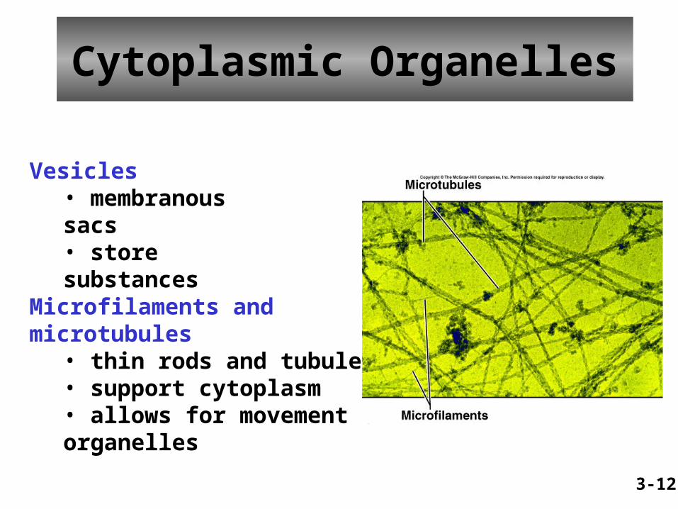
Vesicles• membranous sacs• store substances
Microfilaments and microtubules• thin rods and tubules• support cytoplasm• allows for movement of organelles
3-12
Cytoplasmic Organelles

Cell Nucleus
• control center of cell
• nuclear envelope• porous double membrane• separates nucleoplasm from cytoplasm
• nucleolus• dense collection of RNA and proteins• site of ribosome production
• chromatin• fibers of DNA and proteins• stores information for synthesis of proteins
3-13

Movements Into and Out of the Cell
Passive (Physical) Processes• require no cellular energy• simple diffusion• facilitated diffusion• osmosis• filtration
Active (Physiological) Processes• require cellular energy• active transport• endocytosis• exocytosis• transcytosis
3-14

Simple Diffusion
• movement of substances from regions of higher concentration to regions of lower concentration• oxygen, carbon dioxide and lipid-soluble substances
3-15

Facilitated Diffusion
• diffusion across a membrane with the help of a channel or carrier molecule• glucose
3-16

Osmosis
• movement of water through a selectively permeable membrane from regions of higher concentration to regions of lower concentration• water moves toward a higher concentration of solutes
3-17

Osmosis
Osmotic Pressure – ability of osmosis to generate enough pressure to move a volume of water
Osmotic pressure increases as the concentration of nonpermeable solutes increases
• hypertonic – higher osmotic pressure• hypotonic – lower osmotic pressure• isotonic – same osmotic pressure
3-18
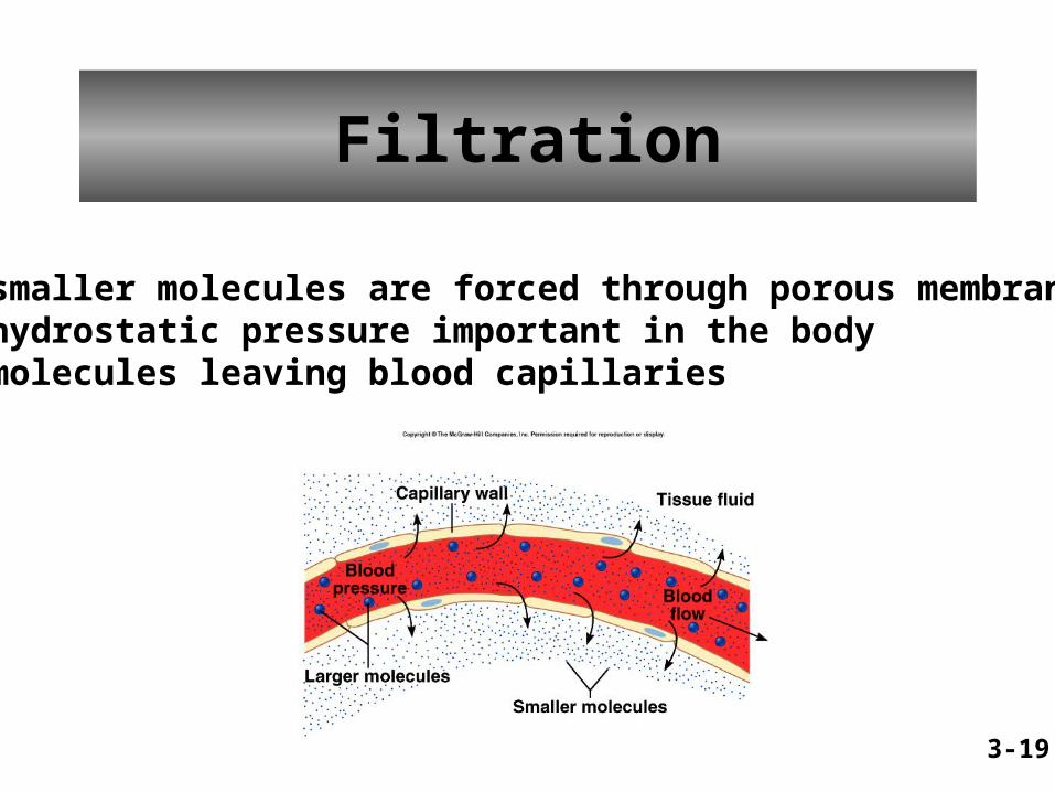
Filtration
• smaller molecules are forced through porous membranes• hydrostatic pressure important in the body• molecules leaving blood capillaries
3-19

Active Transport
• carrier molecules transport substances across a membrane from regions of lower concentration to regions of higher concentration• sugars, amino acids, sodium ions, potassium ions, etc.
3-20

Endocytosis• cell engulfs a substance by forming a vesicle around the substance• three types
• pinocytosis – substance is mostly water• phagocytosis – substance is a solid• receptor-mediated endocytosis – requires the substance to bind to a membrane-bound receptor
3-21

Endocytosis
3-22

Exocytosis• reverse of endocytosis• substances in a vesicle fuse with cell membrane• contents released outside the cell• release of neurotransmitters from nerve cells
3-23

Transcytosis
• endocytosis followed by exocytosis• transports a substance rapidly through a cell• HIV crossing a cell layer
3-24

The Cell Cycle
• series of changes a cell undergoes from the time it forms until the time it divides• stages
• interphase• mitosis• cytoplasmic division• differentiation
3-25

Interphase
• very active period• cell grows• cell maintains routine functions• cell replicates genetic material to prepare for nuclear division• cell synthesizes new organelles to prepare for cytoplasmic division• phases
• G phases – cell grows and synthesizes structures other than DNA• S phase – cell replicates DNA
3-26

Mitosis
• produces two daughter cells from an original cell• nucleus divides – karyonkinesis• cytoplasm divides – cytokinesis • stages
• prophase – chromosomes form; nuclear envelope disappears• metaphase – chromosomes align midway between centrioles• anaphase – chromosomes separate and move to centrioles• telophase – chromatin forms; nuclear envelope forms
3-27
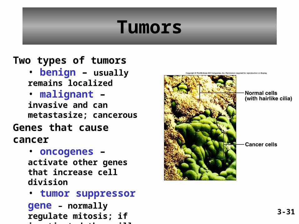
Tumors
Two types of tumors• benign – usually remains localized• malignant – invasive and can metastasize; cancerous
Genes that cause cancer• oncogenes – activate other genes that increase cell division• tumor suppressor gene – normally regulate mitosis; if inactivated they will not regulate mitosis
3-31
Integumentary System
• Integumentary systemIntegumentary system composed of two layers and many different tissues.
• Two distinct layers 1.1. EpidermisEpidermis
a) Stratified keratinized squamous epithelium
2.2. DermisDermis1. Glandular epithelium (sweat, sebaceous glands)2. Dense irregular connective tissue (collagen)3. Smooth muscle tissue (arrector pili muscles)4. Nervous tissue (Meissner’s & Pacinian corpuscles)5. Blood vessels

Subcutaneous Layer
• Distinct layer beneath skin
• Composed of adipose tissue
• Provides insulation and protection


Layers of the Epidermis
• Stratum basale: deepest layer, single row of cells that divide and grow
• Stratum spinosum: flat cells with keratin• Stratum granulosum: flat keratinized cells
with shriveled nuclei• Stratum lucidum: clear cells• Stratum corneum: many layers of flat, dead,
keratinized, nonnucleated cells that are shed


Melanin• Melanin is a dark
pigment produced by melanocytes.
• Melanin absorbs ultraviolet light and helps prevent mutations and damage to cells.
Figure 6.4

Melanin

ABC’s of Melanoma
A.A. Asymmetry Asymmetry the shape of one side does not match the other
B.B. Border Border ragged or irregular edges
C.C. ColorColor color uneven, different shades of brown, black, and tan
D.D. DiameterDiameter greater than 5mm or the size of an eraser head
E.E. ElevationElevation mole is raised or elevated above skin

Melanin
• Determines skin color
• Produced by melanocytes in basal layer
• Everyone has same # of melanocytes
• Inherit amount of melanin produced
• Determines race and/or skin color

Skin Color
Besides melanin other factors affect skin color:
1.1. Carotene yellowCarotene yellow
2.2. Hemoglobin pinkHemoglobin pink
3.3. Cyanosis blueCyanosis blue

Dermis• Epidermal (rete) ridgesEpidermal (rete) ridges project into the dermis. • These dermal papillaedermal papillae are responsible for
fingerprintsfingerprints.• The dermis binds the epidermis to the underlying
tissues.• Nourishment for epidermis• The dermis is composed of irregular denseirregular dense
connective tissue with collagen and elastin in a gel-connective tissue with collagen and elastin in a gel-like ground substance.like ground substance.

Dermis• Dermis contains muscle fibers. Smooth muscles
(arrector pili(arrector pili) are associated with follicles and glands. • Motor and sensory nerves and sensory receptors are
found throughout the dermis.– Meissner’s corpuscles Meissner’s corpuscles light touch
– Pacinian corpusclesPacinian corpuscles pressure sensors
• The dermis contains accessory organsaccessory organs such as blood vessels, hair follicles, sebaceous and sweat glands.
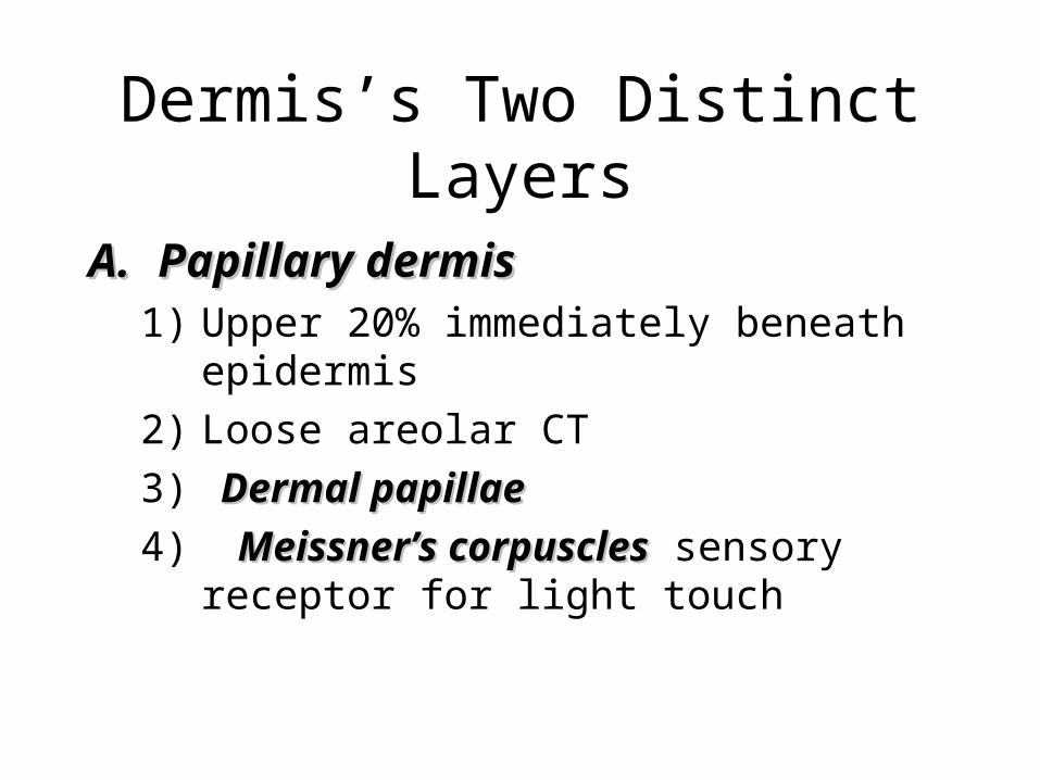
Dermis’s Two Distinct Layers
A.A. Papillary dermis Papillary dermis 1) Upper 20% immediately beneath epidermis
2) Loose areolar CT
3) Dermal papillae Dermal papillae
4) Meissner’s corpusclesMeissner’s corpuscles sensory receptor for light touch

Dermis’s Two Distinct Layers
A.A. Reticular dermis Reticular dermis 1) Lower 80%
2) Composition: dense irregular CT dense irregular CT a) Bundles of collagen, elastic, and reticular collagen, elastic, and reticular
b) Strength, resiliency, and elasticity

Subcutaneous Layer • The subcutaneous layer or hypodermissubcutaneous layer or hypodermis is made
up of loose connective and adipose tissueloose connective and adipose tissue.
• Collagenous and elastic fibers are continuous with those in the dermis.
• The adipose tissue conserves body heatconserves body heat.
• The subcutaneous layer contains the major blood vessels (rete cutaneum) (rete cutaneum) that supply the skin.
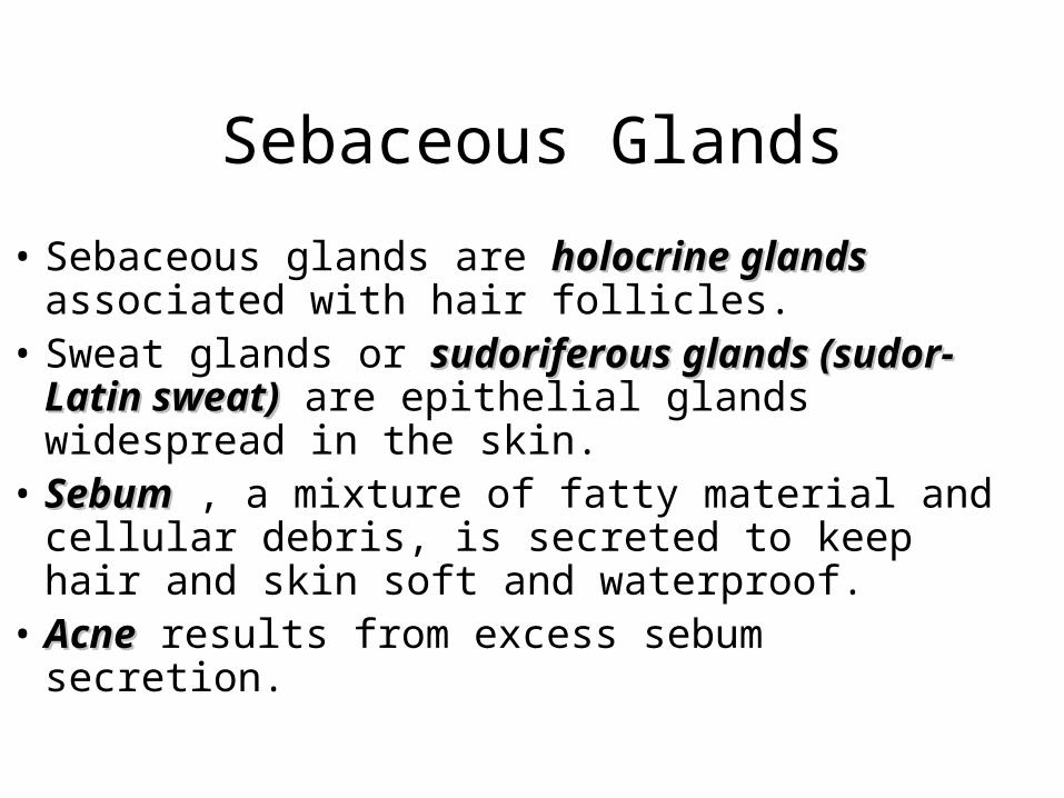
Sebaceous Glands
• Sebaceous glands are holocrine glandsholocrine glands associated with hair follicles.
• Sweat glands or sudoriferous glands (sudor- Latin sudoriferous glands (sudor- Latin sweat)sweat) are epithelial glands widespread in the skin.
• SebumSebum , a mixture of fatty material and cellular debris, is secreted to keep hair and skin soft and waterproof.
• AcneAcne results from excess sebum secretion.


Sweat Glands• Eccrine Eccrine
glandsglands respond to increased body temperature and function in evaporative cooling.
Figure 6.11

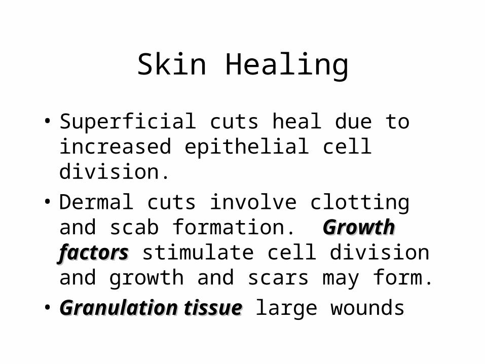
Skin Healing
• Superficial cuts heal due to increased epithelial cell division.
• Dermal cuts involve clotting and scab formation. Growth factorsGrowth factors stimulate cell division and growth and scars may form.
• Granulation tissueGranulation tissue large wounds


Skin Functions
1.1. ProtectionProtection – water loss, injury, chemicals and bacteria
2.2. ExcretionExcretion – minimal but urea urea and uric uric acid acid are secreted in addition to electrolytes
3.3. RegulationRegulation – body temperature

Skin Functions
4. Sensation4. Sensationa)a) Meissner’s corpusclesMeissner’s corpuscles
i. Light touch
ii. Egg-shaped
iii. Dermal papillae
iv. Fingertips, palms, soles, eyelids, tip of tongue, nipples, clitoris, and tip of penis

Skin Functions
4. Sensation 4. Sensation (continued)b) Pacinian corpusclesb) Pacinian corpuscles
i. Pressure
ii. Onion shaped
iii. Deep dermis and subcutaneous
iv. Joints, tendons, muscles, mammary glands, and external genitalia

Skin Functions
5. Vitamin D synthesis5. Vitamin D synthesisa) UV rays activate synthesisb) critical in bone homeostasis
6. Blood reservoir 6. Blood reservoir 10% of body’s blood vessels
7. Immunity 7. Immunity a) Langerhan’s cellsb) T-helper cells

Bone Classification• Bones are classified according to shape• Long bonesLong bones are long with expanded ends, Ex:
forearm and thigh bone• Short bonesShort bones are cube like, Ex: wrist, ankle• Flat bonesFlat bones are broad and plate like, Ex: ribs,
scapulae, and some skull bones• Irregular bonesIrregular bones vary in shape, Ex: vertebrae• SesamoidSesamoid or round bones are small bones
embedded in tendons, Ex: kneecap (patella)


Parts of Bone
Figure 7.2

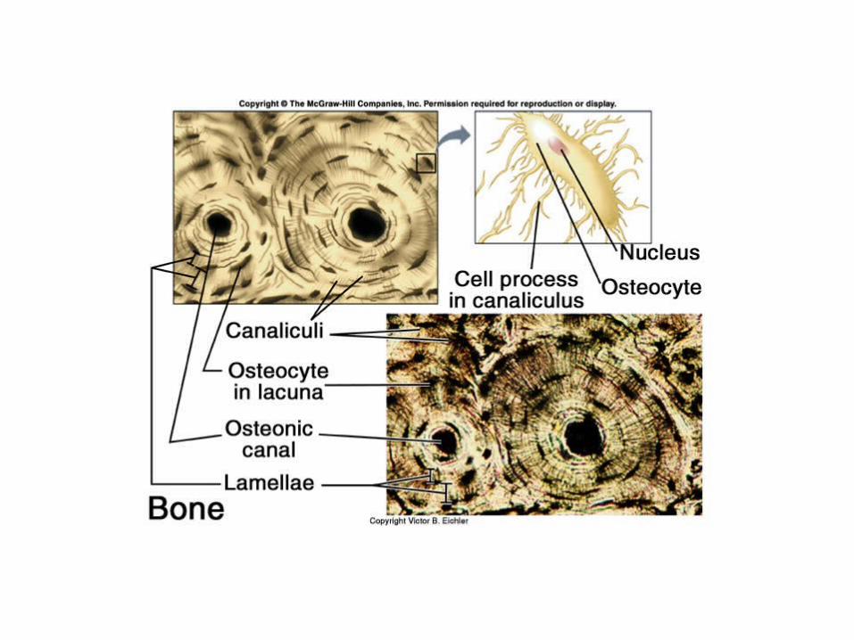
Compact Bone
• Osteocytes Osteocytes and layers of intercellular material lie in concentric rings around an osteonic canalosteonic canal.
• This unit is called an osteonosteon or Haversian system.
• Osteonic canals contain blood vessels and nerve fibers and are interconnected by transverse perforating (Volkmann’sVolkmann’s) canals.
• Intercellular material made of collagen and inorganic salts.


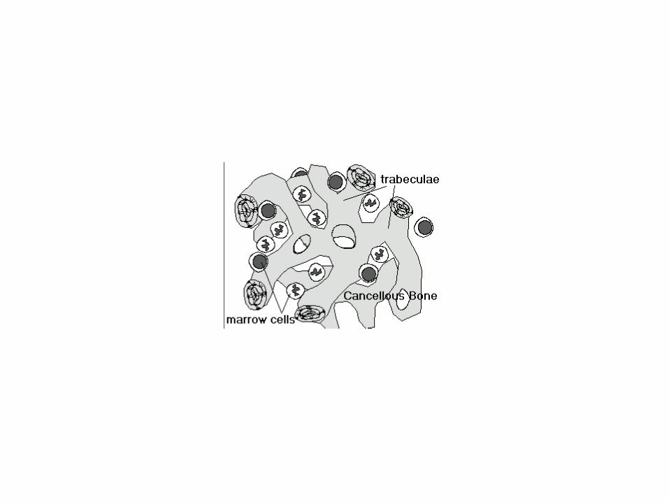
Spongy Bone
• CancellousCancellous bone bone
• Partially fills medullary space/cavitymedullary space/cavity
• Red or yellow
• Yellow – fat
• Red – marrow
• Bone spiculesspicules - trabeculae


Bone Growth and Development
• The skeletal system begins to form during the first weeks of prenatal development.
• Some bones originate within sheets of connective tissue (intramembranous intramembranous bonesbones).
• Some bones begin as models of hyaline cartilage that are replaced by bone (endochrondral bonesendochrondral bones).
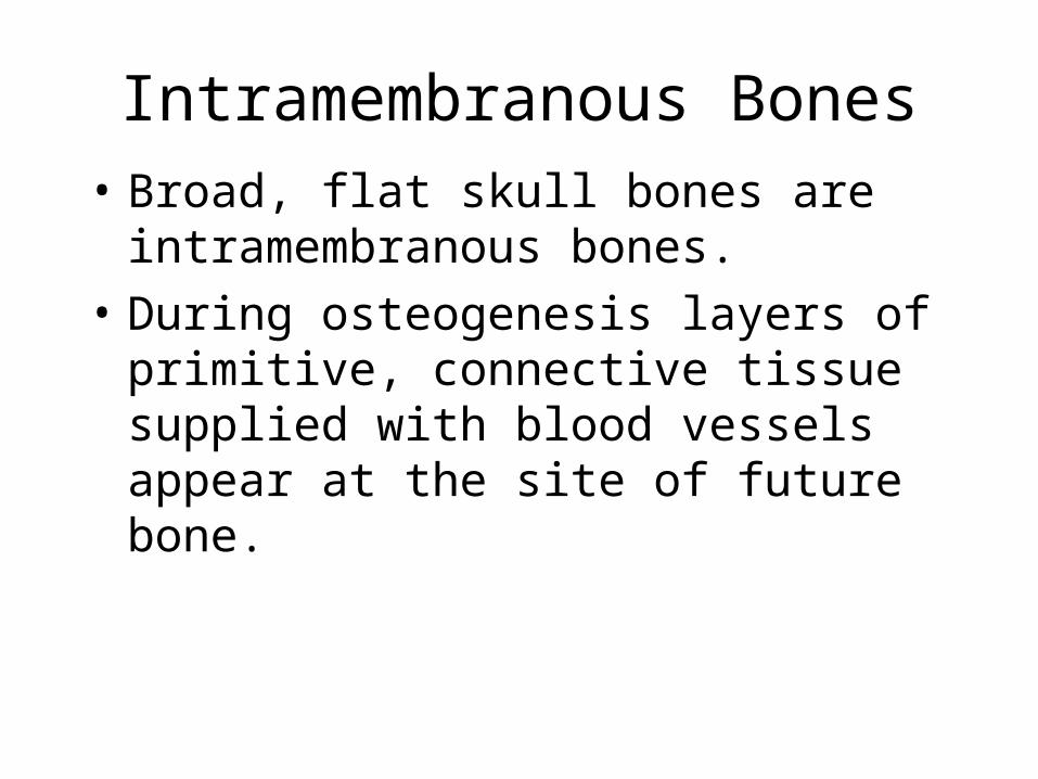
Intramembranous Bones• Broad, flat skull bones are
intramembranous bones.
• During osteogenesis layers of primitive, connective tissue supplied with blood vessels appear at the site of future bone.

Intramembranous Bones• Cells differentiate into osteoblastsosteoblasts (bone-building
cells) which deposit spongy bone.• Boney mineral matrix laid down around
osteoblasts made by osteoblasts.• Spongy bone forms in all directions around
osteoblasts.• Osteoblasts become osteocytesosteocytes when surrounded
by bony matrix in lacunae.• Later some spongy bone may become compact
bone as spaces fill with bone matrix.

Intramembranous Bones
• Connective tissue on the surface of the bone forms the periosteumperiosteum.
• Osteoblasts on the inside of the periosteum deposit compact bone over spongy bone.
• This process is called intramembranous ossification.

Endochondral Bones
• Hyaline cartilageHyaline cartilage forms a model of the bone during embryonic development.
• Cartilage degenerates, periosteum forms.
• Periosteal blood vessels and osteoblasts invade the bone forming a primaryprimary ossification centerossification center in the diaphysisdiaphysis.
• Secondary ossification centersSecondary ossification centers develop in the epiphysesepiphyses.


Endochondral Bones
• Osteoblasts form spongy bone in the space occupied by cartilage.
• Osteoblasts become osteocytes when bony matrix surrounds them.
Endochondral Bones
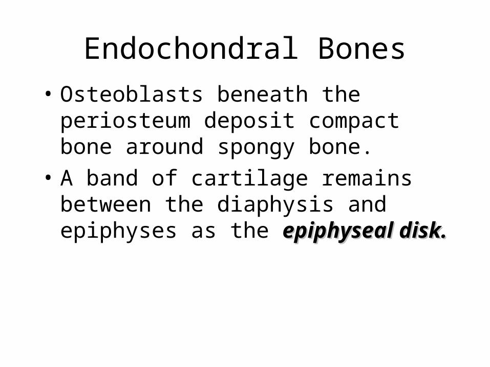
• Osteoblasts beneath the periosteum deposit compact bone around spongy bone.
• A band of cartilage remains between the diaphysis and epiphyses as the epiphyseal epiphyseal disk.disk.

Bone Growth
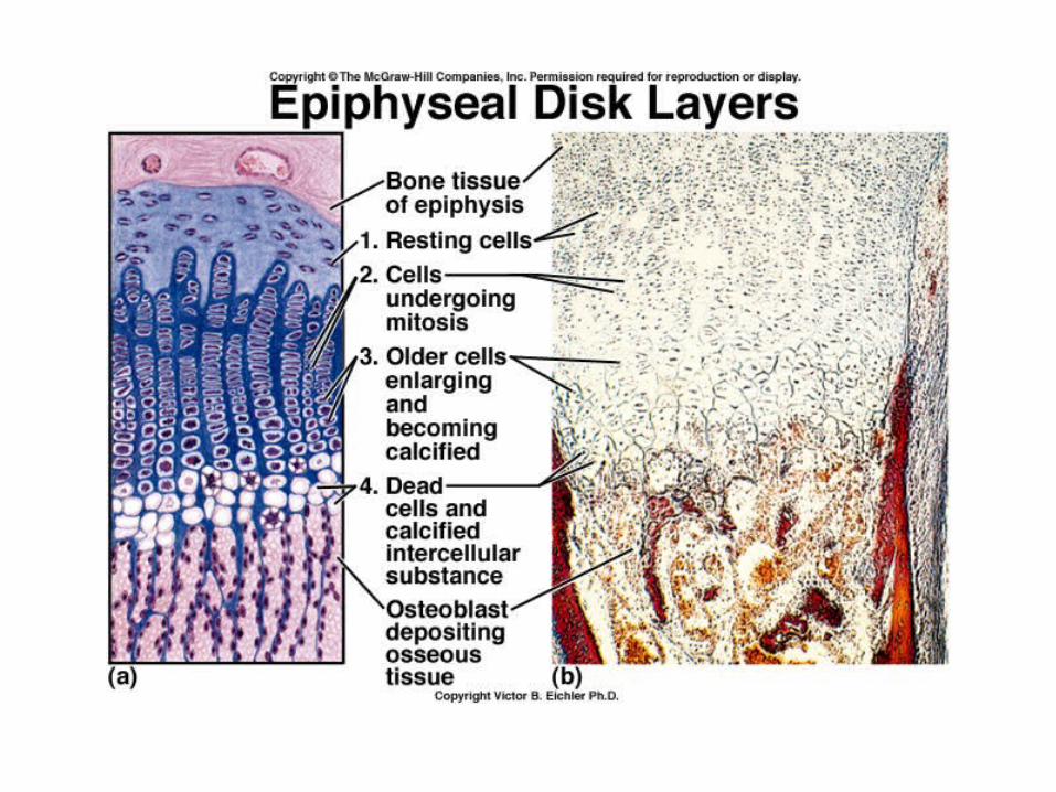
• Growth of long bones occurs along four layers of cartilage in the epiphyseal disk.
• First Layer: resting cells that do not grow.
• Second Layer: young mitotic cells.
• Third Layer: older cells that enlarge.
• Fourth Layer: dead cells and calcified intercellular substances.


Fracture Repair

Fracture Repair
• Blood escapes from damaged blood vessels and forms a hematoma.
• Spongy bone forms in regions near blood vessels and fibrocartilage forms farther away.
• A bony callus replaces the fibrocartilage.
• Osteoclasts remove excess bony tissue, restoring new bone much like the original.

Nutrition and Bone Development
• Vitamin DVitamin D is necessary to absorb calcium in the small intestine.
• Vitamin D deficiency leads in ricketsrickets in children and osteomalaciaosteomalacia in adults.
• DehydrocholesterolDehydrocholesterol made in digestive tract converted to Vit D in the skin by UV rays.

Nutrition and Bone Development
• Vitamin A is necessary for osteoblast and osteoclast activity.
• Vitamin C is necessary for collagen synthesis. – Scurvy [skúrvee]
– disease caused by vitamin deficiency: a disease caused by insufficient vitamin C, the symptoms of which include spongy gums, loosening of the teeth, and bleeding into the skin and mucous membranes
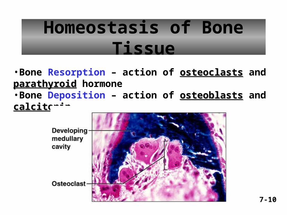
Homeostasis of Bone Tissue
•Bone Resorption – action of osteoclastsosteoclasts and parathyroidparathyroid hormone•Bone Deposition – action of osteoblastsosteoblasts and calcitonincalcitonin
7-10

Hormones and Bone• Growth HormoneGrowth Hormone (GH) stimulates
epiphyseal cartilage cell division.
• Deficiency of G H: pituitary dwarfismpituitary dwarfism. Excess GH: pituitary gigantismgigantism in children and acromegalyacromegaly in adults.

Hormones and Bone• Thyroid hormone stimulates cartilage
replacement in the epiphyseal disks.
• Sex steroids promote formation of bone tissue (ossification) which closes the epiphyseal disk.
• Estrogen more effective than androgens

Physical Factors Affecting Bone
• Physical stress stimulates bone growth.
• Weight bearing exercise stimulates bone tissue to thicken and strengthen (hypertrophy).
• Lack of exercise leads to bone wasting (atrophy).

Levers are categorized into three types –
• First class levers (EFL) e.g. a seesaw – the head on the vertebral column (Figure 11.2a)
• Second-class (FLE) eg. a wheelbarrow(Figure 11.2b)
• Third-class (FEL) (Figure 11.1b) e.g. forceps - the elbow joint (Figure 11.2c).

• Muscle acts on rigid rod (bone)that moves around a fixed point called a fulcrum
• Resistance is weight of bodypart & perhaps an object
• Effort or load is work doneby muscle contraction
• Mechanical advantage– the muscle whose attachment is farther from the joint will
produce the most force
– the muscle attaching closer to the joint has the greater range of motion and the faster the speed it can produce
Lever Systems and Leverage

First - Class Lever• Can produce mechanical
advantage or not depending on location of effort & resistance– if effort is further from fulcrum
than resistance, then a strong resistance can be moved
• Head resting on vertebral column– weight of face is the resistance
– joint between skull & atlas is fulcrum
– posterior neck muscles provide effort

Second - Class Lever• Similar to a wheelbarrow• Always produce mechanical
advantage– resistance is always closer to
fulcrum than the effort
• Sacrifice of speed for force• Raising up on your toes
– resistance is body weight– fulcrum is ball of foot– effort is contraction of calf
muscles which pull heel up off of floor
LL
EE
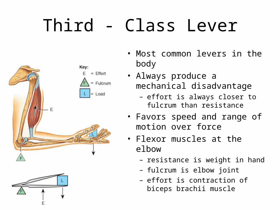
Third - Class Lever
• Most common levers in the body
• Always produce a mechanical disadvantage– effort is always closer to fulcrum
than resistance
• Favors speed and range of motion over force
• Flexor muscles at the elbow– resistance is weight in hand
– fulcrum is elbow joint
– effort is contraction of biceps brachii muscle

Blood Cell Formation• Blood cell formation (hematologists) occurs
in yolk sac in early development.
• Later it occurs in the liver and spleen.
• In the adult red and white blood cells are formed in the red bone marrow.

Blood Cell Formation• Red marrow fills the cavity in the diaphysis
of the long bones in infants. In adults it is replaced with yellow marrow (fat).
• Adult red marrow is found in spongy bone of the skull, ribs, sternum, vertebrae, pelvis.

Inorganic Salt Storage
• Salts account for 70% of the bone matrix.
• These salts are mostly calcium phosphate crystals.
• Calcium phosphate ([Ca3(PO4)2] + calcium hydroxide [Ca(OH)2] called hydroxyapatite
• CaCO3
• Ions – magnesium, fluoride, potassium, & sulfate

Inorganic Salt Storage
• Parathyroid hormone stimulates osteoclasts to break down bone when Ca levels are low.
• Calcitonin stimulates osteoblasts to build bone when Ca levels are high.
• Bone contains Mg, Na, K, and carbonate ions.