chapter 3 cells. cell biology size – 7.5-500 micrometers shape – depends on?...
TRANSCRIPT

CHAPTER 3
CELLS
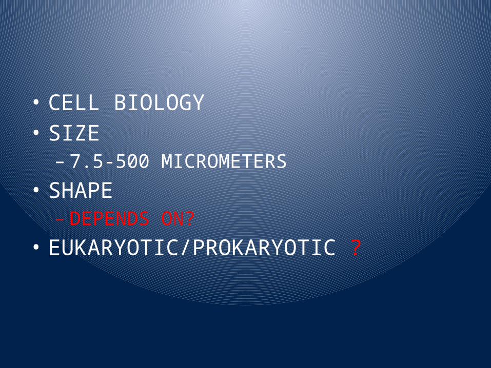
• CELL BIOLOGY• SIZE – 7.5-500 MICROMETERS
• SHAPE– DEPENDS ON?
• EUKARYOTIC/PROKARYOTIC ?
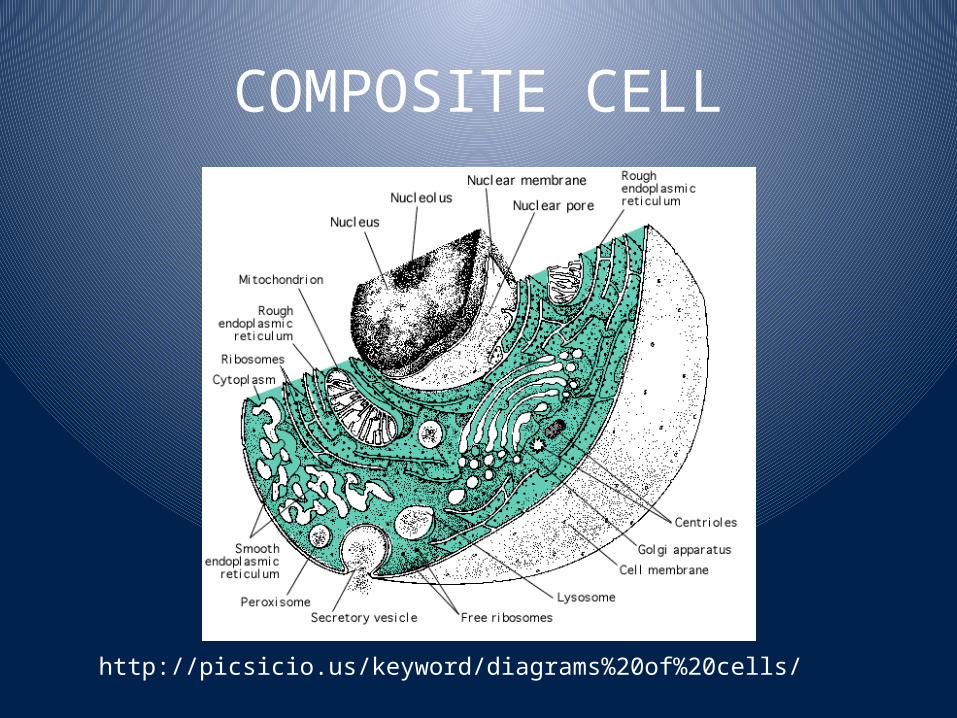
COMPOSITE CELL
http://picsicio.us/keyword/diagrams%20of%20cells/
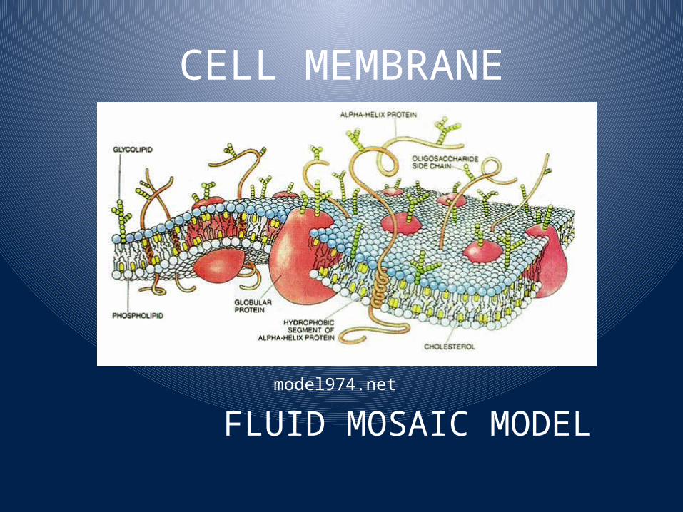
CELL MEMBRANE
model974.net
FLUID MOSAIC MODEL
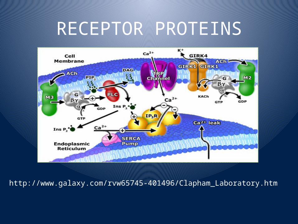
RECEPTOR PROTEINS
http://www.galaxy.com/rvw65745-401496/Clapham_Laboratory.htm

INTEGRAL PROTEIN
users.rcn.com
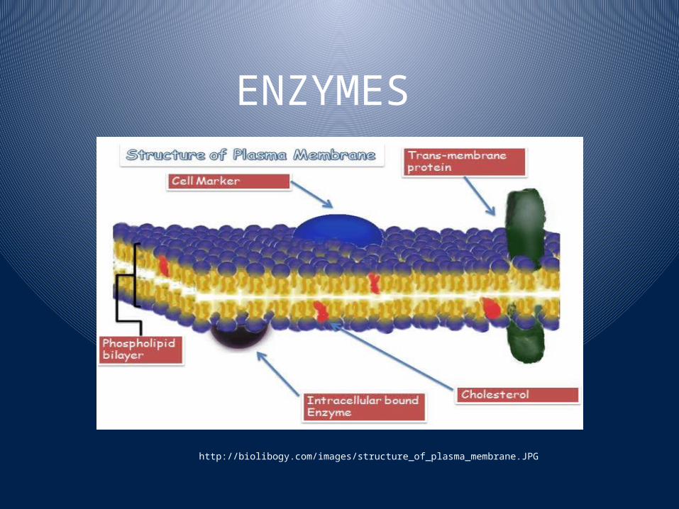
ENZYMES
http://biolibogy.com/images/structure_of_plasma_membrane.JPG

CELL ADHESION MOLECULES
bioweb.wku.edu

CELL SURFACE PROTEINS
http://biolibogy.com/images/structure_of_plasma_membrane.JPG
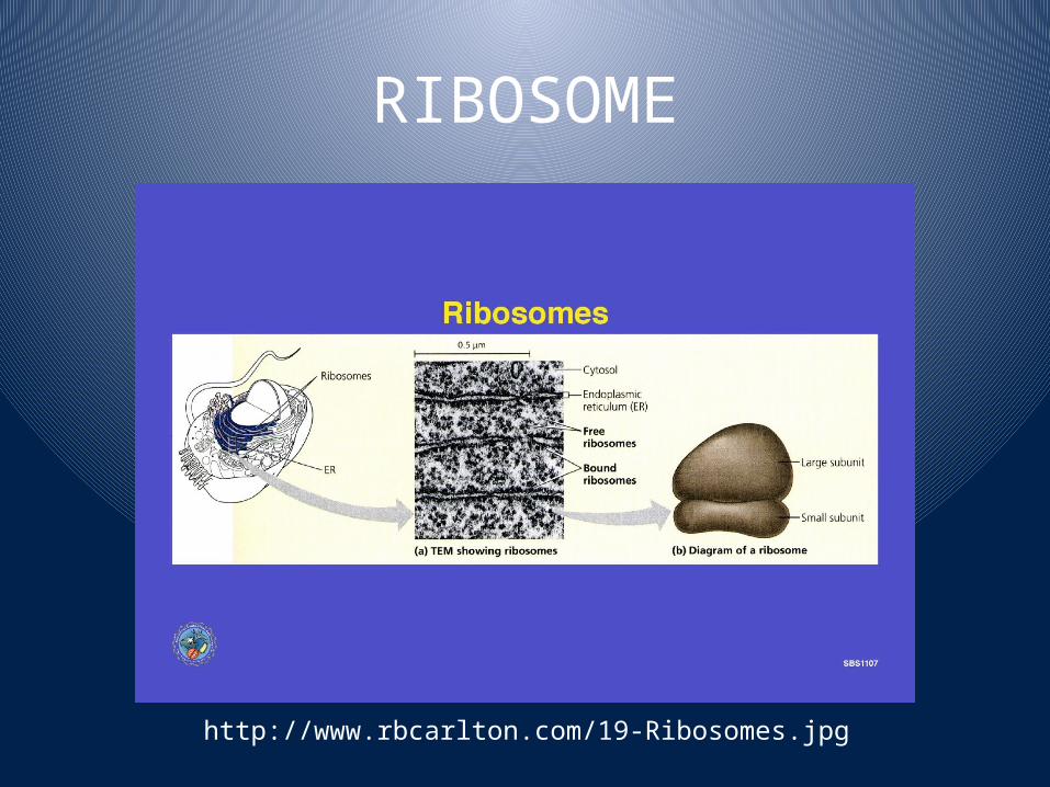
RIBOSOME
http://www.rbcarlton.com/19-Ribosomes.jpg
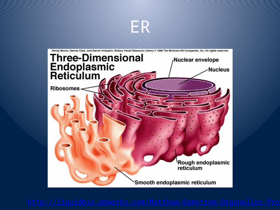
ER
http://liquidbio.pbworks.com/Matthew-Damstrom-Organelles-Project

VESSICLES
http://betarhythm.blogspot.com/2006/09/synaptic-vesicles-message-in-bottle.html

GOLGI APPARATUS
http://kvhs.nbed.nb.ca/gallant/biology/golgi_apparatus.jpg

Mitochondria
http://academic.brooklyn.cuny.edu/biology/bio4fv/page/mito.gif

Lysosomes
http://micro.magnet.fsu.edu/cells/lysosomes/images/lysosomesfigure1.jpg

Peroxisomes
http://micro.magnet.fsu.edu/cells/peroxisomes/images/peroxisomesfigure1.jpg

Centrosome/Centriole
http://micro.magnet.fsu.edu/cells/centrioles/images/centriolesfigure1.jpg

Microfilaments/Microtubules
http://kentsimmons.uwinnipeg.ca/cm1504/Image115.gif
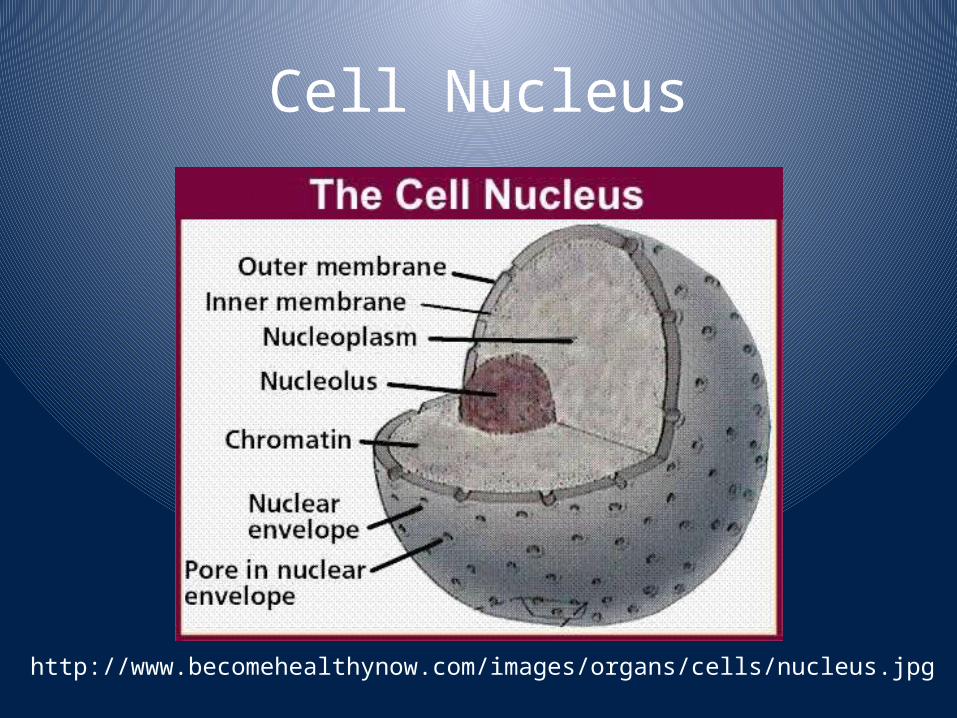
Cell Nucleus
http://www.becomehealthynow.com/images/organs/cells/nucleus.jpg
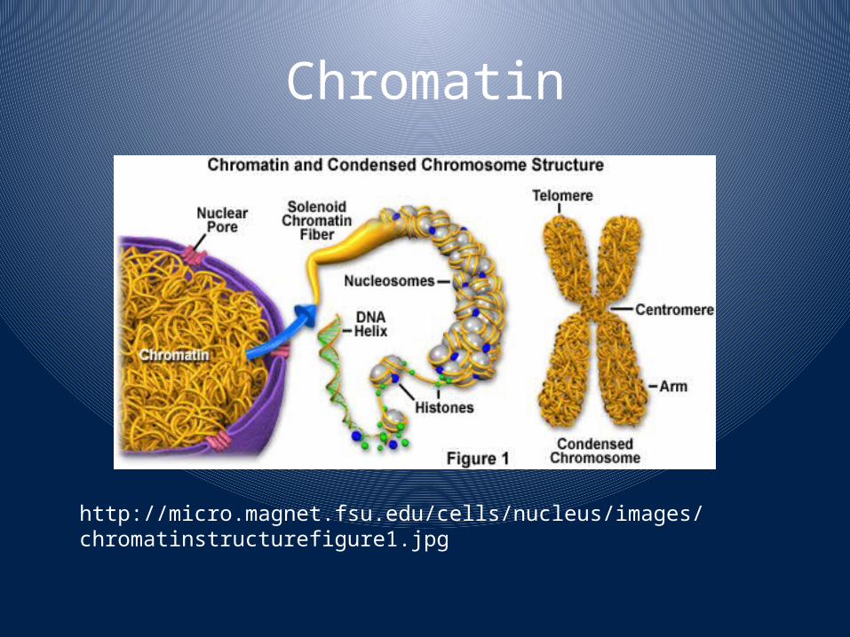
Chromatin
http://micro.magnet.fsu.edu/cells/nucleus/images/chromatinstructurefigure1.jpg
Cell Transport
IMPORTANCE?TYPESDIFFERENCE?

Diffusion
http://upload.wikimedia.org/wikipedia/commons/6/66/Scheme_simple_diffusion_in_cell_membrane-de.png
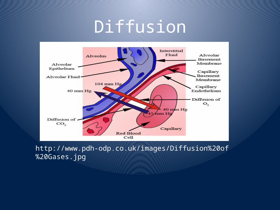
Diffusion
http://www.pdh-odp.co.uk/images/Diffusion%20of%20Gases.jpg

http://www.williamsclass.com/SeventhScienceWork/ImagesCellBricks/facilitatedDiffusion.jpg
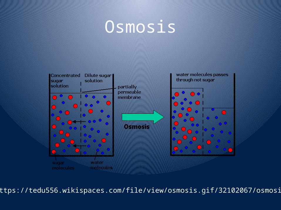
Osmosis
https://tedu556.wikispaces.com/file/view/osmosis.gif/32102067/osmosis.gif

Osmosis
http://biology.unm.edu/ccouncil/Biology_124/Images/tonicity1.jpeg

Filtration
http://herkules.oulu.fi/isbn9514264290/html/graphic55.png
Passive: can be due to force of gravity alone
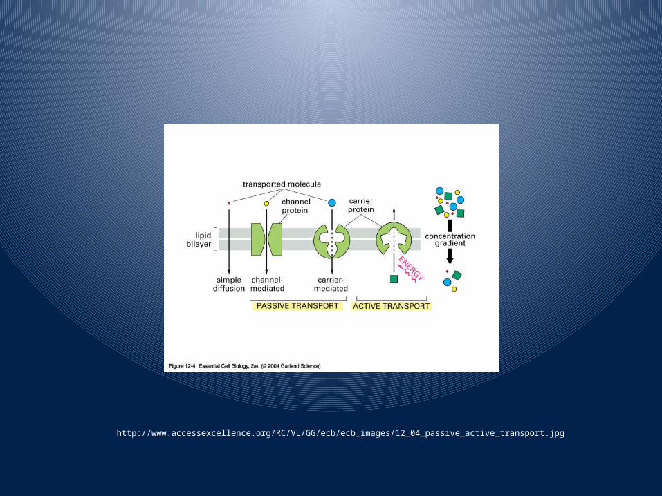
http://www.accessexcellence.org/RC/VL/GG/ecb/ecb_images/12_04_passive_active_transport.jpg

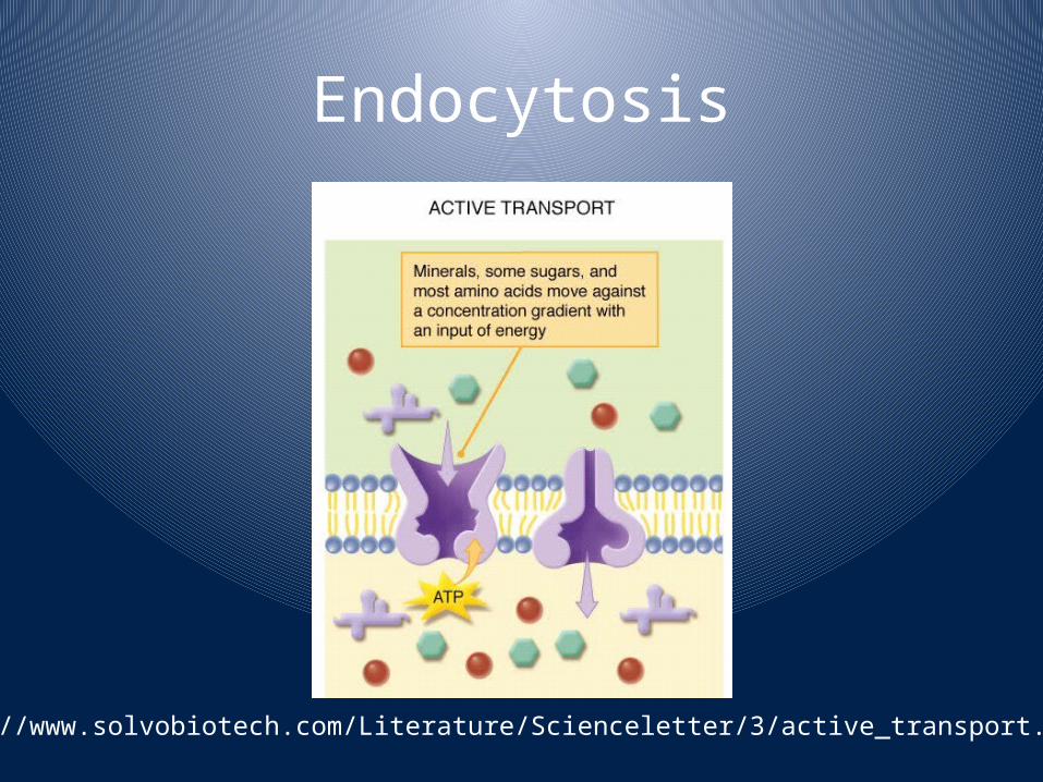
Endocytosis
•Coming into the cell•Pinocytosis•Phagocytosis
Endocytosis
http://www.solvobiotech.com/Literature/Scienceletter/3/active_transport.jpg
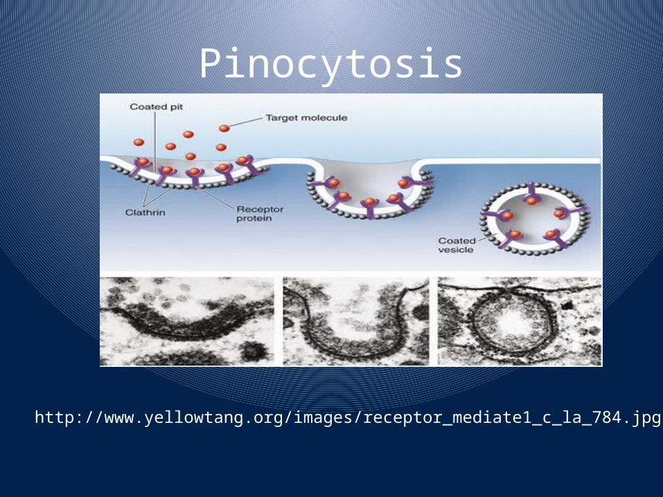
Pinocytosis
http://www.yellowtang.org/images/receptor_mediate1_c_la_784.jpg

Phagocytosis
http://www.ingid.org/teaching-programmes/Introbasicimmune/phagoc1.jpg

Receptor Mediated Endocytosis
http://219.221.200.61/ywwy/zbsw(E)/pic/ech5-19.jpg

Exocytosis
http://s3.images.com/huge.96.481383.JPG
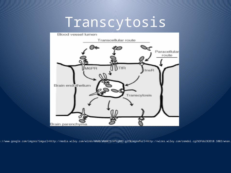
Transcytosis
http://www.google.com/imgres?imgurl=http://media.wiley.com/wires/WNAN/WNAN73/nfig003.gif&imgrefurl=http://wires.wiley.com/remdoi.cgi%3Fdoi%3D10.1002/wnan.73&usg
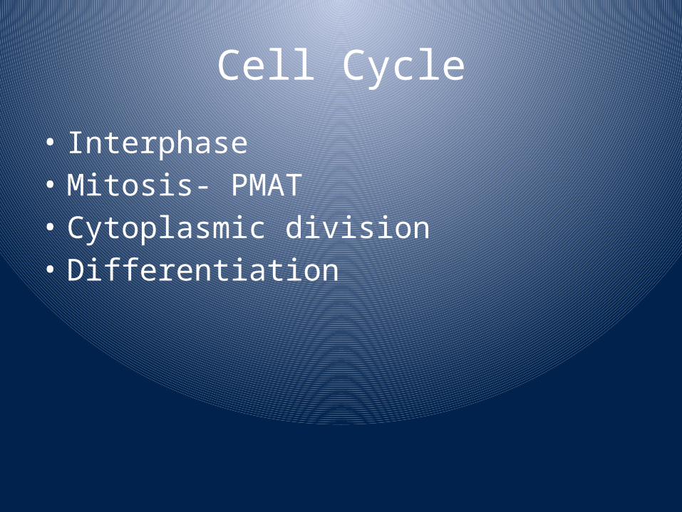
Cell Cycle
• Interphase• Mitosis- PMAT• Cytoplasmic division• Differentiation

Interphase
http://www.obgynacademy.com/basicsciences/fetology/genetics/

MitosisProphaseMetaphaseAnaphaseTelophase
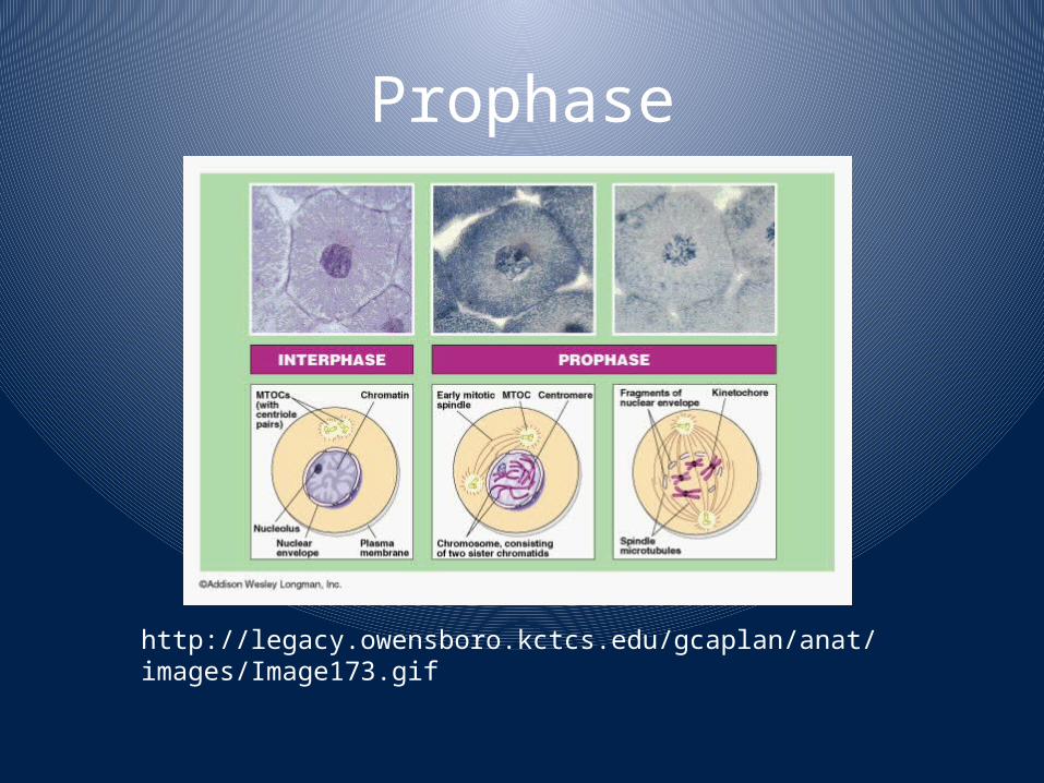
Prophase
http://legacy.owensboro.kctcs.edu/gcaplan/anat/images/Image173.gif

Metaphase
http://wps.aw.com/wps/media/objects/443/453744/metaphase.html

Anaphase
http://micro.magnet.fsu.edu/micro/gallery/mitosis/earlyanaphase.html

Telophase
http://botit.botany.wisc.edu/images/130/mitosis/allium_root_prep._slides/telophase_cytokinesis.html

Cytoplasmic Division
http://www.bing.com/images/search?q=cytoplasmic+division&FORM=BIFD&adlt=strict#

CELL DIVISION CONTROL
• VARIES BY ?• ~50 X• TELOMERES• STRESS CAN SHORTEN LIFESPAN• KINASES AND CYCLINS• CELL SIZE ?• EXTERNAL CONTROLS: HORMONES, GROWTH FACTORS• SPACE– CONTACT INHIBITION

• INFREQUENT MITOSIS =• TOO FRQUENT– TUMOR
• BENIGN• MALIGNANT: METASTISIZE• ONCOGENES• TUMOR SUPRESSOR GENES
STEM CELLS
DIFFERENTIATIONSTEM CELL-> STEM CELL OR PROGENITOR CELLPROGENITOR CELL:
TOTIPOTENT STEM CELLPLURIPOTENT STEM CELL
MOST ORGANS HAVE PLURIPOTENT STEM CELLS OR PROGENITOR CELLSHOW DO CELLS IN BODY DIFFER?

CELL DEATH
• APOPTOSIS• PROGRAMMED CELL DEATH• EMBRYO• DEATH RECEPTOR RELEASES ENZYMES: CASPASES– DESTROY DNA REPLICATION ANDS REPAIR ENZYMES– ACTIVATE ENZYMES THAT SNIP UP DNA– DISSOLVE CYTOSKELETON– DESTROY MITOCHONDRIA ?– SEPARATE CELLS– MEMBRANE TRIGGERS TO ATTRACT PHAGOCYTES

APOPTOSIS
CELL HAS CIRCULAR SHAPE; BULGES: BLEBS; NUCLEUS SHATTERS; CELL SHATTERS;CELL FRAGMENTS ENCAPSULATE SO NO INFLAMMATION; GONE

Necrosis
http://herkules.oulu.fi/isbn9514266676/html/graphic11.jpe