chapter 26 the operon. 26.1 introduction coupled transcription/translation – the phenomena in...
TRANSCRIPT

Chapter 26The Operon

26.1 Introduction
• coupled transcription/translation – The phenomena in bacteria where translation of the mRNA occurs simultaneously with its transcription.
• operon – A unit of bacterial gene expression and regulation, including structural genes and control elements in DNA recognized by regulator gene product(s).
26.1 Introduction
• trans-acting – A product that can function on any copy of its target DNA. This implies that it is a diffusible protein or RNA.
• cis-acting – A site that affects the activity only of sequences on its own molecule of DNA (or RNA); this property usually implies that the site does not code for protein.

26.1 Introduction
• regulator gene – A gene that codes for a product (typically protein) that controls the expression of other genes (usually at the level of transcription).
• structural gene – A gene that codes for any RNA or protein product other than a regulator.
Figure 26.01: A regulator gene codes for a protein that acts at
a target site on DNA.

26.1 Introduction• In negative regulation, a repressor protein binds to an
operator to prevent a gene from being expressed.• In positive regulation, a transcription factor is required
to bind at the promoter in order to enable RNA polymerase to initiate transcription.
Figure 26.02: In negative control, a trans-acting repressor binds to the cis-acting
operator to turn off transcription.
Figure 26.03: In positive control, a trans-acting factor must bind to cis-acting site in
order for RNA polymerase to initiate transcription at the promoter.

26.1 Introduction
• In inducible regulation, the gene is regulated by the presence of its substrate (the inducer).
• In repressible regulation, the gene is regulated by the product of its enzyme pathway (the corepressor).

26.1 Introduction
• Gene regulation in vivo can utilize any of these mechanisms, resulting in all four combinations: negative inducible, negative repressible, positive inducible, and positive repressible.
Figure 26.04: Regulatory circuits can be designed from all possible combinations
of positive and negative control with inducible and repressible control.

26.2 Structural Gene Clusters Are Coordinately Controlled
• Genes coding for proteins that function in the same pathway may be located adjacent to one another and controlled as a single unit that is transcribed into a polycistronic mRNA.
Figure 26.05: The lac operon occupies ~6000 bp of DNA.

26.3 The lac Operon Is Negative Inducible
• Transcription of the lacZYA operon is controlled by a repressor protein (the lac repressor) that binds to an operator that overlaps the promoter at the start of the cluster.
• constitutive expression – A state in which a gene is expressed continuously.
• In the absence of β-galactosides, the lac operon is expressed only at a very low (basal) level.
Figure 26.06: lac repressor and RNA polymerase bind at sites that overlap
around the transcription startpoint of the lac operon.

26.3 The lac Operon Is Negative Inducible
• The repressor protein is a tetramer of identical subunits coded by the lacI gene.
• β-galactoside sugars, the substrates of the lac operon, are its inducer.
• Addition of specific β-galactosides induces transcription of all three genes of the lac operon.
• The lac mRNA is extremely unstable; as a result, induction can be rapidly reversed.

26.3 The lac Operon Is Negative Inducible
Figure 26.07: Addition of inducer results in rapid induction of lac mRNA, and is followed after a short lag by synthesis of the enzymes.

26.4 lac Repressor Is Controlled by a Small-Molecule Inducer
• An inducer functions by converting the repressor protein into a form with lower operator affinity.
• Repressor has two binding sites, one for the operator DNA and another for the inducer.
• gratuitous inducer – Inducers that resemble authentic inducers of transcription, but are not substrates for the induced enzymes.Figure 26.08: lac repressor maintains the
lac operon in the inactive condition by binding to the operator.

26.4 lac Repressor Is Controlled by a Small-Molecule Inducer
• Repressor is inactivated by an allosteric interaction in which binding of inducer at its site changes the properties of the DNA-binding site (allosteric control).
• The true inducer is allolactose, not the actual substrate of β-galactosidase.
Figure 26.09: Addition of inducer converts repressor to a form with low affinity for the
operator. This allows RNA polymerase to initiate transcription.

26.5 cis-Acting Constitutive Mutations Identify the Operator
• Mutations in the operator cause constitutive expression of all three lac structural genes.
• These mutations are cis-acting and affect only those genes on the contiguous stretch of DNA.
• Mutations in the promoter prevent expression of lacZYA and are uninducible and cis-acting.

26.5 cis-Acting Constitutive Mutations Identify the Operator
• cis-dominant – A site or mutation that affects the properties only of its own molecule of DNA, often indicating that a site does not code for a diffusible product.
Figure 26.10: Operator mutations are constitutive because the operator is
unable to bind repressor protein.

26.6 trans-Acting Mutations Identify the Regulator Gene
• Mutations in the lacI gene are trans-acting and affect expression of all lacZYA clusters in the bacterium.
• Mutations that eliminate lacI function cause constitutive expression and are recessive (lacI–).
• Mutations in the DNA-bindingsite of the repressor areconstitutive because therepressor cannot bind theoperator.
Figure 26.11: Mutations that inactivate the lacI gene cause the operon to be
constitutively expressed.

26.6 trans-Acting Mutations Identify the Regulator Gene
• Mutations in the inducer-binding site of the repressor prevent it from being inactivated and cause uninducibility.
• When mutant and wild-type subunits are present, a single lacI–d mutant subunit can inactivate a tetramer whose other subunits are wild-type.– It is dominant negative.

26.6 trans-Acting Mutations Identify the Regulator Gene
• interallelic complementation – The change in the properties of a heteromultimeric protein brought about by the interaction of subunits coded by two different mutant alleles.– The mixed protein may be more or less active than
the protein consisting of subunits of only one or the other type.

26.6 trans-Acting Mutations Identify the Regulator Gene
• negative complementation – This occurs when interallelic complementation allows a mutant subunit to suppress the activity of a wild-type subunit in a multimeric protein.
• lacI–d mutations occur in the DNA-binding site. Their effect is explained by the fact that repressor activity requires all DNA-binding sites in the tetramer to be active.
Figure 26.12: A lacI-d mutant gene makes a monomer that has a damaged DNA binding.

26.7 lac Repressor Is a Tetramer Made of Two Dimers
• A single repressor subunit can be divided into the N-terminal DNA-binding domain, a hinge, and the core of the protein.
• The DNA-binding domain contains two short α-helical regions that bind the major groove of DNA.
• The inducer-binding site and the regions responsible for multimerization are located in the core.

26.7 lac Repressor Is a Tetramer Made of Two Dimers
Figure 26.13: The structure of a monomer of Lac repressor identifies several independent domains.
Structure from Protein Data Bank 1LBG. M. Lewis, et al., Science 271 (1996): 1247-1254. Photo courtesy of Hongli Zhan and Kathleen S. Matthews, Rice University.

26.7 lac Repressor Is a Tetramer Made of Two Dimers
• Monomers form a dimer by making contacts between core subdomains 1 and 2.
• Dimers form a tetramer by interactions between the tetramerization helices.
Figure 26.15: The repressor tetramer consists of two dimers.

26.7 lac Repressor Is a Tetramer Made of Two Dimers
• Different types of mutations occur in different domains of the repressor protein.
Figure 26.16: The locations of three type of mutations in lactose repressor are mapped on
the domain structure of the protein.

26.8 lac Repressor Binding to the Operator Is Regulated by an Allosteric Change in
Conformation • lac repressor protein binds to the double-stranded DNA
sequence of the operator.• The operator is a palindromic sequence of 26 bp.• Each inverted repeat of the operator binds to the DNA-
binding site of one repressor subunit.
Figure 26.17: The lac operator has a symmetrical sequence.

26.8 lac Repressor Binding to the Operator Is Regulated by an Allosteric Change in
Conformation • Inducer binding causes a
change in repressor conformation that reduces its affinity for DNA and releases it from the operator.
Figure 26.18: The inducer changes the structure of the core.

26.9 lac Repressor Binds to Three Operators and Interacts with RNA
Polymerase • Each dimer in a repressor tetramer can
bind an operator, so that the tetramer can bind two operators simultaneously.
• Full repression requires the repressor to bind to an additional operator downstream or upstream as well as to the primary operator at the lacZ promoter.
• Binding of repressor at the operator stimulates binding of RNA polymerase at the promoter but precludes transcription.
Figure 26.21: If both dimers in a repressor tetramer bind to DNA,
the DNA between the two binding sites is held in a loop.

26.10 The Operator Competes with Low-Affinity Sites to Bind Repressor
• Proteins that have a high affinity for a specific DNA sequence also have a low affinity for other DNA sequences.
• Every base pair in the bacterial genome is the start of a low-affinity binding site for repressor.
Figure 26.23: lac repressor binds strongly and specifically to its operator, but is released by inducer. All equilibrium constants are in M–1.

26.10 The Operator Competes with Low-Affinity Sites to Bind Repressor
• The large number of low-affinity sites ensures that all repressor protein is bound to DNA.
• Repressor binds to the operator by moving from a low-affinity site rather than by equilibrating from solution.
Figure 26.24: Virtually all the repressor in the cell is bound to DNA.

26.10 The Operator Competes with Low-Affinity Sites to Bind Repressor
• In the absence of inducer, the operator has an affinity for repressor that is 107 times that of a low-affinity site.
• The level of 10 repressor tetramers per cell ensures that the operator is bound by repressor 96% of the time.
• Induction reduces the affinity for the operator to 104 times that of low-affinity sites, so that operator is bound only 3% of the time.

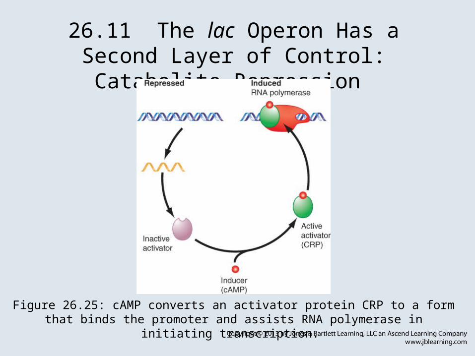
26.11 The lac Operon Has a Second Layer of Control: Catabolite Repression
• catabolite repression – The ability of glucose to prevent the expression of a number of genes. – In bacteria this is a positive control system; in
eukaryotes, it is completely different.• Catabolite repressor protein (CRP) is an
activator protein that binds to a target sequence at a promoter.
26.11 The lac Operon Has a Second Layer of Control: Catabolite Repression
Figure 26.25: cAMP converts an activator protein CRP to a form that binds the promoter and assists RNA polymerase in initiating transcription.

26.11 The lac Operon Has a Second Layer of Control: Catabolite Repression
• A dimer of CRP is activated by a single molecule of cyclic AMP (cAMP).
• cAMP is controlled by the level of glucose in the cell; a low glucose level allows cAMP to be made.
• CRP interacts with the C-terminal domain of the α subunit of RNA polymerase to activate it.Figure 26.27: By reducing the level of
cyclic AMP, glucose inhibits the transcription of operons that require
CRP activity.

26.12 The trp Operon Is a Repressible Operon with Three Transcription Units
• The trp operon is negatively controlled by the level of its product, the amino acid tryptophan (autoregulation).
• The amino acid tryptophan activates an inactive repressor encoded by trpR.
• A repressor (or activator) will act on all loci that have a copy of its target operator sequence.
Figure 26.32: Operators may lie at various positions relative to the
promoter.

26.13 The trp Operon Is Also Controlled by Attenuation
• attenuation – The regulation of bacterial operons by controlling termination of transcription at a site located before the first structural gene.
Figure 26.33: Termination can be controlled via changes in RNA secondary
structure that are determined by ribosome movement.

26.13 The trp Operon Is Also Controlled by Attenuation
• An attenuator (intrinsic terminator) is located between the promoter and the first gene of the trp cluster.
• The absence of Trp-tRNA suppresses termination and results in a 10 increase in transcription.
Figure 26.34: An attenuator controls the progression of RNA polymerase
into the trp genes.
26.14 Attenuation Can Be Controlled by Translation
• The leader region of the trp operon has a 14-codon open reading frame that includes two codons for tryptophan.
• The structure of RNA at the attenuator depends on whether this reading frame is translated.
• In the presence of Trp-tRNA, the leader is translated to a leader peptide, and the attenuator is able to form the hairpin that causes termination.

26.14 Attenuation Can Be Controlled by Translation
Figure 26.35: The trp operon has a short sequence coding for a leader peptide that is located between the operator and the attenuator.

26.14 Attenuation Can Be Controlled by Translation
Figure 26.36: The trp leader region can exist in alternative base-paired conformations.
Figure 26.37: The alternatives for RNA polymerase at the attenuator depend on the location of the ribosome.

26.14 Attenuation Can Be Controlled by Translation
• In the absence of Trp-tRNA, the ribosome stalls at the tryptophan codons and an alternative secondary structure prevents formation of the hairpin, so that transcription continues.
Figure 26.38: In the presence of tryptophan tRNA, ribosomes translate the leader peptide and are released.

26.15 Stringent Control by Stable RNA Transcription
• Poor growth conditions cause bacteria to produce the small-molecule regulators (p)ppGpp.
• Stringent response – The ability of a bacterium to shut down synthesis of ribosomes and tRNA in a poor growth medium.
• The trigger for the reaction is the entry of uncharged tRNA into the ribosomal A site.
• (p)ppGpp competes with ATP during formation of the open complex during transcription initiation by RNA polymerase and inhibits the reaction.

26.15 Stringent Control by Stable RNA Transcription
• Relaxed mutants – In E. coli, these do not display the stringent response to starvation for amino acids (or other nutritional deprivation).
• Stringent factor – The protein RelA, which is associated with ribosomes. It synthesizes ppGpp and pppGpp when an uncharged tRNA enters the ribosome.
Figure 26.40: Stringent factor catalyzes the synthesis of pppGpp and ppGpp; ribosomal proteins can dephosphorylate pppGpp to
ppGpp.

26.16 r-Protein Synthesis Is Controlled by Autoregulation
• Translation of an r-protein operon can be controlled by a product of the operon that binds to a site on the polycistronic mRNA.
Figure 26.42: Translation of the r-protein operons is autogenously controlled and
responds to the level of rRNA.