chapter 26 mandible fractures
TRANSCRIPT

329
Mandible Fractures
Chapter 26
MANDIBLE FRACTURES
JEFFREY A. FAULKNER, MD, DDS*
INTRODUCTION
VARIABLES THAT AFFECT TREATMENT DECISIONS
TREATMENT
STABILIZING THE MANDIBLE
INTERMAXILLARY FIXATION WITH ARCH BARS
ANTIBIOTIC COVERAGE
IMMEDIATE REPAIR VERSUS DELAYED REPAIR
SUMMARY
CASE PRESENTATIONSCase Study 26-1: Immediate TreatmentCase Study 26-2: Delayed TreatmentCase Study 26-3: Immediate Treatment
*Colonel (Retired), Medical Corps, US Army; Physician, Otolaryngology, 499 10th Street, Floresville, Texas 78114
330
Otolaryngology/Head and Neck Combat Casualty Care
INTRODUCTION
4 Maxillofacial Surgery Unit in North Africa and Italy concluded that immediate treatment resulted in fewer complications.17 A noncombat injury study conducted at the San Francisco General Hospital in 2002 concluded that a delay in treatment was as-sociated with an increase in the technical difficulty of repair.18 A study published in 2008 that evaluated gunshot wounds to the face in the American civil-ian community also recommended facial fracture repair within 48 hours unless the wound is complex with comminuted and avulsive bony injuries ac-companied by avulsive soft-tissue injury.19 Perhaps the study that is most reflective of the challenges of managing facial injuries incurred in combat comes from the Maxillofacial Unit of the Specialized Sur-geries Hospital, Medical City, Baghdad, 2003–2004.20
Unlike a current US Army Combat Support Hospital with a robust staff and logistical support, the more constrained resources in the hospitals of Central Baghdad during the early years of the war would dictate what treatments were possible, given local availability of supplies and personnel. In this study, the authors also recommend primary repair and closure within 24 hours. If primary closure is not feasible, then their recommendation is to pack the wounds open. Active treatment was undertaken in 74% of patients. Fifty-two percent of patients were treated with closed reduction and stabilization.
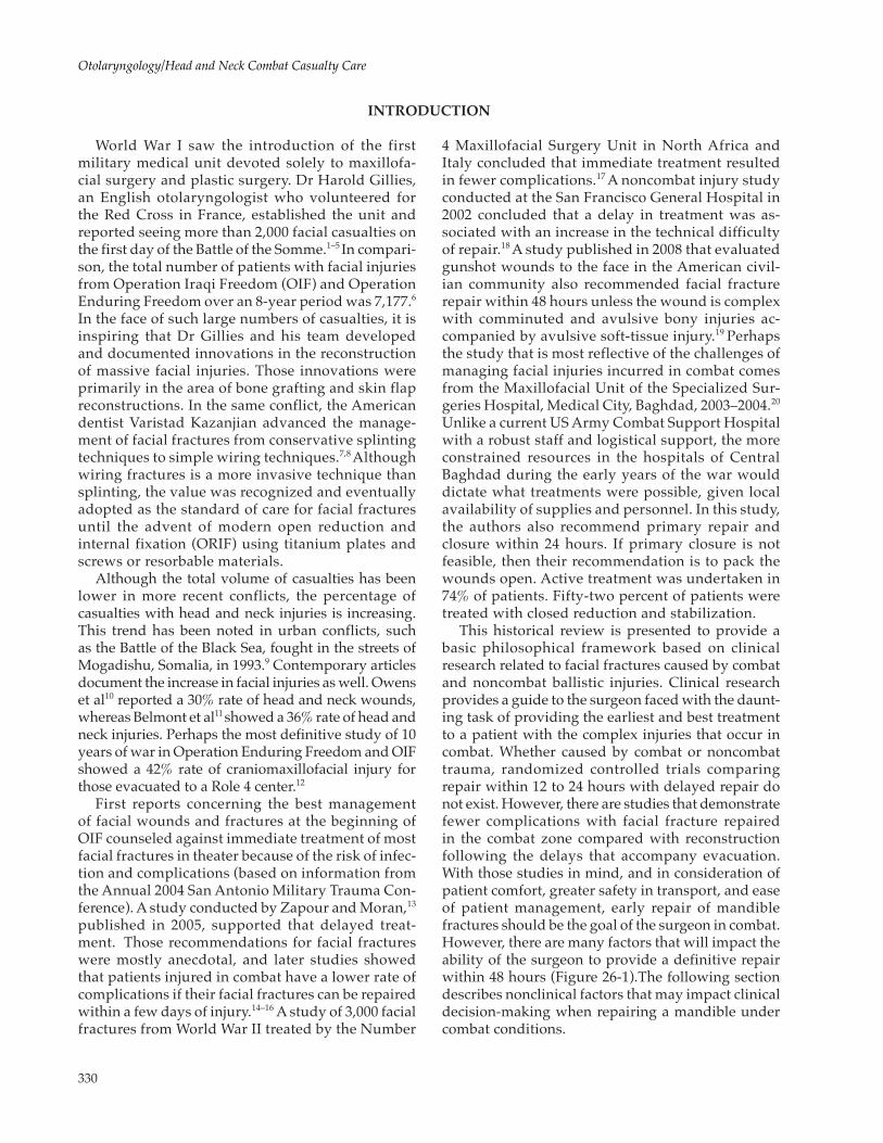
This historical review is presented to provide a basic philosophical framework based on clinical research related to facial fractures caused by combat and noncombat ballistic injuries. Clinical research provides a guide to the surgeon faced with the daunt-ing task of providing the earliest and best treatment to a patient with the complex injuries that occur in combat. Whether caused by combat or noncombat trauma, randomized controlled trials comparing repair within 12 to 24 hours with delayed repair do not exist. However, there are studies that demonstrate fewer complications with facial fracture repaired in the combat zone compared with reconstruction following the delays that accompany evacuation. With those studies in mind, and in consideration of patient comfort, greater safety in transport, and ease of patient management, early repair of mandible fractures should be the goal of the surgeon in combat. However, there are many factors that will impact the ability of the surgeon to provide a definitive repair within 48 hours (Figure 26-1).The following section describes nonclinical factors that may impact clinical decision-making when repairing a mandible under combat conditions.
World War I saw the introduction of the first military medical unit devoted solely to maxillofa-cial surgery and plastic surgery. Dr Harold Gillies, an English otolaryngologist who volunteered for the Red Cross in France, established the unit and reported seeing more than 2,000 facial casualties on the first day of the Battle of the Somme.1–5 In compari-son, the total number of patients with facial injuries from Operation Iraqi Freedom (OIF) and Operation Enduring Freedom over an 8-year period was 7,177.6
In the face of such large numbers of casualties, it is inspiring that Dr Gillies and his team developed and documented innovations in the reconstruction of massive facial injuries. Those innovations were primarily in the area of bone grafting and skin flap reconstructions. In the same conflict, the American dentist Varistad Kazanjian advanced the manage-ment of facial fractures from conservative splinting techniques to simple wiring techniques.7,8 Although wiring fractures is a more invasive technique than splinting, the value was recognized and eventually adopted as the standard of care for facial fractures until the advent of modern open reduction and internal fixation (ORIF) using titanium plates and screws or resorbable materials.
Although the total volume of casualties has been lower in more recent conflicts, the percentage of casualties with head and neck injuries is increasing. This trend has been noted in urban conflicts, such as the Battle of the Black Sea, fought in the streets of Mogadishu, Somalia, in 1993.9 Contemporary articles document the increase in facial injuries as well. Owens et al10 reported a 30% rate of head and neck wounds, whereas Belmont et al11 showed a 36% rate of head and neck injuries. Perhaps the most definitive study of 10 years of war in Operation Enduring Freedom and OIF showed a 42% rate of craniomaxillofacial injury for those evacuated to a Role 4 center.12
First reports concerning the best management of facial wounds and fractures at the beginning of OIF counseled against immediate treatment of most facial fractures in theater because of the risk of infec-tion and complications (based on information from the Annual 2004 San Antonio Military Trauma Con-ference). A study conducted by Zapour and Moran,13
published in 2005, supported that delayed treat-ment. Those recommendations for facial fractures were mostly anecdotal, and later studies showed that patients injured in combat have a lower rate of complications if their facial fractures can be repaired within a few days of injury.14–16 A study of 3,000 facial fractures from World War II treated by the Number

331
Mandible Fractures
Figure 26-1. Concurrent operations in one operating room during Operation Iraqi Freedom (2006).
VARIBLES THAT AFFECT TREATMENT DECISIONS
2003–2004.20 These limitations can occur at any time during combat and require the surgeon to be adept at several techniques for managing fractures.
Hospital Census and Air Evacuation Schedule
During periods of high patient volume, there are often limitations to the availability of operating room suites or hospital beds. Delaying treatment in favor of stabilization and evacuation is preferable, as well as reserving operating room resources for those in the most critical condition.
Surgeon Experience and Training
Although the experience in a combat zone pushes the limits of one’s capability, it is good to know your boundaries. When a case is beyond your surgical
Equipment, Personnel Availability, and Logistical Support
In the initial stages of any armed conflict, there is a period of time where medical facilities are in transition. The rapid advancement of a Combat Support Hospital and the need for mobility will preclude any treatment beyond stabilization and evacuation. Even the larger evacuation hospitals will have a period of growth during which time the option for immediate definitive treatment is not advisable or possible. Brennan21 de-scribes the evolution of the first deployed otolaryngol-ogy team in OIF and documents the decision-making that occurs during the mass casualty that resulted from the Second Battle of Fallujah in November 2004. Local personnel resources and supplies were also mentioned as a critical determinant of treatment options at the Specialized Surgeries Hospital, Medical City, Baghdad,

332
Otolaryngology/Head and Neck Combat Casualty Care
experience or capability, it is in the best interest of the patient to perform stabilization and evacuation.
Patient Condition
In recent armed conflicts, it is not uncommon for a patient to suffer multiple extremity injuries, which may include vascular compromise, traumatic amputa-
tions, and severe blood loss. Those injuries can lead to hypothermia, acidosis, and coagulopathy (commonly called the lethal triad). These patients are often too critically ill to sustain lengthy surgical procedures for reconstruction and are best treated by stabilization and evacuation. Patients with cerebral edema secondary to craniofacial trauma also require a period of conva-lescence prior to undertaking reconstructive surgery.
Figure 26-2. Actual Operation Enduring Freedom critical care airport transport team patient transition at Landstuhl Regional Medical Center occurring during a “mock mass casualty” exercise in 2011. Patient is transferred from a converted school bus to the ground outside the Landstuhl Regional Medical Center emergency department.
TREATMENT
Immediate combat life support management pri-oritizes the control of bleeding prior to addressing the airway. Injuries of the face complicate that recom-mendation because of the intimate association of the airway with the bleeding source and the difficulties associated with applying pressure to wounds that in-volve flail mandibular segments and lacerated oral and pharyngeal mucosae. Mandible fractures caused by ballistic injury are commonly associated with airway compromise. In a review of trauma at the Specialized
Surgeries Hospital, Medical City, Baghdad, Kum-moona et al20 found that 86% of the cases requiring airway management had mandible fractures, and 59% of those needing airway management were injured with high-velocity missiles. As a result, intubation or cricothyrotomy may be required immediately, fol-lowed by control of hemorrhage and stabilization of the patient. Once stabilized, a decision must be made concerning the long-term airway management follow-ing treatment.

333
Mandible Fractures
If the patient will be transported to higher levels of care with intermaxillary fixation (IMF) in place, in most cases that patient should have a tracheostomy. The primary reason for that recommendation is for safety and airway security during transport. The patient will make several transitions from vehicles to helicopters, airplanes, and buses during the evacua-tion process (Figure 26-2). There are many opportu-nities during those transfers for a nasotracheal tube to be dislodged. If that happens, the conditions for reintubation are at their worst with a compromised
airway in a patient with wire fixation, a confined area, and low lighting. If a surgical airway was indicated to repair the facial fractures or if a cricothyrotomy was placed in the field, it is best to keep that airway until the patient has been evacuated out of the com-bat theater.
Definitive control of bleeding is also best performed at the time of repair of the mandible fracture with meticulous dissection to find the bleeding source. This is often in combination with a neck exploration when the fracture results from a ballistic injury.
STABILIZING THE MANDIBLE
Even if immediate repair is not possible because of the clinical or nonclinical indicators, stabilization of the fractured mandible will immediately reduce the pain experienced by the patient and reduce the need for heavy sedation. A tracheotomy placed at the time of IMF will allow for removal of the endotracheal tube. This has many advantages for the patient. Once extu-
bated, the patient will not require as much sedation. Without sedation, the cervical spine can be clinically cleared, and mental status can be better assessed. It also makes transportation easier on the patient and safer with regard to airway management. Techniques for stabilization of the mandible are discussed in the next section.
INTERMAXILLARY FIXATION WITH ARCH BARS
Erich arch bars are commonly used to achieve good IMF when adequate dentition is present. The arch bar is cut to span each arch from the most posterior tooth on each side. It is then wired into position typi-cally capturing all teeth from the canine posteriorly. The fragments are aligned so that the teeth achieve their premorbid dentition, and then the upper and lower arch bars are wired together (Figure 26-3). This typically provides a stable reduction that simplifies the task of open reduction and internal fixation. It is not uncommon for IMF to be used as the definitive treatment for some fractures.20
Ivy loops are also used for temporary fixation (Figure 26-4). Other methods of rapidly achiev-ing IMF involve screws drilled into the maxilla and mandible with wire or rubber band fixation between them. This technique is also discussed in Case Study 26-1.
External fixators are also very useful for stabilization of comminuted fractures that do not lend themselves to immediate repair. The external fixator is less inva-sive than open reduction and internal fixation, and provides stabilization without further compromise to the vascular supply. 22
ANTIBIOTIC COVERAGE
Studies of noncombat mandibular fractures indicate that 24 hours of postoperative antibiotic coverage is sufficient.23–25 In a comprehensive review of combat injuries and head and neck infections, Petersen et al26 recommend perioperative prophy-laxis and postoperative coverage for 24 hours. The primary antibiotic recommendation is Cefazolin
2 grams every 8 hours preoperatively and for 24 hours following surgery. If the patient is beta-lactam allergic, then Clindamycin 600 mg IV can be given every 8 hours preoperatively and for 24 hours post-operatively. An alternative antibiotic is Ceftriaxone 1 gram every 12 hours preoperatively and for 24 hours postoperatively.
IMMEDIATE REPAIR VERSUS DELAYED REPAIR
There is no consensus regarding the optimal timing of repair for mandibular fractures. A literature review by Hermund et al27 concluded that there was no strong evidence for either acute or delayed treatment of man-dibular fractures to minimize healing complications.
The review study was hampered by the fact that none of the reviewed studies were randomized, and only six studies included sufficient documentation to allow for statistical analysis. In addition, none of the studies included cases treated within 12 to 24 hours. A 5-year

334
Otolaryngology/Head and Neck Combat Casualty Care
Figure 26-3. Example of mandibulomaxillary fixation in a patient with a mandible fracture, missing teeth, and a significant premorbid malocclusion.
Figure 26-4. Ivy loops demonstrated on a manikin.
retrospective study by Furr et al,28 published in 2006, concluded that the lag time to repair did not influ-ence the development of postoperative complications. There are many studies reviewing civilian trauma that recommend immediate repair with primary closure, if possible.23,29,30 A few recent studies demonstrate an increase in complications following repair of combat-related facial fractures as the time-to-repair increases.15
Patients with large avulsive injuries of the mandible, especially with concurrent loss of soft-tissue coverage, should undergo immediate debridement and stabiliza-tion with delayed reconstruction.19
Combat injuries seen in the war zone are typically more complex than those seen at a Level I trauma center in the United States. The mandible fractures are typically comminuted or have a significant loss of the bony framework. The soft-tissue envelope is commonly lacerated by projectiles, thus compro-mising the vascular supply; frequently, the skin

335
Mandible Fractures
is burned. These factors complicate the decision-making concerning both the timing and the use of local flaps for reconstruction. In the acute setting, if primary repair cannot be accomplished, the bony fragments should at least be stabilized and soft-tissue coverage provided following debridement of the wound. If the wound is significantly contami-nated or the skin is burned, performing a rotational flap to provide coverage may not be prudent. This is a judgment made by the surgeon based on the
conditions of the wound. With high-velocity ballis-tic injury, the wound tends to worsen over the first few days. This evolution of the wound is because of the delayed effects of the transference of the kinetic energy (KE) of the projectile
KE = 1/2Mass × Velocity2
to the surrounding tissues. It is perfectly acceptable to stage the reconstruction of these complex injuries.22,31
Figure 26-5. CT (computed tomography) scan shows bilateral condylar fractures through the capsule.
SUMMARY
The decision to repair mandibular fractures in the combat environment involves more environmental and logistical consideration than is typically required in a noncombat environment.32 Immediate repair can be provided in theater if the patient’s physiology permits and the operational tempo or transportation
demands do not preclude taking the patient to the operating room. If bone grafting or a vascularized free flap will be required for reconstruction, those patients are best served by stabilization of the fractured seg-ments, adequate soft-tissue coverage, and delayed reconstruction.
CASE PRESENTATIONS
Case Study 26-1: Immediate Treatment
Presentation
A 47-year-old male fell while running in Afghani-stan, sustaining blunt trauma to the mandible.
Preoperative Workup/Radiology
A CT (computerized tomography) scan was per-formed noting bilateral condylar fractures through the capsule and a symphysis fracture with lingual splaying of the mandible anteriorly and widening of the ramus segment (Figures 26-5 to 26-7).
Operative Planning/Timing of Surgery
On the day of injury, his C-spine was cleared clini-cally, and he was taken to the operating room at a For-ward Surgical Hospital.
Operation
The patient was intubated with a nasotracheal tube and placed in IMF using screws placed in the right and left maxilla and mandible (Figure 26-8). The patient was placed on the protocol for ventilator-associated pneu-monia, deep venous thrombosis prophylaxis (sequen-tial compression devices, LOVENOX [Bridgewater, NJ], and a duplex scan), prophylactically treated with a proton pump inhibitor, and then transported sedated and intubated to Landstuhl Regional Medical Center.
On evaluation at Landstuhl Regional Medical Center, the patient was sedated and intubated with a nasotra-cheal tube. He was in IMF using four screws, had a diathesis between his lower incisors, and had a posterior crossbite. The patient was taken to the operating room. The lower IMF screws were removed and an arch bar placed. He was then put into a good class I occlusion,

336
Otolaryngology/Head and Neck Combat Casualty Care
Figure 26-6. Symphysis fracture.
Figure 26-7. Open lingual cortex by 1 to 2 mm translates to posterior cross-bite.
Figure 26-8. Rapid intermaxillary fixation using screws. Note the lever arm created by the extension of the screw laterally, potentially contributing to increased posterior crossbite, and opening of the lingual cortex in a mandible with bilateral condylar instability and a symphysis fracture.
taking care to close the anterior gap and correct the posterior cross-bite. Through an incision in the labial vestibule, the periosteum of the mandible was elevated to the inferior border and laterally to each first bicuspid. A mandible matrix, 6-hole locking titanium plate was secured across the reduced symphysis fracture with three screws on either side of the fracture. The inci-sion was irrigated with bacitracin. The mentalis was resuspended with 3-0 chromic suture and the mucosal incision closed with a running 3-0 chromic suture. The IMF was released to check the occlusion. The occlusion was Class I. Without IMF, he had an anterior open bite consistent with the bilateral condylar fractures. He was left out of IMF for extubation and overnight due to concerns for his airway. The patient did well overnight and was placed in IMF with elastics the following day.
At postoperative day 10, the patient had no open bite, a Class I occlusion, and an oral opening of 1.5 cm. He was placed on an aggressive oral physiotherapy regimen and achieved an oral opening of 40 mm in 2 weeks. At 6 weeks, the elastics were removed. At 12 weeks, there was no relapse. The arch bar and screws were removed in the clinic. A panorex showed good bony healing of the symphysis and a slight shortening of the right condyle. At 16 weeks, the patient reported normal occlusion without pain and was discharged from treatment.
Complications
The patient required a second operation with gen-eral anesthesia.
Lessons Learned
In this case, it would have been best to provide de-finitive treatment for the patient in-theater. The case took 30 minutes in the operating room and would have eliminated the risks of transporting a sedated

337
Mandible Fractures
patient with a nasotracheal intubation, an unstable mandible, and IMF. Immediate treatment would have obviated the need for ventilator-associated pneumonia prophylaxis and deep venous thrombosis prophylaxis, as well. In addition, the choice of IMF screws increased the displacement of the fractures causing the patient more pain and increasing the level of sedation required for transport.
Case Study 26-2: Delayed Treatment
Presentation
A 22-year-old male was injured in an improvised explosive device blast in Iraq. He suffered third-degree burns over 64% of his body, including the head and neck (Figure 26-9).
Preoperative Workup/Radiology
A CT scan showed a comminuted fracture of the mandible with a bilateral condylar fracture, a fractured ulna, a fractured femur, and a penetrating wound to the abdomen (Figure 26-10).
Operative Planning/Timing of Surgery
The patient presented to the hospital at Balad se-dated with a cricothyrotomy in place. He was immedi-ately taken to the operating room for stabilization and debridement of his wounds. Once the life-threatening wounds were addressed, an otolaryngologist was consulted to evaluate his facial injuries. The patient was reasonably stable, but his facial burns had caused significant edema. With multiple injuries and 64% burns, this patient was scheduled for immediate trans-port to the burn unit at Brooke Army Medical Center (San Antonio, TX). It was concluded that it would be in his best interest to stabilize his injuries and prepare him for transport immediately rather than attempt a definitive repair immediately.
Operation
In the operating room, the patient’s cricothyrotomy was converted to a formal tracheostomy, arch bars were placed, and IMF was obtained. His facial wounds were then debrided and closed.
Following air evacuation to the continental United States, he was in IMF for 6 weeks because he was being treated for severe burn injuries, and receiving multiple escharotomies and split-thickness skin grafts. On evaluation for reconstruction of his mandible, he had bilateral temporomandibular joint ankylosis. He
was treated with open reduction and internal fixation of the mandibular symphysis, with a bone graft and bi-lateral costochondral grafts to the condyle and ramus. Six months later through a transcervical approach, an anterior sliding osteotomy was performed along with bilateral neck advancement flaps. Approximately 2 months later, he required excision of fistula tracts in
Figure 26-9. Patient on presentation with severe facial burns, lacerations, open fracture of the mandible, and a cricothy-rotomy in position.
Figure 26-10. CT (computed tomography) scan shows a severely comminuted anterior mandible.

338
Otolaryngology/Head and Neck Combat Casualty Care
the left submandibular region and removal of a broken wire. A month later, he required an additional excision of fistulas, removal of multiple teeth, and removal of the dental splint. One year later, he underwent con-tracture releases of the lower lip and eyelid with full-thickness skin grafts and debulking of a nasolabial flap.
Complications
Late complications included bilateral temporoman-dibular joint ankylosis, multiple fistulas, and infected hardware.
Lessons Learned
This case demonstrates the factors that influence im-mediate versus delayed treatment. Prolonged immobi-lization of a mandible that has sustained condylar and subcondylar trauma has a high risk for ankylosis because
Figure 26-11. Iraqi soldier on presentation with a severely comminuted open fracture of the mandible, fractures of the mid-face, and a left globe injury.
Figure 26-12. CT (computed tomography) scan shows one area of comminution of the mandible.

339
Mandible Fractures
of the scarring within the temporomandibular joint or as a result of trauma to the coronoid process and zygoma, thus creating zygomaticocoronal ankylosis.33 It is very likely that if his mandibular fractures had been addressed immediately and he then underwent oral physiotherapy early, then he may not have suffered the complication of ankylosis. However, he was at a high risk for succumbing to the lethal triad, and it would have been inadvisable to undertake a long reconstructive surgery, including bone grafting from other donor sites under those conditions.
Case Study 26-3: Immediate Treatment
Presentation
An Iraqi soldier presented with injuries sustained by a blast from an improvised explosive device to the face, with multiple facial fractures and injuries throughout his body.
Figure 26-14. Intraoperative placement of the reconstruction bar through a transcervical incision.
Figure 26-13. Displaced segments of the mandible.

340
Otolaryngology/Head and Neck Combat Casualty Care
Preoperative Workup/Radiology
A CT scan on arrival showed that the fragment caused a nasal fracture, left globe laceration, a com-minuted mandible fracture, and multiple facial lacera-tions (Figures 26-11 to 26-13).
Operative Planning/Timing of Surgery
The patient was taken immediately to the operat-ing room.
Operation
Anesthesia was delivered through the cricothy-rotomy. The first procedure was conversion of the cricothyrotomy to a tracheostomy. The ophthalmolo-gist performed repair of the left globe laceration.
Closed reduction of his nasal fracture was performed, and Doyle splints were sutured into position. The oral cavity was inspected. Multiple maxillary and mandibular teeth were extracted, and the oral cavity debrided of devitalized tissue. Erich arch bars were placed on the remaining teeth, and the patient was placed into IMF. The neck was prepped and draped, and a cervical incision was made 4 cm inferior to the border of the mandible. Dissection was carried through the platysma and the Hayes-Martin proce-dure performed to gain access to the inferior border of the mandible. The mandible was reconstructed with a 2.4 mm reconstruction bar, and the wound was irrigated and closed (Figures 26-14 and 26-15). Arch bars were removed at the end of the case to allow normal function. The patient was put on a soft diet, recovered without incident, and was decanulated 1 week later.
Figure 26-15. Postoperative view of reconstruction. (For more details, see article by Güven O. Zygomaticocoronoid ankylosis: a rare clinical condition leading to limitation of mouth opening. J Craniofac Surg. 2012;23:829–830.)

341
Mandible Fractures
Complications
None.
Lessons Learned
This case illustrates some of the indications for immediate treatment. The patient’s physiology was stable, allowing immediate reconstructive surgery. The operating room tempo, although busy, did not preclude scheduling the 3-hour operation, and there was no advantage to delaying the procedures. Because
the patient was Iraqi, evacuation out of theater was not an option, so delaying treatment would not benefit the hospital census and could potentially complicate the patient’s recovery and healing. If this patient had been an American or other coalition soldier with the option of evacuation, the treatment plan would have been the same, while opting for immediate treatment unless there was a pressing logistical reason not to operate. Transporting a patient with this type of open wound would increase the risk of transport, subject the patient to more blood loss, and potentially increase the complication rate.
REFERENCES
1. Chambers JA, Davis MR, Rasmussen TE. A band of surgeons, a long healing line: development of craniofacial surgery in response to armed conflict. J Craniofac Surg. 2010;21:991–997.
2. Crumley R. Some pioneers in plastic surgery of facial region. Arch Facial Plast Surg. 2003;5:9–15.
3. Rosdeutscher J. The history of otolaryngology in plastic surgery. Plast Reconstr Surg. 1969;111:2377–2384.
4. Gillies H. The Principles and Art of Plastic Surgery. Boston, MA: Little Brown; 1957.
5. Simpson D, David J. World War I: the genesis of craniomaxillofacial surgery? ANZ J Surg. 2004;74:71–77.
6. Feldt B, Salinas N, Rasmussen T, Brennan J. The Joint Facial and Invasive Neck Trauma (J-FAINT) Project, Iraq and Afghanistan 2003-2011. Otolaryngol Head Neck Surg. 2013;148:403–408.
7. Hallock G. The plastic surgeon of the 20th century. Plast Reconstr Surg. 2001;107:1014–1024.
8. Anonymous. Massachusetts General Hospital, Department of Plastic Surgery, http://www.brighamandwomens.org/Departments_and_Services/surgery/medical_professionals/surged/PlasticSurgery/AboutKazanjian.aspx. Accessed September 3, 2013.
9. Mabry R, Holcomb J, Baker A, et al. United States Army rangers in Somalia: an analysis of combat casualties on an urban battlefield. J Trauma. 2000;49:515–529.
10. Owens B, Kragh J, Wenke J, Macaitis J, Wade C, Holcomb J. Combat wounds in Operation Iraqi Freedom and Opera-tion Enduring Freedom. J Trauma. 2008;64:295–299.
11. Belmont P, Goodman G, Zacchilli M, Posner M, Evans C, Owens B. Incidence and epidemiology of combat injuries sustained during “the surge” portion of Operation Iraqi Freedom by a U.S. Army Brigade Combat Team. J Trauma. 2010;68:204–210.
12. Chan R, Siller-Jackson A, Verrett A, Wu J, Hale R. Ten years of war: a characterization of craniomaxillofacial injuries incurred during Operations Enduring Freedom and Iraqi Freedom. J Trauma Acute Care Surg. 2012;73:S453–S458.
13. Zapour MJ, Moran KA. Infectious diseases during wartime. Curr Opin Infect Dis. 2005;18:395–399.
14. Lopez M, Arnholt J. Safety of definitive in-theater repair of facial fractures. Arch Facial Plast Surg. 2007;9:400–405.
15. Salinas N, Faulkner J. Facial trauma in Operation Iraqi Freedom casualties: an outcomes study of patients treated from April 2006 through October 2006. J Craniofac Surg. 2010;21:967–970.
16. Petersen K, Colyer M, Hayes D, Hale R, Bell R. Prevention of infections associated with combat-related eye, maxil-lofacial, and neck injuries. J Trauma. 2011;71:S264–S269.

342
Otolaryngology/Head and Neck Combat Casualty Care
17. Clarkson J, Kirkpatrick J, Lawrie R. Prevention by organization: the story of no. 4 maxillofacial surgical unit in North Africa and Italy during the Second World War. Plastic Reconstr Surg. 2008;121:657–668.
18. Biller J, Pletcher S, Goldberg A, Murr A. Complications and the time to repair of mandible fractures. Laryngoscope. 2005;115:769–772.
19. Kaufman Y, Cole P, Hollier L. Contemporary issues in facial gunshot wound management. J Craniofac Surg. 2008;19:421–427.
20. Kummoona R, Muna A. Evaluation of immediate phase of management of missile injuries affecting maxillofacial region in Iraq. J Craniofac Surg. 2006;17:217–223.
21. Brennan J. Experience of first deployed otolaryngology team in Operation Iraqi Freedom: the changing face of combat injuries. Otolaryngol Head Neck Surg. 2006;134:100–105.
22. Maki M. Management outline of oral and maxillofacial missile injuries in Iraq: the value of the intermediate phase. J Craniofac Surg. 2009;20:873–877.
23. Abubaker AO, Rollert MK. Postoperative antibiotic prophylaxis in mandibular fractures: a preliminary randomized, double-blind, and placebo-controlled clinical study. J Oral Maxillofac Surg. 2001;59:1415–1419.
24. Chole RA, Yee J. Antibiotic prophylaxis for facial fractures. A prospective, randomized clinical trial. Arch Otolaryngol-Head Neck Surg. 1987;113:1055–1057.
25. Andreasen JO, Jensen SS, Schwartz O, Hillerup Y. A systematic review of prophylactic antibiotics in the surgical treat-ment of maxillofacial fractures. J Oral Maxillofac Surg. 1987;64:1664–1668.
26. Petersen K, Hayes D, Blice J, Hale R. Prevention and management of infections associated with combat-related head and neck injuries. J Trauma. 2008;64:S265–S276.
27. Hermund N, Hillerup S, Kofod T, Schwartz O, Andreasen J. Effect of early or delayed treatment upon healing of mandibular fractures: a systematic literature review. Dent Traumatol. 2008;24:22–26.
28. Furr A, Schweinfurth J, May W. Factors associated with long-term complications after repair of mandibular fractures. Laryngoscope. 2006;116:427–430.
29. Ellis E III, Muniz O, Anand K. Treatment considerations for comminuted mandibular fractures. J Oral Maxillofac Surg. 2003;61:861–870.
30. Peleg M, Sawatari Y. Management of gunshot wounds to the mandible. J Craniofac Surg. 2010;21:1252–1256.
31. Doctor VS, Farwell DG. Gunshot wounds to the head and neck. Curr Opin Otolaryngol Head Neck Surg. 2007;15:213–218.
32. Salinas N, Eller R, Davis M, Rasmussen T. Mass casualty response of a modern deployed head and neck surgical team. J Craniofac Surg. 2010;21:987–990.
33. Güven O. Zygomaticocoronoid ankylosis: a rare clinical condition leading to limitation of mouth opening. J Craniofac Surg. 2012;23:829–830.