chapter 25: urinary system - north idaho collegecoursecontent.nic.edu/przao/main/htm/pdf/25.pdf1)...
TRANSCRIPT

Chapter 25: Urinary System
I. Kidney anatomy: retroperitoneal from 12th thoracic to 3rd lumbar area
A. External anatomy: hilus is the indentation
1. Adrenal gland: in the fat at the superior end of each kidney
2. Renal capsule: thin dense connective tissue covering
3. Adipose capsule: the fatty mass surrounding each kidney
4. Renal fascia: dense C.T. that helps hold the kidney in place
B. Internal anatomy: 3 main regions
1. Cortex: light colored outer region 2. Medulla: inner region, darker in color a. Renal pyramids: dark, cone-shaped masses

1) Diagram of a pyramid
b. Renal columns: tissue that separate the pyramids
3. Renal pelvis: a funnel-shaped tube that collects urine a. Minor calyces: cup-like structure enclosing the papilla of a pyramid 1) Diagram of minor calyces
b. Major calyces: collects urine from several minor calyces

C. Blood and nerve supply: renal arteries deliver about 25% of total C.O. at any given time 1. Renal plexus: network of sympathetic nerves that control arteriole diameter
D. Nephron: the functional unit of the kidney
1. Parts a. Glomerulus: a tuft of capillaries at beginning of tubule b. Renal tubule 1) Glomerular capsule surrounds the glomerulus 2) Proximal convoluted tubule: cuboidal cells with microvilli
3) Loop of Henle: mostly simple squamous epithelium a) Descending loop b) Ascending loop

4) Distal convoluted tubule: cuboidal cells without microvilli
5) Histology
2. Renal corpuscle: glomerulus plus the glomerular capsule a. Parietal layer: simple squamous epithelium b. Visceral layer: podocytes –the cells with foot-like processes that form filtration slits around the glomerulus

1) Fenestrated capillaries
3. Blood supply to the nephron a. Afferent arteriole: brings blood to the nephron b. Efferent arteriole: smaller diameter
c. Peritubular capillaries: surround the tubule - function to reabsorb solutes into the blood from the nephron tubule
d. Vasa recta: parallels the loop of Henle; helps maintain concentration gradients
4. Juxtaglomerular apparatus: where the DCT contacts the afferent arteriole a. Afferent arteriole cells modified: juxtaglomerular cells
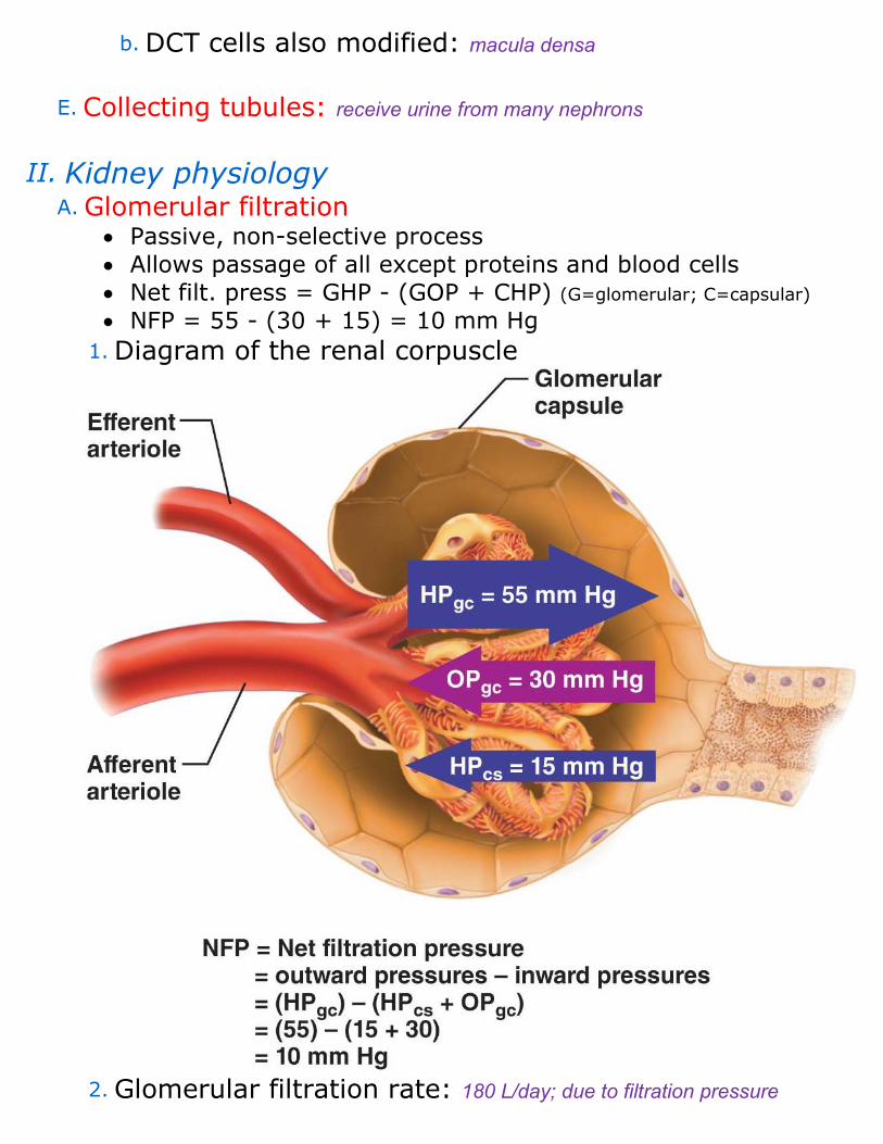
b. DCT cells also modified: macula densa
E. Collecting tubules: receive urine from many nephrons
II. Kidney physiology A. Glomerular filtration
• Passive, non-selective process
• Allows passage of all except proteins and blood cells
• Net filt. press = GHP - (GOP + CHP) (G=glomerular; C=capsular)
• NFP = 55 - (30 + 15) = 10 mm Hg
1. Diagram of the renal corpuscle
2. Glomerular filtration rate: 180 L/day; due to filtration pressure

3. Regulation of filtration a. Autoregulation: controls the afferent arteriole
b. Sympathetic nervous control
1) Stimulates JG cells: to release renin to increase systemic B.P. B. Tubular reabsorption: reabsorbed solutes must pass through the tubule cell and wall of the peritubular capillary before it reaches the blood to be returned to the systemic circulation 1. Active reabsorption: glucose, amino acids, vitamins, most ions a. Most are co-transported with Na+
b. Must have carrier proteins
c. Transport maximum: dependent on the number of carriers available
2. Passive reabsorption: diffusion and osmosis are always down the concentration gradient
3. Areas in tubule that reabsorb a. PCT: most of all substances (75-80%) b. Loop of Henle: mostly control of water c. DCT: mostly Na+ when aldosterone is present (plus some H2O)
C. Tubular secretion: the ability to clear plasma of unwanted substances
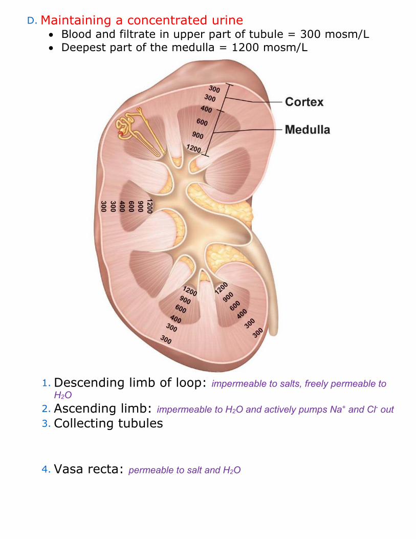
D. Maintaining a concentrated urine • Blood and filtrate in upper part of tubule = 300 mosm/L
• Deepest part of the medulla = 1200 mosm/L
1. Descending limb of loop: impermeable to salts, freely permeable to H2O
2. Ascending limb: impermeable to H2O and actively pumps Na+ and Cl- out 3. Collecting tubules
4. Vasa recta: permeable to salt and H2O

5. Diagram: Countercurrent Mechanism
E. Changing the concentration of urine 1. Forming concentrated urine a. ADH: causes the DCT and collecting duct to become more permeable to H2O
2. Forming dilute urine urine
a. Diuresis: forming a dilute urine 1) Alcohol: inhibits ADH release 2) Caffeine: inhibits Na+ reabsorption
F. Chemical composition of urine: 95% H2O, 5% solutes; mostly urea from the breakdown of proteins
III. Ureters: tubes that conduct urine to the urinary bladder
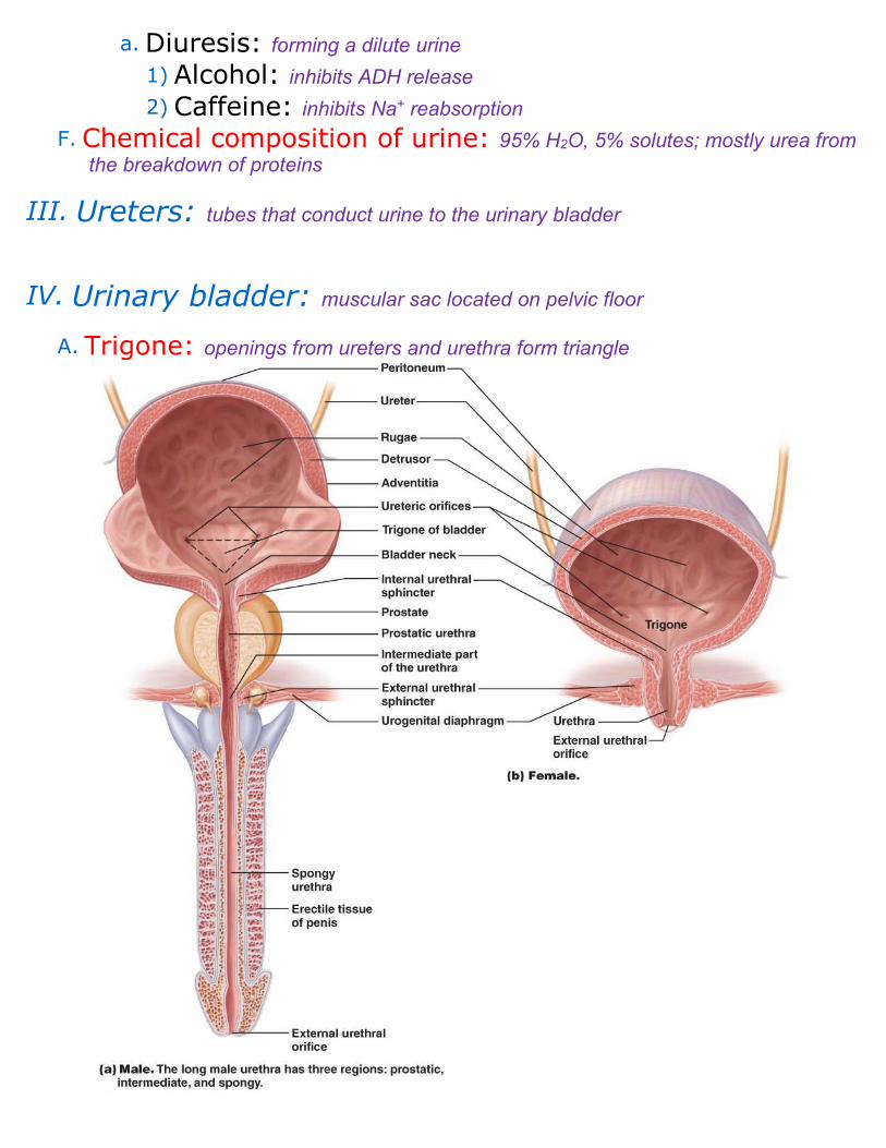
IV. Urinary bladder: muscular sac located on pelvic floor
A. Trigone: openings from ureters and urethra form triangle

B. Bladder wall 1. Transitional epithelium : is folded into rugae
2. Muscularis layer: detrusor muscle
3. Adventitia: the outermost C.T. layer
V. Urethra: a thin-walled tube that drains urine from the bladder
A. Wall 1. Mucosa: mostly pseudostratified columnar
B. Sphincters 1. Internal: smooth muscle
2. External: skeletal muscle
C. Micturition: voiding or urination