chapter 236 /a , furunculosis -...
TRANSCRIPT

Chapter 236 /A , Furunculosis
Definition:
Localized form of otitis externa resulting from infection of a single hair follicle “on the lateralwall of the canal only “
Bacterial invasion of a single hair follicle result in a well circumscribed deep skin infection thena pustule forms and progress to a local abscess formation with cellulitis and edema.
Diagnosis: Histology: is the reference standard for diagnosis but it is not taken on routine practice, Symptoms: similar to sever otitis externa and includes .pain, blocked ear serosangous discharge,
pinna and tragus are tender to touch. Irritation and scratching, lymphadenitis may occur locally. Otoscopy: may be difficult if there is sever edema which is usually restricted to the lateral canal
wall only and tympanic membrane is not affected.
Spread Backwards In advanced cases abscess may be seen pointing into the canal, the edema may spread
to the posterior auricular crease and may be mistaken for mastoiditis. Forwards: into the parotid area. Inwards: through the notch of rivinus to infect the tympanic cavity “rare”. Samples for culture: may guide therapy but they don’t contribute to the diagnosis.
Aetiology and epidemiology: Staphylococcus aureus is the most common organism. Trigger lysis of phagocytic cells Factors which may increase the risk of infection: trauma, maceration, humidity, and heat. Recurrent Furunculosis: associated factors include DM, Dysphagocytosis, and
hypogammaglobulinemia.
Outcome: If untreated it may progress to local abscess and discharge into the external canal and resolve
spontaneously or it may spread to the deeper tissues and cause a diffuse soft tissue infectionspreading into the pinna, post auricular skin and parotid gland.
Repeated infections can cause permanent scarring and fibrosis of the canal and may predisposeto chronic diffuse otitis externa.
Management options:
Analgesia and heating.Oral or systemic anti staph antibiotics:
macrolides, cephalosporines, clindamycine, quinolonesTopical treatment:
antibiotics and hygroscopic dehydrating agents like 10% ichthammol in glycerin applied to agauze wick are anti staph and causes dehydration of the canal.

Incision and drainage: Oral antibiotics in early stage of the disease. Sever spreading soft tissue disease: I.V antibiotics Abscess formation is an indication for incision and drainage then after drainage use topical
antibiotics anti staph. In sever canal edema apply a wick to facilitate treatment.
Recurrent generalized Furunculosis: Eradication therapy with nasal mupirocin Eradication therapy with oral fluxacillin for 14 days Bacterial interference therapy by implanting non pathological strains of staph aureus to colonize
skin.
Cummings
Furunculosis
A furuncle (abscess or boil) is a walled-off collection of pus that is seen as a painful, firm, orfluctuant mass.
It arises from the hair follicles of the lateral ear canal. The causative organism is typically S. aureus. Clinical manifestations include localized pain, particularly to touch. Early manifestations include
a nodular swelling that proceeds to fluctuance. Treatment includes the application of warm compresses and topical antibiotic ointment.
Antistaphylococcal oral antibiotics should be administered. A fluctuant lesion should be incised and drained under local anesthetic.

Chapter 236 /B Bullous Myrengitis
Definition: Myringitis haemorrhagica bullosa : finding of vesicles in the superficial layer of the tympanic
membrane.
Pathology: Vesicles occur between the outer epithelium and the lamina propria of the TM.
Etiology: May be viral or bacterial, influenza virus or mycoplasma pneumonia Others: St.pneumoniae, morexilla, Para influenza virus, adenovirus. Occur in all age groups but children and adolescents are most affected. Bilateral in 5% and recurrent in 5% also.
Symptoms: Sudden onsets sever unilateral throbbing pain during or following upper respiratory tract
infection. Blood stained discharge for a couple of hours. Conductive or less likely S.N.
Signs: Otoscopy: blood filled serous or serosanguinous blisters involving the tympanic membrane and
sometimes the medial wall of the ear canal, tympanic membrane is intact. 30% pure SNHL 50% mixed. 20 % CHL.
Diagnosis: Based on physical examination. Vesicles are present on the tympanic membrane.
Differential diagnosis: Acute otitis media. Herpes zoster oticus. Ramsay Hunt Syndrome.

Investigations: Inspection of the ear. Pneumatic Otoscopy to see if there is middle ear fluid or not. Clinical examination of the facial nerve. Pure tone audiogram. Cultures from the blisters are not needed in management of uncomplicated cases. Serology sample in DDx for herpes zoster.
Outcomes: Complete recovery within days in the majority of cases.
Management options: In cases without middle ear effusion and without SNHL, only analgesic are needed. In SNHL of below two years should be treated like A.O.M and antibiotics are given.
Effects of management: Complete recovery of SNHL within 3 months in 60-100%.
Cummings: Bullous myringitis is an inflammatory/infectious condition involving the lateral surface of the
tympanic membrane and the medial portion of the canal wall. It typically occurs in association with upper respiratory infections and is more common in
winter. The etiology is believed to be primarily viral; however, Mycoplasma has been identified in some
cases. Clinical manifestations include acute, severe otalgia; serosanguinous otorrhea; and hearing loss.
Physical examination reveals bullae on the tympanic membrane that are filled with serous orserosanguinous fluid.
Spontaneous rupture of the bullae leads to the observed discharge. A serous otitis media is a frequently associated finding in 30% to 40% of cases. More significant is the association of this disorder with a sensorineural hearing loss. 65% of
patients manifest a SN or mixed hearing loss, with the SN component recovering completely inapproximately 60%.
Treatment includes analgesics, topical antibiotic/steroid drops to prevent bacterial superinfection, and lancing the bullae, which may result in some pain relief.
Because Mycoplasma sometimes can be associated with this infection, the administration of amacrolide or quinolone antibiotic may be prudent.
The role of systemic steroids has not been established.
Essentials Inflammation of all layers of the tympanic membrane with bulla forming under the epithelia
superficial layer. Classified into
Primary: if no underlying O.M Secondary: form as a squeal of O.M.
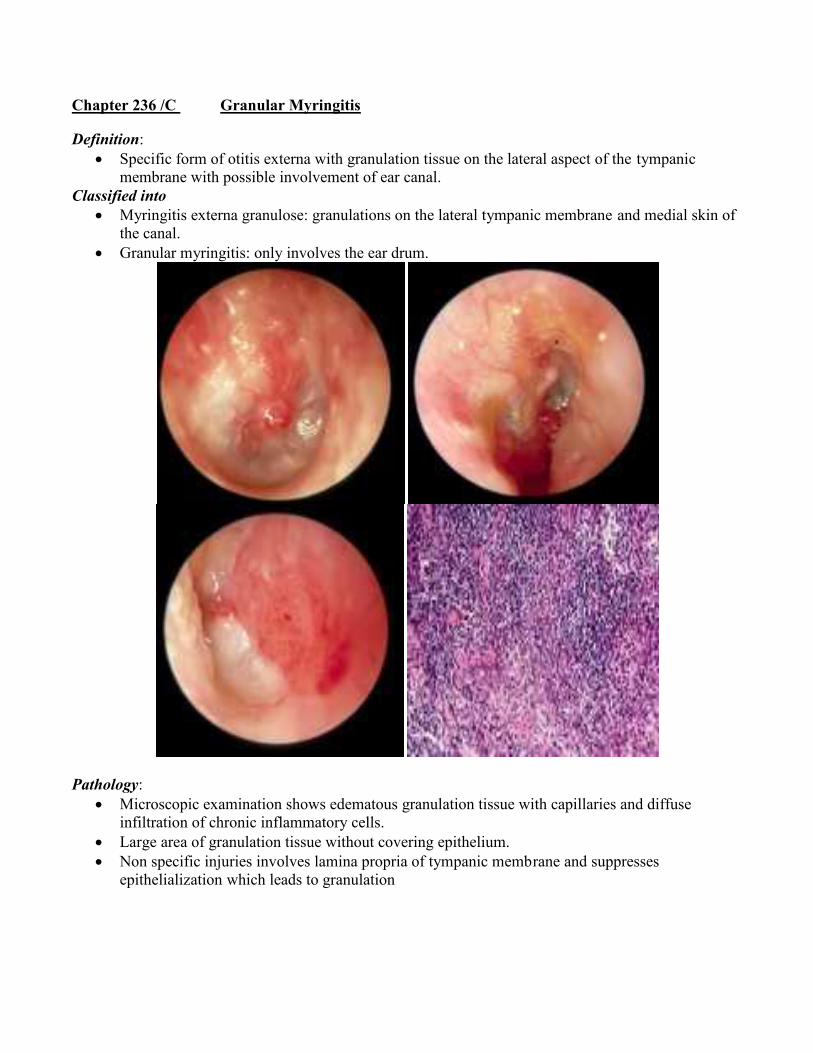
Chapter 236 /C Granular Myringitis
Definition: Specific form of otitis externa with granulation tissue on the lateral aspect of the tympanic
membrane with possible involvement of ear canal.Classified into
Myringitis externa granulose: granulations on the lateral tympanic membrane and medial skin ofthe canal.
Granular myringitis: only involves the ear drum.
Pathology: Microscopic examination shows edematous granulation tissue with capillaries and diffuse
infiltration of chronic inflammatory cells. Large area of granulation tissue without covering epithelium. Non specific injuries involves lamina propria of tympanic membrane and suppresses
epithelialization which leads to granulation

Etiology: Both sexes are equal, systemic disease, more in older patients, poor hygiene, local irritants, FB,
swimming and high temperature. Sometimes seen as post op complication of myrengoplasty (5%) Previous history of O.M, trauma, ventilation tubes.
Symptoms; Foul smelling discharge. Little or no pain. Sensation of fullness or irritation. Hearing may be slightly impaired. Can be asymptomatic.
Signs; Moderate amount of purulent secretion is seen in the affected ear. T.M covered with secretions or crusting. After aural toileting, granulation tissue is revealed.
Classified into: Localized form: most common, small areas are affected, one or more polyps. Diffuse form: slightly raised carpet of granulation covering the TM. Perforation is not present. Drum is mobile on pneumatic examination. Myrengosclerosis or thickened TM may be seen.
Diagnosis: Otoscopy: ear discharge, inflammation and granulation.
Differential Diagnosis: CSOM and Diffuse Otitis Externa. Differentiated by:
No inflammation of the ear canal Normal tympanic membrane movement on pneumatic Otoscopy. Lack of conductive deafness. Normal CT scan.
Investigations: Tympanometry: normal ME, no perforation. PTA: to exclude CHL. CT scan: if middle ear or mastoid involvement is suspected. Biopsy: if granulation doesn’t resolve with treatment to exclude malignancy.
Common pathogens: Gm +ve: S.aureus. Gm –ve: P.aurugenosa, Proteus species. Fungi: Candida Albicans.

Outcome, natural history, and complications: Many patients are asymptomatic. Chronic course and granulation may continue for years. Healing may happen spontaneously. Inflammation in the epithelial layer and lamina propria of TM lead to replacement with
proliferating granulation tissue. Fibrosis and atresia of the medial canal.
Management options: Localized form:
Microscopic debridement, LA or GA. Topical treatment with ear drops containing steroids and antibiotics or antifungal based
on culture results and should continue treatment longer than in otitis externa (2weeks). Diffuse form:
If patient didn’t respond to topical treatment, then a chemical therapy is used (0.5%formalin or trichloroacetic acid applied once a week)
Removal by physical methods if conservative treatment doesn’t work. Like curettage ortympanoplasty or laser evaporization.
Effect of management: Most cases resolve within 3 months
Chapter 236 /D Benign Necrotizing Otitis Externa
Definition: Idiopathic necrosis of localized area of the bone of the tympanic ring with secondary
inflammation of the overlying soft tissue and skin It is also called benign osteonecrosis or sequestration of the tympanic bone.
Pathology: Non specific, necrotic bony sequestrum involves the superficial cortical layer.
Histology: Dead lamellar bone with inflammatory cells filling the marrow spaces. Very limited and mild inflammatory changes of the adjacent skin and soft tissue of EAC. No evidence of vascular necrosis.
Etiology: Unknown but there is some theories:
Vascular insufficiency and microangiopathy. Repeated local trauma “ear bud abuse” or use of hearing aids. Neurotrophic disturbances. Tympanic bone has poor blood supply.
Diagnosis: History:
Mild local inflammation with itching otorrhea or otalgia. One should exclude underlying conditions like immunosuppression, DM, radiotherapy.
Examination: Diagnosed clinically by positive finding of a small area of deficient skin and soft tissue
in the EAC revealing a segment of necrotic superficial bone There may be a mild inflammatory changes or crusting Usually unilateral Clinical examination should exclude granulomas and deep osteitis of temporal bone like
cranial nerve palsies found in malignant otitis externa. Bony necrosis is usually limited but may extend to the annulus with associated TM
perforation.
Differential Diagnosis: Malignant necrotizing otitis externa. Canal cholesteatoma. Osteoradionecrosis of the EAC Tumors of the EAC. Keratosis obturance. The most similar condition is canal cholesteatoma; the only difference is the absence of a lining
of squamous epithelium in the former.
Investigations: Usually not needed and diagnosis is clinical. If gross infection is seen swab may be taken. If it was positive for P.auragenosa then it is
malignant otitis externa. If surgery is planned a CT scan is need to show the extent of bone necrosis. If prominent granulation tissue is seen, granulomatous conditions should be excluded
“TB,Syphilis” Exophytic lesions may require brush cytology to exclude malignancy. PTA: normal or mild CHL due to debris.
Outcome, natural history and complications: Separation of the sequestrum followed by epithelial growth to cover the bony defect “most likely
outcome” Rarely progressive necrosis and exposure of facial nerve and jugular bulb Chlesteatoma may result because squamous epithelium may grow from ulcer’s margin under the
sequstrum.
Management options: Conservative :removal of bony sequestrum with local toileting and locat treatmen to control
infection Aggressive: early removal of the bony sequestrum before it separates spontanousely down to
healthy bone Hyperbaric oxygen is used when there is progression despite intensive local and systemic
treatment and when necrosis is beyond the tympanic bone.
Differential diagnosis of conditions eroding bony external canal: Benign necrotizing otitis externa:
Deficient skin and soft tissue in the EAC. Underlying necrotic, sequestered bone. Mild or no symptoms, itching and discomfort. Lesion is localized no squamous epithelium deep to bony sequestrum.
Malignant necrotizing otitis externa: Pseudomonas isolated Chronic boring pain Granulomas confirming chronic infection with or without soft tissue necrosis and bony
necrosis. DM May in late stages invade deeper in the petrous bone and causes cranial palsies.
Keratosis obturans: Keratin/wax debris filling and packing the canal. Expansion of deeper canal “diffuse not localized” Low ratio of depth of defect to circumference Shallow wide expansion May show bottle neck appearance on the shallow cartilaginous part of EAC
Canal chlesteatoma: Localized skin invasion of bone in burring fashion Squamous epithelium is relatively deep and localized pocket beyond normal plane of
canal bone High ratio of depth to circumference “localized burrow” Squamous epithelium deep to bony sequestrum

Chapter 236 /E Malignant Otitis Externa
Definition: Aggressive and potentially life threatening infection of the soft tissue of the external ear and
surrounding structures which may involve periosteum and skull base bone. If the bone of the skull is involved it is called skull base osteomyelitis and if only soft tissue is
involved it is called necrotizing external otitis.
Staging: Clinicopathological:
Stage I: Clinical evidence of Malignant Otitis Externa with soft tissue infection beyondEAC, but negative Tc99
Stage II: soft tissue infection beyond the canal with positive Tc99 Stage III: as above but with cranial nerve palsy
o Single palsyo Multiple palsies.
Stage IV: meningitis , empymea, sinus thrombosis or brain abscess
Pathology: The infection spreads through cellulitis, chondritis, periosteitits, osteitis and finally
osteomyelitis. Infection spreads from the canal through fissures of Santorini in the floor of the canal. Once periosteitits develops, infection spreads rapidly through skull base and cranial nerve palsies
then develops. Osteomyelitis mainly involves Haversian System of compact bone and involvement of
pneumatized portion is only a late stage. The otic capsule is usually spared. Multiple abscesses are found.
Bacteriology: Pseudomonas auragenosa 95% Rarely Aspergillus.
Predisposing factors: Most commonly affect elderly DM I,II IC and patients with phagocytosis impairment. Cerumen in DM patient has a higher pH which reduces bactericidal effect of it. Histocytosis X or temporal bone malignancy causes refractory cases in children, biopsy is
needed for exclusion. AIDS patints has higher rates.
Diagnosis: Pain granulation otorrhea and resistance to local therapy for at least 8-10 days. DM or other IC condition. Pseudomonas from culture. Positive bone scan and cranial nerve palsies are confirmatory factors. ESR,CRP are raised but not significant.
Radiology: Technetium 99m: detect bony involvement before high resolution CT scan.
Scan may remain positive for 9 months due to osteoblast and osteoclast activity, so Tc99 isuseful for detecting early involvement.
Gallium 67: absorbed by leukocytes and is more sensitive monitor of infection.It goes back to normal quickly so it is a good measure when to terminate treatment.But Gad is taken up by any inflammatory condition so it is useful for monitoring more thandiagnosis.
By combining radiosensetizers with CT ; SPECT is useful but it is an expensive measure MRI: help to differentiate between malignant and inflammatory process.
Outcome and Complications: Mortality now about 10%. If extensive skull base osteomyelitis is established the clivus and contralateral temporal bone can
be involved and infection spread anteriorly to the sphenoid and carotid. Anterior infection can involve TMJ and parapharyngeal space. Facial nerve is the most commonly affected nerve 60%. Then IX, X, XI and rarely IV and XII. Finally disease can spread to central nervous system and sinuses extradural space and meninges. Thrombosis of the internal carotid artery is the usual terminal event If the disease treated ;facial palsy will resolve in 22% completely and 9% partially.
Management options: Aural toilet:
Essential to control granualtions and improve local pain control. Use of topical Abs is controversial and may alter culture results.
Systemic antibiotics: Systemic anti pseudomonas antibiotics.which may be given for 6 weeks to several
months (cipro with or without Af and/or ceftazidime). Once ESR and CRP start to fall then shift to oral antibiotics (cipro or rifampicin).

Hyperbaric oxygen: Could be helpful.
Surgery; Should be reserved to few selected patients in which there is deterioration despite
medical treatment and includes removal of sequestra ,pus and debridement. A normal appearing canal is not a sensitive indicator of resolution and recurrence can
occur in weeks.
Essentials
Progression of the disease: EAC with invasion through fissure of santorini or tympanomastoid suture to the retromandibular
fossa. Involvement of stylomastoid and jugular foramens. Septic thrombosis of the lateral venous sinus. Spread to the petrous apex through the vascular and facial planes, not air cells.
Staging: Stage I: limited to soft tissue and cartilage. Stage II: soft tissue + bony erosion of temporal bone. Stage III: intracranial extension or erosion beyond temporal bone.

Chapter 236 /F Keratosis obturans and primary canal cholesteatoma
Definition: Keratosis obturans: accumulation of a large plug of desquaminated keratin in the EAC. Primary canal cholesteatoma: the invasion of squamous tissue from the ear into localized area of
bony erosion.
Pathology: Keratosis obturans: geometrically patterned keratin plug within the lumen of expanded EAC Keratin is shed from the complete circumference of the deep ear canal forming a lamina “onion
skin”. Primary canal cholesteatoma: keratin is derived from a sac that involves the bone of the ear canal
with bony fragments within it.
Etiology: Keratosis obturans: due to abnormal epithelial migration of the ear canal skin. Keratosis tympanicum: abnormal migration from pars flaccida to the pars tensa across the whole
drum plus tinnitus unilaterally. There are two subtypes of Keratosis obturans:
Inflammatory: which occur secondary to acute problem “viral” causing inflammation ofthe ear canal and alter epithelial migrationCured by removal
Silent: disease that carries on and on, caused by abnormal sepation of the keratin andpersists after the first removal and need subsequent removal many times.
Primary auditory canal cholesteatoma: Uncertain but could be post traumatic or post surgical “stapedectomy” A piece of exposed bone become infected and sequests and the epithelium migrate into this bony
abnormality and cholesteatoma occurs.
Epidemiology: One case of each new 1000 ENT cases for canal cholesteatoma and 5 for each Keratosis
obturans.
Diagnosis: Clinical symptoms and findings: Keratosis obturans:
Acute sever otalgia. CHL. Younger age. Occasionally bilateral. Associated with brochiectasis and sinus disease. Plugged feeling of the ears. Keratin plug occlude the canal. TM thickened.Ear canal ballooned. Hyperemia of the canal skin with granulations.

Primary auditory canal cholesteatoma: Chronic otorrhea “the most common” Normal hearing. Itching or pain “dull”. Older population. 90% unilateral. Keratin in random pattern. T.M normal. Localized bone erosion and osteitis “post infection” Bone sequestration.
CT scan: may be needed to assess bony erosion or bony widening. Histological examination: of any granulation tissue to exclude malignancy,necrotizing otitis
externa
Natural history and complications: Normal ear cana;s can suddenly develop in both diseases. Keratosis obturans can cause extensive bony erosion including automastoidectomy. No intracranial complications in both diseases.
Management options: Keratosis obturans:
Keratinolytic salicylic acid 2% in alcohol, NaHCO3 Removal under G.A Recurrent Keratosis: canaloplasty.
Primary auditory canal cholesteatoma: If the extent of the cholesteatoma is seen then start with conservative treatment “frequent
cleaning ,topical 1% hydrocortisone,3% salicylic acid,betamathasone,NaHCO3” If this is not possible; excision of necrotic bone and cholesteatoma via mastoid and repair
of the defect using temporalis fascia. Other treatments include canal wall up procedure.

Chapter 236 /G Acquired atresia of the external ear
Definition: Absence of or closure of a passage of the ear. Intraluminal sequel of either intra or extra luminal processes of varying etiology resulting in a
blind sac in the EAC. Subdivided into:
Solid: continuous block of either fibrous or fibrous and bony material which iscontinuous with the structure of the T.M.The face of solid atresia can be blunt producing a funnel –shaped medial aspect to thelumen of ear canal.
Membranous: fibrous tissue that has a covering of ear canal skin or both sides; thusseparating the ear canal into a medial and lateral segments.The medial part collects keratin from desquamation of the skin; which may become anerosive process and become an internal canal cholesteatoma.
Diagnosis: CT scan: gives a very detailed image of the bony and soft tissue structure of external ear and
differentiate between solid and membranous types.
Etiology: Inflammation: which may result from:
Otitis externa Psoriasis, eczema, active COM. Trauma: open injury like gunshot wound, closed fracture of the tympanic plate, mainly,
Mandibular condyle, burns. Surgery: including meatal approach “tympanoplasty” or meatal surgery “removal of
osteoma” membranous type more in the lateral canal.
Pathogenesis: Solid type:
In cases associated with otitis externa or media granular medial otitis externa andgranulations in the T.M that persists for many months inspite of treatment.
The granulations become fibrotic and the ear drum thickens as the medial meatus massre-epithelized, the process can repeat many times.

If it is associated with chronic active otitis media the granulations appear around the T.Mperforations and eventually lead to the closure of the defect before the process extendslateral as above.
Membranous type: Originates in the lateral meatus as a web formation, precipitated by a circular irritation
from inflammation, trauma or burns and ulceration of the skin around the entirecircumference of the external ear canal.
The web-like stenosis forms after fibrosis and re epitheliazation as with solid atresias. Repeated attacks or one massive injury such as a tympanic plate fracture leads to a
complete atresia.
Epidemiology: 0.5 Per 100000. Mostly solid type 1:20
Natural history and complications: There is a change from wet to dry phase. Main compliant is CHl. Membranous type is associated with cholesteatoma.
Management options: Medical:
During wet phase, medial granulations can be removed by aspiration and cauterizationwith silver nitrate or trichloroacetic acid. and ear pack with ribbon gauze or a wick.
This may change the phase into dry. CHL may be managed by air conducting hearing aid but may aggregate the conduction. So the use of BAHA is better than the air conduction device.
Surgery: Fibrous atresia:
To remove the fibrous tissue by elevating it from the ear canal bone, fibrousannulus and lamina propria of the T.N.

Using a speculum is usually enough to gain access but sometimes endaural orretroauricular approach is needed.
A circumferential incision is made lateral to the blunt face of the atretic plate anda plane of dissection is established between bony canal and canal skin, followedby atretic late and finally lateral to the fibrous annulus and lamina propria of theT.M.
The epithelial defect is repaired by a fine splint skin graft as single or multiplepieces.
Silastic tube may be inserted to stabilize the epithelial surface. Finally ear canal is packed with ribbon gauze soaked in anti septic. Follow up by suction toileting and repacking to prevent recurrence and medial
granulation. Membranous atresia:
Trans canal using ear speculum for small atretic plate. If it is thick ,retroauricular appraoach is used to preserve lateral and medial
epithelial covering to aid repair of ear canal skin. In trans canal method fibrous plate is excised via a circumferential incision
lateral to its margin. The whole lesin is excised with sacrifies of the minimum of surrounding
epithelium. Silastic sheaths are overlied holding lateral and medial skin over bone. In retroauricular method remove atretic plate and dissect it away from medial
skin covering which will help in final reconstruction providing a skin flap tocover the bare canal bone at the site of membranous atresia.
Atresia surgery with tympanoplasty: In cases of COM with middle ear problem like tympanosclerosis or ossicular discontinuotiy,
tympanotomy may be combined with atresia surgery esp. solid cases.

Outcomes: Aim to decrease CHL and provide mobile T.M. Residual air bone gap in 90% is 15-20 db.
Chapter 236 /H Otitis Externa and Otomycosis
Otitis externa:
Definition: Generalized condition of the EAC skin with general edema and erythema with itchy discomfort
and ear discharge.
Classification: Predisposing factors:
Anatomical: Narrow EAC {hereditary, excestosis} Obstruction of normal meatus “Keratosis obturans”, FB, hearing aid.
Dermatological: Eczema, seborrhoic, dermatitis.
Allergic: Atopy, non atopic, due to topical medication.
Physiologic: Humid environment, immunosuppression.
Traumatic: Skin maceration, ear probing, radiotherapy.
Microbiological: Active chronic otitis media, exposure to pseudomonas or fungi.
Etiology and epidemiology: 0.4% per year. 10% life time. Water and moisture are thought to cause a change from Gm +ve to Gm –ve flora. And as the ear is inflamed, healthy ceumen is removed quickly. Secondary bacterial infection:
Most patients will culture multiple organisms; pseudomonas in the ear has different propertiesthan other body pseudomonas and have special adherence properties.
Bathing:Fresh water lakes increase the risk, but rivers swimming pools or sea doesn’t increase the risk.
Irritant ,allergic reactions:About half of the patients are sensitive to topical agents with neomycin causing most allergy.
Microbiology: Pseudomonas : 50-65% Other Gm –ve: 20-35% Staph aureus: 15-30% Streptococci: 9-15%

Pathology: Clinical course has been subdivided into three stages:
I: pre-inflammatory: The protective lipid/acid balance (Ph4-5) is lost and the stratum corneum becomes edematous,
blocking off the sebaceous and apocrine glands producing aural fullness and itching. With further scratching, disruption of the epithelial layer and invasion of resident or introduced
organisms.
II: acute inflammatory stage: Progressively thickening exudates, more edema and obliteration of the lumen
Mild: little or no obstruction. Moderate: subtotal obstruction. Sever: complete obstruction and increasing pain.
In sever stages auricular changes and cervical lymphadenopathy are seen.
III: chronic inflammation: Chronic otitis externa, resistance inflammation of more than 3 weeks. Others consider it more
than 6 months. Thickening of the external canal skin and fibrous canal stenosis.
Diagnosis: Otalgia ,pruritis, ear discharge, erythema and edema Complete or partial stenosis T.M with significant debris but mobile. Patient with O.M will have fever and otalgia before the ear discharge. Fever is uncommon in otitis externa.
Outcome and complications: Untreated mild attacks resolve spontaneously as the epithelial barrier become re-established. If inflammation was more than repair, pain increases and discharge results and soft tissue
infection can occur which can lead to perichondritis, chondritis, cellulitis and parotitis and orerysipelas and in immune compromised patient malignant otitis externa can result.

Management options: Aural toileting:
The most effective single treatment for otitis externa, with and without microscopicassistance.
Don’t use water irrigation. Topical medication:
Use of wick to hold the medication in the EAC. Glycerol and ichthammol (90:10) for moderate to severe cases. Dehydrating and antimicrobial agents against staph and stept but poor against
pseudomonas. Dehydrating effect help to reduce pain and edema. Oral analgesic in moderate to severe cases. SE of topical agents is burning sensation.
Systemic antibiotics: No evidence for the use of systemic Abs in uncomplicated cases.
Prevention of recurrence: Avoidance of water penetration to the ear by using Vaseline cotton. Use of alcohol or (aqua ear or ear-calm) after swimming will help remove water. Hair dryer ”not on hot setting”
Effect of management and outcomes: Steroid Ag preparations are equal to cipro drops and quinolones are not toxic. Spray based are better than drops.

Otomycosis:
Introduction: 10% of otitis externa cases. More common in hot humid climate and often secondary to prolonged treatment with topical
Abs. DM and IC increase the risk.
Aspergillus 90% Candida 10-20% Other fungi are rare.
Clinical findings: Black, grey, green, yellow, or white discharge with debris resembles wet news papers. Fungal hyphe
Management: Toileting. Removal of debris. Topical antifungal (locorten, vioform). In resistance cases you should exclude fungal infection elsewhere “athletic foot” Rarely fungi can cause invasive otitis externa and aggressive systemic antifungal are needed with
high mortality rates.

Chapter 236 /I perichondritis of the external ear
Definition: Infection or inflammation involving the perichondrium of the external canal, auricle, and canal.
But it is usually used to describe other conditions like “erysipelas,cellulitis or true perichondritits”
Classification: Erysipelas pf the external ear. Cellulitis of the external ear. Perichondritis. Chondritis.
Etiology: Usually secondary to trauma,(surgery, frostbite, burns, chemical injury, infected hematoma, high
piercing of the ear and insertion of earrings) Erysipelas or cellulitis may extend to involve the cartilage or perichondrium. Organism: pseudomonas and staph aureus, Others less common proteus and E.coli.
Pathology: Hyperplasia of the dermal layers. Thickened subcutaneous tissue. Intense infiltration with PMNs. Thickening of the perichondrium. Destruction of the cartilage by phagocytes.
Diagnosis: Dull pain, increasing in severity. Classical signs of inflammation involving cartilaginous pinna. Lobule is spared. Background history of trauma. Diagnosis is clinical, increase pain: chondritis or perichondritis.

Differential diagnosis: Relapsing polychondritis:
Similar but has systemic nature, not only localized “ocular conditions like (scleritis, iritis, andkeratitis)”.
Extranodal non Hodgkin`s lymphoma of the pinna: With or without HIV.
Outcomes: If untreated, subperichondrial abscess develop leading to avascular necrosis of the underlying
cartilage and deformity of the pinna. Infection may spread to the cartilage itself. Rare complications: septicemia due to streptococcal infection, subacute bacterial endocarditis
and necrotizing fascitits of the neck.
Management options: Prevention:
Avoid ear piercing in the cartilage. Ear surgery: avoid cartilage trauma and tight banding. Aseptic drainage of the hematomas. Prophylactic antibiotics in ear burns Gm –ve bacteria and daily dressing and removing
eschcars. First line management:
Mildest form: topical and oral antibiotics. If there is abscess or discharge drainage and swab culture and start I.V. high dose broad
spectrum Abs meanwhile. Subperichondrial abscess need incision and drainage but only if there is fluctuation as
premature incision may result in further spread of the infection Anti pseudomonas quinolones or aminopenicillins for 2-4 weeks orally or until clinical
improvement I.V. Resistant cases:
Nonresponsive to treatment and persistent pain and suppuration necessitates moreintervention.
Aggressive excision and debridement of the necrosed cartilage with significant deformityafter.
Continues drainage and irrigation with Ag and steroid solution twice daily and usingfenestrated polyethylene tubes.
Other forms of management: Iontophoresis: effective local antibiotic delivery without systemic absorption used
exponentially. Low dose radiation: to kill micro organisms 3 times daily for 3 weeks. UV Radiation: significant SE.

Chapter 236/ J relapsing polychondritis
Definition: Rare disease of episodic inflammation and degeneration involving multiple cartilaginous
structures, usually these of upper respiratory tract as well as connective tissue at various othersites.
Other name: polychondropatheis, systemic chondromalacia and chronic atrophic polychondritis.
Pathology: Histology: initial neutophil infiltration early loss of basophils of cartilage and later infiltration by
hitiocytes, plasma cells and lymphocytes and eventually atrophic cartilage with cystic spacescontaining gelatinous material.
Other proteoglycans rich structure such as eye CVS may undergo similar inflammation anddegeneration.
High incidence of vasculitis.
Etiology: Uncertain but may be autoimmune. Serum antibodies to type II collagen in acute phase.
Diagnosis: Macadam’s signs:
Recurrent chondritis of both ears. Non invasive inflammatory polyarthritis. Chondritis of nasal cartilage. Ocular inflammation including conjunctivitis, keratitis, scleritis, episcleritis and uveitis. Chondritis of the respiratory tract involving laryngeal and /or tracheal cartilage. Cochlear and or vestibular damage ”SNHL, tinnitus or vertigo”
I: Three or more of the above signs without histologic confirmation . II: one or more with histologic confirmation. III: involvement of two or more anatomic locations with response to steroids or
dapsone. Clinical picture includes fever in the acute phase. 90% involves the pinna unilaterally or bilaterally.
Acute inflammation leads to resorption of the cartilage and floppy pinna. Nasal chondritis: pain, feeling of fullness, inflammation and sudden painless suddle nose. Larynx and trachea: resorption of the cartilage and hoarsness, tenderness and airway
obstruction, and softening to palpation. Others: costochondral cartilage and Eustachian tube may lead to otitis media with
effusion. Peripheral joints: seronegative non deforming, non erosive ply or oligo chondritis. Non cartilaginous structures:
Ocular inflammation: may affect anywhere in the eye. Inner ear: (vertigo or SNHL) due to vasculitis. CVS and vascular consequences. Skin lesions: vasculitic in nature.
Differential diagnosis: Local inflammatory conditions in the pinna “perichondritis”, or in the nose or larynx. Systemic granulomatous diseases (Wegener`s T cell lymphoma, T.B, sarcoidosis).
Outcomes: Acute exacerbation and remission. Deformity in the pinna, SNHL, saddle nose, and airway compromise.
Management options: Steroids: high doses 30-60 mg prednisone daily to induce remission, and maintenance 5-10 mg
daily to suppress recurrence. Immunosuppressants: to reduce steroids doses, mainly azathioprine or methotrexate or dapsone. NSAIDs and cholchincine: control mild relapses. Anti CD4 monoclonal antibody and minocycline: they are rarely used. Surgical: to maintain airways, tracheotomy or expandable stents.
Effect on outcome: Steroids induce remission in significant proportion but relapse is likely specially if steroids are
stopped.
Chapter 236 /K exostosis of the EAC
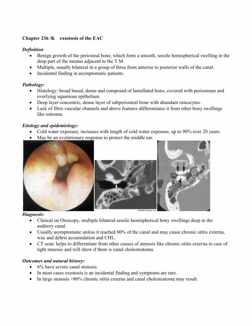
Definition: Benign growth of the periosteal bone, which form a smooth, sessile hemispherical swelling in the
deep part of the meatus adjacent to the T.M. Multiple, usually bilateral in a group of three from anterior to posterior walls of the canal. Incidental finding in asymptomatic patients.
Pathology: Histology: broad based, dense and composed of lamellated bone, covered with periosteum and
overlying squamous epithelium. Deep layer concentric, dense layer of subperiosteal bone with abundant osteocytes. Lack of fibro vascular channels and above features differentiates it from other bony swellings
like osteoma.
Etiology and epidemiology: Cold water exposure, increases with length of cold water exposure, up to 90% over 20 years. May be an evolutionary response to protect the middle ear.
Diagnosis: Clinical on Otoscopy, multiple bilateral sessile hemispherical bony swellings deep in the
auditory canal. Usually asymptomatic unless it reached 80% of the canal and may cause chronic otitis externa,
wax and debris accumulation and CHL. CT scan: helps to differentiate from other causes of stenosis like chronic otitis externa in case of
tight stenosis and will show if there is canal cholesteatoma.
Outcomes and natural history: 6% have severe canal stenosis. In most cases exostosis is an incidental finding and symptoms are rare. In large stenosis >80% chronic otitis externa and canal cholesteatoma may result.

Management options: In most cases no treatment is needed and only advice to use ear plugs in cold water swimmers. Debris is cleaned by aural toileting. Local treatment of infection and inflammation.
Surgery: In cases refractory to medical treatment, meatoplasty procedure, persistent otitis externa
and cerumen obstruction and if wider access is needed for middle ear surgery. Surgery is carries out by post aural or post meatal approach with careful elevation and
preservation of the skin overlying exostosis Bone of exostosis is removed using high speed drill cutting and diamond burr. Complications of surgery: 25% of the transmeatal group needs revision so post aural is better. Infection, T.M perforation, facial palsy, hearing loss due to ossicular chain or inner ear
injury, exposure of the TMJ with chronic pain and sublaxation and early or late soft tissuestenosis of the canal.

Chapter 236 /L foreign bodies in the ear
Pathology: In older: cotton wools ,insects ,beads, paper, small toys and erasors.
Clinical picture: Mostly in children. May be asymptomatic or with pain or discharge caused by otitis externa. Live insects are annoying due to discomfort created by loud noise and movement.
Management options: 2/3 is removed by GP.
Three main aspects should be considered: Nature of FB:
Living insects: should be killed by oil at first. Irregular soft graspable nonliving objects: like dead insects, cotton wools, paper,
small toys are removed by crocodile forceps. Organic objects: beans, etc… shouldn’t be syringed because they absorb water. Button batteries: shouldn’t be syringed because they may leak, they need to be
removed urgently. Inorganic round/ smooth non graspable: beads, erasers, syringing is safe usually
and successful, if not use a blunt hook or microscope.In syringing you should look at an area where there is a gap between the FB andthe canal wall.20% of referred cases need removal under GA.Cyanoacrylate glue can be used as they adhere to the FB.
Location of the FB: Lateral canal FB are easier to remove because it is wider and less sensitive and
due ti its elastic nature. Firmly impacted FB medial to the isthmus especially when previous attempts
caused edema and swelling in the canal may require surgical removal. A post auricular approach and widening of the canal by bone drilling is advised.
Patient considerations: Syringing is better tolerated in children with lower risk of causing trauma. As the FB has been removed it is advisable to check the ears for underlying
pathology as the child may have put the FB due to pain, itching or otorrhea.
Complications: Usually limited to canal lacerations or otitis externa. Rarely facial palsy may occur due to leakage of alkaline material from a button battery and
necrosis of the surrounding tissues. Damage to the T.M or the ossicular chain may occur. GP should refer cases to the ENT if more than one attempt or more than one instrument has been
used.
Chapter 236 /M hematoma auris
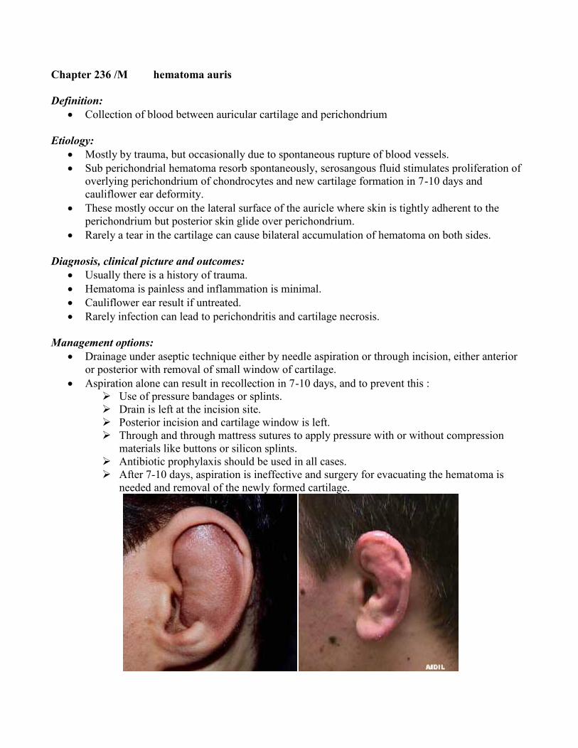
Definition: Collection of blood between auricular cartilage and perichondrium
Etiology: Mostly by trauma, but occasionally due to spontaneous rupture of blood vessels. Sub perichondrial hematoma resorb spontaneously, serosangous fluid stimulates proliferation of
overlying perichondrium of chondrocytes and new cartilage formation in 7-10 days andcauliflower ear deformity.
These mostly occur on the lateral surface of the auricle where skin is tightly adherent to theperichondrium but posterior skin glide over perichondrium.
Rarely a tear in the cartilage can cause bilateral accumulation of hematoma on both sides.
Diagnosis, clinical picture and outcomes: Usually there is a history of trauma. Hematoma is painless and inflammation is minimal. Cauliflower ear result if untreated. Rarely infection can lead to perichondritis and cartilage necrosis.
Management options: Drainage under aseptic technique either by needle aspiration or through incision, either anterior
or posterior with removal of small window of cartilage. Aspiration alone can result in recollection in 7-10 days, and to prevent this :
Use of pressure bandages or splints. Drain is left at the incision site. Posterior incision and cartilage window is left. Through and through mattress sutures to apply pressure with or without compression
materials like buttons or silicon splints. Antibiotic prophylaxis should be used in all cases. After 7-10 days, aspiration is ineffective and surgery for evacuating the hematoma is
needed and removal of the newly formed cartilage.

Chapter 236 /N Osteoradionecrosis of the temporal bone
Definition: Exposure and necrosis of a variable portion of previously irradiated Petrous temporal bone which
fails to heal over a period of 3 months.
Incidence, etiology and pathology: Because of its density, bone absorbs a greater proportion of radiation than soft tissue, More common in the mandible. Also occurs as chondronecrosis of the larynx and osteonecrosis of the maxilla, nasal bone,
nasopharynx, zygoma, palate, clavicle and hyoid. Result from high dose radiotherapy in and around petrous temporal bone for malignancies of the
parotid, EAC, middle ear, maxilla, nasopharynx and pituitary. Radiation causes inhibition of mitosis and the capacity of tissue repair and vasculitis leading to
obliteration of blood vessels and avascular necrosis. The tissue is hypoxic, hypo vascular and hypo cellular. Risk increased by: compact nature and poor blood supply of the Petrous bone and tympanic ring.
Radio therapeutic technique: to reduce the risk includes: Increased energy of beam “mega voltage than kilo voltage” to reduce the dose to
bone. Use of multiple fields to contain dose distribution. Use of heavy charged particles “hadron therapy”
Risk factors: Patients with micro vascular disease(DM, atherosclerosis) Trauma after radiotherapy (dental extraction)
Pathology: Tissue affected includes bone, overlying S.C tissue and skin. Auricular cartilage is rarely affected.
Histological changes: Includes death of osteocytes and osteoblasts resulting in empty lacuna. Demineralization of bone, osteolysis, loss of bone marrow substance Repetitive fibrosis and secondary infection. Soft tissue histological changes:
Epithelial hyperplasia. Atrophy of dermal structures Dermal fibrosis and soft tissue necrosis
Macroscopically: Loss of skin and soft tissue exposing bone, and secondary infection. Tympanic ring mostly affected.
Diagnosis, clinical picture natural history and outcomes: Time between exposure and disease vary between less than 12 months up to 23 years. Divided into:
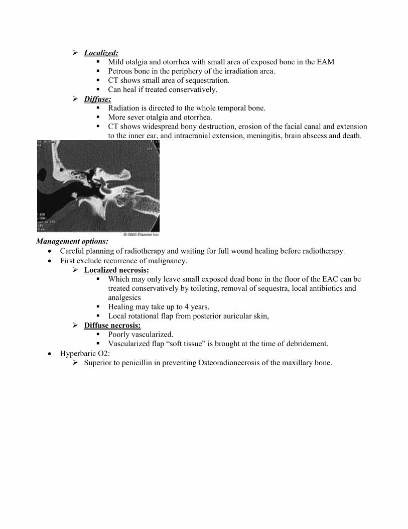
Localized: Mild otalgia and otorrhea with small area of exposed bone in the EAM Petrous bone in the periphery of the irradiation area. CT shows small area of sequestration. Can heal if treated conservatively.
Diffuse: Radiation is directed to the whole temporal bone. More sever otalgia and otorrhea. CT shows widespread bony destruction, erosion of the facial canal and extension
to the inner ear, and intracranial extension, meningitis, brain abscess and death.
Management options: Careful planning of radiotherapy and waiting for full wound healing before radiotherapy. First exclude recurrence of malignancy.
Localized necrosis: Which may only leave small exposed dead bone in the floor of the EAC can be
treated conservatively by toileting, removal of sequestra, local antibiotics andanalgesics
Healing may take up to 4 years. Local rotational flap from posterior auricular skin,
Diffuse necrosis: Poorly vascularized. Vascularized flap “soft tissue” is brought at the time of debridement.
Hyperbaric O2: Superior to penicillin in preventing Osteoradionecrosis of the maxillary bone.

Chapter 236 /O herpes zoster oticus
Definition: Herpetic vesicular rash on the concha, EAC or pinna with lower motor neuron palsy of the
ipsilateral facial nerve.
Pathology: A reactivated varicella zoster infection from dormant viral particles resident in the geniculate
ganglion and the spiral and vestibular ganglia of the VIII nerve. The virus is present in 100% of the conchal vesicles.
Diagnosis: Clinical MRI and CSF exam have no role in the diagnosis or management and in acute phase MRI can be
misleading because it can occur as small vestibular schwannoma. VIII: may be involved to a variable degree resulting in hearing loss, tinnitus and vertigo. Almost all patients have several abnormalities in the auditory pathway in different locations. Auricular pain is the first symptom. Vesicular rash in 14% may develop days later after the pain and facial palsy. Sometimes rash is present on the tongue and pharyngeal mucosa. Spread from VII to VIII nerve is thought to be through vasa nervosum or less commonly
through neurological anastomosis. Facial palsy: can occur without cutaneous or mucosal rash and called zoster sine herpte. Rise in the viral skin titers confirm diagnosis. It is the second most common cause of unilateral facial palsy after idiopathic Bell`s palsy.
Outcomes: If untreated: 60% develop complete facial paralysis in a weak and >60% in older than 50 years. If the palsy is complete 10% will get full return. If the palsy is incomplete 66% will recover completely. Overall: 50% of adults and 80% of children will achieve full recovery to house-brackmann grade
I.
Management options: Acyclovir and prednisone: if given within3 days of the symptoms more than 75% will have
complete recovery. And if treatment started after day 8, about 30% recovery.

If only steroids given the results are less from 90-60% in grade one. Early treatment with antiviral therapy will reduce post herpetic neuralgia. Even suspected cases should be treated.