chapter 20 lecture outline - palm beach state college · 2014-09-18 · chapter 20 lecture outline...
TRANSCRIPT
1
Chapter 20
Lecture Outline
Copyright © McGraw-Hill Education. Permission required for reproduction or display.
See separate PowerPoint slides for all figures and tables pre-
inserted into PowerPoint without notes.

20-2
Introduction
• The route taken by blood was a point of much confusion for many centuries– Chinese emperor Huang Ti (2697–2597 BC) correctly
believed that blood flowed in a circuit around the body and back to the heart
– Roman physician Galen (129–c.199) thought blood flowed back and forth (like air in and out of lungs); he thought the liver created blood out of nutrients and organs consumed it
– English physician William Harvey (1578–1657) performed experiments to show that the heart pumped blood and that it traveled in a circuit
• Many of Harvey’s contemporaries rejected his ideas
• After microscope was invented, capillaries were discovered by van Leeuwenhoek and Malpighi
• Harvey’s work was the start of experimental physiology and it demonstrated how empirical science could overthrow dogma
General Anatomy of
the Blood Vessels
• Expected Learning Outcomes
– Describe the structure of a blood vessel.
– Describe the different types of arteries, capillaries, and
veins.
– Trace the general route usually taken by the blood from
the heart and back again.
– Describe some variations on this route.
20-3
20-4
General Anatomy of
the Blood Vessels
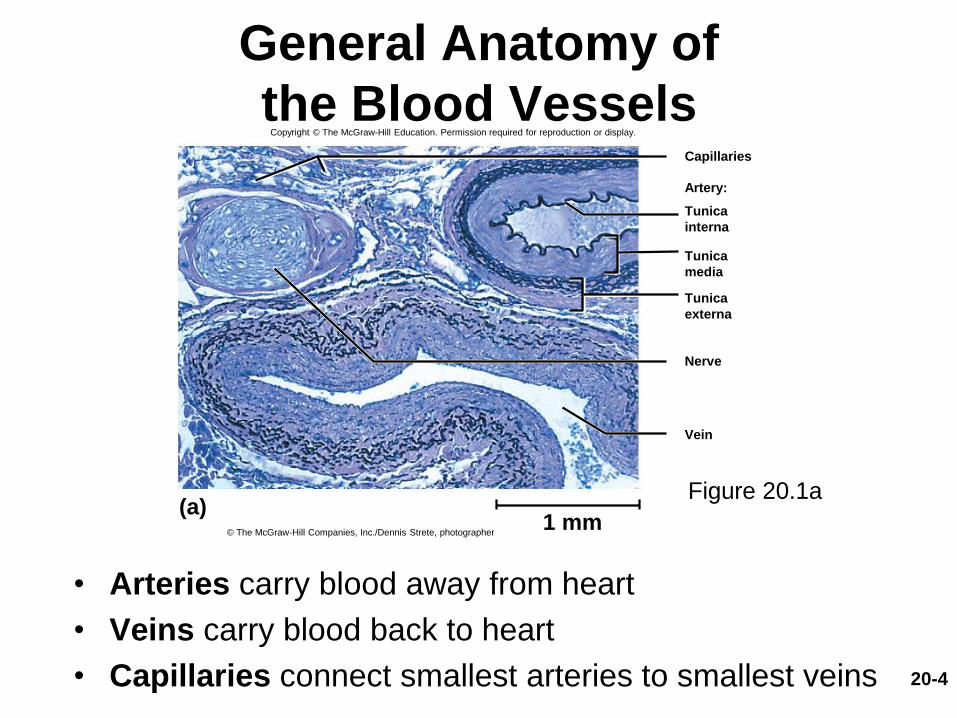
• Arteries carry blood away from heart
• Veins carry blood back to heart
• Capillaries connect smallest arteries to smallest veins
Figure 20.1a
Artery:
Nerve
1 mm(a)
Capillaries
Tunica
interna
Tunica
media
Tunica
externa
© The McGraw-Hill Companies, Inc./Dennis Strete, photographer
Vein
Copyright © The McGraw-Hill Education. Permission required for reproduction or display.

20-5
The Vessel Wall
• Walls of arteries and veins have three layers
(tunics): tunica interna, tunica media, tunica
externa
• Tunica interna (tunica intima)
– Lines the blood vessel and is exposed to blood
– Endothelium: simple squamous epithelium overlying
basement membrane and sparse layer of loose
connective tissue
• Acts as a selectively permeable barrier
• Secretes chemicals that stimulate dilation or
constriction of the vessel
20-6
The Vessel Wall
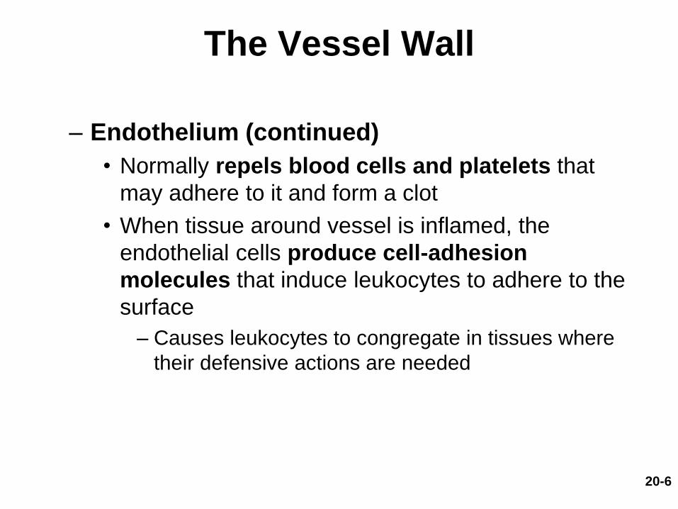
– Endothelium (continued)
• Normally repels blood cells and platelets that
may adhere to it and form a clot
• When tissue around vessel is inflamed, the
endothelial cells produce cell-adhesion
molecules that induce leukocytes to adhere to the
surface
– Causes leukocytes to congregate in tissues where
their defensive actions are needed

20-7
The Vessel Wall
• Tunica media
– Middle layer
– Consists of smooth muscle, collagen, and elastic
tissue
– Strengthens vessels and prevents blood pressure
from rupturing them
– Regulates diameter of the blood vessel
20-8
The Vessel Wall
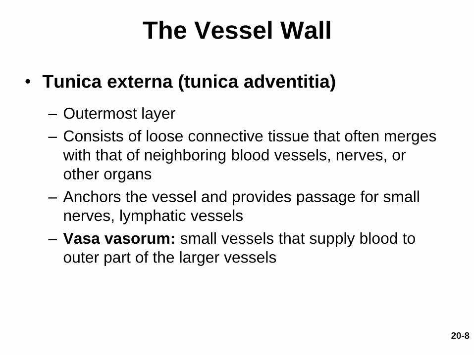
• Tunica externa (tunica adventitia)
– Outermost layer
– Consists of loose connective tissue that often merges
with that of neighboring blood vessels, nerves, or
other organs
– Anchors the vessel and provides passage for small
nerves, lymphatic vessels
– Vasa vasorum: small vessels that supply blood to
outer part of the larger vessels

20-9
The Vessel Wall
Figure 20.2
Copyright © The McGraw-Hill Companies, Inc. Permission required for reproduction or display.
Lumen
Endothelium
Nerve
Lumen
Endothelium
Nerve
Endothelium
Endothelium
Endothelium
Large vein
Medium vein
Capillary
Distributing (medium) artery
Arteriole
Conducting (large) artery
Endothelium
Internal elastic lamina
External elastic lamina
Endothelium
Aorta
Tunica interna:
Basement
membrane
Tunica media
Tunica externa
Vasa
vasorum
Tunica interna:
Basement
membraneValve
Tunica media
Tunica externa
Tunica interna:
Basement
membrane
Tunica media
Tunica externa
Venule
Tunica interna:
Basement
membrane
Tunica media
Tunica externa
Tunica interna:
Basement
membrane
Tunica media
Tunica externa
Basement
membrane
Tunica interna:
Tunica media
Tunica externa
Basement
membrane
Direction
of blood
flow
Inferior
vena
cava
Vasa
vasorum
20-10
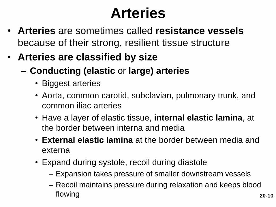
Arteries• Arteries are sometimes called resistance vessels
because of their strong, resilient tissue structure
• Arteries are classified by size
– Conducting (elastic or large) arteries
• Biggest arteries
• Aorta, common carotid, subclavian, pulmonary trunk, and
common iliac arteries
• Have a layer of elastic tissue, internal elastic lamina, at
the border between interna and media
• External elastic lamina at the border between media and
externa
• Expand during systole, recoil during diastole
– Expansion takes pressure of smaller downstream vessels
– Recoil maintains pressure during relaxation and keeps blood
flowing

20-11
Arteries
• Arteries are classified by size (continued)
– Distributing (muscular or medium) arteries
• Distributes blood to specific organs
• Brachial, femoral, renal, and splenic arteries
• Smooth muscle layers constitute three-fourths of
wall thickness
20-12
Arteries
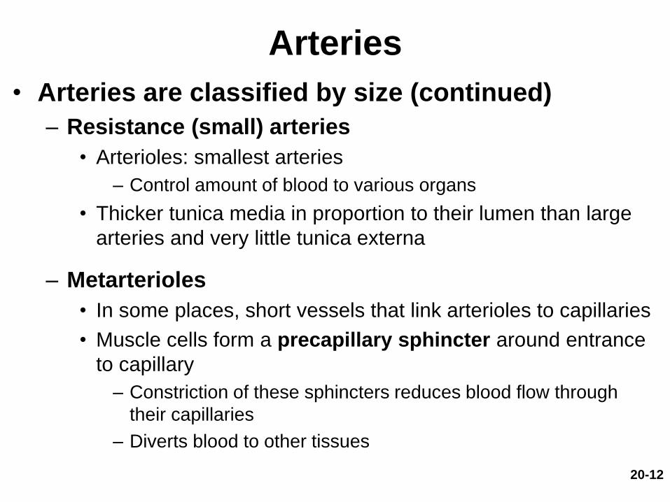
• Arteries are classified by size (continued)
– Resistance (small) arteries
• Arterioles: smallest arteries
– Control amount of blood to various organs
• Thicker tunica media in proportion to their lumen than large
arteries and very little tunica externa
– Metarterioles
• In some places, short vessels that link arterioles to capillaries
• Muscle cells form a precapillary sphincter around entrance
to capillary
– Constriction of these sphincters reduces blood flow through
their capillaries
– Diverts blood to other tissues
20-13
Arteries
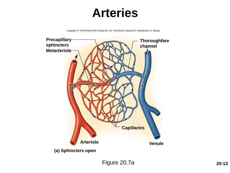
Figure 20.7a
Copyright © The McGraw-Hill Companies, Inc. Permission required for reproduction or display.
Capillaries
Metarteriole
Arteriole
Precapillary
sphinctersThoroughfare
channel
Venule
(a) Sphincters open
20-14
Aneurysm
• Aneurysm—weak point in artery or heart wall
– Forms a thin-walled, bulging sac that pulsates with
each heartbeat and may rupture at any time
– Dissecting aneurysm: blood accumulates between
tunics of artery and separates them, usually because of
degeneration of the tunica media
– Most common sites: abdominal aorta, renal arteries,
and arterial circle at base of brain
– Can cause pain by putting pressure on other structures
– Can rupture causing hemorrhage
– Result from congenital weakness of blood vessels,
trauma, or bacterial infections
• Most common cause is atherosclerosis and hypertension

20-15
Arterial Sense Organs
• Sensory structures in walls of major vessels
that monitor blood pressure and chemistry
– Transmit information to brainstem to regulate heart
rate, blood vessel diameter, and respiration
– Carotid sinuses: baroreceptors
• In walls of internal carotid artery
• Monitor blood pressure
– Transmit signals through glossopharyngeal nerve
– Allow for baroreflex
20-16
Arterial Sense Organs
• Sensory structures (continued)
– Carotid bodies: chemoreceptors
• Oval bodies near branch of common carotids
• Monitor blood chemistry
• Transmit signals through glossopharyngeal nerve to brainstem respiratory centers
• Adjust respiratory rate to stabilize pH, CO2, and O2
– Aortic bodies: chemoreceptors
• One to three bodies in walls of aortic arch
• Same structure and function as carotid bodies, but innervation is by vagus nerve
20-17
Capillaries
• Capillaries—exchange vessels: site where
gasses, nutrients, wastes, and hormones pass
between the blood and tissue fluid
– The “business end” of the cardiovascular system
– Composed of endothelium and basal lamina
– Absent or scarce in tendons, ligaments, epithelia,
cornea, and lens of the eye
– Three capillary types distinguished by ease with
which substances pass through their walls
(permeability): continuous capillaries, fenestrated
capillaries, and sinusoids
20-18
Types of Capillaries
• Three types of capillaries
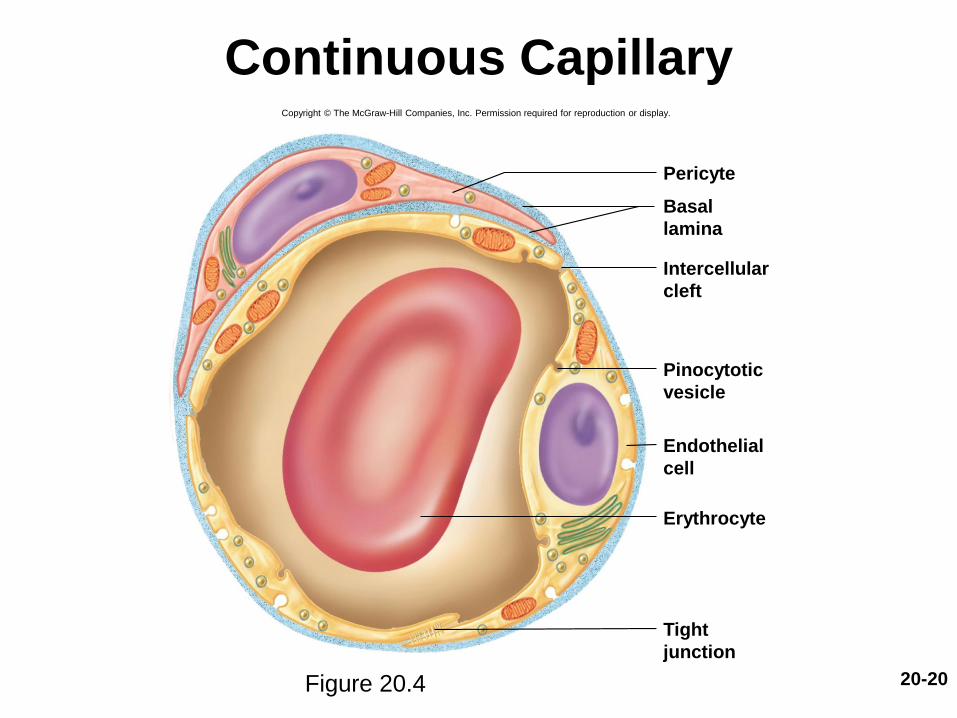
– Continuous capillaries: occur in most tissues
• Endothelial cells have tight junctions forming a
continuous tube with intercellular clefts
• Allow passage of solutes such as glucose
• Pericytes wrap around the capillaries and contain the
same contractile protein as muscle
– Contract and regulate blood flow

20-19
Types of Capillaries
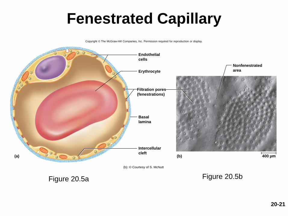
• Three types of capillaries (continued)
– Fenestrated capillaries: kidneys, small intestine
• Organs that require rapid absorption or filtration
• Endothelial cells riddled with holes called filtration
pores (fenestrations)
– Spanned by very thin glycoprotein layer
– Allow passage of only small molecules
– Sinusoids (discontinuous capillaries): liver, bone
marrow, spleen
• Irregular blood-filled spaces with large fenestrations
• Allow proteins (albumin), clotting factors, and new
blood cells to enter the circulation
20-20
Continuous Capillary
Figure 20.4
Copyright © The McGraw-Hill Companies, Inc. Permission required for reproduction or display.
Pericyte
Erythrocyte
Basal
lamina
Intercellular
cleft
Pinocytotic
vesicle
Endothelial
cell
Tight
junction
20-21
Fenestrated Capillary
Figure 20.5a Figure 20.5b
Copyright © The McGraw-Hill Companies, Inc. Permission required for reproduction or display.
(a) (b)
Erythrocyte
Endothelial
cells
Filtration pores
(fenestrations)
Basal
lamina
Intercellular
cleft
Nonfenestrated
area
400 µm
(b): © Courtesy of S. McNutt

20-22
Sinusoid in Liver
Figure 20.6
Copyright © The McGraw-Hill Companies, Inc. Permission required for reproduction or display.
Macrophage
Sinusoid
Microvilli
Endothelial
cells
Erythrocytes
in sinusoid
Liver cell
(hepatocyte)
20-23
Capillary Beds
• Capillary beds are networks of 10-100 capillaries
– Usually supplied by a single arteriole or metarteriole
– At distal end, capillaries transition to venules or drain into a
throroughfare channel (continuation of metarteriole)
– At any given time, three-fourths of body’s capillaries are
shut down
• Most control of flow involves constriction of arterioles that are
upstream from the capillaries
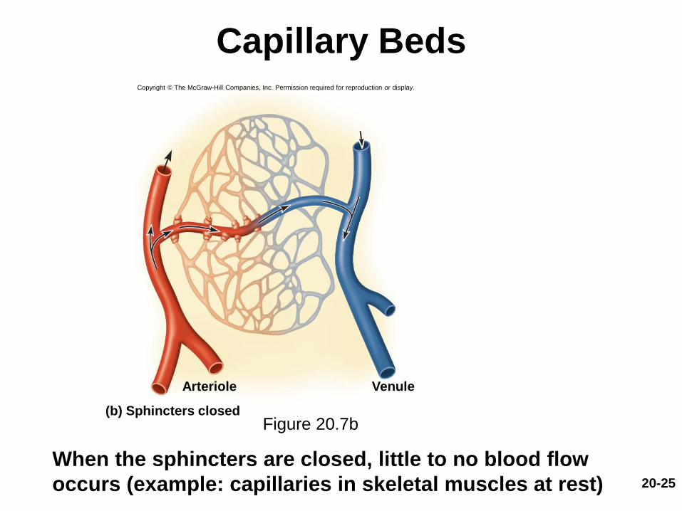
• Within the capillary bed, precapillary shincters control flow
– When sphincters are relaxed, capillaries are well perfused with
blood
– When sphincters contract, they constrict the entry to the
capillary and blood bypasses the capillary

20-24
Capillary Beds
Figure 20.7a
When sphincters are open, the capillaries are well perfused
Copyright © The McGraw-Hill Companies, Inc. Permission required for reproduction or display.
Capillaries
Metarteriole
Arteriole
Precapillary
sphinctersThoroughfare
channel
Venule
(a) Sphincters open
20-25
Capillary Beds
Figure 20.7b
When the sphincters are closed, little to no blood flow
occurs (example: capillaries in skeletal muscles at rest)
VenuleArteriole
Copyright © The McGraw-Hill Companies, Inc. Permission required for reproduction or display.
(b) Sphincters closed
20-26
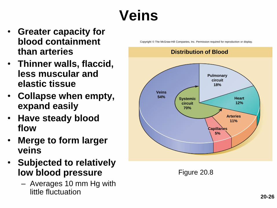
Veins• Greater capacity for
blood containment than arteries
• Thinner walls, flaccid, less muscular and elastic tissue
• Collapse when empty, expand easily
• Have steady blood flow
• Merge to form larger veins
• Subjected to relatively low blood pressure– Averages 10 mm Hg with
little fluctuation
Figure 20.8
Copyright © The McGraw-Hill Companies, Inc. Permission required for reproduction or display.
Distribution of Blood
Pulmonary
circuit
18%
Heart
12%Systemic
circuit
70%
Veins
54%
Arteries
11%
Capillaries
5%
20-27
Veins
• Postcapillary venules—smallest veins
– Even more porous than capillaries so also exchange
fluid with surrounding tissues
– Tunica interna with a few fibroblasts and no muscle
fibers
– Most leukocytes emigrate from the bloodstream
through venule walls

20-28
Veins
• Muscular venules—up to 1 mm in diameter
– One or 2 layers of smooth muscle in tunica media
– Have a thin tunica externa
• Medium veins—up to 10 mm in diameter
– Thin tunica media and thick tunica externa
– Tunica interna forms venous valves
– Varicose veins result in part from the failure of these
valves
– Skeletal muscle pump propels venous blood back
toward the heart
20-29
Veins
• Venous sinuses– Veins with especially thin walls, large lumens, and no
smooth muscle
– Dural venous sinus and coronary sinus of the heart
– Not capable of vasomotor responses
• Large veins—diameter larger than 10 mm– Some smooth muscle in all three tunics
– Thin tunica media with moderate amount of smooth muscle
– Tunica externa is thickest layer
• Contains longitudinal bundles of smooth muscle
– Venae cavae, pulmonary veins, internal jugular veins, and renal veins

20-30
Varicose Veins
• Blood pools in the lower legs of people who
stand for long periods stretching the veins
– Cusps of the valves pull apart in enlarged superficial
veins, further weakening vessels
– Blood backflows and further distends the vessels, their
walls grow weak and develop into varicose veins
• Hereditary weakness, obesity, and pregnancy
also promote problems
• Hemorrhoids are varicose veins of the anal
canal
20-31
Circulatory Routes
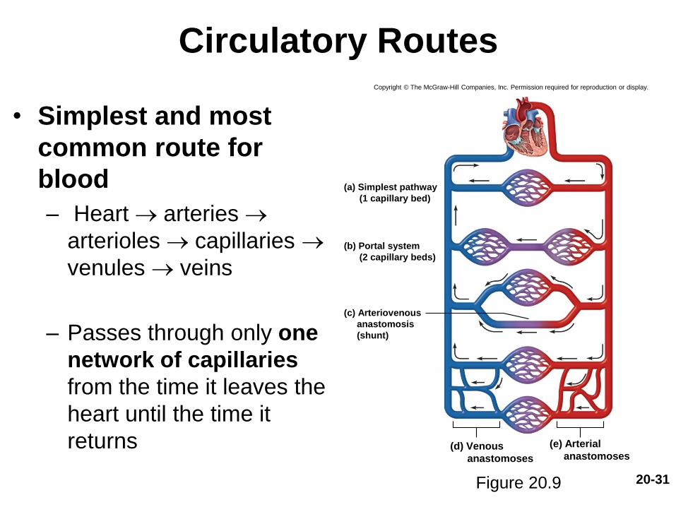
• Simplest and most
common route for
blood
– Heart arteries
arterioles capillaries
venules veins
– Passes through only one
network of capillaries
from the time it leaves the
heart until the time it
returns
Figure 20.9
Copyright © The McGraw-Hill Companies, Inc. Permission required for reproduction or display.
(a) Simplest pathway
(1 capillary bed)
(b) Portal system
(2 capillary beds)
(c) Arteriovenous
anastomosis
(shunt)
(d) Venous
anastomoses
(e) Arterial
anastomoses
20-32
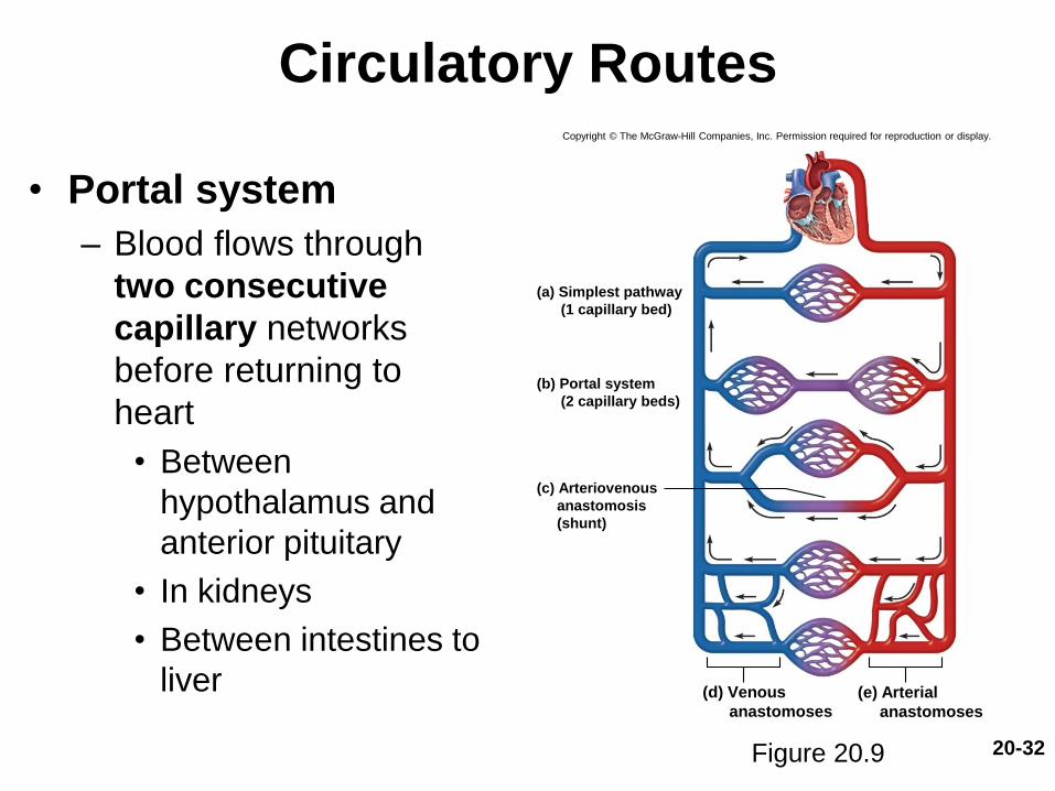
Circulatory Routes
• Portal system
– Blood flows through
two consecutive
capillary networks
before returning to
heart
• Between
hypothalamus and
anterior pituitary
• In kidneys
• Between intestines to
liver
Figure 20.9
Copyright © The McGraw-Hill Companies, Inc. Permission required for reproduction or display.
(a) Simplest pathway
(1 capillary bed)
(b) Portal system
(2 capillary beds)
(c) Arteriovenous
anastomosis
(shunt)
(d) Venous
anastomoses
(e) Arterial
anastomoses

20-33
Circulatory Routes
• Anastomosis—
convergence point
between two vessels
other than capillaries
• Arteriovenous
anastomosis (shunt)
– Artery flows directly into
vein, bypassing capillaries
Copyright © The McGraw-Hill Companies, Inc. Permission required for reproduction or display.
(a) Simplest pathway
(1 capillary bed)
(b) Portal system
(2 capillary beds)
(c) Arteriovenous
anastomosis
(shunt)
(d) Venous
anastomoses
(e) Arterial
anastomoses
Figure 20.9

20-34
Circulatory Routes
• Venous anastomosis
– Most common
– One vein empties directly into
another
– Reason vein blockage is less
serious than arterial blockage
• Arterial anastomosis
– Two arteries merge
– Provides collateral
(alternative) routes of blood
supply to a tissue
– Coronary circulation and
common around joints
Copyright © The McGraw-Hill Companies, Inc. Permission required for reproduction or display.
(a) Simplest pathway
(1 capillary bed)
(b) Portal system
(2 capillary beds)
(c) Arteriovenous
anastomosis
(shunt)
(d) Venous
anastomoses
(e) Arterial
anastomoses
Figure 20.9
Blood Pressure,
Resistance, and Flow
• Expected Learning Outcomes
– Explain the relationship between blood pressure,
resistance, and flow.
– Describe how blood pressure is expressed and how pulse
pressure and mean arterial pressure are calculated.
– Describe three factors that determine resistance to blood
flow.
– Explain how vessel diameter influences blood pressure
and flow.
– Describe some local, neural, and hormonal influences on
vessel diameter.
20-35
20-36
Blood Pressure,
Resistance, and Flow
• Blood supply to a tissue can be expressed in terms
of flow and perfusion
– Blood flow: the amount of blood flowing through an
organ, tissue, or blood vessel in a given time (mL/min.)
– Perfusion: the flow per given volume or mass of tissue in
a given time (mL/min./g)
• At rest, total flow is quite constant, and is equal
to the cardiac output (5.25 L/min)
20-37
Blood Pressure,
Resistance, and Flow
• Important for delivery of nutrients and oxygen, and removal of metabolic wastes
• Hemodynamics
– Physical principles of blood flow based on pressureand resistance
• F P/R (F = flow, P = difference in pressure, R = resistance)
• The greater the pressure difference between two points, the greater the flow; the greater the resistance, the less the flow
20-38
Blood Pressure
• Blood pressure (BP)—the force that blood exerts against a vessel wall
• Measured at brachial artery of arm using sphygmomanometer– A close approximation of pressure at exit of left ventricle
• Two pressures are recorded
– Systolic pressure: peak arterial BP taken during ventricular contraction (ventricular systole)
– Diastolic pressure: minimum arterial BP taken during ventricular relaxation (diastole) between heart beats
• Normal value, young adult: 120/75 mm Hg
20-39
Blood Pressure
• Pulse pressure—difference between systolic and diastolic pressure
– Important measure of driving force on circulation and of stress exerted on small arteries by pressure surges generated by the heart
• Mean arterial pressure (MAP)—the mean pressure one would obtain by taking measurements at several intervals throughout the cardiac cycle
– Diastolic pressure + (one-third of pulse pressure)
– Average blood pressure that most influences risk level for edema, fainting (syncope), atherosclerosis, kidney failure, and aneurysm
20-40
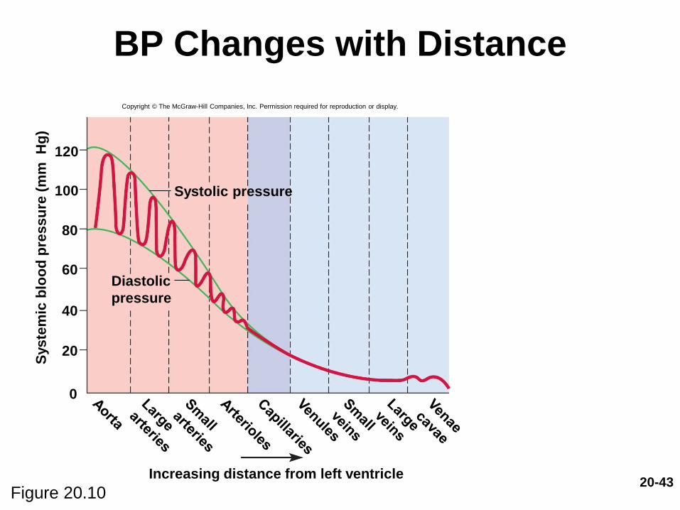
Blood Pressure
• Since pressure varies across the cardiac cycle,
blood flow in arteries is pulsatile
– Speed surges from 40 cm/s to 120 cm/s
– Blood spurts intermittently from an open artery
• In capillaries and veins, blood flows at steady
speed
– Bleeding from veins tends to be slow and steady
• BP tends to rise with age– Arteriosclerosis—stiffening of arteries due to deterioration
of elastic tissues of artery walls
– Atherosclerosis—build up of lipid deposits that become
plaques

20-41
Blood Pressure
• Hypertension—high blood pressure
– Chronic resting BP > 140/90
– Consequences
• Can weaken arteries, cause aneurysms, promote
atherosclerosis
• Hypotension—chronic low resting BP
– Caused by blood loss, dehydration, anemia
20-42
Blood Pressure
• BP determined by cardiac output, blood volume, and resistance to flow– Blood volume regulated mainly by kidneys
20-43
BP Changes with Distance
Figure 20.10
Copyright © The McGraw-Hill Companies, Inc. Permission required for reproduction or display.
Increasing distance from left ventricle
120
100
80
60
40
20
0
Sys
tem
ic b
loo
d p
res
su
re (
mm
H
g)
Systolic pressure
Diastolic
pressure

20-44
Peripheral Resistance
• Peripheral resistance—the opposition to flow that blood encounters in vessels away from the heart
• Resistance hinges on three variables: blood viscosity, vessel length, and vessel radius
– Blood viscosity (“thickness”)
• RBC count and albumin concentration elevate viscosity the most
• Decreased viscosity with anemia and hypoproteinemia speed flow
• Increased viscosity with polycythemia and dehydration slow flow
– Vessel length
• The farther liquid travels through a tube, the more cumulative friction it encounters
• Pressure and flow decline with distance
20-45
Peripheral Resistance
– Vessel radius: most powerful influence over flow
• Only significant way of controlling resistance
• Vasoreflexes—changes in vessel radius
– Vasoconstriction: when smooth muscle of tunica media contracts
– Vasodilation: relaxation of the smooth muscle, allowing blood pressure to expand vessel
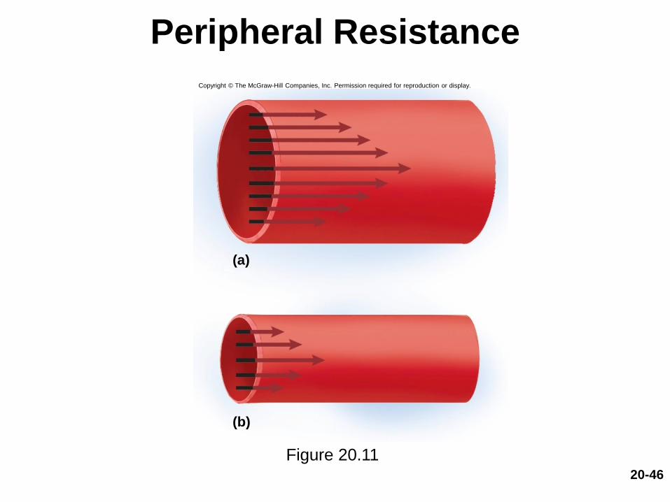
• Vessel radius markedly affects blood velocity
• Laminar flow: flows in layers, faster in center
• Blood flow (F) proportional to the fourth power of
radius (r), F r 4
– Small changes in blood vessel radius can cause large
changes in flow (mL/min)
20-46
Peripheral Resistance
Figure 20.11
(a)
(b)
Copyright © The McGraw-Hill Companies, Inc. Permission required for reproduction or display.
20-47
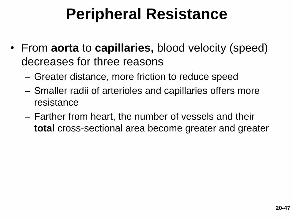
Peripheral Resistance
• From aorta to capillaries, blood velocity (speed)
decreases for three reasons
– Greater distance, more friction to reduce speed
– Smaller radii of arterioles and capillaries offers more
resistance
– Farther from heart, the number of vessels and their
total cross-sectional area become greater and greater
20-48
Peripheral Resistance
• From capillaries to vena cava, velocity
increases again
– Since veins are larger they create less resistance than
capillaries
– Large amount of blood forced into smaller channels
– Blood in veins never regains velocity it had in large
arteries
• Veins are further from the pumping heart
• Veins are more compliant (they stretch more) than
arteries
20-49
Peripheral Resistance
• Arterioles are most significant point of control over
peripheral resistance and flow
– On proximal side of capillary beds and best positioned to
regulate flow into the capillaries
– Outnumber any other type of artery, providing the most
numerous control points
– More muscular in proportion to their diameter
• Highly capable of changing radius
• Arterioles produce half of the total peripheral
resistance
20-50
Regulation of Blood Pressure
and Flow
• Vasoreflexes are quick and powerful ways of
altering blood pressure and flow
• Three ways of controlling vasomotor activity
– Local control
– Neural control
– Hormonal control
20-51
Local Control
• Autoregulation—the ability of tissues to regulate
their own blood supply
– Metabolic theory of autoregulation: If tissue is
inadequately perfused, wastes accumulate, stimulating
vasodilation which increases perfusion
– Bloodstream delivers oxygen and removes metabolites
– When wastes are removed, vessels constrict

20-52
Local Control
• Vasoactive chemicals—substances secreted by
platelets, endothelial cells, and perivascular tissue
to stimulate vasomotor responses
– Histamine, bradykinin, and prostaglandins stimulate
vasodilation
– Endothelial cells secrete prostacyclin and nitric oxide
(vasodilators)
20-53
Local Control
• Reactive hyperemia
– If blood supply cut off then restored, flow increases
above normal
• Angiogenesis—growth of new blood vessels
– Occurs in regrowth of uterine lining, around coronary
artery obstructions, in exercised muscle, and malignant
tumors
– Controlled by several growth factors and inhibitors

20-54
Neural Control
• The central and autonomic nervous systems
also exert control over blood vessel size
• Vasomotor center of medulla exerts sympathetic
control over blood vessels throughout the body
– Stimulates most vessels to constrict, but dilates
vessels in cardiac muscle to meet demands of
exercise
– Vasomotor center is the integrating center for three
autonomic reflexes
• Baroreflexes
• Chemoreflexes
• Medullary ischemic reflex
20-55
Neural Control• Baroreflex—automatic, negative feedback response
to change in blood pressure
– Increases in BP detected by carotid sinuses
– Glossopharyngeal nerve sends signals to brainstem
– Results in 1) inhibition of sympathetic cardiac and
vasomotor neurons, and 2) excitation of vagal fibers that
slow heart rate and thus reduce BP
– Decreases in BP have the opposite effect
• Baroreflexes govern short-term regulation of BP
– Adjustments for rapid changes in posture
– Not helpful in correcting chronic hypertension
– After 2 days or less they adjust their set point
20-56
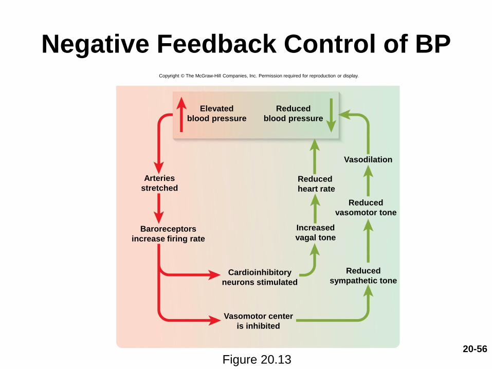
Negative Feedback Control of BP
Figure 20.13
Copyright © The McGraw-Hill Companies, Inc. Permission required for reproduction or display.
Vasodilation
Elevated
blood pressure
Reduced
blood pressure
Reduced
heart rate
Arteries
stretched
Baroreceptors
increase firing rate
Reduced
vasomotor tone
Reduced
sympathetic tone
Increased
vagal tone
Cardioinhibitory
neurons stimulated
Vasomotor center
is inhibited
20-57
Neural Control
• Chemoreflex—an automatic response to
changes in blood chemistry
– Especially pH, and concentrations of O2 and CO2
• Chemoreceptors called aortic bodies and carotid
bodies
– Located in aortic arch, subclavian arteries, external
carotid arteries

20-58
Neural Control
• Primary role: adjust respiration to changes in
blood chemistry
• Secondary role: vasoreflexes
– Hypoxemia, hypercapnia, and acidosis stimulate
chemoreceptors, acting through vasomotor center to
cause widespread vasoconstriction, increasing BP,
increasing lung perfusion, and gas exchange
– Also stimulate breathing

20-59
Neural Control
• Medullary ischemic reflex—automatic response to a
drop in perfusion of the brain
– Medulla oblongata monitors its own blood supply
– Activates corrective reflexes when it senses ischemia
(insufficient perfusion)
– Cardiac and vasomotor centers send sympathetic
signals to heart and blood vessels
– Increases heart rate and contraction force
– Causes widespread vasoconstriction
– Raises BP and restores normal perfusion to the brain
• Other brain centers can affect vasomotor center
– Stress, anger, arousal can also increase BP
20-60
Hormonal Control
• Hormones influence blood pressure– Some through their vasoactive effects
– Some by regulating water balance
• Angiotensin II—potent vasoconstrictor– Raises blood pressure
– Promotes Na+ and water retention by kidneys
– Increases blood volume and pressure
• Atrial natriuretic peptide—increases urinary sodium excretion– Reduces blood volume and promotes vasodilation
– Lowers blood pressure
20-61
Hormonal Control
• ADH promotes water retention and raises BP
– Pathologically high concentrations; also a
vasoconstrictor (aka vasopressin)
• Epinephrine and norepinephrine effects
– Most blood vessels
• Bind to -adrenergic receptors—vasoconstriction
– In cardiac muscle blood vessels
• Bind to -adrenergic receptors—vasodilation
20-62
Two Purposes of Vasoreflexes
• Two purposes of dilation and constriction:
1) general control of BP and 2) routing blood
from one body region to another
• General method of raising or lowering BP
throughout the whole body
– Increasing BP requires medullary vasomotor
center or widespread circulation of a hormone
• Important in supporting cerebral perfusion during
a hemorrhage or dehydration

20-63
Two Purposes of Vasoreflexes
• Method of rerouting blood from one region to
another for perfusion of individual organs
– Either centrally or locally controlled
• During exercise, sympathetic system reduces blood flow
to kidneys and digestive tract and increases blood flow to
skeletal muscles
• Metabolite accumulation in a tissue affects local
circulation without affecting circulation elsewhere in the
body
– If a specific artery constricts, the pressure downstream
drops, pressure upstream rises

20-64
Two Purposes of Vasoreflexes
• Examples
– Vigorous exercise dilates arteries in lungs, heart,
and muscles
• Vasoconstriction occurs in kidneys and digestive
tract
– Dozing in armchair after big meal
• Vasoconstriction in lower limbs raises BP above
the limbs, redirecting blood to intestinal arteries
20-65
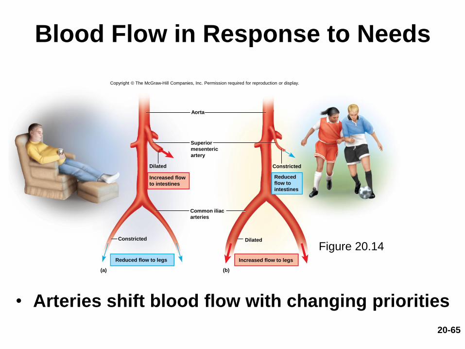
Blood Flow in Response to Needs
• Arteries shift blood flow with changing priorities
Copyright © The McGraw-Hill Companies, Inc. Permission required for reproduction or display.
Constricted
Dilated
Aorta
(a) (b)
Constricted
Dilated
Reduced flow to legs
Superior
mesenteric
artery
Increased flow
to intestines
Common iliac
arteries
Reduced
flow to
intestines
Increased flow to legs
Figure 20.14

20-66
Blood Flow Comparison
• During exercise
– Increased perfusion of lungs, myocardium, and skeletal muscles
– Decreased perfusion of kidneys and digestive tract
Figure 20.15
Copyright © The McGraw-Hill Companies, Inc. Permission required for reproduction or display.
At rest
Total cardiac output 5 L/min
Other
350 mL/min
(7.0%)
Coronary
200 mL/min
(4.0%)
Cutaneous
300 mL/min
(6.0%)
Muscular
1,000 mL/min
(20.0%)
Digestive
1,350 mL/min
(27.0%)
Cerebral
700 mL/min
(14.0%)
Renal
1,100 mL/min
(22.0%)
Moderate exercise
Total cardiac output 17.5 L/min
Other
400 mL/min
(2.3%)
Coronary
750 mL/min
(4.3%)Cutaneous
1,900 mL/min
(10.9%)
Cerebral
750 mL/min
(4.3%)
Renal
600 mL/min
(3.4%)
Digestive
600 mL/min
(3.4%)
Muscular
12,500 mL/min
(71.4%)
Capillary Exchange
• Expected Learning Outcomes
– Describe how materials get from the blood to the
surrounding tissues.
– Describe and calculate the forces that enable capillaries
to give off and reabsorb fluid.
– Describe the causes and effects of edema.
20-67
20-68
Capillary Exchange
• The most important blood in the body is in the
capillaries
• Only through capillary walls are exchanges
made between the blood and surrounding
tissues
• Capillary exchange—two-way movement of fluid
across capillary walls
– Water, oxygen, glucose, amino acids, lipids, minerals,
antibodies, hormones, wastes, carbon dioxide,
ammonia
20-69
Capillary Exchange
• Chemicals pass through the capillary wall by
three routes
– Through endothelial cell cytoplasm
– Intercellular clefts between endothelial cells
– Filtration pores (fenestrations) of the fenestrated
capillaries
• Mechanisms involved
– Diffusion, transcytosis, filtration, and reabsorption

20-70
Diffusion
• Diffusion is the most important form of capillary
exchange
– Glucose and oxygen, being more concentrated in
blood, diffuse out of the blood
– Carbon dioxide and other waste, being more
concentrated in tissue fluid, diffuse into the blood
• Capillary diffusion can only occur if:
– The solute can permeate the plasma membranes of
the endothelial cell, or
– Find passages large enough to pass through
• Filtration pores and intracellular clefts

20-71
Diffusion
• Lipid-soluble substances
– Steroid hormones, O2, and CO2 diffuse easily through
plasma membranes
• Water-soluble substances
– Glucose and electrolytes must pass through filtration
pores and intercellular clefts
• Large particles such as proteins held back
20-72
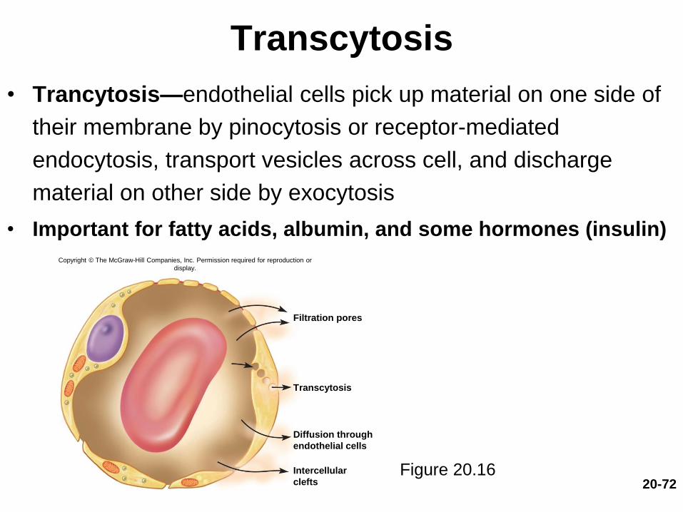
Transcytosis
• Trancytosis—endothelial cells pick up material on one side of
their membrane by pinocytosis or receptor-mediated
endocytosis, transport vesicles across cell, and discharge
material on other side by exocytosis
• Important for fatty acids, albumin, and some hormones (insulin)
Figure 20.16
Copyright © The McGraw-Hill Companies, Inc. Permission required for reproduction or
display.
Filtration pores
Transcytosis
Diffusion through
endothelial cells
Intercellular
clefts
20-73
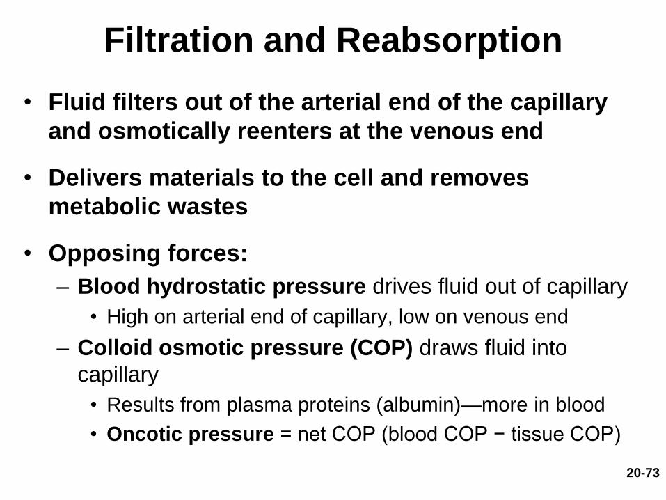
Filtration and Reabsorption
• Fluid filters out of the arterial end of the capillary
and osmotically reenters at the venous end
• Delivers materials to the cell and removes
metabolic wastes
• Opposing forces:
– Blood hydrostatic pressure drives fluid out of capillary
• High on arterial end of capillary, low on venous end
– Colloid osmotic pressure (COP) draws fluid into
capillary
• Results from plasma proteins (albumin)—more in blood
• Oncotic pressure = net COP (blood COP − tissue COP)

20-74
Filtration and Reabsorption
• Hydrostatic pressure
– Physical force exerted against a surface by a liquid
• Blood pressure in vessels is hydrostatic pressure
• Capillaries reabsorb about 85% of the fluid they
filter
• Other 15% is absorbed by the lymphatic system
and returned to the blood
20-75
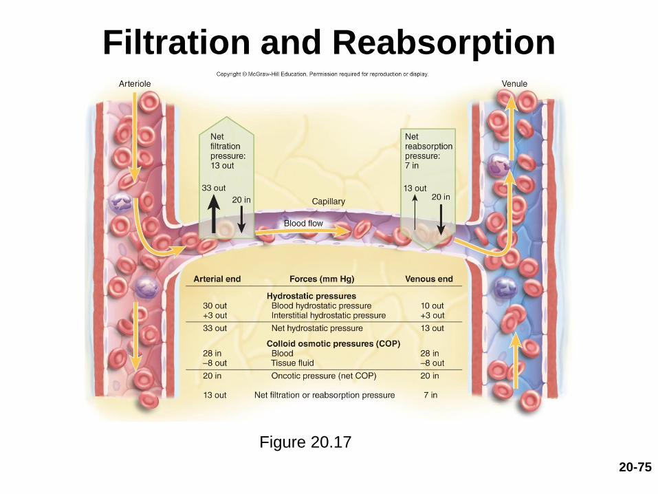
Filtration and Reabsorption
Figure 20.17

20-76
The Forces of Capillary
Filtration and Reabsorption
• Capillary filtration at
arterial end
• Capillary reabsorption
at venous end
• Variations
– Location
• Glomeruli—devoted to
filtration
• Alveolar capillary—devoted
to absorption
– Activity or trauma
• Increases filtration
Figure 20.17

20-77
Variations in Capillary
Filtration and Reabsorption
• Capillaries usually reabsorb most of the fluid
they filter with certain exceptions
– Kidney capillaries in glomeruli do not reabsorb
– Alveolar capillaries in lung absorb completely to keep
fluid out of air spaces
20-78
Variations in Capillary
Filtration and Reabsorption
• Capillary activity varies from moment to
moment
– Collapsed in resting tissue, reabsorption predominates
since BP is low
– Metabolically active tissue has increase in capillary flow
and BP
• Increase in muscular bulk by 25% due to accumulation of
fluid

20-79
Edema
• Edema—accumulation of excess fluid in a tissue
– Occurs when fluid filters into a tissue faster than it is
absorbed
• Three primary causes
– Increased capillary filtration
• Kidney failure, histamine release, old age, poor venous
return
– Reduced capillary absorption
• Hypoproteinemia, liver disease, dietary protein deficiency
– Obstructed lymphatic drainage
• Surgical removal of lymph nodes
20-80
Edema
• Tissue necrosis
– Oxygen delivery and waste removal impaired
• Pulmonary edema
– Suffocation threat
• Cerebral edema
– Headaches, nausea, seizures, and coma
• Severe edema or circulatory shock
– Excess fluid in tissue spaces causes low blood volume
and low blood pressure

Venous Return and
Circulatory Shock
• Expected Learning Outcomes
– Explain how blood in the veins is returned to the heart.
– Discuss the importance of physical activity in venous
return.
– Discuss several causes of circulatory shock.
– Name and describe the stages of shock.
20-81

20-82
Mechanisms of Venous Return
• Venous return—the flow of blood back to the heart; relies on: pressure gradient, gravity, skeletal muscle pump, thoracic pump, and cardiac suction
– Pressure gradient
• Blood pressure is the most important force in venous return
• 7 to 13 mm Hg venous pressure toward heart
• Venules (12 to 18 mm Hg) to central venous pressure:
point where the venae cavae enter the heart (~5 mm Hg)
– Gravity drains blood from head and neck
– Skeletal muscle pump in the limbs
• Contracting muscle squeezes blood out of the compressed
part of the vein

20-83
Mechanisms of Venous Return
Venous Return (Continued)
– Thoracic (respiratory) pump
• Inhalation—thoracic cavity expands and thoracic pressure
decreases, abdominal pressure increases, forcing blood
upward
– Central venous pressure fluctuates
• 2 mm Hg—inhalation, 6 mm Hg—exhalation
• Blood flows faster with inhalation
– Cardiac suction of expanding atrial space

20-84
The Skeletal Muscle Pump
Figure 20.19
Copyright © The McGraw-Hill Companies, Inc. Permission required for reproduction or display.
To heart
Valve open
Valve closed
(a) Contracted skeletal muscles (b) Relaxed skeletal muscles
Venous
blood
20-85
Venous Return and Physical Activity
• Exercise increases venous return in many ways
– Heart beats faster and harder, increasing CO and BP
– Vessels of skeletal muscles, lungs, and heart dilate and
increase flow
– Increased respiratory rate, increased action of thoracic
pump
– Increased skeletal muscle pump
20-86
Venous Return and Physical Activity
• Venous pooling occurs with inactivity
– Venous pressure not enough to force blood upward
– With prolonged standing, CO may be low enough to
cause dizziness
• Prevented by tensing leg muscles, activate skeletal
muscle pump
– Jet pilots wear pressure suits
20-87
Circulatory Shock
• Circulatory shock—any state in which cardiac
output is insufficient to meet the body’s
metabolic needs
– Cardiogenic shock: inadequate pumping of
heart (MI)
– Low venous return (LVR): cardiac output is low
because too little blood is returning to the heart
20-88
Circulatory Shock
(Continued)
– Three principal forms of LVR shock:
• Hypovolemic shock—most common
– Loss of blood volume: trauma, burns, dehydration
• Obstructed venous return shock
– Tumor or aneurysm compresses a vein
• Venous pooling (vascular) shock
– Long periods of standing, sitting, or widespread
vasodilation
20-89
Circulatory Shock
• Neurogenic shock—loss of vasomotor tone,
vasodilation
– Causes from emotional shock to brainstem injury
• Septic shock
– Bacterial toxins trigger vasodilation and increased
capillary permeability
• Anaphylactic shock
– Severe immune reaction to antigen, histamine release,
generalized vasodilation, increased capillary permeability
20-90
Responses to Circulatory Shock
• Compensated shock
– Several homeostatic mechanisms bring about spontaneous recovery
• Example: If a person faints and falls to a horizontal position, gravity restores blood flow to the brain
• Decompensated shock
– When compensation fails
– Life-threatening positive feedback loops occur
– Condition gets worse causing damage to cardiac and brain tissue
Special Circulatory Routes
• Expected Learning Outcomes
– Explain how the brain maintains stable perfusion.
– Discuss the causes and effects of strokes and transient
ischemic attacks.
– Explain the mechanisms that increase muscular perfusion
during exercise.
– Contrast the blood pressure of the pulmonary circuit with
that of the systemic circuit, and explain why the difference
is important in pulmonary function.
20-91
20-92
Brain
• Total blood flow to the brain fluctuates less
than that of any other organ (700 mL/min.)
– Seconds of deprivation causes loss of consciousness
– Four to 5 minutes causes irreversible brain damage
– Though total flow is constant, blood is shifted to active
brain areas from moment to moment
20-93
Brain
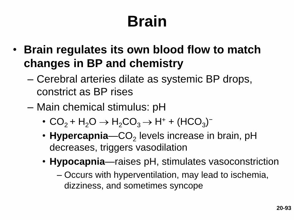
• Brain regulates its own blood flow to match
changes in BP and chemistry
– Cerebral arteries dilate as systemic BP drops,
constrict as BP rises
– Main chemical stimulus: pH
• CO2 + H2O H2CO3 H+ + (HCO3)−
• Hypercapnia—CO2 levels increase in brain, pH
decreases, triggers vasodilation
• Hypocapnia—raises pH, stimulates vasoconstriction
– Occurs with hyperventilation, may lead to ischemia,
dizziness, and sometimes syncope
20-94
Brain
• Transient ischemic attacks (TIAs)—brief
episodes of cerebral ischemia
– Caused by spasms of diseased cerebral arteries
– Dizziness, loss of vision, weakness, paralysis,
headache, or aphasia
– Lasts from a moment to a few hours
– Often early warning of impending stroke
20-95
Brain
• Stroke, or cerebral vascular accident (CVA)
– Sudden death of brain tissue caused by ischemia
• Atherosclerosis, thrombosis, ruptured aneurysm
– Effects range from unnoticeable to fatal
• Blindness, paralysis, loss of sensation, loss of speech
common
– Recovery depends on surrounding neurons, collateral
circulation

20-96
Skeletal Muscles
• Variable blood flow depending on state of exertion
• At rest
– Arterioles constrict, most capillary beds shut down
– Total flow about 1 L/min.
• During exercise
– Arterioles dilate in response to muscle metabolites such
as lactic acid, CO2, and H+
– Blood flow can increase 20-fold
• Blood is diverted from digestive and urinary organs
• Muscular contraction impedes flow
– Isometric contraction causes fatigue faster than
intermittent isotonic contractions

20-97
Lungs
• Low pulmonary blood pressure (25/10 mm Hg)
– Flow slower, more time for gas exchange
– Oncotic pressure overrides blood (hydrostatic) pressure
• Pulmonary capillaries absorb fluid (almost no filtration)
• Prevents fluid accumulation in alveolar walls and lumens
• Unique response to hypoxia
– Pulmonary arteries constrict in diseased area
– Redirects flow to better ventilated region

Anatomy of the Pulmonary Circuit
• Expected Learning Outcome
– Trace the route of blood through the pulmonary circuit.
20-98
Superior lobar arteries
Left pulmonary artery
Inferior lobar artery
Left ventricle
Right ventricle
Pulmonary trunk
(a)
Right pulmonary
artery
Superior lobar
artery
Middle lobar
artery
Inferior lobar
artery
20-99
Anatomy of the Pulmonary Circuit
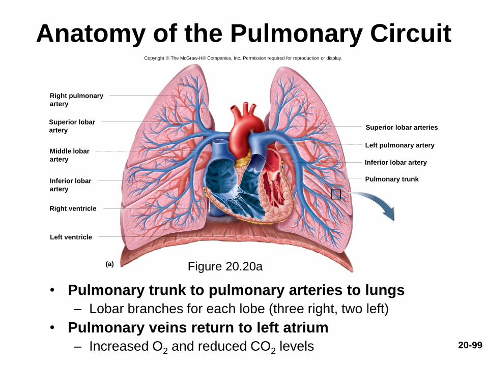
• Pulmonary trunk to pulmonary arteries to lungs
– Lobar branches for each lobe (three right, two left)
• Pulmonary veins return to left atrium
– Increased O2 and reduced CO2 levels
Figure 20.20a
Copyright © The McGraw-Hill Companies, Inc. Permission required for reproduction or display.
20-100
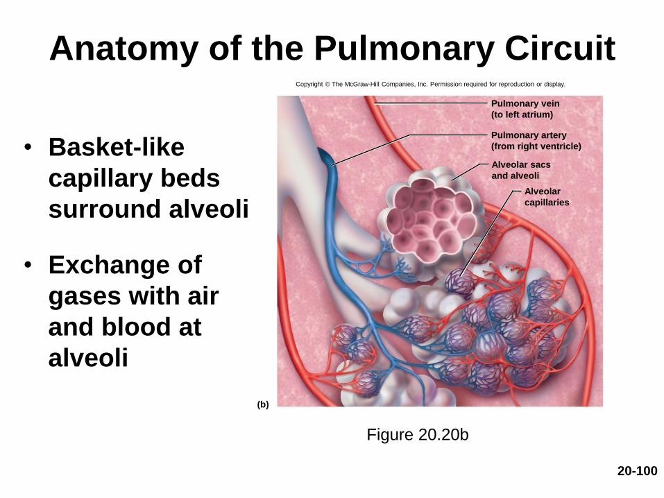
Anatomy of the Pulmonary Circuit
• Basket-like
capillary beds
surround alveoli
• Exchange of
gases with air
and blood at
alveoli
Figure 20.20b
Copyright © The McGraw-Hill Companies, Inc. Permission required for reproduction or display.
(b)
Pulmonary vein
(to left atrium)
Pulmonary artery
(from right ventricle)
Alveolar sacs
and alveoli
Alveolar
capillaries

Systemic Vessels of the
Axial Region
• Expected Learning Outcomes
– Identify the principal systemic arteries and veins
of the axial region.
– Trace the flow of blood from the heart to any
major organ of the axial region and back to the
heart.
20-101

20-102
The Major Systemic Arteries
• Arteries supply oxygen and nutrients to all organs
Figure 20.21
20-103
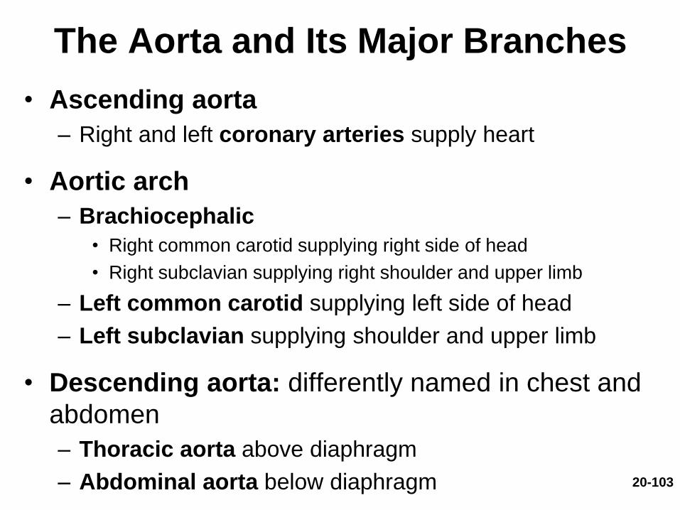
The Aorta and Its Major Branches
• Ascending aorta
– Right and left coronary arteries supply heart
• Aortic arch
– Brachiocephalic
• Right common carotid supplying right side of head
• Right subclavian supplying right shoulder and upper limb
– Left common carotid supplying left side of head
– Left subclavian supplying shoulder and upper limb
• Descending aorta: differently named in chest and
abdomen
– Thoracic aorta above diaphragm
– Abdominal aorta below diaphragm

20-104
The Aorta and Its Major Branches
Figure 20.23
20-105
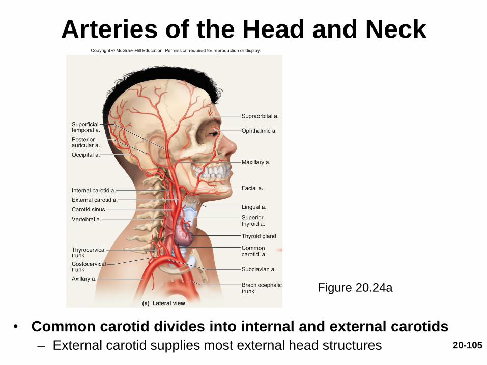
Arteries of the Head and Neck
• Common carotid divides into internal and external carotids
– External carotid supplies most external head structures
Figure 20.24a

20-106
Arteries of the Head and Neck• Paired vertebral arteries combine to form basilar artery on pons
• Circle of Willis arterial anastomosis on base of brain receiving blood from basilar and internal carotid arteries; serves cerebrum– Surrounds pituitary gland and optic chiasm
– Includes anterior and posterior cerebral and communicating arteries
Figure 20.25
Copyright © The McGraw-Hill Companies, Inc. Permission required for reproduction or display.
Cerebellar aa.:SuperiorAnterior inferiorPosterior inferior
Internal carotid a.
Basilar a.
Cerebral arterial circle:
Spinal aa.
Middle cerebral a.
Posterior cerebral a.
(a) Inferior view
(b) Median section
Caudal RostralAnterior
communicating a.
Anterior
cerebral a.
Posterior
communicating a.
Posterior
cerebral a.
Vertebral a.
Anterior
cerebral a.
20-107
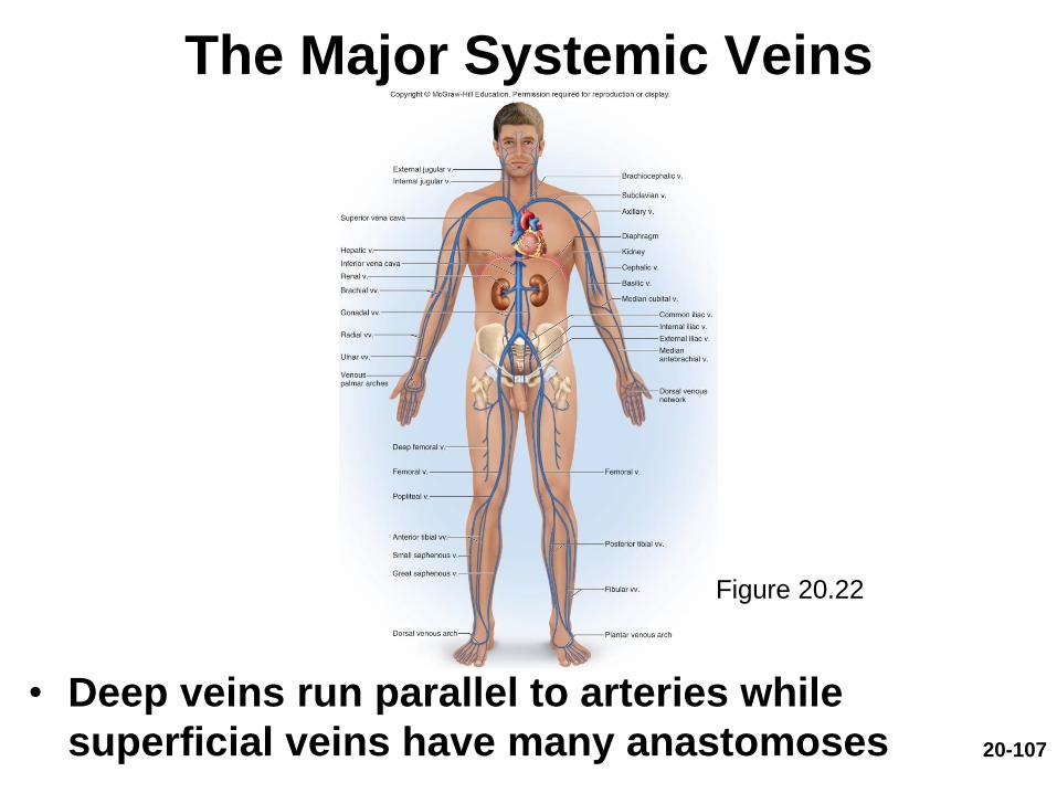
The Major Systemic Veins
• Deep veins run parallel to arteries while
superficial veins have many anastomoses
Figure 20.22

20-108
Veins of the Head and Neck
• Large, thin-walled dural sinuses form between layers of dura mater
• Drain blood from brain to internal jugular vein
Figure 20.26a,b
Copyright © The McGraw-Hill Companies, Inc. Permission required for reproduction or display.
Sigmoid sinus
(a) Dural venous sinuses, medial view
Straight sinus
Corpus callosum
Internal jugularv.
Superior
sagittal sinus
Inferior
sagittal sinusGreat cerebral
vein
Confluence of
sinuses
Transverse
sinus
(b) Dural venous sinuses, inferior view
Straight sinus
v.
Superficial
middle cerebral
vein
Confluence of
sinuses
Transverse
sinus
Sigmoid
sinus
Cavernous
sinus
Superior
ophthalmic vein
To internal
jugular
20-109
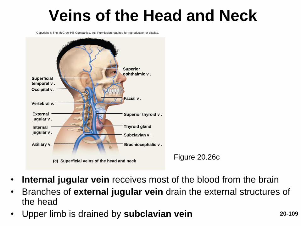
Veins of the Head and Neck
• Internal jugular vein receives most of the blood from the brain
• Branches of external jugular vein drain the external structures of the head
• Upper limb is drained by subclavian vein
Figure 20.26c
Copyright © The McGraw-Hill Companies, Inc. Permission required for reproduction or display.
(c) Superficial veins of the head and neck
Thyroid gland
Superior
ophthalmic v .
Facial v .
Superior thyroid v .
Subclavian v .
Brachiocephalic v .
Superficial
temporal v .
Occipital v.
Vertebral v.
External
jugular v .
Internal
jugular v .
Axillary v.
20-110
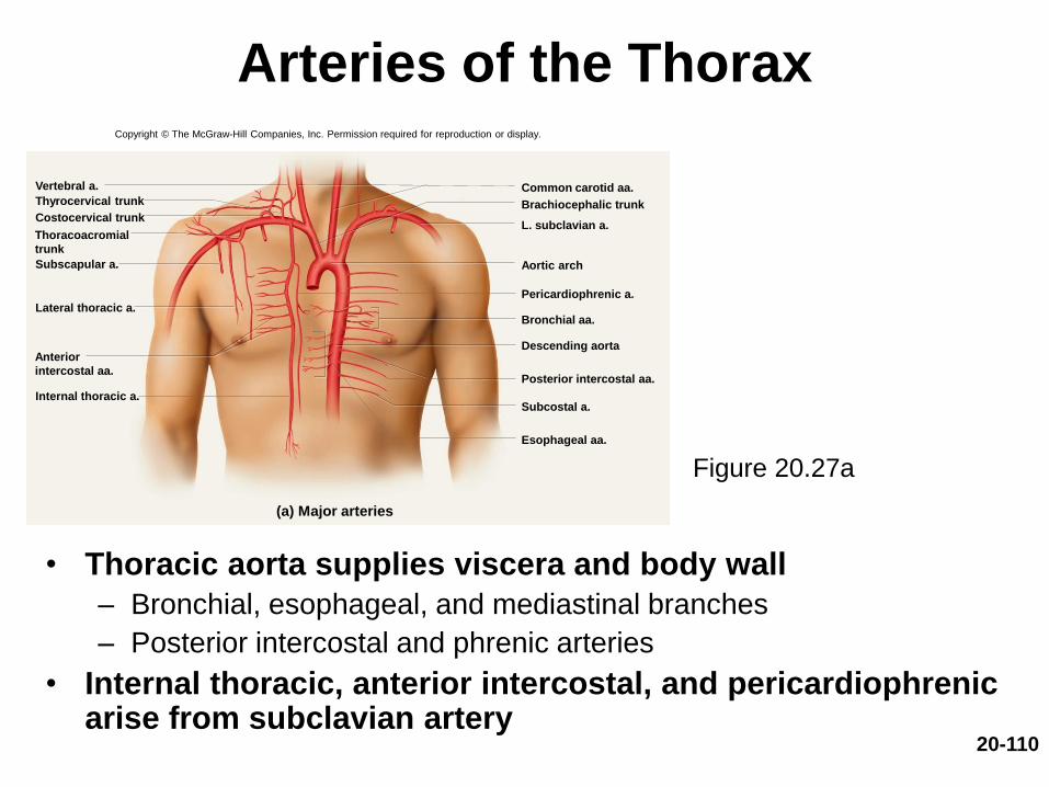
Arteries of the Thorax
• Thoracic aorta supplies viscera and body wall
– Bronchial, esophageal, and mediastinal branches
– Posterior intercostal and phrenic arteries
• Internal thoracic, anterior intercostal, and pericardiophrenic arise from subclavian artery
Figure 20.27a
(a) Major arteries
Costocervical trunk
Lateral thoracic a.
Subscapular a.
Internal thoracic a.
Thyrocervical trunkCommon carotid aa.
Brachiocephalic trunk
L. subclavian a.
Aortic arch
Bronchial aa.
Descending aorta
Posterior intercostal aa.
Esophageal aa.
Pericardiophrenic a.
Vertebral a.
Subcostal a.
Copyright © The McGraw-Hill Companies, Inc. Permission required for reproduction or display.
Anterior
intercostal aa.
Thoracoacromial
trunk

20-111
Arteries of the Abdominal
and Pelvic Region
Figure 20.29
Copyright © The McGraw-Hill Companies, Inc. Permission required for reproduction or display.
Inferior phrenic a.
Aortic hiatus
Celiac trunk
Suprarenal
aa.
Superior
Middle
Inferior
Superior mesenteric a.
Renal a.
Lumbar aa.
Gonadal a.
Inferior mesenteric a.
Common iliac a.
Internal iliac a.
Median sacral a.
20-112
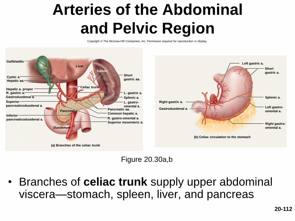
Arteries of the Abdominal
and Pelvic Region
• Branches of celiac trunk supply upper abdominal viscera—stomach, spleen, liver, and pancreas
Figure 20.30a,b
R. gastro-omental a.
Splenic a.
(a) Branches of the celiac trunk
Duodenum
R. gastric a. L. gastric a.Hepatic a. proper
Hepatic aa.Cystic a.
Liver
Gallbladder
Spleen
Aorta
Superior mesenteric a.
Pancreas Pancreatic aa.
Common hepatic a.
Celiac trunk
Short
gastric aa.
L. gastro-
omental a.
Gastroduodenal a.
Superior
pancreaticoduodenal a.
Inferior
pancreaticoduodenal a.
Copyright © The McGraw-Hill Companies, Inc. Permission required for reproduction or display.
(b) Celiac circulation to the stomach
Left gastric a.
Splenic a.
Right gastric a.
Short
gastric a.
Left gastro-
omental a.
Right gastro-
omental a.
Gastroduodenal a.
20-113
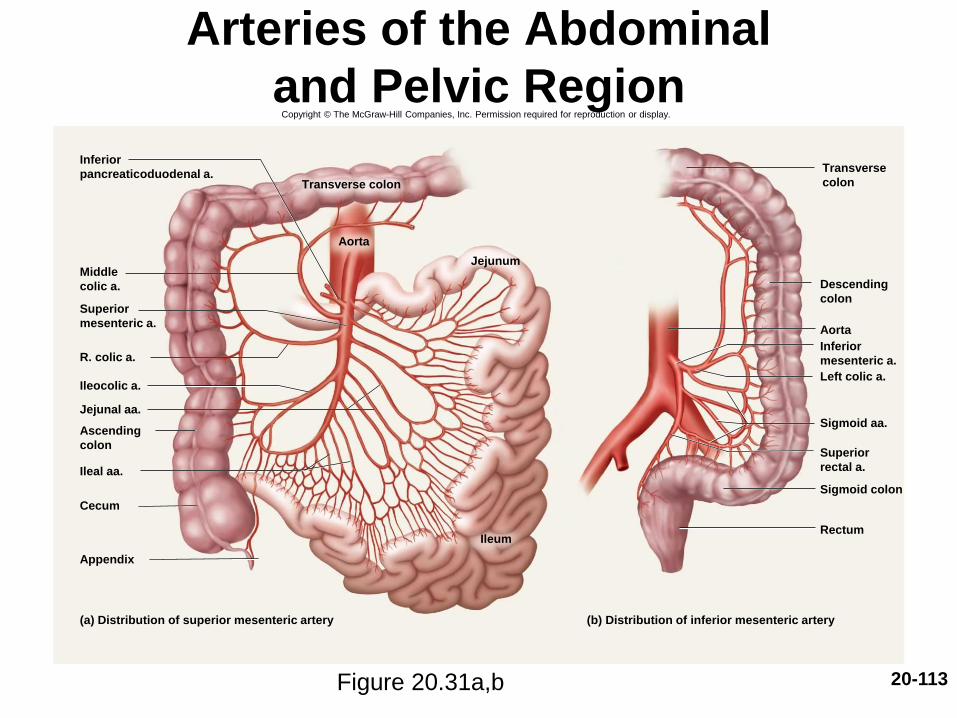
Arteries of the Abdominal
and Pelvic Region
Figure 20.31a,b
Copyright © The McGraw-Hill Companies, Inc. Permission required for reproduction or display.
R. colic a.
Ileocolic a.
Cecum
Ileum
Ileal aa.
Jejunal aa.
Jejunum
Appendix
(a) Distribution of superior mesenteric artery
Aorta
Left colic a.
Aorta
Rectum
Sigmoid colon
Sigmoid aa.
(b) Distribution of inferior mesenteric artery
Inferior
pancreaticoduodenal a.
Middle
colic a.
Superior
mesenteric a.
Ascending
colon
Transverse colon
Transverse
colon
Descending
colon
Inferior
mesenteric a.
Superior
rectal a.
20-114
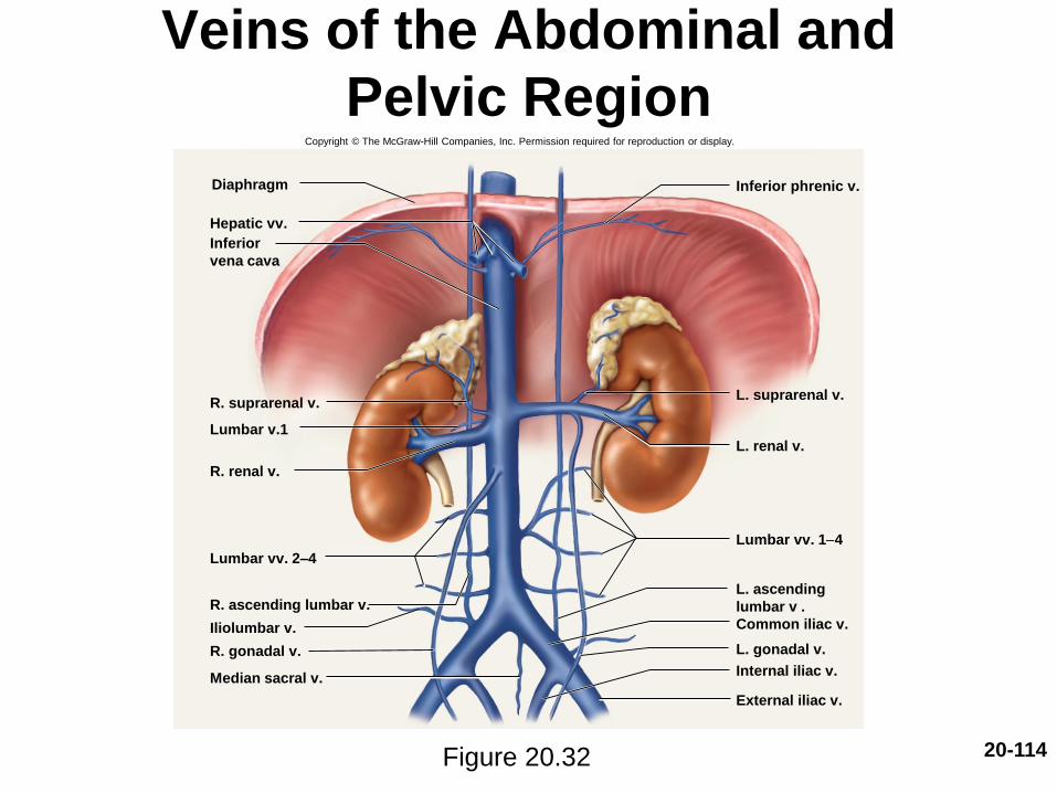
Veins of the Abdominal and
Pelvic Region
Figure 20.32
Copyright © The McGraw-Hill Companies, Inc. Permission required for reproduction or display.
Common iliac v.
Internal iliac v.
External iliac v.
Lumbar vv. 2–4
Diaphragm Inferior phrenic v.
L. suprarenal v.
L. renal v.
Lumbar vv. 1-4
L. ascending
lumbar v .
L. gonadal v.
Hepatic vv.
Inferior
vena cava
R. suprarenal v.
Lumbar v.1
R. renal v.
R. ascending lumbar v.
Iliolumbar v.
R. gonadal v.
Median sacral v.
20-115
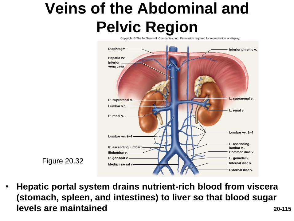
Veins of the Abdominal and
Pelvic Region
• Hepatic portal system drains nutrient-rich blood from viscera
(stomach, spleen, and intestines) to liver so that blood sugar
levels are maintained
Figure 20.32
Copyright © The McGraw-Hill Companies, Inc. Permission required for reproduction or display.
Common iliac v.
Internal iliac v.
External iliac v.
Lumbar vv. 2–4
Diaphragm Inferior phrenic v.
L. suprarenal v.
L. renal v.
Lumbar vv. 1-4
L. ascending
lumbar v .
L. gonadal v.
Hepatic vv.
Inferior
vena cava
R. suprarenal v.
Lumbar v.1
R. renal v.
R. ascending lumbar v.
Iliolumbar v.
R. gonadal v.
Median sacral v.
20-116
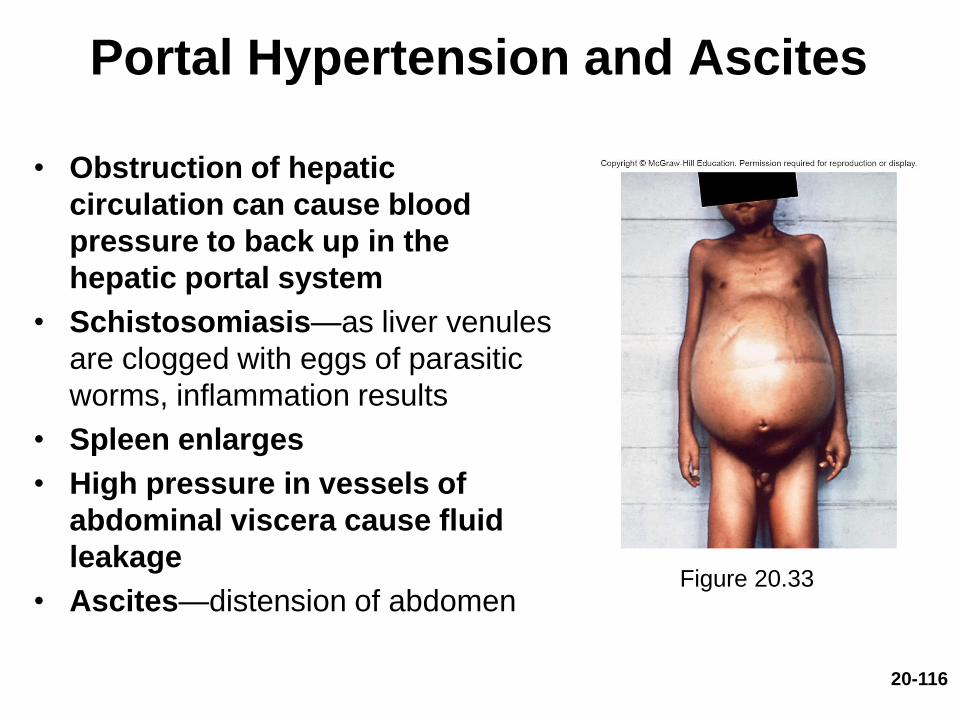
Portal Hypertension and Ascites
• Obstruction of hepatic
circulation can cause blood
pressure to back up in the
hepatic portal system
• Schistosomiasis—as liver venules
are clogged with eggs of parasitic
worms, inflammation results
• Spleen enlarges
• High pressure in vessels of
abdominal viscera cause fluid
leakage
• Ascites—distension of abdomenFigure 20.33

Systemic Vessels of the
Appendicular Region
• Expected Learning Outcomes
– Identify the principal systemic arteries and veins
of the limbs.
– Trace the flow of blood from the heart to any
region of the upper or lower limb and back to the
heart.
20-117
20-118
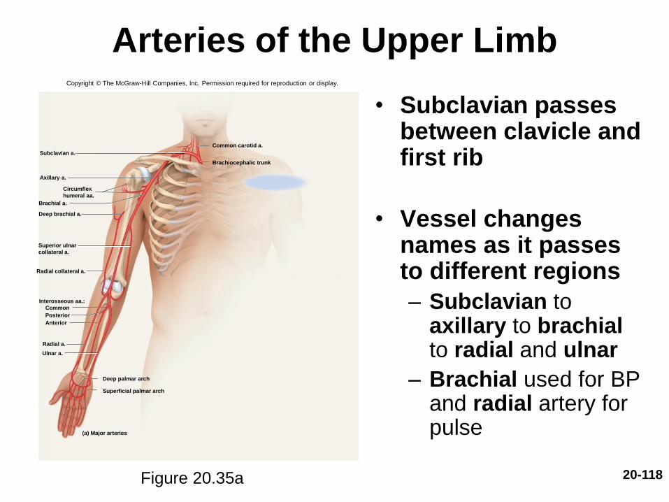
Arteries of the Upper Limb
• Subclavian passes between clavicle and first rib
• Vessel changes names as it passes to different regions
– Subclavian to axillary to brachialto radial and ulnar
– Brachial used for BP and radial artery for pulse
Figure 20.35a
Copyright © The McGraw-Hill Companies, Inc. Permission required for reproduction or display.
Subclavian a.
Axillary a.
Circumflex
humeral aa.
Deep brachial a.
Brachial a.
Radial a.
Ulnar a.
Interosseous aa.:
Common
Anterior
Posterior
Radial collateral a.
Deep palmar arch
Superficial palmar arch
(a) Major arteries
Common carotid a.
Brachiocephalic trunk
Superior ulnar
collateral a.

20-119
Veins of the Upper Limb
Figure 20.36a
Copyright © The McGraw-Hill Companies, Inc. Permission required for reproduction or display.
External
Superior vena cava
Internal
Deep venous palmar arch
(a) Major veins
Superficial venous palmar archDorsal venous network
Superficial veins
Deep veins
Subclavian v.
Axillary v.
Cephalic v.
Basilic v.
Brachial vv.
Median cubital v.
Radial vv.
Ulnar vv.
Cephalic v.
Basilic v.
Jugular vv.
Brachiocephalic vv.
Median
antebrachial v.
20-120
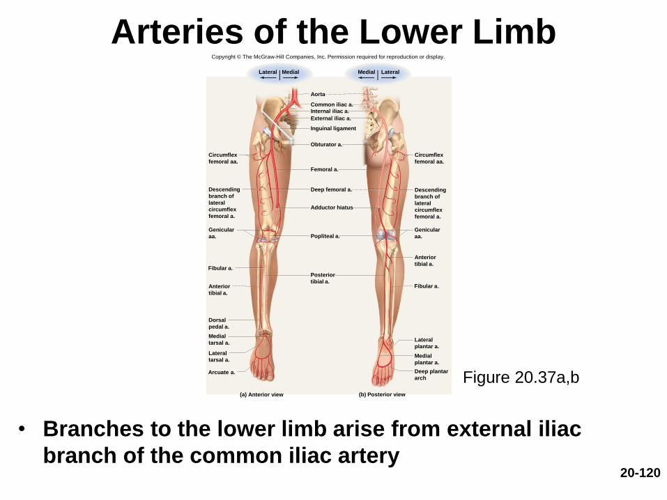
Arteries of the Lower Limb
• Branches to the lower limb arise from external iliac
branch of the common iliac artery
Figure 20.37a,b
Common iliac a.
Femoral a.
Deep femoral a.
Inguinal ligament
Obturator a.
Internal iliac a.
External iliac a.
Aorta
Popliteal a.
Fibular a.
Fibular a.
Arcuate a.
(a) Anterior view (b) Posterior view
Adductor hiatus
Lateral Medial LateralMedial
Circumflex
femoral aa.
Descending
branch of
lateral
circumflex
femoral a.
Genicular
aa.
Anterior
tibial a.
Dorsal
pedal a.
Medial
tarsal a.
Lateral
tarsal a.
Posterior
tibial a.
Deep plantar
arch
Medial
plantar a.
Lateral
plantar a.
Anterior
tibial a.
Genicular
aa.
Descending
branch of
lateral
circumflex
femoral a.
Circumflex
femoral aa.
Copyright © The McGraw-Hill Companies, Inc. Permission required for reproduction or display.

20-121
Veins of the Lower Limb
Figure 20.39a,b
Copyright © The McGraw-Hill Companies, Inc. Permission required for reproduction or display.
(b) Posterior view
Inferior vena cava
Lateral Medial LateralMedial
Superficial veins
Deep veins
Common iliac v.
Internal iliac v .
External iliac v .
Deep femoral v .
Femoral v .
Great saphenous v .
Popliteal v .
Circumflex
femoral vv.
Small
saphenous v.
Anterior
tibial vv.
Dorsal
venous arch
(a) Anterior view
Circumflex
femoral vv.
Anterior tibial v.
Small
saphenous v.
Fibular vv.
Posterior tibial
vv.
Medial plantar v.
Lateral plantar v.
Deep plantar
venous arch
20-122
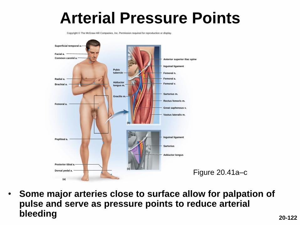
Arterial Pressure Points
• Some major arteries close to surface allow for palpation of pulse and serve as pressure points to reduce arterial bleeding
Figure 20.41a–c
Superficial temporal a.
Facial a.
Common carotid a.
Radial a.
Brachial a.
Femoral a.
Popliteal a.
Dorsal pedal a.
Posterior tibial a.
Inguinal ligament
Anterior superior iliac spine
Inguinal ligament
Femoral n.
Femoral a.
Sartorius m.
Rectus femoris m.
(b)
(c)
(a)
Sartorius
Adductor longus
Gracilis m.
Pubic
tubercle
Adductor
longus m.Femoral v.
Great saphenous v.
Vastus lateralis m.
Copyright © The McGraw-Hill Companies, Inc. Permission required for reproduction or display.
20-123
Hypertension—The “Silent Killer”
• Hypertension—most common cardiovascular disease affecting about 30% of Americans over 50
• “The silent killer”– Major cause of heart failure, stroke, and kidney failure
• Damages heart by increasing afterload
– Myocardium enlarges until overstretched and inefficient
• Renal arterioles thicken in response to stress
– Drop in renal BP leads to salt retention (aldosterone) and worsens the overall hypertension
20-124
Hypertension—The “Silent Killer”
• Primary hypertension– Obesity, sedentary behavior, diet, nicotine
– 90% of cases
• Secondary hypertension—secondary to other disease– Kidney disease, atherosclerosis, hyperthyroidism,
Cushing syndrome
– 10% of cases