chapter 104: malignant neoplasms of the larynx …famona.tripod.com/ent/cummings/cumm104.pdf · 1...
TRANSCRIPT

1
Chapter 104: Malignant Neoplasms of the Larynx
Clarence T. Sasaki, Roy D. Carlson
Cancer of the larynx accounts for approximately 1.2% of all new cancer diagnoses and0.73% of all cancer deaths in the USA according to the National Cancer Institute estimates(Silverberg et al, 1990). These annual cases of laryngeal malignancy represent approximatelyone fifth of all head and neck cancers.
Laryngeal cancer does, however, have a generally favorable prognosis, with overall5-year survival rates of 67%. Moreover, it is largely a preventable disease.
Epidemiology
Incidence
Laryngeal cancer is primarily a disease of middle-aged men, and it has a peakincidence ni the seventh decade (Rothman et al, 1980). The ratio of incidence among mencompared to women is 4.6:1 (Wynder et al, 1976), although this ratio has diminished from14.9:1 reported 20 years earlier by Wynder et al (1956). There is no racial predominance,although the incidence among blacks has increased dramatically during this century. Amongblacks laryngeal cancer tends to occur in younger patients with poorer outcome (Wasfie andNewman, 1988).
Data from the Third National Cancer Survey have demonstrated a slightly greaterprevalence in urban centers (Rothman et al, 1980). Worldwide figures demonstrate a highincidence of laryngeal cancer in men in São Paulo, Brazil, and in Bombay, India.
Risk factors
Environmental risk factors have been associated with cancer of the larynx. Tobacco,especially cigarette, use has been repeatedly implicated in the genesis of laryngeal cancer.Other environmental factors clearly implicated include alcohol, occupational exposure, andradiation. Those implicated but not yet well substantiated include herpes virus infection(Hollinshead et al, 1973) and dietary deficiency (Graham et al, 1981).
Recent studies have identified specific host factors that are linked with laryngealcancer. The presence of a laryngocele is statistically associated with a higher likelihood oflaryngeal cancer (Micheau et al, 1978) but may not be causative (Close et al, 1987).Gastroesophageal reflux has been identified as a probable causative factor, although this isnot proved (Ward and Hanson, 1988). Immunosuppression can contribute to carcinogenesisby interfering with normal immune surveillance. Finally, the presence of juvenile papillomasshould arouse concern regarding possible malignant transformation. Although irradiation ofthe lesions may engender early malignant change, carcinoma has been reported to occur 30years after juvenile papillomatosis in nonirradiated patients (Kashima et al, 1988).

2
Tobacco
Epidemiologic data have without fail demonstrated the strong correlation betweentobacco usage and laryngeal cancer (Burch et al, 1981; Hammond, 1966; Kahn, 1966; Wynderand Stellman, 1977; Wynder et al, 1956). Laryngeal cancer is extremely rare in nonsmokers.Moreover, the risk of laryngeal cancer increases with number of cigarettes smoked per day.Relative risk ratios range from 6.1 to 15.8 compared to nonsmokers. Cigar and pipe smokinghave relative risk ratios in the range of 1.6 to 3.9.
Further evidence of the etiologic relationship of smoking with laryngeal cancer liesin the histologic changes of vocal fold epithelium among smokers. Auerbach et al (1970) andMüller and Krohn (1980) have documented that histologic changes in cadaver larynges arerelated to smoking history. Degenerative changes in the subepithelial mucous glands havebeen related to tobacco consumption by Neilsen and Bak-Pedersen (1984).
Finally, in an animal model, that of the Syrian hamster, laryngeal cancer has beenfound to develop after "smoking" (Homberger, 1975). This provides further evidence inestablishing the causative role of cigarette smoking in laryngeal cancer.
Alcohol
Wynder et al (1956) demonstrated the importance of alcohol in laryngeal cancer,especially that of the "extrinsic" larynx (hypopharynx). He subsequently (1976) demonstratedthe relationship of alcohol to supraglottic carcinoma. Further statistical analysis by others(Flanders and Rothman, 1982) has established both the independent effect of alcohol and thesignificant synergy of alcohol and tobacco. The combination of these two increases theirrelative risk by 50% above that predicted by simple addictive effects. It is likely that thecarcinogens are found in the nonalcoholic components of such beverages (Rothman et al,1989). Experimental support for this epidemiologic observation has been documented by theSyrian hamster model (Stevens, 1979).
Occupation
Efforts to identify occupational risk factors are complicated by the overwhelminginfluence of tobacco and alcohol. Several individual studies in the epidemiologic literatureidentify workers at risk: nickel workers, mustard gas workers, farmers, woodworkers, andmachinists. Other studies have failed to substantiate these findings, however. Asbestos hasfrequently been suspected as a possible causative agent (Burch et al, 1981) with a risk ratioof approximately 2. The review of evidence by Chan and Gee (1988) does not support anetiologic role for asbestos, however. Further investigation may identify other occupational riskfactors; these are also unlikely to be major factors in comparison with tobacco and alcohol.
Radiation
Irradiation, especially in low doses, has long been identified as carcinogenic. Tumorsso induced are usually of soft tissue or superficial glandular structures (eg, thyroid, salivaryglands). Nevertheless, radiation-induced tumors have been reported in the larynx, includingtwo squamous carcinomas (Sakamoto et al, 1979), one fibrosarcoma (Mahmoud, 1980), and

3
one acinic cell carcinoma (Reibel et al, 1981).
Carcinogenesis
Saffiotti and Kaufman (1975) reviewed available data on the biochemistry of laryngealcarcinogenesis. Particular attention has been directed to polynuclear hydrocarbons (eg,benzo(a)pyrene) and to N-nitroso compounds. These compounds are found in significantconcentrations in the particulate and gaseous components of cigarette smoke. MacDonald andJanson (1981) have found that ethyl nitrite, a carcinogen present in cigarette smoke, is foundin greater concentration in the presence of alcohol. Investigations are still under way toidentify the principal carcinogens.
Host factors such as the inducibility of arylhydrocarbon-hydroxylase are likely to playa role in carcinogens (Andréasson et al, 1987). This intracellular enzyme metabolizes andactivates hydrocarbons, such as those found in tobacco smoke, thereby producing potentialcarcinogens. The role played by steroidal sex hormones is still unclear (Virolainen et al,1986).
Although the subcellular events in the initiation and development of laryngeal cancerare still not well understood, the behavior of cells has been well documented. The histologicchanges induced by smoking are well characterized (Auerbach et al, 1970). The naturalhistory of these histologic changes has been well described (Ferlito et al, 1981). Keratosis (ie,abnormal mucosal proliferation) proceeds to carcinoma in 3% to 6% of patients (Crissman,1979; Sllamniku et al, 1989a). The likelihood of malignant transformation is well correlatedwith the degree of cellular atypia: hyperkeratosis without atypia evolves into invasivecarcinoma in only 3% of patients, whereas in hyperkeratosis with severe atypia, this figurerises to 30% (Højslet et al, 1989; Sllamniku et al, 1989a). Carcinoma in situ representsmalignant transformation limited to the epithelium. Careful examination shows 36% coexistentmicroinvasive cancer and in 10% to 15% of patients carcinoma in situ ultimately becomesfrankly invasive carcinoma (Crissman et al, 1988; Miller and Fisher, 1971).
Prevention
Cancer of the larynx is largely a preventable disease. The infrequency of laryngealmalignancy in nonsmokers (less than 5% of all larynx cancer) is ample testimony to this fact.Otolaryngologists should encourage educational programs to eradicate cigarette smoking(Wynder and Hoffman, 1979).
The risk of larynx cancer is decreased by cessation of smoking. The risk diminishesdramatically, however, only after 6 years (Wynder et al, 1976) and approaches that ofnonsmokers only after 15 years. Nonetheless, no-smoking programs could markedly diminishthe incidence of laryngeal cancer.
Finally, "safe" cigarettes may be sought. Cigarette filters diminish the risk of larynxcancer by approximately 50% (Wynder and Stellman, 1979). Low-tar cigarettes may beexpected to diminish risk factors further.

4
Prevention of laryngeal cancer should be a major health concern of otolaryngologistsas well as society at large, to effect the best control of this disease.
Diagnosis
Early diagnosis is key to good survival and cure rates. Laryngeal neoplasms oftenproduce early symptoms that may lead to cure with good preservation of function.
Symptoms
The cardinal symptom of laryngeal cancer is voice change, that is, hoarseness. Thisis usually due to interference of vocal fold mucosal vibration from a glottic tumor thatinvolves the mucosa or thyroarytenoid (vocalis) muscle. Supraglottic lesions generally producea "muffled" voice.
Airway obstruction may also be a presenting symptom, most commonly in subglottictumors. This usually represents a mass effect and suggests that the tumor is large. Othersymptoms of local inflammation may be present, namely, throat discomfort or fullness.Hemoptysis may be present, though this generally occurs only in large ulcerating tumors.
Symptoms relevant to swallowing may also occur. Odynophagia and otalgia arefrequent presenting symptoms of supraglottic lesions. Dysphagia is associated with largetumors and suggests invasion beyond the confines of the larynx.
A neck mass may occur either by direct extension or, more commonly, by nodalmetastasis. Finally, weight loss or other constitutional symptoms may occur. The presence ofthese symptoms is usually indicative of advanced local disease.
Examination
Voice quality is evident during the patient interview. Pitch change or roughness maybe heard, suggesting involvement of the true cord. The muffled quality of supraglottic lesionsmay be detected. Airway obstruction may be noted, especially in subglottic tumors.
Palpation of the neck discloses the presence, location, and fixation of cervical nodes.Fixation of the thyroid cartilage is an ominous sign.
Office examination of the larynx may be performed via indirect mirror examinationor fiberoptic examination. The latter allows photographic or videographic documentation ofthe larynx. The laryngeal examination ought to indicate the appearance of the mucosa, thepresence of submucosal lesions, and mobility of the cords, as well as the status of the airway.Stroboscopy and acoustic analysis may provide helpful information in some patients.
Evaluation for metastatic disease should include a complete physical examination,blood count, chest radiograph, and liver enzyme studies.

5
Radiologic evaluation
To augment the clinical examination, radiologic studies may be undertaken. Thesimplest of these are soft tissue neck films, principally to examine encroachment of theairway.
Contrast studies may be performed. A barium swallow (Fig. 104-1) is useful inevaluating the laryngeal margins, the vallecula and tongue base, the piriform sinuses, and thepostcricoid region. Conventional tomography improves the accuracy of soft tissue films.Evaluation of the endolarynx may be accomplished by a laryngogram (Lehmann and Fletcher,1964). This study provides detailed information, especially about the laryngeal surface of theepiglottis, the ventricle, and the subglottis, regions that may be difficult to examinesatisfactorily (Fig. 104-2). Contrast laryngography has been rendered nearly obsolete by newerimaging techniques.
Recently, computed tomography (CT) and magnetic resonance imaging (MRI) havebecome widely available. These powerful methods have proved useful in supplementingclinical determination of the size and extent of laryngeal tumors. CT scanning is most helpfulin documenting deep invasion (Fig. 104-3) and extension beyond the larynx but is onlymoderately sensitive in determining early cartilage invasion (Archer and Yeager, 1982;Charlin et al, 1989; Werber and Lucente, 1989). MRI, although more expensive and lesswidely available, may be superior in demonstrating cartilage invasion (Castelijas et al, 1990;Hoover et al, 1987). Ultrasonography may prove useful, both in evaluating cartilage invasionand in staging neck metastasis. The choice of imaging techniques depends on the expertiseand facilities available.
Tissue diagnosis
To distinguish malignancy from the panoply of disorders that may have similar grossappearance (fungal and mycobacterial infections, syphilitic gummas, idiopathic granulomatousdisorders, benign neoplasms), a tissue diagnosis must be made. Exfoliative cytologic analysis,which provides correct diagnosis in approximately 90% of cases, may be used (Olofsson,1982). Fine-needle aspiration of presumed metastasis may also aid in establishing thediagnosis (Feldman et al, 1983).
The accepted standard for diagnosis is histopathologic examination of tissue obtainedat laryngoscopy and biopsy. In general this is best performed by direct laryngoscopy with theaid of the operating microscope. Biopsy "mapping" of the observed lesion is helpful in stagingand determining therapy. Toluidine blue has been suggested as a useful adjunct to mappinglesions (Strong et al, 1970), although its limitations are numerous (Lundgren et al, 1979).Microlaryngoscopy under general anesthesia provides the ideal setting, allowing a thoroughexamination of the larynx with the opportunity to obtain a biopsy specimen of the lesion andits margins.
6
Staging and the Biology of Squamous Cancer
Staging of laryngeal malignancy provides a method of succint description of the lesion.This offers insight into the behavior of the tumor as well as a medium for comparison oftreatment outcomes.
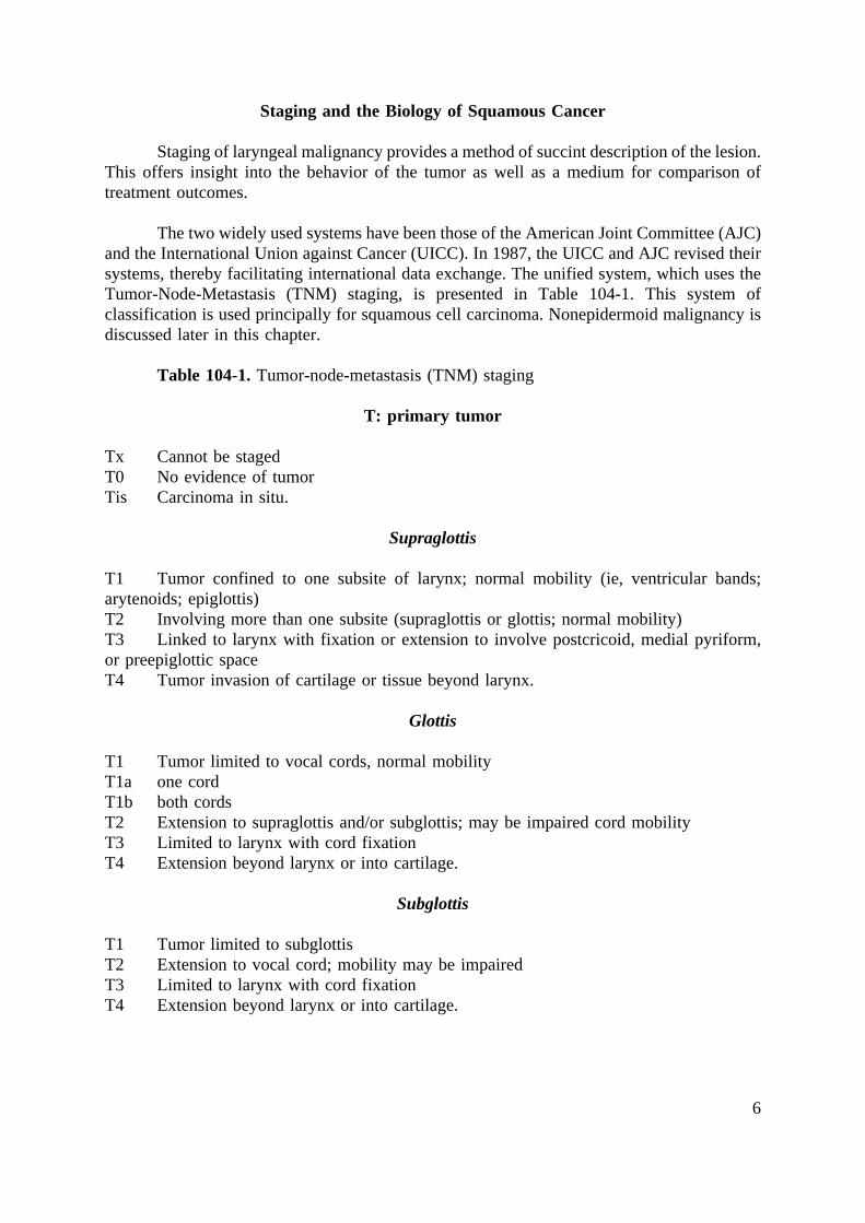
The two widely used systems have been those of the American Joint Committee (AJC)and the International Union against Cancer (UICC). In 1987, the UICC and AJC revised theirsystems, thereby facilitating international data exchange. The unified system, which uses theTumor-Node-Metastasis (TNM) staging, is presented in Table 104-1. This system ofclassification is used principally for squamous cell carcinoma. Nonepidermoid malignancy isdiscussed later in this chapter.
Table 104-1.Tumor-node-metastasis (TNM) staging
T: primary tumor
Tx Cannot be stagedT0 No evidence of tumorTis Carcinoma in situ.
Supraglottis
T1 Tumor confined to one subsite of larynx; normal mobility (ie, ventricular bands;arytenoids; epiglottis)T2 Involving more than one subsite (supraglottis or glottis; normal mobility)T3 Linked to larynx with fixation or extension to involve postcricoid, medial pyriform,or preepiglottic spaceT4 Tumor invasion of cartilage or tissue beyond larynx.
Glottis
T1 Tumor limited to vocal cords, normal mobilityT1a one cordT1b both cordsT2 Extension to supraglottis and/or subglottis; may be impaired cord mobilityT3 Limited to larynx with cord fixationT4 Extension beyond larynx or into cartilage.
Subglottis
T1 Tumor limited to subglottisT2 Extension to vocal cord; mobility may be impairedT3 Limited to larynx with cord fixationT4 Extension beyond larynx or into cartilage.

7
N: regional nodes
Nx Cannot be assessedN0 No regional metastasisN1 Single positive ipsilateral node, less than 3 cmN2 Nodes less than 6 cmN2a Single ipsilateral node 3-6 cmN2b Many ipsilateral nodes less than 6 cmN2c Bilateral and contralateral node less than 6 cmN3 Node(s) greater than 6 cm.
M: distant metastasis
Mx Cannot be assessedM0 No distant metastasisM1 Distant metastasis.
Stage grouping
0 Tis N0 M0I T1 N0 M0II T2 N0 M0III T3 N0 M0
T1-3 N1 M0IV T4 or N2-3 or M1.
T: the anatomy of the primary tumor
The anatomy of the larynx has been carefully delineated. First, the boundaries may beidentified: superiorly, the tip and lateral borders of the epiglottis; anteriorly, the anteriorlingual surface of the epiglottis, thyroid membrane, thyroid cartilage, cricothyroid membrane,and cricoid cartilage; posterolaterally, the aryepiglottic folds, arytenoids, interarytenoid space,and mucosa overlying the cricoid; and inferiorly, the lower border of the cricoid cartilage. Thevallecula is part of the superior hypopharynx; the piriform sinus and postcricoid region arepart of the inferior hypopharynx.
The larynx is divided into three parts: supraglottis, glottis, and subglottis (Fig. 104-4).Most cancers occur in the glottis; the supraglottis is next most frequent; subglottic tumors arerare. The supraglottic/glottic boundary occurs at the level of the apex of the ventricle. Thejunction of the glottis and subglottis occurs 1 cm below the free edge of the vocal fold. Norriset al (1970) and Tucker (1974) have discussed the partitions and boundaries of the larynx,especially in relation to its submucosal compartments.
Transglottic carcinomais a term coined by McGavran et al (1961) to describe tumorsthat cross the laryngeal ventricle, thus involving at least two of the three described portionsof the larynx. Although not included in the AJC or UICC definitions, this concept has beenstressed by several authors, including Mendonca and Bryce (1973), Kirchner et al (1974), andTucker (1974).

8
Anatomic basis of classification
The subdivision of laryngeal (squamous cell) malignancy into anatomic types hasextensive investigative justification. In 1981, Hajek recognized that laryngeal swellingfollowed specific anatomic "rules". His investigations were extended by Pressman and hiscolleagues in two studies published in 1956 using dye injections in living and cadaverlarynges. Welsh et al (1989) have recently duplicated and amplified these earlier studies,correlating their findings with whole organ sections. They determined that the mucosa wascontinuous throughout. The submucosa, however, is compartmentalized. First, the right andleft hemilarynx are distinct throughout from epiglottis to cricoid. Second, several regions werespecifically identified (Fig. 104-5):
1. An epiglottic region corresponding to the cartilage.
2. A marginal epiglottic region lateral to the cartilaginous epiglottis.
3. A posterolateral supraglottic region, which does not transgress the ventricle.
4. A ventricular region, which includes the inferior surface of the vestibular band,superior surface of the vocal fold, and apex between.
5. A sacciform region (possibly part of the ventricle).
6. A bursa on the margin of the true cord (Reinke's space).
7. A subglottic region from the free margin of the cord to the cricoid below.
The laryngeal compartments so described have been demonstrated embryologically byTucker and Smith (1962). These studies provides the ontogenetic basis for the distinctionamong the compartments. Hast (1974) has also related laryngeal embryology to thesubdivision of the larynx. The right and left hemilarynges arise independently. Thesupraglottic larynx arises from the buccopharyngeal anlage, arches III and IV; the subglotticportion is derived from the pulmonary anlage, arch VI. The vascular, lymphatic, andneuroanatomic subdivisions follow these boundaries (Pearson, 1975).
Critical to the understanding of the laryngeal compartments are the connective tissueboundaries, which act as barriers. The cartilaginous epiglottis serves as the anterior limit ofthe epiglottic submucosal compartment. The quadrangular membrane separates the supraglottisfrom the lateral paraglottic space; the ventricle and sacculus form a niche in the paraglotticspace. The conus elasticus serves as the inferior/lateral border of the glottic and subglotticspaces, again separating these from the paraglottic space. The significance of these boundarieslies in the observation that cancer tends first to follow the planes of least resistance, that is,within compartments (Kirchner and Carter, 1987; Pressman et al, 1960). Invasion acrossboundaries generally is associated with a worse prognosis.
The ligamentous and cartilaginous structures of the larynx are of great importance.These connective tissue structures display great resistance to invasion by carcinoma. Oncethey are invaded, however, the survival rates diminish dramatically.

9
The thyrohyoid membrane forms the anterior margin of the preepiglottic space. Oncethe latter space is involved, the tumor may spread through the membrane into the soft tissuesof the neck or into the deep muscle of the tongue base.
The thyroid cartilage surrounds only a small portion of the endolarynx; the piriformregion and paraglottic space about the cartilage on most of its surface. Once tumor hasinvaded the paraglottic space (ie, the intrinsic muscle of the vocal fold), it may invade thecartilage, particularly in regions of ossification.
Of particular importance is the anterior commissure tendon (Fig. 104-6). The tendoncomprises the confluence of the paired vocal ligaments as they insert into the midlineposterior surface of the thyroid cartilage (Broyles, 1943). Parallel to the tendon are numerousvessels and lymphatics (Pearson, 1975; Sessions et al, 1975b). The collagen bundles mayserve as a preformed pathway for extension of tumor (Harrison, 1984; Yeager and Archer,1982). The region of the tendon also represents a distinct margin between the supraglottis andglottis. The vestibular bands loosely converge around the thyroepiglottic ligaments but aredistinct from each other and from the vocal ligaments (Bagatella and Bignardi, 1983; Tuckeret al, 1973). Thus, the anterior commissure region provides a limiting structure of lateralsupraglottic lesions but is a potential pathway for spread of large glottic and midlinesupraglottic lesions.
The cricothyroid membrane represents a vulnerable point below the shield of thethyroid cartilage. The mucosa of the larynx is in direct contact with the cricothyroidmembrane. Subglottic tumors may grow through this membrane into the soft tissues of theneck (van Nostrand and Brodarec, 1982).
Inferiorly and posteriorly, the cricoid serves as a barrier to tumor. That laryngealtumors should reach the cricoid without also reaching the cricothyroid membrane is unlikely.Posterior submucosal spread along the cricoid, however, can reach the arytenoid andinterarytenoid regions. Vocal cord immobility may then ensue. Alternatively, involvement ofthe hypopharyngeal piriform sinus or postcricoid mucosa may also occur (Lam, 1983; vanNostrand and Brodarec, 1982).
Cartilage invasion usually occurs in ossified portions of the cartilage (Kirchner, 1969,1984a; Pittam and Carter, 1982) and rarely in the hyaline regions. The invasion of ossifiedportions may relate to the vascularity associated with ossification. The explanation for theresilience of cartilage is still unknown (Blitzer, 1979), although Folkman (1976) has foundthat cartilage may produce a substance that inhibits tumor angiogenesis factor. In 1988Repássy et al reported the finding of activated connected tissue, especially histiocytes,adjacent to cancer. The stimulus for this increased production of fibrils remains unclear.
Knowledge of the laryngeal compartments and their boundaries provides insight intothe pathways of growth and invasion of laryngeal cancer. The histologic study of excisedlarynges has provided this information (Kirchner, 1989; Tucker, 1961). Retrospective analysisof patient material thus may provide clues to the behavior of new laryngeal malignancies.

10
Staging of supraglottic primary tumors
Supraglottic primary tumors account for 24% to 42% of all laryngeal primary tumors.The description of laryngeal compartments aids in understanding the spread of supraglotticcancer. The supraglottis may be subdivided into (1) three marginal regions, namely, theanterior marginal region or suprahyoid epiglottis, and two lateral marginal regions (ie, alongthe aryepiglottic folds); (2) the midline infrahyoid epiglottis; and (3) the lateral infrahyoidareas of the vestibular bands.
Marginal supraglottic carcinoma should be regarded as distinct by virtue of itspropensity to "spill over" into the vallecula, arytenoids, and piriform sinuses (Fig. 104-7). Assuch it behaves more like an hypopharyngeal lesion and is not "compartmentalized" as readilyas other laryngeal sites. These tend to be asymptomatic for longer intervals and present withmore advanced disease. Laccourreye et al (1983) found that approximately 57% of theirpatients had T3 or T4 primary lesions.
The prognosis of the more inferiorly located supraglottic primary tumors is better thanthat of those on the margin (Bocca, 1975). The pattern of spread of supraglottic cancer isnotable for its restriction to the supraglottic region for long intervals. Supraglottic cancer doesnot invade the vocal cords unless it is, in fact, transglottic, as described later (Kirchner andSom, 1971a). Its growth anterioinferiorly is limited by the anterior commissure tendon so thatonly massive tumors actually invade the anterior glottic and subglottic regions. Moreover,supraglottic cancer rarely invades the thyroid cartilage (unless it has first become transglotticby invading the paraglottic space).
The most important route of spread of supraglottic cancer is anteriorly into thepreepiglottic space. This occurs by means of fenestrae in the elastic cartilage of the epiglottisthrough which cancer may spread. The preepiglottic space is often clinically silent. Thus,underestimation of tumor may occur; T3 supraglottic cancer may appear to be T2. Gregor(1990) has reported that preepiglottic space invasion frequently augurs involvement of thestrap muscles or tongue base and that it may merit reclassification from T3 to T4. Radiologicinvestigation, especially with computed tomography, may greatly improve diagnosis of spreadto this space or beyond and improve staging accuracy (Sagel et al, 1981; Sulfaro et al, 1989).
The pathways of spread of supraglottic cancer are depicted in Figure 104-8. Spreadoccurs (1) outside the endolarynx along mucosal pathways, especially in lesions arising onthe margins; (2) into the preepiglottic space; and (3) across the anterior commissure tendonor through the paraglottic space to become transglottic. The last occurs, in general, only withlarge, ulcerative lesions.
Staging of glottic primary tumors
Glottic carcinomas constitute the majority of laryngeal malignancies, accounting for55% to 75% of primary sites. Since tumors arising from the vocal cord should interfere withmovement of the mucosa, these patients tend to have early symptoms. Indeed most glotticcarcinomas are small: 55% to 65% are T1; 12% to 22% T2; 15% to 19% T3; and only 4%or less T4 (Daly and Strong, 1975; DeSanto et ak, 1977a; Kirchner and Owen, 1977).

11
Spread of glottic cancer first follows Reinke's space along the length of the vocalligament. Since the anterior commissure is composed of the conjoined vocal ligaments andthe mucosa of the anterior commissure is continuous, it is evident that bilateral lesions (so-called horseshoe lesions) can occur.
Involvement of the vestibular structures or subglottis characterizes T2 lesions (Fig.104-9). The preservation of normal cord mobility suggests invasion limited to submucosalcompartments. Deeper invasion, thereby entering the intrinsic laryngeal muscle or paraglotticspace in the region of the ventricle, results in impaired mobility. This clinical observation isvery important. Indeed, several authors have distinguished T2a and T2b tumors with normaland impaired mobility, respectively (Harwood and deBoer, 1980; Kaplan et al, 1984; Olofssonet al, 1973; van den Bogaert et al, 1983). Although this is not included in current stagingsystems, survival statistics are worse with impaired mobility. That this should be true followsfrom the assumption that impaired mobility suggests deep invasion. As well, invasion of theparaglottic or subglottic space may also be associated with clinically undetected invasion ofthe laryngeal cartilage.
Fixation of the cord defines T3 glottic lesions. The principal mechanism of vocal cordfixation has been identified by Kirchner and Som (1971b) and Kirchner (1977) as replacementof the thyroarytenoid muscle (Fig. 104-10). Extensive tumor involving the entire superiorsurface of the cord or the subglottic "shoulder" can also cause fixation though such extensionoccurs infrequently without actual invasion of the muscle. Moreover many lesions with vocalcord fixation also demonstrate invasion of the thyroid cartilage (Kirchner, 1969) Olofsson etal (1973) also cited cricoarytenoid invasion as a cause of cord fixation. Because cord fixationgenerally implies deep invasion, it has a worse prognosis and requires more extensivetreatment.
Invasion of the laryngeal cartilage or invasion beyond the larynx constitutes a T4cancer. Kirchner (1977) found 5 of 52 glottic lesions with cartilage invasion, associated in 4with with extensive (more than 1 cm) infraglottic extension. Van Nostrand and Brodarec(1982) similarly found that extension beyond the larynx occurred in association withsubglottic extension and egress through the cricothyroid membrane. The other route offramework invasion is via the ventricle. These last lesions are, by definition, transglottic.
In summary, the staging of glottic cancer relies principally on the careful observationof cord mobility and extent in the subglottis or ventricle. CT scanning may improve theability to document the full extent of glottic cancers.
Staging of subglottic primary tumors
Primary subglottic tumors are rare, constituting 1% to 5% of laryngeal primary sites(Harrison, 1971; Kirchner and Owen, 1977; Sessions et al, 1975a; Shaha and Shah, 1982;Warde et al, 1987). Most subglottic tumors present with large primary tumors since they areclinically silent until they produce voice change (usually with cord fixation) or airwayobstruction. Combining the small series cited leads to the documentatin of 14% T1, 18% T2,23% T3, and 45% T4 subglottic lesions.

12
The pathway of invasion of subglottic cancer is first into the thyroarytenoid muscleto produce cord fixation. Tumor usually invades the cricothyroid membrane or, moreinferiorly, invades along the trachea. Fifty percent (four of eight) of the subglottic lesions ofKirchner (1977) demonstrated cartilage invasion. Lam (1983) described three of sevensubglottic tumors with framework invasion. Subglottic tumors also demonstrate a highincidence of thyroid gland invasion (Gilbert et al, 1986).
Although subglottic tumors are rare, subglottic extension of glottic or supraglottic (inthe latter case, therefore, transglottic) tumors is not uncommon. The significance of theinferior margin of the thyroid cartilage, generally 10 mm below the anterior and midcord and3 to 4 mm posteriorly, cannot be overemphasized. The mucosa below this line directly abutsthe cricothyroid membrane or cricoid space and provides a direct passage for extralaryngealinvasion.
Staging of transglottic tumors
Transglottic tumors are not included in the UICC or AJC classification. They deserveattention because of the high incidence of framework invasion and extralaryngeal spreadassociated with these lesions. Since, by definition, thransglottic lesions cross the ventricle,they already involve the paraglottic space and are in close proximity to the thyroid alalaterally and to the hypopharynx medially. Kirchner et al (1974) demonstrated cartilageinvasion in 32 of 42 (76%) transglottic cancers. They noted invasion usually in the thyroidala; invasion of the cricoid occurred with extensive subglottic invasion (Fig. 104-11).Cartilage invasion occurred only in tumors larger than 2 cm. Pittam and Carter (1982)conifirmed the latter finding. As well, they documented framework invasion in 80% (21 of26) of transglottic cancers. Lam (1983) identified invasion in 91% of his 22 specimens.Eighteen of the 20 involved the thyroid cartilage, thyrohyoid membrane, and/or cricothyroidmembrane. Moreover, 15 of the 20 spread posteriorly to involve the thyroid ala, piriformsinus, or postcricoid region. Thus, the propensity of these lesions for extralaryngeal spreadis amply demonstrated.
The most significant clinical feature of transglotic cancer is the difficulty ofestablishing that a lesion is truly transglottic. Direct laryngoscopy frequently cannot establishframework invasion or extent of submucosal invasion. Pittam and Carter (1982) foundradiologic techniques (laryngography and CT) also inadequate. Archer and Yeager (1982)have documented similar limitations of CT.
Thus, transglottic invasion of cancer must always be suspected, especially in extensivetumors arising from the glottis. They require aggressive treatment and have a strongpropensity for recurrence.
It is important to stress that staging of primary disease is based on clinical evidence.It is, therefore, not surprising that understaging does occur. Since we often lack the abilityto perceive deep invasion on clinical grounds, lesions are often understaged when comparedwith histopathologic documentation (Norris et al, 1970; Pillsbury and Kirchner, 1979). Thecurrent T classification is designed to reflect invasion, however. T1 and T2 lesions areconfined to their respective mucosal and submucosal regions. T3 lesions demonstrate evidenceof invasion of deeper spaces: into the preepiglottic space (ie, through or around the epiglottis)

13
or into the paraglottic space, as demonstrated by involvement of piriform mucosa or by vocalcord fixation caused by muscle infiltration. T4 lesions demonstrate obvious extralaryngealspread or invasion of cartilage. Errors in clinical staging occur most frequently in T2 and T3lesions in which deep invasion is not clinically apparent.
N: pathways of nodal metastasis
The lymphatic anatomy and patterns of nodal metastasis reflect the location and extentof invasion of the primary tumor. The supraglottic region is well endowed with lymphatics,whereas the glottis demonstrates a paucity of lymphatics. The subglottis behaves more likethe trachea, with which it shares its embryogenesis.
The likelihood of nodal metastasis depends on many factors. The site of tumorinvolvement and the relative extent of lymphatic drainage are the most significant factors.McGavran et al demonstrated in 1961 that perineural infiltration and poor differentiation alsowere associated with nodal metastasis.
Node metastasis from supraglottic tumors
The lymphatics of the supraglottic larynx are numerous. They exit from the larynx viathe thyrohyoid membrane and drain to the upper and midjugular chain. Since manysupraglottic tumors are at or near the midline, tumors may metastasize bilaterally.
The frequency of nodal metastases has been variously reported to be 12% to 54% asindicated by clinical examination, with most series reporting 25% to 35% (Bocca, 1975;DeSanto et al, 1977b; Johns et al, 1982; Kirchner and Som, 1971a; Lindberg, 1972; Oguraet al, 1971; Shah and Tollefsen, 1974). As might be anticipated, larger primary lesions havea higher rate of metastasis, although T1 lesions still had 39% nodal metastasis in Lindberg'sseries (1972) and 40% in Shah and Tollefson's (1974). Moreover, many patients havedemonstrated bilateral nodal metastasis: 7% in the Mayo Clinic series (De Santo et al, 1977b)and 10% in that of Johns et al (1982).
In addition to clinically suspicious nodes, several series have documented a significantincidence of "occult" nodes: 16% in the series of Ogura et al (1971) and 11% in Bocca's(1975). Neck metastasis in untreated necks has been reported to be 25% by DeSanto et al(1977b).
Tumors arising from the marginal supraglottis have a higher incidence of nodemetastasis (perhaps a result of later stage disease). Anterior lesions demonstrated a 60% rateof bilateral node metastasis; the lateral marginal region demonstrated a 62% to 65% incidenceof ipsilateral metastasis (Laccourreye et al, 1983; Lefebvre et al, 1987). Shah and Tollefson(1974) noted a doubling of nodal metastasis with lesions at and over the margin. Kirchner andSom (1971a) have indicated the greater propensity of posterior supraglottic lesions to bemetastatic. This reflects, perhaps, the likelihood of hypopharyngeal involvement.
In summary, supraglottic lesions are frequently metastatic to the neck even in earlystages. This is particularly true of posterior marginal lesions. Anterior lesions especially mayproduce bilateral metastasis. It is not surprising that recurrence in the neck is frequently cited

14
as the cause of failure in treatment of supraglottic cancer.
Node metastasis from glottic tumors
In sharp contrast to neck metastasis in supraglottic cancer is the infrequency ofmetastasis in glottic cancer. The vocal cords have sparse lymphatics, especially in the anteriorportion (Werner et al, 1990). The overall incidence of node metastasis is less than 10%;Ogura et al (1975) reported 8% metastases (36 in 463) of glottic primary tumors. The vastmajority of node metastases occurred in T3 (24 of 113) or T4 (3 of 14) lesions. Kirchner andOwen (1977) reported 1% metastases (3/209).
The incidence of posttreatment failure caused by neck metastasis is also low: 5% to8%, according to the authors cited. Thus, the clinical impression of rare metastasis is borneout.
Node metastasis from subglottic tumors
The lymphatic vessels of the subglottic larynx drain to the cricothyroid region, overwhich lies the prelaryngeal (Delphian) node. Metastasis to the Delphian node portends a pooroutcome (Olsen et al, 1987). Of great importance also is the propensity of node metastasisto occur along the clinically silent paratracheal chains (Fig. 104-12). Harrison (1971) notedthat this plays a part in the high recurrence rate of subglottic tumors. Most studies havedocumented palpable lymph nodes in 18% to 20% of the small series accumulated (McGavranet al, 1961; Sessions et al, 1975a; Shaha and Shah, 1982).
Node metastasis from transglottic tumors
Since transglottic tumors must be supraglottic and glottic (and often subglottic aswell), they should demonstrate a high rate of neck metastasis. Moreover, the high likelihoodof framework invasion or extralaryngeal spread should increase the incidence of metastasis.Clinical experience has confirmed this hypothesis: McGavran et al (1961) found that 52% (13of 25) of transglottic tumors had cervical metastasis (15 of 50). This was more likely to occurin large tumors or those that were poorly differentiated. Mittal et al (1984) documented a 26%rate of metastasis (40 of 152); the rate increased with T stages (to 40% in T4). Furthermore,they demonstrated a 19% neck recurrence rate in untreated necks. Thus, transglottic carcinomacarries a high risk of nodal metastasis.
Staging of neck disease
Table 104-1 lists the staging of nodal disease. The 1987 revisions resulted in uniformAJC and UICC standards. Of note is that contralateral or bilateral neck disease now falls intothe N2 (N2c) category. The most significant factor remains the presence of absence of nodedisease.

15
Node histology
Gilmore et al (1975) demonstrated that histologic analysis of lymph nodes does notaid in prediction of successful host-tumor interaction. Stell (1988) has also stressed that nodehistologic characteristics are far less important than presence or absence of metastasis.Extracapsular spread, however, does lead to a higher likelihood of recurrence (Snyderman etal, 1988).
M: distant metastasis
Distant metastasis from laryngeal cancer is distinctly uncommon. Abramson et al(1975) cited a clinical estimate of 1.3% to 4.1%. Autopsy studies have demonstrated ametastasis rate of up to 88% with a compiled incidence of 26.5% in advanced laryngealmalignancy. Papac (1984) noted a high incidence of distant metastases in advanced laryngealcancer (58.6%).
Kotwall et al (1987) found a 44% incidence of distant metastasis at autopsy; most ofthese (89%) also had residual or recurrent locoregional disease.
The most common site for metastasis is the lung, followed by mediastinal nodes. Lessfrequent are osseous, hepatic, and other distal sites.
The evaluation of patients with laryngeal malignancy ought to include a physicalexamination, chest roentgenogram, and routine blood chemical analyses. Whether chesttomography would improve the yield of information is controversial. Available data suggestthat a vigorous search for distant metastasis is appropriate only in advanced disease.
Clinicians should also be cognizant of the possibility of new (second) primary tumorsof the upper aerodigestive tract. This is particularly true of subsequent pulmonary neoplasms.
Treatment of Invasive Squamous Cell Carcinoma
Since the first laryngectomy for cancer by Theodore Billroth in 1873 and the discoveryof x-rays by Roentgen in 1895, the modalities of laryngectomy and irradiation have servedas the mainstay of treatment of laryngeal cancer. Vast experience has been accumulated forboth methods, and current treatment protocols are largely based on empirical results.Knowledge of the biologic behavior of epidermoid malignancy aids in determining optimaltreatment, providing a sound basis for empirical results.
Many factors must be considered in the determination of optimal treatment for aparticular patient. These include:
1. Age and sex.
2. General health.
3. Personal preferences and social circumstances of the patient and family.

16
4. Treatment facilities available, including the experience of surgeon andradiotherapist.
5. Location and stage of tumor.
The first two categories relate primarily to the relative risk of surgery. Certainlysignificant comorbid illness or extreme age would argue against major surgery. Clinicalpulmonary dysfunction is of special importance in consideration of conservation surgery. Thecapacity of a patient and family to adapt to loss or change of voice must also be considered(Berkowitx and Lucente, 1985; McNeil et al, 1981). Finally, the experience of surgeon andradiotherapist may play a role in the determination of therapy.
Of paramount importance is accurate clinical staging, since determination oftherapeutic options is most dependent on the tumor size and location. For example, thepresence of deep invasion as evidenced by fixation or framework invasion (T3 or T4)diminishes the effectiveness of radiation therapy. Appropriate radiologic studies should beundertaken to provide the most accurate staging.
Irradiation therapy has proved valuable for treatment of laryngeal squamous cancer inmany patients. The effectiveness of irradiation has been correlated with smaller tumor volume(Gilbert et al, 1987). Radionecrosis is a potential complication, however. Radiotherapy alsodoes not permit histopathologic "control" of margins. Moreover, the posttreatment follow-upevaluation of irradiated patients is more difficult, for nests of tumor may remain clinically andeven histologically unrecognized for long intervals. Thus, irradiation therapy possesseslimitations that must be borne in mind.
Surgery also has limitations. The requirement of a general anesthetic has been noted.Perioperative complications may also ensue: mortality in 1% to 2% of patients and othersignificant morbidity in 6% to 8% (Arriaga et al, 1990; Sarkar et al, 1990). Woundcomplications, including fistula formation, can lead to prolonged hospitalization andnecessitate further surgery. As well, most conservation surgical techniques lead to asignificant change in voice that occurs more frequently than it does after irradiation therapy.Moreover, aspiration is a frequent reslt of conservation surgery, requiring completionlaryngectomy in some patients.
Surgery and irradiation techniques are discussed in greater detail elsewhere in Chapter113 through 116. It is important, however, to recognize the general limitations of these twomodalities and to offer a rationale for selection of treatment based on tumor pathophysiology.Some authors (Dimery et al, 1989) have described neoadjuvant chemotherapy plus irradiationfor tumors that would otherwise require total laryngectomy. These results are still preliminary;such treatment is best carried out under the auspices of a formal investigational protocol.
Thus, choice of therapy is contigent on many factors. Specific treatment modalities andresults are discussed with reference to TNM staging. Presence of metastatic diseasedramatically alters management as well.

17
Supraglottic tumors
The major considerations in supraglottic cancer include (1) location, (2) status of thepreepiglottic space, and (3) treatment of the neck. As noted, marginal lesions carry a worseprognosis because of their tendency to escape the confines of the larynx. As well, nodemetastases are more frequent. The significance of the preepiglottic space lies in the highincidence of understaging (29%) produced by unrecognized involvement (Pillsbury andKirchner, 1979). The high incidence of palpable and occult metastasis argues for treatmentof the neck.
Surgical treatment consists of conservation surgical procedures, especially supraglotticlaryngectomy, and total laryngectomy. Marginal lesions may be excised occasionally byultraconservative techniques (eg, epiglottectomy) for very limited suprahyoid lesions(Laccourreye et al, 1983). For supraglottic lesions limited to the supraglottis and preepiglotticspace horizontal supraglottic laryngectomy is oncologically as sound as total laryngectomy(Fig. 104-13). Indeed, part of the tongue may be excised in an extended supraglotticlaryngectomy for some T4 lesions (Bocca et al, 1987). Obviously, extensive supraglotticlesions with subglottic extension (ie, transglottic) usually require total laryngectomy, althoughnear-total laryngectomy may be appropriate for some patients. Extensive posterior or lateralextralaryngeal spread requires laryngopharyngectomy, since these lesions behave ashypopharyngeal cancer does.
A traditional supraglottic laryngectomy can be performed only when the arytenoidsand glottis are not involved. Since the glottis remains intact, voice is preserved. Aspirationis, however, a frequent result. Thus, pulmonary reserve must be adequate. If not, totallaryngectomy is the necessary surgical treatment.
Cervical metastatic disease may be treated by radical or functional neck dissection.Elective (ie, for N0) neck dissection has been advocated by most authors (Bocca, 1975;Bryce, 1979; DeSanto et al, 1977b; Laccourreye et al, 1983), but its rationale has beenchallenged by others (Nadol, 1981; Shah and Tollefsen, 1974). The significant incidence ofbilateral nodes argues for treatment of both sides of the neck, especially in the midlinelesions.
Irradiation is effective for early lesions. The major limitations are spread beyond thelarynx to the preepiglottic space or to the hypopharynx, neither of which does as well withirradiation therapy.
Survival statistics bear out the preceding observations. Early lesions (T1 and T2) doreasonably well with surgery or irradiation. For surgical treatment of T1 and T2 lesions mostauthors have demonstrated cure rates of approximately 70%. Vermund (1970) found that 73%(42 of 58) combined T1 and T2; Kirchner and Som (1971a) cited 68% for both supraglotticand total laryngectomy; Bocca (1975) 90% (105 of 132); Coates et al (1976) 68%; Bryce(1979) 70%; and DeSanto (1985) reported 85% survival in T1 and 82% in T2 tumors treatedsurgically. Moreover, most treatment failures occurred in the neck. This perhaps accounts forthe improved statistics of Bocca, who routinely performed bilateral functional neck dissection.Coates et al (1976) and Bryce (1979) similarly demonstrated excellent control of primarytumors; most recurrences arose in the neck. Thus, surgical treatment is highly effective and

18
preserves the voice, but prophylactic treatment of the neck would seem to be indicated,especially in marginal lesions and those staged T2.
The cure rates of irradiation therapy for T1 and T2 lesions are similar to those ofsurgery. Vermund found that radiation was curative in 73% of patients (53 of 73) in his 1970review. Wang (1983) noted cure of 75% of T1 and 50% of T2 lesions after irradiation.Surgical salvage improved these rates in 82% and 58%, respectively, and 67% overall. Bryce(1979)_ cited a 62% cure rate of irradiation with surgical salvage. Weems et al (1987)reported local control of 92% of T1 and 81% of T2 lesions. Surgical salvage improved thesesurvival rates in 100% (13/13) and 89%. These rates were virtually identical to control ratesof surgery alone. Most recurrences after irradiation alone occur at the primary site or in N2or N3 necks. Surgical salvage is usually effective in this circumstance but frequently requirestotal laryngectomy, thereby resulting in loss of voice.
The treatment results of T3 and T4 tumors demonstrate better cure after surgery ascompared with irradiation. Vermund (1970) found surgical cure in 59% (41 of 69) T3N0 andT4N0 tumors as compared with 25% (9 of 36) by irradiation. Kirchner and Owen (1977)showed that surgery cured 38% (6 of 16) T3 and T4 tumors, whereas irradiation cured only5% (1 of 19); combined treatment was more effective in their series. In 1985 DeSantoreported 60% survival of T3-4 supraglottic cancer patients treated primarily with surgery.Weems et al (1987) found that irradiation produced a 48% control rate (versus 92% in surgeryand postoperative radiotherapy); surgical salvage improved control to 66%. The regimen ofradical irradiation with surgical salvage may be offered to appropriate patients, provided theycan be followed closely and are willing to accept the possibility of increased surgicalmorbidity. Most irradiation failures occurred at the primary site, consistent with the previouslydescribed limitation of radiation. Bocca (1975) advocated supraglottic laryngectomy for manyT3 tumors, especially those with preepiglottic space invasion and, to a more limited degree,base of tongue invasion. When tumor escapes inferiorly to the subglottis or to thehypopharynx, total laryngectomy must be performed.
Marginal supraglottic tumors carry a worse prognosis than the more commonventricular and infrahyoid epiglottic lesions. Laccourreye et al (1983) demonstrated better cureafter surgery than after irradiation for these lesions.
In summary, early lesions may be reasonably well treated either by conservationsurgery or by irradiation therapy. The potential for preepiglottic involvement argues in favorof surgery, but selected patients may undergo radical irradiation (including hyperfractionation)or combined chemotherapy-radiotherapy protocols with surgical salvage in reserve. Surgeryor combined therapy appears to be the choice for more advanced lesions. With any treatmentof the primary tumor, the high incidence of palpable and occult cervical metastasis argues fortreatment of the neck.
Glottic tumors
The most important factor in glottic lesions is cord mobility. Other important factorsare the presence of tumor at the anterior commissure or on the arytenoid. As with supraglotticprimary tumors, understaging is frequent (29% according to Pillsbury and Kirchner, 1979) andusually occurs because of subglottic or paraglottic extension, often with associated cartilage

19
invasion.
Surgical treatment includes a number of options. Surgical therapy may include laserexcision (Shapshay et al, 1990; Strong, 1975) for lesions on the cord. This should beperformed with frozen section control to assure complete tumor excision. Transoral excisionhas been advocated by DeSanto (1982) for appropriate tumors; this permits treatment over avery short interval with good voice preservation. Larger lesions including some T3 lesionsmay be treated by laryngofissure and cordectomy or vertical hemilaryngectomy (Ogura andWhawley, 1980). These provide the opportunity for histopathologic "control" withpreservation of glottic voice, albeit hoarse. Larger lesions may require total laryngectomy,although near-total laryngectomy may be suitable in selected patients.
Irradiation therapy is highly effective for cord lesions with limited invasion. Voicepreservation has been reported to be superior with irradiation therapy and complications rare(Karim et al, 1983). For some patients with larger (ie, T3) tumors, irradiation may be voice-sparing. Moreover, surgical salvage may still be performed. The major limitation is thedifficulty of treating deep invasion, which is often difficult to determine clinically. Follow-upevaluation is more difficult because of potential submucosal tumor. The approximately 6- to7-week course of therapy may prove an inconvenience especially for patients who live farfrom the treatment center or those who may not be compliant with the rigorous treatmentschedule.
Carcinoma in situ or intraepithelial carcinoma represents malignancy still limited tothe mucosa by its basement membrane. It cannot be metastatic but, if untreated, has asignificant likelihood (16%, Miller and Fisher, 1971) of subsequent invasion. Moreover,patients who have in situ carcinomas usually have regional premalignant changes (dysplasia),which may subsequently evolve into in situ or invasive carcinoma.
Both surgery and radiotherapy have proved to be effective treatments. Surgicaltreatment can usually be accomplished by mucosal stripping, either by conventional meansor with the CO2 laser (Vaughan, 1978). As such, voice preservation is good. More extensivedisease may require cordectomy, either transorally or by laryngofissure (DeSanto et al,1977a). When cordectomy is required, hoarseness ensues. This is especially true with bilateraldisease when aspiration also becomes a risk. The other limitations of surgery are therequirement of rigorous follow-up observation, the risk of anterior web formation, and theneed for good exposure of the larynx. Irradiation therapy allows treatment of the entire larynx,theoretically reducing the risk of subsequent malignant evolution of premalignant lesions.Voice preservation is excellent: 76% of patients not a normal voice (Pêne and Fletcher, 1976).The major limitations are the potential for radiation-induced malignancy and the difficulty ofexamination caused by laryngeal edema.
The results of surgical treatment have been excellent. DeSanto et al (1977a) reported98% survival after surgical treatment. Miller and Fisher (1971) noted 25% initial failure afterstripping alone, half of whom subsequently developed invasive carcinoma. Irradiation therapyin their study was effective initially in only 50%; in most of the initial failures invasionsubsequently develioped. Pêne and Fletcher (1976) found that irradiation was curative in 87%of the small lesions and 74% of extensive lesions, all of which could be salvaged bysubsequent surgery. Harwood et al (1980) found that none of their 47 patients had recurrence

20
in 5 years.
Thus, treatment results are excellent with any therapy. Ferlito et al (1981) polled 52international laryngologists; one third used surgery to the exclusion of irradiation, one thirdused irradiation alone, one third used either modality. Most authors in the literature (Crissmanet al, 1988; Espirity and Mathog, 1980; Ogura and Thawley, 1980; Wang, 1983) havesuggested an individualized approach, advocating endoscopic removal for patients who areeasily treated by this method and irradiation for uncooperative patients or those who havebilateral lesions. In either case, long-term close follow-up observation is mandatory for thesignificant possibility of recurrence or second malignancy.
T1 lesions do very well with surgery or irradiation with 5-year cure rates in the rangeof 80% to 95% with both forms of treatment. Irradiation therapy has produced 5-year relativecure rates of 80% (Skolnik et al, 1975); 86% (Vermund, 1970); 89% (Kaplan et al, 1984);90% (Kirchner and Owen, 1977); 92% (Harwood et al, 1980); 95% (Wang, 1983); 96%(Menednhall et al, 1988); and 94% (Kelly et al, 1989). These rates generally include surgicalsalvage for irradiation failures, which accounted for approximately 5% to 10% of these cures.Failures occur more frequently in lesions that are on the posterior portion of the cord (Miller,1975; Wang, 1983). Anterior commissure tumor cure rates are slightly lower after irradiationbut not significantly different from surgical results by hemilaryngectomy or anteriorcommissure techniques (Sessions et al, 1975). Surgery for all T1 lesions has resulted in 5-yearcure rates of 85% to 93% (85%, Vermund, 1970; 86%, Skolnik et al, 1975; 87%, Ogura etal, 1975; 92%, Kirchner and Owen, 1977; 93%, Neel et al, 1980; 93%, Daniilidis et al, 1990).The vast majority of these were treated by conservation techniques, with fair voicepreservation. Posterior membranous cord lesions may be removed by conservation techniques,but involvement of the arytenoid indicates a worse prognosis and total laryngectomy may benecessary. Clinical experience with endoscopic CO2 laser extirpation has shown excellentcontrol rates: 96% (Ossoff et al, 1985); 95% (Hirano and Hirade, 1988); and 90% (Shapshayet al, 1990). These authors note that laser excision is most effective in small midcord lesions;it should be noted that such lesions do well with all forms of therapy and that voice resultsare good in most patients.
Cord mobility is important in determining outcome in T2 lesions. Although overallcure rates are generally 70% to 80%, distinguishing normal and impaired mobility is ofconsiderable diagnostic value, especially in relation to irradiation therapy. This follows fromthe knowledge that impaired mobility implies deeper invasion and, thus, a poorer responseto irradiation. Wang (1983) found cure rates of 92% when cord mobility was normal and 78%with impaired mobility. Harwood et al (1980) found similar results, 87% and 75%, as did vanden Bogaert et al (1983), 85% and 72%; Kaplan et al (1984), 92% and 50%; Kelly et al(1989), 87% and 50%, respectively, Thus T2 lesions with normal cord mobility may beregarded like T1 tumors while those with impaired mobility behave more like T3 lesions.Surgical treatment has produced cure rate of 69% to 88%: 69% (Vermund, 1970), 72%(Skolnik et al, 1975), 75% (Ogura et al, 1975), 86% (Leroux-Robert, 1975), and 88%(Kirchner and Owen, 1977). Kaplan et al (1984) found little difference between cure rates ofnormal and impaired mobility after surgical treatment: 88% and 83%. Conservation surgeryhas traditionally been limited to those patients with subglottic extension less than 1 cmanteriorly and 3 to 4 mm posteriorly. Below this area lies the cricoid, which should not bepartially resected; thus, total laryngectomy must be performed. Cure rates are equivalent for

21
conservation techniques and total laryngectomy.
Fixation of the vocal cord defines T3 lesions and, therefore, deep invasion. As aconsequence, irradiation therapy is less effective. Cure rates have generally been in the rangeof 30% to 60%; 30% (Kaplan et al, 1984); 55% (Harwood et al, 1980; Vermund, 1970); and57% (Wang, 1983). The use of twice-daily radiotherapy (hyperfractionation) has been reportedby Parsons et al (1989) to produce 67% local control and 81% survival in a small group ofpatients with T3 glottic lesions. Interestingly, Harwood et al noted better survival rates amongwomen than among men. These statistics include surgical salvage, which acounts for asubstantial number. Salvage by total laryngectomy results in cure of two thirds of the patientsin whom surgery is feasible (Poncet, 1975) but may be attended by considerable morbidity(DeSanto, 1984). Surgical treatment has been curative in 49% to 87% of patients: 49%(Skolnik et al, 1975); 56% (Leroux-Robert, 1975); 69% (Vermund, 1970); 73% (Ogura et al,1975); 78% (Kaplan et al, 1984); 87% (Kirchner and Owen, 1977); and 76% (DeSanto, 1984).Hemilaryngectomy or extended laryngectomy may be used in selected patients, as Biller andLawson (1986) have shown, producing 73% 2-year local control. Cervical metastasis isinfrequent even in T3 glottic carcinoma. Thus, elective neck dissection would not appear tobe indicated unless transglottic invasion is suspected. Treatment of palpable adenopathyobviously requires neck dissection or irradiation therapy.
Extralaryngeal spread of tumor defines T4 lesions. Irradiation therapy is generallyreserved for palliative treatment, although cure rates of 14%, 20%, and 17% have beenrecorded (Kaplan et al, 1984; Meredith et al, 1987; Vermund, 1970). Surgical treatment hasresulted in cure of 35% to 57%: 35% (Vermund, 1970), 54% (Jesse, 1975), 56% (Kaplan etal, 1984), and 57% (Ogura et al, 1975). Good surgical margins (greater than 2 mmpathologically normal) and appropriate management of the neck are critical for the bestresults. Combined therapy has been recommended by most authors for T4 lesions (Sisson andHendrickson, 1976).
In summary, treatment of glottic lesions is most dependent on cord mobility. Earlylesions (ie, T1 and T2 lesions with normal mobility) may be treated well by either modality,the choice depending on the particular patient and social circumstances. Bilateral cord lesionsand better preservation of voice quality generally argue for irradiation; posterior cord lesionsgenerally do less well with both techniques. More advanced lesions may be best treated bysurgery, although circumstances may predicate combined therapy or irradiation therapy withsurgical salvage, especially in women with T3 lesions who do reasonably well. Finally, faradvanced disease may require either aggressive combined therapy or nonsurgical palliation.
Subglottic tumors
Subglottic tumors are rare. Moreover, they tend to present as advanced lesions,frequently with cervical adenopathy. Because of the proximity of subglottic lesions to thecricothyroid space and the cricoid cartilage, surgical treatment usually requires totallaryngectomy, although hemilaryngectomy may occasionally be feasible (Sessions et al,1975a). Surgery generally includes the soft tissues that may become involved, that is, thethyroid lobe and strap muscles. Curative irradiation may also be used, though the significantlikelihood of cartilage invasion renders this more likely to fail. On the other hand, radiationportals usually include the superior mediastinm, a region less well treated by surgery (Wang,

22
1983).
Vermund in 1970 collated the results of 20 previous authors. He demonstrated cure(5-year survival) of 36% (46 of 122) of patients treated by irradiation and 42% (23 of 58)treated by surgery. Harrison (1971) and Kirchner and Owen (1977) have noted surgical curerates of approximately 40%. Combined therapy as recommended by Shaha and Shah (1982)resulted in 70% cure of their 14 patients. Warde et al (1987) used irradiation therapy aloneand found it highly effective for the 12 T1-3 lesions they treated. They achieved only 36%local control in the 11 patients with T4 tumors, however. Postoperative irradiation is likelyto result in fewer complications than preoperative irradiation (Thawley, 1981) and probablyrepresents the best sequence of treatment. This produces adequate surgical removal of tumorfrom cartilage and extralaryngeal soft tissue with the benefit of treatment of the paratrachealnodes by irradiation.
Stomal recurrence is not uncommon after treatment of subglottic primary lesions andglottic lesions with significant subglottic spread. This is especially true in patient who haverequired emergency preoperative tracheostomy for airway management (Keim et al, 1965).Postoperative irradiation may eradicate submucosal and lymphatic spread, which is a majorcontributor to this complication. Breneman et al (1988) reported that a 5-day course ofpreoperative irradiation is also effective. Alternatively, emergency laser debulking and biopsymay obviate the need for emergency tracheotomy.
In summary, subglottic primary tumors may be treated by irradiation alone orconservation surgical techniques for very limited tumors. Combination therapy is the mostlogical treatment for the more common advanced lesions since it treats the full extent of localdisease, including extralaryngeal soft tissue and the paratracheal nodes.
Transglottic tumors
Transglottic cancer must involve at least the supraglottis and glottis. These tumors areusually advanced and have a high incidence of neck metastasis and extralaryngeal spread. Asa consequence, treatment must generally be radical. Moreover, there is a high incidence ofunderstaging. Pillsbury and Kirchner (1979) found that half of the 42 patients who hadtransglottic lesions were clinically understaged. This results from undetectable cartilageinvasion or from the appearance of cord mobility in anterior transglottic lesions in which themore mobile posterior cord may demonstrate some movement (Kirchner et al, 1974).Nevertheless, some lesions may be superficial (Bryce, 1979), and conservation techniques mayoccasionally be used (Biller and Lawson, 1986).
Kirchner and Owen (1977) reported surgical care by total laryngectomy in 44% of alllesions, the vast majority of which were Y3. Mittal et al (1984) have described theirexperience with 152 transglottic lesions. Most of these lesions were T3 or T4, although 31%were T2. Tumors suitable for conservation surgery were mot common in T2 lesions (43% ofthe T2 lesions); 92% of these patients received irradiation therapy in addition. Most patients(64%) were treated by total laryngectomy and irradiation; 70% also underwent neckdissection. Finally, a small number were treated with irradiation alone. The cure rate withcombined therapy was 55% (adjusted 5-year survival). Survival was better with small lesions(60%), especially those amenable to conservation techniques (67%). Irradiation therapy alone

23
resulted in only 8% 5-year survival. These data provide excellent guidelines. Transglotticlesions should be treated aggressively with combined therapy. Occasionally conservationtechniques may be used, but patients must be chosen with care.
Advanced cancer and palliation
Extensive laryngeal cancer can usually be treated with reasonable expectation of curein up to 50% of patients. The presence of distant metastasis, however, alters treatment plans.Generally, these patients may be offered chemotherapy or radiotherapy or, on some occasions,surgical removal of solitary metastases. As well, patients who have failed standard surgicaland radiotherapeutic management may be improved or, occasionally, cured by chemotherapy.This has been reported by Ervin et al (1984) in stomal recurrence, extensive neck disease, andskin infiltration.
End stage or inoperable laryngeal disease may be amenable only to palliation. Thegoals of palliative treatment are to alleviate pain, allow for adequate airway and nutrition, andprovide emotional and social support. The choice of modality - pharmacologic, radiologic,chemotherapeutic, surgical, or combination - depends primarily on the multipe patient factorsdescribed earlier (Evans, 1976).
Unusual Laryngeal Malignancies
Squamous cell carcinoma is by far the most common histopathologic diagnosis inlaryngeal malignancy, accounting for 90% to 95% of all cases (Ferlito, 1976). Othermalignancies do occur, althoug infrequently (Table 104-2). These may derive from all celltypes present in the larynx: epithelium, melanocytes, neuroendocrine cells, mucous glands,supporting mesenchymal structures, and the lymphoreticular system. The behavior andtreatment of these lesions are largely determined by cell type and tumor location.
Table 104-2.Frequency of laryngeal malignancy histologic types
Invasive squamous cell carcinoma 92.5%Squamous cell variants 3.5
Squamous cell in situ 1.8Verrucous squamous cell carcinoma 1.7Lymphoepithelioma -Melanoma -Carcinosarcoma and other variants 0.8
Carcinoma of glandular origin 1.0Well-differentiated neuroendocrine carcinoma -Moderately differentiated neuroendocrine carcinoma 0.03Poorely differentiated neuroendocrine carcinoma 0.05Adenocarcinoma 0.35Clear cell/giant cell variants -Adenoid cystic 0.23Mucoepidermoid 0.27Adenosquamous 0.07Acinic cell -

24
Sarcomas of structural origin 0.3Fibrosarcoma 0.10Chondrosarcoma 0.10Osteosarcoma -Synovial sarcoma -Rhabdomyosarcoma 0.03Leiomyosarcoma -Liposarcoma -Vasculogenic sarcoma -Neurogenic sarcoma -
Tumors of lymphocytic origin 0.6Lymphoma 0.3Plasmacytoma -Mycosis fungoides -Fibrous histiocytomas 0.2
Metastases to the larynx -Undifferentiated/unclassified 2.1
- Limited to isolated case reports of small number.&
Verrucous squamous cell carcinoma
Verrucous carcinoma accounts for 1% to 4% of the malignancies identified by variousauthors (see Table 104-2). Most verrucous lesions arise on the glottis or supraglottis (Fisher,1975) and are found in cigarette-smoking men above 50 years of age (Batsakis et al, 1982).
The ontogeny of verrucous lesions lies in proliferation of suprabasal cells so that thebasement membrane is apparently uninvaded. Thus, these lesions rarely produce deep invasionor metastasis but do spread along mucous membranes, "pushing" the basement membranesforward, and produce a warty exophytic mass, which may interfere with phonation andrespiratory function. The gross appearance of a warty lesion arising from the glottis isvirtually pathognomonic (Fig. 104-14). Pathologic evaluation necessitates a full-thicknessbiopsy so that the basement membrane may be identified and verrucous carcinomadistinguished frmo invasive squamous cell carcinoma. The histopathologic characteristics havebeen thoroughly discussed by Batsakis et al (1982), Ferlito (1985), Lundgren et al (1986), andSllamniku et al (1989b).
Treatment of verrucous lesions is the subject of controversy. Surgical eradication byconservation techniques is often feasible, although total laryngectomy has been required in6% to 30% of cases (Ryan et al, 1977; Biller et al, 1971; Ferlito and Recher, 1980). Thecollated recurrence rate after surgery was 6.8% (7 of 103); death occurred in 3.9% (4 of 103),according to Batsakis et al (1982). Thus, surgery is highly effective therapy, although rouglyone quarter of patients require total laryngectomy.
Irradiation therapy has been challenged as ineffective and dangerous. Ferlito andRecher (1980) cited the experience of 13 authors: of 90 patients treated by irradiation, 64 hadrecurrence or persistence of the lesion. Moreover, many authors noted that verrucous lesionsbehaved in a far more malignant manner after irradiation, with an increased incidence of

25
cervical metastasis and anaplastic transformation. Nevertheless, some authors have reportedgood results with irradiation therapy (Lundgren et al, 1986; Schwade et al, 1976). The weightof evidence supports the contention that irradiation therapy is far less effective than surgeryand incurs the risk of anaplastic transformation, although the latter may be overstated(Batsakis et al, 1982).
Other epithelial tumors
Lymphoepithelioma
Lymphoepithelioma is a rare tumor. Ferlito (1976) found only 1 among the 2052malignancies seen at Padua. He cites two previous reports and only three more have beenreported since (Stanley et al, 1985). These tumors arise at the laryngeal "tonsil" in theventricle, where clusters of lymphocytes may be found. Accordingly, these tumors should actas transglottic tumors do. Four of the six lymphoepitheliomas described have shown rapidprogression and wide dissemination. The fifth was cured by total laryngectomy with neckdissection and the sixth cured by irradiation therapy.
Melanoma
Malignant melanoma of the larynx has also been identified, although it is extremelyrare. To date, 31 cases have been recorded (Hussain and Whitehead, 1989). Most patientshave been elderly men and have exhibited hoarseness. Melanoma of the larynx usually arisesin the supraglottic larynx and is usually pigmented (15 of 20) and polypoid. In spite oflaryngectomy and/or irradiation, most patients (9 of 17) died of their disease and 4 of theremaining 8 had evidence of disease at the time of follow-up observation. The remaining 3had follow-up study at 5, 17, 21, and 28 months. Since melanoma has a well-known tendencyto recur late, these early follow-up results should not yet be regarded as cures. Thus,melanoma of the larynx, like other mucosal melanomas, has a bleak prognosis.
Pseudosarcomatous carcinoma
Pseudosarcomatous carcinoma (Fig. 104-15) is a puzzling and controversialmalignancy that appears to result from simultaneous squamous cell epithelial malignancy andsarcomatous degeneration. Most authors (Batsakis et al, 1982; Ferlito, 1987; Hyams, 1975;Miller, 1975; Recher, 1985) have concurred that this tumor is principally an epidermoidmalignancy with a pseudosarcomatous stroma or with an epithelial sarcoma. The existenceof true biphasic epithelial and mesenchymal tumors was noted by Batsakis et al (1982) andSrinivasan and Talvalker (1979), but these are probably exceedingly rare. Notwithstanding,these tumors carry a poor prognosis. Irradiation therapy is generally of no avail. Surgicalresection has accounted for some long-term survivors, especially in polypoid glottic tumors.These tumors have an aggressive histopathologic appearance; their behavior mirrors thatappearance. Treatment should be appropriately radical.
Other squamous cell variants have been recognized, including those with papillary,basaloid, and adenoid features (Luna et al, 1990). The implications of these distinctions arestill being investigated.

26
Adenocarcinoma
Adenocarcinoma arises from glandular structures of the larynx. The two major typesare those that arise from neuroendocrine Kulchitsky cells, formerly called carcinoid and smallcell (oat cell) carcinomas, and those that arise from minor salivary glands, namelyadenocarcinmoa and adenoid cystic, acinic cell, mucoepidermoid, adenosquamous, andanaplastic (giant cell) carcinoma. Altogether these account for less than 1% of all laryngealmalignancies (Ferlito, 1976; Spiro et al, 1976).
Neuroendocrine tumors
The nomenclature of neuroendocrine (NEC) tumors, formerly carcinoid, atypicalcarcinoid, and small cell cancer, is not universally adopted as yet. They are thought to arisefrom Kulchitsky cells and their relation to the disseminated amine precursor, uptake, anddecarboxylation (APUD) system is unclear. Well-differentiated NEC (carcinoid tumors)represents low-grade malignancy that has not been shown to be metastatic. Poorlydifferentiated NEC (small cell or oat cell cancer) represents a more aggressive lesion(Paladugu et al, 1982). The moderately differentiated NEC (atypical carcinoid) ischaracterized by malignant potential of intermediate magnitude. The histopathologicdescription of these tumors has been amplified by Ferlito and Friedmann (1989) and Wenigand Gnepp (1989). All are characterized by the electron microscopic finding of neurosecretorygranules.
Well-differentiated neuroendocrine carcinoma is quite rare. Ferlito and Friedman(1989) could document only eight cases, noting that most of the "carcinoid tumors" in theliterature would be reclassified instead as moderately differentiated NEC, that is, atypicalcarcinoids. Immunocytologic studies demonstrate 5-hydroxyindoleacetic acid (5-HIAA) in theplentiful secretory granules; hormonal secretion (carcinoid syndrome) is rare, however. Theclinical course is generally benign and the recommended therapy is surgical excision.
Moderately differentiated tumors have been documented in 69 patients in the reviewof Ferlito and Friedman (1989) and in 54 patients reported by Wenig and Gnepp (1989),probably with some overlap. This tumor behaves more aggressively than its well-differentiatedcounterpart. Metastasis was found in 70% at the time of diagnosis, principally to the cervicalnodes. Surgical excision, often with neck dissection, is the recommended treatment. Disease-free survival after surgical treatment was reported in 33% and 52% of patients (Ferlito andFriedmann, 1989; and Wenig and Gnepp, 1989, respectively).
Poorly differentiated neuroendocrine carcinoma portends a bleak prognosis. Since thefirst report of oat cell cancer of the larynx by Olofsson and van Nostrand in 1972, numerousreports have appeared, totaling approximately 120 patients. Baugh et al (1986) noted thatmetastasis is frequent (67%) and that the result of treatment is poor. Long-term survival hasbeen reported in only 20% to 25% of patients (Baugh et al, 1986; Ferlito, 1986; Wenig andGnepp, 1989). Consonant with the treatment protocols for small cell cancer of other sites, thebest results are obtained by combination therapy (irradiation and chemotherapy, plus surgeryin selected patients), especially in the few patients who have localized disease.

27
Salivary gland tumors
Malignancy arising from the minor salivary glands accounts for 100 (1.0%) of the13.142 laryngeal cancers collated by Batsakis et al (1980) with the addition of the patientsreported by Cohen et al in 1985. Since these malignancies arise from minor salivary glands,they usually are found in the subglottis or supraglottis, especially the aryepiglottic fold.Moreover, their clinical behavior is similar to that of cancers of other minor salivary glands.
Of the 100 mucous gland malignancies encountered in the five large studies collatedby Batsakis et al (1980) and Cohen et al (1985), 47 were classified as adenocarcinoma andits variants. Adenocarcinomas are usually large, bulky lesions and usually arise from thesupraglottis, although they may be transglottic at the time of presentation. Most patients aremen above the age of 60 (Fechner, 1975). Nearly all of the patients have cervical metastasisat the time of presentation and most of the remainder subsequently develop adenopathy.Although the presence of adenocarcinoma in situ has been documented (Ferlito, 1976), mosttumors are locally advanced. The outlook is dismal. In general, these tumors are consideredto be radioresistant. Spiro et al (1976) described the outcome of 12 patients treated foradenocarcinoma. They noted only one survivor. The recommended treatment is aggressivesurgical management. As well, one other patient treated by conservation surgery has beenreported alive and well after 2.5 years (Bloom et al, 1987).
Giant cell and clear cell carcinoma are variants of adenocarcinoma, which areexceedingly rare. Ferlito (1976) described the only patients with giant cell adenocarcinoma.Clear cell carcinoma has been reported in five patients. All were supraglottic; four of thesedemonstrated cervical metastasis on presentation, all of whom died. Pesavento et al (1980)suggested that this tumor is a variant of adenocarcinoma similar to clear cell carcinoma of thelung. Seo et al (1980) hypothesized that this form is related to mucoepidermoid carcinoma,as documented histopathologically. Both clear cell and giant cell tumors are very rare and theprognosis is dismal. Three cases of carcinoma ex pleomorphic adenoma of the larynx havebeen described (Milford et al, 1989). Surprisingly, results of surgical treatment have beenexcellent.
Adenoid cystic carcinoma was found in 29 of the 100 mucous gland carcinomas.Batsakis et al (1980) noted that two thirds of these tumors were subglottic. There is a slightfemale predominance. The clinical behavior mimics adenoid cystic cancers elsewhere withearly perineural spread (Tewfik et al, 1983) and long-term persistance of tumor (Sessions etal, 1975). Cervical metastasis is rare, occurring in 10% to 15% of cases (Tewfik et al, 1983).Most authors have recommended wide local excision (total laryngectomy) with neckdissection for palpable nodes followed by irradiation therapy. The treatment results have beendisappointing, in part because of the tendency of this malignancy to persist or recur manyyears after treatment.
Mucoepidermoid tumors follow a clinical course determined principally by the gradeof the tumor. They are identified by the presence of mucous, squamous, and intermediate cellelements. Histologic grade depends on the morphologic characteristics and predominance ofthe cell types (Ferlito, 1976). Most tumors are supraglottic and have occurred predominantlyin men to daye (Cumberworth et al, 1989). Low-grade mucoepidermoid tumors respond wellto therapy; most patients are cured. On the other hand, high-grade lesions have an ominous

28
prognosis. Treatment is usually effected by total laryngectomy; irradiation may be usedadjunctively for high-grade lesions. Damiani et al (1981) reported that all of their patientswho had low-grade malignancy were free of disease at 1 to 15 years. Of the three patientswith high-grade malignancy, one was cured, one died of metastatic disease, and the third diedof the disease. Thus, prognosis is closely linked to histopathologic features.
Adenosquamous tumors are similar to and occasionally indistinguishable from high-grade mucoepidermoid lesions (Damiani et al, 1981). They appear to be made up ofadenocarcinomatous and epidermoid elements; indeed, some pathologists regard these tumorsas variants of squamous cancer (Batsakis and Huser, 1990). Because of their aggressivenature, treatment should probably include irradiation and surgery. Damiani et al (1981)reoprted their experience with nine patients who had adenosquamous carcinoma. Three of thefive patients who were disease-free at follow-up observation had been treated by surgery, oneby irradiation, and one by combined treatment. Thus, adenosquamous lesions should betreated aggressively, similarly to squamous cell lesions of comparable size and located.
Acinic cell carcinoma is exceedingly rare: five cases have been reported to date. Thesetumors evidently arise frmo the serous cells of the duct acini and should be treated by wide-field excision with irradiation in reserve (Reibel et al, 1981), by analogy with the treatmentof other acinic cell lesions.
Mucous gland cancers of the larynx are rare but highly lethal. Only tumors classifiedas low-grade mucoepidermoid carry a good prognosis. Treatment should generally includewide-field surgery; irradiation is useful in most lesions.
Tumors arising from structural elements of the larynx
Malignancy may arise from any structural element of the larynx. These tumors arequite rare, and for the most part, prognosis depends on the type and grade of the tumor.Collecting the incidence information from Yugoslavia (Krajina, 1975), Padua (Ferlito, 1976),Mayo Clinic (Gorenstein et al, 1980), Turkey (Hacihanefiogly and Öztürk, 1983), ThomasJefferson University Hospital (Gadomski et al, 1986), and Royal Marsden Hospital, London(Hamlyn et al, 1986), supporting tissue sarcomas accounted for 57 of 18.241 malignancies(0.3%). The most common sarcoma type is chondrosarcoma, followed by fibrosarcoma.Osteosarcoma, liposarcoma, rhabdomyosarcoma, leiomyosarcoma, and angiosarcoma have allbeen reported, as have synovial sarcomas, a malignant mesenchynoma, Kaposi's sarcoma, andmalignant neurogenic tumors. Experience with most of these lesions is limited to isolated casereports, and treatment is based primarily on experience with sarcomas in other sites.
Fibrosarcomas (Fig. 104-16) evidently arise from the fibroblasts of the laryngealsupport structure (Gorenstein et al, 1980). Ferlito (1990) has noted, however, that cure shouldbe taken in this histopathologic diagnosis and that immunocytologic studies are imperative.These tumors arise most frequently from the anterior vocal cords and anterior commissureregion and grow as polypoid masses (Batsakis et al, 1982). Well differentiated lesions tendto grow slowly but have a high rate of local recurrence (40%); poorly differentiated lesionstend to metastasize early. Accordingly, 5-year survival rates of 50% and 5% have beenreported for well- and poorly differentiated lesions (Gorenstein et al, 1980). Wide-fieldexcision is recommended as primary therapy; the role of irradiation therapy is as yet poorly

29
defined.
Chondrosarcomas (Fig. 104-17) originate most frequently from the cricoid cartilage(75%); thyroid cartilage is occasionally involved and the arytenoids only rarely (Batsakis,1979; Nicolai et al, 1990). Since they usually arise subglottically, they usually manifestairway obstruction (Gorenstein, 1980). The pathologic criteria have been described by Hyamsand Rabuzzi (1970). Most chondrosarcomas are well differentiated and behave more likebenign neoplasms. Poorly differentiated lesions are rare but may produce early distantmetastasis and tend to recur quickly if inadequately excised. Dedifferentiated chondrosarcomashave also been diagnosed (Nicolai et al, 1990). Their anaplastic appearance is in accord withtheir aggressive malignant behavior. Treatment is based on adequate but conservative surgicalresection. Since most tumors arise from the cricoid, total laryngectomy is often required.Irradiation therapy is of little value.
Osteosarcoma has been described in 10 patients (Pinsolle et al, 1990; van Laer et al,1989) and by Gorenstein et al (1980). These tumors are highly malignant; in spite of wide-field resection, 8 of 10 patients died of pulmonary metastases within 2 years. The other 2were alive and had demonstrated recurrence. Management should consist of wide-fieldresection and, perhaps, chemotherapy or irradiation. Chemotherapy in particular has provedbeneficial in osteosarcoma of other sites.
Synovial sarcoma of the larynx has been identified in five patients (Ferlito et al, 1983;Pruszczynski et al, 1989). Surgical extirpation was curative in three of the four patients forwhom follow-up observation was available. Kawashima et al (1990) reported the only caseof malignant mesenchymoma. This tumor must have at least two distinct sarcomatouselements of soft tissue origin in addition to a base of fibrosarcoma.
Liposarcoma of the larynx is also exceedingly rare. A total of 20 cases can be collated(Gertner et al, 1988; Narula and Jefferis, 1985; Wenig et al, 1990). Most occur in men andtypically arise in the supraglottic larynx. Batsakis et al (1980) noted that prognosis of all headand neck liposarcomas depends on histopathologic subtype: well-differentiated tumors havea 53% recurrence rate, followed by myoid, pleomorphic, and round cell lesions, which havea 85% recurrence rate. Wenig et al (1990) recommended adequate surgical excision for thesetumors, with irradiation therapy decided on an individual basis.
Rhabdomyosarcoma is the most common soft tissue sarcoma of the head and neck,especially in children (Batsakis et al, 1980). Laryngeal rhabdomyosarcoma has been reportedin a newborn (Abramowsky and Wilt, 1983) and in adults. The most common cell subtypeis embryonal, although pleomorphic, alveolar, and botryoid rhabdomyosarcoma have also beenreported (Diehn et al, 1984; Dodd-o et al, 1987). Treatment has usually included surgicalresection. Because these tumors are radiosensitive and susceptible to chemotherapy,individualized combined therapy may be pursued. Results of treatment show that 58% (14 of24) of patients became disease-free; the best results are those after adequate surgery (4 of 7)and combined modality protocols (5 of 9).
Leiomyosarcoma arises from smooth muscle. Friedmann (1975) has included thesetumors in his review of laryngeal sarcomas and was able to find three documented tumors.Treatment is primarily surgical.

30
Sarcoma may rise from vascular tissue. Angiosarcoma of the larynx has been reportedin 20 patients (McRae et al, 1990). It bears a poor prognosis; surgery and adjunctiveradiotherapy are recommended. Hemangiopericytomas differ from angiosarcomas in that theyarise perivascularly rather than intravascularly. Ten cases have been reported to date (Bradleyet al, 1989). Surgery is the recommended treatment; results have been acceptable, althoughfollow-up observation is inadequate in most reports. Kaposi's sarcoma of the larynx has alsobeen described (Coyas et al, 1983). This last lesion was treated by irradiation alone, inkeeping with the efficacy of this treatment in other sites.
Neurogenic malignancy may be theoretically divided into neurofibrosarcoma andmalignant schwannoma. Stanley et al (1987) reported one case of neurofibrosarcoma. Thispatient died with extensive regional and distant metastasis. The single case of malignantschwannoma was reported by DeLozier in 1982. Surgical treatment, perhaps followed byirradiation, is the recommended therapy.
Malignant paragangliomas of the larynx have been alluded to but Batsakis (1979) didnot find any case that was adequately documented as malignant. Marks and Brookes (1983)have subsequently demonstrated one case and discussed the controvery about malignancy.
Tumors arising from lymphoreticular tissue
Malignancies may arise from the lymphoreticular tissue of the larynx. These includenon-Hodgkin's lymphoma, mycosis fungoides, plasmacytomas, and malignant histocytomas.Treatment of these malignancies is based on the treatment regimens for other sites.
Non-Hodgkin's lymphoma occurs in extranodal sites in 25% of cases. Lymphomalimited to the larynx has been documented in 83 cases, as reported by Diebold et al (1990).Local irradiation is the main treatment modality, although partial or total laryngectomy hasoccasionally been required. All patients have achieved excellent local control by these means.
Mycosis fungoides is a T-cell neoplasm that usually presents with cutaneous lesions.Mucosal involvement generally occurs in its terminal phase. Ferlito and Rechner (1986)described a total of three patients in whom mycosis fungoides was demonstrated in thelaryngeal mucosa. Local control was achieved with irradiation in one patient, but shedeveloped widespread cutaneous mycosis fungoides, which led to her death.
Plasmacytoma is a B-cell lymphocyte malignancy whose usual expression is multiplemyeloma. Extramedullary plasmacytoma usually occurs (90%) in the head and neck, includingthe larynx. Gormley et al (1985) related that about 300 cases of laryngeal plasmacytoma hadbeen reported. These tumors occur most frequently in the epiglotis and other supraglottic sites.Localized lesions are best treated by local excision followed by irradiation therapy.Widespread disease, including myeloma, is best treated by irradiation and chemotherapy.
Malignant fibrous histiocytoma is a tumor that arises from tissue histocytes of thelarynx. Sixteen such malignancies have been described (Ferlito et al, 1983); an additional 8isolated case reports have also discussed this unusual tumor. These tend to occur in elderlymen. It was noted to be an aggressive tumor with potential for different morphologic patterns.Nervetheless, long-term control has been achieved in 22 of 23 patients for whom follow-up

31
information is available, usually after wide-field surgical excision. This appears to be thetreatment of choice, with irradiation and chemotherapy held in reserve.
Metastases to the larynx
Metastatic disease to the larynx is rarely diagnosed during life. Ferlito et al (1988)were able to identify 120 cases of metastasis to the larynx. Most primary tumors weremelanomas or renal carcinomas; both spread frequently by hematogenous routes. Tumors ofthe breast, lung, prostate, gut, and reproductive organs have also been reported. The prognosisin these patients is usually dismal, in that laryngeal metastases rarely occur withoutwidespread disease elsewhere.