chapter 10 regulatory strategies. like motor traffic, metabolic pathways flow more efficiently when...
TRANSCRIPT

Chapter 10
Regulatory strategies

Like motor traffic, metabolic pathways flow more efficiently when regulated by signals.

Enzymatic activity is regulated in five principal ways :
1. Allosteric control. Allosteric proteins contain distinct regulatory sites
and multiple functional sites. cooperativity
2. Multiple forms of enzymes. Isozymes, or isoenzymes, provide an
avenue for varying regulation of the same reaction at distinct locations
or times.
3. Reversible covalent modification. The catalytic properties of
enzymes are altered by the covalent attachment of a modifying group.
Kinase - phosphatase
4. Proteolytic activation : Irreversible convert an inactive enzyme into
an active one.
5. Controlling the amount of enzyme present. Transcription level

10.1 Aspartate transcarbamoylase is allosterically inhibited by the end product of its pathway
Aspartate transcarbamoylase catalyzes the first
step in the biosynthesis of pyrimidines(such as
cytidine triphosphate CTP).
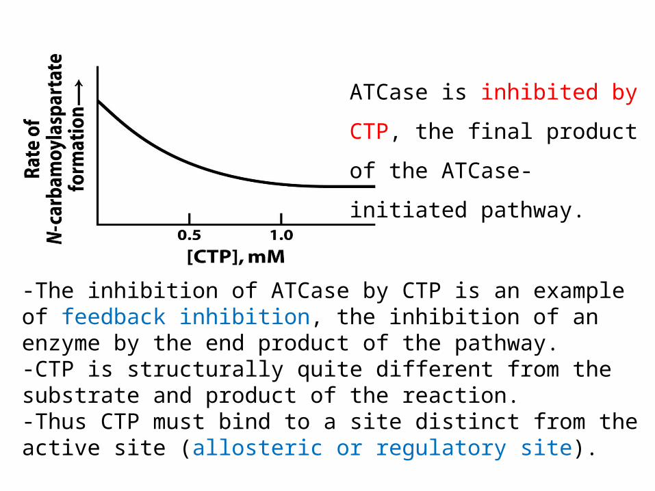
ATCase is inhibited by
CTP, the final product
of the ATCase-
initiated pathway.
-The inhibition of ATCase by CTP is an example of feedback inhibition, the inhibition of an enzyme by the end product of the pathway.-CTP is structurally quite different from the substrate and product of the reaction. -Thus CTP must bind to a site distinct from the active site (allosteric or regulatory site).
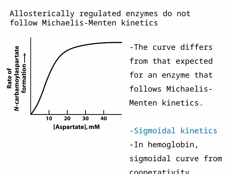
Allosterically regulated enzymes do not follow Michaelis-Menten kinetics
-The curve differs
from that expected
for an enzyme that
follows Michaelis-
Menten kinetics.
-Sigmoidal kinetics
-In hemoglobin,
sigmoidal curve from
cooperativity

ATCase consists of separable catalytic and regulatory subunit
-ATCase can be separated into regulatory(r) and catalytic(c) subunits ; how to know? evidence? -p-hydroxymercuribenzoate reacts with sulfhydryl group of cystein residues in ATCase resulting in separation of subunits.
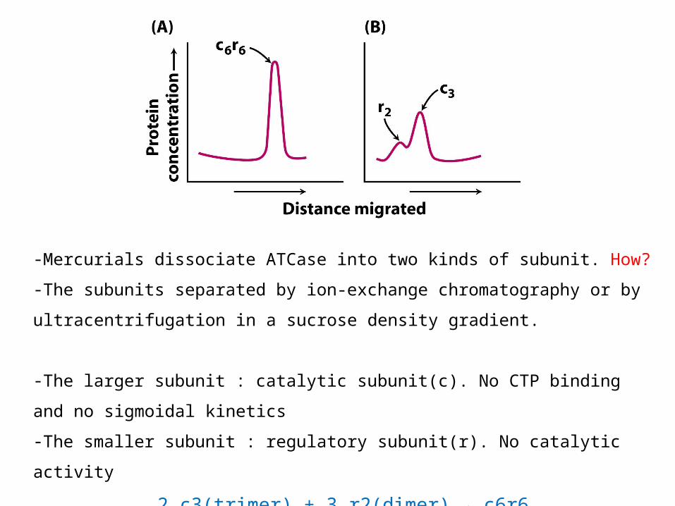
-Mercurials dissociate ATCase into two kinds of subunit. How?
-The subunits separated by ion-exchange chromatography or by
ultracentrifugation in a sucrose density gradient.
-The larger subunit : catalytic subunit(c). No CTP binding and no
sigmoidal kinetics
-The smaller subunit : regulatory subunit(r). No catalytic activity
2 c3(trimer) + 3 r2(dimer) → c6r6
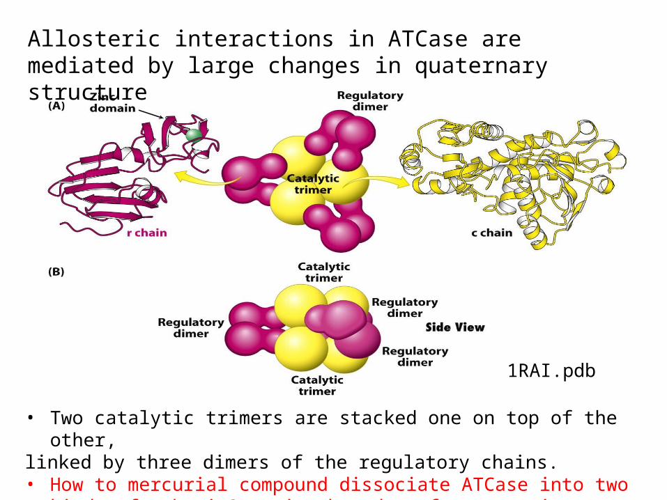
Allosteric interactions in ATCase are mediated by large changes in quaternary structure
• Two catalytic trimers are stacked one on top of the other,linked by three dimers of the regulatory chains.• How to mercurial compound dissociate ATCase into two kinds of
subunit? – zinc bound to four cysteines, destabilize r-subunits
1RAI.pdb

PALA : bisubstrate (two substrates) analog, competitive inhibitor
of ATCase. binds to the active sites and blocks ATCase.
※ ACTase was crystallized in the presence of PALA.
2IPO.pdb

PALA
PALA binds at sites lying at the bounderies between pairs of c chains within a catalytic trimer.

- Conformational change : catalytic trimers move 12Å farther apart, rotate 10 degrees about their threefold axis of symmetry and dimers rotate 15 degrees when PALA(substrate analog) bound.
In the absence of substrateT(tense) state
In the presence of substrateR(relaxed) state

The enzyme exists in an equilibrium between the T and R state.
• In the absence of substrate, almost all the enzyme molecules are in the T state(low affinity for substrates and low catalytic activity).
• Occasional substrate binding to active site → entire enzyme shifts to the R state(higher binding affinity).

• Mechanism for allosteric regulation
1). Concerted mechanism - the change in the enzyme is ‘all or none’(explains the behavior of ATCase well)
2). Sequential model - the binding of ligand to one site on the complex can
affect neighboring sites without T-toR- transition
※ Most other allosteric enzymes have features of both models.

- Mixture of two Michaelis-Menten enzymes.
-As the concentration of substrate is increased, the equilibrium shifts from
the T state to the R state.
-Homotrophic effect by substrate; benefical when small change in substrate
conc is physiologically important
High Km
low Km

Allosteric regulators modulate the T-to-R equilibrium
- The enzyme is in the T state when
bound to CTP.
-Binding sites for CTP exist in regulatory
chains which are far away from each
active sites.
-How can CTP inhibit the catalytic activity
of the enzyme?

-CTP stabilize the T state.
-The binding of CTP shifts the equilibrium toward the T state and
makes it more difficult for substrate binding to convert the
enzyme into the R state.
In the absence of any substrate

In presence of CTP, more substrate is required to attain a given reaction rate.
In presence of ATP, the reaction rate is increased.
Heterotropic effects
High levels of ATP prevent CTP from inhibiting the enzyme. Nonsubstrate effects on allosteric enzyme
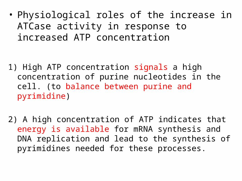
• Physiological roles of the increase in ATCase activity in response to increased ATP concentration
1) High ATP concentration signals a high concentration of purine nucleotides in the cell. (to balance between purine and pyrimidine)
2) A high concentration of ATP indicates that energy is available for mRNA synthesis and DNA replication and lead to the synthesis of pyrimidines needed for these processes.

Equilibrium between the T and the R state
R ↔ T L = [T]/[R] L = the equilibrium constant between the R and the
T forms
CTP-saturated form, the value of L increases from 250 to 1250.
For the ATP saturated form, the value of L decreases to 70.

• Isozymes (or isoenzymes) are enzymes that differ in amino acid sequence yet catalyze the same reaction.
- have Different Km, different regulatory molecules
10.2 Isozymes provide a means of regulation specific to distinct tissues and developmental states
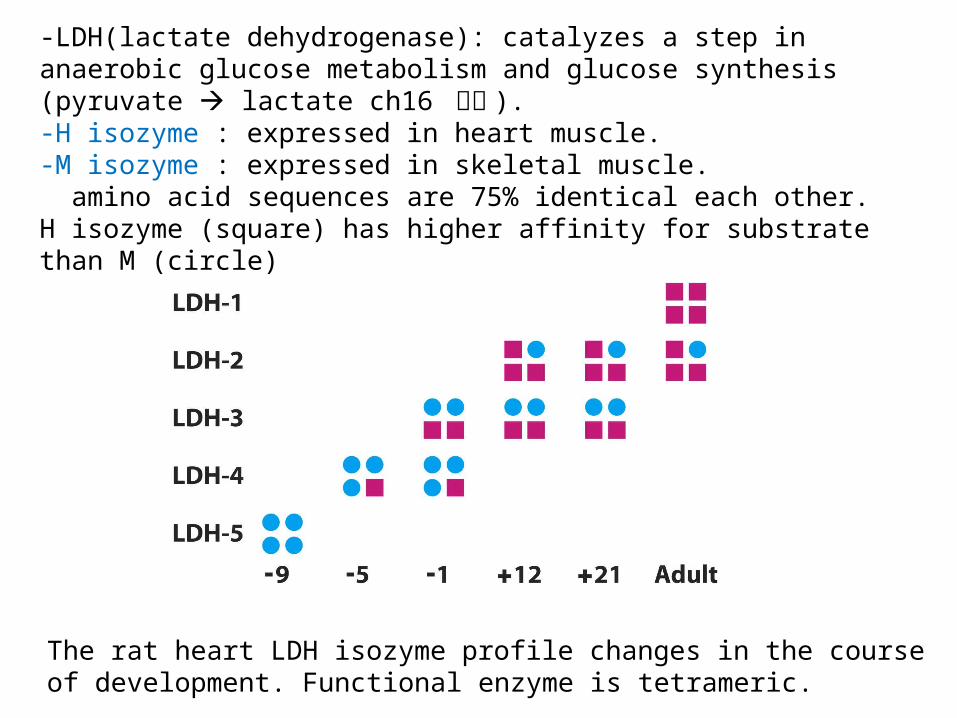
The rat heart LDH isozyme profile changes in the course of development. Functional enzyme is tetrameric.
-LDH(lactate dehydrogenase): catalyzes a step in anaerobic glucose metabolism and glucose synthesis (pyruvate lactate ch16 참조 ). -H isozyme : expressed in heart muscle. -M isozyme : expressed in skeletal muscle. amino acid sequences are 75% identical each other. H isozyme (square) has higher affinity for substrate than M (circle)

1. Sequential displacement - Ordered.
- The coenzyme always binds first and the lactase is always released first.
① ①② ②
Lactate dehydrogenase
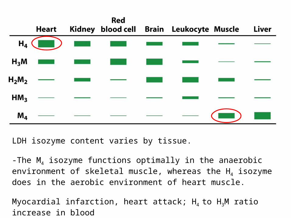
LDH isozyme content varies by tissue.
-The M4 isozyme functions optimally in the anaerobic environment of skeletal muscle, whereas the H4 isozyme does in the aerobic environment of heart muscle.
Myocardial infarction, heart attack; H4 to H3M ratio increase in blood

10.3 Covalent modification is a means of regulating enzyme activity
Some modifications are reversible.

Phosphorylation is a highly effective means of regulating the activities of target proteins- 30% of eukaryotic proteins are phosphorylated.-The enzymes catalyzing phosphorylation reactions are called protein kinases.-ATP is the most common donor of phosphoryl group. αβγ

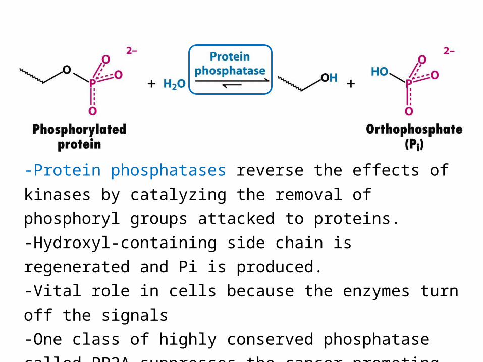
-Protein phosphatases reverse the effects of
kinases by catalyzing the removal of
phosphoryl groups attacked to proteins.-Hydroxyl-containing side chain is
regenerated and Pi is produced.-Vital role in cells because the enzymes turn
off the signals-One class of highly conserved phosphatase
called PP2A suppresses the cancer-promoting
activity of certain kinases.

- The phosphorylation and dephosphorylation are not the reverse of one another; irreversible under physiological conditions without enzymes-With only the help of kinases and phosphatase, take place-The rate of cycling between the phosphorylated and the dephosphorylated states depends on the relative activities of kinases and phosphatases
Kinase
Phosphatase

Phosphorylation is a highly effective means of controlling the activity of proteins for several reasons;1) A phosphoryl group adds two negative charges to a modified protein. New electrostatic
interactions
2) A phosphoryl group can form three or more hydrogen bonds.
3) The free energy of phosphorylation is large. (-12kcalmol-1 by ATP, half is consumed in
making phosphorylation; half (6kcalmol-1) is conserved in the phosphorylated protein.
Phosphorylation can change the conformational equilibrium by the order of 104 ; 1.4kcalmol-1
correspond to 10 fold increase in an equilibrium constant)
4) Phosphorylation and dephosphorylation can take place in less than a second or over a span
of hours.
5) Phosphorylation often highly amplify signaling effects.
6) ATP is the cellular energy currency. Links the energy status of the cell to the regulation of
metabolism.

Cyclic AMP activates protein kinase A (PKA) by altering the quaternary structure
-Flight or fight;
epinephrine (adrenaline)
triggers cAMP formation
-Cyclic AMP subsequently
activates a key enzyme:
protein kinase A.
-The kinase alters the
activities of target
proteins by phosphorylating
specific serine or
threonine residues.
The consensus sequence recognized by PKA is R-R-X-S/T-Z ; (X, small residues; Z, large hydrophobic one)
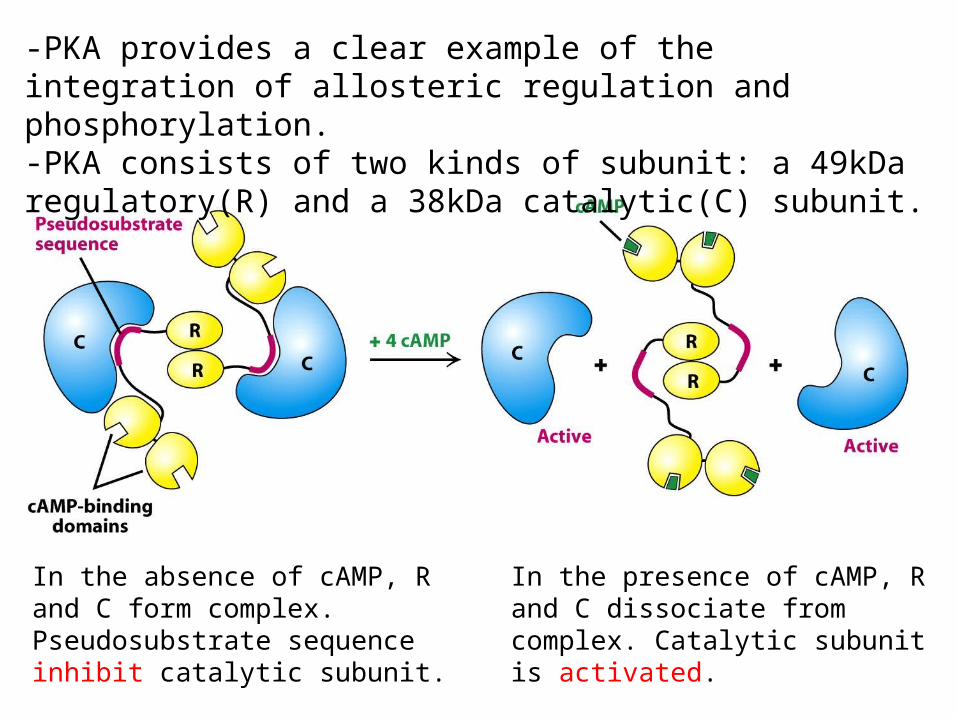
-PKA provides a clear example of the integration of allosteric regulation and phosphorylation.-PKA consists of two kinds of subunit: a 49kDa regulatory(R) and a 38kDa catalytic(C) subunit.
In the absence of cAMP, R and C form complex. Pseudosubstrate sequence inhibit catalytic subunit.
In the presence of cAMP, R and C dissociate from complex. Catalytic subunit is activated.
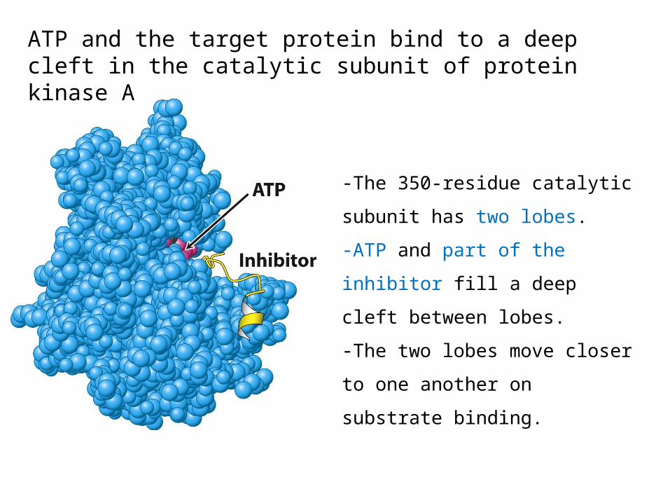
ATP and the target protein bind to a deep cleft in the catalytic subunit of protein kinase A
-The 350-residue catalytic
subunit has two lobes.
-ATP and part of the inhibitor
fill a deep cleft between lobes.
-The two lobes move closer to
one another on substrate
binding.
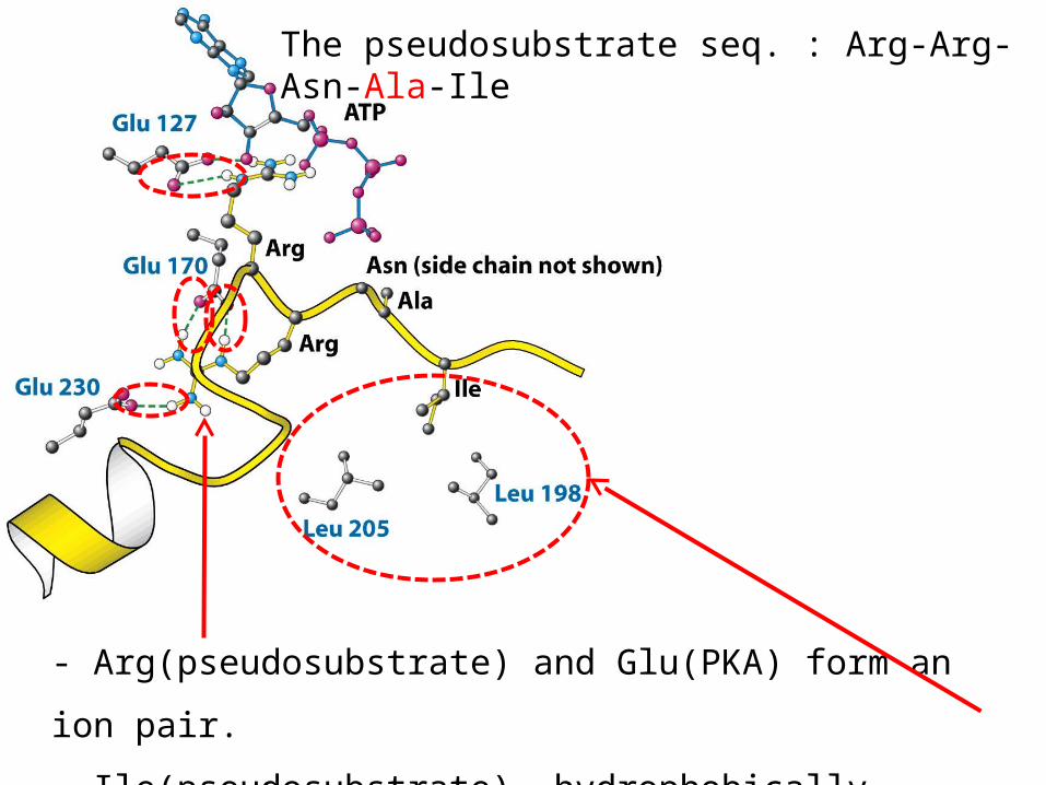
The pseudosubstrate seq. : Arg-Arg-Asn-Ala-Ile
- Arg(pseudosubstrate) and Glu(PKA) form an
ion pair.
- Ile(pseudosubstrate) hydrophobically
interact with leu(KPA)

10.4 Many enzymes are activated by specific proteolytic cleavage
Specific proteolysis is a common means of activating enzymes and other proteins in biological systems. Ex) ; 1) Digestive enzymes2) Blood clotting3) Hormones; proinsuline4) Collagen; procollagen5) Developmental processes: procollagenase (tadpole, 출산 )6). Programmed cell death, apoptosis: procaspases
The inactive precursor is called a zymogen or a proenzyme.Proteolytic activation occurs just once.
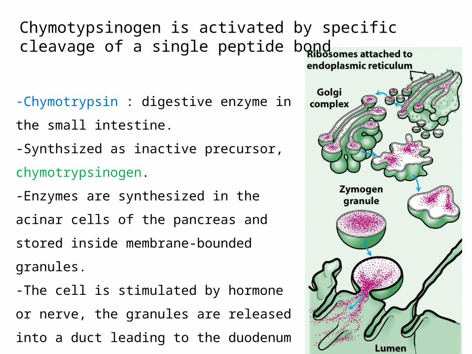
Chymotypsinogen is activated by specific cleavage of a single peptide bond
-Chymotrypsin : digestive enzyme in
the small intestine.
-Synthsized as inactive precursor,
chymotrypsinogen.
-Enzymes are synthesized in the
acinar cells of the pancreas and
stored inside membrane-bounded
granules.
-The cell is stimulated by hormone
or nerve, the granules are released
into a duct leading to the duodenum
( 십이지장 )

Chymotrypsinogen
-Single polypeptide
of 245 amino acids
is cleaved by
trypsin.
Linked by interchain disulfide bonds

Proteolytic activation of chymotrypsinogen leads to the formation of a substrate-binding site
-The cleavage of the peptide
bond between amino acid 15 and
16 triggers key conformational
changes.
-Alteration of the position of
Ile16.
-The electrostatic interaction
between Ile16 and Asp 194 is
essential for the structure of
active chymotrypsin.
-Substrate binding cavity and the oxyanion hole are
formed after proteolytic cleavage.

The generation of typsin from trypsinogen leads to the activation of other zymogens
- The formation of trypsin by
enteropeptidase is the master activation
step.
from the duodenum cells ( 십이지장 )Cleave lysine-isoleucine peptide

Some proteolytic enzymes have specific inhibitors
Pancreatic trypsin
inhibitor :
- 6kda
- Inhibits trypsin by
binding- very tightly to
its active site.
Turnover rate is very
low
- Lys 15 interact with
specificity pocket of
trypsin (Asp189).
Trypsin inhibition → prevention of tissue damage of pancreas or pancreatic ducts.
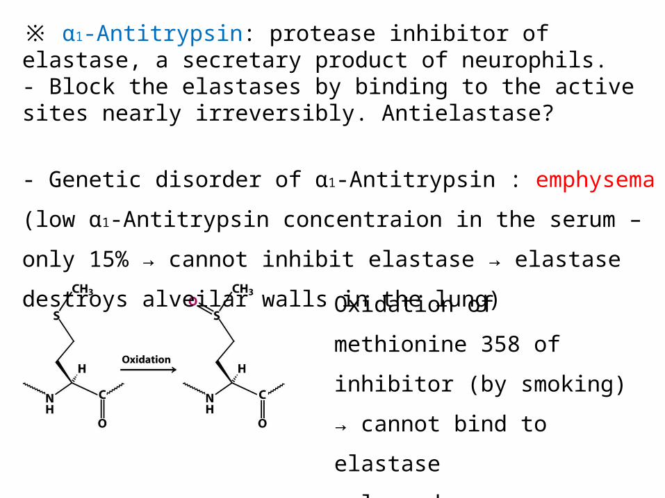
※ α1-Antitrypsin: protease inhibitor of elastase, a secretary product of neurophils.- Block the elastases by binding to the active sites nearly irreversibly. Antielastase?
- Genetic disorder of α1-Antitrypsin : emphysema
(low α1-Antitrypsin concentraion in the serum –
only 15% → cannot inhibit elastase → elastase destroys
alveilar walls in the lung) Oxidation of
methionine 358 of
inhibitor (by smoking)
→ cannot bind to elastase
→ lung damage

Blood clotting is accomplished by a cascade of zymogen activations
-Blood clots are formed by a cascade of zymogen activations. Amplification! rapid response to trauma
-After the tissue factor is exposed, small amounts of thrombin, the key protease in clotting, are generated.
-Thrombin activates enzymes and factors that lead to the generation of yet more thrombin (= positive feedback)-혈우병 ?
Signal amplified
Exposure of anionic surfaces
Integral membrane glycoprotein
Transglutaminase

Fibrinogen is converted by thrombin into a fibrin clot
- Fibrinogen: is made up of three globular units connected by two rods.-Thrombin cleaves four arg-gly peptide bond in the central region of fibrinogen.-The cleaved peptide of Aα and Bβ chain = fibrinopeptide-result in (αβγ)2
6 chain= 340kda
Rod : triple-stranded α–helical coiled coil

-Fibirin monomers assemble into ordered
fibrous arrays called fibrin.
-Fibrin has a periodic structure that repeats
every 23nm.
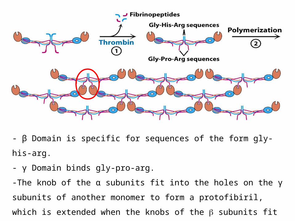
- β Domain is specific for sequences of the form gly-
his-arg.
- γ Domain binds gly-pro-arg.
-The knob of the α subunits fit into the holes on the γ
subunits of another monomer to form a protofibiril,
which is extended when the knobs of the subunits fit
into the holes of subunits of other protofibrils.

Prothrombin is readied for activation by a vitamin K-dependent modification
-Thrombin is synthesized as a zymogen called prothrombin.-Inactive form: 4 major domains. -γ–carboxyglutamate rich domain (Gla domain)
-Activation is begun by proteolytic cleavage
of the bond between arg274 and thr275
-Cleavage of the bond between arg323 and
ile324 yield active thrombin.
① ②

※ Vitamin K (deficiency in this vitamin results in defective blood koagluation) -Essential for the synthesis of prothrombin and other clotting factor.
※ Dicoumarol : vitamin K mimic antagonist. Spoiled clover - Binding of discoumarol results in synthesize an abnormal prothrombin that does not bind Ca2+(cofactor). Strange? Why?
-Amino acid analysis same A.A sequence Why?-NMR!-Vitamin K-dependent γ–carboxylation.

-Normal prothrombin is carboxylated to γ–carboxyglutamate by a vitamin K-dependent enzyme.
- γ–carboxyglutamate can interact with Ca2+,which anchorprothrombin phospholipid membrane.-brings the zymogen into closeproximity to 2 clotting proteins converting into thrombin-During activation, calcium-bindingdomain is removed, freeing the thrombinfrom the membrane so that it cancleave fibrinogen and other targets.
γ–carboxyglutamate

Hemophilia revealed an early step in clotting
-Hemophilia : the best known clotting defect. sex-linked recessive characteristic.
-In classic hemophilia, factor VIII of the intrinsic pathway is missing or has markedly reduced activity.factor VIII is not a protease but it stimulate the activation of factor X by factor Ixa
-Recombinant factor VIII-The activity of factor VIII is markedly increased by limited proteolysis by thrombin positive feed back
The clotting process must be precisely regulated-Clots must form rapidly.
-Activated clotting factors are short-lived
because they diluted by blood flow, removed
by the liver, and degraded by proteases.
-Factor V and VIII are digested by protein C,
switched on by the action of thrombin
-Thrombin has a dual function: catalyzes the
formation of fibrin and it initiates the
deactivation of the clotting cascade
Specific inhibitors of clotting factors are also
critical.
-Tissue factor pathway inhibitor (TFPI) inhibit the
complex of TF-VII-X
-Antithrombin III: a plasma protein, inactivates
thrombin by forming an irreversible complex with it.
Blocks clotting factors such as (9,10,11,12).
-Antithrombin III activity is enhanced by heparin (a
negatively charged polysaccharide found in master
cells and endothelial cells)
- Heparin is anticoagulant by increase the rate of
formation of irreversible complexes between
antithrombin III and clotting factors
-M358R mutation in1-antitrypsin’s binding pocket
change specificity from an elastase inhibitor to a
thrombin inhibitor. 1-antitrypsin increases markedly
after injury to counteract excess elastase. The mutant
1-antitrypsin caused the patient’s thrombin activity
to drop to the level of hemorrhage.
Clots are not permanent structures. How to
desolve clots?
-Fibrin is split by plasmin, a serine
protease that hydrolyzes peptide bonds in the
coiled coil.
-Plasmin is formed by the proteolytic
activation of plasminogen, that has high
affinity for clot.
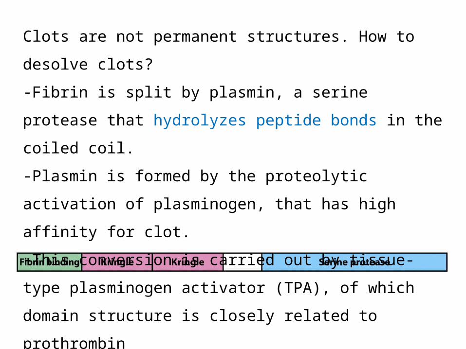
-This conversion is carried out by tissue-
type plasminogen activator (TPA), of which
domain structure is closely related to
prothrombin
-TPA bound to fibrin clots swiftly activate
adhering plasminogen but very slowly free
plasminogen

Plasminogen -> plasmin -> fibrin degradation -> clot removeBy TPA administered intravenously
Before After 3 hour treatment TPA
TPA leads to the dissolution of blood clots, as shown by X-ray images of blood vessels in the heart coronary artery.So fast!