chapter 1 pathophysiology of acute coronary...
TRANSCRIPT

CHAPTER 1
Pathophysiology of acutecoronary syndromes
Alisa B. Rosen and Eli V. Gelfand
Introduction
Acute coronary syndromes (ACS) comprise a spectrum ofclinical conditions, initiated by rupture of an atheroscleroticcoronary plaque with overlying acute thrombosis. The con-sequences of thrombosis include direct obstruction of bloodflow to the coronary beds, as well as distal embolization ofthe platelet-rich thrombus. Both of these processes may leadto myocardial ischemia and may progress to myocytenecrosis and myocardial infarction. The coronary thrombusmay be completely occlusive, as is frequently seen inST-segment-elevation myocardial infarction (STEMI), ornonocclusive, as can be observed in unstable angina ornon-ST-elevation myocardial infarction (UA/NSTEMI). Thelatter two entities are also known collectively as non-ST-elevation acute coronary syndromes (NSTEACS). This chapterdiscusses the basic pathophysiology underlying ACS.
Braunwald has described five processes contributing todevelopment of ACS, or any atherothrombotic event(Figure 1.1). These processes include: (1) thrombus onpreexisting plaque, (2) dynamic obstruction from coronaryspasm or Prinzmetal’s angina, (3) progressive mechanicalobstruction, (4) inflammation and/or infection, and(5) secondary unstable angina due to global myocardialoxygen supply and demand mismatch.
Management of Acute Coronary Syndromes Eli V. Gelfand and Christopher P. Cannon
� 2009 John Wiley & Sons, Ltd
COPYRIG
HTED M
ATERIAL

Formation of atherosclerotic plaque
Complex plaques of mature atherosclerosis are the end-result of a long pathophysiologic process, which typicallybegins in early adulthood. Endothelial dysfunction appearsto play an initial role in atherosclerosis. Injury to theendothelium results in establishment of the cycle of inflam-matory cell migration and proliferation, tissue damage, andrepair, and ultimately leads to plaque growth. Thesemechanisms are outlined in Table 1.1 and are further illu-strated in Figure 1.2 (see color plate for a full-color version).
On histological specimens, early precursors of complex pla-ques include intimal thickening, isolated lipid-containingmacrophage foam cells, and pools of extracellular lipids.These are visible on gross specimens as fatty streaks, and
Secondaryactivation of
plasmacoagulation
systemPlateletactivation,
aggregation, andadhesion
Rupture of avulnerable
atheroscleroticplaque
Coronaryvasoconstriction
Imbalance inmyocardial
oxygenand demand
ACS
Figure 1.1 Processes contributing to the development of ACS.
Table 1.1 Primary components of atherosclerotic plaque formation,
initiated by endothelial dysfunction (data from Ross1)
• Increased endothelial adhesiveness
• Increased endothelial permeability
• Migration and proliferation of smooth muscle cells and macrophages
• Release of hydrolytic enzymes, cytokines, and growth factors
• Focal vessel wall necrosis
• Tissue repair with fibrosis
2 Pathophysiology of acute coronary syndromes

Smooth-musclemigration
B
Foam-cellformation
T-cellactivation
Adherence andaggregation of
platelets
Adherenceand entry
of leukocytesFigure 1.2 The mechanism of atherosclerotic plaque formation
(reproduced from Ross N Engl J Med 1999; 340: 115–26). (A) Early
endothelial dysfunction in atherosclerosis; (B) fatty streak formation;
(C) formation of advanced complex lesion of atherosclerosis;
(D) formation of an unstable fibrous plaque. A full-color version of
this figure appears in the plate section.
Endothelialpermeability
Leukocytemigration
Endothelialadhesion
Leukocyteadhesion
A
Figure 1.2
Formation of atherosclerotic plaque 3

are present in a substantial proportion of young adults wholive in the developed world. Eventually, a reactive fibroticcap and a large lipid core are formed, the lesion may becomeneovascularized, and calcium is deposited within the plaque(Figure 1.2).
Macrophageaccumulation
Formation ofnecrotic core
Fibrous-cap formation
C
Figure 1.2
Plaque rupture Thinning of fibrous cap Hemorrhage formplaque micorovessels
D
Figure 1.2 Continued.
4 Pathophysiology of acute coronary syndromes
Plaque instability and the development of ACS
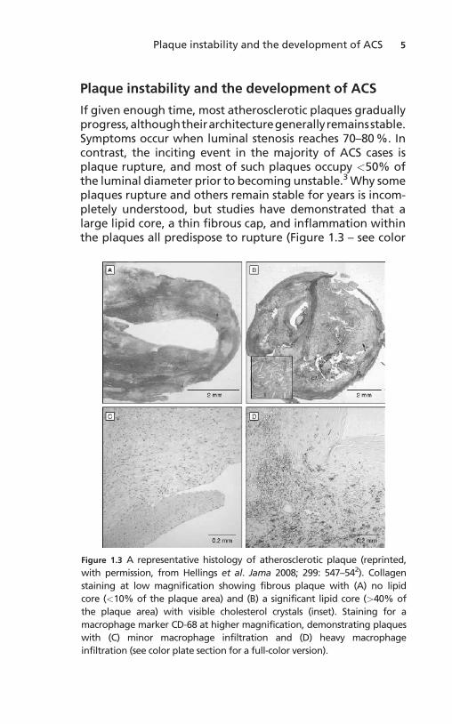
If given enough time, most atherosclerotic plaques graduallyprogress,althoughtheirarchitecturegenerallyremainsstable.Symptoms occur when luminal stenosis reaches 70–80 %. Incontrast, the inciting event in the majority of ACS cases isplaque rupture, and most of such plaques occupy <50% ofthe luminal diameter prior to becoming unstable.3 Why someplaques rupture and others remain stable for years is incom-pletely understood, but studies have demonstrated that alarge lipid core, a thin fibrous cap, and inflammation withinthe plaques all predispose to rupture (Figure 1.3 – see color
Figure 1.3 A representative histology of atherosclerotic plaque (reprinted,
with permission, from Hellings et al. Jama 2008; 299: 547–542). Collagen
staining at low magnification showing fibrous plaque with (A) no lipid
core (<10% of the plaque area) and (B) a significant lipid core (>40% of
the plaque area) with visible cholesterol crystals (inset). Staining for a
macrophage marker CD-68 at higher magnification, demonstrating plaques
with (C) minor macrophage infiltration and (D) heavy macrophage
infiltration (see color plate section for a full-color version).
Plaque instability and the development of ACS 5

plate section for full-color version).4 Inflammation is thoughtto play a central role in actual plaque disruption.1 Indeed, ahigh macrophage content identifies plaques prone to rup-ture,5 and unstable, symptomatic plaques can be identifiedwith molecular imaging targeting inflammation.6 C-reactiveprotein, a marker of inflammation, is a significant, indepen-dentpredictorofmyocardial infarction,stroke,andperipheralarterial disease.7
Exposure of thrombogenic plaque material to flowingblood initiates the endogenous thrombotic response.Actual plaque rupture may precede the clinical syndromeof ACS by several days or even weeks, as evidenced byfindings of both fresh and old thrombus in samples of cor-onary aspirate.8 It seems likely that most plaque erosionsand ruptures are healed with small ‘‘sealing’’ surfacethrombi, and major occlusive thrombosis occurs relativelyrarely. In these latter cases, however, progressive in situ
Increasedmyocardial
oxygen demand
Increased tissue metabolicrate
• Fever• Hyperthyroidism
Increased wall stress
• Hypertension• Left ventricular hypertrophy• Aortic stenosis
Reduced oxygen blood content
Reduced coronary artery flow
• Fixed coronary stenosis• Coronary vasospasm• De novo coronary thrombosis• Hypotension• Tachycardia• Bradycardia• Hypovolemia
• Anemia
• HypoxiaIncreased heart rate
• Atrial tachyarrhythmias
• Ventricular tachyarrhythmias
Decreasedmyocardial
oxygen supply
Figure 1.4 Determinants of myocardial oxygen balance and related
pathophysiologic factors that contribute to acute coronary syndrome.
6 Pathophysiology of acute coronary syndromes

thrombosis, together with plaque and thrombus fragmentembolization to the distal coronary microcirculation, andthe overlying vasospastic response, create conditions formyocardial ischemia.
Myocardial ischemia
Myocardial ischemia occurs when the oxygen demand of themyocardium is greater than its oxygen supply (Figure 1.4).An acute thrombotic coronary occlusion in a previouslypatent vessel abruptly decreases myocardial oxygen supply.Alternatively, in a patient with a stable intracoronary pla-que, elevated heart rate may cause myocardial ischemia byincreasing myocardial oxygen demand without having theability to increase supply. Although most cases of ACS arecaused by decreased myocardial oxygen demand, a thor-ough understanding the components of myocardial oxygendemand and supply is crucial to an understanding of thepathophysiology of myocardial ischemia.
Thrombus formation
Acute coronary syndrome is largely caused by thrombus for-mation on preexisting plaque. This has been shown throughboth autopsies and coronary angiography.9,10 Platelets andthe plasma coagulation system are the two major mechan-isms through which a thrombus is formed (Figure 1.5).
Platelets
Platelets play a major role in primary hemostasis and inthrombus formation. This occurs in three stages: plateletadhesion, platelet activation, and platelet aggregation(Figure 1.5).12
Platelet adhesion1 Plaque rupture exposes collagen and tissue factor to the
bloodstream.2 GP Ib receptor on platelets interacts with von Willabrand
Factor (vWF) to adhere to the damaged endothelialsurface.
Platelets 7
Platelet activation1 Platelet degranulation releases thromboxane A2 (TxA2),
adenosine diphosphate (ADP), and other chemoattrac-tants that mediate platelet aggregation. Thrombin andtissue factors also stimulate platelet aggregation.
2 Platelets undergo a conformational change, from asmooth shape to an irregular shape with a larger surfacearea.
3 Platelets express the GPIIb/IIIa receptor.
Platelet aggregation1 GPIIb/IIIa receptors on the surface of the activated plate-
lets interact with circulating fibrinogen.2 Fibrinogen acts as a bridge between GPIIb/IIIa receptors
on multiple activated platelets, causing the formation of aplatelet plug.
Given that thrombus formation in coronary arteries is themajor pathologic process causing ACS, many of the impor-tant pharmacologic agents used to treat ACS target plateletfunction.
Coagulation cascade
Tissue factor
LMWHUFH
LMWHUFH
Directthrombininhibitors
FondaparinuxFactor Xa
ProthrombinAntithrombin
AntithrombinThrombin
Fibrinogen Fibrin Thrombus
Thrombolytics
GP llb/llainhibitors
Clopidogrel
LMWH
PlasminFibrin
degradation
Platelet aggregation
Fibrinogen crosslinking
Activated platelets
Thromboxane
Asprin
Collagen
A2 vWF ADP
TFPI
Platelets
Platelets
Leukocytes
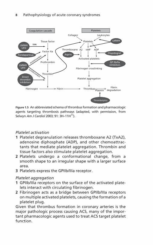
Figure 1.5 An abbreviated schema of thrombus formation and pharmacologic
agents targeting thrombosis pathways (adapted, with permission, from
Selwyn Am J Cardiol 2003; 91: 3H–11H11).
8 Pathophysiology of acute coronary syndromes

Medications that act by interfering with primary hemostasis1 Aspirin inhibits the production of TxA2 by inhibiting
cyclooxygenase, an enzyme in the pathway that convertsarachadonic acid to TxA2 and other prostaglandins.
2 Thienopyridines (ticlopidine, clopidogrel, prasugrel) blockthe ADP receptor on the platelet, which inhibits plateletaggregation and binding of fibrinogen to the GIIb/IIIareceptor on activated platelets.
3 GPIb/IIIa receptor antagonists directly bind to the recep-tors that mediate platelet aggregation.
Secondary hemostasis
Secondary hemostasis (the coagulation cascade) is activatedconcurrently with the platelet-mediated primary hemostaticmechanisms described above (Figure 1.5). Plaque ruptureexposes tissue factor to the bloodstream, which both has arole in platelet adhesion and in activation of the extrinsicsystem of the clotting cascade.
The production of thrombin1 Plaque rupture exposes collagen and tissue factor to the
bloodstream.2 Tissue factor converts factor X to Xa.3 Factor Xa converts prothrombin to thrombin.
The role of thrombin in thrombosis1 Thrombin converts fibrinogen to fibrin, which is the final
step in clot formation.2 It activates factor XIII, which causes crosslinking of fibrin
and stabilization of the clot.3 It stimulates platelet aggregation (as part of primary
hemostasis).
Some medications that act by interfering with the coagula-tion cascade1 Unfractionated heparin activates antithrombin II (ATII),
which inactivates factor Xa and thrombin.2 Low molecular weight heparin (LMWH) also activates
antithrombin, which inactivates factor Xa. However,LMWH has a much lesser effect on thrombin than doesunfractionated heparin.
Secondary hemostasis 9

3 Direct thrombin inhibitors (bivalirudin, argatroban) inhi-bit thrombin and therefore prevent the conversion offibrin to fibrinogen.
4 Factor Xa inhibitor (fondaparinux) inactivates factor Xa,which then prevents the conversion of prothrombin tothrombin.
In the setting of ACS, the normal balance betweenthrombosis and endogenous fibrinolysis is disrupted infavor of thrombosis. In addition to medications aimed atinhibiting formation of a platelet plug (aspirin, clopido-grel, GIIb/IIIa receptor antagonists), anticoagulants suchas unfractionated heparin, LMWH, direct thrombin inhi-bitors, and factor Xa inhibitors are beneficial inthe treatment of ACS (this is discussed further inChapters 3 and 4).
Dynamic obstruction
Dynamic obstruction can occur with epicardial coronaryvasospasm or be limited to the microcirculation.Symptomatic epicardial vasospasm (Prinzmetal’s angina)can occur either at the site of a preexisting nonobstructiveatherosclerotic plaque or in a normal portion of the ves-sel.13 Nonfocal coronary vasospasm can occur in the settingof cocaine use, cold immersion, or emotional stress.14,15
Angiographic evidence of epicardial coronary obstructionmay be absent if the study is performed at a later time, butrecurrent spasm may be demonstrated by asking thepatient to hyperventilate on the angiography table.Microcirculatory angina can occur with vasoconstriction insmall intramural arteries.
Progressive mechanical obstruction
Progressive mechanical obstruction is an unusual cause ofACS. It is most frequently seen when progressive in-stentrestenosis causes decreasing myocardial oxygen supplyover the course of months. Gradual-onset exertional angina,not ACS, is a more typical outcome of progressivemechanical obstruction.
10 Pathophysiology of acute coronary syndromes

Inflammation
As discussed above, inflammation appears to play a majorrole in initiation and progression of atherosclerosis, as wellas in the transition from a stable to an unstable plaque andthe onset of acute atherothrombosis.
Secondary unstable angina
Secondary unstable angina is myocardial ischemia/infarctioncaused by a process other than plaque rupture withthrombus formation. Anemia, bradycardia and severe hypo-tension are common causes of reduced oxygen supply,whereas tachycardia, fever, and hyperthyroidism frequentlyincrease myocardial oxygen demand. Secondary angina isfurther discussed in Chapter 5.
References
1. Ross R. Atherosclerosis—an inflammatory disease. N Engl J Med
1999; 340: 115–26.
2. Hellings WE, Moll FL, De Vries JP, et al. Atherosclerotic plaque
composition and occurrence of restenosis after carotid endart-
erectomy. Jama 2008; 299: 547–54.
3. Giroud D, Li JM, Urban P, Meier B, Rutishauer W. Relation of the site
of acute myocardial infarction to the most severe coronary arterial
stenosis at prior angiography. Am J Cardiol 1992; 69: 729–32.
4. Thieme T, Wernecke KD, Meyer R, et al. Angioscopic evaluation of
atherosclerotic plaques: validation by histomorphologic analysis
and association with stable and unstable coronary syndromes.
J Am Coll Cardiol 1996; 28: 1–6.
5. Fishbein MC, Siegel RJ. How big are coronary atherosclerotic
plaques that rupture? Circulation 1996; 94: 2662–6.
6. Rudd JH, Warburton EA, Fryer TD, et al. Imaging atherosclerotic
plaque inflammation with [18F]-fluorodeoxyglucose positron
emission tomography. Circulation 2002; 105: 2708–11.
7. Ridker PM, Hennekens CH, Buring JE, Rifai N. C-reactive protein and
other markers of inflammation in the prediction of cardiovascular
disease in women. N Engl J Med 2000; 342: 836–43.
8. Rittersma SZ, van der Wal AC, Koch KT, et al. Plaque instability
frequently occurs days or weeks before occlusive coronary thrombosis:
a pathological thrombectomy study in primary percutaneous coronary
intervention. Circulation 2005; 111: 1160–5.
References 11

9. Silva JA, White CJ, Collins TJ, Ramee SR. Morphologic comparison
of atherosclerotic lesions in native coronary arteries and saphenous
vein graphs with intracoronary angioscopy in patients with
unstable angina. Am Heart J 1998; 136: 156–63.
10. Nesto RW, Waxman S, Mittleman MA, et al. Angioscopy of culprit
coronary lesions in unstable angina pectoris and correlation of
clinical presentation with plaque morphology. Am J Cardiol 1998;
81: 225–8.
11. Selwyn AP. Prothrombotic and antithrombotic pathways in acute
coronary syndromes. Am J Cardiol 2003; 91: 3H–11H.
12. Kennon S, Price CP, Mills PG, et al. The central role of platelet
activation in determining the severity of acute coronary
syndromes. Heart 2003; 89: 1253–4.
13. Kaski JC, Tousoulis D, McFadden E, Crea F, Pereira WI, Maseri A.
Variant angina pectoris. Role of coronary spasm in the
development of fixed coronary obstructions. Circulation 1992; 85:
619–26.
14. Pitts WR, Lange RA, Cigarroa JE, Hillis LD. Cocaine-induced
myocardial ischemia and infarction: pathophysiology, recognition,
and management. Prog Cardiovasc Dis 1997; 40: 65–76.
15. Strike PC, Steptoe A. Systematic review of mental stress-induced
myocardial ischaemia. Eur Heart J 2003; 24: 690–703.
12 Pathophysiology of acute coronary syndromes