chapter 1 introduction and literature...
TRANSCRIPT

Chapter 1 Introduction and literature
reviev
INTRODUCTION AND REVIEW OF LITERATURE
In lndia, the science of Aymeda had provided a system of medical treatment
and most of the remedies for treating illness were taken from plants. During the last
few decades, much work has been done in the field of n a t d products. At no time in
the history of mankind there has been more rapid and meaninghl pmyrcss in the
understanding of plant and their constituents tltm during rhe past quarter century.
The development of the science of phyto phnrnlacccuticals and the hopes far the remedies in chronic diseases has generuted more new enthusiasm in mscmh
workers to develop herbal medicines. A considcrnble mount of work has been done
to study the potential of herbul medicines nnd modem scicncc has accepted the plant
kingdom as a source of new hidynamic constituents.
1.1 Importance of the Herbs and Herbal medicine: I-lerbs have been used 8s a
source of drugs to combat diseases since time irnmernorid. 'She cffectivcncss, easy
availability, low cost and non-toxic nature popularim, herbal remdics. In spite of the
dnunatic development of synlhetic drugs and antibiotics as the major thetapeutic
agents, herbs continue to provide basic raw material for some of thc most important
drugs (Ali-Shtayeh et al., 2000).
Ancient health scienccs like Ayu~edfI depend heavily on natural products,
Ayuweda is an important system of medicine (Bhutani, 1999). 'Ihe plants play an
important role in human nutrition, health and disees~. For example, amenloflavone, a
naturally occurring biflavonoid isolated from the leaves of Ginklpa hilrrba selectively
inhibited human secretory phospholipase A2 and proved lo be a novel anti-
inflammatory agent (Moon el a/., 1983). Similarly natural non-saccharide sweetening
agents are low calorific, nowtoxic and 10-1000 times sweeter than sugar and are
capable of overcoming problems associated with sucrose and synthetic sweetenem
when used in diabetic patients. e.g. glycyrrhizin, thnunatin, stcvioside (Prakash el ul.,
2002).
Probenu w&h HnBol hp: A study rrportJ heavy contamination with bectcrie and
fungi in herbtl drugs lik Triphak Avipitittakar, H i i s s t i k , bvan b k b ad

Ameripu churna. Total microbial load of the drug for each group of microorganism,
bacteria, fungi and actinomycetes was found to decline with in dYee months of
storage in some dnrg samples but recorded a tremendous enhancement during sii
months of storage (Roy and Chaurasia, 1989).
Herbal products are widely used in the general population and their use is
often undisclosed to the physician or the pharmacist. There are a number of examples
of herbal therapies, whose multiple active components are known to cause
potentiation or antagonism of the effect of conventional medications. Herbs such as
garlic and devil's claw may cause an increased International Normalized Ratio (INR),
while ginseng and green tea may decrease the INR in patients taking warfarin, thereby
increasing a patient's risk of bleeding or loss of anticoagulant effect, respectively
(Stephen el al., 2002).
1.2 Cancers: Cancer occurs when cell division gets out ofcontrol. Usually, the timing
of cell division is under strict constraint, involving a network of signals that work
together to say when a cell can divide, how often it should happen and how errors can
be fixed. Mutations in one or more of the nodes in this network can trigger cancer, be
it through exposure to some environmental factor (e.g. tobacco smoke) or because of
a genetic predisposition, or both. The predominant mechanisms for the cancers
featured here are (i) impairment of a DNA repair pathway (ii) the transformation of a
normal yenc into an oncogene and (iii) the malfunction of a tumor supressor gene.

Caws for C I Q ~ Cancer is o h perceived as a disease that shikes for no eppannt
reason. But many of the causes of caacer have a l d y been identified. Besides
heredity, scientific studies point to the existence of thne main categories of factors
that contribute to the development of cancers. They an chemicals (e.g., from smoking
or diet), physical (UY radiations) and biological ( v i m or bacteria).
Genes and cancer: Physicallchemicnlhiological factors contribute to the
development of cancer by triggering changes in a cell's genes. Chemicals and
ndiation act by damaging gcnes; Viruses introduce their own genes into cells, and
heredity passes on alterations in genes fmm one generation to the next. These altered
or mutated genes make a petson more suceptihlc to cancer.
Cancer prmenflon: Since exposwr to carcinogens (cancer-causing nycnls) is
responsible for triggering most human cunccrs, people can reduce thcir cancer risk by
taking steps to avoid such agents. Hence the first step in cartccr prcvcntion is to
identify the bchoviours or exposures to particular kinds of carcinogens and viruses
that represent the greatest cnncer hrvard.
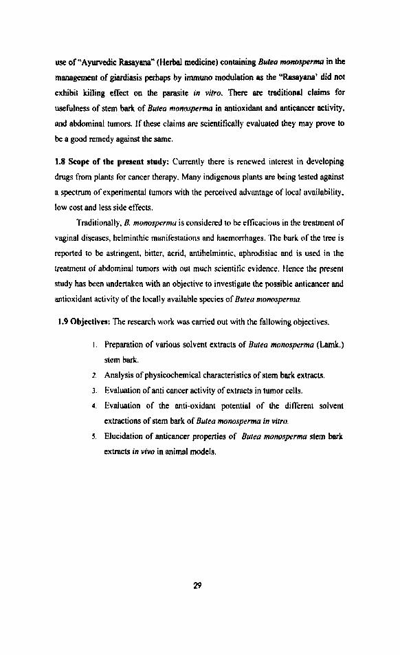
T y p ofcancm: Cancer can originate almost anywhere in the body (Fig I . 1 ) und
there are different kinds of cancer which are discussed below.

Figure 1.1: Leeding sites of cancer world-wide in human9 (Source: American Cancer
Society).
Carcinomas: The most common type of cancers arises fmrn the cells that cover
external and i n t e d body surfaces. I.unp, breast and colon cancers are the most
frequent.
Sarcomas: These tire cancers arising from cells found in the supporting tisues of the
body such as bone, cartilage, fat, connective tissue and muscle.
Lymphomas: These are cancers thut arise in the lymph nodes and tissucs of he
body's immune system. ,
Lahh: These are cancns of the immim blood cells that grow in Lhe bone
marrow and Lend to accumulate in lavge n u m h in the blooclJtream.
l).prs of k W : Mrania. the term coined by Virchow is a cancer of Mle blood
cells. Leukemia o m when a white blood cell whose devclopmcnr is from codma to d u p l i i itself. The T & g p.ogmy of cells are all in b same dage of
devclopmnl and bear the distinctive hallmarks of the type of ancestral white blood

cell that gave rise to thcm. Basad on this undamdiw by 1900 leukemia was no
longer seen as a single disease. Instead i t was imagined akin to a tree with two main
limbs that in turn have two primary branches, all of which reflect from what type of
cell the leukemia originates. One limb, myelogenous leukemia. has its hallmark in the
blood and bone marrow either a prodominance of inmatun mploblasts (acute
myeloid leukemia-AML) or mature myeloid cells (chronic myeloid leukemia-CML).
With the other limb, lyrnphocytic leukemia, the blood and bone marrow is over
populated by either precursor J3 or T cells (acute lymphacytic leukemia-ALL) or
mature B or T cells (chmnic Iymphocytic leukemia-Cl..l.).
Chronic myeloid leukemia: Chmnic myrloid leukemia (CML) is a cancer of
granulocytes (one of the main types of white blood cells). 'hese leukemia cells fill up
the bone marrow and thus lend to an increased risk of infwtion. 'fie disc&. usually
develops very slowly. which is why it is called 'chronic' nlyeloid leukemia. Chronic
myeloid leukemia can occur at any age, but affects more commonly middle-aged and
older people. It is rare in children and it is mainly divided into two phases.
i . The chronic phase: The phasc when most people are diagnosed. At this phasc
CML progresses very slowly and is often stable for long periods. It is also
called as "stable phase" as it lasts for an average of' about 4-5 ycars,
i i . The accelerated and blast phase (advanced phard: Leukemia gradually
develops into the weleratcd phase. during which Be disease develops more
quickly. During this phase immature cells (blast cells) fill up the bone marrow.
After some months leukemia transforms into blast phase, which is more like
an acute leukemia
CML is a disoider characterized by a massive expansion of progenitor cells in
all stages of maturation (Daley el al., 1990). CML is associated with thc Philadelphia
chromosome (Ph), a cytogenic abnormality generated by a reciprocal ttanalocation
bctwetn the bcr gene fimk point cluster region) on chromomme 22q and the c-abl
(ableson leukania virus) protooncogene on chromosome 9q (Fialkow er al.. 1997;
Caopasson et a(. , 1970; Heisterkamp er d., 1983; Oroffcn et al. , 1984).

The fusion gene produces a chimeric 8.5 kb trimdpt that codes for the p210
''''*' protein (Shtivelman et al., 1985). Bcr-abl signaling causes transformation
through several mechanisms (Coretz et al., 1997; Salgia el al., 1997). It has been
postulated that the altered tyrosine kinase activity of p210 stimulates
uncontrolled cell proliferation, leading to the massive clonal expansion of
hematopoietic progenitors detected in CML (Stryckmans et al., 1976; Eaves et al.,
1986). Recent studies revealed that CML progenitors have similar proliferation rates
to their normal counterpart and that p210 bc"*l increases cell survival by inhibiting
apoptosis (Bedi et al., 1994). Thus, p210 """' may act through an anti-apoptotic
mechanism (McGahon et ul., 1994). Furthermore, inhibition of bcr-abl kinase activity
by the tyrosine kinase inhibitor, genestein, induced inhibition of cell growth
associated with apoptosis (Carlo-Stella et 01.. 1996). However, a normal reaction of
CMI, cells to death-inducing stimuli has also been observed (Amos et ul., 1995).
Diagnosis: In the analysis of bone aspirate, if more than 5 % of cells in the
preparation are myeloblasts, this indicates CML: The type of blast, lymphoblast vs.
myeloblast, is usually determined primarily by morphology, with cytochemistry and
immuno phenotyping used as supportive evidence. CML, can be diagnosed from a
peripheral blood smear, but the bone marrow is usually examined for confirmation.
The blood smear will show a raised white blood cell count (30.000-400,00O/pL),
granulocytes at all stages of development, increased eosinophils and basophils, blast
cells, and an elevated platelet count (300,000-600,000). Bone marrow will show
increased cellularity, a relatively low red blood cell count and small megakaryocytes.
'I'he patient's lactete dehydmgenase levels in serum will also be high. A cytogenic
study will show the presence of the Philadelphia chromosome.
General treafmcnr: 'I'he treatment of CML is primarily based on the phase of the
disease. The chronic phase will be treated in a slightly different way than the
accelerated or blast phases. The watment of chronic myeloid leukemia will depend
on the stage or phase of the illness. In the chronic phase the aim of treatment is
usually to control the condition, often for several years. Treatment can be in the form
of cytokine therapy (intedmn alpha) or chemothcrnpy. Sometimes a combination of
both is employed. A bone marrow or stem cell transplant may be a suitable tmbncnt
for some.patients and can cure leukemia in some people. This is more Likely to be

possible in younger patients who have a h t h e r or sister whose bone marrow is a
close match to thew own.
13 The role olrnimsd modtb in drug discovery and drug screening: The process
of cancer drug discovery may begin with empiric screening or rational drug design. In
either case, the necessary steps in druy development which follow the identification of
an interesting lead require appropriate animal model systems. The selection of the
appropriate experimental model is critical to cancer drug discovery and development.
The value of the model depends on its validity, selcvtivity, predictability and
reproducibility. In cnncer drug development the anin~al model is selected to
demonstrate the cytotoxic effect of the druy or biological agent on the tunlor passsp
in the model system. There is no perfwt tumor model for any human cancer. In
selecting the best model system, consideration should hc given lo the ycnctic stability
and heterogeneity of the trnnsplantcd cell line, its immunogcnccity within the host
animal, and the appropriate biologic endpoint (local growth. metastasis, survival)
(Siemnnn, 1987). Cancer research workers selected mouse and rat, bccausc of their
short life span, as the most suitable species. Mice of inbred strains m known tn he
particularly liable to develop distinct forms of cancer, in particular leukemia,
mammary cancer, pulmonary adenomas and hepatomas. These leukemia and
mammary cancer have been used for testing drugs for antitiumor activity. In practice,
however, such spontaneous tumors seemed to have no advantage (Potter. 1961). The
fact that viral agents are responsible for both these type of tumors indicated that it
would be unwise to regard them as models for screening drugs efiective in the
treatment of human cancer. There are many spontaneous tumors for which e viral
etiology has not been demonstrated but their use for test purposes is restricted because
of the following.
Unpredictable time of their appearance and low incidence.
Dificulty of diagnosing nnd measuring unless they happen to arise in accessible
sites and
Only hose mom readily ncognizable at early stage in living animal ate suitable
for ttst
Limitations of the transplanted tumor are development of immunogenecity and
viral infection, which complicates the interpretation of treatment results. Common
viruses that affect laboratory mice are mouse hepatitis viruses (MHV), the pneumonia
virus of the mouse (PVM) and mouse leukemia virus. Recently potential anticancer
agents are evaluated in tumor cells in vitro which are cells derived from human
tumors (Gorelik et al., 1987).
Tumor response evaluation in animal models: A range of methods can be used to
evaluate drug effect on tumours in animal models. Tumour size and tumour weight or
volume changes are simple and easily reproducible parameters. Morphologic changes
and alterations in tumour immunogenicity or invasiveness are the other markers of
response. In addition, many specific assays have been developed for the measurement
of treatment effect on tumours.
1.4 Anti-cancer drugs: 'fie available anticancer drugs have distinct mechanisms of
action which may vary in their effects on differen! types of normal and cancer cells. A
single "cure" far cancer has proved elusive since there is not a single type of cancer
but as many as 100 different types of cancer. In addition, there are very few
demonstrable biochemical differences between cancerous cells and normal cells. For
this reason the effectiveness of many anticancer drugs is limited by their toxicity to
normal rapidly growing cells in the intestinal and bone marrow areas. A final problem
is that cancerous cells which are initially suppressed by a specific drug may develop a
resistance to that drug. For this reason cancer chemotherapy may consist of using
several drugs in combination for varying lengths of time. The result is prevention of
DNA synthesis, inhibition of transcription and induction of mutations. These related
drugs covalently bind to DNA with preferential binding to the N-7 position of guanine
and Pdcnine. They are able to bind to two different sites on DNA producing cross-
links, either intra strand (within the same DNA molecule which results in inhibition of
DNA synthesis and transcription). One of such widely used potent drug is cisplatin.
Clsparh: Cis-diammine dichloroplatinum (Il), commonly known as cisplatin is a
widely used anticancer drug against several animal and human malignancies. Many of
its biological properties and effects have been well documented. It has been suggested
that cisplatin exerts its anticancer activity by reacting with ccllular DNA. In addition
to reacting with DNA, cisplatin affects the host immune response, cell Slpf~lfe, tissue
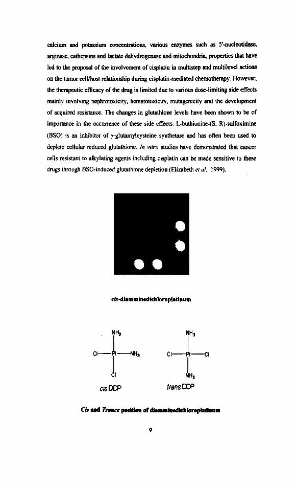
dc ium and potassium concentrations. various eaymes such as 5'-nwleatib,
arginase, catbepsins and lacme dchydrogenase and mitochondria, propaties that have
led to the proposal of the involwment of cisplatin in multistep and multilevel actions
on the tumor ccll/host relationship during cisplatin-mediated chemotherapy. HOWVM,
the therapeutic efficacy of the drug is limited due la various dose-limiting side effects
mainly involving nephratoxicity, hematotoxicity. mutrrgmicily and the development
of acquired resistance. The changes in glutathione levcls have bcen shown to be of
importance in the o c c ~ ~ ~ n c c of these side effects. L-buthionine-ts, R)-sulfoximine
(BSO) is an inhibitor of y-glutamylcysteinc synthetasc and has ofim been used to
deplete cellular reduced glutathione. In virro studies have demonmtcd that cancer
cells resistant to alkylating agents including cisplatin can be made sensitive to these
drugs through BSO-induced glutathione depletion (Elirabeth cr d., 1999).

Systematic name: cis-di ammine dichloro platinum (11)
Molecuhr Ibrmufr: Cp Hg N2 Pt
Molecuhr weight: 300.1
Colac Deep yellow (crystalline solid) and Clear (reconstituted solution)
Melting point: 270' (decomposes)
Cis- Diamminedichlomplatin (11) {cisplatin) is one of the most eIficlive oncoytic
agents against cancers of the ovaries, bladdrt,, und hend and neck. It is also an
important adjunct for cancers of cervix, lung and breast. It is most spcctwular that
success has been in the treatment of testicular cancer, a form of cnnccr previously
resistant to any therapy but now considered to be curable in most cascs ( B ~ h n ef ul..
1991 ).
Dircovety: Cisplatin was first synthesized by Peyrone in 1844 and has been called
Peyrone's chloride. Its structure was first elucidated by Alfred Werner in 1893. The
compound then enjoyed several decades of relative obscurity. In the early 1960'5, a
series of experiments in the laboratories of Barnett Rosenberg at the Michigan Statc
llniversity found some peculiar results. An experiment designed to measure the effect
of electrical cumnts on cell growth yielded i.:schcrichlu coll that werc 300 timca the
normal length. This effect was not due to the electrical fields themselves but to a
chemical agent that was formed in a reaction between the suppsedly inert platinum
electrodes and components of the solution. The chemical agent was later determined
to be cisplatin. Further tests revealed the compound had prev~nted cell division, but
nor other growth processes in the bacteria, leading to the elonyntion, lhis effect
prompted Bamen's group to test cisplatin against tumors in mice. It was found to be
highly effective in eliminating tumors. Human trials produced positive results,
limited, to some extent by toxic side effects. Once the side effects could be made
bearable through the k e of adjuvant therapies, the compound's effectiveness was
proven. It was approved fot use in 1978 (Jamieson el a/., 1999).
Therupeutk eclhrfiy: Cisplatin is widely prescribed for a variety of tumors (genn-cell,
advanced bladder minoma, adred cortex carcinoma, bnasl cancer, head and neck
minoma, lung cankma). It is administad imvcnously for ont to 5 days in a row, followed by a rest period of 2 3 weeks. There arc seriow side tfi?scur pssociawl
witb cispietin, notably rand toxicity, cmcris, newtoxicity, bone msrraw nypnssion
10
and hearing loss. Damage to the kidneys can be minimized through the administration
of continuous IV hydration along with diuretic drugs before and following the
infusion of cisplatin. Similarly, several effective antiemetic drugs protect the patient
from the worst of nausea and vomiting. Testing of patient renal function, blood and
hearing is recommended before each cycle of therapy.
Mechanism of action: Cisplatin is believed to kill cancer cells by binding to DNA
and interfering with its repair mechanism, eventually leading to cell death. The first
step in the process (after the cisplatin molecule penetrates the cell membrane intact) is
for a molecule of water to replace one of the chloride ions. The resulting structure can
then bind to single nitrogen on a DNA nucleotide. Then, the second chloride is
replaced by another 1-120 molecule md the platinum binds to a second nucleotide.
Binding studies of cisplatin with DNA have indicated a preference for nitrogen 7 on
two adjacent guanines on the same strand. It also binds to adenine across strands to a
lesser extent. The cisplatin-DNA complex attracts the attention of I-IMG (high
mobility group)-1 and other DNA repair proteins which become irreversibly bound.
The resulting distortion to the shape of the DNA prevents effective repair (Trans
ivorner of cisplatin is unable to form 1, 2 intrastrand links and lacks antineoplastic
activity). Other antineoplastic agents, such as etoposide, contribute to the platinum-
DNA-protein complex and thus synergistically reinforce the activity of cisplatin
(Elirabeth et ai., 1999).
1.5 Approaches to the discovery of plant derived drugs for cancer: Plants have
been in use for treating various ailments fmm the pre historic times and continue to be
the source of more than 25% of the present range of prescribed drugs. In the
indigenous medicine, all over the world, for 2000 years, plants have been used against
many kinds of cancers. The earliest record of herbal treatment can be traced to ancient
Chinese and Greek texts, (Evans, 1989). Unani and Aywedic systems also used a
large number of plants for the treatment of cancer.
The National cancer institute of the USA began an organized program to
scm anticancerous agents in 1955 with the establishment of Cancer Chemotherapy
National Center (CCNSC). Though, initially, most mataials scnened were pure
compounds of synthetic origin, natural products (of both plant and animal origin)
I I
were also included. Plant extracts from IS5 1 genera and 3394 specits wcrc scnemd;
of these, 4897 plant extracts (4.3%) showed confirmed antineoplastic activity
(Sufhress and Doums, 1979). The screening program of CCNSC brought to light
hundreds of plant species which were used to mt cancer in any system of medicine.
Discovery of an antiturnour drug from plant material is complex one. The
major steps followed for the development of antitumor agents are:
I. Initinl collection and identification of plants,
2. Preparation of plant extracts for screening.
3. Screening of plant extracts for anti (tunor activity.
4. Fractionation studies leading to isolution of cunrpounds.
5 . Characterization of new active compounds.
6. Tumor panel testing.
7. Large scale procurement and production.
8. Formulation and technology.
9. Clinical trials.
The phytochemical examination of plants which have a history of use in
folklore medicine, for the traditional medicine, for the treatment of cancer has oflcn
resulted in the isolation of the principles with antitumor activity, By adopting the
above steps, in a normal course it takes at least 7 years from the lime plant extract is
found active until the pure compound is made available for clinical trials.
Some of the weif ncogniud ~nt&ancer plan&: ' f ie most successful plan1 material
used in cancer chemotherapy is the alkaloids of Cathutanthus roueur. Tnc isolation of
vinca alkaloih - vincristiae and vinblastin and their use in cancer thcrapy was
lightened during their preliminary study as possible an t id iWc ugente ( J o b n el
ul., 1994). The c l i i d succtss of vincristinc has qualified the compound as "Miracle
drug" (Taylor, 1968). tong term disease free survival has bem observod in the
tnstment of various lymphoma and Icukamies, bladder CM*M and testicular cancer,
while significant palliative M t a hove been ~ e e n in patients with bnard ~wcer,
mdamma end anail cell lung cwca (Neuiw el al., 1964). Vinblartinc sml vincristinc
12
are antimitotic and they produce atypical C-mitotic micronuclei (Decorti and Creacy,
1975).
They have been incorporated into combination therapy protocols to treat
Hodgkin's Lymphoma, non-Hodgkins lymphoma, and neuroblastorna. A typical
combination of chemotherapeutic regimens is MOPP (Nitrogen Mustard, Vincristine,
Procarbazine and Prednisone) which has achieved complete responses in more than
70% of patients with Hodgkin's disease. The greatest success in treatment of leukemia
has been obtained by using VAMP (Vincristine, Amethopterin, 6-Mercapyopurine
and Prednisone) (Henderson and Sarnaha, 1969). There is still great interest in the
synthesis of analogs of these agents and new synthetic derivatives such as Vindesine,
Vizolidine and Vinorelbine have been introduced into cancer therapy (Beck et al.,
1995).
Podophyllum hexandrum and Podophyllum peltarum are the two important
species used by ancient Chinese as antitumor drugs and interest in podophyllum was
revived in 1940's by the observation of antimitotic activity and inhibitory effect on
experimental tumors in animals (Belkin, 1948). Two of the semisynthetic derivatives,
etoposide and teniposide of Epipodophyllo toxin, isolated From Podophyllum peltatum
showed clinical use as anticancer agents. These derivatives will act by inhibiting
microtubule assembly and by inhibiting DNA topoisomerase I1 (Thurston et al.,
1989). Etoposide shows clinical activity against smaH cell lung cancer, testicular
cancer as well as lymphomas and leukemias (O'Dwyer el al., 1985). Teniposide is
active against acute lyrnphocytic leukemia, neuroblastoma in children, non-Hodgkins
lymphoma and brain tumors in adults (O'Dwyer et al., 1984).
L.'olchicum awumnale is a rich source of colchicine- 12 which was capable of
producing regression and cytolysis in tumors (Amoroso, 1935). The antitumor activity
of colchicine-12 is a result of its interaction with tubulin, which aggregates to form
the microtubules with M mitotic spindle (Dumont el al., 1987). Colchicine-12 and
Democolcine-13 are used for the treatment of solid tumors and for certain forms of
leukaemia (Rosner et a/. , 198 I).
Taxol, a novel diterpenoid, became one of rhe most promising anticancetous
agents (Wani el al., 1971). It was isolated from Pacific yew, Tmur brev~olia Nun.
The m e b i s m of action of Taxol is thar, it promotes Nbulin polymerization and

stabilizes microtubuks against &cpolymcrization (Landino and Mc Donaid 1995),
Significant e&ct of this drug has been d c m o d on refracting ovarian carcinoma,
breast m i n o m a melanomas and leukemia's (McGuir. 1989).
Camptothecin, an alkaloid derived from Camptorheca acuminara was shorn
to be potent inhibitor of DNA synthesis. Clinical studies in China have shown
responses among patients with liver. gastric. head nnd neck nnd bladder cancers.
Soluble.derivadves of Camptothecin with supcrior anticmcer activity and miuced
toxicity-CPT-1 l (Ohno er al.. 1990) has been used against refractory leukemia and
lymphoma. topatecan against ovarian and lung cancer (Kinysbury er crl., 1991).
Combretastatin A-4 isolated from C'omhretum cqfirrm, n potent inhibitor of
microtuhular action has shown activity against vnrious lruman colon cancer cell lines
(Pettit, 1991). Haningtone and homoharringtone isolated from C'rphalotaxuv
hurringtonia has shown activity against leukemia's hy Chinese authors and was
confirmed by phase 11 clinical trials in lJSA (O'Dwyer el al., 1986). 4-lpomeanol
produced by sweet potatoes infected by fungus Furarium solani has shown to exen
cytotoxicity to human lung cancer cell lines (Falzon clr ol., 1986). Pancmtistatin
isolated from the bulbs of Pancrarium Iirlorttle showed significant in vitro activity
against breast, lung, melanoma and stomach cell lines. as well as the M5076 ovarian
m o m a in mice (Pettit, 1991). Phyllanthoside isolated from the Central American
tree Phylla~htl~ acumimtus exhibited cytotoxicity against tumor cell lincs at low
concentrations (Gordon and Boyd, 1994). Withania somnfiru (Ashwagandha) which
is used for the treatment of a variety of diseases has antitumor nnd radio sensitizing
properties when tested on experimental tumors (Uma Devi er ul., 1993; S h a d a el al.,
1996). The antitumor activity of Plwnabagin, derived from another medicinal plant
Plumbago rosea has been reported by Krishnaswamy and Purushottam (1980). A list
of plant derived compounds and the types of tumor models againsr which they are
active are summarized below:
Jatroptrone P388 lymphocytic leukemia
Triptolide P388 lympkqlk leukemia and L12 10 lymphaid leukemia

Besides these active principles, a number of compounds such as cucurbitacins,
csrotenoids, sesquiterpene - lactones, cardiac glycosides, lipins, alkaloids,
quassinoids, steroid lactones, maytensine have shown significant antitumor activity
against many in vitro models (Suffness and Douros, 1979). The plant kingdom is a
potential source of new chemical compounds. The chemical syntheses of most of the
pure plant derived compounds used in the treatment are either technically or
economically infeasible and hence most of them are still extracted from plants.
Dietary and endogenous antioxidants prevent cellular damage by reacting with
and eliminating oxidizing free radicals. However, in cancer treatment, a mode of
action of certain chemotherapeutic agents involves the generation of free radicals to
cause cellular damage and necrosis of malignant cells. So a concern has logically
developed as to whether exogenous antioxidant compounds taken concurrently during
chemotherapy could reduce the beneficial effect of chemotherapy on malignant cells.
The importance of this concern is underlined by a recent study which estimates 23%
of cancer patients take antioxidants (VandeCieek et a/., 1999). The study of
antioxidant use in cancer treatment is a rapidly evolving area. Antioxidants have been
extensively studied for their ability to prevent cancer in humans (Singh et al., 1998).
Antioxidants are useful as a therapeutic intervention in cancer due to their potential
interactions with radiation and chemotherapy. There has been significant investigation
of this area, with promising findings which indicate continuing investigation is
w m t e d . l'he use of antioxidants as sole cancer therapy was reviewed by Prasad et
ul., (1 999).
Status of Herbal medicine against cancer: Plants have provided mankind with a
large variety of potent drugs to alleviate sufferings from diseases. In spite of
spectacular advances in synthetic drugs, in recent years, many drugs of plant origin
have regained importance. In fact the use of plant-based drugs in the western world is
increasing. Though the worldwide importance of plant derived pharmaceuticals is not
updated and is unpublished. according to earlier surveys, 3000 million dollars worth
of prescriptions containing drugs fiom plants was sold in USA in 1974, about 25% of
prescriptions extracted from plant sources. About SO0! of the prescriptions in West
Oermany and a large prescription in U.K are plant-derived drugs.
Cancer is a major cause of concern for public health service in dewloping
well as industrializsd countries. The world bealth organization reports a steady
increase in the incidence of cancer, with the developing countries accounting for
approximately 2.3 million new cancer cases. Each year ~ l t e mortality due to c a ~ w in
developed countries is ranked second. where as in developing countries it is ranked
fifth, considering the overall world population. The stated goal of the National Cancer
Institute was 50% reduction in the cancer related mor(ality by the year 2000 (Lopez el
al., 19%) and this may have been particularly achieved by the combined efforts of
various international organizations. This is substantiated by the WtIO report of 1998.
which shows that of the 50 million deaths estimated world-wide in 1997, ahout 12 %
were due to cancer.
War against cancer is an international effort which has becn on the fox front
viz. cancer prevention, early detection. regional cancer cure and systemic cancer
control. The potential of gain diminishes with successive category. ('ancer therapy is
ideally expecled to eliminate all cancer cells wit11 no, or minimal injury to the normal
tissues. But none of the therapeutic measures in use today has ken able to achieve
this goal. However, most cancers can be ~uccessfully treated if detected early and
most often failure of canccr theropy is due to delay in diagnosis (Connors and Roe,
1961).
The most commonly employed methods of cancer treatment are: 1. Surgery, 2.
Radiotherapy and 3, Chemotherapy. Resides these, hormone therapy and
immunotherapy are viewed as potential methods for the treatment of cancer and are
finding a significant place in the therapeutic armamentariurn. Toby, most of the
malignant neoplasms are managed by one or more of these modalities, with varying
success rates depending on the type of tumor, its degree of differentiation and spread,
as also with the knowledge and skill with which the treatment plan has has designed
and executed (&ck el al., 1995).
Chcmo* is now employed in the primary tmtmnt of cancer such as
C b o r i d m a , @ate and chronic leukemia, Burkitt'r lymphoma and mulliple
myeloma. It is used as an adjuvant in the tmmcnt of bnajt cancer, Wilm's tumor,
embryonal rbabdomyosatcwra, ogtsogaric sarcoma and malignant melanoma lt ia
also used as an impmnl modslity in du m~~ganmt of many ncqdaans including

Hodgkin's digease and other malignant lymphomas, sarcomas, nelrroblastomas and
cencer of the gastrointestinal and upper respiratory tracts, ovary, prostate, testis,
breast and uterus. Apparent cures have been achieved with chemotherapy in patients
with some forms of cancer and other patients have benefited in terms of palliation of
symptoms and extension of life (Henderson and Sarnaha, 1969).
Cancer chemotherapeutic agents can often provide temporary relief of
symptoms with occasional cures. However, the effective doses of most of the agents
also fall in the range of toxic dose. They are highly reactive and are capable of
inducing varying degrees of cell destruction and this leads to unpleasant side effects
while undergoing treatment. The complications of chemotherapy can be of three types
- early, intermediate and late. Early (hrs) include nausea, vomiting, fever and
hypersensitivity reactions, intermediate (days) include stomatitis, diarrhea, alopecia,
peripheral neuropathy and bone marrow depression. Late (a few months) is
manifested as injury to vital organs, endocrinal changes, teratogenic effects and
psychological effects (Devlen et ul., 1987). Henke the search for a new anti tumor
agent with high chemotherapeutic value to fight against cancer is obviously a medical
priority. The given anti tumor agent must be able to kill or inactivate tumor cells
without damaging normal tissues.
1.6 Conflicting views of antioxidant use in cancer therapy: It was suggested in a
recent publication that no supplementary antioxidants be given concurrently with
chemotherapy agents, which employ a free radical mechanism (Labriola and
Livingston. 1999). Combination of antioxidants and chemotherapy agents needs more
investigation, and should serve as a wake-up call regarding how much we need further
definition of the actions of specific antioxidants with chemotherapeutic agents.
However, it should not serve as scientific closure on on adjunctive treatment of
possible great promise in cancer therapy. It was suggested that antioxidants might
interfere with the oxi,dative mechanisms of alkylating agents (Labriola and
Livingston, 1999). These drugs create substantial DNA damage, resulting in cell
necrosis. However, recent evidence indicates a sizeable amount of chemotherapy
damage is by other mechanisms, which t r i w apoptosis (Schmitt and Lowe, 1999).
Antioxidants have bten shown to increase cell death by this mechanism (SchmiU and
Lowe, 1999; Chinery el d , 1997). Oiven this, any argument that mtioxidants an

likely to intmkrc with most cbemo-y is too simplistic and probably untrue.
Nummus animal studis have been published demonstrating d t c d tumor sia
and/or i n c d longevity with the combination of chemotherapy and antioxidants
(Berry el al., 1984). A recent study was condwctcd on small-cell lung cancer in
humans using combination chemotherapy of cyclophospharnide. adriamycinrn
(doxorubicin), and vincristine with radiation and a combination of antioxidanls.
vitamins, trace elements, and fatty acids. 'he conclusion was "antioxidant treatment,
in comdination with chemotherapy and irradiation, prolonged the survival timc of
patients" conipared to expected outcome without the composite oral therapy (Jaakkoln
ef al.. 1992). Two human studies found melatonin plus chemothcrapy to induce
greater tumor response than chemotherapy alone (1,issoni i t / a/., 1995; 1.issoni CI al.,
1997). The treatments producing t h e positive msults would have bcen advised
against by those advocating no antioxidant use during chemothcrapy. 'I'tie fact
remains that physicians must he aware of the nvailablc ~ s e a r c h to help thcir patients
take advantage of positive interactions existing betwccn antioxidants and
chemotherapy or radiation. Additionally, physicians nccd to remain aware of the luge
body of evidence showing a positive effect of antioxidants in the period following
chemotherapy administration. 'fie general protoccll with standard oncc)logic thcrapics
is to follow a watch and wait strategy aHcr therapeutic administration is concluded.
This is a period when supplemental therapies are highly indicated and have been
demonstrated to result in a higher percentage of successful outcomes (Whelan er ul.,
1999; Lamm el al.. 1994).
Potentid mechanirms of antloxldants in cancer therapy: How fould antioxidant
therapy protect normal cells against damage from cancer therapies, while o h
increasing their cytoloxic effect against malignant cells? Though there is no suitublc
answer for further, there are concepts which might help us understand. Thcrc is
evidence (Schmin and Lowe, 1999) that radiation and chemotherapy often harm DNA
to a relatively minor extent, which cawrcs the cells to undergo apoptosis. ralhcr than
m s i s . Since many antioxidant treatments stimulate apoptotic pathways (Chinery el
al., 1W; McdiaviIla el al., 1999) the potential exists for a synergistic effwt with
radiation or chemotherapy with antioxidnnts. A second concept is that the defcnsivc
macbaniaru of many canca cells an known to be impaired (&nsde et d., 1969).
This pmumbly makes h u ~ w cdls unable to use du: extra antioxidants in a repair
capacity; this has been illustrated (Oberley and Oberley, 1997) m viiro. An
experimental murine ascites tumor cell line was found to have 10-100 times less
cataiase than normal cells. This led to a build-up of hydrogen peroxide in the cells
upon treatment with vitamin C, in turn leading to cell death. The cytotoxic effects of
vitamin C were completely eliminated by addition of catalase to the cell culture.
Glutathione as cancer treatment: Glutathione is a tri-peptide thiol (sulfhydryl-
containing) compound which is the major intracellular antioxidant in the body. A
study in human suggests that oral glutathione is poorly absorbed, with negligible
plasma concentrations found after administration of a single 3g oral dose (Witschi et
ul., 1992). This conclusion is contradicted by a study in rats, in which it was found
that dietary ylutathione was absorbed in a dose-dependent manner and remained
elevated in the plasma for three hours after administration (Hagen et al., 1990).
Aerosol administration of glutathione is an effective means of delivery to the plasma,
as is intravenous administration. Glutathione is thought to be non-toxic to humans,
although one study found a Sg oral daily dose was associated with Gastro-intestinal
(01) irritation and sulfur odor (tiospers el of., 1999). A trial of six hepato carcinoma
patients on 5g oral glutathione daily found regression or stagnation of tumor growth
in three patients. One patient also had a reduction in alpha-fetoprotein (a. tumor
marker). Two patients of the six survived for one year. These patients were both
women, raising the possibility of n sex-dependent effect. In a study, (Novi, 1981) oral
administration of glutathione in rats caused regression of liver tumors, and increased
survival of tumor-bearing animals. The usefulness of glutathione as an anti-tumor
agent may be limited to the liver, kidney, and peripheral neurons. as these are the only
tissues believed to have sufficient transport enzymes for celldar uptake (Kidd 1997).
Gluruthlonc with rudiation: A randomized pilot trial (De Maria el al., 1992) with 45
participants investigated the d i o protective effect of glutathione. Patients were
administered 1200mg glutathione or saline placebo intravenously IS minutes prior to
pelvic radiotherapy. Patients receiving glutnthione suffered less from post-therapy
diarrhea (28%, compared to 52% of controls) and were more likely to complete the
treatment cycle (71% to 52%). Although the sample size was too small to sbow
significance, the authon concluded glutathione was unlikely to intafkm with the
effect of radiation on neoplasms @e Maria et dl 1992). Ttae tugmeat was not based
on patient outcome.
19
G I m h h e wlitL &emu&-. I n c W cellular concentrations of g l d o m
havc bcen associated with resistam to both anthnryclines and platinum agnt~.
Given the suggestion of the inability of most cell types to take up exogenous
glutathione (Kidd 1997). d o c d chernothuapy efficacy due to glutathione
administration may be limited to liver, kidney. and neurological tumors. The use of
cisplatin and glutathionc concurrently has been studied in several small human trials.
One human trial (Bogliun er a/.. 1996; Cascinu CI al., 1995) found 3ylml intravenous
glutathione given 20 minutes prior to cisplatin (100mg/ml) led to a significant
reduction in nephro toxicity in patients with ovnrian cancer compared to those
receiving cisplatin alone. There was a trend towards grater tumor nsponsc in the
glutathione gmup (73%) compared to the control gmup (62%). A similar trial using
smaller doses of glutathione (2500mdml) and cisplntin (50-75mdml) did not find the
reduction in nephrotoxicity as repaned nbove. tlowvever, the trend toward grcntcr
tumor response with glutathione treatment (72% response, cornpad to 52% in
controls) was compnmble.
A double blind trial studied the neum protective effect of intravenous
glutathione (1500 mg/m2) during cisplatin treatment for gastric cancer. APter nine
weeks, no patient of the 24 receiving glutathionc, but I6 of 18 patients w i v i n g
placebo, had developed neuropathy symptoms. Again, a trend towards grwltcr tumor
response (76%. compared to 52% in controls) was secn with glutathione treatment
(Cascinu el al.. 1995).
FIownoids as cancer treatmeal: Flavonoids arc plant compounds known to havc
antioxidant properties in vitro and in viw (Bushman el al., 1998). Many of the
thousands of flavonoids in nature have been studied for anticancer properties. The
most well characterized anti-tumor flavonoids arc epigallocatechin gallate (from
g m tea), genistein (from soy and red clover), cwcumin (from turmeric), silibinin
(hom milk thistle), and qwcctin (fn>m many yellow vegetables). Several n u t h
have recently p q a r d reviews on the use of qucrcetin as canocr therapy (Li et uf.,
1999, M i d i ef al., 1999).
FCcrwrroIdr WM rd&&#: Little is lrnown about the e f f w of flavorwids on
radiodvrapy. An in vho expetiment AomA pm-traraacnt application of qucrcctin
causrd lprattr ccU death in ladistioMrrsted bepaloma cells than radiation alone (van

S j n and van den Berg, 1997). In the same experiment, g&in was shown to be
associated with increased cell death from radiation when applied during or after
mtment. Many different mtosides (flavonoids with similar structum to quercetin)
were found to have neither a protective nor sensitizing effect on radiotherapy in
experimental mouse tumors. There is not enough evidence currently to support or
argue against the use of therapeutic doses of flavonoids together with radiation (Fritz-
Niggli and Rao, 1977).
Flavonoids with chemotherapy: Recent research has focused on the ability of
flavonoids to increase the concentration of chemotherapeutics in tumor cells.
Resistance due to many chemotherapy agents is thought to be due to reduced
accumulation in tumor cells (Robert, 1999). Oral administration of green tea in mice
increased the concentration of doxorubicin in two tumor types, but not in normal
tissue. The anti-tumor activity of doxorubicin was enhanced 2.5 times (Sadzuka et al.,
1998). Another report confirmed this action of green tea, finding that the tumor
inhibition of doxorubicin increased from negligible to 62 percent. This report,
however, determined that the activity of green tea could be due to an amino acid,
theanine, rather than its flavonoid content (Sugiyarna and Sadzuka, 1998). Quercetin
has been shown in vitro to increase the concentration of doxorubicin in multidrug-
resistant human breast cancer cells. Conversely, quercetin decreased the concentration
of doxombicin in a resistant human colon cancer cell line (Critchfield el al., 1997).
Quercetin and genistein both increased the concentration of daunombicin in some
multidrug-resistant cell lines, but had no effect in others (Versantvoort et al., 1993).
Genistein In vitro increased the concentration of cisplatin in resistant cell lines
(Marverti and Andrew, 1996). Other than the green tea studies, none of the studies
analyzed the cell death due to flavonaid administration. In mice with transplanted
human tumors, quercetin (20mglkg) given with cisplatin reduced tumor growth to a
greater degree than cisplatin alone (Hohann er al., 1990). In a separate experiment,
quercetin enhanced the effect of cisplatin and busulfan in vitro and in vivo. No
cnhanccment or reduction of the anti-tumor activity of doxorubicin or etoposide was
seur (Scambia el al., 1992). In in vitro study it was found that quercetin increased the
effect of doxorubicin against resistant breast cancer cells. It should be cautioned,
howcvu, that a ncent study (Bracke et al., 1999) showed a potential adverse

intgection. Tangcretin, a flavonoid fowd in c i m fiuits, completely blocked the
inhibitory effect of tamoxifen on mammary cancer in mice. '
Cumbinatiom of antiad8~ts: Given that many antioxidants have been shown to
have anti-tumor properties, it is worth exploring their use in combination. A study in
mice found that co-administration of betacarotene and alpha-tocopheml led to much
greater tumor regression than either agent alone. 'The effwt was synergistic, king
much greater than the sum of the mild tumor inhibition of hcln-cwotene and alphn-
tocopherol (Shklar, el al., 1989). Other studies have shown that multivitamin
supplements were associated with fewer rrmrtvnces of solid tumors d e r remission
following standard oncologic therapies (Whelan ct crl., 1999: h m ct al., 1994). A
small double-blind trial of a mixture of antioxidants, including 600 mp vitamin E. Ig
vitamin C, and ZOOmg NAC taken only during treatment, looked at Lhe potential of
this mixture to prevent cardio toxicity during chcmo nnd radiotherapy. No pntienr
taking the antioxidant mixture had a fall in ejection fraction greater thnn 10 percent.
In patients taking placebo, four of six patients undergoing radiotheropy and two of
seven patients treated with chemotherapy had nn ejection fraction reduction of 10
percent or more. Treatment outcomes in patients laking antioxidants v c m placebo
were not discussed (Bracke et al., 1999). An open trial of combination antioxidant
treatment along with chemotherapy and radiation in patients with small-cell lung
cancer had encouraging results. Patients laking the supplement, which contained at
least 15,000 IU vitnmin A, 10,000 IU beta-carotcnc, 300 I U alpha-tocopheml. 2g
vitamin C and 800pg selenium, were able to tolerate chemotherapy and radiation well.
Their two-year survival rate was greater than that of historical controls ( ~ 3 3 % to
<IS%), with 44 percent still alive at the end of the study (mean survival lime for
survivors = 32 months). No side effects from nutritional treatment were noted
(Jaakkola el al,, 1992). .
1.7 Brrteu munmpmno (Lorn): It is commonly known as !lame of fomt and belongs
to the family Fabaceat (Putil, a al., 2006). It is I d l y called aa palas, Pal&,
Mutthug, Bijasneha, Dbalr, KWam, Chichra, Bastard Teak, Bengal Kino and
Nourouc and is commody available throughout India, Bwna and Ceylon except in
very arid pans. Gcnnally it gpows gngariowly on open grasslands and scannod in
mixed forest. Plantations can k r a i d both on irrigated and dry lands. The pods
sbould be oollectcd ad sown beforc the -1 of rains, root suckera arc
freely produced and help in vegetative pn,paga!ion. In Wa, palas ranks next to
kusum (Schleickm trijuga) as a host t r e ~ for lac insect (Kspoor, 2005). Almost all
the purts of the plant are being lLced since decades in maticine and for other p v .
Burea monosperma (h) kuntze is one among fwr spccics belonging to the genus
Bulea brig, three species of which accur in I d a (Ihe Wealth of India, 1988). It
holds an important place because of its medicinal and other misocllantous uses of
economic value. Bark. fibers arc obtained from stem for d i n g cordage (mpc and
string). Stem bark (Fig 1.3) powder is used to stupcfy fishes. Young roots are usad for
making ropes. G m leaves are good fodder for domestic animals. 1,cavcs arc d
for making platters, cups, bowls and becdi wrappers (Ambasta, 1994). Lcsvcs are also
used for making Ghongda to protect from rains and an aten by bufhlocs and
elephants. Tribals use flowers and young fruits as vegetables. Flowers are boilcd in
water to obtain a dye. Orange or red dye is used for wlouring garments and for
making skin antiseptic ointments (Knstw el a/., 2002). Frcsh twigs an tied on horns
of bullocks, on occasion of 'pola' and dry twigs an uscd to fwd the s a c d fire.
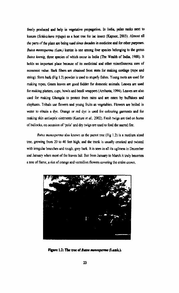
Butea monosperma also known ar the parrot trce (Fig 1.2) is a medium siuxl
w e , growing from 20 to 40 feet high, and the trunk is usually crooked and twisted
with irregular branches and rough, grey hark. It is som in all its ugliness in December
and January when most of the leaves fdl. But fram January to March it truly bocornea
a tree of flame, a riot of orange and vermilion flowers covcring the entire crown.
Rgam 13: Tk tree of Bvtar rnorurqmm~ (Lurk).
Figure 13: B ~ l ~ l l monmpem (Lmk) rkm bark.
PhytuchemIrq: 'he phytochemical constituents of B. monospermu are given helow.
Flower: Contains l'riterpene, butein, hutin, isuhutrin, compsin, iuocompsin (hutin
7-ylucoside), sulphurein, monospennoside (butrin 3-e-D-ylucoside) and isomom)
spermoside, chalcones, aurones, flavonoids (palasitrin, prunctin) and steroids
(Lavhale and Mishra, 2007; Gupta el al., 1970).
Gum: Contains of Tannins, mucilaginous material and pyracatcchin (Burlia and
Khadeb, 2007).
Snd: Contains Oil (yellow. tasteless), proteolytic and lypolytic cnzymea, plant
protcinase and polypeptidase (Similar to yeas1 trypsin), A nitrogenous acidic
compound, along with palaponin is presmt in s e d (Gupta et d, 1970). It elw
contains mono spermoside (butein 3-c-D-glumside) and isornono spemroside. F m
d coat allophanic acid has bcen isolated and identified (Jawaharlal el al., 1978;
Rastogi and Mehmtra, 1979). An irnide fium rhe pod was isoM (Chiha el a1 1990),
R a k Comairw of JMc CSICIS I, 11 and laccijalaiic esters 111. IV; 2- mytin, C-
s i m i a glucoside ad arro~e; lactone-nheneicosanoic wid-(-lactone (Singh et
al.. 1974; Nsdkarni, 2002).
Sup: Contains Chalconcs, butcih, butin, coiourless isomeric flavanom and its
glumides butrin (The Wealth of India 1988).
Lcorvs: Contains Glumside, Kino-oil containing oleic and linoleic acid, palmitic and
lignoceric acid (Nadkami, 2002).
Stem bork: Kino-tannic acid, gallic acid. pyrocalcchin (Shah el al., 1 W2) arc present
in stem bark. The plant also contains polmirrin. and major glycosides as butrin,
danind, allophanic acid, butolic acid, cyanidin, histidine, lupcnonc, lupcwl, I-) medicarpin, mimstrol, palasimidr and shellolic acid (Madhav ef a/., 1967; Porwal el
al., 1988; Bandara ef ol., 1990).
Biological activity: Mishra el ol., (2000) evaluated frrx rndical scavenging activity of
various extracts of flowers of Bu~ea monosprma in vifro such as rcduciny power
assay, scavenging of 2.2 diphenyl-I- picryl hydrnzyl (DPPH) radical, nitric oxide
radical, super oxide anion radical, hydmxyl radical and inhibition of erythrocytes
hemolysis using 2,2' wo-bis (midinopropane) dihydmhloride-AAPl.1). Mcthanolic
extract along with its ethyl acetote d butanol fructions showed potent frcc radical
scavenging activity (Iavhalc and Mishra, 2007). 'lhc obscrvcd activity could bc due
to higher phenolic contents in the extracts.
Rhatwadekar el ul., (1999) rcported anti strcss activity of flowcrs of Butea
monosperma. Water soluble part of ethanolic extract attenuated water immersion
stress and increased brain serotonin and plasma corticosteronc levels. 'The ulcer index
was also decreased in dose dependent manner. Thc observed effects may be attributed
to its nonspecific anti-stress activity. Kasture el al., (2002) r c p o d anticonvulsivc
activity of flowers. The anticonvulsive principle was found to be a triterpenc present
in the n-hexane: ethyl acetak (1 : I ) fraction of the petroleum eiher extract. Tritcrpcne
exhibited enticonvulsant activity against seizures induced by Maximum Eloctm Shock
(MES). It also inhibited seizures induced by pentylem tctnuole, electrical kindling
and the combition of lithium sulphatt and pilowpine nitrate.
Wagner e! d, (1986) n?portad Isobutrin and Butrin, the antiheparotoxic
prkiples of flowas. Activity was monitored by means of CClr and GaIN-induced
liva lesion in vtbo. The dhqmmoxic prlrrciplu isolsied m i s t a d of two known

flavonoids, isobutrin (3,4,2', 4'-tetrahydroxychalcone-3,4'- diglucoside), and the less
active butrin (7,3', 4'-trihydroxyflavanone-7,3'diglucoside).
Gawale e! al., (2001) reported effect of flowers in memory and behaviour
mediated via mono amine neuro-transmitten. The acetone soluble part of petroleum
ether and ethanolic extract exhibited nootropic activity in the elevated plus maze
paradigm and active avoidance learning
Mishra et ul., (2000) reported the presence of flavonoids in ethyl acetate
fraction of methanol extractives. Shah el ul., (1990) reported phytochemical studies
and antiestrogenic activity of flowers. Phytochemical investigations of the dried
flowers of Buteafrondosu Roxb revealed the presence of at least seven flavones and
flavonoid constituents including butrin and isobutrin and also four free amino acids.
Purified alcoholic extract at lower dose level and ethereal and water extracts at higher
dose level have been found to exhibit significant antiestrogenic activity in immature
mice, while ethyl acetate extract containing butrin and isobutrin exhibited poor
activity (Shah el ul., 1990). I'here was a significant inhibition of uterus weight gain,
va~inal epithelium cornification and characteristic histological changes.
Gupta el ul., (1970) camed out a reinvestigation of the flowers of B u m
monospermu and revealed the presence of seven flavonoid glucosides. Two of them
are butrin and isobutrin, which have been isolated earlier from the plant. Three
ylucosides have been identified as coreapsin, isocoreopsin and sulphurein. The
remaining two are new and have been assigned the structures monospermoside and
isomonospermoside. Shah rr ul., (1992) isolated and identified free sugars and free
amino acids fmrn the petroleum ether e x h c t of flowers.
See&: Powdered seeds are consumed by children as remedy against intestinal worms
(Kirtikar and b u , 1991).Seeds are crushed in milk and this mixture about 2 spoons
is taken orally to treat urinal compldnts and also against urinary stones. Fruit and
seed an digestible, aperient, cure 'Vata' and 'Kapha', skin diseases, turnours,
abdominal troubles and are given for Scorpion-sting as per Ayur~eda Fmit and seed
are useful in piles, eye diseases and inflammation. When pounded with lemon juice
md applied seeds act as powerlid mbefacient and they have been SINXYSSI!~~ ustd in
curing a form of herpes, h w s as Dbobie's itch (Patil et al., 2006). Prashan! el al.,
(2001) n p o d in vim antihelmintic activity of melfianol exbact of scad. Pandey ef
d., (2001)-reported the use of seed oil as traditional sexual toncr and contmwptive.
Singh el d., (1974) reported components of SOA nsin. They isolated four
essentially pure acid esters, which together constitute the bulk of soft rcsin. They
termed these acid m e n , jalaric ester-I, jalaric ester-11, laccijalaric ester-1 and
laccijalaric ester-11. Bhargava (1986) isolated butin fmm the seed and reported
antiimplantation activity in rat.
Leaves: Leaves are good for the disease of the eye. Leaf is an nppetir~r, astringent,
carminative, anthelmintic, aphrodisiac, tonic, lessens inflammation and lumbago.
cures boils and piles (Patil el (11.. 2006). Petiole is chewed nnd the juice is sucked to
cure cough, cold and stomach disorders. Leaf powder nbout 2 spoons per day for a
month is drunk mixed with a cup of wakr to cure diabctes (Kirtikar and Basu 1935).
Leaf extract is used as gargle in case of sore thmat. I..eaf extract about 3-4 spoons is
drunk at night for 2-3 months. It checks irregular bleeding during menstmation.
Mishra et al.. (2000) reported 3, 9 dimethoxy pterocapan from ethyl acetate fraction
of methanol extractives from leaves and hexanc fraction of methanol extractives
yielded 3-alpha hydroxycuph- 25-enylhcptacosanoate.
Gum: Gum is applied for cracks on foot sole. Two spoons of diluted gum are advised
for dysentery until cwc. Gum is astringent to bowel, good in stomatitis, cough,
pterygium, corneal opacities and cures excessive perspiration (Patil el ul., 2006).
ROO& The root cures nigh1 blindness and other defects of sights, useful in
elephantiasis. Root pieces are heated and then 2-3 spoons of extract are advised at
night as a remedy against impotency and it is administered for one month. Spoonful
of root powder mixed with water is drunk as an antidote for snake bite (Patil ~t a!.,
2006). Bodakhe and Ahuja (2004) reported in virro lens protective and antimicrobial
activity of roots of Butea monosperma.
S&m bark: Stem bark powder of Butea monasperma is used to apply on injury caused
due to axe. Stem juice is applied on goitn of human being. Paste of stem bark ir
applied in case of brdy swellings (Savitri Kumar end Swarna Samaranayskc, 1989).
Bark is acrid, bit-, appetiscr, aphrodisiac, laxative, anthelmiitic, useful in fractum
of the bones, dkascs of the anus, dysentery, piles, hydrocelc, cures u l m and

turnours. Bark is useful in biliousness, dysmenorrhea, liver disorder, gonorrhoea and
it also purifies the blood. The ash of young branch is prescribed in combination with
other drugs in case of scorpion sting (Savitri Kumar and Swarna Samaranayake,
1989).
Antifungal constituents from petroleum and ethyl acetate extracts of stem bark
extract exhibited significant antifungal activity against Candida cladosporioa'es
(Savitri Kumar and Swarna Samaranayake, 1989). Ethanolic extract of stem bark of
Buteu monospermu has antidiarrhoea1 potential in castor oil induced diarrhoea model
(Sharma el al., 2005).PGE2 induced enteropoolingin in rats. Extract also reduced
gastrointestinal motility aAer charcoal meal administration (Sharma el al., 2005).
Suguna et ul., (2005) investigated the effect of alcoholic bark extract on
cutaneous wound healing in rats. Excision wounds were made on the back of rat and
extract was applied topically. 'The granulation tissue formed on days 4, 8, 12 and 16
(post-wound) was used to estimate total collagen hexosaamine, protein, DNA and
uronic acid. Further epithelialization and wound contraction was confirmed by
histopathalogical examination.
In ancient Ayuwcdic literature extensive mention of this plant (stem bark and
flowers of Buleu monosprcrma) is available in the treatment of Krimi Roga (worm
infestations). It enters into the composition of some very important and widely used
recipes of Ayuwedic medicines used in the treatment of Krimi Roga. In Sushruta
samhita this drug has been described under four different groups of herbal medicines
eg. Rudaradigana, Musakadigana, Amabasatadigana and Nyagro dhabigana dealing
with different disorders eg. Medoroga, Striroga, Prameha and also credited with
Kapha and Pittnnasak properties. The first mention of its Krimighna property is
available in Sushruta samhita and the later Ayurvedic authors have also described its
efficacy in Netraroyn and its astringent action in different conditions. In ancient and
later Ayurvcdic literature this drug has been mentioned either alone or as a constituent
of many prepared medicines used in the treabnent of Krimi Roga (Agarwal er al.,
1994).
A clinical trial of the plant in worm infestation proved its effectiveness in
cases of round worm and thrcad wonn infistations and the drug was found to be
ineffective in the only case of tapeworm infestation. A g a r 4 er d , (1994) rcpotted