chapter 1 chapter 4 fluorophores and biotin and their ... · hapten derivatives molecular probes™...
TRANSCRIPT
CHAPTER 4
Biotin and Hapten Derivatives
Molecular Probes™ HandbookA Guide to Fluorescent Probes and Labeling Technologies
11th Edition (2010)
CHAPTER 1
Fluorophores and Their Amine-Reactive Derivatives
The Molecular Probes® HandbookA GUIDE TO FLUORESCENT PROBES AND LABELING TECHNOLOGIES11th Edition (2010)
Molecular Probes® Resources
Molecular Probes® Handbook (online version)Comprehensive guide to �uorescent probes and labeling technologies
lifetechnologies.com/handbook
Fluorescence SpectraViewerIdentify compatible sets of �uorescent dyes and cell structure probes
lifetechnologies.com/spectraviewer
BioProbes® Journal of Cell Biology ApplicationsAward-winning magazine highlighting cell biology products and applications
lifetechnologies.com/bioprobes
Access all Molecular Probes® educational resources at lifetechnologies.com/mpeducate
Molecular Probes ResourcesMolecular Probes Handbook (online version)Comprehensive guide to fl uorescent probes and labeling technologiesthermofi sher.com/handbook
Molecular Probes Fluorescence SpectraViewerIdentify compatible sets of fl uorescent dyes and cell structure probesthermofi sher.com/spectraviewer
BioProbes Journal of Cell Biology ApplicationsAward-winning magazine highlighting cell biology products and applicationsthermofi sher.com/bioprobes
Access all Molecular Probes educational resources at thermofi sher.com/probes
151www.invitrogen.com/probes
The Molecular Probes® Handbook: A Guide to Fluorescent Probes and Labeling TechnologiesIMPORTANT NOTICE: The products described in this manual are covered by one or more Limited Use Label License(s). Please refer to the Appendix on page 971 and Master Product List on page 975. Products are For Research Use Only. Not intended for any animal or human therapeutic or diagnostic use.
FO
UR
CHAPTER 4
Biotin and Hapten Derivatives
4.1 Introduction to Avidin–Biotin and Antibody–Hapten Techniques . . . . . . . . . . . . . . . . . . . . . . 153
Avidin–Biotin and Antibody–Hapten Techniques and Their Applications . . . . . . . . . . . . . . . . . . . . . . . . . . . . . . . . . . . . . . . . . . . . . . . . . . . . . 153
Detection Methods Compatible with Avidin–Biotin Techniques . . . . . . . . . . . . . . . . . . . . . . . . . . . . . . . . . . . . . . . . . . . . . . . . . . . . . . . . . . . 154
Endogenous Biotin and Biotinidase. . . . . . . . . . . . . . . . . . . . . . . . . . . . . . . . . . . . . . . . . . . . . . . . . . . . . . . . . . . . . . . . . . . . . . . . . . . . . . . . . 154
Modi�ed Avidin and Biotin . . . . . . . . . . . . . . . . . . . . . . . . . . . . . . . . . . . . . . . . . . . . . . . . . . . . . . . . . . . . . . . . . . . . . . . . . . . . . . . . . . . . . . . . . . 154
CaptAvidin™ Biotin-Binding Protein. . . . . . . . . . . . . . . . . . . . . . . . . . . . . . . . . . . . . . . . . . . . . . . . . . . . . . . . . . . . . . . . . . . . . . . . . . . . . . . . . 154
DSB-X™ Biotin . . . . . . . . . . . . . . . . . . . . . . . . . . . . . . . . . . . . . . . . . . . . . . . . . . . . . . . . . . . . . . . . . . . . . . . . . . . . . . . . . . . . . . . . . . . . . . . . . 154
4.2 Biotinylation and Haptenylation Reagents . . . . . . . . . . . . . . . . . . . . . . . . . . . . . . . . . . . . . . . 155
Biotin and Biotinylation Reagents . . . . . . . . . . . . . . . . . . . . . . . . . . . . . . . . . . . . . . . . . . . . . . . . . . . . . . . . . . . . . . . . . . . . . . . . . . . . . . . . . . . . . 155
Amine-Reactive Biotinylation Reagents. . . . . . . . . . . . . . . . . . . . . . . . . . . . . . . . . . . . . . . . . . . . . . . . . . . . . . . . . . . . . . . . . . . . . . . . . . . . . . 155
Amine-Reactive Chromophoric Biotin Derivative . . . . . . . . . . . . . . . . . . . . . . . . . . . . . . . . . . . . . . . . . . . . . . . . . . . . . . . . . . . . . . . . . . . . . . 156
Thiol-Reactive Biotinylation Reagents . . . . . . . . . . . . . . . . . . . . . . . . . . . . . . . . . . . . . . . . . . . . . . . . . . . . . . . . . . . . . . . . . . . . . . . . . . . . . . . 156
Click-iT® Biotin Alkyne and Biotin Azide Reagents . . . . . . . . . . . . . . . . . . . . . . . . . . . . . . . . . . . . . . . . . . . . . . . . . . . . . . . . . . . . . . . . . . . . . . 156
Histidine, Serine and Threonine Modi�cation with Biotin Derivatives . . . . . . . . . . . . . . . . . . . . . . . . . . . . . . . . . . . . . . . . . . . . . . . . . . . . . . . 157
Aldehyde Modi�cation with Biotin Hydrazides and Biotin Hydroxylamine (ARP) . . . . . . . . . . . . . . . . . . . . . . . . . . . . . . . . . . . . . . . . . . . . . . 157
Biotinylation of Carboxylic Acids . . . . . . . . . . . . . . . . . . . . . . . . . . . . . . . . . . . . . . . . . . . . . . . . . . . . . . . . . . . . . . . . . . . . . . . . . . . . . . . . . . . 157
Reactive DSB-X™ Biotin Derivatives . . . . . . . . . . . . . . . . . . . . . . . . . . . . . . . . . . . . . . . . . . . . . . . . . . . . . . . . . . . . . . . . . . . . . . . . . . . . . . . . . 158
Convenient Kits for Biotinylating Proteins . . . . . . . . . . . . . . . . . . . . . . . . . . . . . . . . . . . . . . . . . . . . . . . . . . . . . . . . . . . . . . . . . . . . . . . . . . . . . . 158
Biotin-XX Microscale Protein Labeling Kit . . . . . . . . . . . . . . . . . . . . . . . . . . . . . . . . . . . . . . . . . . . . . . . . . . . . . . . . . . . . . . . . . . . . . . . . . . . . 158
FluoReporter® Mini-Biotin-XX Protein Labeling Kit . . . . . . . . . . . . . . . . . . . . . . . . . . . . . . . . . . . . . . . . . . . . . . . . . . . . . . . . . . . . . . . . . . . . . 158
FluoReporter® Biotin-XX Protein Labeling Kit . . . . . . . . . . . . . . . . . . . . . . . . . . . . . . . . . . . . . . . . . . . . . . . . . . . . . . . . . . . . . . . . . . . . . . . . . 159
FluoReporter® Biotin/DNP Protein Labeling Kit . . . . . . . . . . . . . . . . . . . . . . . . . . . . . . . . . . . . . . . . . . . . . . . . . . . . . . . . . . . . . . . . . . . . . . . . 159
DSB-X™ Biotin Protein Labeling Kit . . . . . . . . . . . . . . . . . . . . . . . . . . . . . . . . . . . . . . . . . . . . . . . . . . . . . . . . . . . . . . . . . . . . . . . . . . . . . . . . . 159
FluoReporter® Cell-Surface Biotinylation Kit . . . . . . . . . . . . . . . . . . . . . . . . . . . . . . . . . . . . . . . . . . . . . . . . . . . . . . . . . . . . . . . . . . . . . . . . . . 159
Zenon® Biotin Antibody Labeling Kits . . . . . . . . . . . . . . . . . . . . . . . . . . . . . . . . . . . . . . . . . . . . . . . . . . . . . . . . . . . . . . . . . . . . . . . . . . . . . . . 159
Biotin Quantitation Assay Kits . . . . . . . . . . . . . . . . . . . . . . . . . . . . . . . . . . . . . . . . . . . . . . . . . . . . . . . . . . . . . . . . . . . . . . . . . . . . . . . . . . . . . . . . 160
FluoReporter® Biotin Quantitation Assay Kit for Biotinylated Proteins . . . . . . . . . . . . . . . . . . . . . . . . . . . . . . . . . . . . . . . . . . . . . . . . . . . . . . 160
FluoReporter® Biotin Quantitation Assay Kit for Biotinylated Nucleic Acids . . . . . . . . . . . . . . . . . . . . . . . . . . . . . . . . . . . . . . . . . . . . . . . . . . 161
Haptenylation Reagents. . . . . . . . . . . . . . . . . . . . . . . . . . . . . . . . . . . . . . . . . . . . . . . . . . . . . . . . . . . . . . . . . . . . . . . . . . . . . . . . . . . . . . . . . . . . . 161
Product List 4.2 Biotinylation and Haptenylation Reagents. . . . . . . . . . . . . . . . . . . . . . . . . . . . . . . . . . . . . . . . . . . . . . . . . . . . . . . . . . . . . . . . 163
4.3 Biotin and Desthiobiotin Conjugates . . . . . . . . . . . . . . . . . . . . . . . . . . . . . . . . . . . . . . . . . . . 164
Fluorescent Biotin Derivatives . . . . . . . . . . . . . . . . . . . . . . . . . . . . . . . . . . . . . . . . . . . . . . . . . . . . . . . . . . . . . . . . . . . . . . . . . . . . . . . . . . . . . . . . 164
Fluorescein Biotin . . . . . . . . . . . . . . . . . . . . . . . . . . . . . . . . . . . . . . . . . . . . . . . . . . . . . . . . . . . . . . . . . . . . . . . . . . . . . . . . . . . . . . . . . . . . . . 164
Biotin-4-Fluorescein. . . . . . . . . . . . . . . . . . . . . . . . . . . . . . . . . . . . . . . . . . . . . . . . . . . . . . . . . . . . . . . . . . . . . . . . . . . . . . . . . . . . . . . . . . . . . 164
Other Fluorescent Biotin Derivatives . . . . . . . . . . . . . . . . . . . . . . . . . . . . . . . . . . . . . . . . . . . . . . . . . . . . . . . . . . . . . . . . . . . . . . . . . . . . . . . . 165
Biotinylated Dextrans . . . . . . . . . . . . . . . . . . . . . . . . . . . . . . . . . . . . . . . . . . . . . . . . . . . . . . . . . . . . . . . . . . . . . . . . . . . . . . . . . . . . . . . . . . . . . . . 165
The Molecular Probes™ Handbook: A Guide to Fluorescent Probes and Labeling Technologies
IMPORTANT NOTICE : The products described in this manual are covered by one or more Limited Use Label License(s). Please refer to the Appendix on page 971 and Master Product List on page 975. Products are For Research Use Only. Not intended for any animal or human therapeutic or diagnostic use.
thermofi sher.com/probes
152www.invitrogen.com/probes
The Molecular Probes® Handbook: A Guide to Fluorescent Probes and Labeling TechnologiesIMPORTANT NOTICE: The products described in this manual are covered by one or more Limited Use Label License(s). Please refer to the Appendix on page 971 and Master Product List on page 975. Products are For Research Use Only. Not intended for any animal or human therapeutic or diagnostic use.
Chapter 4 — Biotin and Hapten Derivatives
Biotinylated and DSB-X™ Biotin–Labeled Proteins. . . . . . . . . . . . . . . . . . . . . . . . . . . . . . . . . . . . . . . . . . . . . . . . . . . . . . . . . . . . . . . . . . . . . . . . 166
Biotinylated Primary Antibodies . . . . . . . . . . . . . . . . . . . . . . . . . . . . . . . . . . . . . . . . . . . . . . . . . . . . . . . . . . . . . . . . . . . . . . . . . . . . . . . . . . . 166
Biotinylated and DSB-X™ Biotin–Labeled Secondary Antibodies . . . . . . . . . . . . . . . . . . . . . . . . . . . . . . . . . . . . . . . . . . . . . . . . . . . . . . . . . . 166
BioGEE: A Biotinylated Glutathione Analog . . . . . . . . . . . . . . . . . . . . . . . . . . . . . . . . . . . . . . . . . . . . . . . . . . . . . . . . . . . . . . . . . . . . . . . . . . . 166
Biotinylated Microspheres . . . . . . . . . . . . . . . . . . . . . . . . . . . . . . . . . . . . . . . . . . . . . . . . . . . . . . . . . . . . . . . . . . . . . . . . . . . . . . . . . . . . . . . . . . . 167
Biotinylated Qdot® Nanocrystals . . . . . . . . . . . . . . . . . . . . . . . . . . . . . . . . . . . . . . . . . . . . . . . . . . . . . . . . . . . . . . . . . . . . . . . . . . . . . . . . . . . . . . 167
Biotinylated Nucleotides . . . . . . . . . . . . . . . . . . . . . . . . . . . . . . . . . . . . . . . . . . . . . . . . . . . . . . . . . . . . . . . . . . . . . . . . . . . . . . . . . . . . . . . . . . . . 167
Biotinylated Site-Selective Probes. . . . . . . . . . . . . . . . . . . . . . . . . . . . . . . . . . . . . . . . . . . . . . . . . . . . . . . . . . . . . . . . . . . . . . . . . . . . . . . . . . . . . 167
Biotinylated Lipids . . . . . . . . . . . . . . . . . . . . . . . . . . . . . . . . . . . . . . . . . . . . . . . . . . . . . . . . . . . . . . . . . . . . . . . . . . . . . . . . . . . . . . . . . . . . . . . . . 168
Data Table 4.3 Biotin and Desthiobiotin Conjugates . . . . . . . . . . . . . . . . . . . . . . . . . . . . . . . . . . . . . . . . . . . . . . . . . . . . . . . . . . . . . . . . . . . . . 169
Product List 4.3 Biotin and Desthiobiotin Conjugates . . . . . . . . . . . . . . . . . . . . . . . . . . . . . . . . . . . . . . . . . . . . . . . . . . . . . . . . . . . . . . . . . . . . 170
The Molecular Probes™ Handbook: A Guide to Fluorescent Probes and Labeling Technologies
IMPORTANT NOTICE : The products described in this manual are covered by one or more Limited Use Label License(s). Please refer to the Appendix on page 971 and Master Product List on page 975. Products are For Research Use Only. Not intended for any animal or human therapeutic or diagnostic use.thermofisher.com/probes

Chapter 4 — Biotin and Hapten Derivatives
153www.invitrogen.com/probes
The Molecular Probes® Handbook: A Guide to Fluorescent Probes and Labeling TechnologiesIMPORTANT NOTICE: The products described in this manual are covered by one or more Limited Use Label License(s). Please refer to the Appendix on page 971 and Master Product List on page 975. Products are For Research Use Only. Not intended for any animal or human therapeutic or diagnostic use.
Section 4.1 Introduction to Avidin–Biotin and Antibody–Hapten Techniques
�is chapter is devoted to our biotinylation, desthiobiotinylation and haptenylation reagents (Section 4.2) and our biotin and desthiobiotin (DSB-X™ biotin) conjugates (Section 4.3). For the detection of biotin and hapten conjugates, we prepare a large assortment of labeled avidin and an-tibody probes, which are described in Section 7.6 and Section 7.4, respec-tively. Our avidin- and biotin-coated FluoSpheres® microspheres (Section 6.5) and Qdot® nanocrystals (Section 6.6) provide alternative detection technologies that o�er a combination of �uorescence intensity and photo-stability far superior to that of any simple dye conjugate.
Avidin–Biotin and Antibody–Hapten Techniques and Their Applications
�e high a�nity and speci�city of avidin–biotin and antibody–hapten interactions have been exploited for diverse applications in immunology, histochemistry, in situ hybridization, a�nity chromatography and many other areas.1–5 Biotinylation (Table 4.1) and haptenylation (Table 4.2) re-agents provide the “tag” that transforms poorly detectable molecules into probes that can be recognized by a labeled detection reagent or an a�nity-capture matrix. Once tagged with biotin or a hapten, a molecule of inter-est—such as an antibody, drug, oligonucleotide, polysaccharide or receptor ligand—can be used to probe cells and tissues, as well as protein and nucleic acid blots and arrays.6,7 A�er �nding its target, this tagged molecule can be
4.1 Introduction to Avidin–Biotin and Antibody–Hapten TechniquesTable 4.1 Biotinylation and desthiobiotinylation reagents.
Reactive Moiety Biotin DerivativeDSB-X™ Biotin Derivative
Aliphatic amine A1593, A1594, B1596 (XX), N6356
Alkyne B10185
Azide B10184
Carboxylic acid B1595, B20656, B1592 D20657
DNP-X–biocytin-X, SE B2604, F6348 (F)
Hydrazide B1603, B2600 (XX) D20653 (X)
Hydroxylamine A10550
Iodoacetamide B1591 D30753
Maleimide M1602
Succinimidyl ester (SE) B1513, B1582 (X), B1606 (XX), B6353 (SSE)(X), B6352 (SSE)(XX), F2610 (F), F6347 (F), B30010 (Micro), B30756 (Micro)
D20655 (D)
TS-Link™ thiosulfate T30754
(D) = DSB-X™ Biotin Protein Labeling Kit. (F) = FluoReporter® Protein Labeling Kit. (Micro) = Biotin-XX Microscale Protein Labeling Kit. (SE) = Succinimidtyl ester. (SSE) = Sulfosuccinimidyl ester. (X) = Aminohexanoyl (7-atom) spacer separating the biotin or desthiobiotin and the reactive moiety. (XX) = Aminohexanoylaminohexanoyl (14-atom) spacer separating the biotin and the reactive moiety.
Table 4.2 Selected haptenylation reagents and their anti-hapten antibodies.
Cat. No. Preferred Reactive Hapten(s) Unlabeled and Labeled Anti-Hapten Antibodies (Cat. No.) *
A30000A30100
Alexa Fluor® 405, SE Anti–Alexa Fluor® 405/Cascade Blue® dye (A5760)
A20000A20100A30005A30052
Alexa Fluor® 488, SEAlexa Fluor® 488, SEAlexa Fluor® 488, 5-TFPAlexa Fluor® 488, 5-SDP
Anti–Alexa Fluor® 488 dye (A11094)
A2952 3-Amino-3-deoxydigoxigenin hemisuccinamide, SE
Anti-digoxigenin (available from other suppliers)
B1582B1606B6353B6352
Biotin-X, SEBiotin-XX, SEBiotin-X, SSEBiotin-XX, SSE
Anti-biotin (03-3700)
D6102B10006
BODIPY® FL-X, SEBODIPY® FL, STP ester
Anti–BODIPY® FL dye (A5770) †
C2284 Cascade Blue® acetyl azide Anti–Alexa Fluor® 405/Cascade Blue® dye (A5760)
D6104 Dansyl-X, SE Anti-dansyl (A6398)
D2248B2604
DNP-X, SEDNP-X–biocytin-X, SE
Anti-DNP (A6423, A6430, A6435, A11097, Q17421)
F2181F6130
Fluorescein 5(6)-SFXFluorescein-EX, SE
Anti–�uorescein/Oregon Green® dye (A889, A982, A6413, A6421, A11090, A11091, A11095, A21253, Q15421, Q15431) †
L1338 Lucifer yellow iodoacetamide Anti–lucifer yellow dye (A5750, A5751)
O6185 Oregon Green® 488-X, SE Anti–�uorescein/Oregon Green® dye (A889, A982, A6413, A6421, A11090, A11091, A11095, A21253, Q15421, Q15431) †
T6105 5(6)-TAMRA-X, SE Anti-tetramethylrhodamine (A6397), anti–Texas Red® dye (A6399) †
R6160 Rhodamine Red™-X, SE Anti-tetramethylrhodamine (A6397), anti–Texas Red® dye (A6399) †
T6134T20175
Texas Red®-X, SE Anti-tetramethylrhodamine (A6397), anti–Texas Red® dye (A6399) †
* See Section 7.4 for a description of these anti-hapten antibodies. † Both the anti-tetramethylrhodamine and the anti–Texas Red® dye antibodies cross-react with tetramethylrhodamine, Lissamine rhodamine, Rhodamine Red™ and Texas Red® �uorophores. Therefore, these �uorophores should not be used simultaneously to generate separate signals in a multicolor experiment. Similarly, the anti–BODIPY® FL dye antibody may cross-react with other BODIPY® dyes, and the anti–�uorescein/Oregon Green® dye antibody cross-reacts with both �uorescein and Oregon Green® dyes.
The Molecular Probes™ Handbook: A Guide to Fluorescent Probes and Labeling Technologies
IMPORTANT NOTICE : The products described in this manual are covered by one or more Limited Use Label License(s). Please refer to the Appendix on page 971 and Master Product List on page 975. Products are For Research Use Only. Not intended for any animal or human therapeutic or diagnostic use.
thermofisher.com/probes

Chapter 4 — Biotin and Hapten Derivatives
154www.invitrogen.com/probes
The Molecular Probes® Handbook: A Guide to Fluorescent Probes and Labeling TechnologiesIMPORTANT NOTICE: The products described in this manual are covered by one or more Limited Use Label License(s). Please refer to the Appendix on page 971 and Master Product List on page 975. Products are For Research Use Only. Not intended for any animal or human therapeutic or diagnostic use.
Section 4.1 Introduction to Avidin–Biotin and Antibody–Hapten Techniques
detected with the appropriate avidin or anti-hapten antibody conjugate labeled with a �uoro-phore, �uorescent microsphere, enzyme, magnetic particle or colloidal gold. Biotinylated mol-ecules can also be captured with various forms of immobilized streptavidin, such as streptavidin agarose (S951), CaptAvidin™ agarose (C21386) or streptavidin-coupled magnetic Dynabeads® (www.invitrogen.com/handbook/dynabeads). Biotinylated probes can be developed for elec-tron microscopy with NANOGOLD® or Alexa Fluor® FluoroNanogold™ streptavidin (N24918, A24926, A24927) or streptavidin-coupled Qdot® nanocrystals 8–10 (Section 6.6). Our extensive array of avidin and streptavidin conjugates are described in Section 7.6.
Detection Methods Compatible with Avidin–Biotin TechniquesAvidin–biotin and antibody–hapten techniques are compatible with �ow cytometry and
light, electron and �uorescence microscopy, as well as with solution-based methods such as en-zyme-linked immunosorbent assays (ELISAs). Moreover, avidin–biotin and antibody–hapten techniques are frequently combined for simultaneous, multicolor detection of multiple targets in complex tissue samples.11 By judicious choice of detection reagents and sandwich protocols, these techniques can be employed to amplify signals from low-abundance analytes.12 For example, the bridging method is a common immunohistochemical technique for signal ampli�cation and improved tissue penetration in which avidin or streptavidin serves as a bridge between two bio-tinylated molecules.13 Other ampli�cation strategies include the tyramide signal ampli�cation (TSA™) technology (Section 6.2).
Endogenous Biotin and BiotinidaseMammalian cells and tissues contain biotin-dependent carboxylases, which are required
for a variety of metabolic functions. �ese biotin-containing enzymes o�en produce substan-tial background signals when biotin–avidin or biotin–streptavidin detection systems are used to identify cellular targets 14,15 (Figure 4.1.1, Figure 4.1.2). Endogenous biotin is particularly prevalent in mitochondria 16,17 and in kidney, liver and brain tissues.14,18 �e reagents in the Endogenous Biotin-Blocking Kit (E21390), which is described in Section 7.6, can be used to minimize interference from endogenous biotin in these techniques. In mammalian serum and plasma, biotinylated proteins are susceptible to cleavage by endogenous biotinidases, producing free biotin and unlabeled protein.19
Modi�ed Avidin and BiotinCaptAvidin™ Biotin-Binding Protein
Although binding of biotin to native avidin or streptavidin is essentially irreversible, appro-priately modi�ed avidins can bind biotinylated probes reversibly, making them valuable reagents for isolating and purifying biotinylated molecules from complex mixtures. In the CaptAvidin™ biotin-binding protein (C21385, Section 7.6), selective nitration of tyrosine residues in the four biotin-binding sites of avidin considerably reduces the a�nity of this protein for biotinylated molecules above pH 9.20 Consequently, biotinylated probes can be adsorbed to CaptAvidin™ biotin-binding protein at neutral pH or below and released at ~pH 10.20,21 CaptAvidin™ agarose (C21386, Section 7.6) is particularly useful for separating and purifying biotin conjugates from complex mixtures.22,23
DSB-X™ BiotinIn contrast to the modi�ed avidin of the CaptAvidin™ products, our DSB-X™ biotin tech-
nology employs a modi�ed biotin to provide a means of labeling and separating biomolecules, including live cells, under extremely gentle conditions.24 �e DSB-X™ biotin reagents, which are derivatives of desthiobiotin (Figure 4.1.3) with an additional seven-atom ‘X’ spacer, have mod-erate a�nity for avidin and streptavidin that is rapidly reversed by low concentrations of free biotin or desthiobiotin at neutral pH and room temperature; the Kd for desthiobiotin binding to streptavidin has been reported to be 1.9 nM.25 �e DSB-X™ Biotin Protein Labeling Kit (D20655, Section 4.2) provides a convenient method for labeling proteins with the amine-reactive suc-cinimidyl ester of DSB-X™ biotin and purifying the conjugate.Figure 4.1.3 Comparison of the structures of D-biotin (top)
and D-desthiobiotin (bottom).
OH
NH
H
H
C
(CH2)4O
SH
NH
O
NH
NH
OH3C
(CH2)5CO
OH
Figure 4.1.2 The intermediate �laments in bovine pul-monary artery endothelial cells, localized using our anti-desmin antibody (A21283), which was visualized with the Alexa Fluor® 647 goat anti–mouse IgG antibody (A21235). Endogenous biotin in the mitochondria was la-beled with Alexa Fluor® 546 streptavidin (S11225) and DNA in the cell was stained with blue-�uorescent DAPI (D1306, D3571, D21490).
Figure 4.1.1 The cytoskeleton of a �xed and permeabilized bovine pulmonary artery endothelial cell detected using mouse monoclonal anti–α-tubulin antibody (A11126), visu-alized with Alexa Fluor® 647 goat anti–mouse IgG antibody (A21235) and pseudocolored magenta. Endogenous biotin in the mitochondria was labeled with green-�uorescent Alexa Fluor® 488 streptavidin (S11223, S32354) and DNA was stained with blue-�uorescent DAPI (D1306, D3571, D21490).
The Molecular Probes™ Handbook: A Guide to Fluorescent Probes and Labeling Technologies
IMPORTANT NOTICE : The products described in this manual are covered by one or more Limited Use Label License(s). Please refer to the Appendix on page 971 and Master Product List on page 975. Products are For Research Use Only. Not intended for any animal or human therapeutic or diagnostic use.thermofisher.com/probes
Chapter 4 — Biotin and Hapten Derivatives
155www.invitrogen.com/probes
The Molecular Probes® Handbook: A Guide to Fluorescent Probes and Labeling TechnologiesIMPORTANT NOTICE: The products described in this manual are covered by one or more Limited Use Label License(s). Please refer to the Appendix on page 971 and Master Product List on page 975. Products are For Research Use Only. Not intended for any animal or human therapeutic or diagnostic use.
Section 4.2 Biotinylation and Haptenylation Reagents
REFERENCES
4.2 Biotinylation and Haptenylation ReagentsWe are a primary manufacturer of a diverse array of biotinylation (Table 4.1) and haptenyl-
ation (Table 4.2) reagents for labeling biomolecules. Reviews of the methods that we use to pre-pare biotinylated 1 and �uorescent 2 conjugates of antibodies have been published. To make the labeling reactions particularly easy, we have developed some very useful kits for labeling proteins with biotin, DSB-X™ biotin, 2,4-dinitrophenyl (DNP) or a choice of several di�erent �uorophores, as described below. Each of the protein labeling kits contains the preferred reactive dye or hap-ten—many of which have spacers to reduce interactions between the label and the biomolecule—along with a detailed protocol for preparing the conjugates. In most cases, these kits also provide the separation media for purifying labeled protein conjugates from the reaction mixture.
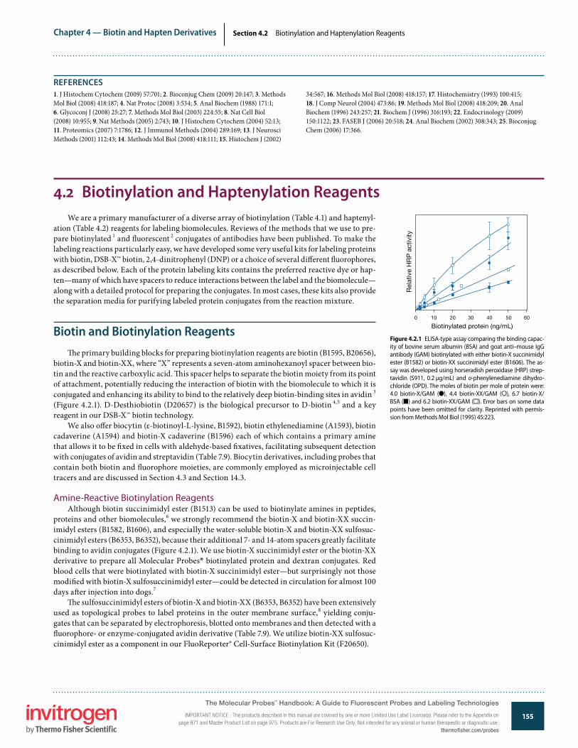
Biotin and Biotinylation Reagents�e primary building blocks for preparing biotinylation reagents are biotin (B1595, B20656),
biotin-X and biotin-XX, where “X” represents a seven-atom aminohexanoyl spacer between bio-tin and the reactive carboxylic acid. �is spacer helps to separate the biotin moiety from its point of attachment, potentially reducing the interaction of biotin with the biomolecule to which it is conjugated and enhancing its ability to bind to the relatively deep biotin-binding sites in avidin 3 (Figure 4.2.1). D-Desthiobiotin (D20657) is the biological precursor to D-biotin 4,5 and a key reagent in our DSB-X™ biotin technology.
We also o�er biocytin (ε-biotinoyl-L-lysine, B1592), biotin ethylenediamine (A1593), biotin cadaverine (A1594) and biotin-X cadaverine (B1596) each of which contains a primary amine that allows it to be �xed in cells with aldehyde-based �xatives, facilitating subsequent detection with conjugates of avidin and streptavidin (Table 7.9). Biocytin derivatives, including probes that contain both biotin and �uorophore moieties, are commonly employed as microinjectable cell tracers and are discussed in Section 4.3 and Section 14.3.
Amine-Reactive Biotinylation ReagentsAlthough biotin succinimidyl ester (B1513) can be used to biotinylate amines in peptides,
proteins and other biomolecules,6 we strongly recommend the biotin-X and biotin-XX succin-imidyl esters (B1582, B1606), and especially the water-soluble biotin-X and biotin-XX sulfosuc-cinimidyl esters (B6353, B6352), because their additional 7- and 14-atom spacers greatly facilitate binding to avidin conjugates (Figure 4.2.1). We use biotin-X succinimidyl ester or the biotin-XX derivative to prepare all Molecular Probes® biotinylated protein and dextran conjugates. Red blood cells that were biotinylated with biotin-X succinimidyl ester—but surprisingly not those modi�ed with biotin-X sulfosuccinimidyl ester—could be detected in circulation for almost 100 days a�er injection into dogs.7
�e sulfosuccinimidyl esters of biotin-X and biotin-XX (B6353, B6352) have been extensively used as topological probes to label proteins in the outer membrane surface,8 yielding conju-gates that can be separated by electrophoresis, blotted onto membranes and then detected with a �uorophore- or enzyme-conjugated avidin derivative (Table 7.9). We utilize biotin-XX sulfosuc-cinimidyl ester as a component in our FluoReporter® Cell-Surface Biotinylation Kit (F20650).
Figure 4.2.1 ELISA-type assay comparing the binding capac-ity of bovine serum albumin (BSA) and goat anti–mouse IgG antibody (GAM) biotinylated with either biotin-X succinimidyl ester (B1582) or biotin-XX succinimidyl ester (B1606). The as-say was developed using horseradish peroxidase (HRP) strep-tavidin (S911, 0.2 µg/mL) and o-phenylenediamine dihydro-chloride (OPD). The moles of biotin per mole of protein were: 4.0 biotin-X/GAM (d), 4.4 biotin-XX/GAM (s), 6.7 biotin-X/BSA (j) and 6.2 biotin-XX/GAM (h). Error bars on some data points have been omitted for clarity. Reprinted with permis-sion from Methods Mol Biol (1995) 45:223.
Rel
ativ
e H
RP
act
ivity
0
Biotinylated protein (ng/mL)10 20 30 40 50 60
1. J Histochem Cytochem (2009) 57:701; 2. Bioconjug Chem (2009) 20:147; 3. Methods Mol Biol (2008) 418:187; 4. Nat Protoc (2008) 3:534; 5. Anal Biochem (1988) 171:1; 6. Glycoconj J (2008) 25:27; 7. Methods Mol Biol (2003) 224:55; 8. Nat Cell Biol (2008) 10:955; 9. Nat Methods (2005) 2:743; 10. J Histochem Cytochem (2004) 52:13; 11. Proteomics (2007) 7:1786; 12. J Immunol Methods (2004) 289:169; 13. J Neurosci Methods (2001) 112:43; 14. Methods Mol Biol (2008) 418:111; 15. Histochem J (2002)
34:567; 16. Methods Mol Biol (2008) 418:157; 17. Histochemistry (1993) 100:415; 18. J Comp Neurol (2004) 473:86; 19. Methods Mol Biol (2008) 418:209; 20. Anal Biochem (1996) 243:257; 21. Biochem J (1996) 316:193; 22. Endocrinology (2009) 150:1122; 23. FASEB J (2006) 20:518; 24. Anal Biochem (2002) 308:343; 25. Bioconjug Chem (2006) 17:366.
The Molecular Probes™ Handbook: A Guide to Fluorescent Probes and Labeling Technologies
IMPORTANT NOTICE : The products described in this manual are covered by one or more Limited Use Label License(s). Please refer to the Appendix on page 971 and Master Product List on page 975. Products are For Research Use Only. Not intended for any animal or human therapeutic or diagnostic use.
thermofisher.com/probes

Chapter 4 — Biotin and Hapten Derivatives
156www.invitrogen.com/probes
The Molecular Probes® Handbook: A Guide to Fluorescent Probes and Labeling TechnologiesIMPORTANT NOTICE: The products described in this manual are covered by one or more Limited Use Label License(s). Please refer to the Appendix on page 971 and Master Product List on page 975. Products are For Research Use Only. Not intended for any animal or human therapeutic or diagnostic use.
Section 4.2 Biotinylation and Haptenylation Reagents
Figure 4.2.4 Biotin alkyne (PEG4 carboxamide-propargyl biotin, B10185).
(CH2)4
S
HN
NH
O
H
H
H
CO
NH(CH2CH2O)4CH2CH2 NHCH2C CHC
O
Figure 4.2.2 Biotin-X 2,4-dinitrophenyl-X-L-lysine, succin-imidyl ester (DNP-X-biocytin-X, SE, B2604).
Figure 4.2.3 Reaction of a TS-Link™ reagent with a thiol, followed by removal of the label with a reducing agent.
SH
R2SH
C
O
CH2S SO3R1NH
C
O
CH2S SR2R1NH
C
O
CH2R1NH
DTT or TCEP
+ R2SH
Figure 4.2.5 Biotin azide (PEG4 carboxamide-6-azidohexa-nyl biotin, B10184).
(CH2)4
S
HN
NH
O
H
H
H
CO
NH(CH2CH2O)4CH2CH2 NH(CH2)6 N NNC
O
Figure 4.2.6 Nucleophilic attack of serine on the carbonyl group (C=O) of biotin-X, SSE (B6353) results in the stable O-acylated derivative. In addition to histidine-x-serine, this stable intermediate can be formed in the presence of linear se-quences of histidine-x-tyrosine and histidine-x-threonine, where "x" refers to any amino acid.
R2H
C
H
N
O
C
X
C
H
N
O
C
H
N N
CH2CN
CH2
O
H
H
R1
H
R2H
C
H
N
O
C
X
C
H
N
O
C
H
N N
CH2CN
CH2
O
C
O
(CH2)5NH
R1H
2)4
NH
N
H
H
H
O
C
O
(CH2)5NH
CH)
NH
N
H
H
H
O
(CH2)4NH(CH2)5 C
O
NH(CH2)5 C
O
O
O
O
SO3_
N
+
Amine-Reactive Chromophoric Biotin DerivativeDetermining a protein’s degree of biotinylation is relatively di�cult because of the lack
of visible absorbance by the biotin molecule. To facilitate this determination, we o�er an amine-reactive chromophoric derivative, biotin-X 2,4-dinitrophenyl-X-L-lysine succinimi-dyl ester (DNP-X–biocytin-X, SE; B2604; Figure 4.2.2). Following protein conjugation, the extent of biotinylation is easily determined from the absorbance of the DNP chromophore (EC360 = 15,000 cm–1M–1). Incorporation of the DNP moiety into the biotinylating reagent does not a�ect its complexation with avidin or with anti-biotin antibodies. Our FluoReporter® Biotin/DNP Protein Labeling Kit (F6348), described below, contains su�cient DNP-X–biocytin-X, SE for 5 to 10 protein labeling reactions of 0.2–2 mg each.
Thiol-Reactive Biotinylation ReagentsAlthough amine-reactive reagents are more commonly employed, the thiol-reactive biotin
iodoacetamide, frequently identi�ed in the literature by the acronym BIAM (B1591), biotin ma-leimide (M1602) and DSB-X™ biotin C2-iodoacetamide (D30753) derivatives can also be used to label proteins and thiol-modi�ed oligonucleotides.9 Biotin iodoacetamide and biotin maleimide are primarily used for biotinylation of free protein thiols in relation to investigations of thiol–disul�de exchange, disul�de isomerization, S-glutathionylation and other posttranslational modi�cation processes.10–13
We also o�er TS-Link™ DSB-X™ biotin C5-thiosulfate (TS-Link™ desthiobiotin-X C5-thiosulfate, T30754), which is a water-soluble thiosulfate that reacts readily and selectively with a free thiol to form a disul�de bond (Figure 4.2.3). In contrast to the thioether bonds formed by maleimides and iodoacetamides, the disul�de bond formed by this TS-Link™ reagent is revers-ible—the TS-Link™ DSB-X™ hapten can easily be removed using a reducing agent such as dithio-threitol or tris-(2-carboxyethyl)phosphine (DTT, D1532; TCEP, T2556; Section 2.1), leaving the molecule of interest unchanged.
Click-iT® Biotin Alkyne and Biotin Azide ReagentsClick-iT® labeling and detection technology uses bioorthogonal reactive chemistry in which
the reaction partners have no endogenous representation in biological molecules, cells, tissues or model organisms.14–16 �e click reaction comprises a copper-catalyzed cycloaddition between an alkyne and an azide, forming a stable triazole conjugate (Section 3.1). �e azide and alkyne moieties can be used interchangeably; either one can be used to tag the biomolecule of interest, whereas the other is used for subsequent detection. Moreover, the click chemistry labels—either the alkyne or the azide—provide a functional group that typically neither reacts with other cell components nor disrupts normal cell processes. �e relative transparency to cell machinery of these labels means that tagged molecules are o�en acceptable substrates for enzymes that as-semble these building blocks into biopolymers.
The Molecular Probes™ Handbook: A Guide to Fluorescent Probes and Labeling Technologies
IMPORTANT NOTICE : The products described in this manual are covered by one or more Limited Use Label License(s). Please refer to the Appendix on page 971 and Master Product List on page 975. Products are For Research Use Only. Not intended for any animal or human therapeutic or diagnostic use.thermofisher.com/probes

Chapter 4 — Biotin and Hapten Derivatives
157www.invitrogen.com/probes
The Molecular Probes® Handbook: A Guide to Fluorescent Probes and Labeling TechnologiesIMPORTANT NOTICE: The products described in this manual are covered by one or more Limited Use Label License(s). Please refer to the Appendix on page 971 and Master Product List on page 975. Products are For Research Use Only. Not intended for any animal or human therapeutic or diagnostic use.
Section 4.2 Biotinylation and Haptenylation Reagents
Biotin alkyne (B10185, Figure 4.2.4) and biotin azide (B10184, Figure 4.2.5) are available for use in the detection and a�nity capture of azide- and alkyne-modi�ed biomolecules, respec-tively, using click chemistry.17–19 �e biotin conjugate formed by the click reaction can subse-quently be detected with a labeled avidin or streptavidin (Table 7.9). We also o�er a Click-iT® Biotin Protein Analysis Detection Kit (C33372, Section 9.4) for detection of azide-functionalized glycoproteins in 1D or 2D electrophoresis gels or western blots.
Histidine, Serine and Threonine Modi�cation with Biotin DerivativesTripeptide sequences of certain peptides such as gonadotropin releasing hormone (GnRH),
wherein serine, threonine or tyrosine residues are separated from a histidine residue by a single amino acid, can be selectively acylated by the succinimidyl ester or sulfosuccinimidyl ester of biotin-X (B1582, B6353). �is reaction probably involves formation of an acyl histidine interme-diate, followed by intramolecular transfer of the label (Figure 4.2.6). O-acylation can be detected by treating the conjugate with hydroxylamine, which cleaves esters of biotin but not amides.20 N-terminal serine and threonine residues of proteins can be oxidized by periodate and then bio-tinylated with biotin hydrazine derivatives 21 (B1603, B2600, D20653), which are described below.
Aldehyde Modi�cation with Biotin Hydrazides and Biotin Hydroxylamine (ARP)As described in Section 3.3, aldehydes generated by periodate oxidation of vicinal diols in
glycoproteins, polysaccharides and RNA or of N-terminal serine and threonine residues in pro-teins can be biotinylated using biotin-XX hydrazide (B2600). In cases where structural integrity may be compromised by periodate oxidation, derivatization with biotin hydrazide via reductive amination at the reducing end provides an alternative method for biotinylating carbohydrates.22 Biocytin hydrazide (B1603) may be preferred over biotin-XX hydrazide in some labeling proto-cols because of its higher water solubility.23 As with our other DSB-X™ biotin reagents, DSB-X™ biotin hydrazide (desthiobiotin-X hydrazide, D20653) can be used to produce a DSB-X™ bio-tin–labeled molecule that exhibits easily reversible binding to avidin- or streptavidin-labeled reagents. Biotin hydrazides are also o�en used for detection and a�nity capture of oxidatively damaged proteins via coupling to carbonyl groups.24
�e biotin-containing hydroxylamine derivative ARP (aldehyde-reactive probe, A10550) has been used to modify the exposed aldehyde group at abasic lesions in DNA 25,26 (Figure 4.2.7). Abasic sites are generated spontaneously or can be caused by free radicals, ionizing radia-tion or mutagens like methyl methanesulfonate (MMS). A quick and sensitive microplate assay for abasic sites can be performed using ARP.27
In addition, ARP is membrane permeant, permitting detection of abasic sites in live cells.28,29 Once the aldehyde group in an abasic site is modi�ed by ARP and the cells are �xed and per-meabilized, the resulting biotinylated DNA can be detected with �uorescent dye–, Qdot® nano-crystal– or enzyme-conjugated streptavidin conjugates (Table 7.9). Likewise, ARP can be used to detect and capture 4-hydroxynonenal (HNE)–modi�ed proteins.30 ARP has also been used to immobilize IgG antibodies on streptavidin-coated monolayer surfaces with their binding sites oriented toward the solution phase.31
Biotinylation of Carboxylic Acids�e biotin amines and hydrazides can be coupled to chemically activated carboxylic acids 32
(Figure 4.2.8). �e amine-containing biotin derivatives (B1592, A1593, A1594, B1596, N6356) are versatile intermediates for coupling biotin to DNA, carboxylic acids and array support sur-faces.33 �e biotin cadaverines (A1594, B1596) and potentially our unique norbiotinamine 34 (N6356, Figure 4.2.9) are useful for transglutaminase-mediated modi�cation of glutamine
Figure 4.2.8 Conversion of a carboxylic acid group into an aliphatic amine. The activated carboxylic acid is derivatized with a half-protected aliphatic diamine (mono-N-(t-BOC)-propylenedi-amine, M6248), usually in an organic solvent, followed by removal of the t-BOC–protecting group with tri�uoroacetic acid.
+ RC OH
O ED C CF3COOHH2N(CH2)3NH CR
O
NH(CH2)3NH2C
O
(CH3)3CO NH(CH2)3NHC
O
(CH3)3CO CR
OA
Figure 4.2.7 Aldehyde-reactive probe (ARP) used to de-tect DNA damage. The biotin hydroxylamine ARP (A10550) reacts with aldehyde groups formed when reactive oxygen species depurinate DNA. This reaction forms a covalent bond linking the DNA to biotin. The biotin can then be de-tected using �uorophore- or enzyme-linked streptavidin.
5′
3′
NH
NHNH
SH
(CH2)4C
O
H
H
NH
C
O
NH2OCH2
O
+
ARP
5′
3′
5′
3′
N
N
N
N
NH2
CH2O
O
O
P
O
O O–
CH2
CHOH
O
O
P
O
O O–
O
O
O
C
NH
H
H
O
C
(CH2)4
HS
NHNH
NH
NOCH2
O–
O
O
P
O
O
OHCH
CH2
Reactive Oxygen Species (ROS)or Radiation
Figure 4.2.9 Norbiotinamine, hydrochloride (N6356).
The Molecular Probes™ Handbook: A Guide to Fluorescent Probes and Labeling Technologies
IMPORTANT NOTICE : The products described in this manual are covered by one or more Limited Use Label License(s). Please refer to the Appendix on page 971 and Master Product List on page 975. Products are For Research Use Only. Not intended for any animal or human therapeutic or diagnostic use.
thermofisher.com/probes
Chapter 4 — Biotin and Hapten Derivatives
158www.invitrogen.com/probes
The Molecular Probes® Handbook: A Guide to Fluorescent Probes and Labeling TechnologiesIMPORTANT NOTICE: The products described in this manual are covered by one or more Limited Use Label License(s). Please refer to the Appendix on page 971 and Master Product List on page 975. Products are For Research Use Only. Not intended for any animal or human therapeutic or diagnostic use.
Section 4.2 Biotinylation and Haptenylation Reagents
residues in cells and certain proteins 35,36 (Section 3.4, Figure 4.2.10) and for the microplate assay of transglutaminase activity.37–39
Reactive DSB-X™ Biotin DerivativesOur unique DSB-X™ biotin technology (Section 4.1) permits the readily reversible binding
of DSB-X™ biotin–labeled biomolecules to avidin and streptavidin derivatives.40 �e DSB-X™ biotin reagents, which are derivatives of desthiobiotin (Figure 4.2.11), have a moderate a�nity for avidins (Kd for DSB binding to streptavidin is 1.9 nM 41), making DSB-X™ biotin an ideal ligand for transient immobilization of avidin and streptavidin conjugates. DSB-X™ biotin succinimi-dyl ester, which is a component of the DSB-X™ Biotin Protein Labeling Kit (D20655) described below, can be conjugated to amine-containing molecules in the same way as the biotin suc-cinimidyl esters. Our DSB-X™ biotin C2-iodoacetamide (D30753) and TS-Link™ DSB-X™ biotin C5-thiosulfate (T30754) are thiol-reactive derivatives for labeling proteins and thiol-modi�ed oligonucleotides. In addition, we o�er aldehyde-reactive DSB-X™ biotin hydrazide (D20653).
Convenient Kits for Biotinylating ProteinsBiotin-XX Microscale Protein Labeling Kit
�e Biotin-XX Microscale Protein Labeling Kit (B30010) provides a convenient means for biotinylating small amounts (20–100 µg) of puri�ed protein. �e kit has been optimized for labeling proteins with molecular weights between 12,000 and 150,000 daltons, and contains everything needed to perform three labeling reactions and to separate the resulting conjugates from excess reactive biotin. Convenient spin columns are used to purify the labeled protein with yields between 60 and 90%, depending primarily on the molecular weight of the starting mate-rial. Labeling and puri�cation can be completed in as little as 30 minutes.
Each Biotin-XX Microscale Protein Labeling Kit contains:
• Biotin-XX sulfosuccinimidyl ester• Sodium bicarbonate• Reaction tubes• Puri�cation resin and spin �lters• Protocols for preparing and purifying the biotinylated protein
For determining the degree of labeling, the FluoReporter® Biotin Quantitation Assay Kit for proteins is available separately (F30751) or in combination with the Biotin-XX Microscale Protein Labeling Kit (B30756). When biotinylating larger amounts of protein, we recommend the FluoReporter® Mini-Biotin-XX Protein Labeling Kit, which is optimized for 0.1–3 mg samples of >40,000-dalton proteins, or the FluoReporter® Biotin-XX Protein Labeling Kit, which is opti-mized for 5–20 mg samples; see below for a description of these kits.
FluoReporter® Mini-Biotin-XX Protein Labeling Kit�e FluoReporter® Mini-Biotin-XX Protein Labeling Kit (F6347) provides a method for e�-
ciently biotinylating small amounts of antibodies or other proteins. �e water-soluble biotin-XX sulfosuccinimidyl ester contained in this kit readily reacts with a protein’s amines to yield a bio-tin moiety that is linked to the protein through two tandem aminohexanoyl chains (“XX”). �is 14-atom spacer has been shown to enhance the binding of biotin derivatives to avidin’s relatively deep binding sites (Figure 4.2.1).
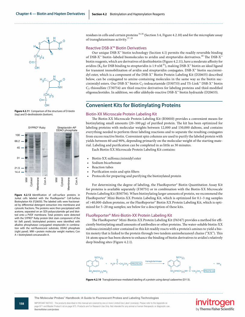
Figure 4.2.11 Comparison of the structures of D-biotin (top) and D-desthiobiotin (bottom).
OH
NH
H
H
C
(CH2)4O
SH
NH
O
NH
NH
OH3C
(CH2)5CO
OH
Figure 4.2.10 Transglutaminase-mediated labeling of a protein using dansyl cadaverine (D113).
N(CH3)2
SO2NH(CH2)5NH2
+ + NH3
N(CH3)2
SO2NH(CH2)5NH C
O
CH2CH2 PROTE I N
H2N C
O
CH2CH2 PROTE INtransglutaminase
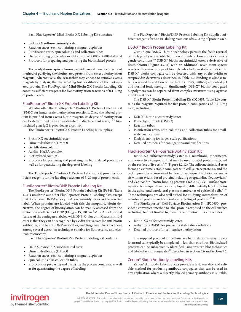
Figure 4.2.12 Identi�cation of cell-surface proteins in Jurkat cells labeled with the FluoReporter® Cell-Surface Biotinylation Kit (F20650). The labeled cells were fractionat-ed by di�erential detergent extraction into membrane and cytosolic fractions. The proteins were then precipitated with acetone, separated on an SDS-polyacrylamide gel and blot-ted onto a PVDF membrane. Total proteins were detected with the SYPRO® Ruby protein blot stain component of the kit (left panel); biotinylated proteins were identi�ed with alkaline phosphatase–conjugated streptavidin in combina-tion with the red-�uorescent substrate, DDAO phosphate (right panel). MW = protein molecular weight markers; Con A = biotinylated concanavalin A.
SYPRO® Ruby Streptavidin-APDDAO phosphate
100
66.2
45
31
21.5
14.4
MW
Cyt
osol
Mem
bra
ne
Con
A
MW
Cyt
osol
Mem
bra
ne
Con
A
The Molecular Probes™ Handbook: A Guide to Fluorescent Probes and Labeling Technologies
IMPORTANT NOTICE : The products described in this manual are covered by one or more Limited Use Label License(s). Please refer to the Appendix on page 971 and Master Product List on page 975. Products are For Research Use Only. Not intended for any animal or human therapeutic or diagnostic use.thermofisher.com/probes
Chapter 4 — Biotin and Hapten Derivatives
159www.invitrogen.com/probes
The Molecular Probes® Handbook: A Guide to Fluorescent Probes and Labeling TechnologiesIMPORTANT NOTICE: The products described in this manual are covered by one or more Limited Use Label License(s). Please refer to the Appendix on page 971 and Master Product List on page 975. Products are For Research Use Only. Not intended for any animal or human therapeutic or diagnostic use.
Section 4.2 Biotinylation and Haptenylation Reagents
Each FluoReporter® Mini-Biotin-XX Labeling Kit contains:
• Biotin-XX sulfosuccinimidyl ester• Reaction tubes, each containing a magnetic spin bar• Puri�cation resin, spin columns and collection tubes• Dialysis tubing (molecular weight cut-o� ~12,000–14,000 daltons)• Protocols for preparing and purifying the biotinylated protein
�e ready-to-use spin columns provide an extremely convenient method of purifying the biotinylated protein from excess biotinylation reagents. Alternatively, the researcher may choose to remove excess reagents by dialysis, thereby avoiding further dilution of the biotinyl-ated protein. �e FluoReporter® Mini-Biotin-XX Protein Labeling Kit contains su�cient reagents for �ve biotinylation reactions of 0.1–3 mg of protein each.
FluoReporter® Biotin-XX Protein Labeling KitWe also o�er the FluoReporter® Biotin-XX Protein Labeling Kit
(F2610) for larger-scale biotinylation reactions. Once the labeled pro-tein is puri�ed from excess biotin reagent, its degree of biotinylation can be determined using an avidin–biotin displacement assay; 42,43 bio-tinylated goat IgG is provided as a control.
�e FluoReporter® Biotin-XX Protein Labeling Kit supplies:
• Biotin-XX succinimidyl ester• Dimethylsulfoxide (DMSO)• Gel �ltration column• Avidin–HABA complex• Biotinylated goat IgG• Protocols for preparing and purifying the biotinylated protein, as
well as for quantitating the degree of labeling
�e FluoReporter® Biotin-XX Protein Labeling Kit provides suf-�cient reagents for �ve labeling reactions of 5–20 mg of protein each.
FluoReporter® Biotin/DNP Protein Labeling Kit�e FluoReporter® Biotin/DNP Protein Labeling Kit (F6348, Table
1.3) is similar to our other FluoReporter® Protein Labeling Kits, except that it contains DNP-X–biocytin-X succinimidyl ester as the reactive label. When proteins are labeled with this chromophoric biotin de-rivative, the degree of biotinylation can be readily assessed from the extinction coe�cient of DNP (EC364 = 15,000 cm–1M–1). An additional feature of the conjugates labeled with DNP-X–biocytin-X succinimidyl ester is that they can be recognized by avidin derivatives (or anti-biotin antibodies) and by anti-DNP antibodies, enabling researchers to choose among several detection techniques suitable for �uorescence and elec-tron microscopy.
Each FluoReporter® Biotin/DNP Protein Labeling Kit contains:
• DNP-X–biocytin-X succinimidyl ester• Dimethylsulfoxide (DMSO)• Reaction tubes, each containing a magnetic spin bar• Spin columns plus collection tubes• Protocols for preparing and purifying the protein conjugate, as well
as for quantitating the degree of labeling
�e FluoReporter® Biotin/DNP Protein Labeling Kit supplies suf-�cient reagents for 5 to 10 labeling reactions of 0.2–2 mg of protein each.
DSB-X™ Biotin Protein Labeling KitOur unique DSB-X™ biotin technology permits the facile reversal
of the typically irreversible biotin–avidin interaction under extremely gentle conditions.40 DSB-X™ biotin succinimidyl ester, a derivative of desthiobiotin (Figure 4.2.11) with an additional seven-atom spacer, reacts with amine groups of biomolecules to form stable amides. �e DSB-X™ biotin conjugate can be detected with any of the avidin or streptavidin derivatives described in Table 7.9. Binding is almost to-tally reversed by addition of free biotin (B1595, B20656) at neutral pH and normal ionic strength. Signi�cantly, DSB-X™ biotin–conjugated biopolymers can be separated from complex mixtures using agarose a�nity matrices.
�e DSB-X™ Biotin Protein Labeling Kit (D20655, Table 1.3) con-tains the reagents required for �ve protein conjugations of 0.5–3 mg each, including:
• DSB-X™ biotin succinimidyl ester• Dimethylsulfoxide (DMSO)• Reaction tubes• Puri�cation resin, spin columns and collection tubes for small-
scale puri�cations• Dialysis tubing for larger-scale puri�cations• Detailed protocols for conjugations and puri�cations
FluoReporter® Cell-Surface Biotinylation KitBiotin-XX sulfosuccinimidyl ester is a membrane-impermeant,
amine-reactive compound that may be used to label proteins exposed on the surface of live cells 8,44 (Figure 4.2.12). �e sulfosuccinimidyl ester forms an extremely stable conjugate with cell-surface proteins, and the biotin provides a convenient hapten for subsequent isolation or analy-sis with an avidin-based protein, including streptavidin, NeutrAvidin™ and CaptAvidin™ biotin-binding proteins (Table 7.9). Cell-surface bioti-nylation techniques have been employed to di�erentially label proteins in the apical and basolateral plasma membranes of epithelial cells.45,46 �ese techniques are also well suited for studying internalization of membrane proteins and cell-surface targeting of proteins.47–49
�e FluoReporter® Cell-Surface Biotinylation Kit (F20650) pro-vides a convenient method to label proteins exposed on the cell surface including, but not limited to, membrane proteins. �is kit includes:
• Biotin-XX sulfosuccinimidyl ester• Anhydrous DMSO for preparing stable stock solutions• Detailed protocols for cell-surface biotinylation
�e supplied protocol for cell-surface biotinylation is easy to per-form and can typically be completed in less than one hour. Biotinylated proteins can be subsequently identi�ed using western blot techniques and labeled avidin conjugates44 described in Section 6.6 and Section 7.6.
Zenon® Biotin Antibody Labeling KitsZenon® Antibody Labeling Kits provide a fast, versatile and reli-
able method for producing antibody conjugates that can be used in any application where a directly labeled primary antibody is suitable,
The Molecular Probes™ Handbook: A Guide to Fluorescent Probes and Labeling Technologies
IMPORTANT NOTICE : The products described in this manual are covered by one or more Limited Use Label License(s). Please refer to the Appendix on page 971 and Master Product List on page 975. Products are For Research Use Only. Not intended for any animal or human therapeutic or diagnostic use.
thermofisher.com/probes
Chapter 4 — Biotin and Hapten Derivatives
160www.invitrogen.com/probes
The Molecular Probes® Handbook: A Guide to Fluorescent Probes and Labeling TechnologiesIMPORTANT NOTICE: The products described in this manual are covered by one or more Limited Use Label License(s). Please refer to the Appendix on page 971 and Master Product List on page 975. Products are For Research Use Only. Not intended for any animal or human therapeutic or diagnostic use.
Section 4.2 Biotinylation and Haptenylation Reagents
including microscopy, �ow cytometry, high-throughput screening, and others. �is enabling technology not only eliminates the need for secondary detection reagents in many protocols, but also simpli�es immunolabeling protocols that previously were time consuming or impractical, such as use of multiple antibodies derived from the same species in the same protocol.50,51 We have applied our exclusive Zenon® technology (Section 7.3) in several Zenon® Biotin-XX Antibody Labeling Kits, which permit the quantitative biotinylation of even submicrogram quantities of an antibody typically in less than 10 minutes.
�e Zenon® Biotin-XX Antibody Labeling Kits include:
• Zenon® Biotin-XX Mouse IgG1 Labeling Kit (Z25052)• Zenon® Biotin XX Mouse IgG2a Labeling Kit (Z25152)• Zenon® Biotin-XX Mouse IgG2b Labeling Kit (Z25252)• Zenon® Biotin-XX Rabbit IgG Labeling Kit (Z25352)• Zenon® Biotin-XX Human IgG Labeling Kit (Z25452)
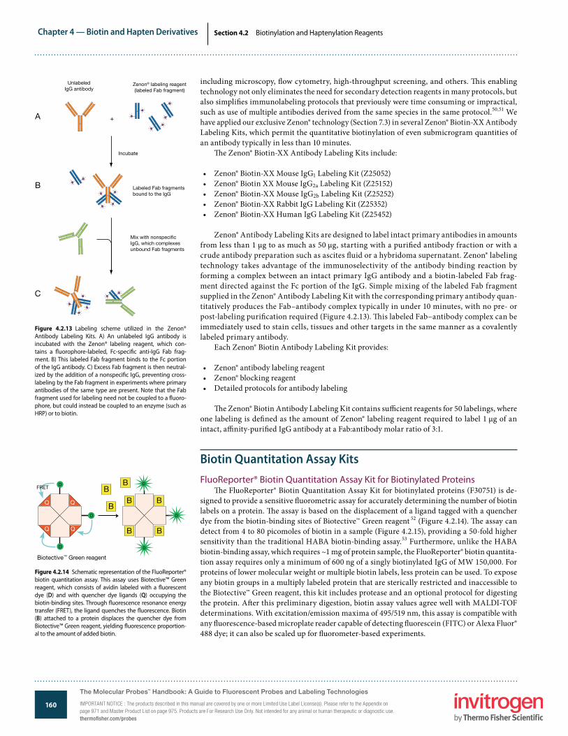
Zenon® Antibody Labeling Kits are designed to label intact primary antibodies in amounts from less than 1 µg to as much as 50 µg, starting with a puri�ed antibody fraction or with a crude antibody preparation such as ascites �uid or a hybridoma supernatant. Zenon® labeling technology takes advantage of the immunoselectivity of the antibody binding reaction by forming a complex between an intact primary IgG antibody and a biotin-labeled Fab frag-ment directed against the Fc portion of the IgG. Simple mixing of the labeled Fab fragment supplied in the Zenon® Antibody Labeling Kit with the corresponding primary antibody quan-titatively produces the Fab–antibody complex typically in under 10 minutes, with no pre- or post-labeling puri�cation required (Figure 4.2.13). �is labeled Fab–antibody complex can be immediately used to stain cells, tissues and other targets in the same manner as a covalently labeled primary antibody.
Each Zenon® Biotin Antibody Labeling Kit provides:
• Zenon® antibody labeling reagent• Zenon® blocking reagent• Detailed protocols for antibody labeling
�e Zenon® Biotin Antibody Labeling Kit contains su�cient reagents for 50 labelings, where one labeling is de�ned as the amount of Zenon® labeling reagent required to label 1 µg of an intact, a�nity-puri�ed IgG antibody at a Fab:antibody molar ratio of 3:1.
Biotin Quantitation Assay KitsFluoReporter® Biotin Quantitation Assay Kit for Biotinylated Proteins
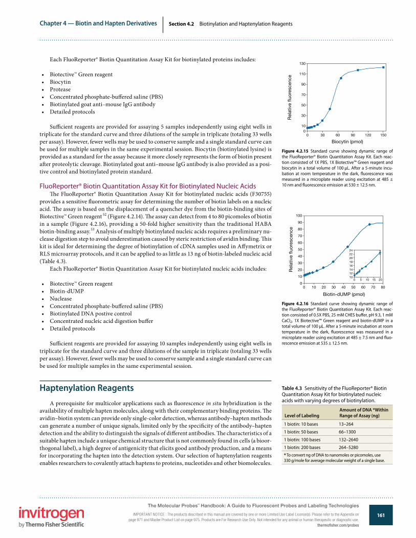
�e FluoReporter® Biotin Quantitation Assay Kit for biotinylated proteins (F30751) is de-signed to provide a sensitive �uorometric assay for accurately determining the number of biotin labels on a protein. �e assay is based on the displacement of a ligand tagged with a quencher dye from the biotin-binding sites of Biotective™ Green reagent 52 (Figure 4.2.14). �e assay can detect from 4 to 80 picomoles of biotin in a sample (Figure 4.2.15), providing a 50-fold higher sensitivity than the traditional HABA biotin-binding assay.53 Furthermore, unlike the HABA biotin-binding assay, which requires ~1 mg of protein sample, the FluoReporter® biotin quantita-tion assay requires only a minimum of 600 ng of a singly biotinylated IgG of MW 150,000. For proteins of lower molecular weight or multiple biotin labels, less protein can be used. To expose any biotin groups in a multiply labeled protein that are sterically restricted and inaccessible to the Biotective™ Green reagent, this kit includes protease and an optional protocol for digesting the protein. A�er this preliminary digestion, biotin assay values agree well with MALDI-TOF determinations. With excitation/emission maxima of 495/519 nm, this assay is compatible with any �uorescence-based microplate reader capable of detecting �uorescein (FITC) or Alexa Fluor® 488 dye; it can also be scaled up for �uorometer-based experiments.
Figure 4.2.13 Labeling scheme utilized in the Zenon® Antibody Labeling Kits. A) An unlabeled IgG antibody is incubated with the Zenon® labeling reagent, which con-tains a �uorophore-labeled, Fc-speci�c anti-IgG Fab frag-ment. B) This labeled Fab fragment binds to the Fc portion of the IgG antibody. C) Excess Fab fragment is then neutral-ized by the addition of a nonspeci�c IgG, preventing cross-labeling by the Fab fragment in experiments where primary antibodies of the same type are present. Note that the Fab fragment used for labeling need not be coupled to a �uoro-phore, but could instead be coupled to an enzyme (such as HRP) or to biotin.
UnlabeledIgG antibody
Zenon® labeling reagent(labeled Fab fragment)
Incubate
Labeled Fab fragmentsbound to the IgG
Mix with nonspeci�cIgG, which complexesunbound Fab fragments
A
B
C
Figure 4.2.14 Schematic representation of the FluoReporter® biotin quantitation assay. This assay uses Biotective™ Green reagent, which consists of avidin labeled with a �uorescent dye (D) and with quencher dye ligands (Q) occupying the biotin-binding sites. Through �uorescence resonance energy transfer (FRET), the ligand quenches the �uorescence. Biotin (B) attached to a protein displaces the quencher dye from Biotective™ Green reagent, yielding �uorescence proportion-al to the amount of added biotin.
Biotective™ Green reagent
B
Q
Q
Q
Q
B
B
D
D
D
B
B
B
B
D
D
D
FRET
The Molecular Probes™ Handbook: A Guide to Fluorescent Probes and Labeling Technologies
IMPORTANT NOTICE : The products described in this manual are covered by one or more Limited Use Label License(s). Please refer to the Appendix on page 971 and Master Product List on page 975. Products are For Research Use Only. Not intended for any animal or human therapeutic or diagnostic use.thermofisher.com/probes
Chapter 4 — Biotin and Hapten Derivatives
161www.invitrogen.com/probes
The Molecular Probes® Handbook: A Guide to Fluorescent Probes and Labeling TechnologiesIMPORTANT NOTICE: The products described in this manual are covered by one or more Limited Use Label License(s). Please refer to the Appendix on page 971 and Master Product List on page 975. Products are For Research Use Only. Not intended for any animal or human therapeutic or diagnostic use.
Section 4.2 Biotinylation and Haptenylation Reagents
Each FluoReporter® Biotin Quantitation Assay Kit for biotinylated proteins includes:
• Biotective™ Green reagent• Biocytin• Protease• Concentrated phosphate-bu�ered saline (PBS)• Biotinylated goat anti–mouse IgG antibody• Detailed protocols
Su�cient reagents are provided for assaying 5 samples independently using eight wells in triplicate for the standard curve and three dilutions of the sample in triplicate (totaling 33 wells per assay). However, fewer wells may be used to conserve sample and a single standard curve can be used for multiple samples in the same experimental session. Biocytin (biotinylated lysine) is provided as a standard for the assay because it more closely represents the form of biotin present a�er proteolytic cleavage. Biotinylated goat anti–mouse IgG antibody is also provided as a posi-tive control and biotinylated protein standard.
FluoReporter® Biotin Quantitation Assay Kit for Biotinylated Nucleic Acids�e FluoReporter® Biotin Quantitation Assay Kit for biotinylated nucleic acids (F30755)
provides a sensitive �uorometric assay for determining the number of biotin labels on a nucleic acid. �e assay is based on the displacement of a quencher dye from the biotin-binding sites of Biotective™ Green reagent 52 (Figure 4.2.14). �e assay can detect from 4 to 80 picomoles of biotin in a sample (Figure 4.2.16), providing a 50-fold higher sensitivity than the traditional HABA biotin-binding assay.53 Analysis of multiply biotinylated nucleic acids requires a preliminary nu-clease digestion step to avoid underestimation caused by steric restriction of avidin binding. �is kit is ideal for determining the degree of biotinylation of cDNA samples used in A�ymetrix or RLS microarray protocols, and it can be applied to as little as 13 ng of biotin-labeled nucleic acid (Table 4.3).
Each FluoReporter® Biotin Quantitation Assay Kit for biotinylated nucleic acids includes:
• Biotective™ Green reagent• Biotin-dUMP• Nuclease• Concentrated phosphate-bu�ered saline (PBS)• Biotinylated DNA postive control• Concentrated nucleic acid digestion bu�er• Detailed protocols
Su�cient reagents are provided for assaying 10 samples independently using eight wells in triplicate for the standard curve and three dilutions of the sample in triplicate (totaling 33 wells per assay). However, fewer wells may be used to conserve sample and a single standard curve can be used for multiple samples in the same experimental session.
Haptenylation ReagentsA prerequisite for multicolor applications such as �uorescence in situ hybridization is the
availability of multiple hapten molecules, along with their complementary binding proteins. �e avidin–biotin system can provide only single-color detection, whereas antibody–hapten methods can generate a number of unique signals, limited only by the speci�city of the antibody–hapten detection and the ability to distinguish the signals of di�erent antibodies. �e characteristics of a suitable hapten include a unique chemical structure that is not commonly found in cells (a bioor-thogonal label), a high degree of antigenicity that elicits good antibody production, and a means for incorporating the hapten into the detection system. Our selection of haptenylation reagents enables researchers to covalently attach haptens to proteins, nucleotides and other biomolecules.
Figure 4.2.15 Standard curve showing dynamic range of the FluoReporter® Biotin Quantitation Assay Kit. Each reac-tion consisted of 1X PBS, 1X Biotective™ Green reagent and biocytin in a total volume of 100 µL. After a 5-minute incu-bation at room temperature in the dark, �uorescence was measured in a microplate reader using excitation at 485 ± 10 nm and �uorescence emission at 530 ± 12.5 nm.
Biocytin (pmol)
0 30 60 120 15090
Rel
ativ
e �u
ores
cenc
e
0
70
50
30
10
110
90
130
Figure 4.2.16 Standard curve showing dynamic range of the FluoReporter® Biotin Quantitation Assay Kit. Each reac-tion consisted of 0.5X PBS, 25 mM CHES bu�er, pH 9.3, 1 mM CaCl2, 1X Biotective™ Green reagent and biotin-dUMP in a total volume of 100 µL. After a 5-minute incubation at room temperature in the dark, �uorescence was measured in a microplate reader using excitation at 485 ± 7.5 nm and �uo-rescence emission at 535 ± 12.5 nm.
Biotin-dUMP (pmol)
0 40302010 50 60 70 80R
elat
ive
�uor
esce
nce
0
40
30
20
10
50
60
80
90
100
70
20
2224
20
16
12
18
14
10151050
Table 4.3 Sensitivity of the FluoReporter® Biotin Quantitation Assay Kit for biotinylated nucleic acids with varying degrees of biotinylation.
Level of LabelingAmount of DNA *Within Range of Assay (ng)
1 biotin: 10 bases 13–264
1 biotin: 50 bases 66–1300
1 biotin: 100 bases 132–2640
1 biotin: 200 bases 264–5280
* To convert ng of DNA to nanomoles or picomoles, use 330 g/mole for average molecular weight of a single base.
The Molecular Probes™ Handbook: A Guide to Fluorescent Probes and Labeling Technologies
IMPORTANT NOTICE : The products described in this manual are covered by one or more Limited Use Label License(s). Please refer to the Appendix on page 971 and Master Product List on page 975. Products are For Research Use Only. Not intended for any animal or human therapeutic or diagnostic use.
thermofisher.com/probes

Chapter 4 — Biotin and Hapten Derivatives
162www.invitrogen.com/probes
The Molecular Probes® Handbook: A Guide to Fluorescent Probes and Labeling TechnologiesIMPORTANT NOTICE: The products described in this manual are covered by one or more Limited Use Label License(s). Please refer to the Appendix on page 971 and Master Product List on page 975. Products are For Research Use Only. Not intended for any animal or human therapeutic or diagnostic use.
Section 4.2 Biotinylation and Haptenylation Reagents
In addition to our wide range of biotinylation reagents discussed above, we provide many unique haptenylation reagents, including an amine-reactive version of digoxigenin (A2952), dinitrophenyl-X 54 (DNP-X, SE; D2248) and several �uorophores (Table 4.2). We usually recommend haptenylation reagents that contain spacers between the hapten and the reactive groups to reduce potential interactions with the biomolecule to which it is conjugated and to make the hapten maxi-mally available to secondary detection reagents. Most of the preferred haptenylation reagents in Table 4.2 possess this feature.
Fluorescein has been found to be an excellent hapten for in situ hybridization because it binds with high a�nity to its anti-�uorescein antibody.55–57 Anti-�uorescein antibodies cross-react with all of the Oregon Green® dyes (Section 1.5), permitting their use with conju-gates prepared from these dyes. By adding antibodies that recognize the Alexa Fluor® 488, dansyl, tetramethylrhodamine and Texas Red® �uorophores to our line of detection reagents (Section 7.4), we have greatly expanded the number of potential haptens. Because the anti-tetramethylrhodamine and anti–Texas Red® dye antibodies cross-react with the tetramethylrhodamine, Lissamine rhodamine, Rhodamine Red™ and Texas Red® �uorophores, these antibody–�uorophore combi-nations should not be used simultaneously to generate separate signals in a multicolor experiment. Similarly, our antibody to the BODIPY® FL dye cross-reacts with some of the other BODIPY® dyes (Section 1.4).
REFERENCES1. Methods Mol Biol (1998) 80:173; 2. Methods Mol Biol (1995) 45:205; 3. Biochemistry (1982) 21:978; 4. Biochemistry (2001) 40:8343; 5. Biochemistry (2001) 40:8352; 6. Proc Natl Acad Sci U S A (1974) 71:3537; 7. Ann Hematol (1997) 74:231; 8. Proteomics (2008) 8:4012; 9. Org Biomol Chem (2008) 6:908; 10. Mol Cell Proteomics (2003) 2:242; 11. J Biol Chem (2009) 284:22213; 12. Methods Cell Biol (2007) 80:417; 13. Proc Natl Acad Sci U S A (2006) 103:13932; 14. Angew Chem Int Ed Engl (2009) 48:6974; 15. Biochemistry (2009) 48:6571; 16. ACS Chem Biol (2006) 1:644; 17. J Am Chem Soc (2010) 132:2504; 18. Chem Res Toxicol (2008) 21:432; 19. Nat Cell Biol (2008) 10:1224; 20. J Biol Chem (1992) 267:5060; 21. Bioconjug Chem (2003) 14:205; 22. Anal Biochem (2006) 354:54; 23. Anal Biochem (1988) 170:271; 24. Methods Mol Biol (2009) 536:457; 25. Photochem Photobiol (2002) 76:123; 26. Anal Chem (2001) 73:2229; 27. Methods (2000) 22:164; 28. Proc Natl Acad Sci U S A (2000) 97:686; 29. J Biol Chem (2000) 275:6741; 30. Anal Chem (2006) 78:6847; 31. Anal Biochem (2003) 312:113; 32. Anal Biochem (2002) 304:266; 33. Proc Natl Acad Sci U S A (2009) 106:405; 34. Bioconjug Chem (1996) 7:271; 35. J Biol Chem (2003) 278:35184; 36. J Biol Chem (2003) 278:4227; 37. Anal Biochem (1994) 223:88; 38. J Biol Chem (1994) 269:28309; 39. Anal Biochem (1992) 205:166; 40. Anal Biochem (2002) 308:343; 41. Bioconjug Chem (2006) 17:366; 42. FEBS Lett (1993) 328:165; 43. Methods Enzymol (1970) 18:418; 44. Methods Mol Biol (2005) 303:35; 45. J Neurochem (2001) 77:1301; 46. J Cell Sci (1996) 109:3025; 47. Infect Immun (2006) 74:1148; 48. J Virol (2001) 75:4744; 49. J Biol Chem (1999) 274:36801; 50. J Cell Biol (2009) 185:903; 51. J Neurosci (2007) 27:1836; 52. Biotechniques (2007) 43:503; 53. Biochem J (1965) 94:23C; 54. Eur J Cell Biol (1991) 56:223; 55. Nucleic Acids Res (1991) 19:3237; 56. J Histochem Cytochem (1990) 38:467; 57. Nucleic Acids Res (2008) 36:4047.
DATA TABLE 4.2 BIOTINYLATION AND HAPTENYLATION REAGENTSCat. No. MW Storage Soluble Abs EC Em Solvent NotesA1593 367.30 NC DMF, DMSO <300 noneA1594 442.50 NC DMF, DMSO <300 noneA2952 586.68 F,D DMF, DMSO <300 noneA10550 445.41 F,D H2O, DMSO <300 noneA20000 643.41 F,DD,L H2O, DMSO 494 73,000 517 pH 7 1, 2, 3, 4A20100 643.41 F,DD,L H2O, DMSO 494 73,000 517 pH 7 1, 2, 3, 4A30000 1028.26 F,DD,L H2O, DMSO 400 35,000 424 pH 7 5, 6A30005 884.91 F,DD,L H2O, DMSO 494 72,000 520 pH 7 2, 4, 7A30052 825.46 F,DD,L H2O, DMSO 493 73,000 520 pH 7 2, 4, 7A30100 1028.26 F,DD,L H2O, DMSO 400 35,000 424 pH 7 5, 6B1513 341.38 F,D DMF, DMSO <300 noneB1582 454.54 F,D DMF, DMSO <300 noneB1591 454.33 F,D DMF, DMSO <300 none 8B1592 372.48 NC H2O <300 noneB1595 244.31 NC pH >6, DMF <300 noneB1596 555.65 NC DMF, DMSO <300 noneB1603 386.51 D pH >6, DMF <300 noneB1606 567.70 F,D DMF, DMSO <300 noneB2600 484.66 D DMF, DMSO <300 noneB2604 861.97 F,D,L DMF 362 15,000 none pH 8B6352 669.74 F,D DMF, pH >6 <300 none 1B6353 556.58 F,D DMF, pH >6 <300 none 1B10006 542.19 F,D,L H2O, DMSO 502 80,000 510 MeOH 11, 15B10184 615.79 F,D,L <300 noneB10185 528.66 F,D <300 noneB20656 244.31 RO pH >6 <300 none 9C2284 607.42 F,D,LL H2O, MeOH 396 29,000 410 MeOH 5, 10D2248 394.34 F,D,L DMF, DMSO 348 18,000 none MeOHD6102 502.32 F,D,L DMSO, MeCN 504 85,000 510 MeOH 11D6104 461.53 F,D,L DMF, MeCN 335 4200 518 MeOHD20653 341.45 D DMSO <300 none 12D20657 214.26 RO pH >6 <300 none 9, 12D30753 537.44 F,D DMSO <300 none 8, 12F2181 586.55 F,D,L DMF, DMSO 494 74,000 520 pH 9 13F6130 590.56 F,D,L DMF, DMSO 491 86,000 515 pH 9 13
The Molecular Probes™ Handbook: A Guide to Fluorescent Probes and Labeling Technologies
IMPORTANT NOTICE : The products described in this manual are covered by one or more Limited Use Label License(s). Please refer to the Appendix on page 971 and Master Product List on page 975. Products are For Research Use Only. Not intended for any animal or human therapeutic or diagnostic use.thermofisher.com/probes

Chapter 4 — Biotin and Hapten Derivatives
163www.invitrogen.com/probes
The Molecular Probes® Handbook: A Guide to Fluorescent Probes and Labeling TechnologiesIMPORTANT NOTICE: The products described in this manual are covered by one or more Limited Use Label License(s). Please refer to the Appendix on page 971 and Master Product List on page 975. Products are For Research Use Only. Not intended for any animal or human therapeutic or diagnostic use.
Section 4.2 Biotinylation and Haptenylation Reagents
L1338 659.51 F,D,L H2O 426 11,000 531 pH 7 8M1602 523.60 F,D pH >6, DMF <300 noneN6356 251.77 D DMF, pH <6 <300 noneO6185 622.53 F,D,L DMF, DMSO 494 84,000 517 pH 9 14R6160 768.90 F,D,L DMF, DMSO 560 129,000 580 MeOHT6105 640.69 F,D,L DMF, DMSO 543 92,000 571 MeOHT6134 816.94 F,D,L DMF, DMSO 583 112,000 603 MeOHT20175 816.94 F,D,L DMF, DMSO 587 96,000 602 MeOHT30754 587.72 F,D DMSO <300 none 12For de�nitions of the contents of this data table, see “Using The Molecular Probes® Handbook” in the introductory pages.Notes
1. This sulfonated succinimidyl ester derivative is water soluble and may be dissolved in bu�er at ~pH 8 for reaction with amines. Long-term storage in water is NOT recommended due to hydrolysis.2. The �uorescence lifetime (τ) of the Alexa Fluor® 488 dye in pH 7.4 bu�er at 20°C is 4.1 nanoseconds. Data provided by the SPEX Fluorescence Group, Horiba Jobin Yvon Inc.3. A20100 is an alternative packaging of A20000 but is otherwise identical.4. Abs and Em of the Alexa Fluor® 488 dye are red-shifted by as much as 16 nm and 25 nm respectively on microarrays relative to aqueous solution values. The magnitude of the spectral shift
depends on the array substrate material. (Biotechniques (2005) 38:127)5. The Alexa Fluor® 405 and Cascade Blue® dyes have a second absorption peak at about 376 nm with EC ~80% of the 395–400 nm peak.6. A30100 is an alternative packaging of A30000 but is otherwise identical.7. TFP and SDP ester derivatives are water-soluble and may be dissolved in bu�er at ~pH 8 for reaction with amines. Long-term storage in water is NOT recommended due to hydrolysis.8. Iodoacetamides in solution undergo rapid photodecomposition to unreactive products. Minimize exposure to light prior to reaction.9. This product is supplied as a ready-made solution in the solvent indicated under "Soluble."10. Unstable in water. Use immediately.11. The absorption and �uorescence spectra of BODIPY® derivatives are relatively insensitive to the solvent.12. The dissociation constant (Kd) for desthiobiotin binding to streptavidin is 1.9 nM. (Bioconjug Chem (2006) 17:366)13. Absorption and �uorescence of �uorescein derivatives are pH dependent. Extinction coe�cients and �uorescence quantum yields decrease markedly at pH <7.14. Absorption and �uorescence of Oregon Green® 488 derivatives are pH dependent only in moderately acidic solutions (pH <5).15. This sulfotetra�uorophenyl (STP) ester derivative is water soluble and may be dissolved in bu�er at ~pH 8 for reaction with amines. Long-term storage in water is NOT recommended due to
hydrolysis.
Cat. No. Description QuantityA30000 Alexa Fluor® 405 carboxylic acid, succinimidyl ester 1 mgA30100 Alexa Fluor® 405 carboxylic acid, succinimidyl ester 5 mgA20000 Alexa Fluor® 488 carboxylic acid, succinimidyl ester *mixed isomers* 1 mgA20100 Alexa Fluor® 488 carboxylic acid, succinimidyl ester *mixed isomers* 5 mgA30005 Alexa Fluor® 488 carboxylic acid, 2,3,5,6-tetra�uorophenyl ester (Alexa Fluor® 488 5-TFP) *5-isomer* 1 mgA30052 Alexa Fluor® 488 5-SDP ester (Alexa Fluor® 488 sulfodichlorophenol ester) 1 mgA2952 3-amino-3-deoxydigoxigenin hemisuccinamide, succinimidyl ester 5 mgA1593 N-(2-aminoethyl)biotinamide, hydrobromide (biotin ethylenediamine) 25 mgA10550 N-(aminooxyacetyl)-N’-(D-biotinoyl) hydrazine, tri�uoroacetic acid salt (ARP) 10 mgA1594 N-(5-aminopentyl)biotinamide, tri�uoroacetic acid salt (biotin cadaverine) 25 mgB1592 biocytin (ε-biotinoyl-L-lysine) 100 mgB1603 biocytin hydrazide 25 mgB1595 D-biotin 1 gB20656 D-biotin *50 mM aqueous solution* 10 mLB10185 biotin alkyne (PEG4 carboxamide-propargyl biotin) 1 mgB10184 biotin azide (PEG4 carboxamide-6-azidohexanyl biotin) 1 mgB1582 6-((biotinoyl)amino)hexanoic acid, succinimidyl ester (biotin-X, SE; biotinamidocaproate, N-hydroxysuccinimidyl ester) 100 mgB6353 6-((biotinoyl)amino)hexanoic acid, sulfosuccinimidyl ester, sodium salt (Sulfo-NHS-LC-Biotin; biotin-X, SSE) 25 mgB1606 6-((6-((biotinoyl)amino)hexanoyl)amino)hexanoic acid, succinimidyl ester (biotin-XX, SE) 100 mgB6352 6-((6-((biotinoyl)amino)hexanoyl)amino)hexanoic acid, sulfosuccinimidyl ester, sodium salt (biotin-XX, SSE) 25 mgB1591 N-(biotinoyl)-N’-(iodoacetyl)ethylenediamine 25 mgB1513 D-biotin, succinimidyl ester (succinimidyl D-biotin) 100 mgB1596 biotin-X cadaverine (5-(((N-(biotinoyl)amino)hexanoyl)amino)pentylamine, tri�uoroacetic acid salt) 10 mgB2604 biotin-X 2,4-dinitrophenyl-X-L-lysine, succinimidyl ester (DNP-X-biocytin-X, SE) 5 mgB2600 biotin-XX hydrazide (6-((6-((biotinoyl)amino)hexanoyl)amino)hexanoic acid, hydrazide) 25 mgB30010 Biotin-XX Microscale Protein Labeling Kit *for 20–100 µg protein* *3 labelings* 1 kitB30756 Biotin-XX Microscale Protein Labeling Kit with FluoReporter® Biotin Quantitation Assay Kit *includes B30010 and F30751* 1 kitB10006 BODIPY® FL, STP ester, sodium salt 5 mgC2284 Cascade Blue® acetyl azide, trisodium salt 5 mg
PRODUCT LIST 4.2 BIOTINYLATION AND HAPTENYLATION REAGENTS
DATA TABLE 4.2 BIOTINYLATION AND HAPTENYLATION REAGENTS—continuedCat. No. MW Storage Soluble Abs EC Em Solvent Notes
continued on next page
The Molecular Probes™ Handbook: A Guide to Fluorescent Probes and Labeling Technologies
IMPORTANT NOTICE : The products described in this manual are covered by one or more Limited Use Label License(s). Please refer to the Appendix on page 971 and Master Product List on page 975. Products are For Research Use Only. Not intended for any animal or human therapeutic or diagnostic use.
thermofisher.com/probes

Chapter 4 — Biotin and Hapten Derivatives
164www.invitrogen.com/probes
The Molecular Probes® Handbook: A Guide to Fluorescent Probes and Labeling TechnologiesIMPORTANT NOTICE: The products described in this manual are covered by one or more Limited Use Label License(s). Please refer to the Appendix on page 971 and Master Product List on page 975. Products are For Research Use Only. Not intended for any animal or human therapeutic or diagnostic use.
Section 4.3 Biotin and Desthiobiotin Conjugates
We prepare a wide array of biotin and desthiobiotin conjugates, all of which are included in this section’s product list. We will also custom-conjugate biotin, desthiobiotin (DSB-X™), �uoro-phores or other haptens to proteins or other biomolecules of interest; contact Invitrogen Custom Services to request a quote.
Fluorescent Biotin DerivativesFluorescein Biotin
Fluorescein biotin (B1370, Figure 4.3.1) was developed as an alternative to radioactive bio-tin for detecting and quantitating biotin-binding sites by either �uorescence or absorbance.1 A �uorescence polarization–based assay that employs competitive binding of �uorescein biotin to assess the degree of protein biotinylation has been reported 2 (Fluorescence Polarization (FP)—Note 1.4). A similar derivative was used for determining avidin and biotin concentrations by �uorescence depolarization.3
Biotin-4-FluoresceinOur biotin-4-�uorescein (B10570, Figure 4.3.2) o�ers a substantially improved method for
quantitating biotin-binding sites. Biotin-4-�uorescein binds to avidin much faster than does conventional �uorescein biotin, allowing for rapid analysis.4 �e strong quenching associated with avidin binding to biotin-4-�uorescein can be used to accurately measure the concentration of avidin or streptavidin 5 (Figure 4.3.3). Engineered single-chain dimers created by circular
4.3 Biotin and Desthiobiotin Conjugates
Figure 4.3.1 5-((N-(5-(N-(6-(biotinoyl)amino)hexanoyl)amino) pentyl)thioureidyl)�uorescein (�uorescein biotin, B1370).
Figure 4.3.2 Biotin-4-�uorescein (B10570).
Cat. No. Description QuantityD20657 D-desthiobiotin *50 mM aqueous solution* 10 mLD6102 6-((4,4-di�uoro-5,7-dimethyl-4-bora-3a,4a-diaza-s-indacene-3-propionyl)amino)hexanoic acid, succinimidyl ester (BODIPY® FL-X, SE) 5 mgD6104 6-((5-dimethylaminonaphthalene-1-sulfonyl)amino)hexanoic acid, succinimidyl ester (dansyl-X, SE) 25 mgD2248 6-(2,4-dinitrophenyl)aminohexanoic acid, succinimidyl ester (DNP-X, SE) 25 mgD30753 DSB-X™ biotin C2-iodoacetamide (desthiobiotin-X C2-iodoacetamide) 5 mgD20653 DSB-X™ biotin hydrazide 5 mgD20655 DSB-X™ Biotin Protein Labeling Kit *5 labelings* 1 kitF30755 FluoReporter® Biotin Quantitation Assay Kit *for biotinylated nucleic acids* *10 determinations* 1 kitF30751 FluoReporter® Biotin Quantitation Assay Kit *for biotinylated proteins* *5 determinations* 1 kitF6348 FluoReporter® Biotin/DNP Protein Labeling Kit *5–10 labelings* 1 kitF2610 FluoReporter® Biotin-XX Protein Labeling Kit *5 labelings of 5–20 mg protein each* 1 kitF20650 FluoReporter® Cell-Surface Biotinylation Kit 1 kitF6347 FluoReporter® Mini-biotin-XX Protein Labeling Kit *5 labelings of 0.1–3 mg protein each* 1 kitF2181 6-(�uorescein-5-(and-6)-carboxamido)hexanoic acid, succinimidyl ester (5(6)-SFX) *mixed isomers* 10 mgF6130 �uorescein-5-EX, succinimidyl ester 10 mgL1338 lucifer yellow iodoacetamide, dipotassium salt 25 mgM1602 Nα-(3-maleimidylpropionyl)biocytin 25 mgN6356 norbiotinamine, hydrochloride 10 mgO6185 Oregon Green® 488-X, succinimidyl ester *6-isomer* 5 mgR6160 Rhodamine Red™-X, succinimidyl ester *5-isomer* 5 mgT6105 6-(tetramethylrhodamine-5-(and-6)-carboxamido)hexanoic acid, succinimidyl ester (5(6)-TAMRA-X, SE) *mixed isomers* 10 mgT6134 Texas Red®-X, succinimidyl ester *mixed isomers* 5 mgT20175 Texas Red®-X, succinimidyl ester *single isomer* 2 mgT30754 TS-Link™ DSB-X™ biotin C5-thiosulfate (TS-Link™ desthiobiotin-X C5-thiosulfate, sodium salt) 5 mgZ25452 Zenon® Biotin-XX Human IgG Labeling Kit *50 labelings* 1 kitZ25052 Zenon® Biotin-XX Mouse IgG1 Labeling Kit *50 labelings* 1 kitZ25152 Zenon® Biotin-XX Mouse IgG2a Labeling Kit *50 labelings* 1 kitZ25252 Zenon® Biotin-XX Mouse IgG2b Labeling Kit *50 labelings* 1 kitZ25352 Zenon® Biotin-XX Rabbit IgG Labeling Kit *50 labelings* 1 kit
PRODUCT LIST 4.2 BIOTINYLATION AND HAPTENYLATION REAGENTS—continued
The Molecular Probes™ Handbook: A Guide to Fluorescent Probes and Labeling Technologies
IMPORTANT NOTICE : The products described in this manual are covered by one or more Limited Use Label License(s). Please refer to the Appendix on page 971 and Master Product List on page 975. Products are For Research Use Only. Not intended for any animal or human therapeutic or diagnostic use.thermofisher.com/probes
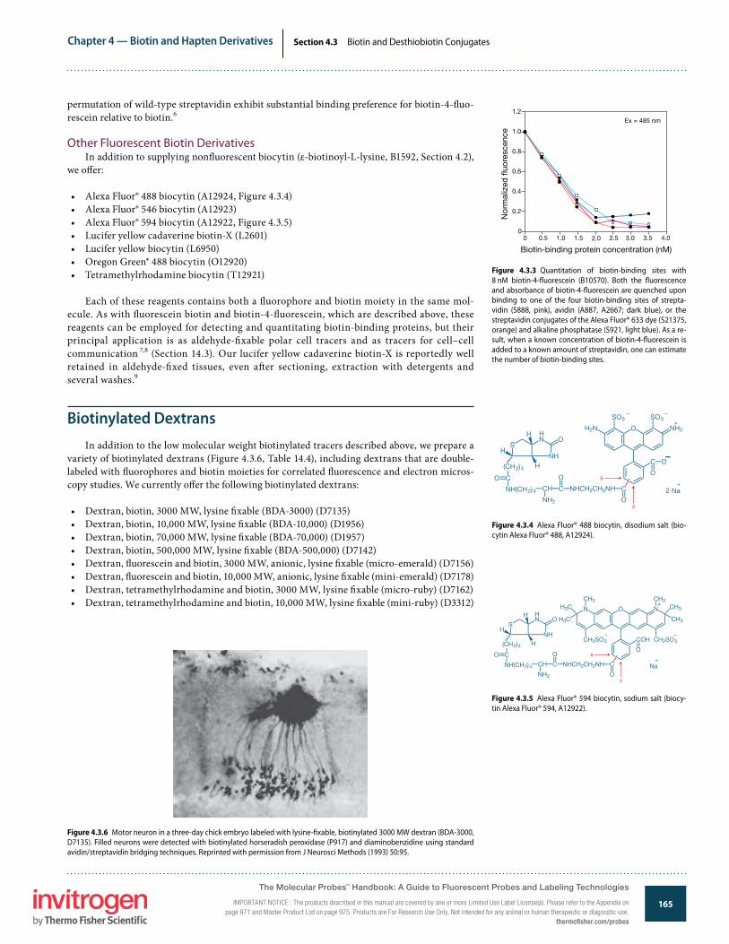
Chapter 4 — Biotin and Hapten Derivatives
165www.invitrogen.com/probes
The Molecular Probes® Handbook: A Guide to Fluorescent Probes and Labeling TechnologiesIMPORTANT NOTICE: The products described in this manual are covered by one or more Limited Use Label License(s). Please refer to the Appendix on page 971 and Master Product List on page 975. Products are For Research Use Only. Not intended for any animal or human therapeutic or diagnostic use.
Section 4.3 Biotin and Desthiobiotin Conjugates
permutation of wild-type streptavidin exhibit substantial binding preference for biotin-4-�uo-rescein relative to biotin.6
Other Fluorescent Biotin DerivativesIn addition to supplying non�uorescent biocytin (ε-biotinoyl-L-lysine, B1592, Section 4.2),
we o�er:
• Alexa Fluor® 488 biocytin (A12924, Figure 4.3.4)• Alexa Fluor® 546 biocytin (A12923)• Alexa Fluor® 594 biocytin (A12922, Figure 4.3.5)• Lucifer yellow cadaverine biotin-X (L2601)• Lucifer yellow biocytin (L6950)• Oregon Green® 488 biocytin (O12920)• Tetramethylrhodamine biocytin (T12921)
Each of these reagents contains both a �uorophore and biotin moiety in the same mol-ecule. As with �uorescein biotin and biotin-4-�uorescein, which are described above, these reagents can be employed for detecting and quantitating biotin-binding proteins, but their principal application is as aldehyde-�xable polar cell tracers and as tracers for cell–cell communication 7,8 (Section 14.3). Our lucifer yellow cadaverine biotin-X is reportedly well retained in aldehyde-�xed tissues, even a�er sectioning, extraction with detergents and several washes.9
Biotinylated DextransIn addition to the low molecular weight biotinylated tracers described above, we prepare a
variety of biotinylated dextrans (Figure 4.3.6, Table 14.4), including dextrans that are double-labeled with �uorophores and biotin moieties for correlated �uorescence and electron micros-copy studies. We currently o�er the following biotinylated dextrans:
• Dextran, biotin, 3000 MW, lysine �xable (BDA-3000) (D7135)• Dextran, biotin, 10,000 MW, lysine �xable (BDA-10,000) (D1956)• Dextran, biotin, 70,000 MW, lysine �xable (BDA-70,000) (D1957)• Dextran, biotin, 500,000 MW, lysine �xable (BDA-500,000) (D7142)• Dextran, �uorescein and biotin, 3000 MW, anionic, lysine �xable (micro-emerald) (D7156)• Dextran, �uorescein and biotin, 10,000 MW, anionic, lysine �xable (mini-emerald) (D7178)• Dextran, tetramethylrhodamine and biotin, 3000 MW, lysine �xable (micro-ruby) (D7162)• Dextran, tetramethylrhodamine and biotin, 10,000 MW, lysine �xable (mini-ruby) (D3312)
Figure 4.3.3 Quantitation of biotin-binding sites with 8 nM biotin-4-�uorescein (B10570). Both the �uorescence and absorbance of biotin-4-�uorescein are quenched upon binding to one of the four biotin-binding sites of strepta-vidin (S888, pink), avidin (A887, A2667; dark blue), or the streptavidin conjugates of the Alexa Fluor® 633 dye (S21375, orange) and alkaline phosphatase (S921, light blue). As a re-sult, when a known concentration of biotin-4-�uorescein is added to a known amount of streptavidin, one can estimate the number of biotin-binding sites.
1.2
1.0
0.8
0.6
0.4
0.2
00 0.5 1.5 2.0 2.5 4.0
Biotin-binding protein concentration (nM)
Nor
mal
ized
�uo
resc
ence
1.0 3.0 3.5
Ex = 485 nm
Figure 4.3.4 Alexa Fluor® 488 biocytin, disodium salt (bio-cytin Alexa Fluor® 488, A12924).
O
H2N O NH2
C
O
O
SO3SO3
2 Na
6
5
(CH2)4
S
HN
NH
O
H
H
H
O
NHCH2CH2NHC CCH
O
NH(CH2)4
NH2
C
Figure 4.3.5 Alexa Fluor® 594 biocytin, sodium salt (biocy-tin Alexa Fluor® 594, A12922).
O
COH
Na
6
5
(CH2)4
S
HN
NH
O
H
H
H
O
NHCH2CH2NHC CCH
O
NH(CH2)4
NH2
C
N O N
CH2SO3
CH3CH3
CH3H3C
H3C CH3
CH2SO3
O
Figure 4.3.6 Motor neuron in a three-day chick embryo labeled with lysine-�xable, biotinylated 3000 MW dextran (BDA-3000, D7135). Filled neurons were detected with biotinylated horseradish peroxidase (P917) and diaminobenzidine using standard avidin/streptavidin bridging techniques. Reprinted with permission from J Neurosci Methods (1993) 50:95.
The Molecular Probes™ Handbook: A Guide to Fluorescent Probes and Labeling Technologies
IMPORTANT NOTICE : The products described in this manual are covered by one or more Limited Use Label License(s). Please refer to the Appendix on page 971 and Master Product List on page 975. Products are For Research Use Only. Not intended for any animal or human therapeutic or diagnostic use.
thermofisher.com/probes

Chapter 4 — Biotin and Hapten Derivatives
166www.invitrogen.com/probes
The Molecular Probes® Handbook: A Guide to Fluorescent Probes and Labeling TechnologiesIMPORTANT NOTICE: The products described in this manual are covered by one or more Limited Use Label License(s). Please refer to the Appendix on page 971 and Master Product List on page 975. Products are For Research Use Only. Not intended for any animal or human therapeutic or diagnostic use.
Section 4.3 Biotin and Desthiobiotin Conjugates
Dextrans are hydrophilic polysaccharides characterized by their moderate to high molecular weight, good water solubility and low toxicity. �ey are biologically inert due to their uncommon poly-(α-D-1,6-glucose) linkages, which render them resistant to cleavage by most endogenous cellular glycosidases. Dextrans are widely used as both anterograde and retrograde tracers in neurons and for many other di-verse applications; see Section 14.5 for a discussion of the applications of these reagents, particularly as cell tracers.
Biotinylated and DSB-X™ Biotin–Labeled Proteins
Our biotinylated primary and secondary antibodies, F(ab´)2 frag-ments, phycobiliproteins and enzymes are reliable detection reagents for a broad assortment of assays; for more information, see Chapter 6 and Chapter 7. Biotinylated R-phycoerythrin (P811) and biotinylated horse-radish peroxidase (P917) can be used in combination with an avidin or streptavidin bridge to amplify the detection of biotinylated targets.
Biotinylated Primary AntibodiesWe prepare biotin conjugates of several primary antibodies,
including:
• Anti-bromodeoxyuridine, mouse IgG1, monoclonal MoBU-1, bio-tin conjugate (biotinylated anti-BrdU, B35138)
• Anti–HuC/HuD neuronal protein (human), mouse IgG2b, mono-clonal 16A11, biotin-XX conjugate (A21272)
• Anti–α-tubulin (bovine), mouse IgG1, monoclonal 236-10501, bio-tin-XX conjugate (A21371)
• Anti–dinitrophenyl-KLH, rabbit IgG fraction, biotin-XX conjugate (A6435)
Figure 4.3.7 Diagram illustrating the use of streptavidin agarose and a DSB-X™ biotin bio-conjugate in a�nity chromatography. A DSB-X™ biotin–labeled IgG antibody and its target antigen are used as an example.
Figure 4.3.8 Glutathione ethyl ester, biotin amide (BioGEE, G36000).
(CH2)4
C
NHCHCH2CH2
S
O
HN
NH
O
H
H
H
C NHCH C NHCH2 C OCH2CH3
O O O
CH2SHC
OH
O
• Anti–�uorescein/Oregon Green®, rabbit IgG fraction, biotin-XX conjugate (A982)
• Anti–lucifer yellow, rabbit IgG fraction, biotin-XX conjugate (A5751)
• Anti–Green Fluorescent Protein (GFP), rabbit IgG fraction, biotin-XX conjugate (A10259)
• Anti–Green Fluorescent Protein (GFP), chicken IgY fraction, bio-tin-XX conjugate (A10263)
For a complete list of primary antibodies available from Invitrogen, go to www.invitrogen.com/handbook/antibodies.
Biotinylated and DSB-X™ Biotin–Labeled Secondary Antibodies
We prepare biotin and DSB-X™ biotin conjugates of several com-monly used secondary antibodies, including:
• Biotin-XX F(ab´)2 fragment of goat anti–mouse IgG (H+L) (B11027)• Biotin-XX F(ab´)2 fragment of goat anti–rabbit IgG (H+L) (B21078)• Biotin-XX goat anti–mouse IgG (H+L) (B2763)• Biotin-XX goat anti–rabbit IgG (H+L) (B2770)• DSB-X™ biotin goat anti–mouse IgG (H+L) (D20691)• DSB-X™ biotin goat anti–mouse IgM (µ chain) (D20693)• DSB-X™ biotin goat anti–rat IgG (H+L) (D20697)• DSB-X™ biotin goat anti–chicken IgG (H+L) (D20701)• DSB-X™ biotin donkey anti–goat IgG (H+L) (D20698)
Targets complexed with DSB-X™ biotin–labeled antibodies can be selectively detected with avidin or streptavidin conjugates or isolated on a�nity matrices, including streptavidin agarose (S951, D20658; Section 7.6), and then rapidly released with D-biotin (B1595, B20656; Section 4.2) under extremely gentle conditions 10 (Figure 4.3.7). See Section 4.1 for a complete description of our unique DSB-X™ biotin technology.
BioGEE: A Biotinylated Glutathione AnalogBiotinylated glutathione ethyl ester (BioGEE, G36000; Figure 4.3.8)
is a cell-permeant, biotinylated glutathione analog for detecting gluta-thiolation. Under conditions of oxidative stress, cells may transiently incorporate glutathione into proteins. Stressed cells incubated with BioGEE will also incorporate this biotinylated glutathione derivative into proteins, facilitating the identi�cation of oxidation-sensitive pro-teins.11,12 Once these cells are �xed and permeabilized, glutathiolation levels can be detected with a �uorescent streptavidin conjugate (Section 7.6, Table 7.9) using either �ow cytometry or �uorescence microsco-py. Proteins glutathiolated with BioGEE can be captured using strepta-vidin agarose (S951, Section 7.6) and analyzed by mass spectrometry or by western blotting methods.13,14
The Molecular Probes™ Handbook: A Guide to Fluorescent Probes and Labeling Technologies
IMPORTANT NOTICE : The products described in this manual are covered by one or more Limited Use Label License(s). Please refer to the Appendix on page 971 and Master Product List on page 975. Products are For Research Use Only. Not intended for any animal or human therapeutic or diagnostic use.thermofisher.com/probes
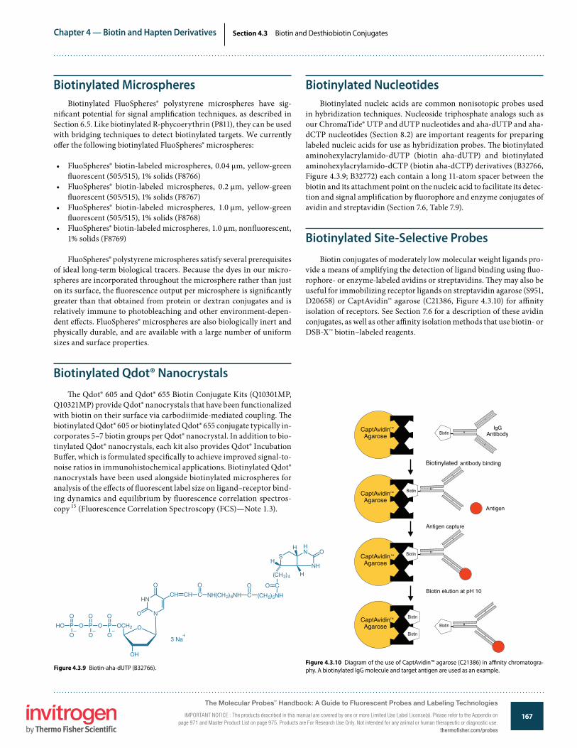
Chapter 4 — Biotin and Hapten Derivatives
167www.invitrogen.com/probes
The Molecular Probes® Handbook: A Guide to Fluorescent Probes and Labeling TechnologiesIMPORTANT NOTICE: The products described in this manual are covered by one or more Limited Use Label License(s). Please refer to the Appendix on page 971 and Master Product List on page 975. Products are For Research Use Only. Not intended for any animal or human therapeutic or diagnostic use.
Section 4.3 Biotin and Desthiobiotin Conjugates
Figure 4.3.9 Biotin-aha-dUTP (B32766).
3 Na
(CH2)4
S
HN
NH
O
H
H
H
O
POPOP
O
HO
O O O
O O
HN
N
O
OH
OCH2
O
CH CH C
O
NH(CH2)6NH C
O
(CH2)5NH
CO
Figure 4.3.10 Diagram of the use of CaptAvidin™ agarose (C21386) in a�nity chromatogra-phy. A biotinylated IgG molecule and target antigen are used as an example.
Biotinylated MicrospheresBiotinylated FluoSpheres® polystyrene microspheres have sig-
ni�cant potential for signal ampli�cation techniques, as described in Section 6.5. Like biotinylated R-phycoerythrin (P811), they can be used with bridging techniques to detect biotinylated targets. We currently o�er the following biotinylated FluoSpheres® microspheres:
• FluoSpheres® biotin-labeled microspheres, 0.04 µm, yellow-green �uorescent (505/515), 1% solids (F8766)
• FluoSpheres® biotin-labeled microspheres, 0.2 µm, yellow-green �uorescent (505/515), 1% solids (F8767)
• FluoSpheres® biotin-labeled microspheres, 1.0 µm, yellow-green �uorescent (505/515), 1% solids (F8768)
• FluoSpheres® biotin-labeled microspheres, 1.0 µm, non�uorescent, 1% solids (F8769)
FluoSpheres® polystyrene microspheres satisfy several prerequisites of ideal long-term biological tracers. Because the dyes in our micro-spheres are incorporated throughout the microsphere rather than just on its surface, the �uorescence output per microsphere is signi�cantly greater than that obtained from protein or dextran conjugates and is relatively immune to photobleaching and other environment-depen-dent e�ects. FluoSpheres® microspheres are also biologically inert and physically durable, and are available with a large number of uniform sizes and surface properties.
Biotinylated Qdot® Nanocrystals�e Qdot® 605 and Qdot® 655 Biotin Conjugate Kits (Q10301MP,
Q10321MP) provide Qdot® nanocrystals that have been functionalized with biotin on their surface via carbodiimide-mediated coupling. �e biotinylated Qdot® 605 or biotinylated Qdot® 655 conjugate typically in-corporates 5–7 biotin groups per Qdot® nanocrystal. In addition to bio-tinylated Qdot® nanocrystals, each kit also provides Qdot® Incubation Bu�er, which is formulated speci�cally to achieve improved signal-to-noise ratios in immunohistochemical applications. Biotinylated Qdot® nanocrystals have been used alongside biotinylated microspheres for analysis of the e�ects of �uorescent label size on ligand–receptor bind-ing dynamics and equilibrium by �uorescence correlation spectros-copy 15 (Fluorescence Correlation Spectroscopy (FCS)—Note 1.3).
Biotinylated NucleotidesBiotinylated nucleic acids are common nonisotopic probes used
in hybridization techniques. Nucleoside triphosphate analogs such as our ChromaTide® UTP and dUTP nucleotides and aha-dUTP and aha-dCTP nucleotides (Section 8.2) are important reagents for preparing labeled nucleic acids for use as hybridization probes. �e biotinylated aminohexylacrylamido-dUTP (biotin aha-dUTP) and biotinylated aminohexylacrylamido-dCTP (biotin aha-dCTP) derivatives (B32766, Figure 4.3.9; B32772) each contain a long 11-atom spacer between the biotin and its attachment point on the nucleic acid to facilitate its detec-tion and signal ampli�cation by �uorophore and enzyme conjugates of avidin and streptavidin (Section 7.6, Table 7.9).
Biotinylated Site-Selective ProbesBiotin conjugates of moderately low molecular weight ligands pro-
vide a means of amplifying the detection of ligand binding using �uo-rophore- or enzyme-labeled avidins or streptavidins. �ey may also be useful for immobilizing receptor ligands on streptavidin agarose (S951, D20658) or CaptAvidin™ agarose (C21386, Figure 4.3.10) for a�nity isolation of receptors. See Section 7.6 for a description of these avidin conjugates, as well as other a�nity isolation methods that use biotin- or DSB-X™ biotin–labeled reagents.
The Molecular Probes™ Handbook: A Guide to Fluorescent Probes and Labeling Technologies
IMPORTANT NOTICE : The products described in this manual are covered by one or more Limited Use Label License(s). Please refer to the Appendix on page 971 and Master Product List on page 975. Products are For Research Use Only. Not intended for any animal or human therapeutic or diagnostic use.
thermofisher.com/probes
Chapter 4 — Biotin and Hapten Derivatives
168www.invitrogen.com/probes
The Molecular Probes® Handbook: A Guide to Fluorescent Probes and Labeling TechnologiesIMPORTANT NOTICE: The products described in this manual are covered by one or more Limited Use Label License(s). Please refer to the Appendix on page 971 and Master Product List on page 975. Products are For Research Use Only. Not intended for any animal or human therapeutic or diagnostic use.
Section 4.3 Biotin and Desthiobiotin Conjugates
Our biotinylated ligands include:
• Biotin-X conjugate of annexin V (A13204, Section 15.5), for detecting the externalization of phosphatidylserine, an early indicator of apoptosis
• Biotin-XX conjugate of α-bungarotoxin 16–19 (B1196, Section 16.2), for labeling the nicotinic acetylcholine receptor (nAChR)
• Biotin-XX cholera toxin subunit B (C34779, Section 7.7), which binds to cell-surface ganglio-side GM1 receptors and is a marker of lipid ra�s 20
• Biotin-XX conjugate of epidermal growth factor 21–23 (EGF, E3477), as well as biotinylated EGF complexed with Alexa Fluor® 488 streptavidin (E13345, Figure 4.3.11), Alexa Fluor® 555 streptavidin (E35350), Alexa Fluor® 647 streptavidin (E35351) or Texas Red® streptavidin (E3480, Figure 4.3.12), for labeling EGF receptors (Section 16.1)
• Biotin-XX conjugate of isolectin IB4 from Gri�onia simplicifolia24 (I21414, Section 7.7), for detecting α-galactosyl moieties in microglia and other glycoproteins
• Biotin-XX conjugate of phalloidin 25,26 (B7474, Section 11.1), for labeling F-actin• Biotin-XX conjugate of transferrin 27 (T23363, Section 16.1), for following intracellular traf-
�cking of this iron-carrying protein• Biotin-XX tyramide, which is an ampli�cation reagent used in several of our TSA™ Kits
(Section 6.2, Table 6.1) for high-sensitivity detection of targets in cells and tissues
Biotinylated LipidsOur extensive selection of labeled phospholipids includes two phospholipid derivatives of
biotin and biotin-X:
• Biotin DHPE (N-(biotinoyl)-1,2-dihexadecanoyl-sn-glycero-3-phosphoethanolamine (B1550, Figure 4.3.13)
• Biotin-X DHPE (N-((6-(biotinoyl)amino)hexanoyl)-1,2-dihexadecanoyl-sn-glycero-3-phos-phoethanolamine (B1616, Figure 4.3.14)
�ese biotinylated lipids, which are also described in Section 13.2, can be used to prepare liposomes that retain high a�nity for avidin conjugates and can be captured for downstream analysis using streptavidin agarose 28 (S951, Section 7.6).
Figure 4.3.13 Biotin DHPE (N-(biotinoyl)-1,2-dihexadecanoyl-sn-glycero-3-phosphoethanolamine, triethylammonium salt, B1550).
C OCH2
C OCH
CH2O P OCH2CH2NH
O
O
CH3(CH2)14
O
CH3(CH2)14
O
(CH2)4
S
HN
NH
O
H
H
H
C
O
(CH3CH2)3NH
Figure 4.3.12 Lightly �xed human epidermoid carcinoma cells (A431) stained with biotinylated epidermal growth fac-tor (EGF) complexed to Texas Red® streptavidin (E3480). An identical cell preparation stained in the presence of a 100-fold excess of unlabeled EGF (E3476) showed no �uorescent signal (data not shown).
Figure 4.3.11 Early endosomes in live HeLa cells identi�ed after a 10-minute incubation with green-�uorescent Alexa Fluor® 488 epidermal growth factor (E13345). The cells were subsequently �xed with formaldehyde and labeled with an antibody to the late endosomal protein, RhoB. That anti-body was visualized with a red-orange–�uorescent second-ary antibody. Nuclei were stained with TO-PRO®-3 iodide (T3605, pseudocolored blue). The image was contributed by Harry Mellor, University of Bristol.
The Molecular Probes™ Handbook: A Guide to Fluorescent Probes and Labeling Technologies
IMPORTANT NOTICE : The products described in this manual are covered by one or more Limited Use Label License(s). Please refer to the Appendix on page 971 and Master Product List on page 975. Products are For Research Use Only. Not intended for any animal or human therapeutic or diagnostic use.thermofisher.com/probes

Chapter 4 — Biotin and Hapten Derivatives
169www.invitrogen.com/probes
The Molecular Probes® Handbook: A Guide to Fluorescent Probes and Labeling TechnologiesIMPORTANT NOTICE: The products described in this manual are covered by one or more Limited Use Label License(s). Please refer to the Appendix on page 971 and Master Product List on page 975. Products are For Research Use Only. Not intended for any animal or human therapeutic or diagnostic use.
Section 4.3 Biotin and Desthiobiotin Conjugates
Figure 4.3.14 Biotin-X DHPE (N-((6-(biotinoyl)amino)hexanoyl)-1,2-dihexadecanoyl-sn-glycero-3-phosphoethanolamine, triethylammonium salt, B1616).
C OCH2
C OCH
CH2O P OCH2CH2NH
O
O
CH3(CH2)14
O
CH3(CH2)14
O
(CH2)4
S
HN
NH
O
H
H
H
C
O
(CH3CH2)3NH
(CH2)5NHC
O
REFERENCES1. Biochim Biophys Acta (1998) 1381:203; 2. Clin Chem (1994) 40:2112; 3. Anal Chem (1988) 60:853; 4. Biochim Biophys Acta (1999) 1427:33; 5. Methods Mol Biol (2008) 418:73; 6. Proc Natl Acad Sci U S A (2005) 102:8507; 7. Science (2008) 321:417; 8. Nat Neurosci (2005) 8:916; 9. J Neurosci Methods (1993) 46:59; 10. Anal Biochem (2002) 308:343; 11. Amino Acids (2007) 33:51; 12. Biochemistry (2000) 39:11121; 13. J Biol Chem (2009) 284:22213; 14. Nat Immunol (2008) 9:866; 15. Biophys J (2008) 95:865; 16. BMC Neurosci (2009) 10:80; 17. Nano Lett (2008) 8:780; 18. Mol Brain (2008) 1:18; 19. J Neurosci (2005) 25:9949; 20. Am J Pathol (2008) 172:1683; 21. Methods Mol Biol (2007) 374:69; 22. J Microsc (2006) 222:22; 23. Nat Biotechnol (2004) 22:198; 24. Nat Neurosci (2008) 11:429; 25. J Cell Biol (1995) 130:591; 26. Anal Biochem (1992) 200:199; 27. J Cell Biol (2000) 149:901; 28. Biotechniques (2005) 38:858.
DATA TABLE 4.3 BIOTIN AND DESTHIOBIOTIN CONJUGATESCat. No. MW Storage Soluble Abs EC Em Solvent NotesA12922 1141.31 D,L DMSO, H2O 591 80,000 618 pH 7A12923 1209.66 D,L DMSO, H2O 556 99,000 572 pH 7A12924 974.98 D,L DMSO, H2O 494 62,000 520 pH 7B1196 ~8400 F,D H2O <300 none 1B1370 831.01 L DMF, pH >6 494 75,000 518 pH 9 2B1550 1019.45 FF,D see Notes <300 none 3B1616 1132.61 FF,D see Notes <300 none 3B7474 ~1300 F MeOH, H2O <300 none 1, 4B10570 644.70 L DMSO 494 68,000 523 pH 9 2B32766 1041.78 FF H2O <300 none 5B32772 ~1050 FF H2O <300 none 5E3477 ~6600 FF,D H2O <300 none 1E3480 see Notes FF,D,L H2O 596 ND 612 pH 7 6, 7E13345 see Notes FF,D,L H2O 497 ND 520 pH 8 6, 8E35350 see Notes FF,D,L H2O 554 ND 568 pH 7 6, 9E35351 see Notes FF,D,L H2O 653 ND 671 pH 7 6, 10G36000 561.67 F,D DMSO <300 noneL2601 873.10 D,L H2O 428 11,000 531 H2OL6950 850.03 D,L H2O 428 11,000 532 pH 7O12920 887.39 L DMSO, H2O 495 66,000 522 pH 9 11T12921 869.09 D,L DMSO 554 103,000 581 pH 7For de�nitions of the contents of this data table, see “Using The Molecular Probes® Handbook” in the introductory pages.Notes
1. α-Bungarotoxin, EGF and phallotoxin conjugates have approximately 1 label per peptide.2. Absorption and �uorescence of �uorescein derivatives are pH dependent. Extinction coe�cients and �uorescence quantum yields decrease markedly at pH <7.3. Chloroform is the most generally useful solvent for preparing stock solutions of phospholipids (including sphingomyelins). Glycerophosphocholines are usually freely soluble in ethanol. Most
other glycerophospholipids (phosphoethanolamines, phosphatidic acids and phosphoglycerols) are less soluble in ethanol, but solutions up to 1–2 mg/mL should be obtainable, using sonica-tion to aid dispersion if necessary. Labeling of cells with �uorescent phospholipids can be enhanced by addition of cyclodextrins during incubation. (J Biol Chem (1999) 274:35359)
4. Although this phallotoxin is water soluble, storage in water is not recommended, particularly in dilute solution.5. This product is supplied as a ready-made solution in the solvent indicated under "Soluble."6. ND = not determined.7. E3480 is a complex of E3477 with Texas Red® streptavidin, which typically incorporates 3 dyes/streptavidin (MW ~52,800).8. E13345 is a complex of E3477 with Alexa Fluor® 488 streptavidin, which typically incorporates 5 dyes/streptavidin (MW ~52,800).9. E35350 is a complex of E3477 with Alexa Fluor® 555 streptavidin, which typically incorporates 3 dyes/streptavidin (MW ~52,800).10. E35351 is a complex of E3477 with Alexa Fluor® 647 streptavidin, which typically incorporates 3 dyes/streptavidin (MW ~52,800).11. Absorption and �uorescence of Oregon Green® 488 derivatives are pH dependent only in moderately acidic solutions (pH <5).
The Molecular Probes™ Handbook: A Guide to Fluorescent Probes and Labeling Technologies
IMPORTANT NOTICE : The products described in this manual are covered by one or more Limited Use Label License(s). Please refer to the Appendix on page 971 and Master Product List on page 975. Products are For Research Use Only. Not intended for any animal or human therapeutic or diagnostic use.
thermofisher.com/probes

Chapter 4 — Biotin and Hapten Derivatives
170www.invitrogen.com/probes
The Molecular Probes® Handbook: A Guide to Fluorescent Probes and Labeling TechnologiesIMPORTANT NOTICE: The products described in this manual are covered by one or more Limited Use Label License(s). Please refer to the Appendix on page 971 and Master Product List on page 975. Products are For Research Use Only. Not intended for any animal or human therapeutic or diagnostic use.
Section 4.3 Biotin and Desthiobiotin Conjugates
Cat. No. Description QuantityA12924 Alexa Fluor® 488 biocytin, disodium salt (biocytin Alexa Fluor® 488) 250 µgA12923 Alexa Fluor® 546 biocytin, sodium salt (biocytin Alexa Fluor® 546) 250 µgA12922 Alexa Fluor® 594 biocytin, sodium salt (biocytin Alexa Fluor® 594) 250 µgA13204 annexin V, biotin-X conjugate *100 assays* 500 µLA6435 anti–dinitrophenyl-KLH, rabbit IgG fraction, biotin-XX conjugate *2 mg/mL* 0.5 mLA982 anti–�uorescein/Oregon Green®, rabbit IgG fraction, biotin-XX conjugate *1 mg/mL* 0.5 mLA10263 anti–Green Fluorescent Protein, chicken IgY fraction, biotin-XX conjugate (anti-GFP, IgY, biotin-XX conjugate) *2 mg/mL* 100 µLA10259 anti–Green Fluorescent Protein, rabbit IgG fraction, biotin-XX conjugate (anti-GFP, IgG, biotin-XX conjugate) *2 mg/mL* 100 µLA21272 anti–HuC/HuD neuronal protein (human), mouse IgG2b, monoclonal 16A11, biotin-XX conjugate 100 µgA5751 anti–lucifer yellow, rabbit IgG fraction, biotin-XX conjugate *3 mg/mL* 0.5 mLA21371 anti–α-tubulin (bovine), mouse IgG1, monoclonal 236-10501, biotin-XX conjugate 50 µgB32772 biotin-aha-dCTP *1 mM in TE bu�er* 25 µLB32766 biotin-aha-dUTP *1 mM in TE bu�er* 25 µLB1550 biotin DHPE (N-(biotinoyl)-1,2-dihexadecanoyl-sn-glycero-3-phosphoethanolamine, triethylammonium salt) 10 mgB10570 biotin-4-�uorescein 5 mgB1370 5-((N-(5-(N-(6-(biotinoyl)amino)hexanoyl)amino)pentyl)thioureidyl)�uorescein (�uorescein biotin) 5 mgB1616 biotin-X DHPE (N-((6-(biotinoyl)amino)hexanoyl)-1,2-dihexadecanoyl-sn-glycero-3-phosphoethanolamine, triethylammonium salt) 5 mgB11027 biotin-XX F(ab’)2 fragment of goat anti-mouse IgG (H+L) *2 mg/mL* 250 µLB21078 biotin-XX F(ab’)2 fragment of goat anti-rabbit IgG (H+L) *2 mg/mL* 250 µLB2763 biotin-XX goat anti-mouse IgG (H+L) *2 mg/mL* 0.5 mLB2770 biotin-XX goat anti-rabbit IgG (H+L) *2 mg/mL* 0.5 mLB7474 biotin-XX phalloidin 50 UB35138 BrdU, mouse monoclonal antibody (Clone MoBU-1), biotin conjugate (anti-BrdU, biotin conjugate) *0.2 mg/mL* 350 µLB1196 α-bungarotoxin, biotin-XX conjugate 500 µgC34779 cholera toxin subunit B (recombinant), biotin-XX conjugate 100 µgD7135 dextran, biotin, 3000 MW, lysine �xable (BDA-3000) 10 mgD1956 dextran, biotin, 10,000 MW, lysine �xable (BDA-10,000) 25 mgD1957 dextran, biotin, 70,000 MW, lysine �xable (BDA-70,000) 25 mgD7142 dextran, biotin, 500,000 MW, lysine �xable (BDA-500,000) 10 mgD7156 dextran, �uorescein and biotin, 3000 MW, anionic, lysine �xable (micro-emerald) 5 mgD7178 dextran, �uorescein and biotin, 10,000 MW, anionic, lysine �xable (mini-emerald) 10 mgD7162 dextran, tetramethylrhodamine and biotin, 3000 MW, lysine �xable (micro-ruby) 5 mgD3312 dextran, tetramethylrhodamine and biotin, 10,000 MW, lysine �xable (mini-ruby) 10 mgD20658 DSB-X™ Bioconjugate Isolation Kit #1 *with streptavidin agarose* *5 isolations* 1 kitD20698 DSB-X™ biotin donkey anti–goat IgG (H+L) *2 mg/mL* 0.5 mLD20701 DSB-X™ biotin goat anti–chicken IgG (H+L) *2 mg/mL* 0.5 mLD20691 DSB-X™ biotin goat anti–mouse IgG (H+L) *highly cross-adsorbed* *2 mg/mL* 0.5 mLD20693 DSB-X™ biotin goat anti–mouse IgM (µ chain) *2 mg/mL* 250 µLD20697 DSB-X™ biotin goat anti–rat IgG (H+L) *2 mg/mL* 0.5 mLE3477 epidermal growth factor, biotin-XX conjugate (biotin EGF) 20 µgE13345 epidermal growth factor, biotinylated, complexed to Alexa Fluor® 488 streptavidin (Alexa Fluor® 488 EGF complex) 100 µgE35350 epidermal growth factor, biotinylated, complexed to Alexa Fluor® 555 streptavidin (Alexa Fluor® 555 EGF complex) 100 µgE35351 epidermal growth factor, biotinylated, complexed to Alexa Fluor® 647 streptavidin (Alexa Fluor® 647 EGF complex) 100 µgE3480 epidermal growth factor, biotinylated, complexed to Texas Red® streptavidin (Texas Red® EGF complex) 100 µgF8769 FluoSpheres® biotin-labeled microspheres, 1.0 µm, non�uorescent *1% solids* 0.4 mLF8766 FluoSpheres® biotin-labeled microspheres, 0.04 µm, yellow-green �uorescent (505/515) *1% solids* 0.4 mLF8767 FluoSpheres® biotin-labeled microspheres, 0.2 µm, yellow-green �uorescent (505/515) *1% solids* 0.4 mLF8768 FluoSpheres® biotin-labeled microspheres, 1.0 µm, yellow-green �uorescent (505/515) *1% solids* 0.4 mLG36000 glutathione ethyl ester, biotin amide (BioGEE) *glutathiolation detection reagent* *special packaging* 10 x 100 µgI21414 isolectin GS-IB4 from Gri�onia simplicifolia, biotin-XX conjugate 500 µgL6950 lucifer yellow biocytin, potassium salt (biocytin lucifer yellow) 5 mgL2601 lucifer yellow cadaverine biotin-X, dipotassium salt 10 mgO12920 Oregon Green® 488 biocytin (biocytin Oregon Green® 488) 5 mgP917 peroxidase from horseradish, biotin-XX conjugate 10 mgP811 R-phycoerythrin, biotin-XX conjugate *4 mg/mL* 0.5 mLQ10301MP Qdot® 605 Biotin Conjugate Kit 1 kitQ10321MP Qdot® 655 Biotin Conjugate Kit 1 kitT12921 5-(and-6)-tetramethylrhodamine biocytin (biocytin TMR) 5 mgT23363 transferrin from human serum, biotin-XX conjugate 5 mg
PRODUCT LIST 4.3 BIOTIN AND DESTHIOBIOTIN CONJUGATES
The Molecular Probes™ Handbook: A Guide to Fluorescent Probes and Labeling Technologies
IMPORTANT NOTICE : The products described in this manual are covered by one or more Limited Use Label License(s). Please refer to the Appendix on page 971 and Master Product List on page 975. Products are For Research Use Only. Not intended for any animal or human therapeutic or diagnostic use.thermofisher.com/probes