chapter 1 beginnings: the molecular pathology of hemoglobin · chapter 1 beginnings: the molecular...
TRANSCRIPT

1
Chapter 1 Beginnings: the molecular pathology of hemoglobinDavid Weatherall
once pinpointed, the appropriate bacterial colonies could be grown to generate larger quantities of DNA carrying a par-ticular gene. Later it became possible to sequence these genes, persuade them to synthesize their products in microorgan-isms, cultured cells or even other species, and hence to defi ne their key regulatory regions.
The early work in the fi eld of human molecular genetics fo-cussed on diseases in which there was some knowledge of the genetic defect at the protein or biochemical level. However, once linkage maps of the human genome became available, following the identifi cation of highly polymorphic regions of DNA, it was possible to search for any gene for a disease, even where the cause was completely unknown. This approach, fi rst called ‘reverse genetics’ and later rechristened ‘positional cloning’, led to the discovery of genes for many important diseases.
As even more DNA markers became available and as meth-ods for sequencing were improved and automated, thoughts turned to the next major goal in this fi eld, which was to de-termine the complete sequence of the bases that constitute our 30 000 or so genes and all that lies between them: the Human Genome Project. This remarkable endeavor was par-tially completed recently and should be fi nished within the next few years. The further understanding of the functions and regulation of our genes will require multidisciplinary research encompassing many different fi elds. The next stage in the Human Genome Project, called ‘genome annotation’, entails analyzing the raw DNA sequence in order to determine its biological signifi cance. One of the main ventures in the era of functional genomics will be in what is termed ‘proteomics’, the large-scale analysis of the protein products of genes. The ultimate goal will be to try to defi ne the protein complement, or proteome, of cells and how the many different proteins interact with one another. To this end, large-scale facilities are being established for isolating and purifying the protein
Historical background
Linus Pauling fi rst used the term ‘molecular disease’ in 1949, after the discovery that the structure of sickle cell hemoglobin differed from that of normal hemoglobin. Indeed, it was this seminal observation that led to the concept of molecular medi-cine; that is, the description of disease mechanisms at the level of cells and molecules. However, until the development of recombinant DNA technology in the mid-1970s, knowledge of events inside the cell nucleus, notably how genes function, could only be the subject of guesswork based on the structure and function of their protein products. However, as soon as it became possible to isolate human genes and to study their properties, the picture changed dramatically.
Progress over the last 20 years has been driven by techno-logical advances in molecular biology. At fi rst it was possible only to obtain indirect information about the structure and function of genes by DNA/DNA and DNA/RNA hybridiza-tion; that is, by probing the quantity or structure of RNA or DNA by annealing reactions with molecular probes. The next major advance was the ability to fractionate DNA into pieces of predictable size with bacterial restriction enzymes. This led to the invention of a technique that played a central role in the early development of human molecular genetics, called ‘Southern blotting’ after the name of its developer, Edwin Southern. This method allowed the structure and organiza-tion of genes to be studied directly for the fi rst time and led to the defi nition of a number of different forms of molecular pathology.
Once it was possible to fractionate DNA, it soon became feasible to insert the pieces into vectors that are able to divide within bacteria. The steady improvement in the properties of cloning vectors made it possible to generate libraries of human DNA growing in bacterial cultures. Ingenious approaches were developed to scan the libraries to detect genes of interest;
Historical background, 1The structure, genetic control and synthesis of normal hemoglobin, 2The molecular pathology of hemoglobin, 6
Genotype–phenotype relationships in the inherited disorders of hemoglobin, 12
Postscript, 16Further reading, 17

Chapter 1

2 Molecular Hematology
products of genes that have been expressed in bacteria. Their structure can then be studied by a variety of different tech-niques, notably X-ray crystallography and nuclear magnetic resonance spectroscopy. The crystallographic analysis of pro-teins is being greatly facilitated by the use of X-ray beams from a synchrotron radiation source.
During this remarkable period of technical advance, con-siderable progress has been made towards an understanding of the pathology of disease at the molecular level. This has had a particular impact on hematology, leading to advances in the understanding of gene function and disease mechanisms in almost every aspect of the fi eld.
The inherited disorders of hemoglobin, the thalassemias and structural hemoglobin variants, the commonest human monogenic diseases, were the fi rst to be studied systematically at the molecular level and a great deal is known about their genotype–phenotype relationships. This fi eld led the way to molecular hematology and, indeed, to the development of molecular medicine. Thus, even though the genetics of he-moglobin is complicated by the fact that different varieties are produced at particular stages of human development, the molecular pathology of the hemoglobinopathies provides an excellent model system for understanding any monogenic disease and the complex interactions between genotype and environment that underlie many multigenic disorders.
In this chapter we will consider the structure, synthesis and genetic control of the human hemoglobins, describe the molecular pathology of the hemoglobin disorders individu-ally, and discuss briefl y how the complex interactions of their different genotypes produce a remarkably diverse family of clinical phenotypes. Readers who wish to learn more about the methods of molecular genetics, particularly as applied to the study of hemoglobin disorders, are referred to the reviews cited at the end of this chapter.
The structure, genetic control and synthesis of normal hemoglobin
Structure and function
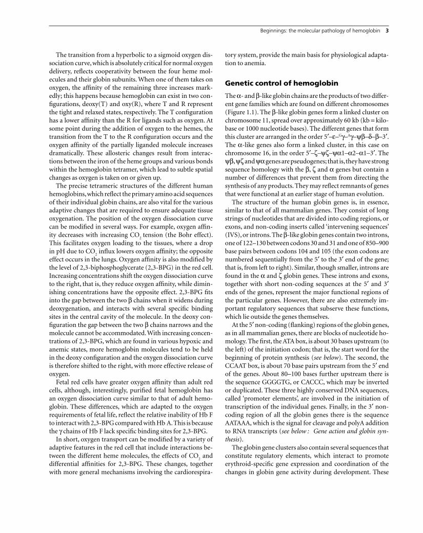
The varying oxygen requirements during embryonic, fetal and adult life are refl ected in the synthesis of different struc-tural hemoglobins at each stage of human development. They all have the same general tetrameric structure, however, con-sisting of two different pairs of globin chains, each attached to one heme molecule. Adult and fetal hemoglobins have α chains combined with β chains (Hb A, α
2β
2), δ chains (Hb A
2,
α2δ
2) and γ chains (Hb F, α
2γ
2). In embryos, α-like chains
called ζ chains combine with γ chains to produce Hb Portland (ζ
2γ
2), or with ε chains to make Hb Gower 1 (ζ
2ε
2), while α and
ε chains form Hb Gower 2 (α2ε
2). Fetal hemoglobin is het-
erogeneous; there are two varieties of γ chain that differ only in their amino acid composition at position 136, which may be occupied by either glycine or alanine; γ chains containing glycine at this position are called Gγ chains, those with alanine, Aγ chains (Figure 1.1).
The synthesis of hemoglobin tetramers consisting of two unlike pairs of globin chains is absolutely essential for the effective function of hemoglobin as an oxygen carrier. The classical sigmoid shape of the oxygen dissociation curve, which refl ects the allosteric properties of the hemoglobin molecule, ensures that, at high oxygen tensions in the lungs, oxygen is readily taken up and later released effectively at the lower tensions encountered in the tissues. The shape of the curve is quite different to that of myoglobin, a molecule which consists of a single globin chain with heme attached to it, which, like abnormal hemoglobins that consist of ho-motetramers of like-chains, has a hyperbolic oxygen disso-ciation curve.
Fig. 1.1 The genetic control of human hemoglobin production in embryonic, fetal and adult life
1 Kb
30 3131 32 104 10599 100
11βδψβAγGγεα1 θ1α2ψα2 ψα1ψζζ
16
ζ2ε2Hb Gower 1
ζ2γ2Hb Portland
α2ε2Hb Gower 2
α2γ2HbF
α2β2HbA
α2δ2HbA2
Embryo Fetus Adult

1 Kb
30 3131 32 104 10599 100
11βδψβAγGγεα1 θ1α2ψα2 ψα1ψζζ
16
ζ2ε2Hb Gower 1
ζ2γ2Hb Portland
α2ε2Hb Gower 2
α2γ2HbF
α2β2HbA
α2δ2HbA2
Embryo Fetus Adult
Beginnings: the molecular pathology of hemoglobin 3
The transition from a hyperbolic to a sigmoid oxygen dis-sociation curve, which is absolutely critical for normal oxygen delivery, refl ects cooperativity between the four heme mol-ecules and their globin subunits. When one of them takes on oxygen, the affi nity of the remaining three increases mark-edly; this happens because hemoglobin can exist in two con-fi gurations, deoxy(T) and oxy(R), where T and R represent the tight and relaxed states, respectively. The T confi guration has a lower affi nity than the R for ligands such as oxygen. At some point during the addition of oxygen to the hemes, the transition from the T to the R confi guration occurs and the oxygen affi nity of the partially liganded molecule increases dramatically. These allosteric changes result from interac-tions between the iron of the heme groups and various bonds within the hemoglobin tetramer, which lead to subtle spatial changes as oxygen is taken on or given up.
The precise tetrameric structures of the different human hemoglobins, which refl ect the primary amino acid sequences of their individual globin chains, are also vital for the various adaptive changes that are required to ensure adequate tissue oxygenation. The position of the oxygen dissociation curve can be modifi ed in several ways. For example, oxygen affi n-ity decreases with increasing CO
2 tension (the Bohr effect).
This facilitates oxygen loading to the tissues, where a drop in pH due to CO
2 infl ux lowers oxygen affi nity; the opposite
effect occurs in the lungs. Oxygen affi nity is also modifi ed by the level of 2,3-biphosphoglycerate (2,3-BPG) in the red cell. Increasing concentrations shift the oxygen dissociation curve to the right, that is, they reduce oxygen affi nity, while dimin-ishing concentrations have the opposite effect. 2,3-BPG fi ts into the gap between the two β chains when it widens during deoxygenation, and interacts with several specifi c binding sites in the central cavity of the molecule. In the deoxy con-fi guration the gap between the two β chains narrows and the molecule cannot be accommodated. With increasing concen-trations of 2,3-BPG, which are found in various hypoxic and anemic states, more hemoglobin molecules tend to be held in the deoxy confi guration and the oxygen dissociation curve is therefore shifted to the right, with more effective release of oxygen.
Fetal red cells have greater oxygen affi nity than adult red cells, although, interestingly, purifi ed fetal hemoglobin has an oxygen dissociation curve similar to that of adult hemo-globin. These differences, which are adapted to the oxygen requirements of fetal life, refl ect the relative inability of Hb F to interact with 2,3-BPG compared with Hb A. This is because the γ chains of Hb F lack specifi c binding sites for 2,3-BPG.
In short, oxygen transport can be modifi ed by a variety of adaptive features in the red cell that include interactions be-tween the different heme molecules, the effects of CO
2 and
differential affi nities for 2,3-BPG. These changes, together with more general mechanisms involving the cardiorespira-
tory system, provide the main basis for physiological adapta-tion to anemia.
Genetic control of hemoglobin
The α- and β-like globin chains are the products of two differ-ent gene families which are found on different chromosomes (Figure 1.1). The β-like globin genes form a linked cluster on chromosome 11, spread over approximately 60 kb (kb = kilo-base or 1000 nucleotide bases). The different genes that form this cluster are arranged in the order 5′–ε–Gγ–Aγ–ψβ–δ–β–3′. The α-like genes also form a linked cluster, in this case on chromosome 16, in the order 5′–ζ–ψζ–ψα1–α2–α1–3′. The ψβ, ψζ and ψα genes are pseudogenes; that is, they have strong sequence homology with the β, ζ and α genes but contain a number of differences that prevent them from directing the synthesis of any products. They may refl ect remnants of genes that were functional at an earlier stage of human evolution.
The structure of the human globin genes is, in essence, similar to that of all mammalian genes. They consist of long strings of nucleotides that are divided into coding regions, or exons, and non-coding inserts called ‘intervening sequences’ (IVS), or introns. The β-like globin genes contain two introns, one of 122–130 between codons 30 and 31 and one of 850–900 base pairs between codons 104 and 105 (the exon codons are numbered sequentially from the 5′ to the 3′ end of the gene; that is, from left to right). Similar, though smaller, introns are found in the α and ζ globin genes. These introns and exons, together with short non-coding sequences at the 5′ and 3′ ends of the genes, represent the major functional regions of the particular genes. However, there are also extremely im-portant regulatory sequences that subserve these functions, which lie outside the genes themselves.
At the 5′ non-coding (fl anking) regions of the globin genes, as in all mammalian genes, there are blocks of nucleotide ho-mology. The fi rst, the ATA box, is about 30 bases upstream (to the left) of the initiation codon; that is, the start word for the beginning of protein synthesis (see below). The second, the CCAAT box, is about 70 base pairs upstream from the 5′ end of the genes. About 80–100 bases further upstream there is the sequence GGGGTG, or CACCC, which may be inverted or duplicated. These three highly conserved DNA sequences, called ‘promoter elements’, are involved in the initiation of transcription of the individual genes. Finally, in the 3′ non-coding region of all the globin genes there is the sequence AATAAA, which is the signal for cleavage and polyA addition to RNA transcripts (see below : Gene action and globin syn-thesis).
The globin gene clusters also contain several sequences that constitute regulatory elements, which interact to promote erythroid-specifi c gene expression and coordination of the changes in globin gene activity during development. These


4 Molecular Hematology
include the globin genes themselves and their promoter ele-ments—enhancers (regulatory sequences that increase gene expression despite being located at a considerable distance from the genes) and ‘master’ regulatory sequences called, in the case of the β globin gene cluster, the ‘locus control region’ (LCR); and, in the case of the α genes, HS40 (a nuclease-hy-persensitive site in DNA 40 kb from the α globin genes). Each of these sequences has a modular structure made up of an array of short motifs that represent the binding sites for tran-scriptional activators or repressors.
Gene action and globin synthesis
The fl ow of information between DNA and protein is sum-marized in Figure 1.2. When a globin gene is transcribed, mes-senger RNA (mRNA) is synthesized from one of its strands, a process which begins with the formation of a transcription complex consisting of a variety of regulatory proteins to-gether with an enzyme called RNA polymerase (see below). The primary transcript is a large mRNA precursor which con-tains both intron and exon sequences. While in the nucleus, this molecule undergoes a variety of modifi cations. First, the introns are removed and the exons are spliced together. The intron/exon junctions always have the same sequence: GT at their 5′ end, and AG at their 3′ end. This appears to be essential
for accurate splicing; if there is a mutation at these sites this process does not occur. Splicing refl ects a complex series of intermediary stages and the interaction of a number of differ-ent nuclear proteins. After the exons are joined, the mRNAs are modifi ed and stabilized; at their 5′ end a complex CAP structure is formed, while at their 3′ end a string of adenylic acid residues (polyA) is added. The mRNA processed in this way moves into the cytoplasm, where it acts as a template for globin chain production. Because of the rules of base pair-ing—that is, cytosine always pairs with thymine, and guanine with adenine—the structure of the mRNA refl ects a faithful copy of the DNA codons from which it is synthesized; the only difference is that, in RNA, uracil (U) replaces thymine (T).
Amino acids are transported to the mRNA template on car-riers called transfer RNAs (tRNAs); there are specifi c tRNAs for each amino acid. Furthermore, because the genetic code is redundant (that is, more than one codon can encode a particular amino acid), for some of the amino acids there are several different individual tRNAs. Their order in the globin chain is determined by the order of codons in the mRNA. The tRNAs contain three bases, which together constitute an anticodon; these anticodons are complementary to mRNA codons for particular amino acids. They carry amino acids to the template, where they fi nd the appropriate positioning by codon–anticodon base-pairing. When the fi rst tRNA is in po-
Fig. 1.2 The mechanisms of globin gene transcription and translation
Gene
mRNA precursor
Excision of intronsSplicing of exons
Processed mRNA
TranslationAAAA-A
AAAA-AAAAA-A
Nucleus
Cytoplasm
5' CAP
5' 3'
UAARibosome
Transfer RNA
Aminoacid Growing
chainFinished
chain
UGC UUCACG AAG
A U GU A C
Processedchain
CACCC
CCAAT
TATA
ATG
AATAAA
TAA
NCAGGTAGGTNCFlankingFlanking IVS 1 IVS 2

Beginnings: the molecular pathology of hemoglobin 5
sition, an initiation complex is formed between several pro-tein initiation factors together with the two subunits which constitute the ribosomes. A second tRNA moves in alongside and the two amino acids that they are carrying form a pep-tide bond between them; the globin chain is now two amino acid residues long. This process is continued along the mRNA from left to right, and the growing peptide chain is transferred from one incoming tRNA to the next; that is, the mRNA is translated from 5′ to 3′. During this time the tRNAs are held in appropriate steric confi guration with the mRNA by the two ribosomal subunits. There are specifi c initiation (AUG) and termination (UAA, UAG and UGA) codons. When the ribosomes reach the termination codon, translation ceases, the completed globin chains are released, and the ribosomal subunits are recycled. Individual globin chains combine with heme, which has been synthesized through a separate pathway, and then interact with one like chain and two unlike chains to form a complete hemoglobin tetramer.
Regulation of hemoglobin synthesis
The regulation of globin gene expression is mediated mainly at the transcriptional level, with some fi ne tuning during translation and post-translational modifi cation of the gene products. DNA that is not involved in transcription is held tightly packaged in a compact, chemically modifi ed form that is inaccessible to transcription factors and polymerases and
which is heavily methylated. Activation of a particular gene is refl ected by changes in the structure of the surrounding chromatin, which can be identifi ed by enhanced sensitivity to nucleases. Erythroid lineage-specifi c nuclease-hypersensitive sites are found at several locations in the β globin gene cluster. Four are distributed over 20 kb upstream from the ε globin gene in the region of the β globin LCR (Figure 1.3). This vital regulatory region is able to establish a transcriptionally active domain spanning the entire β globin gene cluster. Several en-hancer sequences have been identifi ed in this cluster. A variety of regulatory proteins bind to the LCR, and to the promoter regions of the globin genes and to the enhancer sequences. It is thought that the LCR and other enhancer regions become opposed to the promoters to increase the rate of transcription of the genes to which they are related.
These regulatory regions contain sequence motifs for vari-ous ubiquitous and erythroid-restricted transcription factors. Binding sites for these factors have been identifi ed in each of the globin gene promoters and at the hypersensitive-site re-gions of the various regulatory elements. A number of the fac-tors which bind to these areas are found in all cell types. They include Sp1, Yy1 and Usf. In contrast, a number of transcrip-tion factors have been identifi ed, including GATA-1, EKLF and NF-E2, which are restricted in their distribution to erythroid cells and, in some cases, megakaryocytes and mast cells. The overlapping of erythroid-specifi c and ubiquitous-factor bind-ing sites in several cases suggests that competitive binding may
Fig. 1.3 The positions of the major regulatory regions in the β and α globin gene clustersThe arrows indicate the position of the erythroid lineage-specifi c nuclease-hypersensitive sites. HS = hypersensitive.
–20
–40 –20
0
LCR
0
20
20 40 kb
40 60 kb
3' HS5' HS
4 3 2 1
ε Gγ Aγ δ β
1
HS–40
ζ α2 α1Chromosome 16
Chromosome 11

–20
–40 –20
0
LCR
0
20
20 40 kb
40 60 kb
3' HS5' HS
4 3 2 1
ε Gγ Aγ δ β
1
HS–40
ζ α2 α1Chromosome 16
Chromosome 11
6 Molecular Hematology
play an important part in the regulation of erythroid-specifi c genes. Another binding factor, SSP, the stage selector protein, appears to interact specifi cally with ε and γ genes.
The binding of hematopoietic-specifi c factors activates the LCR, which renders the entire β globin gene cluster transcrip-tionally active. These factors also bind to the enhancer and promoter sequences, which work in tandem to regulate the expression of the individual genes in the clusters. It is likely that some of the transcriptional factors are developmental-stage-specifi c, and hence may be responsible for the differ-ential expression of the embryonic, fetal and adult globin genes. The α globin gene cluster also contains an element, HS40, which has some structural features in common with the β LCR, although it is different in aspects of its structure. A number of enhancer-like sequences have also been identi-fi ed, although it is becoming clear that there are fundamental differences in the pattern of regulation of the two globin gene clusters.
In addition to the different regulatory sequences outlined above, there are also sequences which may be involved specifi -cally with ‘silencing’ of genes, notably those for the embryonic hemoglobins, during development.
Some degree of regulation is also mediated by differences in the rates of initiation and translation of the different mRNAs, and at the post-transcriptional level by differential affi nity for different protein subunits. However, this kind of post-tran-scriptional fi ne tuning probably plays a relatively small role in determining the overall output of the globin gene products.
Regulation of developmental changes in globin gene expression
During development, the site of red cell production moves from the yolk sac to the fetal liver and spleen, and thence to bone marrow in the adult. Embryonic, fetal and adult he-moglobin synthesis is approximately related in time to these changes in the site of erythropoiesis, although it is quite clear that the various switches, between embryonic and fetal and between fetal and adult hemoglobin synthesis, are beautifully synchronized throughout these different sites. Fetal hemo-globin synthesis declines during the later months of gestation and Hb F is replaced by Hbs A and A
2 by the end of the fi rst
year of life.Despite a great deal of research, very little is known about
the regulation of these different switches from one globin gene to another during development. Work from a variety of different sources suggests that there may be specifi c regions in the α and β globin gene clusters that are responsive to the ac-tion of transcription factors, some of which may be develop-mental-stage-specifi c. However, proteins of this type have not yet been isolated, and nothing is known about their regulation and how it is mediated during development.
The molecular pathology of hemoglobin
As is the case for most monogenic diseases, the inherited dis-orders of hemoglobin fall into two major classes. First, there are those that result from a reduced output of one or other globin genes, the thalassemias. Second, there is a wide range of conditions that result from the production of structurally abnormal globin chains; the type of disease depends on how the particular alteration in protein structure interferes with its stability or function. Of course, no biological classifi ca-tion is entirely satisfactory; those which attempt to defi ne the hemoglobin disorders are no exception. There are some structural hemoglobin variants which happen to be synthe-sized at a reduced rate and hence are associated with a clinical picture similar to thalassemia. And there are other classes of mutations which simply interfere with the normal transition from fetal to adult hemoglobin synthesis, a family of condi-tions that is given the general title ‘hereditary persistence of fetal hemoglobin’. Furthermore, because these diseases are all so common and occur together in particular populations, it is not uncommon for an individual to inherit a gene for one or other form of thalassemia and a structural hemoglobin variant. The rather heterogeneous group of conditions that results from all these different mutations and interactions is summarized in Table 1.1.
Table 1.1 The thalassemias and related disorders.
α Thalassemiaα0
α+
Deletion (−α) Non-deletion (αT)β Thalassemiaβ0
β+
Normal Hb A2
‘Silent’δβ Thalassemia(δβ)+
(δβ)0
(Aγδβ)0
γ Thalassemiaδ Thalassemiaεγδβ ThalassemiaHereditary persistence of fetal hemoglobin Deletion (δβ)0
Non-deletion Linked to β globin genes Gγβ+
Aγβ+
Unlinked to β globin genes


Beginnings: the molecular pathology of hemoglobin 7
Over recent years, the determination of the molecular pa-thology of the two common forms of thalassemia, α and β, has provided a remarkable picture of the repertoire of mutations that can underlie human monogenic disease. Similarly, stud-ies of the relationship between structure and function in the structurally abnormal hemoglobins have provided a great deal of information about normal human hemoglobin function.
In the sections that follow we will describe, in outline, the different forms of molecular pathology that underlie these conditions.
The β thalassemias
There are two main classes of β thalassemia, β0 thalassemia, in which there is an absence of β globin chain production, and β+ thalassemia, in which there is a variable reduction in the out-put of β globin chains. As shown in Figure 1.4, mutations of the β globin genes may cause a reduced output of gene prod-uct at the level of transcription or mRNA processing, transla-tion, or through the stability of the globin gene product.
Defective β globin gene transcription
There are a variety of mechanisms that interfere with the nor-mal transcription of the β globin genes. First, the genes may be either completely or partially deleted. Overall, deletions of the β globin genes are not commonly found in patients with β thalassemia, with one exception: a 619 bp deletion involving the 3′ end of the gene is found frequently in the Sind popula-tions of India and Pakistan, where it constitutes about 30% of the β thalassemia alleles. Other deletions are extremely rare.
A much more common group of mutations, which results in a moderate decrease in the rate of transcription of the β
globin genes, involves single nucleotide substitutions in or near the TATA box at about −30 nucleotides (nt) from the transcription start site, or in the proximal or distal promoter elements at −90 nt and −105 nt. These mutations result in de-creased β globin mRNA production, ranging from 10 to 25% of the normal output. Thus, they are usually associated with the mild forms of β+ thalassemia. They are particularly com-mon in African populations, an observation which explains the unusual mildness of β thalassemia in this racial group. One particular mutation, C→T at position −101 nt to the β globin gene, causes an extremely mild defi cit of β globin mRNA. In-deed, this allele is so mild that it is completely silent in carriers and can only be identifi ed by its interaction with more severe β thalassemia alleles in compound heterozygotes.
Mutations that cause abnormal processing of mRNA
As mentioned earlier, the boundaries between exons and in-trons are marked by the invariant dinucleotides GT at the do-nor (5′) site and AG at the acceptor (3′) site. Mutations (base changes) that affect either of these sites completely abolish normal splicing and produce the phenotype of β0 thalassemia. The transcription of genes carrying these mutations appears to be normal, but there is complete inactivation of splicing at the altered junction.
Another family of mutations involves what are called ‘splice site consensus sequences’. Although only the GT dinucleotide is invariant at the donor splice site, there is conservation of adjacent nucleotides and a common, or consensus, sequence of these regions can be identifi ed. Mutations within this se-quence can reduce the effi ciency of splicing to varying degrees because they lead to alternate splicing at the surrounding cryptic sites. For example, mutations of the nucleotide at
Fig. 1.4 The mutations of the β globin gene that underlie β thalassemiaThe heavy black lines indicate the length of the deletions. The point mutations are designated as follows: PR, promoter; C, CAP site; I, initiation codon; FS, NS, frameshift and nonsense mutations; SPL, splice mutations; Poly A, poly A addition site mutations.
Deletions
IVS 2IVSI
PR C I FSNS
SPL SPL FSNS
SPL SPL FSNS
Poly A
100 bp
32I
Point mutations

Deletions
IVS 2IVSI
PR C I FSNS
SPL SPL FSNS
SPL SPL FSNS
Poly A
100 bp
32I
Point mutations

8 Molecular Hematology
position 5 of IVS-1 (the fi rst intervening sequence), G→C or T, result in a marked reduction of β chain production and in the phenotype of severe β+ thalassemia. On the other hand, the substitution of C for T at position 6 in IVS-1 leads to only a mild reduction in the output of β chains.
Another mechanism that leads to abnormal splicing in-volves ‘cryptic splice sites’. These are regions of DNA which, if mutated, assume the function of a splice site at an inappropri-ate region of the mRNA precursor. For example, a variety of mutations activate a cryptic site which spans codons 24–27 of exon 1 of the β globin gene. This site contains a GT di-nucleotide, and adjacent substitutions that alter it so that it more closely resembles the consensus donor splice site result in its activation, even though the normal splice site is intact. A mutation at codon 24 GGT→GGA, though it does not alter the amino acid which is normally found in this position in the β globin chain (glycine), allows some splicing to occur at this site instead of the exon–intron boundary. This results in the production of both normal and abnormally spliced β globin mRNA and hence in the clinical phenotype of severe β thalassemia. Interestingly, mutations at codons 19, 26 and 27 result in both reduced production of normal mRNA (due to abnormal splicing) and an amino acid substitution when the mRNA which is spliced normally is translated into pro-tein. The abnormal hemoglobins produced are hemoglobins Malay, E and Knossos, respectively. All these variants are as-sociated with a mild β+ thalassemia-like phenotype. These mutations illustrate how sequence changes in coding rather than intervening sequences infl uence RNA processing, and underline the importance of competition between potential
splice site sequences in generating both normal and abnormal varieties of β globin mRNA.
Cryptic splice sites in introns may also carry mutations that activate them even though the normal splice sites remain in-tact. A common mutation of this kind in Mediterranean pop-ulations involves a base substitution at position 110 in IVS-1. This region contains a sequence similar to a 3′ acceptor site, though it lacks the invariant AG dinucleotide. The change of the G to A at position 110 creates this dinucleotide. The result is that about 90% of the RNA transcript splices to this particu-lar site and only 10% to the normal site, again producing the phenotype of severe β+ thalassemia (Figure 1.5). Several other β thalassemia mutations have been described which generate new donor sites within IVS-2 of the β globin gene.
Another family of mutations that interferes with β globin gene processing involves the sequence AAUAAA in the 3′ un-translated regions, which is the signal for cleavage and poly-adenylation of the β globin gene transcript. Somehow, these mutations destabilize the transcript. For example, a T→C substitution in this sequence leads to only one-tenth of the normal amount of β globin mRNA transcript and hence to the phenotype of a moderately severe β+ thalassemia. Anoth-er example of a mutation which probably leads to defective processing of function of β globin mRNA is the single base substitution, A→C, in the CAP site. It is not yet understood how this mutation causes a reduced rate of transcription of the β globin gene.
There is another small subset of rare mutations which in-volve the 3′ untranslated region of the β globin gene and are associated with relatively mild forms of β thalassemia. It is
Fig. 1.5 The generation of a new splice site in an intron as the mechanism for a form of β+ thalassemiaFor details see text.
Normal splicing
β gene
β+ thalassemia
90%
10%
A
TTGGTCT
GT IVS1 AG GT IVS2 AG

Normal splicing
β gene
β+ thalassemia
90%
10%
A
TTGGTCT
GT IVS1 AG GT IVS2 AG

Beginnings: the molecular pathology of hemoglobin 9
thought that these interfere in some way with transcription but the mechanism is unknown.
Mutations that result in abnormal translation of β globin mRNA
There are three main classes of mutations of this kind. Base substitutions that change an amino acid codon to a chain ter-mination codon prevent the translation of β globin mRNA and result in the phenotype of β0 thalassemia. Several muta-tions of this kind have been described; the commonest, in-volving codon 17, occurs widely throughout Southeast Asia. Similarly, a codon 39 mutation is encountered frequently in the Mediterranean region.
The second class involves the insertion or deletion of one, two or four nucleotides in the coding region of the β globin gene. These disrupt the normal reading frame, cause a frameshift, and hence interfere with the translation of β globin mRNA. The end result is the insertion of anomalous amino acids after the frameshift until a termination codon is reached in the new reading frame. This type of mutation always leads to the phenotype of β0 thalassemia.
Finally, there are several mutations which involve the β globin gene initiation codon and which, presumably, reduce the effi ciency of translation.
Unstable β globin chain variants
Some forms of β thalassemia result from the synthesis of highly unstable β globin chains which are incapable of form-ing hemoglobin tetramers, and which are rapidly degraded, leading to the phenotype of β0 thalassemia. Indeed, in many of these conditions no abnormal globin chain product can be demonstrated by protein analysis and the molecular pa-thology has to be interpreted simply on the basis of a derived sequence of the variant β chain obtained by DNA analysis.
Recent studies have provided some interesting insights into how complex clinical phenotypes may result from the synthesis of unstable β globin products. For example, there is a spectrum of disorders that result from mutations in exon 3 which give rise to a moderately severe form of β thalassemia in heterozygotes. It has been found that nonsense or frameshift mutations in exons I and II are associated with the absence of messenger RNA from the cytoplasm of red cell precursors. This appears to be an adaptive mechanism, called ‘nonsense-mediated decay’, whereby abnormal messenger RNA of this type is not transported to the cytoplasm, where it would act as a template for the production of truncated gene products. However, in the case of exon III mutations, apparently be-cause this process requires the presence of an intact upstream exon, the abnormal messenger RNA is transported into the cytoplasm and hence can act as a template for the production
of unstable β globin chains. The latter precipitate in the red cell precursors together with excess α chains to form large inclusion bodies, and hence there is enough globin chain imbalance in heterozygotes to produce a moderately severe degree of anemia.
The molecular pathology of the α thalassemias
The molecular pathology of the α thalassemias is more com-plicated than that of the β thalassemias, simply because there are two α globin genes per haploid genome. Thus, the normal α globin genotype can be written αα/αα. As in the case of β thalassemia, there are two major varieties of α thalassemia, α+ and α0 thalassemia. In α+ thalassemia one of the linked α globin genes is lost, either by deletion (−) or mutation (T); the heterozygous genotype can be written −α/αα or αTα/αα. In α0 thalassemia the loss of both α globin genes nearly al-ways results from a deletion; the heterozygous genotype is therefore written − −/αα. In populations where specifi c de-letions are particularly common—Southeast Asia (SEA) or the Mediterranean region (MED)—it is useful to add the ap-propriate superscript, as follows: − − SEA/αα or − − MED/αα. It follows that when we speak of an ‘α thalassemia gene’ what we are really referring to is a haplotype; that is, the state and function of both of the linked α globin genes.
α0 Thalassemia
Three main molecular pathologies, all involving deletions, have been found to underlie the α0 thalassemia phenotype. The majority of cases result from deletions that remove both α globin genes and a varying length of the α globin gene cluster (Figure 1.6). Occasionally, however, the α globin gene cluster is intact but is inactivated by a deletion which involves the major regulatory region HS40, 40 kb upstream from the α globin genes. Finally, the α globin genes may be lost as part of a truncation of the tip of the short arm of chromosome 16.
As well as providing us with an understanding of the molecular basis for α0 thalassemia, detailed studies of these deletions have yielded more general information about the mechanisms that underlie this form of molecular pathology. For example, it has been found that the 5′ breakpoints of a number of deletions of the α globin gene cluster are located approximately the same distance apart and in the same order along the chromosome as their respective 3′ breakpoints; sim-ilar fi ndings have been observed in deletions of the β globin gene cluster. These deletions seem to have resulted from il-legitimate recombination events which have led to the dele-tion of an integral number of chromatin loops as they pass through their nuclear attachment points during chromosom-al replication. Another long deletion has been characterized


10 Molecular Hematology
in which a new piece of DNA bridges the two breakpoints in the α globin gene cluster. The inserted sequence originates upstream from the α globin gene cluster, where it normally is found in an inverted orientation with respect to that found between the breakpoints of the deletion. Thus it appears to have been incorporated into the junction in a way that refl ects its close proximity to the deletion breakpoint region during replication. Other deletions seem to be related to the family of Alu-repeats, simple repeat sequences that are widely dis-persed throughout the genome; one deletion appears to have resulted from a simple homologous recombination between two repeats of this kind that are usually 62 kb apart.
A number of forms of α0 thalassemia result from terminal truncations of the short arm of chromosome 16 to a site about 50 kb distal to the α globin genes. The telomeric consensus sequence TTAGGGn has been added directly to the site of the break. Since these mutations are stably inherited, it appears that telomeric DNA alone is suffi cient to stabilize the ends of broken chromosomes.
The molecular pathology of α+ thalassemia
As mentioned earlier, the α+ thalassemias result from the in-activation of one of the duplicated α globin genes, either by deletion or point mutation.
α+ Thalassemia due to gene deletions
There are two common forms of α+ thalassemia that are due to loss of one or other of the duplicated α globin genes, −α3.7 and − α4.2, where 3.7 and 4.2 indicate the sizes of the deletions. The way in which these deletions have been generated refl ects the underlying structure of the α globin gene complex (Figure 1.7). Each α gene lies within a boundary of homology, approxi-mately 4 kb long, probably generated by an ancient duplication event. The homologous regions, which are divided by small
inserts, are designated X, Y and Z. The duplicated Z boxes are 3.7 kb apart and the X boxes are 4.2 kb apart. As the result of misalignment and reciprocal crossover between these segments at meiosis, a chromosome is produced with either a single (− α) or triplicated (ααα) α globin gene. As shown in Figure 1.7, if a crossover occurs between homologous Z boxes 3.7 kb of DNA are lost, an event which is described as a rightward deletion, −α3.7. A similar crossover between the two X boxes deletes 4.2 kb, the leftward deletion −α4.2. The corresponding tripli-cated α gene arrangements are called αααanti 3.7 and αααanti 4.2. A variety of different points of crossing over within the Z boxes give rise to different length deletions, still involving 3.7 kb.
Non-deletion types of α+ thalassemia
These disorders result from single or oligonucleotide muta-tions of the particular α globin gene. Most of them involve the α2 gene but, since the output from this locus is two to three times greater than that from the α1 gene, this may simply re-fl ect ascertainment bias due to the greater phenotypic effect and, possibly, a greater selective advantage.
Overall, these mutations interfere with α globin gene func-tion in a similar way to those that affect the β globin genes. They affect the transcription, translation or post-translation-al stability of the gene product. Since the principles are the same as for β thalassemia, we do not need to describe them in detail with one exception, a mutation which has not been observed in the β globin gene cluster. It turns out that there is a family of mutations that involves the α2 globin gene termina-tion codon, TAA. Each specifi cally changes this codon so that an amino acid is inserted instead of the chain terminating. This is followed by ‘read-through’ of α globin mRNA, which is not normally translated until another in-phase termination codon is reached. The result is an elongated α chain with 31 additional residues at the C terminal end. Five hemoglobin variants of this type have been identifi ed. The commonest,
Fig. 1.6 Some of the deletions that underlie α0 and α+ thalassemiaThe heavy red lines indicate the lengths of the deletions. The unshaded regions indicate uncertainty about the precise breakpoints. The three small deletions at the bottom of the fi gure represent the common α+ thalassemia deletions.
ψα2ψζ1 ψα1ζ2
Inter-ζHVR
α2 α1 θ1
3'HVR
30–50 –10 0 10 20

ψα2ψζ1 ψα1ζ2
Inter-ζHVR
α2 α1 θ1
3'HVR
30–50 –10 0 10 20

Beginnings: the molecular pathology of hemoglobin 11
hemoglobin Constant Spring, occurs at a high frequency in many parts of Southeast Asia. It is not absolutely clear why the read-through of normally untranslated mRNAs leads to a reduced output from the α2 gene, although there is consider-able evidence that it in some way destabilizes the mRNA.
α Thalassemia/mental retardation syndromes
There is a family of mild forms of α thalassemia which is quite different to that described in the previous section and which is associated with varying degrees of mental retardation. Recent studies indicate that there are two quite different varieties of this condition, one encoded on chromosome 16 (ATR-16) and the other on the X chromosome (ATR-X).
The ATR-16 syndrome is characterized by a relatively mild mental handicap with a variable constellation of facial and skeletal dysmorphisms. These individuals have long deletions involving the α globin gene cluster, but removing at least 1–2 Mb. This condition can arise in several ways, including un-balanced translocation involving chromosome 16, truncation of the tip of chromosome 16, and the loss of the α globin gene cluster and parts of its fl anking regions by other mecha-nisms.
The ATR-X syndrome results from mutations in a gene on the X chromosome, Xq13.1-q21.1. The product of this gene is one of a family of proteins that are involved in chromatin-me-diated transcriptional regulation. It is expressed ubiquitously during development and at interphase it is found entirely
within the nucleus in association with pericentomeric het-erochromatin. In metaphase, it is similarly found close to the centromeres of many chromosomes but, in addition, occurs at the stalks of acrocentric chromosomes, where the ribos-omal (r) RNA is located. These locations provide important clues to the potential role of this protein in the establishment and/or maintenance of methylation of the genome. Although it is clear that ATR-X is involved in α globin transcription, it also must be an important player in early fetal development, particularly of the urogenital system and brain. Many differ-ent mutations of this gene have been discovered in associa-tion with the widespread morphological and developmental abnormalities which characterize the ATR-X syndrome.
α Thalassemia and the myelodysplastic syndrome
Since the fi rst description of the fi nding of Hb H in the red cells of a patient with leukemia, many examples of this asso-ciation have been reported. The condition usually is refl ected in a mild form of Hb H disease, with typical Hb H inclusions in a proportion of the red cells and varying amounts of Hb H demonstrable by hemoglobin electrophoresis. The hemato-logical fi ndings are usually those of one or other form of the myelodysplastic syndrome. The condition occurs predomi-nantly in males in older age groups. Very recently it has been found that some patients with this condition have mutations involving ATR-X. The relationship of these mutations to the associated myelodysplasia remains to be determined.
Fig. 1.7 Mechanisms of the generation of the common deletion forms of α+ thalassemia(a) The normal arrangement of the α globin genes, with the regions of homology X, Y and Z.(b) The crossover that generates the −α3.7 deletion.(c) The crossover that generates the −α4.2 deletion.
ψα1
ψα1
ψα1
ψα1
ψα1
α2
α2
α2
α2
α2
α1
α1
α1
α1
α1
(a)X Y Z X Y Z
(b) Rightward crossover
αααanti3.7
–α3.7
αααanti4.2
–α4.2
(c) Leftward crossover

ψα1
ψα1
ψα1
ψα1
ψα1
α2
α2
α2
α2
α2
α1
α1
α1
α1
α1
(a)X Y Z X Y Z
(b) Rightward crossover
αααanti3.7
–α3.7
αααanti4.2
–α4.2
(c) Leftward crossover

12 Molecular Hematology
Rarer forms of thalassemia and related disorders
There are a variety of other conditions that involve the β globin gene cluster which, although less common than the β thalassemias, provide some important information about mechanisms of molecular pathology and therefore should be mentioned briefl y.
The δβ thalassemias
Like the β thalassemias, the δβ thalassemias, which result from defective δ and β chain synthesis, are subdivided into the (δβ)+ and (δβ)0 forms.
The (δβ)+ thalassemias result from unequal crossing over between the δ and β globin gene loci at meiosis with the production of δβ fusion genes. The resulting δβ fusion chain products combine with α chains to form a family of hemoglobin variants called the hemoglobin Lepores, after the family name of the fi rst patient of this kind to be discovered. Because the synthesis of these variants is directed by genes with the 5′ sequences of the δ globin genes, which have de-fective promoters, they are synthesized at a reduced rate and result in the phenotype of a moderately severe form of δβ thalassemia.
The (δβ)0 thalassemias nearly all result from long deletions involving the β globin gene complex. Sometimes they involve the Aγ globin chains and hence the only active locus remaining is the Gγ locus. In other cases the Gγ and Aγ loci are left intact and the deletion simply removes the δ and β globin genes; in these cases both the Gγ and the Aγ globin gene remains func-tional. For some reason, these long deletions allow persistent synthesis of the γ globin genes at a relatively high level during adult life, which helps to compensate for the absence of β and δ globin chain production. They are classifi ed according to the kind of fetal hemoglobin that is produced, and hence into two varieties, Gγ(Aγδβ)0 and GγAγ(δβ)0 thalassemia; in line with other forms of thalassemia, they are best described by what is not produced: (Aγδβ)0 and (δβ)0 thalassemia, respectively. Homozygotes produce only fetal hemoglobin, while hetero-zygotes have a thalassemic blood picture together with about 5–15% hemoglobin F.
Hereditary persistence of fetal hemoglobin (HPFH)
Genetically determined persistent fetal hemoglobin synthesis in adult life is of no clinical importance except that its genetic determinants can interact with the β thalassemias or structural hemoglobin variants; the resulting high level of Hb F produc-tion often ameliorates these conditions. The different forms of HPFH result from either long deletions involving the δβ globin gene cluster, similar to those that cause (δβ)0 thalassemia, or
from point mutations that involve the promoters of the Gγ or Aγ globin gene. In the former case there is no β globin chain synthesis and therefore these conditions are classifi ed as (δβ)0 HPFH. In cases in which there are promoter mutations involv-ing the γ globin genes, there is increased γ globin chain produc-tion in adult life associated with some β and δ chain synthesis in cis, i.e. directed by the same chromosome, to the HPFH mutations. Thus, depending on whether the point mutations involve the promoter of the Gγ or Aγ globin gene, these condi-tions are called Gγ β+ HPFH and Aγ β+ HPFH, respectively.
There is another family of HPFH-like disorders in which the genetic determinant is not encoded in the β chain cluster. In one case the determinant encodes on chromosome 6, al-though its nature has not yet been determined.
It should be pointed out that all these conditions are very heterogeneous and that many different deletions or point mutations have been discovered that produce the rather simi-lar phenotypes of (δβ)0 or Gγ or Aγ β+ HPFH.
Structural hemoglobin variants
Over 700 structural hemoglobin variants have been described, most of which are of no clinical signifi cance. Only if the un-derlying mutation interferes with the stability or function of the hemoglobin molecule is there any important clinical ac-companiment.
The majority of these variants result from missense muta-tions; that is, base substitutions which produce a codon change which encodes a different amino acid in the affected globin chain. Rarely, structural variants result from more subtle al-terations in the structure of the α or β globin genes. Shortened chains may result from internal deletions of their particular genes, while elongated chains result either from duplications within genes or frameshift mutations which allow the chain termination codon to be read through and additional amino acids to be added to the C terminal end.
Genotype–phenotype relationships in the inherited disorders of hemoglobin
It is now necessary briefl y to relate the remarkably diverse molecular pathology described in the previous sections to the phenotypes observed in patients with these diseases. It will not be possible to describe all these complex issues here. Rather we shall focus on those aspects that illustrate the more general principles of how abnormal gene action is refl ected in a particular clinical picture. Perhaps the most important question that we will address is why patients with apparently identical genetic lesions have widely differing disorders, a problem that still bedevils the whole fi eld of medical genetics, even in the molecular era.

Beginnings: the molecular pathology of hemoglobin 13
The β thalassemias
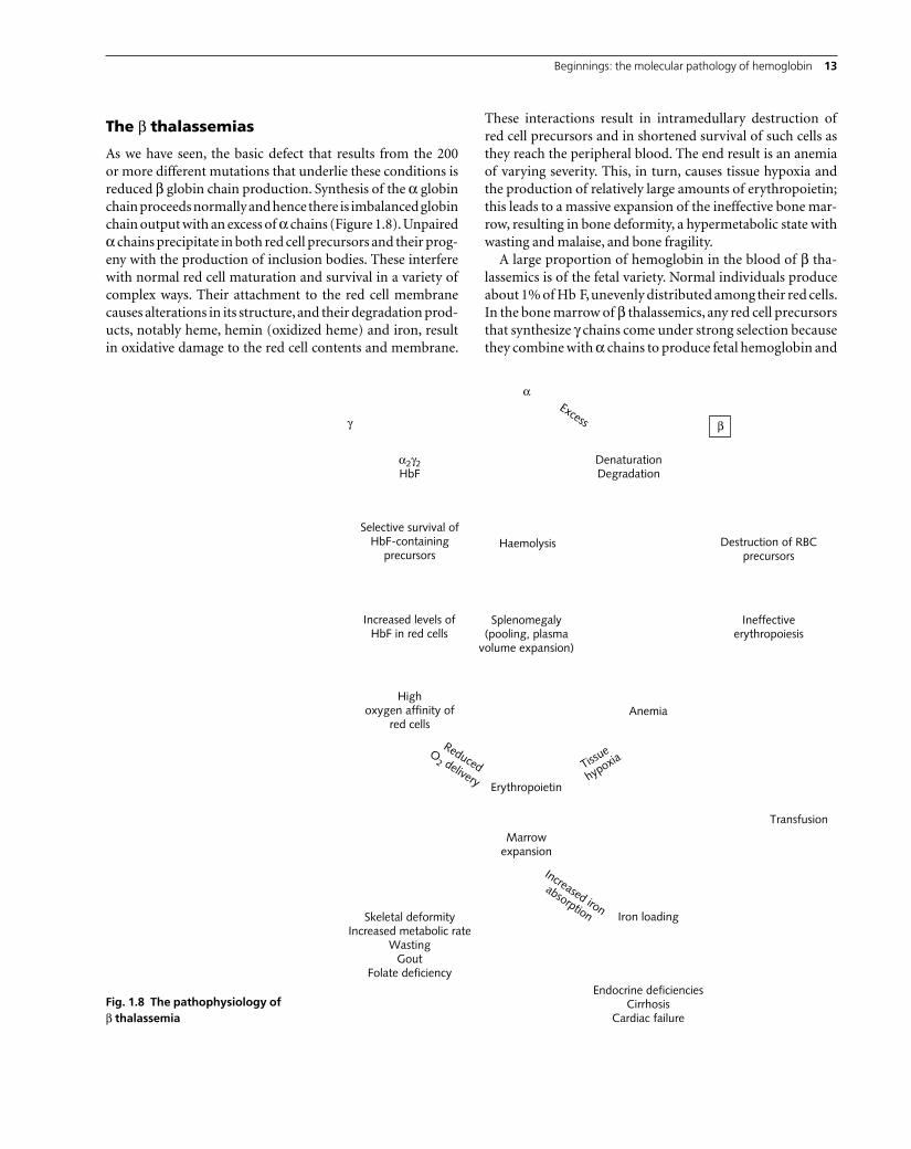
As we have seen, the basic defect that results from the 200 or more different mutations that underlie these conditions is reduced β globin chain production. Synthesis of the α globin chain proceeds normally and hence there is imbalanced globin chain output with an excess of α chains (Figure 1.8). Unpaired α chains precipitate in both red cell precursors and their prog-eny with the production of inclusion bodies. These interfere with normal red cell maturation and survival in a variety of complex ways. Their attachment to the red cell membrane causes alterations in its structure, and their degradation prod-ucts, notably heme, hemin (oxidized heme) and iron, result in oxidative damage to the red cell contents and membrane.
These interactions result in intramedullary destruction of red cell precursors and in shortened survival of such cells as they reach the peripheral blood. The end result is an anemia of varying severity. This, in turn, causes tissue hypoxia and the production of relatively large amounts of erythropoietin; this leads to a massive expansion of the ineffective bone mar-row, resulting in bone deformity, a hypermetabolic state with wasting and malaise, and bone fragility.
A large proportion of hemoglobin in the blood of β tha-lassemics is of the fetal variety. Normal individuals produce about 1% of Hb F, unevenly distributed among their red cells. In the bone marrow of β thalassemics, any red cell precursors that synthesize γ chains come under strong selection because they combine with α chains to produce fetal hemoglobin and
Fig. 1.8 The pathophysiology of β thalassemia
γ
α2γ2HbF
α
β
Excess
DenaturationDegradation
Destruction of RBCprecursors
Ineffectiveerythropoiesis
Anemia
HaemolysisSelective survival of
HbF-containingprecursors
Increased levels ofHbF in red cells
Highoxygen affinity of
red cells
Splenomegaly(pooling, plasma
volume expansion)
Erythropoietin
Marrowexpansion
Transfusion
Iron loading
Fi
Skeletal deformityIncreased metabolic rate
WastingGout
Folate deficiencyEndocrine deficiencies
CirrhosisCardiac failure
Reduced
Increased iron
absorption
O2 delivery
Tissue
hypoxia

14 Molecular Hematology
therefore the degree of globin chain imbalance is reduced. Furthermore, the likelihood of γ chain production seems to be increased in a highly stimulated erythroid bone marrow. It seems likely that these two factors combine to increase the relative output of hemoglobin F in this disorder. However, it has a higher oxygen affi nity than hemoglobin A and hence patients with β thalassemia are not able to adapt to low hemo-globin levels as well as those who have adult hemoglobin.
The greatly expanded, ineffective erythron leads to an in-creased rate of iron absorption; this, combined with iron re-ceived by blood transfusion, leads to progressive iron loading of the tissues, with subsequent liver, cardiac and endocrine damage.
The constant bombardment of the spleen with abnormal red cells leads to its hypertrophy. Hence there is progressive splenomegaly with an increased plasma volume and trap-ping of part of the circulating red cell mass in the spleen. This leads to worsening of the anemia. All these pathophysiological mechanisms, except for iron loading, can be reversed by regu-lar blood transfusion which, in effect, shuts off the ineffective bone marrow and its consequences.
Thus it is possible to relate nearly all the important features of the severe forms of β thalassemia to the primary defect in globin gene action. But can we also explain their remarkable clinical diversity? Part of it refl ects the different mutations of the β globin genes. For example, some of the promoter or splice mutations cause an extremely mild form of β+ tha-lassemia. Many β thalassemics are compound heterozygotes for either two severe β thalassemia alleles, a severe and mild allele, or different mild alleles, and this also accounts for a considerable amount of clinical diversity of the disease.
What of patients who have the same mutations at their β loci yet have completely different clinical phenotypes? The co-inheritance of α thalassemia, which reduces the magnitude of the excess of α globin chains in β thalassemia, may ameliorate the clinical course. This remarkable experiment of nature pro-vides unequivocal confi rmation that the major pathophysio-logical mechanism that underlies β thalassemia is imbalanced globin chain synthesis. In other patients, especially those who are homozygous for β0 thalassemia yet run a particularly mild course, it is apparent that unusually increased production of fetal hemoglobin is the main ameliorating factor. Although the precise mechanism is not yet understood, it is becoming apparent that genetic determinants both within the β globin gene cluster and on other chromosomes may be involved in this more effective production of Hb F. For example, a pro-moter polymorphism of the Gγ globin gene may be associated with increased propensity to synthesize fetal hemoglobin, particularly in states of hemopoietic stress. Similarly, there is good evidence that a so-far unidentifi ed gene on chromosome 6 may be involved in modifying the β thalassemia phenotype
in this way. Undoubtedly other polymorphisms of this kind will be discovered.
There is increasing evidence that the complications of β thalassemia may be modifi ed by polymorphisms at different loci. For example, variations at several loci that determine bone metabolism may be involved in modifying the sever-ity of osteoporosis, a common complication of this disorder. Similarly, the occurrence of jaundice and iron loading from the intestine, both common complications of the intermediate forms of β thalassemia, are related to polymorphisms of genes involved in bilirubin and iron metabolism, respectively.
Recently it has been suggested that these complex layers of modifi ers of β thalassemia should be placed in three classes: primary, including the different mutations that involve the β globin gene; secondary, including variation at the α globin locus or in fetal hemoglobin production; and tertiary, includ-ing modifi ers that are involved in varying the severity of the different complications of the disease.
In short, while we have a reasonable idea of how the β tha-lassemia phenotype is modifi ed, many questions remain and a considerable amount of the clinical variability of the disease remains unexplained.
The α thalassemias
The pathophysiology of the α thalassemias differs from that of the β thalassemias mainly because of the properties of the excess globin chains that are produced as a result of defective α chain synthesis. While the excess α chains produced in β thalassemia are unstable and precipitate, this is not the case in the α thalassemias, in which excess γ chains or β chains are able to form the soluble homotetramers γ
4 (Hb Bart’s) and
β4 (Hb H) (Figure 1.9). Although these variants, particularly
Fig. 1.9 The pathophysiology of α thalassemia
γα
β
α2γ2 α2β2
ExcessExcess
Fetus Adult
γ4Hb Bart's
β4Hb H
High oxygen affinity—hypoxiaInstability of homotetramers
Inclusion bodies. Membrane damageShortened red cell survival—hemolysis
Splenomegaly—hypersplenism

γα
β
α2γ2 α2β2
ExcessExcess
Fetus Adult
γ4Hb Bart's
β4Hb H
High oxygen affinity—hypoxiaInstability of homotetramers
Inclusion bodies. Membrane damageShortened red cell survival—hemolysis
Splenomegaly—hypersplenism

Beginnings: the molecular pathology of hemoglobin 15
Hb H, are unstable and precipitate in older red cell popula-tions, they remain soluble suffi ciently long for the red cells to mature and develop relatively normally. Hence there is far less ineffective erythropoiesis in the α thalassemias and the main cause of the anemia is hemolysis associated with the precipi-tation of Hb H in older red cells. In addition, of course, there is a reduction in normal hemoglobin synthesis, which results in hypochromic, microcytic erythrocytes. Another important factor in the pathophysiology of the α thalassemias is the fact that Hb Bart’s and Hb H are useless oxygen carriers, having an oxygen dissociation curve similar to that of myoglobin. Hence the circulating hemoglobin level may give a false impression of the oxygen-delivering capacity of the blood and patients may be symptomatic at relatively high hemoglobin levels.
The different clinical phenotypes of the α thalassemias are an elegant example of the effects of gene dosage (Figure 1.10). The heterozygous state for α+ thalassemia is associated with minimal hematological changes. That for α0 thalassemia (the loss of two α globin genes) is characterized by moderate hypochromia and microcytosis, similar to that of the β tha-lassemia trait. It does not matter whether the α genes are lost on the same chromosome or on opposite pairs of homolo-gous chromosomes. Hence the homozygous state for α+ tha-lassemia, − α/−α, has a similar phenotype to the heterozygous state for α0 thalassemia (− − /αα).
The loss of three α globin genes, which usually results from the compound heterozygous states for α0 and α+ thalassemia, is associated with a moderately severe anemia with the pro-duction of varying levels of hemoglobin H. This condition, hemoglobin H disease, is characterized by varying anemia and splenomegaly with a marked shortening of red cell survival.
Finally, the homozygous state for α0 thalassemia (− − /− − ) is characterized by death in utero or just after birth, with the clinical picture of hydrops fetalis. These babies produce no α chains and their hemoglobin consists mainly of Bart’s with variable persistence of embryonic hemoglobin. This is re-fl ected in gross intrauterine hypoxia; although these babies may have hemoglobin values as high as 8–9 g/dl, most of it is unable to release its oxygen. This is refl ected in the hydropic changes, a massive outpouring of nucleated red cells, and hepatosplenomegaly with persistent hematopoiesis in the liver and spleen.
Structural hemoglobin variants
While most structural hemoglobin variants produce no clini-cal disability, a few, notably the sickling, and the rare variants are associated with instability or abnormal oxygen transport (discussed in detail in Chapter 14).
The sickling disorders
The sickling disorders represent the homozygous state for the sickle cell gene, sickle cell anemia, and the compound het-erozygous state for the sickle cell gene and various structural hemoglobin variants, or β thalassemia. The chronic hemo-lysis and episodes of vascular occlusion and red cell seques-tration that characterize sickle cell anemia can all be related to the replacement of the normal β6 glutamic acid by valine in hemoglobin S. This causes a hydrophobic interaction with another hemoglobin molecule, triggering aggregation into
Fig. 1.10 The genetics of the common forms of α thalassemiaThe light boxes represent normal α genes and the shaded boxes deleted α genes. The mating shown at the top shows how two α0 thalassemia heterozygotes can produce a baby with the hemoglobin Bart’s hydrops syndrome. In the mating at the bottom, between individuals with α0 and α+ thalassemia, one in four of the offspring will have hemoglobin H disease.
α0 Thal. trait
α0 Thal.trait
α0 Thal.trait
α+ Thal.trait
α0 Thal.trait
α0 Thal. trait
α0 Thal. trait
α+ Thal. trait
Normal
Normal
Hb Bart'shydrops
Hb Hdisease
X
X

α0 Thal. trait
α0 Thal.trait
α0 Thal.trait
α+ Thal.trait
α0 Thal.trait
α0 Thal. trait
α0 Thal. trait
α+ Thal. trait
Normal
Normal
Hb Bart'shydrops
Hb Hdisease
X
X

16 Molecular Hematology
large polymers. It is this change that causes the sickling distor-tion of the red blood cell and hence a marked decrease in its deformability. The resulting rigidity of the red cells is respon-sible for the vaso-occlusive changes that lead to many of the most serious aspects of all the sickling disorders.
The different conformations of sickle cells (banana-shaped or resembling a holly leaf) refl ect different orientations of bundles of fi bres along the long axis of the cell, the three-dimensional structure of which is constituted by a rope-like polymer composed of 14 strands. The rate and extent of poly-mer formation depend on the degree of oxygenation, the cel-lular hemoglobin concentration, and the presence or absence of Hb F. The latter inhibits polymerization and hence tends to ameliorate sickling. Polymerization of Hb S causes damage to the red cell membrane, the result of which is an irrevers-ibly sickled cell. Probably the most important mechanism is cellular dehydration resulting from abnormalities of potas-sium/chloride cotransport and Ca2+-activated potassium ef-fl ux. This is suffi cient to trigger the Ca2+-dependent (Gardos) potassium channel, providing a mechanism for the loss of po-tassium and water and leading to cellular dehydration.
The vascular pathology of the sickling disorders is not en-tirely related to the rigidity of sickled red cells, however. There is now a wealth of evidence that abnormal interactions be-tween sickled cells and the vascular endothelium play a major role in the pathophysiology of the sickling disorders. Recently it has been demonstrated that nitric oxide may also play a role in some of the vascular complications of this disease. It has been found that nitric oxide reacts much more rapidly with free hemoglobin than with hemoglobin in erythrocytes and therefore it is possible that such decompartmentalization of hemoglobin into plasma, as occurs in sickle cell disease and other hemolytic anemias, diverts nitric oxide from its homeo-static vascular function.
These issues are discussed in greater detail in Chapter 14.
Unstable hemoglobin variants
There are a variety of different mechanisms underlying he-moglobin stability resulting from amino acid substitutions in different parts of the molecule. The fi rst is typifi ed by amino acid substitutions in the vicinity of the heme pocket, all of which lead to a decrease in the stability of the bind-ing of heme to globin. A second group of unstable variants results from amino acids that simply disrupt the secondary structure of the globin chains. About 75% of globin is in the form of α helix, in which proline cannot participate except as part of one of the initial three residues. At least 11 un-stable hemoglobin variants have been described that result from the substitution of proline for leucine, fi ve that are caused by an alanine-to-proline change, and three in which
proline is substituted for histidine. Another group of vari-ants that causes disruption of the normal confi guration of the hemoglobin molecule involves internal substitutions that somehow interfere with its stabilization by hydrophobic interactions. Finally, there are two groups of unstable hemo-globins that result from gross structural abnormalities of the globin subunits; many are due to deletions involving regions at or near interhelical corners. A few of the elongated globin chain variants are also unstable.
Abnormal oxygen transport
There is a family of hemoglobin variants that are associated with high oxygen affi nity and hereditary polycythemia. Most result from amino acid substitutions that affect the equilib-rium between the R and T states (see earlier section—Structure and function). Thus, many of them result from amino acid substitutions at the α
1/β
2 interface, the C terminal end of the
β chain, and at the 2,3-DPG binding sites.
Congenital cyanosis due to hemoglobin variants
There is a family of structural hemoglobin variants that is designated hemoglobin M, to indicate congenital methemo-globinemia, and is further defi ned by their place of discovery. The iron atom of heme is normally linked to the imidazole group of the proximal histidine residue of the α and β chains. There is another histidine residue on the opposite side, near the sixth coordination position of the heme iron; this, the so-called distal histidine residue, is the normal site of binding of oxygen. Several M hemoglobins result from the substitution of a tyrosine for either the proximal or distal histidine residue in the α or β chain.
Postscript
In this short account of the molecular pathology of hemo-globin we have considered how mutations at or close to the α or β globin genes result in a diverse family of clinical disorders due to the defective synthesis of hemoglobin or its abnormal structure. Work in this fi eld over the last 20 years has given us a fairly good idea of the repertoire of different mutations that underlie single-gene disorders and how these are expressed as discrete clinical phenotypes. Perhaps more importantly, however, the globin fi eld has taught us how the interaction of a limited number of genes can produce a remarkably di-verse series of clinical pictures, and something of the basis for how monogenic diseases due to the same mutation may vary widely in their clinical expression.


Beginnings: the molecular pathology of hemoglobin 17
Further reading
Huisman THJ, Carver MFH, Erol Baysal E. (1997) A Syllabus of Tha-lassemia Mutations. Augusta, GA: The Sickle Cell Anemia Founda-tion.
Stamatoyannopoulos G, Grosveld F. (2001) Hemoglobin switching. In: Stamatoyannopoulos G, Nienhuis AW, Majerus PW et al., eds. The Molecular Basis of Blood Diseases, 3rd edn. Philadelphia: W.B. Saun-ders, pp. 136–182.
Steinberg MH, Forget BG, Higgs DR et al. (2001) Disorders of Hemo-globin. Cambridge: Cambridge University Press.
Weatherall DJ. (2001) Phenotype–genotype relations in monogenic dis-ease: lessons from the thalassaemias. Nature Genetics, 2, 245–255.
Weatherall DJ. (2001) Thalassemia. In: Stamatoyannopoulos G, Nienhu-is AW, Majerus PW et al., eds. The Molecular Basis of Blood Diseases, 3rd edn. Philadelphia: W.B. Saunders, pp. 183–226.
Weatherall DJ, Clegg JB. (2001) The Thalassaemia Syndromes. Oxford: Blackwell.
Weatherall DJ, Clegg JB, Higgs DR et al. (2001) The hemoglobinopathies. In: Scriver CR, Beaudet AL, Sly WS et al., eds. The Metabolic Basis of Inherited Disease, 8th edn. New York: McGraw-Hill.
