chapter – 1 - shodhgangashodhganga.inflibnet.ac.in/bitstream/10603/92684/7/07_chapter1.pdf ·...
TRANSCRIPT

1
CHAPTER – 1
Introduction

2
CHAPTER - 1
Introduction
1.1 General Introduction:
Medicinal plants are an important part of our natural wealth. In many developing
countries, traditional medicines are still the mainstay of health care and drugs. Even in
developed countries, the raw materials for manufacturing essential drugs are extracted
from medicinal plants. The popularity of herbal medicines is connected with their easy
access, therapeutic efficiency, relatively low cost and the assumption for absence of toxic
side effects.
About 80% of the world’s inhabitants rely mainly on traditional medicines for
their primary health care (Owolabi et al., 2007). Modern pharmacopoeia still contains at
least 25% drugs derived from plants and many others, which are synthetic analogues,
built on prototype compounds isolated from plants.
India has a rich heritage of traditional medicines and the traditional health care
system. It has several traditional medical systems such as Ayurveda and Unani, which
has survived through more than 3000 years, mainly using plant-based drugs. Over the
centuries, the use of medicinal herbs has become an important part of daily life despite
the progress in modern medical and pharmaceuticals research. Approximately 3000
plants species are known to have medicinal properties in India (Prakasha et al., 2010).
The Botanical Survey of India records over 15,000 plant species occurring in the country,
of which at least 7,500 species have been used for medicinal purposes (Attisso M.A.,
1983)
In recent years, the increasing demand for herbal medicines becomes as an alternative
conventional medicine even in the industrialized countries and the adoption of crude
extracts of plants for self-medication has gained more importance. Each medicinal plant
species has its own nutrient composition besides having pharmacologically important
phytochemicals. These nutrients are essential for the physiological functions of human
body. The phytotherapy acts as a bridge between traditional and modern medicines. The
3
developments of plant derived drugs have always been a multi-step procedure starting
with a crude extract followed by the standardized extract and ending up with isolated
constituents.
The present thesis deals with the analysis of medicinal plants for their proximate
composition, antioxidant activity and elemental content.
1.2 Proximate composition:
1.2.1Extractable matter:
Extractable matter is the amount of active constituents extracted with different
solvents from the medicinally important plant material. These solvents are normally
water, alcohol, ether and chloroform. The extractive values provide information of the
extent of polar, medium polar and non-polar components present in medicinal plant
material.
1.2.2 Moisture content:
Moisture content is the quantity of water present in a material. The presence of
excess water in medicinal plants will encourage microbial growth. Limits for water
content should therefore be set for every given plant material. The accurate method for
determining the amount of water is the Karl Fischer titration method.
1.2.3 Ash content:
It is the nonvolatile inorganic matter of a compound which remains after subjecting
it to a high decomposition temperature. Ash represents the mineral salt or inorganic
matter content of the drug. The total ash includes both physiological ash, which is derived
from the plant tissue itself and non-physiological ash which is the residue of the
extraneous matter adhering to the plant. Low amount of total ash indicates that the
inorganic matter and non-physiological matter such as silica is less in medicinal plant
material.

4
1.2.4 Crude fibers:
Fibers include cellulose, hemicelluloses, pectin and lignin. It represents only 60%
to 80% of the cellulose and 4% to 6% of the lignin. Most of them are polysaccharides.
Within the past decade food composition databases have reflected technological advances
by listing specific values for total, soluble and insoluble dietary fibers. Wheat, rye, rice
and most other grains are primarily composed of insoluble fiber (Englyst et al., 1982).
1.2.5 Fats and Waxes (Lipids):
Lipids are of great importance to the body as they are main storage form of
energy. All Lipids are hydrophobic in nature. This group of molecules includes fats and
oils, waxes, phospholipids and steroids. Waxes are esters of fatty acids with long chain
monohydric alcohols. Natural waxes are often mixtures of such esters and may also
contain hydrocarbons (Gidez L. I., 1984).
1.3 Phytochemicals:
Phytochemicals are defined as non-nutritive bioactive plant chemicals in fruits,
vegetables, grains and other plants having protective or disease preventive properties and
are considered to reduce the risk of chronic diseases. Some well-known phytochemicals
include carotenoids, phenolics, alkaloids, nitrogen-including compounds and organo-
sulfur compounds. It was proven that phytochemicals working together with nutrients
found in fruits, vegetables and nuts, helps in slowing down the aging process and reduce
the risk of many diseases including cancer, heart disease, stroke, high blood pressure,
cataracts, osteoporosis and urinary tract infections.
1.3.1 Phenolic compounds:
Phenolic compounds are natural antioxidants having an aromatic ring with one or
more hydroxyl groups. Numerous types of phenolics are found in nature, including
simple phenol, phenylproponoids, benzoic acid derivatives, flavonoids, stilbenes, tannins,
lignans and lignins. Phenolic compounds in fruits and vegetables are the secondary
metabolites in plants that are derived from the metabolism phenylalanine and tyrosine
(Van Sumere C. F., 1989). Phenolic compounds play a crucial role in the growth and

5
reproduction of plants, and also act as antifeedants and antipathogens (Butler L. G.,
1992). The presence of phenols is considered to be potentially toxic to the growth and
development of pathogens. Phenolic function as antibiotics, natural pesticides, signaling
substances, protective substance against ultraviolet light, insulating materials to make cell
walls impermeable to gas and water, and give structural stability to plants. Many
properties of plant products, such as the astringency of foods or their potential
antinutritional properties are associated with the presence and content of phenolics
(Butler L.G., 1992). Among the variety of polyphenolic compounds, phenolic acids have
attracted considerable interest in the past few years because they exhibit many potential
health benefits such as antiallergenic, antiatherogenic, antiinflammatory, antimicrobial,
antioxidant, antithrombotic cardioprotective and vasodilatory effects (Manach and
Mazur, 2005).
1.3.2 Alkaloids:
Alkaloids are the natural compounds found in all plants that include nitrogen.
The alkaloids are the plant bases. They are essentially basic nitrogenous compounds of
vegetable origin, possessing physiological actions. They are found in all parts of plants,
but mostly in fruits, stem, bark, roots, leaves and seeds. Alkaloids are generally insoluble
in water and soluble in ether or chloroform and other non polar solvents. Along with that
alkaloids have significant therapeutic value and form the ingredients of many important
medicines. It has been associated with medicinal uses for centuries and one of their
common biological properties is their cytotoxicity (Nobori et al., 1994). Many alkaloids
derived from plants have anticancer properties.
1.3.3 Flavonoids:
Flavonoids are polyphenolic compounds that are ubiquitous in nature and are
categorized, according to chemical structure into flavonols, flavones, flavanones,
isoflavones, catechins, anthocyanidins and chalcones. Over 4,000 flavonoids have been
identified many of which occur in fruits, vegetables and beverages. The flavonoids have
been reported to have antiviral, antiallergic, antiplatelet, antiinflammatory, antitumor and
antioxidant activities. Besides this flavonoids have also been reported to act as anticancer

6
agents via regulation of signal transduction pathways of cell growth and proliferation,
suppression of oncogenes and tumor formation, induction of apoptosis, modulation of
enzyme activity related to detoxification, oxidation and reduction, stimulation of the
immune system and DNA repair and regulation of hormone metabolism (Aron et al.,
2008).
1.3.4 Amino acids:
Amino acids are basic unit of protein containing an amino group and a carboxylic
group and plays major role in regulating multiple processes related to gene expression,
including modulation of the function of the proteins that mediate messenger RNA
(mRNA) translation (Scot and Leonard, 2006). They also help in tissue protein formation.
Few amino acids are involved in enzyme formation. Hormones like insulin, growth
hormone and glucagon are made up of amino acids. Adrenaline, nor-adrenaline and
thyroxin are made up of single amino acid. Glutathione, a physiologically active peptide
is also made up of amino acids. Amino acids are involved in synthesis of melanin. It has
been reported that amino-acid balance in cancer patients often differs from that in healthy
individuals, because of metabolic changes (Jun et al., 2010). Amino acids are absorbed
through stomas in plants. It has been observed that amino acids influence the
physiological activities of the plant. The basic classification of amino acid is shown in
Table 1.1.

7
Table 1.1: Amino acids classification
Essential Amino acid Non- essential Amino acid Special amino acid
Lysine Cysteine GABA
Methionine Tyrosine DOPA
Valine Serine Citrulline
Tryptophan Alanine Ornithine
Isoleucine Asparagines Taurine
Histidine Aspartic acid
Phenylalanine Glutamic acid
Threonine Glycine
Leucine Hydroxylysine
Arginine Proline
1.3.5 Oxidative stress:
Oxidative stress is a condition resulting from an imbalance between the
production of free radicals and antioxidant defense systems in which oxidation
predominates (Halliwell and Whiteman, 2004). It plays a role in cellular processes, such
as aging and apoptosis. In a balanced cell state, ROS are produced as a byproduct of
metabolic processes and the level of ROS can be controlled with antioxidants, such as
vitamin E and vitamin C (Spitz et al., 2004). In a state of cellular imbalance damage is
caused to nuclear and mitochondrial DNA, proteins and lipids. If this damage is
irreparable then mutagenesis, carcinogenesis and cell death can occur. Oxidative stress
has been linked to diseases, including some allergic and inflammatory skin diseases
(Okayama Y., 2005).
1.3.6 Free radicals:
Free radicals are reactive species which contain one or more unpaired electrons and
are capable of independent existence. There are many types of radicals, but those which
concerned in biological systems are derived from oxygen, and known as reactive oxygen
species (ROS). Free radicals and other reactive species are continuously produced in the

8
body and have important functions for the immune system. Free radicals can also be
generated in non-enzymatic reactions of oxygen with organic compounds as well as those
initiated by ionizing radiations (Fig. 1.1). ROS are potentially very toxic to cells.
Oxidative stress might cause damage to the biomolecules such as lipids, protein and
DNA, which may increase the risk of development of several diseases.
Free radicals and other reactive oxygen species are derived either from normal
essential metabolic processes in the human body or from external sources. Some
internally generated sources are mitochondria (Balaban et al., 2005), phagocytic cells,
reactions involving iron and other transition metals, peroxisomes, exercise and
inflammation while the externally generated sources of free radicals are cigarette smoke,
environmental pollutants, radiation, ultraviolet light, certain drugs, pesticides, Ozone,
alcohol consumption, viral infections (Halliwell B., 1996).
Figure 1.1: Different ways for formation of free radicals
1.3.6.1 Types of free radicals:
There are numerous types of free radicals that can be formed within the body. The
most common ROS which are biologically significant are superoxide radical (O2⁻),
hydroxyl radical (·OH), peroxyl radical (ROO·), hydrogen peroxide (H2O2), singlet
oxygen (1O2), nitric oxide (NO), peroxynitrite (ONOO⁻) and hypochlorous acid (HOCl).

9
In the human body O2-· is continuously formed during metabolism. The rate of
formation depends on the amount of oxygen flowing through the mitochondria at any
given time. Hydroxyl radicals are short-lived but the most damaging radicals within the
body. This type of free radical can be formed from O2- and H2O2 via the Harber-Weiss
reaction. The interaction of copper or iron and H2O2 also produces OH·. Hydrogen
peroxide is produced in vivo by many reactions. Transition metal ions are important in
the production of ROS. The ability of metal ions to donate and accept single electrons is
the basis for the formation and propagation of many ROS. Both copper and iron gain or
lose electrons during redox reactions.
1.4 Antioxidants:
Antioxidants in the broad sense are all substances that can protect materials
against auto-oxidation, irrespective of the mechanism of action. They have the ability to
repair damage done by free radicals and this is thought to reduce cancer risk and aging.
They vary widely in chemical structure and have diverse mechanisms of action.
Antioxidants can inhibit or retard oxidation in two ways: either by scavenging free
radicals, where the compound is described as a primary antioxidant, or by a mechanism
that does not involve direct scavenging of free radicals, in which case the compound is a
secondary antioxidant. The components of primary antioxidants are consumed during the
induction period. Secondary antioxidants operate by a variety of mechanisms including
binding mental ion catalysts, scavenging oxygen, absorbing UV radiation and converting
hydroperoxides to non-radical species or intercepting single oxygen. Different
mechanisms of antioxidant activity are shown in Table 1.2.

10
Table 1.2 – Different mechanisms of antioxidant activity (Hall C., 2001)
Antioxidant class Mechanism of antioxidant
activity
Examples of antioxidants
Proper antioxidants Inactivating lipid free radicals Phenolic compounds
Hydroperoxide
stabilizers
Preventing decomposition of
hydroperoxides into free
radicals
Phenolic compounds
Synergists Promoting activity of proper
antioxidants
Citric acid, ascorbic acid
Metal chelators Binding heavy metals into
inactive compounds
Phosphoric acid,
Maillard reaction
compounds,
citric acid
Singlet oxygen
quenchers
Transforming singlet
oxygen into triplet oxygen
Carotenes
Substances reducing
Hydroperoxides
Reducing hydroperoxides
in a non-radical way
Proteins, amino acids
1.4.1 Types of Antioxidants:
According to their origin antioxidants are divided in two groups namely, natural
antioxidants and synthetic antioxidants. Whereas, based on the nature of antioxidants, the
human antioxidant system can be divided into two main groups, enzymatic antioxidants
and non-enzymatic antioxidants.
1.4.1.1 Enzymatic antioxidants:
The major primary intracellular endogenous antioxidant defenses are called as
enzymatic system. This enzymatic antioxidant system includes superoxide dismutase
(SODs), catalase (CAT) and glutathione peroxidase (GSHPx).

11
1.4.1.2. Non-enzymatic antioxidants:
Non-enzymatic antioxidants are classified into two groups: endogenous
antioxidants and exogenous antioxidants.
Endogenous antioxidants:
The major endogenous antioxidants found in human plasma are the transition
metal binding proteins. This includes ceruloplasmin, transferring, hepatoglobin and
albumin.
Exogenous antioxidants
Antioxidants from our diet play an important role in helping endogenous
antioxidants for the neutralization of oxidative stress (Lien Ai et al., 2008). The best
known examples are vitamins such as ascorbic acid, vitamin E, carotenoids, quinines, and
polyphenols.
1.4.2 Types of antioxidant assays:
On the basis of the chemical reactions involved, major antioxidant activity assays
can be divided roughly into two categories (Huang et al., 2005):
1) Hydrogen atom transfer (HAT) and
2) Single-electron transfer (SET) reaction–based assays
These two mechanisms yield identical results, but they differ in terms of kinetics
and the potential for side reactions to occur.
1.4.2.1 Hydrogen atom transfer (HAT) assay:
HAT-based procedures measure the classical ability of an antioxidant to quench
free radicals by hydrogen donation:
……………………….1.1
Where, (AH = any H donor). Antioxidant activity measurements of HAT assays are
based on competition kinetics. HAT reactions are solvent and pH independent and
12
usually are quite rapid—typically they are completed in seconds to minutes. A
disadvantage of the procedure, however, is that the presence of reducing agents,
including metals, is a complication that can lead to high apparent reactivity. Total
phenolic and total flavonoids assays fall in this category because they donate hydrogen
ions while maintaining a stable structure (Chance B., 1979).
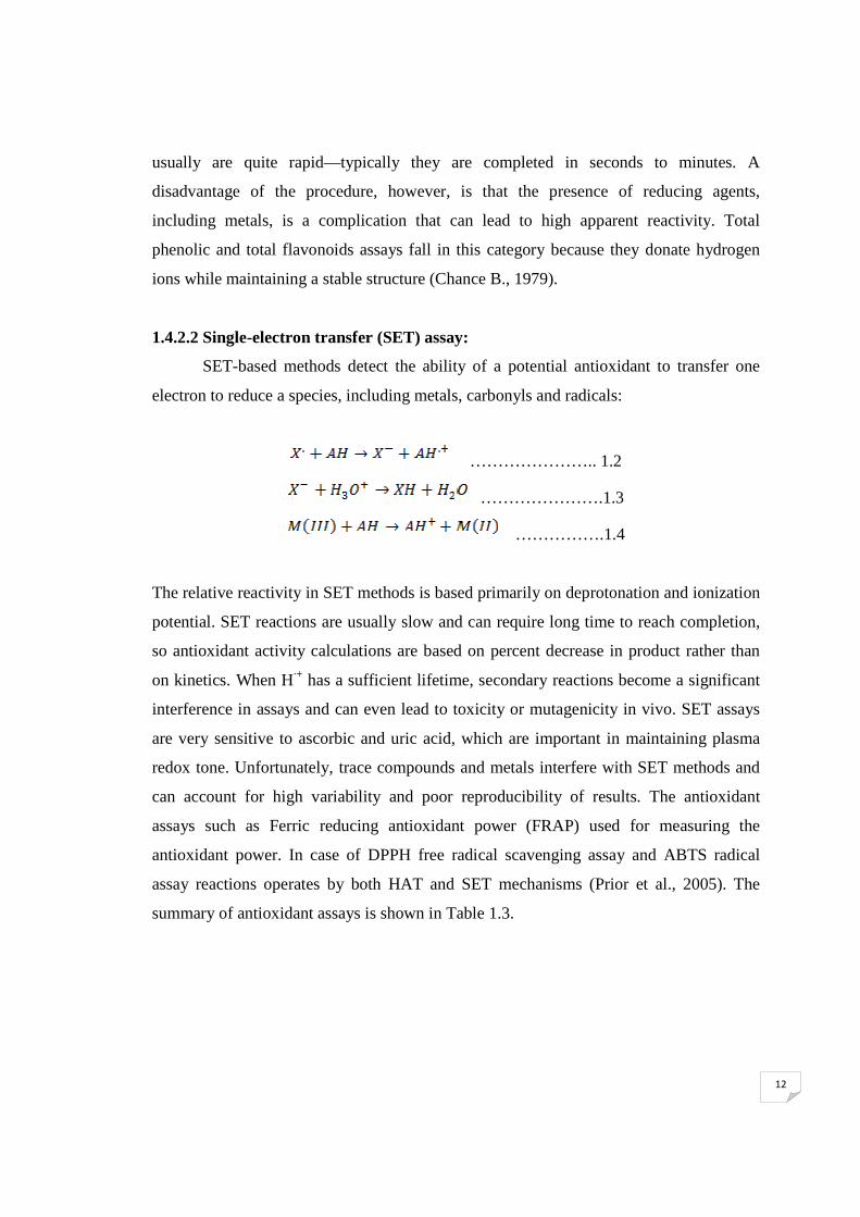
1.4.2.2 Single-electron transfer (SET) assay:
SET-based methods detect the ability of a potential antioxidant to transfer one
electron to reduce a species, including metals, carbonyls and radicals:
………………….. 1.2
………………….1.3
…………….1.4
The relative reactivity in SET methods is based primarily on deprotonation and ionization
potential. SET reactions are usually slow and can require long time to reach completion,
so antioxidant activity calculations are based on percent decrease in product rather than
on kinetics. When H·+ has a sufficient lifetime, secondary reactions become a significant
interference in assays and can even lead to toxicity or mutagenicity in vivo. SET assays
are very sensitive to ascorbic and uric acid, which are important in maintaining plasma
redox tone. Unfortunately, trace compounds and metals interfere with SET methods and
can account for high variability and poor reproducibility of results. The antioxidant
assays such as Ferric reducing antioxidant power (FRAP) used for measuring the
antioxidant power. In case of DPPH free radical scavenging assay and ABTS radical
assay reactions operates by both HAT and SET mechanisms (Prior et al., 2005). The
summary of antioxidant assays is shown in Table 1.3.

13
Table 1.3: Summary of Antioxidant Assays (Badarinath et al., 2010)
Antioxidant
assay
Simplicity Instrumentation
required
Biological
relevance
Mechanism Time
required
ORAC + + + + + + HAT ++
TRAP - - - - + + + HAT +++
FRAP + + + + + + - - SET - -
TEAC + + - SET -
FC + + + - - SET +
ABTS + + + HAT +
+, ++, +++ = desirable to highly desirable characteristic.
—,——, — — — = less desirable to highly undesirable characteristic.
1.4.3 Antioxidant assays:
1.4.3.1 Phenolic content:
Singleton and Rossi (Singleton and Rossi, 1965) adapted this assay first time for
analysis of food products. Since then, the total phenols assay has been used in many
studies and is now commonly known as the total phenols (or phenolics) assay. Total
phenolics methodology consists of the addition of Folin-Ciocalteau reagent to a sample
held in the dark for two hours, followed by measurement of the absorption. Intensity of
the light absorption is directly proportional to the concentration of phenols. Results are
expressed as ferulic acid equivalents of dry weight of the sample using the standard
curve. The total phenols assay by FCR method is convenient, simple and reproducible.
1.4.3.2 Total flavonoids:
Flavonoids are most common and widely distributed group of plant phenolic
compounds that are characterized by a benzo-y-pyrone structure, which is ubiquitous in
fruits and vegetables. The AlCl3 method is used for quantification of the total flavonoids
content of the plant extracts. Total flavonoids can be determined in the sample extract by

14
reaction with sodium nitrite, followed by the development of colored flavonoid aluminum
complex formation using aluminum chloride which can be monitored
spectrophotometrically at 510 nm. Flavonoids are important for human health because of
their high pharmacological activities as radical scavenger.
1.4.3.3 DPPH radical scavenging assay:
A rapid, simple and inexpensive method to measure antioxidant capacity of food
involves the use of the free radical 2, 2-Diphenyl-1-picrylhydrazyl (DPPH). It is widely
used to test the ability of compounds to act as free radical scavengers or hydrogen donors
and to evaluate antioxidant activity.
The DPPH radical is long-lived organic nitrogen radical and has a deep purple
color. It is commercially available and does not have to be generated before assay. The
molecule of 1, 1-diphenyl-2-picrylhydrazyl is characterized as a stable free radical by
virtue of the delocalisation of the spare electron over the molecule as a whole, so that the
molecules do not dimerise. The delocalization also gives rise to the deep violet colour,
characterized by an absorption band in ethanol solution centered at about 520 nm. When
a solution of DPPH is mixed with that of a substance it donate a hydrogen atom, then this
gives rise to the reduced form with the loss of this violet colour.

15
1.4.3.4 ABTS radical scavenging assay:
The ABTS assay was first reported by Miller et al. (Miller et al., 1993). ABTS·- is
generated by mixing ABTS solution with potassium persulfate under darkness at room
temperature (23 0C) for 16 h. The solution can then be diluted with 50 % ethanol and the
absorbance can be measured at 734 nm. The mechanism by which the ABTS•+ radical
cation reacts with the antioxidant extract is shown below.
ABTS assay has been used in many research laboratories for studying antioxidant
capacity. The advantage of using this method is that it is rapid and can be used over a
wide range of pH values (Arnao et al., 1999) in both aqueous and organic solvent
systems.
1.4.3.5 Ferrous reducing antioxidant power assay:
The method is based on the reduction of Fe3+-TPTZ complex (colorless complex)
to Fe2+-tripyridyltriazine (blue colored complex) formed by the action of electron
donating antioxidants at low pH. This reaction is monitored by measuring the change in
absorbance at 593 nm. The mechanism is as shown below.

16
N
N
N
NN
NN
N NN
NN
Fe (III)Antioxidant
N
N
N
NN
NN
N NN
NN
Fe (II)
[Fe(III)(TPTZ)2]3+[Fe(II)(TPTZ)2]2+, λmax = 593 nm
-e
The method is simple, rapid, inexpensive and does not require sophisticated
instrumentation. TPTZ is deficient as the ideal reaction stoichiometry between Fe (III) and
TPTZ is 1 to 2. The oxidant is not just Fe(III)(TPTZ)2, it also contains other Fe (III)
species which can lead to potential problems as many metal chelators in food extract
could bind Fe(III) and form complexes that are also capable of reacting with antioxidants.
1.4.3.6 Reducing power assay:
The reducing power of a compound may serve as a significant indicator of its
potential antioxidant activity. In this assay, the yellow color of the test solution changes
to green depending on the reducing power of test specimen. The antioxidant ability of
certain compounds is associated with their reducing power. Reducing power assay is
based on the principle that substances, which have reduction potential, react with
potassium ferricyanide (Fe+3) to form potassium ferrocyanide (Fe+2), which then reacts
with ferric chloride to form ferric ferrous complex that has an absorption maximum at
700 nm. Increased absorbance of the reaction mixture indicates increase in reducing
power.
1.5 Trace elements:
A trace element is defined as an element in a sample that has an average
concentration in the range of 1 part per billion to 100 parts per million (Skoog et al.,
2003). Trace elements can be classified as essential and non-essential elements. Essential
trace elements are also known as micronutrients and are vitally important for proper
maintenance of various metabolic body functions. They are required by human beings in

17
the range of 50 µg/day to 18 mg/day. The insufficient intake of these elements by human
beings and other living bodies over a long period of time results in the impairment of
normal body functions. Supplementation of the required elements helps in preventing or
curing the corresponding deficiency (Mertz W., 1981). The safe and acceptable intake of
some of the essential trace elements by human beings as recommended by WHO (WHO,
1996) are given in Table 1.4.
Table 1.4 WHO recommended safe and adequate dietary intake of some elements
for adults (WHO, 1996)
Mineral Recommended daily allowance Tolerated upper limit
Calcium 1200 mg 2500 mg
Magnesium 320-420 mg 750 mg
Iron 15-10 mg 45 mg
Zinc 12-15 mg 40 mg
Selenium 55-70 µg 400 µg
Copper 0.9 mg 10 mg
Manganese 1.8-2.3 mg 11 mg
Nickel 400-700 µg 1000 µg
Cobalt 10-20 µg 250 µg
Sodium 1500 mg 2300 mg
Potassium 4700 mg N/A
The importance of trace elements in human being is discussed below:
1.5.1 Calcium:
It is one of the most abundant elements in the body, 99 % being found in the
skeleton. It is primarily present in bone tissue as the hydroxyapatite form of calcium
phosphate. The basic function of calcium is to provide a strong framework supporting
and protecting delicate organs. On the cellular level, calcium is used to regulate the
permeability and electrical properties of biological membranes, which in turn control

18
muscle and nerve functions, glandular secretions, and blood vessel dilation and
contraction.
1.5.2 Magnesium:
It is the fourth most abundant mineral in the body. Numerous biochemical and
physiological processes require magnesium, including energy production, protein
synthesis, muscle contractions and vascular tone. It is a component of several enzymes
implicated in the metabolism of carbohydrates, lipids and proteins. Hence the deficiency
may lead to a range of serious biochemical and functional problems. There is an
increased interest in the role of magnesium in preventing and managing disorders such as
hypertension, cardiovascular disease and diabetes.
1.5.3 Iron:
It plays a key role in many biochemical reactions. Haemoglobin iron represents
approximately 60% of total body iron, whereas myoglobin represents only about 3-7% of
total iron (McDowell L.R., 1992). In humans, iron is an essential component of proteins
involved in oxygen transport. A deficiency of iron limits oxygen delivery to cells,
resulting in fatigue, poor work performance, and decreased immunity. On the other hand,
excess amounts of iron can result in toxicity and even death (Corbett J.V., 1995).
1.5.4 Selenium:
It is an important component of antioxidant enzymes such as glutathione peroxidase
(GPx), thioredoxin reductase (TrxR) and iodothyronine deiodinases (IDD). It may also
protect the animal organism from detrimental effects of heavy metals including cadmium,
mercury and silver (McDowell L. R., 1992). It is toxic if taken in large quantities and
may result in hair loss, tooth decay, brittle nails, white spots, poor appetite, sour taste in
the mouth and change in skin pigmentation.
1.5.5 Zinc:
It activates several enzymes and is a component of many important
metalloenzymes, DNA and RNA. The element is critically involved in cell replication

19
and in the development of cartilage and bone. The deficiency of zinc leads to an
underperforming immune system open to infections, allergies, night blindness, loss of
smell, falling hair, white spots under finger nails, skin problems, sleep disturbances.
1.5.6 Nickel:
It is found in the enzyme superoxide dismutases, which is an important
antioxidant. It influences absorption and metabolism of iron. Acute nickel exposure is
associated with a variety of clinical symptoms and signs which include gastrointestinal
disturbances (nausea, vomiting, abdominal discomfort and diarrhoea), visual disturbance
(temporary left homonymous hemianopia), headache, giddiness, wheezing and cough.
Excess nickel in the body is also associated with a high incidence of heart disease,
thyroid disease and cancer.
1.5.7 Sodium:
It is an element that is vital to human life. Together with potassium and chlorine, it
forms a very important part of blood plasma. It also allows our body to maintain the right
blood chemistry and the correct amount of water in our blood. Normal functioning of our
nervous system also depends on this important element. Too much sodium can damage
kidney and increases the chances of high blood pressure.
1.5.8 Manganese:
It is actually an extremely important element that the body uses for a variety of
things. It supports the immune system, regulates blood sugar levels and is involved in the
production of energy and cell reproduction. Additionally, manganese works with vitamin
K to support blood clotting and with B-complex vitamins it helps to control the effects of
stress.
1.5.9 Copper:
It is an element that is very important for our good health. It along with vitamin C
is important for keeping blood vessels and skin elastic and flexible. This important

20
element is also required by the brain to form chemicals that keep us awake and alert. The
deficiencies of copper include anemia and arthritis.
1.5.10 Potassium:
It is an important electrolyte in the body which is intimately associated with sodium
metabolism. It is essential for the transport of nutrients into each cell and waste products
out of each cell and helps normalize the heartbeat. It also plays an important role of a
catalyst for many types of enzymes inside the human body. Deficiency of potassium may
lead to nervous disorders, constipation, slow irregular heartbeat and muscle damage.
1.5.11 Cobalt:
It forms the core of vitamin B-12 and required for normal functioning of the
pancreas. It helps in repair of myelin sheath, increase the effectiveness of glucose
transport and building of red blood cells. Cobalt has metabolic links with iron and copper
which can be depressed at high levels of cobalt intake leading to anemia, nerve disorders
and abnormalities in cell formation.
1.6 Bioaccessibility:
Bioaccessibility is a term used to describe the proportion of a nutrient in food that
can be utilized for normal body function (Judprasong et al., 2005). In general,
bioaccessibility is affected by the type and/or composition of food and also by the
simulated gastrointestinal conditions which may affect the distribution of initial species.
Many factors affect the bioaccessibility of a compound; these may be divided into
exogenous factors such as the complexity of the food matrix, the chemical form of the
compound of interest, structure and amount of co-ingested compounds (Scholz and
Williamson., 2007) as well as endogenous factors including mucosal mass, intestinal
transit time, rate of gastric emptying, metabolism and extent of conjugation and protein-
binding in blood and tissues. There are two approaches to estimate the bioaccessibility of
minerals for the animal, in -vivo and in -vitro techniques.

21
1.6.1 In- vivo techniques:
In -vivo studies are both expensive and laborious, and the possibility of measuring
certain parameters during the experiments are often limited (Danielsson et al., 1995). In
this technique, bioavailable amount of an element of interest is estimated as the
difference in the concentration of the element in ingest and excreta, using radiotracers.
Basic disadvantage of the method is the exposure of ionizing radiations (Welch and
House, 1984). Most of the in vivo studies were carried out on Fe and Zn (McCance and
Widdowson, 1942).
1.6.2 In - vitro techniques:
In-vitro methods are rapid and inexpensive (Miller, et al. 1981). It involves the
simulation of the gastric and intestinal digestive conditions in the laboratory. As the
experiments are carried out under ‘simulated’ digestive conditions, the results may not be
as accurate as those obtained by in-vivo studies. The results obtained by in vitro methods
are based on the formation of digestive products that are soluble or dialyzable. However,
these methods are efficient to identify potential food products as nutrient supplements
(Van Campen and Glahn, 1999). In-vitro method is routinely used to estimate the
bioaccesible concentrations of essential elements in the diet. It is shown that the
bioaccesible values obtained by these methods can be well co-related with that of human
subjects (Menson and Cook, 1979) and many animal models (Forbs et al., 1989).
1.7 Inductively coupled plasma atomic emission spectrometry (ICP-AES):
It is an analytical technique used for the detection of trace metals. It is a type of
emission spectroscopy that uses the inductively coupled plasma to produce excited atoms
that emit electromagnetic radiation at wavelengths characteristic of a particular element.
The intensity of this emission is indicative of the concentration of the element within the
sample. A schematic diagram of ICP-AES is shown in Fig. 1.2.

22
Figure 1.2: Schematic diagram of Inductively coupled plasma atomic emission
spectrometry (ICP-AES)
The ICP-AES is composed of two parts: the ICP torch and the optical
spectrometer. The ICP torch consists of 3 concentric quartz glass tubes. Argon gas is
typically used to create the plasma. When the torch is turned on, an intense
electromagnetic field is created within the coil by the high power radio frequency signal
flowing in the coil. This RF signal is created by the RF generator. The argon gas flowing
through the torch is ignited with a Tesla unit that creates a brief discharge arc through the
argon flow to initiate the ionization process. Once the plasma is ignited, the Tesla unit is
turned off.
A peristaltic pump delivers sample into a nebulizer where it is changed into mist
and introduced directly inside the plasma flame. The sample immediately collides with
the electrons and charged ions in the plasma and is itself broken down into charged ions.
The various molecules break up into their respective atoms which then lose electrons and
recombine repeatedly in the plasma, giving off radiation at the characteristic wavelengths
of the elements involved. Within the optical chamber, after the light is separated into its
different wavelengths the light intensity is measured with a photomultiplier tube. Using

23
these detector arrays, the intensities of all wavelengths can be measured simultaneously
or sequentially.
1.8 Atomic absorption spectroscopy (AAS):
Atomic absorption spectrophotometry analyzes the concentration of elements in a
liquid sample based on energy absorbed from certain wavelengths of light (usually 190 to
900 nm). It typically include a flame burner to atomize the sample, hollow cathode lamp,
a monochromator and a photon detector (Fig. 1.3)
Figure 1.3: A schematic diagram of atomic absorption spectrometer (AAS)
In atomic absorption spectrometry, light of a specific wavelength is passed
through the atomic vapor of an element of interest and measurement is made of the
attenuation of the intensity of the light as a result of absorption.
Radiation from the source passes through a flame into which sample is aspirated.
The metallic compounds are decomposed in the flame forming cloud of atoms. Atomic
absorption measures the amount of light at the resonant wavelength which is absorbed as
it passes through a cloud of atoms. By measuring the amount of light absorbed, a
quantitative determination of the amount of element present can be made. The use of
hollow cathode lamp and careful selection of wavelength allow the specific quantitative

24
determination of individual elements in the presence of others. The ease and speed at
which precise and accurate determinations can be made with this technique have made
atomic absorption one of the most popular methods for determination of metals.
1.9 High performance liquid chromatography:
Chromatographic process can be defined as separation technique involving mass-
transfer between stationary and mobile phase. In HPLC a liquid mobile phase is used to
separate the components of a mixture. Stationary phase can be a liquid or a solid phase.
The components to be separated are first dissolved in a solvent, and then forced to flow
through a chromatographic column under a high pressure. In the column, the mixture
separates into its components. The amount of resolution is important, and is dependent
upon the extent of interaction between the solute components and the stationary phase.
The interaction of the solute with mobile and stationary phases can be manipulated
through different choices of both solvents and stationary phases. As a result, HPLC
acquires a high degree of versatility and it has the ability to easily separate a wide variety
of chemical mixtures. A schematic diagram of HPLC instrument is shown in Fig. 1.4.
Figure 1.4: A schematic diagram of High performance liquid
Chromatography (HPLC)

25
HPLC instrumentation includes a pump, injector, column, detector and data
system. The heart of the system is the column where separation occurs. Since the
stationary phase is composed of micrometer size porous particles, a high pressure pump
is required to move the mobile phase through the column. Detection of the components
depends upon the detector used. The response of the detector to each component is
displayed on a chart recorder or computer screen and is known as a chromatogram. To
collect, store and analyze the chromatographic data, computer, integrator, and other data
processing equipment are frequently used.
1.10 High performance thin layer chromatography:
High performance thin layer chromatography (HPTLC) is a sophisticated
instrumental technique based on the full capabilities of thin layer chromatography. The
advantages of automation, scanning, full optimization, selective detection principle,
minimum sample preparation, hyphenation enable it to be a powerful analytical tool for
chromatographic information of complex mixtures of inorganic, organic and
biomolecules. The modern HPTLC technique is sensitive and suitable for use in
qualitative and quantitative analysis. HPTLC is a valuable tool for identification because
it can provide chromatographic fingerprints that can be visualized and stored as electronic
images. A schematic diagram of HPTLC analysis is shown in Fig. 1.5.

26
Figure 1.5: A schematic diagram of HPTLC analysis.
1.11 Review of literature:
In India, medicinal plants are widely used by all sections of the population, either
directly in different indigenous systems of medicine or indirectly in the pharmaceutical
preparations of modern medicines. The first step towards pharmaceutical preparations is
phytochemical screening of plant extracts and/or extracts from traditional preparations
used in popular medicine (Alonso Paz et al., 1995). There are many reports in the
literature on proximate composition and phytochemical contents of various medicinal
plants of different origins. Pal et al. (Pal et al., 2005) and Donya et al. (Donya et al.,
2007) reported the proximate composition of whole bitter melon.
Survey of the literature shows that plants are endowed with free radical
scavenging molecules, such as vitamins, terpenoids, phenolic acids, lignins, stilbenes,
Sample preparation
Method creation
Sample application
Chromatographic development
Visualization
Scanning (Quantification
and identification)
Photo documentation Post chromatographic development
Scanning

27
tannins, flavonoids, quinones, coumarin, alkaloids, amines, betalains, and other
metabolites, which are rich in antioxidant activity (Zheng and Wang, 2001; Cai et al.,
2003). Netzel et al. (Netzel et al., 2007) have investigated seven native fruits of Australia
in terms of their antioxidant activities and phytochemical components and the results
showed that some fruits displayed high level of total phenolics as well as antioxidant
properties. Rajurkar and Hande (Rajurkar and Hande, 2011) analyzed 11 Indian
medicinal plants for their phytochemical content and antioxidant activity. They have
observed that all medicinal plants under study are good source of phytochemicals.
Prior et al. (Prior et al., 2005) observed that there are several factors that may
impact the antioxidant activity of foods, this include genetics, harvest season, geographic
and environmental conditions. Twenty four Indian medicinal herbs were studied by Ali et
al. (Ali et al., 2008) they observed that all studied plants have great antioxidant potential.
Ljubuncic et al. (Ljubuncic et al., 2006) observed that the aqueous extract of T. polium
can effectively inhibit oxidative processes and has substantial antioxidant activity in
vitro. Carini et al. (Carini et al., 2001) reported that the polar fraction isolated from the
flowering tops of H. stoechas displays radical scavenging properties with potency
comparable to that of Trolox.
Horváthová et al. (Horváthová et al., 2007) and Suhaj et al. (Suhaj et al., 2006)
reported that irradiation at certain doses can facilitate the antioxidant activities of some
dietary plants. Bergers W.W.A. (Bergers W.W.A., 1981), Patil et al. (Patil et al., 1999)
and Penner and Fromm (Penner and Fromm, 1972) reported a time-dependent change
with irradiation and storage in the antioxidant-rich phenolics, chlorogenic acid, scopoletin
and quercetin. Pendharker and Nair (Pendharker and Nair, 1975) also reported an
increase in PAL activity with irradiation. Jo et al. (Jo et al., 2003) reported an increase in
DPPH scavenging activity in irradiated raw and cooked pork patties with added freeze-
dried green tea leaf extract powder. Adamo et al. (Adamo et al., 2004) observed a
degradation of polyphenolic compounds upon irradiation at 1.0–1.5 kGy range.
Trace elements play a very important role in the formation of the active chemical
constituents present in medicinal plants and are responsible for their medicinal as well as
toxic proprieties. Garg et al. (Garg et al., 2007) studied 15 Indian medicinal herbs for
their elemental concentrations and found wide variation in elemental concentration.

28
Liang et al. (1998) measured various metallic elements in commercial Chinese medicines
using AAS.
Rajurkar and Vinchurkar (Rajurkar and Vinchurkar, 1992) have analyzed some
Ayurvedic preparations, incorporating different medicinal plants, by INAA technique
using 252Cf source. Using the same technique Rajurkar and Pardeshi (Rajurkar and
Pardeshi, 1997) have also analyzed several medicinal plants used in the treatment of
diabetes mellitus and heart diseases.
Tokalıoglu S. (Tokalıoglu S., 2012) analyzed ten elements in thirty medicinal
herb samples from Kayseri, Turkey by using ICP-MS after microwave digestion and
observed the decreasing sequence of the mean metal levels in medicinal herbs as follows:
Fe > Sr > Mn > Zn > Rb > Cu > - Ni > Cr > Co > Pb. Desideri et al. (Desideri et al.,
2010) analyzed 23 elements by polarised X ray fluorescence spectrometer (EDPXRF) in
35 medicinal plants used in Italy. Saiki et al. (Saiki et al., 1990) have determined 15
elements in the extract of Brazilian medicinal plants and discussed the therapeutic action
of the corresponding elements from the plants. Whereas Faknkun et al. (Faknkun et al.,
1993) have determined several elements in Nigerian medicinal plants.
The mean Cr level in four types of Chinese medicinal plants by Fei et al. (Fei et
al., 2010) was found to be 2.00 µg/gm. Hemalatha et al. (Hemalatha et al., 2007),
Carbonaro et al. (Carbonaro et al., 2001), Garcia et al. (Garcia et al., 2009) and Camara et
al. (Camara et al., 2005) have observed that even though the total iron content in the
vegetables and fruit is relatively high, only a small fraction is bioaccessible. Kulkarni et
al. (Kulkarni et al., 2006) have studied bioaccessibility of elements in wheatgrass by in-
vitro gastrointestinal digestion method combined with neutron activation analysis (NAA).
Amino acids are necessary for protein synthesis and have various functions in the
body. The role of neurotransmitter amino acids in the function of the nervous system has
been the focus of increasingly intense research over the past several years. Hur et al. (Hur
et al., 1989) observed the high concentration of glutamic acid in medicinal mushrooms.
Phenolic compounds are secondary plant metabolites, diverse in structure and
with a wide phylogenetic distribution. Van Sumere et al. (Van Sumere et al., 1993)
developed a method for separation of nearly 50 phenolic compounds from the rose flower
pedals whereas, Escarpa and Gonzalez (Escarpa and Gonzalez, 1998) separated multiple

29
groups of the most prominent phenolics with a relatively short analysis time. Hakkinen et
al. (Hakkinen et al., 1999) reported that the ferulic acid is most abundant phenolic acid in
cranberry and blueberry fruit. Kahkonen et al. (Kahkonen et al., 1999) studied wheat bran
extracts and observed that it contain several phenolic acids, including vanillic, p-
coumaric and largely ferulic acid.
1.12 Scope of the work:
Medicinal plants were the unique source of medicines in past. After first half of
20th century therapeutics was based mostly on synthetic medicines. However, there is
recently a trend again to return to natural sources of medicine and there is an increasing
tendency to use ayurvedic medicines mainly of herbal origin. Hence a systematic study
on phytochemical screening, elemental content and their antioxidant activity will help to
create a base line data of the medicinal plants and in the preparation of ayurvedic drugs.
The present study was undertaken with following objectives:
� Phytochemical screening of different medicinal plants.
� Determination of antioxidant activity of plant extracts and effect of gamma
irradiation on it.
� Estimation of elemental concentration in medicinal plants and their
bioaccessibility.
� Speciation of selenium in different medicinal plants.
� Identification and quantification of essential amino acids from medicinal plants
under study by using High Performance Thin Layer Chromatography (HPTLC).
� Identification and quantification of phenolic acids from medicinal plant under
study by using High Performance Liquid Chromatography (HPLC).