changes in the anterior nasal spine and the alveolar ...ila.ilsl.br/pdfs/v20n3a02.pdf · changes in...
TRANSCRIPT

CHANGES IN THE ANTERIOR NASAL SPINE AND THE ALVEOLAR PROCESS OF THE MAXILLARY BONE
IN LEPROSY
V. M~LLER-CHRISTENSEN Samlingen af Middelalderlig Skeletter
fra Nestved Set. JfJrgensgilrd, Denmark 1
S. N. BAKKE
Roentgen-Radium Department Univer.~ity of Bergen
R. S. MELSOM
Pleiestiftelsen for Spedalske No. 1 Bergen
AND E. WAALER
Department of Pathology, Gade's Institute University of Bergen, Norway
The most well-known bone lesions in leprosy are those which are seen in the hands and feet of patients suffering from the maculo-anesthetic type of the disease. These 'changes were studied by Heiberg (4) as early as 1886, and were assumed to be due to neurotrophic disturbances. There develops a concentric atrophy of the bones, mainly the phalanges, and later publications-among others by Harbitz (3)-seem to indicate that the leprous affection of the nerves is the cause of these bone changes and that there is no real leprous inflammation of the bones themselves. However, leprous bone disease with cyst formation and leprous periostitis have also been described~.g., by Erickson and Johansen (2)-but apparently that condition is not very common.
In later years there have appeared x-ray studies of skeletons of leprous persons, some of them on quite large numbers. In these studies some attention has been paid to the bones of the skull and face, but with the techniques used the authors have not been able to demonstrate any characteristic changes in leprosy. Both Chamberlain, Wayson and Garland (1) in 1931, and Murdock and Hutter (9) in 1932, were unable to find changes in the maxillary bone, and particularly not of the anterior nasal spine, in the material which they studied.
One of us (Meller-Christensen, 5, 6, 7, 8), in material from Naestved, Sct. Jorgensgaard, where leprous persons were buried
1 The Collection of Medieval Skeletons from Saint George Court, Naestved.
335

336 International Journal of Leprosy 1952
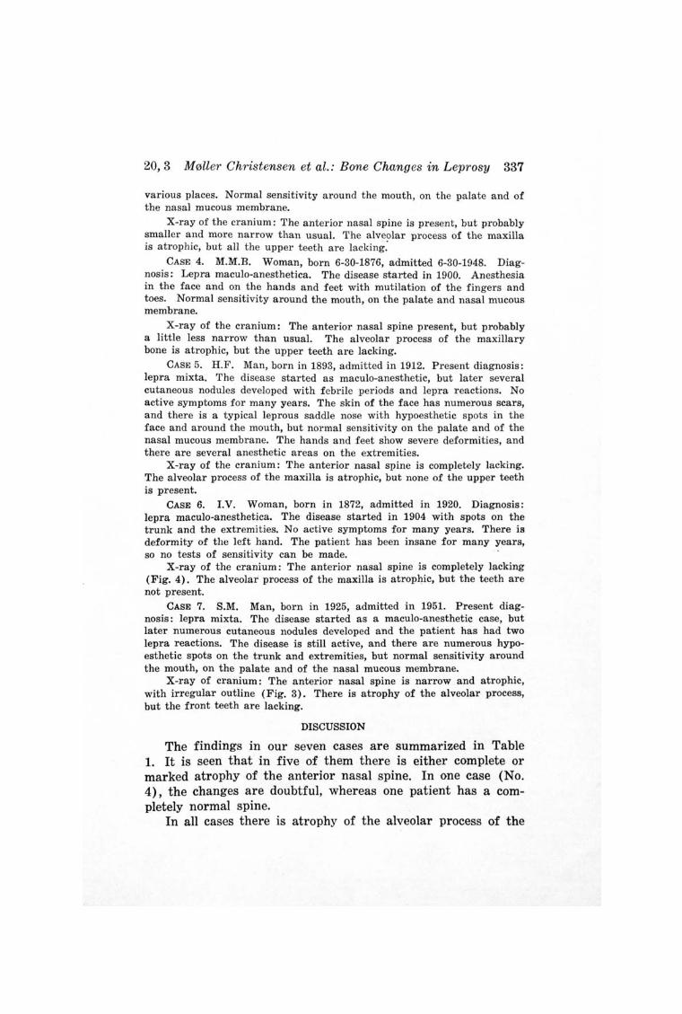
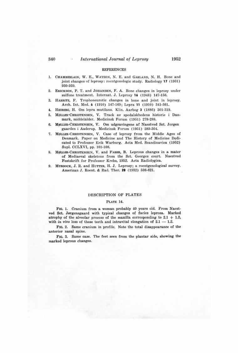
between the years 1260 and 1540, has demonstrated a marked atrophy of the alveolar process of the maxillary bone and also atrophy of the anterior nasal spine. These changes are clearly demonstrated in Figs. 1 and 2. This cranium came from a woman probably 40 years of age in whom there were mutilations and concentric atrophy of the first phalanges (Fig. 3), so that the diagnosis of leprosy was beyond doubt. These characteristic cranial changes, particularly the atrophy of the anterior nasal spine, are present in 110 of 150 craniums of the Naestved material, and as they are found in 80 per cent of the skeletons where leprosy has been diagnosed by the typical changes of the hands and feet, it is taken to be a change typical of the disease, and M0Iler-Christensen has given it the name "facies leprosa."
In living patients this atrophy may be observed either by palpation or by x-ray examination. It is possible that in order to observe these changes by roentgenologic examination it may be necessary to take soft pictures, otherwise they may be difficult to detect.
Because of these observations by M011er-Christensen we have studied seven patients in the Pleiestiftelsen No. 1 in Bergen. The case histories and the roentgenologic findings are briefly reported.
CASE 1. I.L. Woman, born in 1882, admitted in 1892, having had symptoms since 1890. On admission a typical case of the maculo-anesthetic type, with multiple anesthetic spots on the trunk and limbs. The disease has been very slight and is now completely burned out, resulting only in very slight atrophy of the one hand and a few hypo esthetic spots in the face and on the extremities. There is no loss of sensitivity around the mouth, on the palate, or in the mucous membrane of the nose.
X-ray of the cranium: The anterior nasal spine is present. There is atrophy of the alveolar process of the maxillary bone, but all teeth are lacking.
CASE 2. N.A. Man, born in 1866, admitted in 1884. On admission a case of the maculo-anesthetic type. Now only remnants of the disease with anesthetic spots on the face, trunk and hands, but the sensitivity around the mouth, on the palate and of the mucous membrane of the nose is not abnormal.
X-ray of the cranium: The anterior nasal spine is lacking. There is also atrophy of the alveolar process of the maxilla, but the upper teeth are lacking.
CASE 3. M.B. Woman, born in 1885, admitted in 1919, duration two years. On admission she was a typical case of lepra maculo-anesthetica, with spots on the trunk and extremities. Now inactive, with the usual remnants of the disease: atrophy of hands and feet; loss of sensitivity in
20,3 M0ller Christensen et al.: Bone Changes in Leprosy 337
various places. Normal sensitivity around the mouth, on the palate and of the nasal mucous membrane.
X-ray of the cranium: The anterior nasal spine is present, but probably smaller and more narrow than usual. The alveolar process of the maxilla is atrophic, but all the upper teeth are lacking:
CASE 4. M.M.B. Woman, born 6-30-1876, admitted 6-30-1948. Diagnosis: Lepra maculo-anesthetica. The disease started in 1900. Anesthesia in the face and on the hands and feet with mutilation of the fingers and toes. Normal sensitivity around the mouth, on the palate and nasal mucous membrane.
X-ray of the cranium: The anterior nasal spine present, but probably a little less narrow than usual. The alveolar process of the maxillary bone is atrophic, but the upper teeth are lacking.
CASE 5. H.F. Man, born in 1893, admitted in 1912. Present diagnosis: lepra mixta. The disease started as maculo-anesthetic, but later several cutaneous nodules developed with f ebrile periods and lepra reactions. No active symptoms for many years. The skin of the face has numerous scars, and there is a typical leprous saddle nose with hypoesthetic spots in the face and around the mouth, but normal sensitivity on the palate and of the nasal mucous membrane. The hands and feet show severe deformities, and there are several anesthetic areas on the extremities.
X-ray of the cranium: The anterior nasal spine is completely lacking. The alveolar process of the maxilla is atrophic, but none of the upper teeth is present.
CASE 6. I.V. Woman, born in 1872, admitted in 1920. Diagnosis: lepra maculo-anesthetica. The disease started in 1904 with spots on the trunk and the extremities. No active symptoms for many years. There is deformity of the left hand. The patient has been insane for many years, so no tests of sensitivity can be made.
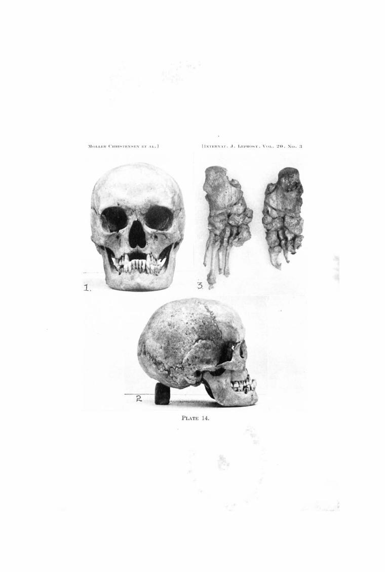
X-ray of the cranium: The anterior nasal spine is completely lacking (Fig. 4). The alveolar" process of the maxilla is atrophic, but the teeth are not present.
CASE 7. S.M. Man, born in 1925, admitted in 1951. Present diagnosis: lepra mixta. The disease started as a maculo-anesthetic case, but later numerous cutaneous nodules developed and the patient has had two lepra reactions. The disease is still active, and there are numerous hypoesthetic spots on the trunk and extremities, but normal sensitivity around the mouth, on the palate and of the nasal mucous membrane.
X-ray of cranium: The anterior nasal spine is narrow and atrophic, with irregular outline (Fig. 3). There is atrophy of the alveolar process, but the front teeth are lacking.
DISCUSSION
The findings in our seven cases are summarized in Table 1. It is seen that in five of them there is either complete or marked atrophy of the anterior nasal spine. In one case (No. 4), the changes are doubtful, whereas one patient has a completely normal spine.
In all cases there is atrophy of the alveolar process of the

338 International Journal of Leprosy 1952
TABLE l.-Roentgenological findings in the skulls of seven leprosy patients in Pleiestiftelsen No.1.
Case Age Sex Duration Type of Anterior nasal No. (years) of disease disease spine
1 70 F. 62 years Maculo-anesthetic Present
2 86 M. 70 years Maculo-anesthetic Lacking
3 67 F. 35 years Maculo-anesthetic Very small
4 76 F. 52 years Maculo-anesthetic Possibly small?
5 59 M. 51 years Mixed Lacking
6 80 F. 48 years Maculo-anesthetic Lacking
7 27 M. 12 years Mixed Narrow, atrophic
maxillary bone, but there is no reason to stress this finding except probably in Case 7. The first six cases are all either old or very old people and have been without teeth for many years, and there is therefore reason to look upon their atrophy as secondary, due to loss of the teeth after caries. We cannot be definitely sure, either, that the atrophy of the alveolar · process in Case 7 has anything to do with the patient's leprosy. The patient lost his front teeth at the age of 15, at the same time as the first symptoms of leprosy occurred.
The atrophy of the anterior nasal spine, on the other hand, cannot be regarded as a fortuitous finding, and there is reason to consider the possibility that this affection is in some way caused by leprosy. In one of the cases (No.7, Fig. 3), the original x-ray picture shows that the atrophic nasal spine has an irregular outline with a worm-eaten appearance. The characteristic atrophy is found both in the maculo-anesthetic type of the disease and in cases of lepra mixta.
It is interesting to note that patient No.1, with a completely normal anterior nasal spine, had had a very slight attack of the disease which has been burned out for many years, leaving only insignificant sequelae as compared with the other patients in whom the disease had been more severe.
With regard to the pathogenesis of the atrophy of the anterior nasal spine, it might be thought reasonable to assume that it is due to a neurotrophic disturbance, caused by a leprous neuritis, the pathogenesis thus being the same as that of the neurotrophic affection of the phalanges. The neurological examinations which we have carried out have, however,

20,3 M0ller Christensen et al.: Bone Changes in Leprosy 339
not given any support to this theory. In none of the cases have we been able to demonstrate anesthesia which would indicate that any of the nerves in this region were affected. Another possibility is that the lesion is· due to a leprous affection of the bone, or erosion of the bone because of the leprous changes in its immediate vicinity. The only case with a short duration of the disease in our material may point to one of the latter explanations as being correct. It is also possible that atrophy of the anterior nasal spine is a rather early sign. The leprosy bacillus is probably present in the nasal mucosa at an early stage of the disease, and for this reason the underlying parts might be affected relatively early. Our material in Bergen cannot throw any light on these questions. These problems should be studied in materials with younger and fresher cases. In such materials one should also pay attention to the alveolar processes in individuals in whom the teeth are present.
SUMMARY AND CONCLUSIONS
The authors report seven cases of leprosy (five of the maculo-anesthetic type and two lepra mixta), in five of which atrophy of the anterior nasal spine was found in the x-ray pictures. All seven had atrophy of the alveolar process of the maxillary bone, but that was probably due to loss of teeth several years earlier.
The pathogenesis of the atrophy of the anterior nasal spine is discussed. No changes of sensitivity were found which might indicate that the atrophy was due to neurotrophic disturbance of the bone. The possibility that the atrophy of the anterior nasal spine may be a rather early manifestation of leprosy is discussed, and as it can easily be demonstrated by palpation or by x-ray examination, it may play a role in the diagnosis of the disease.
RESUMEN
Los autores hacen un recuento de los hallazgos radiogriificos en los esqueletos de pacientes leprosos enterrados en N aestved, Sct. J orgensgaard durante los anos 1260 a 1540. En 110 de 150 criineos, se demostro atrofia de Ia espina nasal anterior. En 5 de 7 casos clinicos de los autores, estudios radiogriificos · demostraron Iesion·es simiIares y por tanto los auto res concluyen que este signo radiogriifico puede usarse como una ayuda en el diagnostico de Ia lepra particularmente del tipo maculo-anestesico.
340 International Journal of Leprosy 1952
REFERENCES
1. CHAMBERLAIN, W. E., WAYSON, N. E. and GARLAND, N. H. Bone and joint changes of leprosy: roentgenologic study. Radiology 17 (1931) 930-939.
2. ERICKSON, P. T. and JOHANSEN, F. A. Bone changes in leprosy under sulfone treatment. Internat. J. Leprosy 16 (1948) 147-156.
3. HARBITZ, F. Trophoneurotic changes in bone and joint in leprosy. Arch. Int. Med. 6 (1910) 147-169; Lepra 11 (1910) 341-361.
4. HEIBERG, H . Om lepra mutilans. Klin. Aarbog 3 (1886) 301-319.
5. M~LLER-CHRISTENSEN, V. Traek av spedalskhedens hi storie i Danmark, middelalder. Medicinsk Forum (1951) 278-288.
6. M91LLER-CHRISTENSEN, V. Om udgravingene af Naestved Sct. Jorgen gaarden i Aaderup. Medicinsk Forum (1951) 289-304.
7. M~LLER-CHRISTENSEN, V. Case of leprosy from the Middle Ages of Denmark. Paper on Medicine and The History of Medicine D~dicated to Professor Erik Warburg. Acta Med. Scandinavica (1952) Supl. CCLXVI, pp. 101-108.
8. M~LLER-CHRISTENSEN, V. and FABER, B. Leprous changes in a mater of Mediaeval skeletons from the Sct. Georges court. Naestved Festskrift for Professor Krebs, 1952. Acta Radiol<;>gica.
9. MURDOCK, J. R. and HUTTER, H. J. Leprosy; a roentgenological survey. American J. Roent. & Rad. Ther. 28 (1932) 598-621.
DESCRIPTION OF PLATES
PLATE 14.
FIG. 1. Cranium from a woman probably 40 years old. From N aestved Sct. J9rgensgaard with typical changes of facies leprosa. Marked atrophy of the alveolar process of the maxilla corresponding to 2.1 + 1.2, with in vivo loss of these teeth and intravital elongation of 2.1 - 1.2.
FIG. 2. Same cranium in profile. Note the total disappearance of the anterior nasal spine.
FIG. 3. Same case. The feet seen from the plantar side, showing the marked leprous changes.
-'i C) "!.I·: !.' ( ' lIln ~TI': S~Jo::\' EJ' ,\1 / .1
PLATE 14.

PLATE 15.
FIG. 4. Case 6. X-ray of cranium. Total disappearance of anterior nasal spine.
FIG. 5. Case 7. X-ray of cranium. The anterior nasal spine is atrophic, with irregular outline and worm-eaten appearance ; total atrophy of alveolar process.
-'I I) I . I . I-: Il ('IIHISTI-:SSES 1':'1' AI..I
PJ.ATE 15.