ch 13 viruses and prions. student learning outcomes differentiate a virus from a bacterium. explain...
TRANSCRIPT

Ch 13
Viruses and
Prions

Student Learning OutcomesDifferentiate a virus from a bacterium.Explain the difference between
enveloped and nonenveloped viruses.Define viral species.Describe how bacteriophages and
animal viruses are cultured.Compare and contrast the lytic and
lysogenic cycles of bacteriophages.Define oncogene and transformed cell.Discuss the relationship between viruses
and cancer.Explain latent viral infections and give an
example.Discuss how a proteins can be infectious.
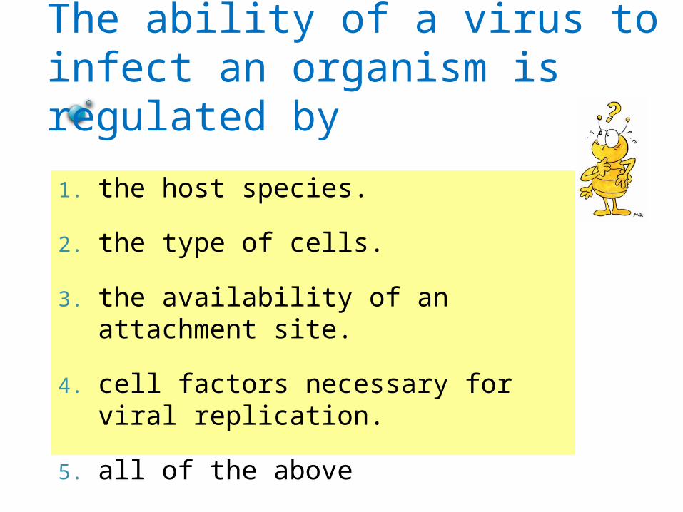
The ability of a virus to infect an organism is regulated by
1. the host species.
2. the type of cells.
3. the availability of an attachment site.
4. cell factors necessary for viral replication.
5. all of the above

Foundations of VirologyNon-living agents that infect all life forms (phages vs. animal viruses)
Viral cultivation differs from bacterial cultivation
1,500 known viruses (estimates: 400,000 exist)
Advent of EM allowed for visualization of viruses
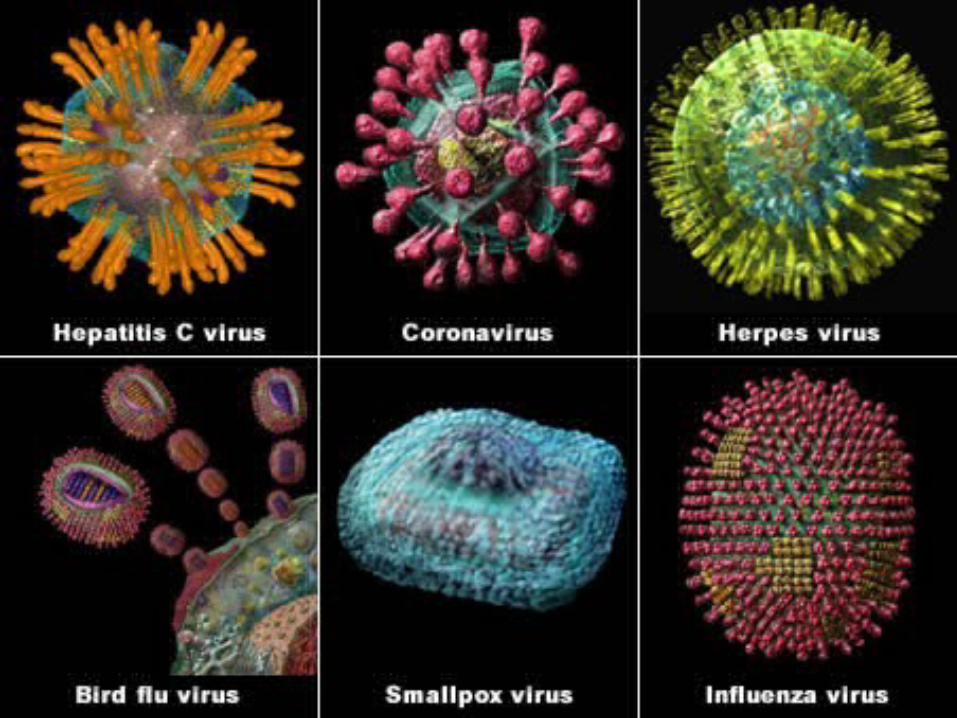

General Characteristics of Viruses
Obligatory intracellular parasitesFilterable Virus = Latin for poisonContain DNA or RNAContain a protein coat = capsid made up of
capsomeres. Various shapes Some are enclosed by an envelope (naked vs.
enveloped)
Some viruses have spikes (COH/protein)Most viruses are tissue specificHost range is determined by specific host
attachment sites and cellular factors

Host Range and SpecificityUsually narrow host range –
due to?
Tissue tropism
Phage Therapy
Oncolytic viruses

Fig 13.1
Virus Shapes and Sizes

Which of the following statements about viruses is FALSE?
A. Viruses use their own catabolic enzymes
B. Viruses contain a protein coat
C. Viruses contain DNA or RNA but never both
D. Viruses use the anabolic machinery of the cell

Virion Structure
Nucleic acid◦DNA or RNA
Capsid◦Capsomeres
Envelope
Spikes
Fig 13.2

Morphology of an enveloped helical virus
Example of a enveloped polyhedral virus: Hepes simplex

Polyhedral
Smallpox virusComplex symmetry
Compare to Figs 13.3 - 13.5

Electron micrograph of Aeromonas virus 31, an unassigned virus in the family Myoviridae
photograph by Dr Hans Ackermann.

Taxonomy of VirusesNo evidence for common viral ancestor. Classification based on genomics and
structure. ◦Family names end in –viridae◦Genus and species names end in -virus.
Viral species: A group of viruses sharing the same genetic information and ecological niche (host). Common names are used for species.
Subspecies are designated by a number.

Examples of Naming VirusesFamily: HerpesviridaeGenus: VaricellovirusSpecies and subspecies:
Human herpes virus 3 (HHV-3
Family: RetroviridaeGenus: LentivirusSpecies and
subspecies: Human immunodeficiency virus 1 and 2 (HIV-1, HIV-2)
Family: Picornaviridae
Genus: Hepatovirus
Species and subspecies: Hepatitis A virus
Briefly review Table 13.2


The viral envelope closely resembles the
1. Capsomere
2. Cytoplasm
3. Prokaryotic cell wall
4. Eukaryotic cell membrane
5. None of the above

Isolation, Cultivation, and Identification of Viruses
Viruses must be grown in living cellsBacteriophages
form plaques on a lawn of bacteria
Animal viruses may be grown in cell culture, embryonated eggs (Fig 13.7), or living animals
Fig 13.6
Fig 13.8
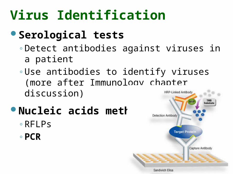
Virus IdentificationSerological tests
◦Detect antibodies against viruses in a patient
◦Use antibodies to identify viruses (more after Immunology chapter discussion)
Nucleic acids methods◦RFLPs◦PCR

Viral MultiplicationObligate intracellular parasites
using host cell machinery
Very limited number of genes encode proteins for ◦Capsid formation
◦Viral nucleic acid replication
◦Movement of virus into and out of cell
Kill or live in harmony within the host cell – Outside the cell, viruses are inert

2 Mechanisms of Bacteriophage MultiplicationLytic cycle (by lytic or virulent phage)
Phage multiplies, eventually causing lysis and death of host cell
Lysogenic cycle (by lysogenic or temperate phage)Phage DNA incorporated in host DNA
Prophage. No host cell lysis, cell lives. 3 results of lysogeny:
1. Lysogenic cell immune to reinfection by same phage
2. Phage conversion3. Possibility for specialized transduction
Mastering: Viral Multiplication

T-Even Bacteriophage:The Lytic Cycle
1. Attachment to cell surface receptors (chance encounter – no active movement)
2. Penetration – only genome enters
3. Biosynthesis – Production of phage DNA and proteins
4. Maturation – assembly to form intact phage
5. Release due to phage induced lysozyme production See Fig 13.11

1
2
3
Lytic Cycle of a T-Even Bacteriophage
Fig 13.11
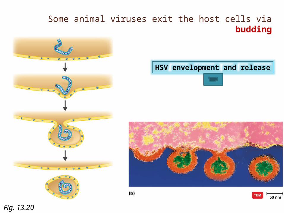
Some animal viruses exit the host cells via budding
HSV envelopment and release
Fig. 13.20

Fig 13.12
Lytic and Lysogenic Cycles ( Phage)

Place the following in the order in which they are found in a host cell: (1) capsid proteins; (2) infective phage particles; (3) phage nucleic acid.
A. 1, 2, 3B. 3, 2, 1C. 2, 1, 3D. 3, 1, 2E. 1, 3, 2

Multiplication of Animal DNA Viruses
Foundation Fig 13.15

Fig 13.19
Multiplication of a Retrovirus

Cancer - OncologyCancer uncontrolled mitotic divisions
Benign vs. malignant tumors
Carcinoma vs. Sarcoma
Adenocarcinoma
3 important characteristics of cancer cells:
1. Rapid cell division2. Loss of anchoring junctions and contact
inhibition ______________3. Dedifferentiation of cells

Viruses and CancerRoot of all cancers:Chemicals and ___________ directly
damage the genes through mutation rate
Viruses damage/alter genes by bringing new genes into the cell. what kinds of genes?
Normal cell cycle ends in cell division. Necessary for normalgrowth & development and wound healing….

Viruses and CancerNormal cell cycle regulator genes
1. Proto-oncogenes
2. Tumor suppressor genes
Genetic material of oncogenic viruses becomes integrated into the host cell’s DNA _____ virus.

Provirus leads to….
……conversion of proto-oncogenes to oncogenes or suppression of Tumor suppressor genes1. Foot on accelerator model:Proto-oncogenes turned ______
2. Foot off brake model:Inhibitors of tumor suppressor proteins

Oncogenic Viruses are responsible for 10 % of human cancers DNA Viruses
HPV _________cancer
Epstein-Barr virus (EBV) Burkitt’s lymphoma
HHV8 Kaposi’s sarcoma
HBV _________cancer
Hepatitis C virus (HCV) liver cancer
human T-cell leukemia virus (HTLV-1)
RNA Viruses

Proto-oncogenes can be activated to become oncogenes and cause cancer by
1. carcinogens in cigarette smoke.
2. overexposure to UV radiation in sunlight
3. spontaneous mutations.
4. virus infection.
5. all of the above.

Fig 13.21
Latent and Persistent Viral InfectionsLatent:
Virus remains in asymptomatic host cell for long periods
Persistent:Disease processes occurs over a long period; generally is fatal
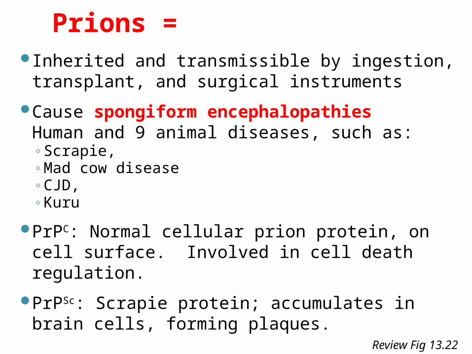
Prions = Inherited and transmissible by ingestion,
transplant, and surgical instruments
Cause spongiform encephalopathies Human and 9 animal diseases, such as:◦Scrapie, ◦Mad cow disease◦CJD, ◦Kuru
PrPC: Normal cellular prion protein, on cell surface. Involved in cell death regulation.
PrPSc: Scrapie protein; accumulates in brain cells, forming plaques. Review Fig 13.22

Spongiform EncephalopatiesCaused by altered protein:
◦Mutation in normal PrPc gene (sporadic CJD), or
◦contact with the abnormal PrPSc protein (Kuru)
Mastering: Prions

Fig 13.22
1 PrPc produced by cells is secreted to the cell surface.
2 PrPSc may be acquired or produced by analtered PrPc gene.
PrPSc
PrPSc reacts with PrPc
on the cell surface.4 PrPSc converts the PrPc
to PrPSc.
The new PrPSc converts more PrPc.
5 The new PrPSc is taken in, possibly by receptor-mediated endocytosis.
6
PrPc
Lysosome
PrPSc accumulates inendosomes.
7 PrPSc continues to accumulate as the endosome contents are transferred to lysosomes. The result is cell death.
8
Endosome
3
