cerebral anoxia and its residuals: pt. ii. respiration
TRANSCRIPT

Medical Arts and Sciences: A Scientific Journal of the College ofMedical Evangelists
Volume 1 | Number 2 Article 3
7-1947
Cerebral Anoxia and Its Residuals: Pt. II.Respiration, Normal and PathologicalCyril B. CourvilleCollege of Medical Evangelists
Follow this and additional works at: http://scholarsrepository.llu.edu/medartssciences
Part of the Cell and Developmental Biology Commons, Circulatory and Respiratory PhysiologyCommons, and the Medical Cell Biology Commons
This Article is brought to you for free and open access by TheScholarsRepository@LLU: Digital Archive of Research, Scholarship & Creative Works. Ithas been accepted for inclusion in Medical Arts and Sciences: A Scientific Journal of the College of Medical Evangelists by an authorized administratorof TheScholarsRepository@LLU: Digital Archive of Research, Scholarship & Creative Works. For more information, please [email protected].
Recommended CitationCourville, Cyril B. (1947) "Cerebral Anoxia and Its Residuals: Pt. II. Respiration, Normal and Pathological," Medical Arts and Sciences:A Scientific Journal of the College of Medical Evangelists: Vol. 1: No. 2, Article 3.Available at: http://scholarsrepository.llu.edu/medartssciences/vol1/iss2/3

MEDICAL ARTS AND SCIENCES VOLUME 1 JULY, 1947 NUMBER 2
CEREBRAL ANOXIA AND ITS RESIDUALS*
. II. RESPIRATION) NORMAL AND PATHOLOGICAL
CYRIL B. COURVILLE, M.D.
From our present viewpoint it is not surprising that the story of the clinical development of asphyxia had its inception at the same time as that of the physiology of respiration. No sooner does the inquisitive physician discover a new disease entity th~n he begins to investigate the phenomena upon which it is based. It was Aristotle who first wrote of difficulty of respiration in high altitudes; he also postulated that the prime purpose of respiration was to cool the blood. Although his idea of respiratory physiology wa a little na!ve , it was but a token of the long interval of ignorance which wa to intervene before the physics and chemistry of respiration were to be understood.
This long wait was due to man's slowness in comprehension of the chemical aspects of_ living matter. And this in tum was a result of his inability to cope with the manifold problems presented by even his most superficial investigation in the realm of biology. Inventive genius was then too deeply buried beneath the snows of dogma and authority. But when Boyle invented the air pump (1666), it was only a step to prove that air was an un-
* From the Department of ervous Disease , College of Medical Evangelists, and the Cajal Laboratory of Neuropathology, Los Angeles County Hospital , Los Angeles, California.
equivocally fundamental necessity of life. Thi was easily demonstrated by exhausting a chamber containing a living animal. The fact was also proved in another way by Hooke (1867)? who found that an animal could be kept alive by passing air through its perforated and collapsed lung.
Lower (1669) and Mayow (1673) concluded that the action of air in respiration and combustion was similar to that of niter in the combustion of gunpowder. They believed that the "nitro-aerial spirit" in the air was absorbed into the blood from the lungs, whence (according to the theory of Descartes) it was carried to the brain, distilled into the ventricles, to reach ultimately the muscles via the nerve tubules.
It was almost a century later before the next development in the physiology of respiration oc urred. In 17 54 Joseph Black discovered that "fixed air" (carbon dioxide) was given off from the lungs in respiration. Shortly t~ereafter this concept was enlarged by Priestley (1774), who found that "dephlogisticated air" (oxygen) disappears in both animal respiration and combustion, but it is produced by green plants. Lavoisier ( 1777) unified these two isolated ideas by demonstrating the chemi-
Copyright, 1947, by Review and Herald Publishing Association
35
36 Courville-Cerebral Ano. ia
cal formation of carbon dioxide from oxygen and carbon, a product which i formed by an animal in approximately equivalent amount to the oxygen consumed (Lavoisier and Laplace [1783]). But all that Lavoi ier got for his efforts wa the doubtful privileg of being guillotined in the Place de la Revolution on a sunny morning of May, 1794. The new republic, so they said, had no use for bourgeoisie investigators.
The next step in the physiology of respiration wa th discovery by Leaalloi ( 1812) that respiratory failure and death followed the destruction of a mall area in the floor of th fourth ventri le. Thus was identified the "re -piratory center." But this discovery was somewhat of an e topic departure, for there wa still much to be learned about the chemical aspect of respiration. Magnus (1 37, 1 45) found a way to liberate th a e from the blood (by expo ing it to a va uum), pro ing thereby that les oxygen and m r carbon dioxide wer given off from venou than from art rial blood. With the dev lopment of the mer urial aa pump by L. Meyer, Ludwia, and Pfluger, it was found that en i taken up from the lungs by the blood; that it form an easily di o iabl omp und with hemoglobin; and that thi aa i taken up from th ti sue as the blood cir ulate thr ugh the capillary ystem. It wa 1 arned that the oxyaen-depleted
venou blood al ·o accumulated a upply of carbon dio ide arri d in a loo e h mi al combination to be discharu d in the lung .
But thi la t bit of hi t ri al lore ound tranael lik our pre ent-da phy i loay of
re piration, whi h inde d it i . L tu ee, then, what our modern cone pt of th me hanic , the phy i , and hemi tr of r piration really in ol e.
R 1AL RE PIR Tl
A tudy of normal r piration rev al that a number of fa t r ar in olv d: (1) the me-
• 1
\
chanics of chest movement, which re ult in the inhalation and exhalation of air, (2) the physical principles governing exchange of ga es, (3) the chemical rea tions that have to do with transportation of oxygen and carbon dio, ide in the blood and tissue respiration,
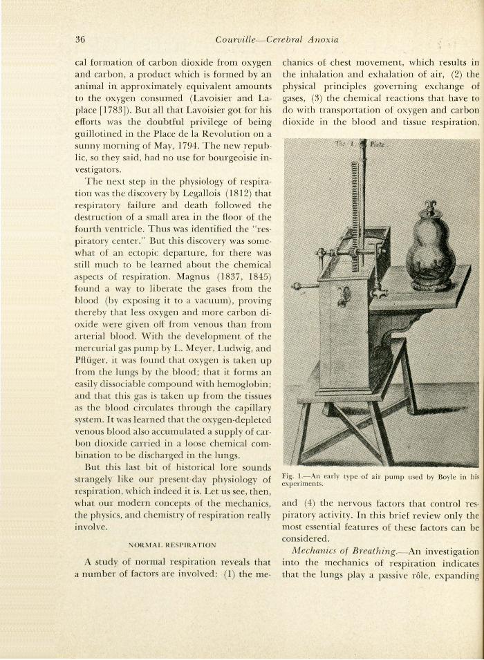
Fig. 1.- n earl type of air pump u eel by Bo le in hi ·periment.
and (4) the nervou fa tor that ontrol re -pirator a tivit . In thi bri f r iei onl the mo t ential featur oE th e fa tor an be on id red.
Mechanic of Breathing.- An in e tigation into th me bani f r piration indi ate that the lung pl a pa i e role, e pandincr

Medical Arts and czences 37
and contracting with the chest walls; that inspiration i largely an active process, and expiration i almost wholly pa ive ; that the che t is enlarged in all it diameters during inspiration, wherea during expiration the thoracic cacre of it own wei ht re umes it restincr position; and that the di tended lungs recoil, and the now relaxed muscular diaphragm is pulled upward into the chest.
Thi alternating recurrence of active and passive movement result in the in-and-out passage of air chemically altered in the lung b the gaseous exchange through the alveolar and capillary walls. The gases of this alveolar air come into a balanced tate with that of the capillaries in the alveolar wall. The e small pockets of air must be con tantl y renewed by the addition of fre hair from the out ide. Thi i accompli hed as follow :
By the encl of each expiratory movement most of the air in the di tended acs i xp lled into the bronchiole' and thence into the bronchi. With the next inspiration thi air i again for d back into the alveoli, while the fr sh air now fill the air pa sages. The oxygen from the new air i diffu eel throughout th alveolar air, and the carbon dioxide in turn i diffused into the air in the air pa age . '.[ ixing of th e alveolar air with the in pired air eerns to be fundamentally mechanical and incident to re piratory movement and the chang in temperature betw en th residual anu in pired air.
It i a urned that at omplete re t there i about 150 c . of air in th re piratory pa sages where the thickne of the wall preclude any ga eou e, hange. In expiration mo t of thi air i exhal d. On an ordinar in piration 500 cc. oE air i inhaled, 150 c . of which fill thi "dead spa e" of the br nchial tree. he remaining 350 cc. i mixed with the air whi h has remained in the lung after expiration. With the next expiration the now well-mix d air i fore d out of the al eoli into th bronchial ystem. Part of it i exhaled; the remainder o u pi the bronchial pa age . We thu learn that all the air from the previou inhalation i not exhaled in turn but that ev ral
respiratory act are nece sary to completely exchange all the air which is in the lungs at a given time.
The Phy ical Law of Gases and Their lnf/,uence on Respiration.- In the exchange of oxygen and carbon dioxide through the alveolar-capillary wall, four laws croverning the a tivity of gase are known to play a part. It is recognized that if under constant temperatures the olume of a ga is altered , the pressure of that gas varies inversely (Boyle's law). On the other hand, if the temperature of a gas i raised 1 ° Centigrade it volume at 0° Centi- · grade expands by 1/ 273 if the pressure is kept con tant (Gay-Lu sac' law). The pre ure exerted by any given gas in a mixture oE gases i that of th gas in its unmixed state (Dalton' law). The amount of a gas which goe int solution i proportional to the partial pressur of that ga and it relative solubilit (Henry's law).
In appl ing the e law to the e, hange of oxycren and arbon dioxide between the alveolar air and the blood pla ma, we find that the oxyg n is f reed into the pla ma because of it higher partial pr ur in th alveolar air. In turn, the higher con entration of carbon dio, id in the blood plasma results in e cape of thi a int th alv olar air (Henry' law).
In thi int ra tion each era behaves a though it were the only one pr sent, exerting it own partial pres ure (Dalton' law). In a ord with the prin ipl of Boyle' law, oxygen pas into olution in the blood plasma of th pulmonary apillaries b cau e of th lower temperature, but in r pon e to th higher temperature in th ti ues it i more readily given off. Also becau of thi high r temperature in the ti u increa ed molecular a tion produced thereby fa or the olu ~
tion of arbon dioxid in the blood plasma (la' of Ga -Lu a ).
Chemical Factor in Re piration.- The transportation of ox en from the lungs to th
38 Courville- Cerebral Anoxia
tissues is accomplished through the complex ubstance, hemoglobin, resident in the red
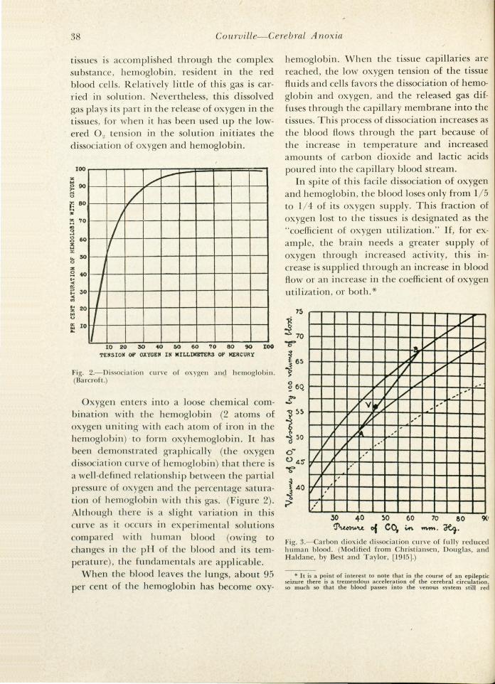
blood cells. Relatively little of this .gas is carried in olution. Nevertheless, this dissolved gas plays its part in the release of oxygen in the tissues, for when it has been u ed up the lowered 0 2 tension in the solution initiates the dissociation of oJ ygen and hemoglobin.
IOO
~ 80
"" ~ ?O co 0 M
g 60 x lol :x;
~ ~ ;z:
~ 40
"" < !5 ... 30 < en
~ 20 w u
gj IO ...
/ i,....----
/ "' I
I I
' 1 I I l
IO 20 ~ 40 SO 60 '10 80 90 IOO TENSION OF OXYGEN IN MILLIMBTERS OF MERCURY
Fig. 2.- Di ocialion curve o( oxygen and hemoglobin. (Barcroft.)
Oxygen enter into a loose chemi al combination with the hemoglobin (2 atoms of oxygen uniting with each atom of iron in the hemoglobin) to form oxyhemoglobin. It has been demonstrated graphicall (th o ' ygen diss iation curve of hemoglobin) that there i a well-defined relationship b tween the partial pre ure of oxygen and the percentage aturation of h moglobin with thi gas. (Fi<Yure 2). Al though there i a light variation in thi urve a it occur in experimental olution omparecl with human blood (owin<Y to
change in the pH of the blood and it temperature), the fundamental are applicable.
When the blood leave the lungs, about 911 per cent of th hemoglobin ha be me oxy-
hemoglobin. When the tissue capillaries are reached, the low oxygen tension of the tissue fluids and cells favors the dissociation of hemoglobin and oxygen, and the released gas diffuses through the capillary membrane into the tissues. This process of dissociation increases as the blood flows through the part because of the mcrease m temperature and increased amount of carbon dioxide and lactic acids poured into the capillary blood stream.
In spite of this facile dissociation of oxygen and hemoglobin, the blood lose only from 1/5 to 1/4 of its oxygen supply. This fraction of oxygen lost to the tissues is designated as the "coefficient of oxygen utilization." If, for example, the brain needs a greater supply of o ygen through increased activity, this mcrease is supplied through an increase in blood flow or an increase in the coefficient of oxygen utilization, or both.*
75
~ 70 t-t-+--t--+--+-+--11-J-+..-..J-/..Jv~t:::l/-L.....J T .. V f es r-r-;-;--t--t--+-+~.9"~~~~/~v-l.--1 i /Lj/ .v
0.. v o 4 s- I . ~ v v ~ 40 /, ,' '
;>
, .,.
70 60
"""'· ~· C)(t
Fig. 3.- Carbon dioxide di ocialion urve of full reduced human blood. (Modified from Christian en, Dougla , and Haldane, by Be t and Taylor, [1945).)
* It is a point of interest to note that in the course of an epileptic seizure there is a tremendou acceleration of the cerebral circulation so mu h so that the blood pa ses into the venou system still red

Medical Arts and Sciences 39
Much experimental work has been done on the problem of tissue respiration, but as yet there is no precise information available a to the exact methods of oxygen utilization by the cell. It is obvious, of course, that the chief source of cellular energy is the oxidation of metabolite . When this process ceases, cell death follows, as is seen in ca es of serious degrees of anoxemia in which profound damage to the cerebral tissues results. It is believed that thi process involves the removal of electrons from the involved metabolite and that glutathione and ascorbic acid may assist somehow in the proce s. Just how thi i actually accomplished is as yet unknown.
Be all this as it may, it is clear that the venous blood which returns to the lungs is deficient in oxyhemoglobin and is surcharged with carbon dioxide. When carbon dioxide enters the blood from the tissue , it combine with water of the plasma to form H 2C03. This action is facilitated by an enzyme, carbonic anhydrase, found in the erythrocytes. Most of this vveak acid combines with a base to form bicarbonates.
The role played by hemoglobin in the transportation of carbon dioxide ha not been fully appreciated until quite recently. It was long presumed that this ga was carried alone in chemical combination in solution. It is now recognized that hemoglobin actually ha a double action in this respect: (1) it carrie a considerable amount of ba e, which it yield up on losing its oxygen, and (2) it actually unites with carbon dioxide to form carbhemoglobin (2 to 10 per cent of carbon dioxide is carried in th is way). About 5 per cent of this gas i carried in simple solution, and the r -maining 85 to 93 per ent i carried as a bicarbonate.
In the lung the carbon dioxide is rapidly unloaded, a necessity, for the blood remain only one second on an average in the capillaries of the rungs as in other body tissues. This rapid release of carbon dioxide is made possible by (1) the action of carbonic anhydrase
with its high percentage of oxyhemoglobin. This too rapid circulation docs not permit an adequate release of oxygen to the tissues, and a state of partial tissue anoxia results. It has been assumed that the advanced degrees of cerebral atrophy found in chronic epileptics j the end result of repeated episodes of partial anoxia.
(which serves in both phases of the reversible reaction) and (2) by the rapid release of this gas from combination with hemoglobin.
Nervous Regulation.- As the result of animal experimentation, Markwald (1887) came to the conclusion that respiration was under specific nervous control. Lumsden ( 1923) sugaested that the respiratory center was located in the medulla at the level of the striae medullares acusticae) and that this center was nor-
-n---A J3 ,__,,__ __ c.
--#---D
\
Fig. 4.- Location and extent of the respiratory center in the cat. (According to Pitts, Magoun , and Ran on .)
mally dominated by an inhibitory or pneumotoxic center in the upper end of the pons. Pitts, Magoun, and Ranson (1939) recently found that the respiratory center (in the cat) was lo-ated in the reticular formation of the me
dulla, and that it was divided into two portions, an inspiratory (apneustic center of Lum den) and an expiratory division (Figure 4). It was learned that regular respiration can be p~oduced by timulation of either divison.
Although it was for a time denied by some,

40 Courville- Cerebral A no -ia
it is now known that the re pirator center may act pontaneously, and thi activity i inherent on the inspiratory portion of the center. It i recoo-nized, of cour e, that thi enter i al o influenced by emotional factors and, further, that it may be controlled temporarily by volition.
CAROTID SINUS
man ( 1927) discovered the carotid reflex. In the carotid and aortic bodie two type of receptors were i olated. The pressoreceptors were capable of being stimulated by mechanical m an , while the chemoreceptor could b timulated by chemical means. Comroe and Schmidt ( 1938) demonstrated that
LAR~HX
~
lNTERCOSTAL MUSCLES
DJAPHUOM ABDOMIN~L
MUS CL ts
Fig. ~ .-The nervou control of re piration . (Be t and Ta lor.)
he· major ontrol , however, seem to be on a r flex ba i , a wa hown a long ago a 1 6 (Hering and Breuer), b ing influen d by r -pirator a ti ity. The e inv tigator howed that inflation of the lungs inhibit d inspiration, and, conver ely, that deflation inhibit d expirati n. Mor re entl He man and Hey-
the hemor flex mechani m of the carotid and aorti bodi wa re i tant to the. influence of anoxia · thi m hani m i th re fore the la t
lin of d f n e a ain t r pirator failure. It ha long b en known that carbon dio -
ide ha had a p ifi action on the re pirator nter. ·with an increa in thi ga in the

Medical Arts and Sciences 41
blood, the center was stimulated to increased activity. It would seem as though the cells in the center are highly specialized, capable of ampling the blood for it oxygen content (Best and Taylor).
At thi point one might investigate the pathologic phy iology of a variety of respiratory episode (Cheyne-Stokes or Biot's respiration), which are o often a part of certain disea e syndrome . But even though anoxia, at lea t to a relative degree, may play it part in the production of ome of them, these conditions per e are not of major concern to us in thi conne tion. \!Ve are interested, to the contrary, in tho e more serious, even lethal, condition which leave their unmi takable stamp on the tis ue of the brain to .produce the characten tic ano ' i ymptom- omplexe . But before we tud the e cerebral le ions them elve , a word about the variou types of anoxia is in order.
A OXIA- IT TYP AND CAU S
We are now in a position to inve tigate the pathologi phy iology of anoxia, the ubje t here beino- on idered. W hall find that ther ar many cau e for an ultimate de rea e in the amount of oxygen d livered to the ti sue oE th bod in o-eneral and th brain in particular. Barcroft ( 1920) de ribed three type of ano ia ba ed on th fun tional di turbance in the ph iology of re pi ration: ( 1) the anoxic type (due to def cti ve o ygenation of th blood in th 1 u ng ) ; (2) the an mic type (in ident to a lowered apacity of the blood to arr ox gen)· and (3) the tagnant type (the
re ult of a lowing of movement of blood through the apillary tern). To th e thre type Peter and Van lyke (19 2) ha e added another: (4) the hi tiotoxic type (in which there o cur an int rferen e with interval re -piration, the oxygenation of th ti sue them- · elve ) (Figure 6). he p ific etiology and
mechani m of these typ will be given brief attention.
A no ic A noxia.-In the production of this type of ano ia we have a con iderable number of causes which may be responsible. In it are to be found a variety of conditions not uncommonly experienced in medi al practice. Three ubgroup of cause may be di tinguished: (a) mechanical interference with passage of air into the alveolar sacs, (b) a decrease in oxygen tension in the inspired air, and ( c) certain congenital cardiac lesions which limit the amount of blood reaching the lungs. In any of these ituations both the o ' ygen and cai;-bon dio id tension in the blood become lowered, so that even that o, ygen which is present in the blood i not readily available to the ti ue .
Mechanical defects in the respiratory apparatus include ob tru tive occlusion of the upper air pas ages by foreign bodie , by acute inflammatory le ions of the throat (tonsillar .ab ce s with rupture) , ervical tistue (relluliti of th neck) or of the larynx and trachea (diphtheria), or by throttling in as ault or hanging. Intcrf erence with respiration in the lungs themselv may oc ur in uffusion of tb se spa e by fluids (drowning, pulmonary edema, pu from rupture of an ab ces of the lung or adjacent ti sues, blood from ruptured ancury rn) , or by thickening of the alveolar wall or o du ion of the entire ac by di ea proce e (pn umonia, tuberculosi , emphy erna, a thma, col-
lapse of the lung). Failure of the nervou centers would, of coune, play an important part in the production of anoxia. h pre ent writer ha con idered the e po ibilitie in ca e of depression of th re piratory center by certain anesth tic agent notably nitrous oxide (Courville [1937, 1939]), but al o by ether ( ourvill [1941 ]). ongenital defe t in th lung are rarely responsible for asphyxia in th newborn.
( r a le ened oxygen ten ion in in pired air, ano, ia may re ult from the pre en e of a number of ga ub tances su h a nitrogen, or the presence of fume of ulfur, fermenting liquor , or decaying sub-tance . Ev n the pre ence of foul air in mines (a ha
already b n referr d to in the ection on hi tory) may cau serious if not fatal a phyxia. Suffocation by m ke in fire i another xam ple of thi mechani m.
noth r mechani m whi h cau e thi t p of anoxia i that occurring in high altitudes. The long-recognized form of mountain ickne ha b n briefl referred to; th more modern t pc incident to air travel in rarefied atmo phere has re eived attention in the recent war.
Insufficient aeration of the blood may occur in ongenital le ion of the heart and great ve el (septa!
d ( ct , patent ductus) be ause much of the blood hunted by the Jung field.

42 Courville- Cerebral Ano ia
Anemic A noxia.- This occur characteristically in any form of anemia, resulting either acutely from hemorrhage or chronically by destruction of red cells or by fa ilure to produce them. Under these circumstances the oxygen
anoxia after carbon monoxide, which interferes with the oxygen-carrying power of the blood by production of methemoglobin. Poi-oning by nitrate , chlor ides, and certain other
chemicals also produces anemic anoxia.
10 20 3040 50 60 70S> 90 Or11pn Ten~ion mm.
NORMAL
I 0 Z0 30 40 50 60 70 80 90
ANOXIC
10 zo 30 ..a 50607080.,
STAGNANT
10 zo 30 40 so 6010 50 90
H 15T0Toi1c
Fig. 6.- Diagram illustrating types of anoxia. Column represen ting arterial blood ( ) and veno us blood (V) are uperimposed upon the oxygen di sociation curve. The black portion of the columns represents reduced hemoglobin, and the shaded portion, o ygenated hemoglobin. Jn the case of anemic anoxia th e dotted portion of the columns repre ent hemoglobin that i. either lost, as in true anem ia , or unfit for oxygen transport, a in arbon monoxide poi oning. Th perpendicu lar arrows denote the vo lume of oxygen give n up to the ti ssues from a unit of blood. (From Best and Taylor. Modified from Mean .)
tension i normal, but not enough of thi ga can be carried ·by the deficient amount of hemoglobin. In this type of anoxia the ituation i often relative, the evidence of impaired oxygenation making it appearance only on exertion.
nder this de ignation may also be included
tagnant A no ia.- Thi type i characteri -tically seen in cardiac failure with slowed circulation. The oxygen content and the oxygen ten ion in the blood are normal , but the oxygen a tuall supplied to the tissue i reduced incident to the slowed blood current. The failure of the circulation consequent to

Medical Art and Science 43
surgical shock, if long continued, will produce tissue changes incident to the anoxia produced (Rand and Courville [1936]).
Histiotoxic A noxia.- This has been described by Peters and Van Slyke and is attrib-
A
THE PATHOLOGIC PHYSIOLOGY OF
CEREBRAL NECROSIS
It has been recognized that the evil effects of anoxia are most evident in the tissues of the brain. It is true, however, that minor changes
B'
Fig. 7.- Phys iologi statt1 oE cerebral change incident to asphyxia. A. Pressure o[ oxygen in tissues. B. Increased circulator activity under conditions o[ minor oxygen want. C. Focal cortical necrosis after asphyxia showing changes about a dilated capi llary with preservation o[ intercapillary tissues. D. howing relationship o[ necrosis to capillary blood vessel.
uted to an interference with ti sue respiration by toxic substances such as cyanide. The cells are apparently unable to utilize the oxygen brought to them even though it i sufficient in amount and is under adequate tension.*
are found in the viscera. For example, thick-
* This concept has al o been utilized to explain the action of alcohol (a well as of certain anesthetic agents) on the brain. The presence of alcohol in the tissue fluids is presumed to result in an interference with the use of oxygen. The resultant mild and recurrent form of anoxia is attributed to be the cause of cereb(al atrophy in chronic alcoholics.

44 Courville- Cerebral Anoxia
erring and cellular infiltration oE the walls of the pulmonary alveoli, brown atrophy and focal necro is of the cardiac muscle fibers, necrosis of the liver lobules, chiefly in the region of the efferent vein, acute degeneration of the renal epithelium, cellular infiltration of the spleen, and hemorrhages in these organs or the membranous structure (pericardium, pleura, meninges) have been described (Courville [1939], literature). But these changes are not, as a rule, sufficient to cau e death. It is the cerebral lesions at least which produce the most cripplino- and fatal residual . To the production of these lesions brief attention should be given. .
It was learned experimentally by Gildea and Cobb ( 1930) that a temporary interference with the cerebral circulation produces areas of focal necrosis in the cerebral cortex. These H er de or areas of necrosis are now recognized to be the characteristic cerebral le ion of asphyxia (Courville [ 1937]). A car ful analysis of these le ions makes clear that ( 1) the earliest evidence of the lesion is found in an enlargement of the perineuronal space associated with moderate shrinkage of the nerve cell and degenerative changes in th urrounding interstitial tis ues; (2) the area of focal necrosi are u uall y found urrounding a dil_ated blood ve el; and (3) the larger cortical le ion are but a progressive fusion of these areas of focal n crosis.
The co-ordination of impres ion as to the physiology or oxygen on umption by the ti ue and ph ical clamag to the brain incident to anoxia may be as i ted by comparing the accompanying diagram by Barcroft (Figure 7, ) with th photomicrograph (Figure 7, C)
showing the vidence of focal damage to the cerebral cortex in ca e of asphyxia. Barcroft' diagram was designed to how the effect of oxygen pressure in the tis-u . The dot in th center of ea h erie of concentric
circle in repre nts a capillary containing oxygen with an oxygen pre ure of 30 mm. Hg. B tween the su ceeding cone ntric circles the oxygen pre ure falls 5 mm. Hg. It is vident that at the point marked x the oxygen pre ure i zer_o and under ordinary circumstance , evidence of tis ue damage would o cur at the e point . It is as urned that additional capillaries
(Figure 7, B) would open up to supply the deficiency. A tudy of photomicrographs. taken of_ the 5er_ebral
cortex after asphyxia (as with mtrous oxide) mdicat~s that this po tulate doc not hold un~er _patholo_g1c conditions. The localization of destruct10n m the immediate environs of the capillary implies to the contrary that the vessel, once the source of life-giving oxygen, has now become the ource of a noxiou pr?duct which i histiotoxic and hi tiolytic in its behavior. Since, however, this va cular change is not uniform, only cattered areas of necrosi occur. Becau e nitrou oxide per se i not toxic, one mu. t ass_ume that .some additional factor must be responsible m the ultimate analysi . It may be that the accumulation of ca~bon dioxide in the pericapillary tissues because of a fa1l:ire to ab orb it by the chemically altered plasma flowmg with markedly reduced speed through the vessel (sta~nant anoxia) is primarily re ponsible. At any rate, it become visually evident that in anoxia there is a profound disturbance in tissue re piration, in which there is a reverse o( the usual diffusion of oxygen through the tis ues. In this disturbed proce s the tissue fluid seem to play an important role.
Therefore, in the ase of anoxia following nitrous oxide anesthe ia the pre ent writer ha presum d that these cerebral lesions occur primarily as the result of a triple mechani m: ( 1) th general reduction of oxygen tension in the blood, (2) a temporary cessation (or at least a marked slowing) of the blood current incident to the often attendant ardiac failure and (~) a dilatation of the small corti al blood ve els. It i the last of the e factor that apparently determines the focalization of the early area of cortical ne ro i , a ugge ted b . the presence of an enlarged entral ve sel. As the situation grow more eriou , more or le of the va cular bed become dilated and exten i e le ions re ult from a onfiuen e of area of focal n crosi to form laminar degen-ration, with ultimate fu ion of the e lamina
to form ubt tal de tru tion f th corte . In hart, the cortical le ion ar the apparent
combined result of two type of ano ' ia: the anox1 form and the ta nant form. But the details of the nature of the re ultant le ion will b re erved for th following e tion.
OTE.- The bibliography will appear at the end of the article.
(To be continued)
ompleted