cerebellum and adhd
TRANSCRIPT

The Practitioner Scholar: Journal of Counseling and Professional Psychology 1 Volume 1, 2012
1
The Cerebellum and Attention Deficit Hyperactivity Disorder A Case Study of a Cerebellar Chiari 1 Malformation
Robert Eme
Erin Sheffer M.A. Illinois School of Professional Psychology at Argosy University, Schaumburg
Abstract
The cerebellum is the most consistently implicated and the most robustly deviant brain structure in the pathophysiology of Attention-Deficit/Hyperactivity Disorder (ADHD). Its role in the neurobiology of ADHD is also buttressed by many recent ana-tomical, functional neuroimaging, and human lesion studies that have implicated it in the neural network that mediates selective attention. This article may add to the evidence for the role of the cerebellum in the pathophysiology of ADHD by presenting a case history of a young female adult with a cerebellar disorder called a Chiari Malformation type 1. She developed a severe impairment in selective attention, which resulted in an adult onset of ADHD. This case study provides a unique exploration of the pathophysiology of ADHD because all prior clinical findings from human lesion studies implicating the in-volvement of the cerebellum in the neural circuit for selective attention have overlooked the relevance of Chiari Malformation type 1.
Attention-Deficit/Hyperactivity Disorder (ADHD) is characterized by developmentally inappropriate levels of inattention and hyperactivity/impulsivity, which adversely affects many facets of an individual’s life, and tends to be chronic (American Academy of Pediatrics, 2011; Barkley, 2006, Barkley, Murphy & Fischer, 2008). Its importance is also underscored by the fact that it is the most commonly diagnosed juvenile neurodevelopment disorder, with a prevalence of approximately 8% of juveniles between the ages of 5-17 in the United States (American Academy of Pediatrics, 2011). Its adult prevalence is approximately 5% (Barkley et al., 2008).
It is generally accepted that genetic and neurological factors are the most common causes of ADHD (Barkley, 2006; Nigg, 2006). Structural, functional, and clinical findings indicate that these factors affect multiple brain structures and neural circuits that are involved in the patho-physiology of ADHD (Bledsoe, Semrud-Clikeman, & Pliska, 2011; Durston, van Belle, & de Zeeuw, 2011; Nigg, 2006, 2010; Sonuga-Barke, 2010). Although the cerebellum which has his-torically been thought of as a structure involved primarily if not exclusively in motor control, it is the most consistently implicated and also the most robustly abnormal structure in the patho-physiology of ADHD (Bledsoe, Semrud-Clikeman, & Pliska, 2011; Kieling et al., 2008; Krain & Castellanos, 2006; Mackie et al., 2007).1 Its involvement beyond that of a simple controller of motor acts stems from the fact that it is second in size to only the cerebral cortex, contains more neurons than the rest of the brain combined, and is massively connected to the cerebral cortex (Bower & Parsons, 2003; Stick, Dum, & Fiez, 2009). Its role in the neurobiology of ADHD is also buttressed by many recent anatomical, functional neuroimaging, and human lesion studies of individuals without ADHD that have implicated it in numerous non-motor functions (e.g. ex- 1 Note that neuroimaging techniques that study brain structures implicated in the pathophysiolo-gy of ADHD such as the cerebellum cannot currently be used in clinical diagnosis (Zametkin, Schroth, & Faden, 2005).

The Practitioner Scholar: Journal of Counseling and Professional Psychology 2 Volume 1, 2012
2
ecutive control, language, working memory) as well as selective attention (Casey & Riddle, 2012; Cherkasova & Hechtman, 2009; Strick, Dum, & Fiez, 2009; Stoodley, 2011; Timmann & Daum, 2007).
The purpose of this article is to add to the evidence for the role of the cerebellum in the pathophysiology of ADHD by presenting the case history of an individual with a cerebellar dis-order called a Chiari Malformation type I (CMI). It will do so by first briefly reviewing the evi-dence establishing a selective attention network and the role of the cerebellum in this network. It will then discuss the onset of CM1 in young adulthood that so adversely affected the selective attention that it resulted in a secondary attention-deficit/hyperactivity disorder (SADHD) which is the term that was coined for ADHD that develops after traumatic brain injuries (Max, 2011). This clinical case study provides a unique exploration to the pathophysiology of ADHD because all prior clinical findings from human lesion studies implicating the involvement of the cerebel-lum in the neural circuit for selective attention have overlooked the relevance of this disorder (National Institute of Neurological Disorders and Strokes, NINDS, 2011; Timmann & Daum, 2007).
Selective Attention and the Cerebellum
Cognitive neuroscience has resoundingly rejected the previously held view that attention is a uniform, monolithic concept (Raz & Buhle, 2006). It is now recognized that attention, like any other aspect of human cognition, is a multilevel phenomenon and that there are different kinds of attention mediated by different discreet, independent, though overlapping neural circuits (Raz & Buhle, 2006; Wang, Liu, & Fan, 2012). The Posner three-network model of attention has the most empirical support (Raz & Buhle, 2006). In this model, there is compelling evidence at both a functional and anatomical level for a selective attention network (Raz & Buhle, 2006).
Selective or focused attention is the ability to select target information from a broad field of stimuli and inhibit irrelevant stimuli (Nigg, 2006). It receives its classic description from Wil-liam James: “Everyone know what attention is…the taking possession by the mind, in clear and vivid form, of one out of what seem several simultaneously possible objects or trains of thought. Focalization, concentration, of consciousness is of its essence “(cited in Bisley & Goldberg, 2010, p.2).
The possibility that the cerebellum might be part of an attentional network was first se-riously considered in the late 1980s (Haarmeier & Thier, 2007). Since then numerous anatomical studies have demonstrated that the cerebellum is connected to brain structures that are part of the selective attention network such as the parietal lobe and prefrontal cortex (Casey & Riddle, 2012; Cherkasova & Hechtman, 2009; Posner & Rothbart, 2007; Stoodley, 2011; Strick, Dum, & Fiez, 2009). In addition, studies of patients with various cerebellar abnormalities have found var-ious cognitive deficits including disorders of attention control (Schmanmann, Weilburg, & Sherman, 2007). However, as previously mentioned, none of these patient studies has included cases of CM1.
Case History
Erin was a 25 year old graduate first year student in a doctoral program of clinical psy-chology when she was granted a leave of absence from the program because she developed a number of physical symptoms that made it impossible for her to function in the program. Prior to the onset of the symptoms she was in excellent physical and mental health, had no prior signifi-cant medical or psychological problems, and was functioning as a straight “A” student. There
The Practitioner Scholar: Journal of Counseling and Professional Psychology 3 Volume 1, 2012
3
was no family history of CM1 which is not an uncommon finding because the extent to which genes are implicated in the etiology of the disorder is still not clear (Labuda, Loth, & Slavin, 2011).
The symptoms included: • Severe, excruciating, daily headaches which she reported felt like an “ice pick going
through my skull” and often forced her to go to the emergency room to manage the pain • Burning neck pain at the base of her skull • Burning pain in shoulders and behind shoulder blades • Random jerking throughout her body
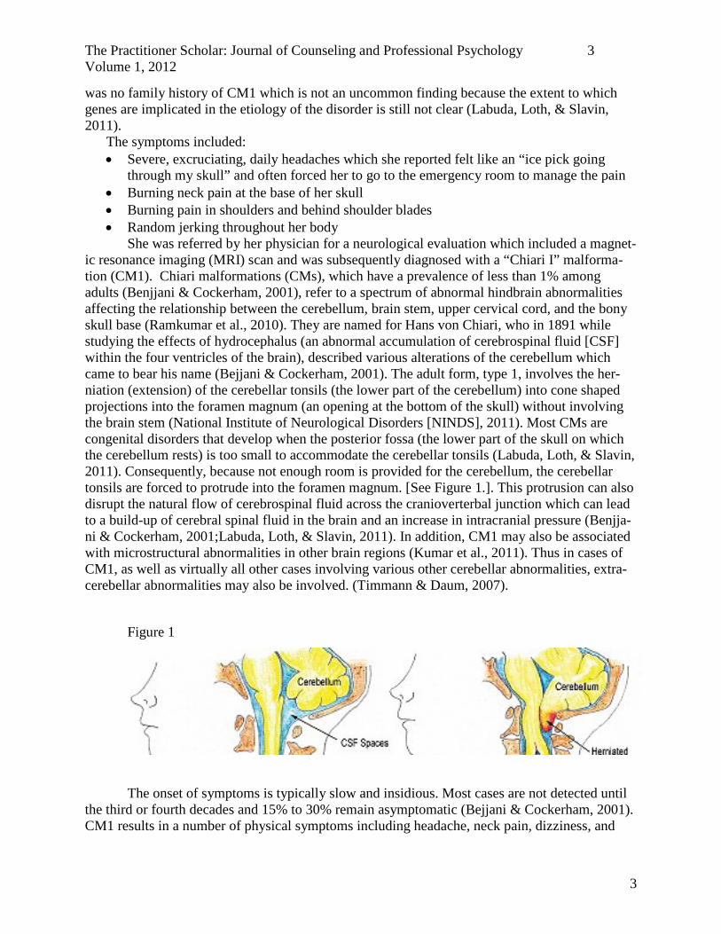
She was referred by her physician for a neurological evaluation which included a magnet-ic resonance imaging (MRI) scan and was subsequently diagnosed with a “Chiari I” malforma-tion (CM1). Chiari malformations (CMs), which have a prevalence of less than 1% among adults (Benjjani & Cockerham, 2001), refer to a spectrum of abnormal hindbrain abnormalities affecting the relationship between the cerebellum, brain stem, upper cervical cord, and the bony skull base (Ramkumar et al., 2010). They are named for Hans von Chiari, who in 1891 while studying the effects of hydrocephalus (an abnormal accumulation of cerebrospinal fluid [CSF] within the four ventricles of the brain), described various alterations of the cerebellum which came to bear his name (Bejjani & Cockerham, 2001). The adult form, type 1, involves the her-niation (extension) of the cerebellar tonsils (the lower part of the cerebellum) into cone shaped projections into the foramen magnum (an opening at the bottom of the skull) without involving the brain stem (National Institute of Neurological Disorders [NINDS], 2011). Most CMs are congenital disorders that develop when the posterior fossa (the lower part of the skull on which the cerebellum rests) is too small to accommodate the cerebellar tonsils (Labuda, Loth, & Slavin, 2011). Consequently, because not enough room is provided for the cerebellum, the cerebellar tonsils are forced to protrude into the foramen magnum. [See Figure 1.]. This protrusion can also disrupt the natural flow of cerebrospinal fluid across the cranioverterbal junction which can lead to a build-up of cerebral spinal fluid in the brain and an increase in intracranial pressure (Benjja-ni & Cockerham, 2001;Labuda, Loth, & Slavin, 2011). In addition, CM1 may also be associated with microstructural abnormalities in other brain regions (Kumar et al., 2011). Thus in cases of CM1, as well as virtually all other cases involving various other cerebellar abnormalities, extra-cerebellar abnormalities may also be involved. (Timmann & Daum, 2007).
Figure 1
The onset of symptoms is typically slow and insidious. Most cases are not detected until the third or fourth decades and 15% to 30% remain asymptomatic (Bejjani & Cockerham, 2001). CM1 results in a number of physical symptoms including headache, neck pain, dizziness, and

The Practitioner Scholar: Journal of Counseling and Professional Psychology 4 Volume 1, 2012
4
impaired balance (NINDS, 2011). It can also result in a severe impairment in selective attention, as the case history will document. Additional support for this causal relationship will be dis-cussed subsequent to the case history presentation.
Selective Attention Impairment
After finally discovering a medication (a 2-year quest) that successfully treated the pain, Erin returned to the program. She had decided against a surgical procedure because of the risks involved. Pain free for the first time in two years, she was happy and excited to return to her doc-toral program. However, despite the absence of pain, she experienced grave difficulties in resum-ing her high level of cognitive functioning because of a severe impairment in selective attention. She reported that she was constantly being bombarded by inner and outer stimuli, which she could not filter out and which affected her in many ways. Her major symptoms were
• Severe difficulty in filtering out external extraneous sounds, which, for example, made it painful for her to be in situations with a lot of noise (e.g., graduation reception for her MA in clinical psychology).
• Severe difficulty in filtering out internal stimuli such that her mind was constantly full of racing, random thoughts. These difficulties caused her to
o become exhausted taking exams because of the tremendous amount of energy re-quired to concentrate.
o have grave difficulty listening to someone or learning as she reported that the new information is “added to the internal mass of information and usually gets lost right away.”
o have significant word retrieval problems. • Severe difficulty in task persistence and reengagement because of the distractibility.
Because of these symptoms, she struggled with academic demands that she previously had accomplished rather effortlessly and with great success. For example, accurate note taking became virtually impossible and she had to resort to taping lectures and then laboriously review-ing them to extract the relevant information. Likewise, the multiple reading demands required much more time as she constantly had to struggle with focus and continually needed to reread.
Erin’s grave deficit in selective attention was by no means a unique finding with regard to CM1. Despite the failure of the authoritative NINDS (2011) fact sheet to list an attention im-pairment as a common symptom in CM1, there is significant anecdotal evidence of patients re-porting “foggy thinking, poor memory and concentration” (Wisconsin Chiari Center, 2011, p.2) a direct quote needs a page or paragraph number). There is also some scientific support, though in general this issue has not been studied. Frim (2010) evaluated 17 adult patients with a battery of neuropsychological tests prior to surgery for the disorder and reported finding ‘subtle’ atten-tion deficits compared to a control group. In Erin’s case however, these deficits in selective at-tention were so severe that she was diagnosed with SADHD.
SADHD
As previously discussed, despite the successful management of her pain, which enabled an enthusiastic return to her doctoral studies, she experienced grave difficulties in resuming her high level of cognitive functioning because of the severe impairment in selective attention. Be-

The Practitioner Scholar: Journal of Counseling and Professional Psychology 5 Volume 1, 2012
5
cause impaired attention very commonly occurs after traumatic brain injury (McCullagh & Feinstein, 2011) with the result that 20% to 50% of individuals who sustain a severe brain injury develop SADHD (Yeates et al., 2005), this prompted a referral to a neurologist for an evaluation. Erin was diagnosed with Attention Deficit/Hyperactivity Disorder Not Otherwise Specified which is given to individuals whose symptoms and impairment meet the criteria for Atten-tion-Deficit/Hyperactivity Disorder, Predominantly Inattentive Type but whose age at on-set is beyond 7 years (American Psychiatric Association, 2000). Because impairment of selec-tive attention, as indexed by the criterion “is easily distracted by extraneous stimuli or irrelevant thoughts,” is the single most sensitive indicator of adult ADHD (Barkley, Murphy, & Fischer, 2008, p. 113), it is not surprising that she was so diagnosed.
Following the diagnosis, she was treated with stimulant medication (Adderall) for the SADHD. This treatment was very successful as it greatly enhanced her ability to concentrate and remain on task as it typically does in most cases (Swanson, Baler, Volow, 2011). She returned to her prior high level of academic functioning as a straight “A” student, which she has maintained for a period of three years as of this writing.
Conclusion
This case study may provide additional support for the involvement of the cerebellum in the pathophysiology of ADHD from an overlooked domain of human lesion studies of the cere-bellum. Future research with CM1 cases needs to focus on two areas. First, the symptom of im-pairment in selective attention warrants much greater study to investigate in a methodologically sophisticated way the numerous patient reports of such impairment. Second, because CM1 in-volves not only cerebellar impairment (i.e., compression) but also associated extra-cerebellar im-pairments, a better understanding of the exact mechanisms, which result in a deficit in selective attention, is needed. You discuss implications for future research, what about implications for practice? Here it is
Lastly, with regard to practice, the most important implication is that the case study pro-vides an important reminder that although ADHD is a highly hereditable disorder, there are a number of biological non-genetic risk factors associated with ADHD (Nigg, 2006) such as lead ingestion (Nigg et al., 2010), prenatal exposure to alcohol (Eme & Millard, 2012), and severe traumatic brain injury (Eme, 2012). Hence, an evidence-based assessment for ADHD requires that a clinician be highly knowledgeable regarding the non-genetic biological risks factors asso-ciated with ADHD and conduct an evaluation that is informed by this knowledge.
References
American Academy of Pediatrics (2011). ADHD: Clinical practice guidelines for the diagnosis, evaluation, and treatment of Attention-Deficit/Hyperactivity Disorder in children and adolescents. Pediatrics, 128, 1007-1022. Confirm page numbers.
American Psychiatric Association (2000). Diagnostic and statistical manual of mental disorders. 4th edition, text revised. Washington, DC: American Psychiatric Association, 2000.
Barkley, R. (2006). Attention-Deficit Hyperactivity Disorder (3rd ed.) Guilford Press, New York (2006).
Barkley, R., Murphy, K., & Fischer, M. (2008). ADHD in adults. New York: Guilford Press.

The Practitioner Scholar: Journal of Counseling and Professional Psychology 6 Volume 1, 2012
6
Bejjani, G. & Cockerman, K. (2001). Adult chiari malformation. Contemporary Neurosurgery, 23, 1-8.
Bisley, J., & Goldberg, M. (2010). Attention, inattention, and priority in the parietal lobe. Annual Review of Neuroscience, 33, 1-21.
Bledsoe, J., Semrud-Clikeman, M., & Pliszka, S. (2009). A magnetic resonance imaging study of the cerebellar vermis in chronically treated and treatment-naïve children with Attention-Deficit/Hyperactivity Disorder Combined Type. Biological Psychiatry, 65, 620-624.
Bower, J., & Parsons, L. (2003). Rethinking the ‘lesser brain’. Scientific American, August, 51-57.
Casey, B. & Riddle, M. (2012). Typical and atypical development of attention. In M. Posner (Ed.), Cognitive neuroscience of attention, (pp. 344-356). New York: Guilford Press.
Cherkasova, M. & Hechtman, L. (2009). Neuroimaging in attention-deficit hyperactivity disord-er: Beyond the frontostriatal circuitry. The Canadian Journal of Psychiatry, 54, 651-664.
Durston, S., van Belle, J. & de Zeeuw, P. (2011). Differentiating frontostraital and fronto-cerebellar circuits in Attention-Deficit/Hyperactivity Disorder. Biological Psychiatry, 69, 1178-1184.
Eme, R. (2012). Attention-Deficit/Hyperactivity Disorder: An Integration with Pediatric Trau-matic Brain Injury. Expert Review of Neurotherapeutics, 12, 475-483.
Eme, R., & Millard, E. (2012). Fetal alcohol spectrum disorders: a literature review with screen-ing recommendations. The School Psychologist, Winter, 12-20.
Frim, D. (2010). Cognitive functions in Chiari 1. Conquer Chiari Research Conference 2010: New Developments and Controversies, November 11, Chicago, IL.
Kieling, C., Goncalves, R., Tannock, R., & Castellanos, F. (2008). Neurobiology of Attention-Deficit/Hyperactivity Disorder. Child and Adolescent Psychiatric Clinics of North America, 17, 285-307.
Krain, A., & Castellanos, F. (2006). Brain development and ADHD. Clinical Psychology Re-view, 26, 433-444.
Kumar, M., Rathore, R., Srivastava, A., Yadav, S., Behari, S., & Gupta, R. (2011). Correlation of diffusion tensor imaging metrics with neurocognitive function in the Chiari 1 malformation. World Neurosurgery, 76, 189-194.
Labuda, R., Loth, F., & Slavin, K. (2011). National Institutes of Health Chiari Research Confe-rence: State of the research and directions. Neurological Research, 33, 227-231.
Mackie, S., Shaw, P., Lenroot, R., Pierson, R., Greenstein, D., Nugent, T., et al. (2007). Cerebellar development and clinical outcome in Attention-Deficit/Hyperactivity Disorder. The American Journal of Psychiatry, 164, 647-655.
Max, J. (2011). Children and adolescents. In J. Silver, T. McAllister, & S. Yudofsky (Eds.), Textbook of traumatic brain injury (pp. 477-494). Washington D.C.: American Psychiatric Association.

The Practitioner Scholar: Journal of Counseling and Professional Psychology 7 Volume 1, 2012
7
McCullagh, S., & Feinstein, A. (2011). Cognitive changes. In J. Silver, T. McAllister, & S. Yduofsky (Eds.), Textbook of traumatic brain injury, 2nd ed. (pp. 279-294). Washington DC: American Psychiatric Publishing, Inc.
National Institute of Neurological Disorders and Strokes (2011). Chiari malformation fact sheet. http://www.ninds.nih.gov/disorders/chiari/detail_chiari.htm Accessed 11/19/2011
Nigg, J. (2006). What causes ADHD? Understanding what goes wrong and why. New York: Guilford Press.
Nigg, J (2010). Attention-Deficit/Hyperactivity Disorder: Endophenotypes, structure, and etio-logical pathways. Current Directions in Psychological Science, 19, 24-29.
Nigg, J., Nikolas, M., Knottnerus, G., Cavanagh, K., & Fridecrici, K. (2010). Confirmation and extension of association of blood lead with Attention-Deficit/Hyperactivity Disorder (ADHD) and ADHD symptom domains at population-typical exposure levels. Journal of the American Academy of Child and Adolescent Psychiatry, 51, 58-65.
Posner, M., & Rothbart, M. (2007). Research on attention networks as a model for the integra-tion of psychological science. Annual review of psychology, 58, 1-23.
Ramkumar, P., Kanodia, A., Ananthakrishnan, G. & Roberts, R. (2010). Chiari II malformation mimicking partial rhombencephalosynapsis? A case report. Cerebellum, 9, 111-114.
Raz, A. & Buhle, J. (2006). Typologies of attentional networks. Nature Reviews, 7, 367-379.
Schmahmann, J., Weilburg, J., & Sherman, J. (2007). The neuropsychiatry of the cerebellum – insights from the clinic. The Cerebellum, 6, 254-267.
Sonuga-Barke, E. (2010). Disambiguating inhibitory dysfunction in Attention-Deficit/Hyperactivity Disorder: Toward the decomposition of developmental brain phe-notypes. Biological Psychiatry, 67, 599-601.
Stoodley, C. (2011). The cerebellum and cognition: evidence from functional imaging studies. Cerebellum, March, 1-20. doi:10.1007/s/12311-011-0260-7.
Strick, P., Dum, R. & Fiez, J. (2009). Cerebellum and nonmotor function. Annual review of neu-roscience, 32, 413-434.
Swanson, J., Baler,R., & Volkow, N. (2011). Understanding the effects of stimulant medications on cognition in individuals with Attention-Deficit Hyperactivity Disorder: A decade of progress. Neuropsychopharmacology, 36, 207-226.
Timmann, D., & Daum, I. (2007). Cerebellar contributions to cognitive functions. The Cerebel-lum, 6, 159-162.
Wang, H., Liu, X., & Fan, J. (2010). Symbolic and connectionist models of attention. In M. Posner (Ed.), Cognitive neuroscience of attention, (pp. 47-56). New York: Guilford Press.
Wisconsin Chiari Center (2011). Symptoms of Chiari malformation. http://www.wichiaricenter.org/?gclid=CN61g_jayqwCFQ1x5QodszUEpw Accessed No-vember 22, 2011.
The Practitioner Scholar: Journal of Counseling and Professional Psychology 8 Volume 1, 2012
8
Yeates, K., Armstrong, K., Janusz, J., et al. (2005). Long-term attention problems in children with traumatic brain injury. Journal of the American Academy of Child and Adolescent Psychiatry, 44, 574-584.
Zametkin, A., Schroth, E., & Faden, D. (2005). The role of brain imaging in the diagnosis and management of ADHD. ADHD Report, October, 11-14.