central annals of sports medicine and research · central annals of sports medicine and research....
TRANSCRIPT

Central Annals of Sports Medicine and Research
Cite this article: De Crée C (2016) A Sting in the Tail ―Sacral Stress Fractures as a Cause of Lower Back Pain in Jūdōka: A Case Report. Ann Sports Med Res 3(4): 1074.
*Corresponding authorProfessor Carl De Crée, Sports Medicine Research Laboratory, P.O. Box 125, B-2800 Malines, Belgium, Fax: 44-870-762-1701; Email:
Submitted: 15 May 2016
Accepted: 31 May 2016
Published: 02 June 2016
ISSN: 2379-0571
Copyright© 2016 De Crée
OPEN ACCESS
Keywords•Athletic injuries•Judo•Low back pain•Lumbar vertebrae•Martial arts•Overuse injury•Sacral region•Sacrum•Spondylolysis•Spondylolisthesis•Sports injuries•Stress fractures
Case Report
A Sting in the Tail -Sacral Stress Fractures as a Cause of Lower Back Pain in Jūdōka: A Case ReportCarl De Crée1,2*1Division of South and East Asia: Japanology, Ghent University, Belgium2Sports Medicine Research Laboratory, B-2800 Malines, Belgium
Abstract
Introduction: Rubens-Duval et al. in 1960 reported finding a high prevalence of back problems in black-belt-level senior jūdōka. Since then many more authors have found evidence in support of long-time jūdō practice being associated with degenerative vertebral changes. The present paper introduces and discusses a recent case of a sacral stress fracture in a jūdōka.
Case presentation: A 27-year-old Caucasian male jūdō black belt presented with lower-back pain. The subject was competitively active at the non-elite level in the middleweight category (-81 kg). Flexion-abduction-external-rotation (FABER), Gaenslen’s, and Scour test were all positive, but squish test was not. Clinical and MRI findings were consistent with a stress fracture of the right hemi-sacrum “Denis zone-I”.
Differential diagnosis: Nonspecific low-back pain, sacral joint dysfunction, ligamentous or muscular strains, Scheuermann’s Disease, sciatica, spondylolysis, spondylolisthesis, tendinitis, Paget’s disease, Cushing’s syndrome, primary bone cancer, and spinal metastases.
Treatment: Reduction of sports competitive activities and training load temporarily improved symptoms. When deemed appropriate and training intensity was increased the pain quickly returned. Physical therapy produced no significant improvement in symptoms.
Uniqueness of the study: Sacral stress fractures have not previously been described in association with jūdō practice.
Conclusion: Sacral stress fractures should be considered when jūdōka present with lingering lower-back pain. Prompt and correct diagnosis is crucial to adapt the subject’s training regime and ensure proper healing. Currently, there is no established best treatment for sacral stress fractures. Whilst prolonged bed rest is generally effective, it is considered a poor option for athletes, especially for those involved in weight class events. Noninvasive treatment options include pulsed electromagnetic fields (PEMF), low-intensity pulsed ultrasound (LIPU), and especially, extracorporeal shockwave therapy (ESWT), but large-scale randomized clinical studies validating their therapeutic effectiveness are still lacking.
ABBREVIATIONSBMI: Body Mass Index; CT: Computed Tomography; DVT: Deep
Venous Thrombosis; ESWT: Extra-Corporeal Shockwave Therapy; FABER: Flexion-Abduction-External-Rotation Test; LIPU: Low-Intensity Pulsed Ultrasound; LRA: Radiologic Abnormalities Of The Lumbar Spine; MDP: Medronate Methylene Diphosphonate; MRI: Magnetic Resonance Imaging; Nslbp: Nonspecific Low-Back Pain Syndromes; PEMF: Pulsed Electromagnetic Fields; PMMA: Polymethylmethacrylate; PTH: Parathyroid Hormone; ROM: Range of Motion
INTRODUCTIONSince Rubens-Duval et al. in 1960 [1] reported finding a high
prevalence of back problems in black-belt-level senior jūdōka many more authors have found evidence in support of long-time jūdō practice being associated with degenerative vertebral changes. The majority of these studies originated in France and Japan and resulted in several non-English language articles [2-7] and dissertations [8-14]. English language publications discussing degenerative vertebral changes in jūdōka are fairly recent and have remained few in number [15-21].
Central
De Crée (2016)Email:
Ann Sports Med Res 3(4): 1074 (2016) 2/7
Among the chronic back problems that were observed most frequently in jūdōka authors reported: spondylolysis (stress fracture in the pars interarticularis of the vertebral arch) [5,18,20,21], spondylolisthesis (forward displacement of a vertebra, often at the level of the fifth lumbar vertebra) [18] [3], Scheuermann’s Disease (juvenile kyphosis) [8], in addition to musculotendinous and nonspecific low-back pain syndromes (nsLBP) [7, 20]. For, example, in a study of 82 Japanese young adult jūdōka recruited from a collegiate jūdō club, 35.4% (34.5% in the lightweight, 32.3% in the middleweight, and 40.9% in the heavyweight categories, respectively) reported nonspecific low-back pain (nsLBP), while 81.7% (65.5% in the light-, 90.3%, in the middle-, and 90.9% in the heavyweight categories) had radiologic abnormalities of the lumbar spine (LRA) [7]. Lumbar intervertebral disc degeneration and spondylolysis were frequently identified (56.1% and 34.1%, respectively). However, the prevalence of LRA in those with nsLBP (79.3%) was similar to the prevalence of LRA for those without nsLBP (83%), suggesting a lack of direct association between back pain and medical imaging findings [7].
Expanding the number of more commonly known back problems associated with jūdō practice, the present paper introduces and discusses a recent case of a jūdōka presenting with a sacral stress fracture. Currently, to the best of our knowledge, no literature data on the prevalence of this specific injury in jūdō are available.
CASE PRESENTATIONA 27-year-old Caucasian male presented with lower-back
pain. The subject had approximately 17 years of jūdō experience and became a jūdō black belt 6.5 years ago; he currently holds the rank of nidan (second degree black belt). He was competitively active at the non-elite level as a middleweight category (-81 kg) fighter. He complained of a localized pain that stretched out from the right hip over the right buttocks to the right sacral area. He was, however, unable to identify a specific traumatic event or movement when the pain first manifested itself.
After implementing a 4-week resting period during which physical activities were limited to walking and daily chores, pain had significantly decreased. However, as soon as he restarted his jūdō practice the pain promptly returned to the full extent, including during casual walking.
Investigations
The patient reported to be in overall good health and denied having any other symptoms, including fatigue, fever, insomnia, nausea, numbness, tingling, unexplained weight loss, weakness, sweating or chills. The patient denied having any gastrointestinal or urogenital symptoms. He did not have a history of alcohol or tobacco consumption, and was not taking any drugs. His surgical history was limited to a previous bariatric surgical intervention 9 years ago during which a temporary gastric band was inserted, which was removed 2.8 years later. Recorded anthropometric data included height (1.80m), body mass (84kg), and BMI [22, 23].
The patient described the pain as a gnawing pain which worsened, for example, while in supine position and attempting to lift his right leg. The pain was also constantly present during
walking and was the most intense whenever he tried running. To the best of our knowledge there are no meta-analyses or well-controlled studies that have assessed the diagnostic accuracy of clinical tests specifically for sacral fractures. However, there exist several pain provocation tests which aim to challenge the anatomical structures involved in an attempt to reproduce the patient’s symptoms. Flexion-abduction-external-rotation (FABER) test, Gaenslen’s test, Scour test, Squish test, and one-leg hop test were administered as part of the clinical physical examination. The reliability of pain provocation tests for the sacroiliac joint have been observed as moderate to good. Clusters of three or more sacral pain provocation tests previously have been found to be reliable, with a sensitivity of 94% and specificity of 78%, respectively. When all such clinical provocation tests fail to produce sacral pain, the sacral iliac joint can be ruled out as the patient’s source of lower back pain [24, 25].
The FABER test is performed on the patient in supine position with the affected-side knee 90° flexed and the foot leant on the contro-lateral knee. The affected-side knee is then pushed by the clinician towards the examining table. For Gaenslen’s test the patient needs to be in supine position with the hip and the knee of the affected side flexed. The clinician then hyper-extends the opposite hip. The goal of the Gaenslen test is to stress both sacroiliac joints simultaneously. The FABER and Gaenslen’s test should be interpreted with care as they also stress the psoas muscle, the hip joint and the femoral nerve and not just the sacrum or sacroiliac joint [24]. The Scour test is a hip functional test that involves the clinician moving the patient’s hip through a range of motion (ROM) from hip flexion and adduction to hip extension and abduction, while adding compressive force through the hip joint as well as movement into the hip internal and external rotation. The FABER, Gaenslen, and Scour test are considered positive if the maneuver evokes pain and/or intra-articular joint clicking.
The squish test also is performed with the patient in a supine position and has a sensitivity and specificity of approximately 69% [24]. The test involves the clinician placing his/her hands on each anterior superior iliac spine, while maintaining his/her body over the hands. A shearing force is then created by pushing inward at a 45 degree angle on both. While maintaining the position on one side, one then increases the pressure on the opposite to determine the mobility of the joint. The test is then repeated on the opposite side, and the anterior-superior iliac spines are palpated. In this way the shearing force produced informs us about the ROM is available at the sacroiliac joint. A positive squish test is suggestive of posterior sacroilliac joing injury.
While the diagnostic accuracy of any of these functional tests remains debatable [24, 25], they are helpful in getting an idea of the degree of discomfort the patient is experiencing, his joint flexibility, and whether and what further imaging tests to order. In the present case, hip active and passive ROM testing, FABER, Gaenslen’s and hip Scour test were positive and aggravated the patient’s pain and revealed symmetric hypomobility. Palpation and posterior-anterior pressure over the right sacral base did not worsen pain symptoms, although the patient reported the area feeling tender. The patient’s most significant impairments were his right sacroiliac joint hypomobility and right-hip adduction

Central
De Crée (2016)Email:
Ann Sports Med Res 3(4): 1074 (2016) 3/7
ROM deficit observed with the hip internally rotated and flexed to 90°. The one-legged hop test for detecting sacral fractures was negative. There was no local erythema, mass or fluctuance anywhere in the iliosacral and hip regions. The patient was a febrile and the remainder of the physical exam was normal.
Magnetic resonance imaging (MRI) was performed for the purpose of evaluation of stress or full fractures, irregular zones, edema, or ligamentous injury, and showed localized bone edema at the ventral cranial sacral part of the right sacroiliac joint with the imposed discrete hypo-intense line running parallel with the right sacroiliac joint being suggestive of a stress fracture of the right hemi-sacrum “Denis zone-I” (Figure 1). Denis zone-I fractures occur in the most lateral portion of the sacrum, the sacral wing. These injuries are not normally complicated by neurological symptoms, but occasionally nerve roots may be involved [26] (Figure 2). No technetium Tc-99 medronate methylene diphosphonate (MDP) bone scintigraphy was performed, even though its sensitivity for detecting sacral stress fractures is not questioned.
Differential diagnosis
• NsLBP [7, 27]
• Sacral joint dysfunction [28]
• Sacroiliac, iliolumbar, sacrospinous, or sacrotuberous ligamentous strains [28]
• Scheuermann’s Disease [8]
• Sciatica [28]
• Spondylolysis [7]
• Spondylolisthesis [7]
• Tendinitis or acute strain of the sacrospinalis (erector spinae), gluteus maximus, of iriformis muscle(s) [28].
• Underlying metabolic disease (Paget’s disease, Cushing’s syndrome), or corticosteroid abuse [28]
• Primary bone cancer (spinalosteoid osteoma, chondrosarcoma, osteosarcoma, Ewing’s sarcoma) (fortunately rare) or secondary metastases from breast, lung or prostate cancer [7, 28].
TREATMENT After an initial 4-week resting period had brought only
temporary relief the patient chose to not again refrain from jūdō practice in order to not further jeopardize his physical condition and jūdō technical skills. He also declined pain medication.
Figure 1 Anterior view of the anatomy of the sacrum and sacroiliac joint with the Denis classification [10] for vertical sacral fractures. Denis zone-I fractures (approximately 50% of sacral fractures) involve the alar region and are entirely lateral to the neuroforamina, zone-II (approximately 34% of sacral fractures) occur in the sacral foraminal area but do not involve the spinal canal, and zone-III fractures (approximately 16% of sacral fractures) occur in the vicinity of the central canal. Zone-III fractures because of their location have a high incidence of neurological deficits including incontinence, impotence and unilateral sacral anesthesia. Transverse sacral fractures (<5% of all sacral fractures) are traditionally included as zone III-fractures. (Modified after Marieb [28], by kind permission of Pearson Benjamin Cummings, Inc., All rights reserved).
Figure 2 Sacrum-aligned multi-oblique reformatted coronal plane magnetic resonance imaging (MRI) (field strength: 3 T) with fat-saturated T2-weighted short-tau inversion recovery (STIR) sequences to demonstrate bone edema and bruising are shown in the upper image, while T1-weighted fat-unsaturated sequences identifying the fracture line and ruling out involvement of the iliosacral joint, neuroforamina and step formation are shown in the lower image. MRI findings include localized bone edema at the ventral cranial sacral part of the right sacroiliac joint with the imposed discrete hypo-intense line running parallel with the right sacroiliac joint being suggestive of a stress fracture of the right hemi-sacrum “Denis zone-I”. Denis zone-I fractures occur in the most lateral portion of the sacrum, the sacral wing.
Central
De Crée (2016)Email:
Ann Sports Med Res 3(4): 1074 (2016) 4/7
Weekly manual physical therapy interventions involving positional release therapy, as recommended by the patient’s primary care physician were utilized to treat the right sacroiliac joint, hypomobility and associated soft tissue tenderness. These techniques were followed by passively stretching the right hip into flexion, medial rotation, and adduction.
Outcome and follow-up
The patient found the manual therapy treatment sessions very painful and four months later reported little improvement with regard to his pain or mobility. Extracorporeal shockwave therapy (ESWT) was suggested as an alternative, but was not followed through by the patient.
DISCUSSION
Diagnosis
Today many physicians will request MRI or computed tomography (CT) to supplement standard X-rays when fractures or stress fractures of the sacrum are suspected. The advantage of MRI is that it can well evaluate potential transverse fractures of the sacral body, subtle ventral cortical disruption of the sacral alae, sclerotic bands or irregular zones in the sacral alae during the healing phase, or to detect sacral marrow edema. Furthermore, MRI allows for risk factor assessment of lumbosacral fusion (even retrospectively due to its characteristic patterns) [26,29]. Multi-oblique reformatted plane MRIs are important as sacrum fractures otherwise may be missed on lumbar spine MRIs taken for the purpose of lower nsLBP. General MRI findings for sacral stress fractures include hypointensity on T1-weighted sequences and hyperintensity on T2-weighted sequences, with conspicuity for stress fractures being particularly noticeable on short-tau inversion recovery (STIR) or frequency-selective fat saturation (FS) T2-weighted images (T2WI). The results of contrast-enhanced weighted T1-imaging may be debatable in this context. MRI is also useful in detecting the variable presence of a “Honda sign or butterfly” sign, which in case of being accompanied by fractures elsewhere or no evidence of metastatic disease can be considered as solid evidence for representing a sacral insufficiency fracture [26]. MRI therefore remains the gold standard for detecting sacral stress and other sacral fractures and has replaced conventional X-rays, which only had a low 50% sensitivity.
However, it is noteworthy to point out the advantages of traditional Technetium Tc-99 MDP bone scintigraphy in showing abnormalities early in the course of a stress fracture (as early as 6 to 72 hours after the onset of symptoms). That being said, the image resulting from the increased radioactive tracer uptake despite its high sensitivity (74-100%) often is fairly nonspecific (33-76%) in identifying anatomic detail [30]. Therefore, despite their role as a useful follow-up diagnostic examination tool for stress fractures, MRI (sensitivity and specificity approx. 90% or more) and CT (sensitivity 42% and specificity 90-100%) are recommended to enhance specificity [30-32]. One of the advantages of CT is that it can show vacuum phenomena, i.e. gas within a fracture or sacroiliac joint. However, these phenomena are typically observed only in the presence of insufficiency fractures, not pathologic fractures.
Injury mechanism
Fatigue stress fractures are caused by the application of abnormal muscular stress or mechanical torque to a bone that has normal elasticity and mineral content [30, 33]. Bone, a dynamic structure, responds to the external forces placed upon it, according to Wolff’s Law. However, this response in bone may be delayed compared to muscle, as muscles typically adjust to increased demands at a faster rate than bone [28]. Hence, fatigue fractures of the sacrum have previously been reported in various special populations subject to rapid changes in anthropometry, such as active children [16], or pregnant women [27, 34-37]. In jūdō, such situations of abnormal muscular stress or mechanical torque can be caused by virtually every movement in nage-waza (throwing techniques) to every movement in newaza (groundwork), especially uchi-komi (repetitive movement drilling exercises), and the repeated contact with the tatami (practice mats) caused by notoriously heavy-impact throws such as ura-nage (reverse throw) or yoko-gake (side hook) performed during randori (free exercises) or nage-no-kata (prearranged forms of throwing).
In addition, the practice of jūdō requires abrupt rotation of the hips and lumbopelvic region for leverage in order to execute throws. An association between lower-back pain in jūdōka and reduced hip range of motion has been identified [15]. In a study of young jūdōka (15-23 years) with at least 4 years of experience, those with reduced range of motion, particularly in their nondominant limb, were more likely to report a history of nonspecific low-back pain [15]. It is postulated that the reduction in hip range of motion leads to an increased load on lumbopelvic structures due to compensatory increase in lumbopelvic rotation [15]. It is also possible that the lumbar symptoms cause adaptations at the hip secondary to altered movement. In the past, further research has been recommended to determine if improving hip flexibility and range of motion helps prevent some cases of lower-back pain in young jūdōka [15].
Because stress fractures are a component of the very broad “overuse syndrome” category and can be confused with many other medical conditions, athletic trainers, sports and family physicians and physical therapists should have a thorough understanding of the etiology, clinical manifestations, and diagnosis of stress fractures.
Injury rate
Of all full sacral fractures 50% are thought to be entirely lateral to the neuroforamina (Denis zone-I), 34% are located in the foraminal area (Denis zone-II), and 16% involve the vicinity of the spinal canal (Denis zone-III); less than 5% of all sacral fractures are transverse fractures (also Denis zone-II) [38]. Fatigue stress fractures are not thought to be common in the sacrum. Longhino et al. [26], however, have suggested that their occurrence is probably underestimated due to the lack of specific symptoms. Although the exact frequency of fatigue stress fractures is not known, these injuries are commonly observed in athletes as well as military recruits and account for 5% to 6% of overuse injuries. In a longitudinal study only 1.7% (3 out of 175) stress fractures of the lower extremity were found to occur in the pelvis of female athletes [32]. Most sports practice-induced
Central
De Crée (2016)Email:
Ann Sports Med Res 3(4): 1074 (2016) 5/7
sacral stress fractures reported in the medical literature have been associated with long-distance running [39, 40].
Injury risk factors
Athletes may present with both extrinsic and intrinsic risk factors associated with stress fractures. Intrinsic factors in sports in general include: eating disorders, loss of body mass [41] gender (women have a higher incidence) [42], insufficient muscle strength, leg length discrepancy, and low aerobic fitness, menstrual irregularities, osteopenia [42]. Extrinsic risk factors typically comprise improper footwear, sudden changes in training regimen or surface. However, since most sports-related cases of sacral stress fractures discussed in the literature involve runners or other endurance sports, it is likely that the extrinsic risk factors associated with jūdō are different. For example, there is no footwear in jūdō, and muscular efforts are typically sudden, explosive and powerful rather than sustained and rhythmic. It is also uncertain if the same stress fracture gender imbalance (females > males) exists in jūdō. Jūdō at the competitive level, as well as its supporting exercises (i.e., lifting weights), mainly involves explosive power and maximal power. Males, in doing so generate considerably more power than female jūdōka. Male jūdōka also tend to throw much harder due to their higher muscular force and body mass, and because they more often choose techniques where the opponent is thrown from increased height (e.g.: ura-nage [reverse throw], kata-guruma [shoulder wheel], daki-wakare [high-lift and –separating throw], te-guruma [hand wheel], morote-gari [double-leg-reaping throw]).
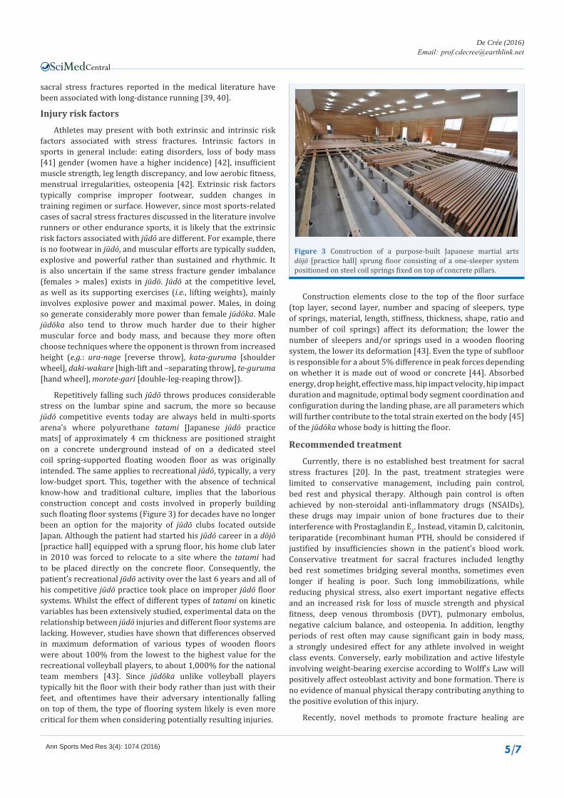
Repetitively falling such jūdō throws produces considerable stress on the lumbar spine and sacrum, the more so because jūdō competitive events today are always held in multi-sports arena’s where polyurethane tatami [Japanese jūdō practice mats] of approximately 4 cm thickness are positioned straight on a concrete underground instead of on a dedicated steel coil spring-supported floating wooden floor as was originally intended. The same applies to recreational jūdō, typically, a very low-budget sport. This, together with the absence of technical know-how and traditional culture, implies that the laborious construction concept and costs involved in properly building such floating floor systems (Figure 3) for decades have no longer been an option for the majority of jūdō clubs located outside Japan. Although the patient had started his jūdō career in a dōjō [practice hall] equipped with a sprung floor, his home club later in 2010 was forced to relocate to a site where the tatami had to be placed directly on the concrete floor. Consequently, the patient’s recreational jūdō activity over the last 6 years and all of his competitive jūdō practice took place on improper jūdō floor systems. Whilst the effect of different types of tatami on kinetic variables has been extensively studied, experimental data on the relationship between jūdō injuries and different floor systems are lacking. However, studies have shown that differences observed in maximum deformation of various types of wooden floors were about 100% from the lowest to the highest value for the recreational volleyball players, to about 1,000% for the national team members [43]. Since jūdōka unlike volleyball players typically hit the floor with their body rather than just with their feet, and oftentimes have their adversary intentionally falling on top of them, the type of flooring system likely is even more critical for them when considering potentially resulting injuries.
Construction elements close to the top of the floor surface (top layer, second layer, number and spacing of sleepers, type of springs, material, length, stiffness, thickness, shape, ratio and number of coil springs) affect its deformation; the lower the number of sleepers and/or springs used in a wooden flooring system, the lower its deformation [43]. Even the type of subfloor is responsible for a about 5% difference in peak forces depending on whether it is made out of wood or concrete [44]. Absorbed energy, drop height, effective mass, hip impact velocity, hip impact duration and magnitude, optimal body segment coordination and configuration during the landing phase, are all parameters which will further contribute to the total strain exerted on the body [45] of the jūdōka whose body is hitting the floor.
Recommended treatment
Currently, there is no established best treatment for sacral stress fractures [20]. In the past, treatment strategies were limited to conservative management, including pain control, bed rest and physical therapy. Although pain control is often achieved by non-steroidal anti-inflammatory drugs (NSAIDs), these drugs may impair union of bone fractures due to their interference with Prostaglandin E2. Instead, vitamin D, calcitonin, teriparatide (recombinant human PTH, should be considered if justified by insufficiencies shown in the patient’s blood work. Conservative treatment for sacral fractures included lengthy bed rest sometimes bridging several months, sometimes even longer if healing is poor. Such long immobilizations, while reducing physical stress, also exert important negative effects and an increased risk for loss of muscle strength and physical fitness, deep venous thrombosis (DVT), pulmonary embolus, negative calcium balance, and osteopenia. In addition, lengthy periods of rest often may cause significant gain in body mass, a strongly undesired effect for any athlete involved in weight class events. Conversely, early mobilization and active lifestyle involving weight-bearing exercise according to Wolff’s Law will positively affect osteoblast activity and bone formation. There is no evidence of manual physical therapy contributing anything to the positive evolution of this injury.
Recently, novel methods to promote fracture healing are
Figure 3 Construction of a purpose-built Japanese martial arts dōjō [practice hall] sprung floor consisting of a one-sleeper system positioned on steel coil springs fixed on top of concrete pillars.

Central
De Crée (2016)Email:
Ann Sports Med Res 3(4): 1074 (2016) 6/7
under study [27]. These include noninvasive treatment using pulsed electromagnetic fields (PEMF), low-intensity pulsed ultrasound (LIPU) [46], and especially, results with ESWT have been promising [47]. However, the lack of major randomized clinical trials and the recent inflationary use of ESWT especially in non-hospital settings necessitate a cautious approach except in low-risk patients with poor response to conventional treatments [48].
Surgery is not a first option in the management of sacral stress fractures and remains reserved for instable full fractures that are midline or transverse, or that involve a considerable disruption of sacral alignment, or those that cause neurological defects. Surgical methods include sacroplasty where sacral stress fractures are being manually repaired using lumbosacral arthrodesis [49] or CT-guided percutaneously injected small quantities of bone cements such as polymethylmethacrylate (PMMA). While these surgical interventions have been reported to quickly reduce pain, most data are obtained from middle-aged people leading relatively sedentary lifestyles. Potential exothermic reactions of PMMA on nerves and bone eventually causing necrosis and nerve damage if the cement spreads, are some of the most serious potential consequences, especially in active young people and athletes. There are, however, no controlled studies that have investigated the success rate of such therapeutic interventions in martial arts practitioners such as jūdōka who will then promptly return to taking heavy falls and extreme high stresses in that area.
In practice, many active athletes instead tend to keep practicing their sport at a reduced level, and keep improving their injury during time-outs from their sport caused by summer vacation or winter breaks. No literature data are available on the successful outcome of athletes with sacral stress fractures returning to their sport and competitively performing at the same or better level than before the therapeutic interventions.
Uniqueness of the study
Sacral stress fractures have not been previously described in association with jūdō, which as a combat sport creates specific etiological circumstances and predisposing factors.
CONCLUSIONSSacral stress fractures should be considered when jūdōka
present with lingering lower-back pain. Timely and correct diagnosis should involve MRI, CT-scan and blood work, and is crucial to promptly adapt the subject’s training regime and ensure proper healing. Fracture lines evolve over weeks to months and show central bone absorption. The fractures can heal after persisting for over a year without significant changes or they can progress to pseudoarthrosis with bone destruction similar to neuropathic joint disease. Whilst prolonged bed rest is generally effective, it is considered a poor option for athletes (i.e. negative effects on physical condition and gain in body fat and body mass), especially for those involved in weight class events [50, 51].
Learning points/Take-home messages
• Sacral stress fractures in otherwise healthy young people and athletes present with several numerous screening,
diagnostic, and management challenges.
• Currently, there is no established best treatment for sacral stress fractures, but several noninvasive treatment options are available although characterized by a lack of large-scale randomized clinical studies attesting to their therapeutic effectiveness.
• The patient response to treatment should be the most important factor that guides the practitioner’s clinical decision making.
ACKNOWLEDGEMENTSFilip Vanhoenacker, MD, Academic Hospital St. Maarten,
Mechelen, and Department of Radiology and nuclear medicine, Ghent University, performed the MRI scans.
REFERENCES1. Rubens-duval A, Bellin A, Ficheux JM, Villiaumey J, Souchet B. The
spine of black-belt jūdō experts. Rev Rhum Mal Osteoartic. 1960; 27: 233-241.
2. Frey A, Rousseau D, Vesselle B, Hervouet des Forges Y, Egoumenides M. Neuf saisons de surveillance médicale de compétitions de jūdō [Medical surveillance in jūdō competition: nine seasons]. J Traumatol Sport. 2004; 21:100-109.
3. Higuchi J , Chōsa E, Tajima N, Sonoda N, Kuroki T. Secular changes on X-ray findings of jūdō players: about lumbar separation and degenerative changes. Jpn J Orthop Sports Med. 1997; 17: 23-30.
4. Higuchi J, Tajima N, Kuroki T, Chosa E, Sonoda N. X-ray findings of the lumbar spine of jūdō players. Jpn J Orthop Sports Med. 1996; 16: 33-39.
5. Kuroki T, Tajima N, Tanabe R, Matsumoto M, Higuchi J, Hirohashi K. Lumbar disorders of jūdō players. Jpn J Orthop Sports Med. 1994; 14: 387-90.
6. Kuroki T, Tajima N. Lumbar injuries in jūdō athletes and long-distance runners. Jpn J Orthop Sports Med. 1993;13:105.
7. Yan CX, Vautour L, Martin MH. Postpartum sacral insufficiency fractures. Skeletal Radiol. 2016; 45: 413-417.
8. Barre P. Scheuermann’s disease: about 82 radiographies in 40 Jūdōka and 42 swimmers from 13 to 18 years. Doctoral dissertation medicine. Reims, France: University of Reims, 1986.
9. Brondani J-C. The effect of judo practice on the spine during growth period. Doctoral dissertation medicine. Paris: University of Paris VII, Faculty of medicine Lariboisière-Saint-Louis, 1974.
10. Creusillet Ch. The lumbo-sacral spine of the judoka. Doctoral dissertation medicine. Tours, France: Université François-Rabelais Tours. 1975.
11. Meas Y (Tchang Chi-Cheng). Spine of the judoka and manual therapies. Doctoral dissertation medicine. Nantes, France: University of Nantes; 1986; 129.
12. Rumîlly O. Dorso-lumbar pain in the elite jūdōka. Doctoral dissertation medicine. Paris: University of Paris Descartes, Cochin Faculty of Medicine; 1990; 180.
13. Thomas F. Influence of jūdō practice on the spine. Doctoral dissertation medicine. Bordeaux, France: Université de Bordeaux-Segalen; 1982.
14. Wiedemann D. Statistical study of risk factors of spinal pain in the Jūdōka: survey of 640 black belts of the Alsacian Judo Federation: characteristics of the female population. Doctoral dissertation medicine. Strasbourg, France; 1988.

Central
De Crée (2016)Email:
Ann Sports Med Res 3(4): 1074 (2016) 7/7
15. Almeida GP, De Souza VL, Sano SS, Saccol MF, Cohen M. Comparison of hip rotation range of motion in judo athletes with and without history of low back pain. Man Ther. 2012; 17: 231-235.
16. Catanese AJ. The medical care of the jūdōka : A guide for athletes, coaches and referees to common medical problems in judo. Tucson, AZ: Wheatmark, Inc; 2012;114-126.
17. Harmer PA. Judo. In: Epidemiology of Injury in Olympic Sports, Volume XVI (eds D. J. Caine, P. A. Harmer and M A. Schiff). Oxford, UK: Wiley-Blackwell, 2009.
18. Okada T, Nakazato K, Iwai K, Tanabe M, Irie K, Nakajima H. Body mass, nonspecific low back pain, and anatomical changes in the lumbar spine in judo athletes. J Orthop Sports Phys Ther. 2007; 37: 688-693.
19. Pocecco E, Ruedl G, Stankovic N, Sterkowicz S, Del Vecchio FB, Gutiérrez-García C, et al. Injuries in judo: a systematic literature review including suggestions for prevention. Br J Sports Med. 2013; 47: 1139-1143.
20. Sakai T, Sairyo K, Suzue N, Kosaka H, Yasui N. Incidence and etiology of lumbar spondylolysis: review of the literature. J Orthop Sci. 2010; 15: 281-288.
21. Zetaruk MN. Chapter 11 – Spinal injuries in combat Sports. In: Lyle Micheli, Cynthia Stein, Michael O’Brien, Pierre d’Hemecourt (eds.). Spinal Injuries and Conditions in Young Athletes. New York, NY: Springer, 2014; 105-114.
22. Mutō Y, Yamashita T, Tanaka Y. Martial arts sports medicine. Tokyo: Bēsubōru Magazin-sha [Baseball Magazine Company]. 2016; 1-191.
23. Linstrom NJ, Heiserman JE, Kortman KE, Crawford NR, Baek S, Anderson RL, Pitt AM. Anatomical and biomechanical analyses of the unique and consistent locations of sacral insufficiency fractures. Spine (Phila Pa 1976). 2009; 34: 309-315.
24. Laslett M, Aprill CN, McDonald B, Young SB. Diagnosis of sacroiliac joint pain: validity of individual provocation tests and composites of tests. Man Ther. 2005; 10: 207-218.
25. Robinson HS, Brox JI, Robinson R, Bjelland E, Solem S, Telje T. The reliability of selected motion- and pain provocation tests for the sacroiliac joint. Man Ther. 2007; 12: 72-79.
26. Longhino V, Bonora C, Sansone V. The management of sacral stress fractures: current concepts. Clin Cases Miner Bone Metab. 2011; 8: 19-23.
27. Yamaji O, Imai N, Arima T, Miyazaki S. Lumber disorders of the jūdō players. Tokai J Sports Med Sci.1992; 4: 46-51.
28. Boissonnault WG, Thein-Nissenbaum JM. Differential diagnosis of a sacral stress fracture. J Orthop Sports Phys Ther. 2002; 32: 613-621.
29. Fujii M, Abe K, Hayashi K, Kosuda S, Yano F, Watanabe S, et al. Honda sign and variants in patients suspected of having a sacral insufficiency fracture. Clin Nucl Med. 2005; 30: 165-169.
30. Gaeta M, Minutoli F, Scribano E, Ascenti G, Vinci S, Bruschetta D, et al. CT and MR imaging findings in athletes with early tibial stress injuries: comparison with bone scintigraphy findings and emphasis on cortical abnormalities. Radiology. 2005; 235: 553-561.
31. Fanciullo JJ, Bell CL. Stress fractures of the sacrum and lower extremity. Curr Opin Rheumatol. 1996; 8: 158-162.
32. Matheson GO, Clement DB, McKenzie DC, Taunton JE, Lloyd-Smith DR,
MacIntyre JG. Stress fractures in athletes. A study of 320 cases. Am J Sports Med. 1987; 15: 46-58.
33. Whiting W, Zernicke R. Biomechanics of Musculoskeletal Injury – 2nd Edition. Champaign, IL: Human Kinetics; 2008; 1-360.
34. Celik EC, Oflouglu D, Arioglu PF. Postpartum bilateral stress fractures of the sacrum. Int J Gynaecol Obstet. 2013; 121: 178-179.
35. Hilal N, Nassar AH. Postpartum sacral stress fracture: a case report. BMC Pregnancy Childbirth. 2016; 16: 96.
36. Oztürk G, Külcü DG, Aydoğ E. Intrapartum sacral stress fracture due to pregnancy-related osteoporosis: a case report. Arch Osteoporos. 2013; 8: 139.
37. Schmid L, Pfirrmann C, Hess T, Schlumpf U. Bilateral fracture of the sacrum associated with pregnancy: a case report. Osteoporos Int. 1999; 10: 91-93.
38. Denis F, Davis S, Comfort T. Sacral fractures: an important problem. Retrospective analysis of 236 cases. Clin Orthop Relat Res. 1988; 227: 67-81.
39. Atwell EA, Jackson DW. Stress fractures of the sacrum in runners. Two case reports. Am J Sports Med. 1991; 19: 531-533.
40. Holtzhausen LM, Noakes TD. Stress fracture of the sacrum in two distance runners. Clin J Sport Med. 1992; 2:139-42.
41. Green CM, Petrou MJ, Fogarty-Hover ML, Rolf CG. Injuries among judokas during competition. Scand J Med Sci Sports. 2007; 17: 205-210.
42. Grier D, Wardell S, Sarwark J, Poznanski AK. Fatigue fractures of the sacrum in children: two case reports and a review of the literature. Skeletal Radiol. 1993; 22: 515-518.
43. Nigg BM, Yeadon MR, Herzog W. The influence of construction strategies of sprung surfaces on deformation during vertical jumps. Med Sci Sports Exerc. 1988; 20: 396-402.
44. Wilson BD, Neal RJ, Swannell PD. The response of gymnastic sports floors to dynamic loading. Aust J Sci Med Sport. 1989; 21:14-19.
45. Van den Kroonenberg AJ, Hayes WC, McMahon TA. Hip impact velocities and body configurations for voluntary falls from standing height. J Biomech. 1996; 29: 807-811.
46. Behrens SB, Deren ME, Matson A, Fadale PD, Monchik KO. Stress fractures of the pelvis and legs in athletes: a review. Sports Health. 2013; 5: 165-174.
47. Moretti B, Notarnicola A, Garofalo R, Moretti L, Patella S, Marlinghaus E, et al. Shock waves in the treatment of stress fractures. Ultrasound Med Biol. 2009; 35: 1042-1049.
48. Leal C, D’Agostino C, Gomez Garcia S, Fernandez A. Current concepts of shockwave therapy in stress fractures. Int J Surg. 2015; 24: 195-200.
49. Meredith DS, Taher F, Cammisa FP, Jr., Girardi FP. Incidence, diagnosis, and management of sacral fractures following multilevel spinal arthrodesis. Spine J. 2013; 13: 1464-1469.
50. Marieb EN. Human anatomy and physiology. 6th ed. San Francisco (CA): Pearson Benjamin Cummings, Inc.; 2004.
51. Wilde GE, Miller TT, Schneider R, Girardi FP. Sacral fractures after lumbosacral fusion: a characteristic fracture pattern. AJR Am J Roentgenol. 2011; 197: 184-188.
De Crée C (2016) A Sting in the Tail ―Sacral Stress Fractures as a Cause of Lower Back Pain in Jūdōka: A Case Report. Ann Sports Med Res 3(4): 1074.
Cite this article