cellulose based bio- dd interfaces for immunodiagnostic...
TRANSCRIPT
9HSTFMG*aeidfc+
ISBN 978-952-60-4835-2 ISBN 978-952-60-4836-9 (pdf) ISSN-L 1799-4934 ISSN 1799-4934 ISSN 1799-4942 (pdf) Aalto University School of Chemical Technology Department of Forest Products Technology www.aalto.fi
BUSINESS + ECONOMY ART + DESIGN + ARCHITECTURE SCIENCE + TECHNOLOGY CROSSOVER DOCTORAL DISSERTATIONS
Aalto-D
D 13
8/2
012
Hannes O
relma
Cellulose based bio-interfaces for im
munodiagnostic applications
Aalto
Unive
rsity
Department of Forest Products Technology
Cellulose based bio-interfaces for immunodiagnostic applications
Hannes Orelma
DOCTORAL DISSERTATIONS

Aalto University publication series DOCTORAL DISSERTATIONS 138/2012
Cellulose based bio-interfaces for immunodiagnostic applications
Hannes Orelma
Doctoral dissertation for the degree of Doctor of Science in Technology to be presented with due permission of the School of Chemical Technology for public examination and debate in Auditorium Puu2 at the Aalto University School of Chemical Technology (Espoo, Finland) on the 9th of November 2012 at 12 noon.
Aalto University School of Chemical Technology Department of Forest Products Technology Forest Products Surface Chemistry
Supervising professor Professor Janne Laine Thesis advisor Dr. Ilari Filpponen Preliminary examiners Professor Kevin Edgar, Virginia Polytechnic Institute and State University, USA Professor Gil Garnier, Monash University, Australia Opponent Professor Robert Pelton, McMaster University, Canada
Aalto University publication series DOCTORAL DISSERTATIONS 138/2012 © Hannes Orelma ISBN 978-952-60-4835-2 (printed) ISBN 978-952-60-4836-9 (pdf) ISSN-L 1799-4934 ISSN 1799-4934 (printed) ISSN 1799-4942 (pdf) http://urn.fi/URN:ISBN:978-952-60-4836-9 Unigrafia Oy Helsinki 2012 Finland
Abstract Aalto University, P.O. Box 11000, FI-00076 Aalto www.aalto.fi
Author Hannes Orelma Name of the doctoral dissertation Cellulose based bio-interfaces for immunodiagnostic applications Publisher School of Chemical Technology Unit Department of Forest Products Technology
Series Aalto University publication series DOCTORAL DISSERTATIONS 138/2012
Field of research Forest Products Chemistry
Manuscript submitted 5 June 2012 Date of the defence 9 November 2012
Permission to publish granted (date) 25 September 2012 Language English
Monograph Article dissertation (summary + original articles)
Abstract In this work, the interactions between various proteins and modified cellulose surfaces were investigated. The work focused on the development of immobilization methods for the covalent attachment of specific immunological antibodies (proteins) onto cellulose substrate. The immobilization methods were explored using cellulose model surfaces and surface sensitive techniques, such as quartz crystal microbalance with dissipation monitoring (QCM-D) and surface plasmon resonance (SPR).
The highest adsorption of globular proteins on unmodified cellulose surfaces occurred at their respective isoelectric points, suggesting a non-electrostatic adsorption mechanism. An increased surface charge at the cellulose substrate was found to enhance the adsorption of all the proteins investigated. This indicated the presence of attractive electrostatic interactions and the adsorption was found to be mainly irreversible. In addition, the effect of oligosaccharide regions of proteins on their adsorption on cellulose was examined with one glycoprotein, avidin. The adsorption of avidin on cellulose was driven by a combination of electrostatic and non-electrostatic forces, and the adsorption was mainly irreversible. Moreover, the oligosaccharide regions of avidin decreased its adsorption strength to cellulose.
In this work, several strategies for covalent immobilization of antibodies onto functionalized cellulose matrices were developed. The novel biointerfaces were capable of sensing antigens both selectively and quantitatively. The use of traditional conjugation chemistries typically leads to a random conformation of immobilized antibodies on the surfaces which in turn may decrease the ability of immobilized antibodies to bind antigens due to the sterical hindrances. Therefore, in this work, the antibodies were immobilized onto cellulose in more oriented manner using avidin-biotin linkage. This approach resulted in over two-fold higher antigen response when compared to those of the traditional conjugation chemistry. In the last part of this work, a biointerface was prepared on a water-resistant nanofibrillar cellulose (NFC) film. The NFC film was made amine reactive by using sequential TEMPO-mediated oxidation and EDC/NHS activation. Activated NFC-films were observed to bind antibodies covalently, and the antibodies could be deposited using standard inkjet printing techniques. The developed NFC-based biointerfaces are expected to open new venues for using cellulose in immunodiagnostic applications.
Keywords Cellulose, antibodies, immobilization, immunodiagnostic
ISBN (printed) 978-952-60-4835-2 ISBN (pdf) 978-952-60-4836-9
ISSN-L 1799-4934 ISSN (printed) 1799-4934 ISSN (pdf) 1799-4942
Location of publisher Espoo Location of printing Helsinki Year 2012
Pages 150 urn http://urn.fi/URN:ISBN:978-952-60-4836-9

Tiivistelmä Aalto-yliopisto, PL 11000, 00076 Aalto www.aalto.fi
Tekijä Hannes Orelma Väitöskirjan nimi Selluloosapohjaiset biointerfaasit immunodiagnostisiin aplikaatioihin Julkaisija Kemian tekniikan korkeakoulu Yksikkö Puunjalostustekniikan laitos
Sarja Aalto University publication series DOCTORAL DISSERTATIONS 138/2012
Tutkimusala Puunjalostuksen kemia
Käsikirjoituksen pvm 05.06.2012 Väitöspäivä 09.11.2012
Julkaisuluvan myöntämispäivä 25.09.2012 Kieli Englanti
Monografia Yhdistelmäväitöskirja (yhteenveto-osa + erillisartikkelit)
Tiivistelmä Työssä tutkittiin proteiinien vuorovaikutusta pintamuokattujen selluloosamateriaalien kanssa hyödynnettäväksi immunodiagnostisissa sovelluksissa. Erityisesti työssä tutkittiin immunologisten vasta-aineiden kiinnittämistä pysyvästi selluloosapintoihin, ja kiinnitettyjen vasta-aineiden aktiivisuutta tunnistaa antigeenejä, vasta-aineille spesifisiä proteiineja. Edellä mainittujen proteiinien adsorptiota selluloosamateriaaleihin tutkittiin käyttäen pintaherkkiä menetelmiä, kuten kvartsikidevaakaa (QCM-D) ja pintaplasmoniresonanssi-instrumenttia (SPR) selluloosamallipintojen kanssa.
Tutkitut proteiinit sitoutuivat modifioimattomiin selluloosapintoihin parhaiten niiden isoelektrisissä pisteissä, mikä osoittaa adsorption tapahtuvan pääasiassa muiden kuin sähköisten vuorovaikutusvoimien välityksellä. Selluloosapinnan kasvanut varaus kasvatti kaikkien tutkittujen proteiinien adsorptiota modifioiduille selluloosapinnoille. Tutkimuksessa selvitettiin myös yhden glykoproteiinin, avidiinin, adsorptiota muokatuille selluloosapinnoille, sekä avidiinin proteiinikuoren oligosakkaridiketjujen vaikutusta adsorptiomekanismeihin. Avidinin adsorptio selluloosapinnalla tapahtui sekä sähköisten ja ei-sähköisten vuorovaikutusten välityksellä ja sitoutuminen oli pysyvää. Tutkimuksessa havaittiin myös oligosakkaridiketjujen vähentävän avidinin adsorptiota selluloosapinnalle.
Työssä kehitettiin menetelmiä sitoa vasta-aineita selluloosapintoihin vesifaasissa hyödyntäen ionisia polysakkarideja (karboksymetyyliselluloosa ja kitosaani), jotka adsorboituvat irreversiibelisti selluloosapintoihin. Tutkimuksissa havaittiin, että adsorboimalla tutkittuja ionisia polysakkarideja selluloosapintoihin, voidaan vasta-aineita sitoa selluloosapintaan kovalenttisten sidosten välityksellä. Kehitetyillä biointerfaasilla pystyttiin detektoimaan antigeenejä spesifisesti. Vasta-aineen konformaatio kiinteällä pinnalla vaikuttaa sen kykyyn sitoa tunnistettavaa antigeeniä. Perinteisessä kovalenttisessa immobilisaatiossa vasta-aineen konformaatiota ei pystytä hallitsemaan. Työssä tutkittiin menetelmää kiinnittää vasta-aineita selluloosapintoihin avidini-biotiinisidoksen avulla. Avidini-biotiinisidoksen avulla biointerfaasin kyky tunnistaa antigeenejä saatiin kaksinkertaistettua. Tutkimuksen viimeisessä vaiheessa kehitettiin biointerfaasi hyödyntäen nanoselluloosafilmejä, joiden pinnat modifioitiin käyttäen TEMPO-hapetusta ja EDC/NHS-aktivointia. Tutkitun aktivoidun nanoselluloosakalvon havaittiin sitovan vasta-aineita kovalenttisesti, ja niiden kiinnitys demonstroitiin mustesuihkutulostusta hyödyntäen. Kehitetty biointefaasi tarjoaa lujan ja kestävän alustan tulevaisuuden immunodiagnostisille sovelluksille.
Avainsanat Selluloosa, vasta-aine, immobilisointi, immunodiagnostiikka
ISBN (painettu) 978-952-60-4835-2 ISBN (pdf) 978-952-60-4836-9
ISSN-L 1799-4934 ISSN (painettu) 1799-4934 ISSN (pdf) 1799-4942
Julkaisupaikka Espoo Painopaikka Espoo Vuosi 2012
Sivumäärä 150 urn http://urn.fi/URN:ISBN:978-952-60-4836-9

i
Preface
This work was carried out in the Department of Forest Products Technology at Aalto Uni-versity, School of Science and Technology, during the years of 2008 – 2012. The work was performed as a part of the BioActive paper I and II projects funded by the National Agency for Technology and Innovation (TEKES) with industrial partners.
I am grateful to my supervisor Professor Janne Laine for giving me the opportunity to work under his supervision. My instructors, Ilari Filpponen, Leena-Sisko Johansson, and Susanna Holappa are thanked for supporting and guiding my research and scientific writ-ing during my studies. Professor Orlando Rojas is thanked for giving me the opportunity to visit in his surface nanomaterial research group at North Carolina State University (NCSU), USA, and guiding my research and writing work.
I am thankful for my co-authors Tuija Teerinen, Professor Monika Österberg, and Terhi Saarinen, for giving the opportunity to work with you. I am thankful to Docent Eero Kontturi for guiding me in the scientific world of cellulose. Tuomas Hänninen is thanked for introducing the TEMPO-oxidation chemistry to me and soulful discussions in work and elsewhere. I am also grateful to Karoliina Junka for fruitful discussions and sharing a room with me.
I want to thank all the past and present colleagues of my research group. Special thanks go to Niko Aarne, Tiina Nypelö, Laura Taajamaa, Paula Eronen, Elli Niinivaara, Petri Myllytie, Susanna Ahola, Tero Taipale, Katri Kontturi, Ania Olszewska, Ola Sundsman, Miro Suchy, and Timo Pääkkönen. The members in the colloids and interface group in NCSU are thanked for supporting me during my visit at NCSU. Special thanks go for Justin Zoppe, Soledad Peresin, Ingrid Hoeger, Carlos I-III, Nafisa Islam, and Julio Arboleda. The tech-nical staff, Risu, Rita, Anu, Marja, and Aila, is thanked for assistance in practicalities in laboratories. Riitta Hynynen is thanked for helping me in many practicalities. Joe Camp-bell is thanked for performing XPS-tests, and reading and correcting my manuscripts.
The members of TEPS are acknowledged for keeping me in touch with the normal life. My parents, brother, relatives, and friends are thanked for supporting me during my doctoral studies.
Finally, my deepest thanks go for my wife Petra and son Eelis, for understanding and sup-porting me during this work.
Helsinki, October 15th, 2012
Hannes Orelma
ii
List of publications
This thesis is mainly based on the results presented in four publications which are referred
as Roman numerals in the text. Some additional data related to the work is also discussed.
Paper I Orelma H., Filpponen I., Johansson L.-S., Laine J., and Rojas O. (2011) Modifica-
tion of Cellulose Films by Adsorption of CMC and Chitosan for Controlled Attachment of
Biomolecules, Biomacromolecules, 12 (12), 4311 - 4318.
Paper II Orelma H., Teerinen T., Johansson L.-S., Holappa S., and Laine J. (2012) CMC-
modified cellulose biointerface for antibody conjugation. Biomacromolecules, 13 (3), 1051
- 1058.
Paper III Orelma H., Filpponen I., Johansson L.-S., and Laine J. (2012) Generic Method
for Attaching Biomolecules via Avidin-Biotin Complexes Immobilized on Films of Regen-
erated and Nanofibrillar Cellulose. Biomacromolecules, 13 (9), 2802 - 2810.
Paper IV Orelma H., Filpponen I., Österberg M., Johansson L.-S., and Laine J. (2012)
Surface functionalized nanofibrillar cellulose (NFC) film as a platform for immunoassays
and diagnostics. Biointerphases, 7 (61), 1 - 12.
Author’s contribution to the appended joint publications:
I-IV Hannes Orelma was responsible for the experimental design, performed the main
part of the experimental work, analysed the corresponding results, and wrote the manu-
scripts.
Other publications that are not included in this thesis but to which the author has contrib-
uted:
Paper V Saarinen T., Orelma, H., Grönqvist, S., Andberg, M., Holappa, S., Laine, J. (2009) Ad-
sorption of different laccases on cellulose and lignin surfaces, BioResources, 4 (1), 94 - 110.
Paper VI Marjamaa, K., Orelma, H., Andberg, M., Holappa, S., Laine, J., Kruus, K. (2012) Ad-
sorption and catalytic action of Trichoderma reesei cellulases on cellulose model surfaces revealed
by QCM-D, Biotechnology and bioengineering, Submitted.
Paper VII Virtanen H., Orelma, H., Erho, T., Smolander, M. (2012) Development of printable
bioactive paper containing laccase. Process biochemistry, 47 (10), 1496 - 1502.
iii
List of abbreviations
AFM atomic force microscopy
BSA bovine serum albumin
CMC carboxymethyl cellulose
DS degree of substitution
DP degree of polymerization
EDC 1-ethyl-3-[3-dimethylaminopropyl]carbodiimide hydrochloride
hIgG human Immunoglobulin G
LS Langmuir-Schaeffer
MFC microfibrillar cellulose
Mw molecular weight
NFC nanofibrillar cellulose
NHS n-hydroxysuccinimide
RMS root mean square
SPR surface plasmon resonance
TEMPO 2,2,6,6-tetramethylpiperidine-1-oxyl free radical
TMSC trimethylsilyl cellulose
QCM-D quartz crystal microbalance with dissipation
XPS X-ray photoelectron spectroscopy
iv
Table of contents
Preface ................................................................................................................................. i
List of publications ............................................................................................................. ii
List of abbreviations .......................................................................................................... iii
1 Introduction and outline of the study ............................................................................... 1
2 Background...................................................................................................................... 3
2.1 Cellulosic supports in immunodiagnostic applications ............................................ 3
2.1.1 Immunodiagnostic assays and traditional support materials ............................ 3
2.1.2 Natural fiber based supports in immunodiagnostic applications .......................5
2.1.3 Chemistry of natural cellulosic materials ............................................................ 7
2.1.4 Microfibrillar cellulose (MFC) ........................................................................... 12
2.1.5 Cellulose model surfaces ................................................................................... 13
2.2 Sensing antibodies ................................................................................................... 15
2.2.1 The chemistry of antibodies .............................................................................. 16
2.2.2 Conjugation sites on antibodies ........................................................................ 19
2.3 Immobilization of sensing antibodies on cellulosic supports ................................ 20
2.3.1 Protein adsorption on cellulosic materials ........................................................ 21
2.3.2 Chemical conjugation of antibodies on cellulose ............................................. 23
2.3.3 Functionalization of cellulosic materials ......................................................... 25
3 Experimental ................................................................................................................. 28
3.1 Materials .................................................................................................................. 28
3.1.1 Cellulose model surfaces and NFC-films .......................................................... 28
3.1.2 Antibodies and other proteins .......................................................................... 29
3.1.3 Polysaccharides and chemicals for surface functionalization .......................... 30
3.1.4 Conjugation chemicals ..................................................................................... 30
3.2 Methods ................................................................................................................... 31
3.2.1 Model cellulose film preparation techniques .................................................... 31
3.2.2 Surface Plasmon Resonance (SPR) .................................................................. 33
v
3.2.3 Quartz Crystal Microbalance with Dissipation (QCM-D) ................................ 35
3.2.4 Atomic Force Microscopy imaging (AFM) ....................................................... 38
3.2.5 Additional techniques ...................................................................................... 39
4 Results and discussion ................................................................................................... 41
4.1 Surface functionalization of cellulose ...................................................................... 41
4.1.1 Carboxylation of cellulose by CMC adsorption .................................................. 41
4.1.2 Amination of cellulose by chitosan adsorption ................................................ 44
4.1.3 Carboxylation of cellulose by TEMPO-oxidation ............................................. 46
4.2 Adsorption of proteins on modified cellulose ........................................................ 48
4.2.1 Adsorption of globular proteins on surface modified cellulose ....................... 48
4.2.2 Adsorption of glycoproteins on surface modified cellulose ............................. 50
4.2.3 Adsorption of antibodies on surface modified cellulose .................................. 53
4.3 Conjugation of antibodies on cellulose in random orientation ............................... 55
4.3.1 Anti-hemoglobin conjugation on CMC functionalized cellulose ...................... 56
4.3.2 hIgG conjugation on chitosan-functionalized cellulose ................................... 59
4.3.3 Anti-hIgG conjugation on TEMPO-oxidized NFC ........................................... 62
4.3.4 Attachment of proteins on cellulose via pre-conjugation with CMC ............... 64
4.4 Oriented attachment of antibodies on cellulose ..................................................... 65
4.4.1 Immobilization of Avidin and neutravidin on surface modified cellulose ....... 66
4.4.2 Effect of the oriented immobilization strategy on antigen binding of antibodies
...................................................................................................................................... 69
4.5 Preparation of a biointerface on NFC-films ............................................................ 71
5 Concluding remarks ....................................................................................................... 75
6 References ...................................................................................................................... 77
1
1 Introduction and outline of the study
Studies on the preparation of biointerfaces on cellulosic materials are presented in this
thesis. Cellulose is an abundant and renewable material in the biosphere, and its chemis-
try and material properties are rather well characterized. It belongs to a group of carbon
neutral materials, and therefore it can be used to compensate for the use of fossil based
materials. The most important sources of cellulose are woods and plants. However, in the
Nordic countries, the wood based materials have been the major cellulose source due to
the large forest regions available. The dissolved cellulose based materials have traditional-
ly been utilized in immunodiagnostic applications (Elias 2000). Recently, the utilization of
natural cellulose fibers, and its substructures (nanofibrils), on diagnostical applications
have gained more attention (Pelton 2009). Compared to the homogenously dissolved cel-
lulose, natural wood fibers are heterogeneous substrates containing several constituents.
Therefore, analysis of the surface interactions with natural fiber materials could be benefi-
cial for leading to a better understanding of the limits and possibilities when natural fiber
based materials are utilized in immunodiagnostic applications.
Cellulose is rather stable and unreactive material that contains only a limited selection of
free conjugation sites. Traditionally, the immobilization of antibodies on cellulose has
been carried out using two main immobilization methods: direct adsorption and covalent
conjugation. The direct adsorption is the simplest route to attach antibodies to cellulose,
but its disadvantages, such as irreversible binding and reduced activity of antibodies, have
led to a demand for more sophisticated conjugation chemistries (Rusmini, Zhong & Feijen
2007). Cellulosic materials contain a large number of hydroxyl groups that can contribute
to the covalent conjugation of antibodies in non-polar solvents. The conjugation reactions
in aqueous media require surface functionalization strategies for introducing suitable con-
jugation sites on cellulose, which can contribute to the formation of covalent linkages.
The aim of this thesis was to prepare and evaluate several different routes for antibody
immobilization on cellulosic supports. All the work was carried out in aqueous solutions
using chemistries with low toxicity. In Paper I, the effect of the surface charge of cellulose
surfaces on protein adsorption was investigated. This was carried out using the surface
plasmon resonance (SPR) technique with surface tailored cellulose model surfaces. The
presence of electrostatic interactions was observed in the adsorption of proteins on hydro-
philic surfaces. In Paper II, an immobilization strategy to covalently conjugate anti-
2
hemoglobin on cellulose using carboxymethyl cellulose (CMC) acting as a linker molecule
was investigated. The covalent coupling reaction between antibody and CMC-modified
cellulose was analyzed by combining the quartz crystal microbalance with dissipation
(QCM-D) and SPR techniques. The specificity and detection limits of the prepared anti-
hemoglobin biointerface were analyzed. In Paper III, a strategy to use avidin-biotin link-
ages for orienting the antibodies conformation on cellulose surfaces was investigated. The
immobilization of avidin on carboxylated cellulose, and the ability of the immobilized avi-
din to detect biotinylated proteins were investigated by using QCM-D, atomic force mi-
croscopy (AFM), and X-ray photoelectron spectroscopy (XPS). Specific binding of bioti-
nylated molecules on avidin modified cellulose was investigated with biotinylated bovine
serum albumin (BSA) and anti-human immunoglobulin G (anti-hIgG). The influence of
the oriented conjugation chemistry on the detection of antigens was investigated on nano-
fibrillated cellulose (NFC) surfaces by using QCM-D. In Paper IV, the methods developed
in Papers I – III were applied to immobilize antibodies on NFC-films. TEMPO-oxidation
chemistry was used to introduce carboxyl groups toposelectively on NFC-films, which in
the later stage were converted to amine reactive esters. The carboxylation reaction was
verified using conductometric titration and contact angle measurements (CAM). The
amine reactivity of the activated NFC-films was characterized using fluorescence-stained
anti-human IgG. The inkjet printing technique was utilized for depositing antibodies on
activated NFC-films.
In general, this work presents several routes to immobilize antibodies on cellulosic ma-
terials in a controllable way. This knowledge could be useful in preparing more sophisti-
cated immunodiagnostic tests based on the natural fiber support materials.
3
2 Background
2.1 Cellulosic supports in immunodiagnostic applications
Immunodiagnostic assays, also referred as to immunoassays, are quantitative or qualita-
tive tests that utilize antibodies to measure the presence of soluble analytes, antigens, in
complex sample fluids. The antibodies, obtained by either isolation of human body fluids
or growing them in the host animals, are conjugated onto solid supports. The basic re-
quirements for all diagnostic assays are similar; low detection limit, high analyte selectivi-
ty, small sample preparation, and high cost-effectiveness. Hence, the optimized support
materials and specific conjugation chemistries define the accuracy and usability of the
assay in the clinical testing. Traditionally, immunodiagnostic assays are based on the use
of synthetic polymeric materials produced mainly from oil. Recently, sustainable renewa-
ble support materials such as natural cellulosic fibers have gained significant attention.
2.1.1 Immunodiagnostic assays and traditional support materials
The preliminary studies of immunodiagnostic testing were carried out in the later 1950s
by Yalow and Berson with radioactively-labeled peptide hormone insulin (Yalow, Berson
1959). This was first time when a rapid diagnostic test was utilized to qualitatively meas-
ure the soluble analytes. Soon after that it was observed that there is a significant health
risk with the radio-labeled antibodies of radio-immunodiagnostic assays (RIA). The en-
zyme linked immunosorbent assay (ELISA) was developed in the early 1970´s by Engvall
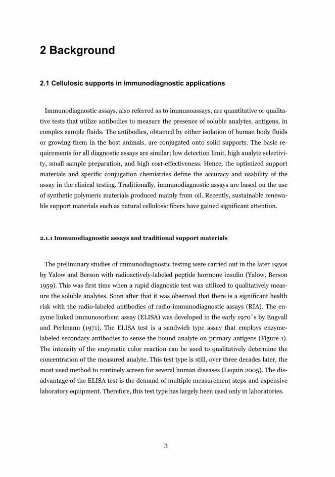
and Perlmann (1971). The ELISA test is a sandwich type assay that employs enzyme-
labeled secondary antibodies to sense the bound analyte on primary antigens (Figure 1).
The intensity of the enzymatic color reaction can be used to qualitatively determine the
concentration of the measured analyte. This test type is still, over three decades later, the
most used method to routinely screen for several human diseases (Lequin 2005). The dis-
advantage of the ELISA test is the demand of multiple measurement steps and expensive
laboratory equipment. Therefore, this test type has largely been used only in laboratories.
4
Figure 1. Enzyme labeled immunosorbent assay (ELISA). Antigens are bound onto a solid sup-
port via conjugated primary antibodies. The binding of the antigen is exposed using enzyme
labeled secondary-antibodies that causes a visible color reaction when the substrate material
of the enzyme is applied in the test.
The ELISA and radiolabeled immunoassay (RIA) methods need multiple measurement
steps, which are sometimes too complicated in commercial use. Therefore, an immunodi-
agnostic test, which could simultaneously sense the analytes and illustrate the result of the
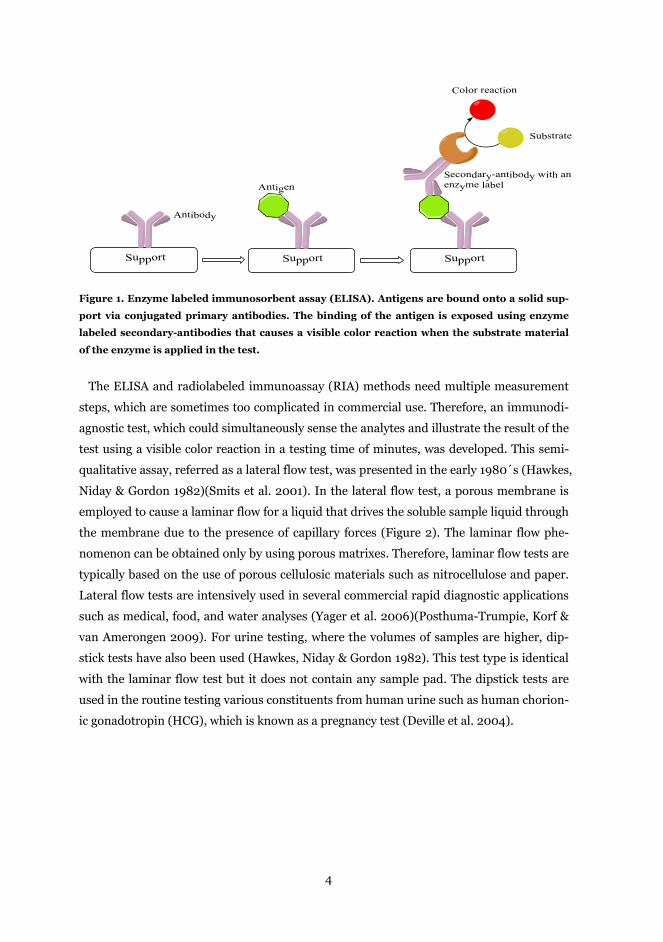
test using a visible color reaction in a testing time of minutes, was developed. This semi-
qualitative assay, referred as a lateral flow test, was presented in the early 1980´s (Hawkes,
Niday & Gordon 1982)(Smits et al. 2001). In the lateral flow test, a porous membrane is
employed to cause a laminar flow for a liquid that drives the soluble sample liquid through
the membrane due to the presence of capillary forces (Figure 2). The laminar flow phe-
nomenon can be obtained only by using porous matrixes. Therefore, laminar flow tests are
typically based on the use of porous cellulosic materials such as nitrocellulose and paper.
Lateral flow tests are intensively used in several commercial rapid diagnostic applications
such as medical, food, and water analyses (Yager et al. 2006)(Posthuma-Trumpie, Korf &
van Amerongen 2009). For urine testing, where the volumes of samples are higher, dip-
stick tests have also been used (Hawkes, Niday & Gordon 1982). This test type is identical
with the laminar flow test but it does not contain any sample pad. The dipstick tests are
used in the routine testing various constituents from human urine such as human chorion-
ic gonadotropin (HCG), which is known as a pregnancy test (Deville et al. 2004).
Support Support Support
Color reaction
Antibody
AntigenSecondary-antibody with an enzyme label
Substrate
5
Figure 2. Typical lateral flow test. (a) A sample liquid with a soluble analyte is applied into the
sample pad where the liquid flows through the membrane. The analyte in liquid first reacts
with gold nanoparticle stained secondary antibodies, and then the conjugate flows through the
migration membrane where it is caught in the test line by primary antibodies. (b) The visible
mark in the test line is caused by gathering of nanoparticles in the small area. The validity of
the test is verified with a control line, where both reacted and unreacted antibodies are caught.
(Adapted from (Yager et al. 2006)).
The basic requirement for stable immunodiagnostic assays is that the supporting mate-
rial does not cross-interact with analytes. Traditionally, the solid supports in the ELISA
and RIA assays have widely been based on synthetic polymeric materials. In that case, the
cross-reactivity between analytes and the plastic supports have been blocked by adsorbing
proteins such as bovine serum albumin (BSA). The protein fills the “holes” on the surface,
preventing the non-specific binding of antigens (Kenna, Major & Williams 1985)(Sverre-
Henning 1997). Similar treatment steps have also been used for the semi-synthetic nitro-
cellulose membranes in laminar flow tests. Otherwise, the anionic charge of nitrocellulose
electrostatically binds proteins, causing a false response (Spinola, Cannon 1985). Addi-
tional, clear disadvantages of nitrocellulose based matrices are their brittle nature and
their high flammability. By the virtue of those properties, renewable totally natural fiber-
based materials could be a competing platform to be used as a supporting material in rap-
id diagnostic assays.
2.1.2 Natural fiber based supports in immunodiagnostic applications
Natural cellulose fiber-based materials exhibit interesting characteristics, such as their
hydrophilic nature, high stability, low toxicity, and high industrial availability (Pelton
6
2009). Therefore, the development of natural fiber-based immunodiagnostic assays has
attracted significant attention. The first paper-based “diagnostic test” for the detection of
glucose in urine was demonstrated in the late 1950´s by Free and coworkers (Free et al.
1957). This pioneering test was based on the use of immobilized glucose oxidase enzymes,
which catalyzed the reduction of glucose to D-glucono-δ-lactone. The use of the capillary
flow properties of porous paper in immunodiagnostic testing was first demonstrated by
Zuk and coworkers (Zuk et al. 1985). This test was an enzyme immunochromatographic
test, where the antigen binding in the paper strip was visualized by an external substrate
addition step. The complete paper-based laminar flow test for immunodiagnostic analysis
has been demonstrated in several reports (Abe, Suzuki & Citterio 2008)(Fenton et al.
2009)(Lappalainen et al. 2010). Moreover, in these studies non-contact dispersing tech-
niques were used to manufacture these low-cost diagnostic paper assays (Figure 3). The
multicomponent paper based laminar flow test, manufactured by inkjet printing of filter
paper with hydrophobic polymer walls, has also been demonstrated (Bruzewicz, Reches &
Whitesides 2008). Splitting of the laminar flow to separate flow channels allows analysis
of multiple analytes simultaneously. In addition, fluid channeling in paper has been ap-
plied to the size-based extraction of molecules from complex mixtures (Osborn et al. 2010).
A multicomponent paper-based microfluidic test on the testing of glucose and BSA simul-
taneously from urine has been demonstrated (Martinez et al. 2010). The use of a paper
matrix in the routine ELISA-testing has also been represented (Cheng et al. 2010). In this
case, the paper-based support was prepared on a chemically treated paper using photoli-
thography with UV-light. The use of rapid immunodiagnostic testing for screening for the
quality of foods can be found in the review paper by Krska and Molinelli (Krska, Molinelli
2009). The examples presented above show the growing potential for immunodiagnostic
tests using natural cellulosic fiber based materials. However, the knowledge of surface
interactions and immobilization reactions on the wood fiber surfaces is presently imper-
fect. Many of those papers were targeted to demonstrate the action of prepared test with-
out focusing on the detailed analysis of conjugation and adsorption reactions. Hence, a
deeper understanding of interfacial reactions could be beneficial when more effective and
sophisticated immunodiagnostic tests are developed.
7
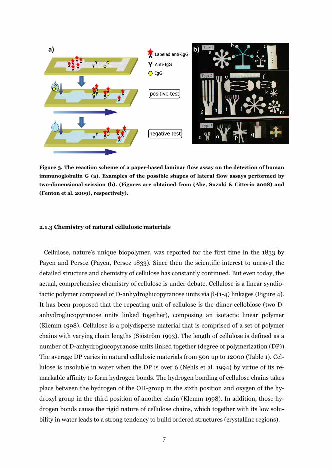
Figure 3. The reaction scheme of a paper-based laminar flow assay on the detection of human
immunoglobulin G (a). Examples of the possible shapes of lateral flow assays performed by
two-dimensional scission (b). (Figures are obtained from (Abe, Suzuki & Citterio 2008) and
(Fenton et al. 2009), respectively).
2.1.3 Chemistry of natural cellulosic materials
Cellulose, nature’s unique biopolymer, was reported for the first time in the 1833 by
Payen and Persoz (Payen, Persoz 1833). Since then the scientific interest to unravel the
detailed structure and chemistry of cellulose has constantly continued. But even today, the
actual, comprehensive chemistry of cellulose is under debate. Cellulose is a linear syndio-
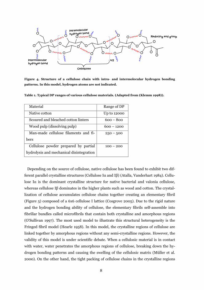
tactic polymer composed of D-anhydroglucopyranose units via β-(1-4) linkages (Figure 4).
It has been proposed that the repeating unit of cellulose is the dimer cellobiose (two D-
anhydroglucopyranose units linked together), composing an isotactic linear polymer
(Klemm 1998). Cellulose is a polydisperse material that is comprised of a set of polymer
chains with varying chain lengths (Sjöström 1993). The length of cellulose is defined as a
number of D-anhydroglucopyranose units linked together (degree of polymerization (DP)).
The average DP varies in natural cellulosic materials from 500 up to 12000 (Table 1). Cel-
lulose is insoluble in water when the DP is over 6 (Nehls et al. 1994) by virtue of its re-
markable affinity to form hydrogen bonds. The hydrogen bonding of cellulose chains takes
place between the hydrogen of the OH-group in the sixth position and oxygen of the hy-
droxyl group in the third position of another chain (Klemm 1998). In addition, those hy-
drogen bonds cause the rigid nature of cellulose chains, which together with its low solu-
bility in water leads to a strong tendency to build ordered structures (crystalline regions).
a) b)
8
Figure 4. Structure of a cellulose chain with intra- and intermolecular hydrogen bonding
patterns. In this model, hydrogen atoms are not indicated.
Table 1. Typical DP ranges of various cellulose materials. (Adapted from (Klemm 1998)).
Material Range of DP
Native cotton Up to 12000
Scoured and bleached cotton linters 600 – 800
Wood pulp (dissolving pulp) 600 – 1200
Man-made cellulose filaments and fi-
bers
250 – 500
Cellulose powder prepared by partial
hydrolysis and mechanical disintegration
100 – 200
Depending on the source of cellulose, native cellulose has been found to exhibit two dif-
ferent parallel crystalline structures (Cellulose Iα and Iβ) (Atalla, Vanderhart 1984). Cellu-
lose Iα is the dominant crystalline structure for native bacterial and valonia cellulose,
whereas cellulose Iβ dominates in the higher plants such as wood and cotton. The crystal-
lization of cellulose accumulates cellulose chains together creating an elementary fibril
(Figure 5) composed of a 6x6 cellulose I lattice (Cosgrove 2005). Due to the rigid nature
and the hydrogen bonding ability of cellulose, the elementary fibrils self-assemble into
fibrillar bundles called microfibrils that contain both crystalline and amorphous regions
(O'Sullivan 1997). The most used model to illustrate this structural heterogeneity is the
Fringed fibril model (Hearle 1958). In this model, the crystalline regions of cellulose are
linked together by amorphous regions without any semi-crystalline regions. However, the
validity of this model is under scientific debate. When a cellulosic material is in contact
with water, water penetrates the amorphous regions of cellulose, breaking down the hy-
drogen bonding patterns and causing the swelling of the cellulosic matrix (Müller et al.
2000). On the other hand, the tight packing of cellulose chains in the crystalline regions
O OO O
O OOO O
O
OO
O
O
O
OO
O
O
O
O
O
O
O
O
O
n/2
Cellobiose
Reducing end group
Intramolecular hydrogen bond
Intermolecular hydrogen bond
9
prevents the penetration of water into the microfibrils. The crystalline structure can be
separated using non-polar solvent systems (Dawsey, McCormick 1990), such as lithium
chloride/dimethylacetamide (LiCl/DMAc), which breaks the intermolecular hydrogen
bonding patters between the cellulose chains. The dissolved cellulose can be recrystallized
in an aqueous solvent system leading to twisting of the cellulose chains to the anti-parallel
cellulose II conformation (Fink, Philipp 1985). This cellulose II lattice is also referred as
regenerated cellulose, and it is widely used in various applications on, e.g., non-woven
cellulose fibers, fabrics, and cellulosic membranes.
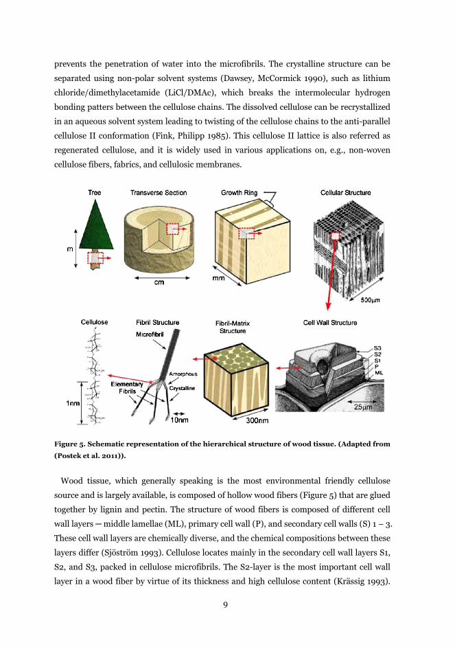
Figure 5. Schematic representation of the hierarchical structure of wood tissue. (Adapted from
(Postek et al. 2011)).
Wood tissue, which generally speaking is the most environmental friendly cellulose
source and is largely available, is composed of hollow wood fibers (Figure 5) that are glued
together by lignin and pectin. The structure of wood fibers is composed of different cell
wall layers ─ middle lamellae (ML), primary cell wall (P), and secondary cell walls (S) 1 – 3.
These cell wall layers are chemically diverse, and the chemical compositions between these
layers differ (Sjöström 1993). Cellulose locates mainly in the secondary cell wall layers S1,
S2, and S3, packed in cellulose microfibrils. The S2-layer is the most important cell wall
layer in a wood fiber by virtue of its thickness and high cellulose content (Krässig 1993).
10
Therefore, this layer has the most influence on the material properties of cellulosic end
products. The middle lamellae glues fibers together, and it is mainly composed of lignin
and pectin. A flexible primary wall layer is sandwiched between the S1 and the middle la-
mellae. It is composed mainly of amorphous hemicelluloses and lignin, but also a small
amount of cellulose microfibrils in random orientations has been found (Sjöström 1993).
Hemicelluloses are a diverse group of water soluble branched heteropolymers, composed
of various monosaccharides with a low degree of polymerization (DP of 50 - 300). The
high solubility of hemicelluloses in water is caused mainly by both the branched structure
and the presence of charged side groups. The chemical composition of hemicelluloses in
different plant species varies (Sjöström 1993). In generally, hemicelluloses that are pre-
sent in hardwood fibers are glucuronoxylan and glucomannans, whereas softwood species
contain mainly galactoglucomannans and arabinoglucuronoxylan (Table 2). The role of
hemicellulose in wood cell wall layers is to fill the voids, provide coupling to lignin, and
regulate the structure of cell walls (Atalla et al. 1993). Lignin, in all wood tissues, has a
three dimensional amorphous random network; the lignin precursors, p-coumaryl alcohol
(hardwoods), coniferyl alcohol (softwoods), and sinapyl alcohol (hardwood), are connect-
ed via radical polymerization reactions. The extent of lignin in different plant species var-
ies significantly, but it has similar functions in all wood species ─ glue individual wood
fibrils together, provide compression strength, and protect plants against pathogens
(Boerjan, Ralp & Baucher 2003).

11
Table 2. The major hemicellulose components in softwood (SW) and hardwood (HW).
(Adapted from (Sjöström 1993)).
The surface chemistry of cellulosic fibers and microfibrils is important since all chemical
reactions take place in the interface between a surface and bulk media. When natural cel-
lulose-based materials are utilized in applications where the high cellulose content is
needed, only chemical pulping with multiple bleaching steps can provide high quality cel-
lulosic fibers. The chemical disintegration, pulp cooking, selectively dissolves lignin, main-
ly from middle lamella that separates the wood fibers (Sjöström 1993)(Duchesne, Daniel
2000). However, in pulp cooking a part of lignin and hemicellulose from the secondary
wall layers is also removed, which produces small pores on the fiber surface (Maloney,
Paulapuro 1999). After removal of lignin from a softwood (Norway Spruce) in the KRAFT-
process, which is the most used industrial delignification process, it has been observed
that cellulose microfibrils are visible on the fiber surface (Figure 6), and the microfibrils
are uncovered or only covered with a few molecular layers of hemicelluloses (Duchesne et
al. 2001). The average pore size on the fiber surfaces is approximately 20 nm measured by
AFM (Fahlen, Salmen 2005). The lignin content on softwood fibers after KRAFT-process
with bleaching steps have been measured by XPS to be about 0.5 % or lower (Laine,
Stenius 1994)(Risén, Hultén & Paulsson 2004). Those results demonstrate that the surfac-
es of delignified wood fibers after delignification steps have a cellulosic structure. But
12
there are a small amount of hemicelluloses present, introducing a slight negative charge
for wood fibers.
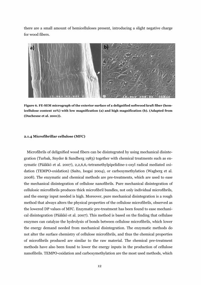
Figure 6. FE-SEM micrograph of the exterior surface of a delignified softwood kraft fiber (hem-
icellulose content 10%) with low magnification (a) and high magnification (b). (Adapted from
(Duchesne et al. 2001)).
2.1.4 Microfibrillar cellulose (MFC)
Microfibrils of delignified wood fibers can be disintegrated by using mechanical disinte-
gration (Turbak, Snyder & Sandberg 1983) together with chemical treatments such as en-
zymatic (Pääkkö et al. 2007), 2,2,6,6,-tetramethylpipelidine-1-oxyl radical mediated oxi-
dation (TEMPO-oxidation) (Saito, Isogai 2004), or carboxymethylation (Wagberg et al.
2008). The enzymatic and chemical methods are pre-treatments, which are used to ease
the mechanical disintegration of cellulose nanofibrils. Pure mechanical disintegration of
cellulosic microfibrils produces thick microfibril bundles, not only individual microfibrils,
and the energy input needed is high. Moreover, pure mechanical disintegration is a rough
method that always alters the physical properties of the cellulose microfibrils, observed as
the lowered DP values of MFC. Enzymatic pre-treatment has been found to ease mechani-
cal disintegration (Pääkkö et al. 2007). This method is based on the finding that cellulase
enzymes can catalyze the hydrolysis of bonds between cellulose microfibrils, which lower
the energy demand needed from mechanical disintegration. The enzymatic methods do
not alter the surface chemistry of cellulose microfibrils, and thus the chemical properties
of microfibrils produced are similar to the raw material. The chemical pre-treatment
methods have also been found to lower the energy inputs in the production of cellulose
nanofibrils. TEMPO-oxidation and carboxymethylation are the most used methods, which
a) b)
13
are based on the introduction of anionic functional groups to the surfaces of cellulose mi-
crofibrils. The anionic charge causes electrostatic repulsion between cellulose microfibrils
that drastically ease mechanical disintegration (Saito et al. 2007). Carboxylated cellulose
microfibrils, produced using the prescribed methods, are found to carry a high surface
charge that stabilizes them in aqueous suspensions (Wagberg et al. 2008)(Saito, Isogai
2004). In general, all cellulose microfibrils prepared from natural fiber materials have a
slight anionic surface charge by virtue of a small amount of hemicellulose on the fibril sur-
faces (Fall et al. 2011).
Cellulose microfibrils have very interesting characteristics, such as high aspect ratio (100
- 150), high strength properties, and low toxicity towards to living organisms. In aqueous
suspensions, cellulose microfibrils are found to form a network that behaves like a pseudo-
plastic gel due to the high hydrogen bonding and entanglement of cellulose microfibrils
(Herrick et al. 1983). This microfibrillar cellulose (MFC) gel has been employed to cast
strong translucent cellulose films (Henriksson et al. 2008)(Syverud, Stenius 2009)(Aulin,
Gällstedt & Lindström 2010)(Fujisawa et al. 2011). The properties of the NFC-films are
mainly adopted from the raw material used, and thus, a homogenous cellulose microfibril-
lar gel has high transparency (Nogi et al. 2009)(Fujisawa et al. 2011)(Aulin, Gällstedt &
Lindström 2010). NFC-films have good strength and oxygen barrier properties only in dry
atmosphere. But, due to the hydrophilic nature of cellulose, those properties are signifi-
cantly disturbed in the wet state. However, NFC-films, cast from mechanical disintegrated
MFC (low charged MFC) have been observed to exhibit such a low water uptake that they
have rather good strength properties also in high humidity conditions (Spence et al. 2010).
The hydrophobicity of NFC-films can be improved using various grafting and surface func-
tionalization chemistries (Siró, Plackett 2010). But then, almost every time, the surface
properties of cellulosic microfibrils are altered causing a loosely bound fibrillar network
that reduces the strength properties of the NFC-film. The potential applications for cast
NFC-films are food and pharmaceutical packaging materials, where the high oxygen barri-
er properties with low toxicity are beneficial. However, the inherent characteristics of cel-
lulosic nanomaterials might also be utilized in medical and diagnostic applications.
2.1.5 Cellulose model surfaces
Cellulosic fibers always have a complex surface structure due to their varying character-
istics e.g. roughness, porosity, and chemical composition that make the analysis of the
adsorption and conjugation of proteins to be a challenging task to study. To fundamentally
14
study adsorption phenomena on cellulosic fibers, cellulose thin films can be utilized. These
model surfaces exhibit similar chemistry as the native cellulose fibers. Moreover, these
ultrathin model cellulose surfaces enable in-situ studies with surface sensitive methods e.g.
Quartz Crystal Microbalance with Dissipation monitoring (QCM-D), Surface Plasmon
Resonance (SPR), Atomic Force Microscopy (AFM), and X-Ray Photoelectron Spectrosco-
py (XPS). The first cellulose model surfaces were reported in 1972 (Agnihotri, Giles 1972).
There, cadoxen(tri(ethylenediamine)cadmium hydroxide was successfully employed to
cast a cellulosic monolayer on water. The development of cellulose model surfaces has
continued for several decades, and various cellulose thin films has been introduced, e.g.,
regenerated cellulose, nanofibrillated cellulose, nanocrystalline cellulose, and cellulose bi-
layer model surfaces with various characteristics (Kontturi, Tammelin & Österberg 2006).
In this chapter, only the model cellulose surfaces, which were utilized in this work, are
discussed in detail i.e. the regenerated cellulose films with low and high crystallinity, and
nanofibrillar cellulose films.
Cellulose model surfaces are usually prepared using spin coating or Langmuir-Schaefer
(LS) techniques. Both methods are scientifically well established, and allow deposition of
ultrathin cellulose layers with thicknesses varying from 5 to 100 nm. Normally, about a 15
nm thick cellulose film has been found to be enough to evenly cover the supporting mate-
rial (Kontturi, Tammelin & Österberg 2006). In prescribed methods, the cellulosic materi-
al is first either dissolved or dispersed in a liquid medium before deposition on the solid
support. The mostly used solvent systems to dissolve cellulose are dimethylacetamide with
lithium chloride (DMAc-LiCl) (Dawsey, McCormick 1990) and N-methylmorpholine-N-
oxide (NMMO) (Rosenau et al. 2001). The deposition of dissolved cellulose can be carried
out either directly or by first derivatizing the cellulose using, as an example, hexamethyl-
disilazane (Cooper, Sandberg & Hinck 1981) to produce a cellulose derivative soluble in
organic solvents. The Langmuir-Schaefer method (LS) is based on the preparation of a
hydrophobic monolayer on a water surface whose properties can be controlled by adjust-
ing the surface tension (Langmuir, Schaefer 1938). Theoretically, when the hydrophobic
support is in the contact with the monolayer, this monolayer layer is transferred onto the
surface of the support. In general, the deposited monolayer is not uniform and often sev-
eral dips are needed to insure the uniformity of the film. Cellulose II surfaces from trime-
thylsilylated cellulose (TMSC) using the LB-method has been demonstrated earlier by
Schaub and co-workers (Schaub et al. 1993). The TMSC-surfaces can be converted into
cellulose form with an HCl-vapor treatment that causes a vapor-phase transition from
TMSC to cellulose. LS-deposited cellulose II films have been investigated in detail by
Tammelin and co-workers (Tammelin et al. 2006). The degree of crystallinity of deposited
15
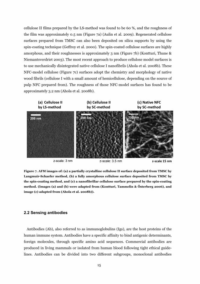
cellulose II films prepared by the LS-method was found to be 60 %, and the roughness of
the film was approximately 0.5 nm (Figure 7a) (Aulin et al. 2009). Regenerated cellulose
surfaces prepared from TMSC can also been deposited on silica supports by using the
spin-coating technique (Geffroy et al. 2000). The spin-coated cellulose surfaces are highly
amorphous, and their roughnesses is approximately 3 nm (Figure 7b) (Kontturi, Thune &
Niemantsverdriet 2003). The most recent approach to produce cellulose model surfaces is
to use mechanically disintegrated native cellulose I nanofibrils (Ahola et al. 2008b). These
NFC-model cellulose (Figure 7c) surfaces adopt the chemistry and morphology of native
wood fibrils (cellulose I with a small amount of hemicellulose, depending on the source of
pulp NFC prepared from). The roughness of those NFC-model surfaces has found to be
approximately 3.2 nm (Ahola et al. 2008b).
Figure 7. AFM images of: (a) a partially crystalline cellulose II surface deposited from TMSC by
Langmuir-Schaefer method, (b) a fully amorphous cellulose surface deposited from TMSC by
the spin-coating method, and (c) a nanofibrillar cellulose surface prepared by the spin-coating
method. (Images (a) and (b) were adapted from (Kontturi, Tammelin & Österberg 2006), and
image (c) adapted from (Ahola et al. 2008b)).
2.2 Sensing antibodies
Antibodies (Ab), also referred to as immunoglobulins (Igs), are the host proteins of the
human immune system. Antibodies have a specific affinity to bind antigenic determinants,
foreign molecules, through specific amino acid sequences. Commercial antibodies are
produced in living mammals or isolated from human blood following tight ethical guide-
lines. Antibodies can be divided into two different subgroups, monoclonal antibodies
200 nm
z-scale 15 nm
(a) Cellulose II by LS-method
(b) Cellulose II by SC-method
(c) Native NFC by SC-method
16
(mAb) and polyclonal antibodies (pAb), according to their specificity to bind antigenic
determinants. Polyclonal antibodies are collected directly from serum, whereas the pro-
duction of monoclonal antibodies requires multiple production and purification steps.
Polyclonal antibodies are a collection of heterogeneous mixtures of antibodies with vary-
ing affinities, but also a small amount of several other Ig classes is present. Thus polyclo-
nal antibodies may cross-react with antigens other than the target antigen, whereas mono-
clonal antibodies contain only a single Ig-class with negligible non-specific cross-reactivity.
2.2.1 The chemistry of antibodies
Immunoglobulins (Igs) are glycoproteins, which are composed of one or more identical
Y-shaped units. Immunoglobulins (Igs) can be separated into five major subclasses (IgG,
IgA, IgM, IgD, and IgE) on the basis of their physical, chemical, and immunological prop-
erties (Figure 8). The IgG (γ-heavy chain), IgD (δ-heavy chain), and IgE (ε-heavy chain)
are Y-shaped antibodies composed of four polypeptide chains (two light chains and two
heavy chains). IgA (α-heavy chain) is a dimer and IgM (μ-heavy chain) is typically a pen-
tamer. Approximately 75 % of human serum immunoglobulin belongs to the IgG-class
(Hamilton 1987), and this type is the most used antibody subclass. In this thesis, only the
IgG-type antibodies were exploited, and thus this sub-type is discussed in more detailed
here. IgG has four subclasses IgG>IgG2>IgG3>IgG4, classified mainly on the length of the
heavy chains (Hamilton 1987). However, the antigenic binding point of these IgG sub-
classes are equivalent, and thus their antigen binding ability is almost a constant. The Y-
shaped immunoglobulin G units are comprised of two identical heavy chains (Mw of each ̴
50,000) and two identical light chains (Mw of each ̴ 25,000) linked together through both
sulfur bridges and non-covalent interactions between polypeptide chains (Madigan, Mar-
tinko & Brock 2009). Moreover, the individual polypeptide chains are composed of vari-
ous numbers of amino acids (22 common amino acids) through the peptide (amide) bonds,
and each light and heavy chain has been analyzed to contain approximately 220 and 440
amino acids, respectively (Amit et al. 1986). The amino terminal ends of the polypeptide
chains have considerable variation in amino acid composition. Therefore, they are refer to
variable (V) regions, whereas the constant (C) regions have rather constant amino acid
composition. The composition of the variable regions of heavy and light chains determines
which antigens can be detected. The antigens, in turn, are specific for a type of antibody.
The protein and polysaccharide content of the IgG class has been found to be 82-96 % and
4-18 %, respectively (Peakman et al. 2009).

17
Figure 8. Schematic illustrations of structures of five major Immunoglobulin classes. All clas-
ses have similar VH and VL domains that bind antigen. In the illustration, light chains are green,
heavy chains are blue, and circles denote the sites of glycosylation. (Adapted from (Rojas, Apo-
daca 2002)).
Immunoglobulin G antibodies have the similar substructure of all proteins ─ the prima-
ry structure (an amino acid sequence of a polypeptide chain), secondary (twisting and
folding of a polypeptide chain, α-helix or β-sheet folding), tertiary (three dimensional
structure composed of two polypeptide chains), and quaternary structure (three dimen-
sional structure composed of many polypeptide chains) (Branden, Tooze cop. 1999). The
heavy and light chains interact together with non-covalent interactions between side-chain
functionalities (residues) and sulfur bridges, defining the three dimensional folding of an
IgG in water (Figure 9a). In an aqueous environment, the hydrophobic amino acid resi-
dues tend to be oriented inside in the structure of an antibody, whereas ionic amino acid
residues (hydrophilic) are oriented in bulk water (Thanki, Thornton & Goodfellow 1988).
Due to the ionic (hydrophilic) side chain functionalities, the conformation of an immuno-
globulin molecule alters as a function of the properties of the solvent. Low ionic strength
conditions stabilize the structure of a protein, whereas higher salt concentrations lead to
precipitation due to the reduced stability and denaturation of proteins (salting out) (Ara-
18
kawa, Timasheff 1984). Each antibody has a specific pH where its surface net charge is
zero (sum of negative and positive charges), i.e. the point of isoelectricity (pI) (Patrickios,
Yamasaki 1995). Below the pI, positively charged amino acid residues dominate, leading to
positive net charge. Conversely, above the pI, the antibody has a negative net charge due
to the domination of ionized anionic amino acid residues.
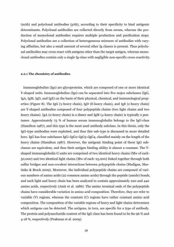
Figure 9. Three dimensional illustration of folding of a human IgG (Adapted from (Jefferis
2009)) (a). Two dimensional schematic illustration of the structure of the basic Y-shaped unit
composed of two identical heavy chains (red line) and two identical light chains (black line) by
sulfuric bridges (b). The constant domains (CH1, CH2, and CH3) are identical in all IgG antibod-
ies. The fragment antigen-binding (FAB) is a part of antibody that can separated by digestion of
the hinge region.
Antigens binding to antibodies take place through the variable VH and VL regions of
fragment-antigen binding (FAB fragments) (Figure 9b). The specific point on the core of
an antigen which reacts with the FAB-fragment is called as an epitope. The antigen-
antibody interaction is always highly specific due to the variations in the amino acid se-
quences of VH and VL regions, which are specific for the type of the antibody (Peakman et
al. 2009)(Davies, Sheriff & Padlan 1988). X-ray crystallographic studies have shown that
antibody-antigen interactions are caused mainly by van der Waals interactions, formation
of hydrogen bonds, and to a lesser extent the formation of salt-bridges (Braden, Poljak
1995). Therefore, the binding of antigen to antibody is reversible, allowing the regenera-
tion of the antigen-antibody bond in suitable conditions (mainly at low pH and high ionic
strength). The strength of this binding, also referred as to an affinity, is the sum of the
attractive and repulsive forces, which can be analyzed using indirect methods by obtain
the affinity constant (Friguet et al. 1985). Recently, atomic force microscopy has also been
exploited for direct analysis of the strength of the antigen-antibody interaction (Allen et al.
1997). The affinity is not the only characteristic that defines antigen binding to antibodies.
b)a)
Fc
19
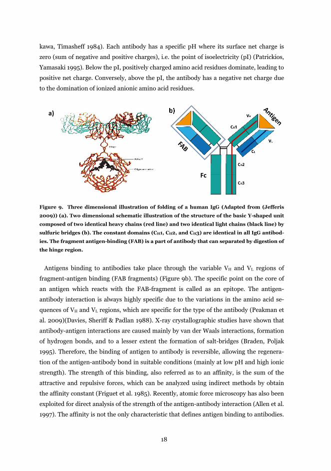
The specificity, the ability of an individual antibody FAB fragment to react with only one
antigenic determinant, is also important. In general, antibodies specificity varies signifi-
cantly between different species. IgGs can also react through the Fc-regions with a variety
of host effector molecules. In the human immune system antibody molecules serve as an
immunologic bridge in the recognition of foreign pathogens and other species responsible
for triggering the host response system (Canfield, Morrison 1991).
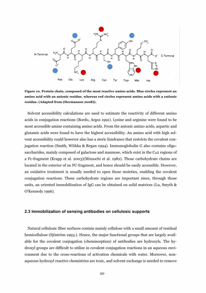
2.2.2 Conjugation sites on antibodies
The covalent conjugation of antibodies onto solid surfaces takes place through the resi-
dues of amino acids. Hence, residues that can orientate on the outside of the IgG are more
accessible for the covalent linking reactions (Rusmini, Zhong & Feijen 2007)(Jameson,
Wong 2009). Polar (hydrophilic) amino acid residues have a higher affinity to orientate
out into the aqueous media, whereas non-polar (hydrophobic) amino acid residues are
hidden in the protein structure (Manavalan, Ponnuswamy 1978). Amino acids with ioniz-
able residues (Figure 10) are significant from the conjugational point of view because co-
valent conjugation reactions take place through those accessible side functionalities. As-
partic acid and glutamic acid are carboxyl-containing amino acids that have a negative
charge. The theoretical pKa of aspartic (Asp) and glutamic (Glu) acids are 3.7-4.0 and 4.2-
4.5, respectively. When these groups are ionized and they can act as a nucleophile in addi-
tion reactions. Cysteine (Cys) is only amino acid that contains a sulfhydryl group, which is
ionized at high pH (pKa of 7.7 – 9.1); however, cysteine has a high hydrophobicity, and
hence, it is usually partially inaccessible for conjugation reactions (Hermanson 2008).
Lysine (Lys) and arginine (Arg) are amine containing amino acids, and they are protonat-
ed at acidic and neutral pH (pKa of 9.3 – 9.5 and 12-12.4, respectively). Histidine (His) has
an imidazole ring that is protonated at alkaline pH (pKa of 6.7 – 7.1). Tyrosine (Tyr) and
tryptophan (Tryp) are aromatic ring-containing amino acids that are normally located
inside an IgG, and therefore their accessibility in water is low (Hermanson 2008).
20
Figure 10. Protein chain, composed of the most reactive amino acids. Blue circles represent an
amino acid with an anionic residue, whereas red circles represent amino acids with a cationic
residue. (Adapted from (Hermanson 2008)).
Solvent accessibility calculations are used to estimate the reactivity of different amino
acids in conjugation reactions (Bordo, Argos 1991). Lysine and arginine were found to be
most accessible amine containing amino acids. From the anionic amino acids, aspartic and
glutamic acids were found to have the highest accessibility. An amino acid with high sol-
vent accessibility could however also has a steric hindrance that restricts the covalent con-
jugation reaction (Smith, Withka & Regan 1994). Immunoglobulin G also contains oligo-
saccharides, mainly composed of galactose and mannose, which exist in the CH2 regions of
a Fc-fragment (Krapp et al. 2003)(Mizuochi et al. 1982). Those carbohydrate chains are
located in the exterior of an FC-fragment, and hence should be easily accessible. However,
an oxidative treatment is usually needed to open those moieties, enabling the covalent
conjugation reactions. These carbohydrate regions are important since, through those
units, an oriented immobilization of IgG can be obtained on solid matrices (Lu, Smyth &
O'Kennedy 1996).
2.3 Immobilization of sensing antibodies on cellulosic supports
Natural cellulosic fiber surfaces contain mainly cellulose with a small amount of residual
hemicellulose (Sjöström 1993.). Hence, the major functional groups that are largely avail-
able for the covalent conjugation (chemisorption) of antibodies are hydroxyls. The hy-
droxyl groups are difficult to utilize in covalent conjugation reactions in an aqueous envi-
ronment due to the cross-reactions of activation chemicals with water. Moreover, non-
aqueous hydroxyl reactive chemistries are toxic, and solvent exchange is needed to remove
21
the residual substances. Therefore, the direct adsorption is the only realistic approach to
bind antibodies to native cellulosic materials in an aqueous solution. However, cellulosic
surfaces can be modified to introduce new functional groups on the cellulosic fiber surfac-
es (Klemm, Philipp & Heinze 1998). Given functional groups can then be utilized in the
covalent linking of antibodies to cellulose even in water. Normally, chemical conjugation
leads to random orientation of antibodies, where their biological activity to bind antigenic
determinants is reduced. Therefore, immobilization strategies to secure oriented binding
of antibodies on solid have been developed. In the following chapter, the most common
immobilization strategies are discussed.
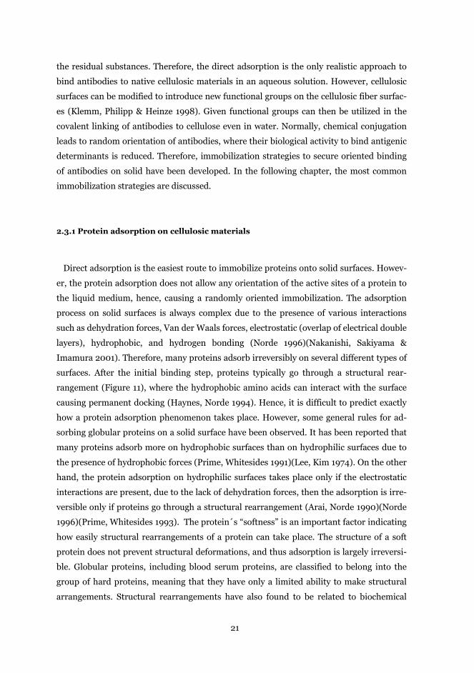
2.3.1 Protein adsorption on cellulosic materials
Direct adsorption is the easiest route to immobilize proteins onto solid surfaces. Howev-
er, the protein adsorption does not allow any orientation of the active sites of a protein to
the liquid medium, hence, causing a randomly oriented immobilization. The adsorption
process on solid surfaces is always complex due to the presence of various interactions
such as dehydration forces, Van der Waals forces, electrostatic (overlap of electrical double
layers), hydrophobic, and hydrogen bonding (Norde 1996)(Nakanishi, Sakiyama &
Imamura 2001). Therefore, many proteins adsorb irreversibly on several different types of
surfaces. After the initial binding step, proteins typically go through a structural rear-
rangement (Figure 11), where the hydrophobic amino acids can interact with the surface
causing permanent docking (Haynes, Norde 1994). Hence, it is difficult to predict exactly
how a protein adsorption phenomenon takes place. However, some general rules for ad-
sorbing globular proteins on a solid surface have been observed. It has been reported that
many proteins adsorb more on hydrophobic surfaces than on hydrophilic surfaces due to
the presence of hydrophobic forces (Prime, Whitesides 1991)(Lee, Kim 1974). On the other
hand, the protein adsorption on hydrophilic surfaces takes place only if the electrostatic
interactions are present, due to the lack of dehydration forces, then the adsorption is irre-
versible only if proteins go through a structural rearrangement (Arai, Norde 1990)(Norde
1996)(Prime, Whitesides 1993). The protein´s “softness” is an important factor indicating
how easily structural rearrangements of a protein can take place. The structure of a soft
protein does not prevent structural deformations, and thus adsorption is largely irreversi-
ble. Globular proteins, including blood serum proteins, are classified to belong into the
group of hard proteins, meaning that they have only a limited ability to make structural
arrangements. Structural rearrangements have also found to be related to biochemical
22
activity of proteins. Adsorbed proteins that have gone through remarkable structural ar-
rangements are to lose their biochemical activities (Norde, Zoungrana 1998)(Sethuraman,
Belfort 2005). On the other hand, at least a part of biochemical activity is present when
hard proteins are adhered to surfaces.
Figure 11. Schematic representation of a protein molecule and a sorbent surface before (a) and
after adsorption (b). Shaded areas represent hydrophobic regions. (Adapted from (Norde
1996)).
Surfaces of cellulosic materials always have a hydrophilic nature due to the presence of
polar hydroxyl groups, which can contribute to hydrogen bonding with proteins or the
binding of water to the surface. Cellulosic surfaces are typically far from the smoothness
and flatness of metallic or synthetic polymer surfaces. The knowledge of protein adsorp-
tion on cellulosic materials have largely been achieved with regenerated cellulosic materi-
als in chromatographic applications (Hage 1999). It has been observed earlier that in-
creased anionic surface charge increases the adsorption of cationically charged proteins on
cellulose, but in that case the adsorption was irreversible to a large extent (Peterson, Sober
1956). Recently, on-line adsorption measurement tools have been utilized in the analysis
of adsorption where the heterogeneity of the cellulosic materials can be taken into account.
However, those analyses have intensively focused on the analysis of the adsorption of cel-
lulose specific enzymes on cellulosic surfaces (Turon, Rojas & Deinhammer 2008)(Ahola
et al. 2008c)(Saarinen et al. 2009). Detailed analysis of the adsorption of immunoglobu-
lins and other serum proteins on natural cellulose has not earlier been reported.
a) b)
23
2.3.2 Chemical conjugation of antibodies on cellulose
Covalent linking reactions take place through the nucleophilic and electrophilic groups
that can contribute to the formation of covalent bonds. A nucleophile is any atom that con-
tains an unshared pair of electrons or an excess of electrons available to donate an elec-
tron pair to an electrophile (Ritchie 1972). The relative order of the nucleophilicity of the
major groups in biological materials can be summarizes as follows: R-S- > R-NH2 > R-
COO- = R-O- >> -OH (Hermanson 2008). The external surfaces of cellulosic materials
contain a significant amount of hydroxyl groups, which can be exploited as conjugation
sites for the immobilization of proteins. On the other hand, amine groups are abundant
functional groups in the exterior areas of proteins (Bordo, Argos 1991). The covalent con-
jugation of proteins to cellulose through the hydroxyl groups can be carried out using var-
ious chemistries such as carbonyldiimidazole (CDI) (Stöllner, Scheller & Warsinke 2002),
epoxide (Uy, Wold 1977), and periodate oxidation (Van Leemputies, Horisberger 1974).
However, these hydroxyl reactive chemistries always need at least one activation step in a
non-polar solvent. The use of carboxyl reactive chemistries on the covalent immobilization
of proteins and other molecules to cellulose has been demonstrated in the literature (Arola
et al. 2012, Filpponen, Argyropoulos 2010). Covalent bonding through the carboxyl group
does not take place spontaneously in water, due to the low nucleophilicity of carboxyls in
water (Hermanson 2008). Therefore, an activator is needed to achieve bond formation.
The most used linker chemistries to achieve bond formation through the carboxyl group
are CDI (Fernandez-Lafuente et al. 1993) and 1-ethyl-3-(3-
dimethylaminopropyl)carbodiimide (EDC) (Grabarek, Gergely 1990). However, only EDC
is a water soluble activator, and hence it has been widely utilized on the conjugation of
proteins. EDC reacts with a carboxyl anion forming an active O-acylisourea intermediate
that is easily displaced by a nucleophilic attack by the amine containing molecule (Figure
12). The O-acylisourea intermediate is unstable in aqueous solutions, and therefore, if the
intermediate does not react in seconds with amine containing molecules, the N-
unsubstituted urea will be released into the medium (Nakajima, Ikada 1995)(Gilles, Hud-
son & Borders Jr. 1990). This hydrolysis failure can be prevented by adding N-hydroxyl
succinimide (NHS) in the reaction mixture, which stabilizes the amine-reactive intermedi-
ate by converting it to an amine-reactive NHS-ester intermediate (Staros, Wright & Swin-
gle 1986). This amine reactive NHS-ester intermediate is semi-stable in water, and the
yield of amide bond formation is significantly increased. In this work the EDC/NHS chem-
istry was employed to link proteins to cellulose.
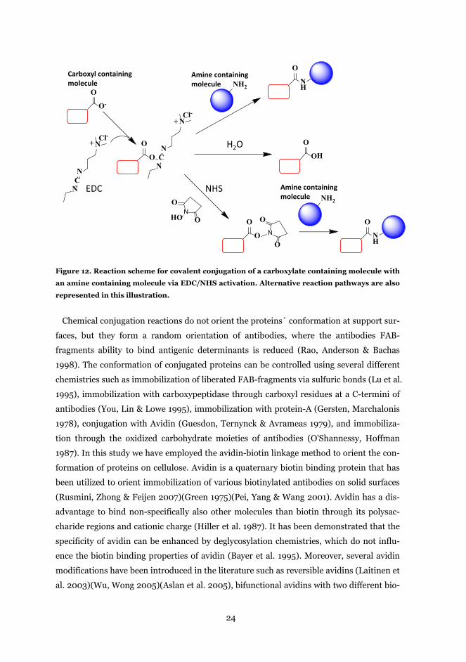
24
Figure 12. Reaction scheme for covalent conjugation of a carboxylate containing molecule with
an amine containing molecule via EDC/NHS activation. Alternative reaction pathways are also
represented in this illustration.
Chemical conjugation reactions do not orient the proteins´ conformation at support sur-
faces, but they form a random orientation of antibodies, where the antibodies FAB-
fragments ability to bind antigenic determinants is reduced (Rao, Anderson & Bachas
1998). The conformation of conjugated proteins can be controlled using several different
chemistries such as immobilization of liberated FAB-fragments via sulfuric bonds (Lu et al.
1995), immobilization with carboxypeptidase through carboxyl residues at a C-termini of
antibodies (You, Lin & Lowe 1995), immobilization with protein-A (Gersten, Marchalonis
1978), conjugation with Avidin (Guesdon, Ternynck & Avrameas 1979), and immobiliza-
tion through the oxidized carbohydrate moieties of antibodies (O'Shannessy, Hoffman
1987). In this study we have employed the avidin-biotin linkage method to orient the con-
formation of proteins on cellulose. Avidin is a quaternary biotin binding protein that has
been utilized to orient immobilization of various biotinylated antibodies on solid surfaces
(Rusmini, Zhong & Feijen 2007)(Green 1975)(Pei, Yang & Wang 2001). Avidin has a dis-
advantage to bind non-specifically also other molecules than biotin through its polysac-
charide regions and cationic charge (Hiller et al. 1987). It has been demonstrated that the
specificity of avidin can be enhanced by deglycosylation chemistries, which do not influ-
ence the biotin binding properties of avidin (Bayer et al. 1995). Moreover, several avidin
modifications have been introduced in the literature such as reversible avidins (Laitinen et
al. 2003)(Wu, Wong 2005)(Aslan et al. 2005), bifunctional avidins with two different bio-
H2O
Carboxyl containing molecule
EDC NHS
Amine containing molecule
Amine containing molecule
O-
O
NCN
N+Cl-
O
O
NCN
N+Cl-
NH2
OH
O
NH
O
HON
O
O
O
ON
O
O
NH
O
NH2
25
tin binding pockets (Nordlund et al. 2004)(Riihimaki et al. 2011), bifunctional avidin that
binds biotin covalently (Leppiniemi et al. 2011), and avidin modification with high thermal
stability (denaturation temperature over 90ºC) (Nordlund et al. 2003).
2.3.3 Functionalization of cellulosic materials
The aim of the surface functionalization of cellulose is to introduce new functional
groups (conjugation sites) on cellulose, which can contribute to covalent conjugation reac-
tions. The cellulosic surface always contains –OH groups (hydroxyls), which enable a pos-
sibility to chemically convert hydroxyls groups of the anhydroglucopyranose units to vari-
ous functional groups using functionalization and grafting chemistries (Klemm et al.
1997)(Roy et al. 2009)(Bhattacharya, Misra 2004). The chemistries most used for func-
tionalization of native cellulose are carboxymethylation, TEMPO-mediated oxidation, and
amination. These chemistries are not surface sensitive, and due to the chemicals penetra-
tion through the cell wall of wood fibers, the substructures of wood fibers are also perma-
nently altered. Carboxymethylation converts the –OH groups of cellulose to carboxyme-
thyl form (Walecka 1956)(Klemm, Philipp & Heinze 1998). A disadvantage of the carbox-
ymethylation treatment is that the carboxymethylation reaction can be carried out only in
non-aqueous solvent systems. Therefore, aqueous based 2,2,6,6,-tetramethylpipelidine-1-
oxyl radical mediated oxidation (TEMPO-oxidation) has gained more attention (De Nooy,
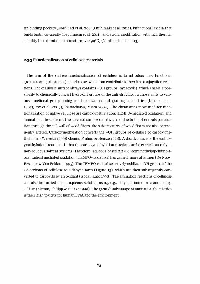
Besemer & Van Bekkum 1995). The TEMPO-radical selectively oxidizes –OH groups of the
C6-carbons of cellulose to aldehyde form (Figure 13), which are then subsequently con-
verted to carboxyls by an oxidant (Isogai, Kato 1998). The amination reactions of cellulose
can also be carried out in aqueous solution using, e.g., ethylene imine or 2-aminoethyl
sulfate (Klemm, Philipp & Heinze 1998). The great disadvantage of amination chemistries
is their high toxicity for human DNA and the environment.
26
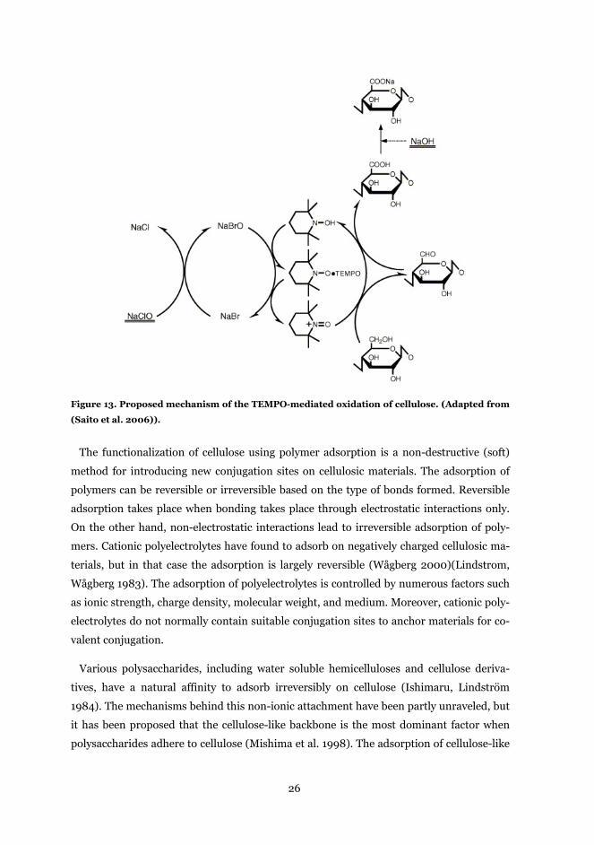
Figure 13. Proposed mechanism of the TEMPO-mediated oxidation of cellulose. (Adapted from
(Saito et al. 2006)).
The functionalization of cellulose using polymer adsorption is a non-destructive (soft)
method for introducing new conjugation sites on cellulosic materials. The adsorption of
polymers can be reversible or irreversible based on the type of bonds formed. Reversible
adsorption takes place when bonding takes place through electrostatic interactions only.
On the other hand, non-electrostatic interactions lead to irreversible adsorption of poly-
mers. Cationic polyelectrolytes have found to adsorb on negatively charged cellulosic ma-
terials, but in that case the adsorption is largely reversible (Wågberg 2000)(Lindstrom,
Wågberg 1983). The adsorption of polyelectrolytes is controlled by numerous factors such
as ionic strength, charge density, molecular weight, and medium. Moreover, cationic poly-
electrolytes do not normally contain suitable conjugation sites to anchor materials for co-
valent conjugation.
Various polysaccharides, including water soluble hemicelluloses and cellulose deriva-
tives, have a natural affinity to adsorb irreversibly on cellulose (Ishimaru, Lindström
1984). The mechanisms behind this non-ionic attachment have been partly unraveled, but
it has been proposed that the cellulose-like backbone is the most dominant factor when
polysaccharides adhere to cellulose (Mishima et al. 1998). The adsorption of cellulose-like
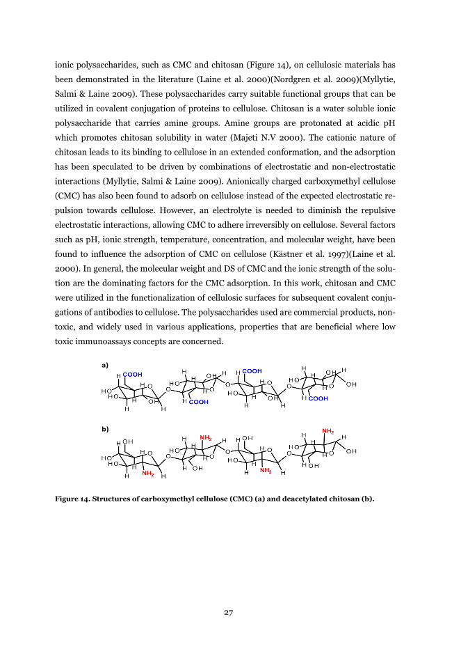
27
ionic polysaccharides, such as CMC and chitosan (Figure 14), on cellulosic materials has
been demonstrated in the literature (Laine et al. 2000)(Nordgren et al. 2009)(Myllytie,
Salmi & Laine 2009). These polysaccharides carry suitable functional groups that can be
utilized in covalent conjugation of proteins to cellulose. Chitosan is a water soluble ionic
polysaccharide that carries amine groups. Amine groups are protonated at acidic pH
which promotes chitosan solubility in water (Majeti N.V 2000). The cationic nature of
chitosan leads to its binding to cellulose in an extended conformation, and the adsorption
has been speculated to be driven by combinations of electrostatic and non-electrostatic
interactions (Myllytie, Salmi & Laine 2009). Anionically charged carboxymethyl cellulose
(CMC) has also been found to adsorb on cellulose instead of the expected electrostatic re-
pulsion towards cellulose. However, an electrolyte is needed to diminish the repulsive
electrostatic interactions, allowing CMC to adhere irreversibly on cellulose. Several factors
such as pH, ionic strength, temperature, concentration, and molecular weight, have been
found to influence the adsorption of CMC on cellulose (Kästner et al. 1997)(Laine et al.
2000). In general, the molecular weight and DS of CMC and the ionic strength of the solu-
tion are the dominating factors for the CMC adsorption. In this work, chitosan and CMC
were utilized in the functionalization of cellulosic surfaces for subsequent covalent conju-
gations of antibodies to cellulose. The polysaccharides used are commercial products, non-
toxic, and widely used in various applications, properties that are beneficial where low
toxic immunoassays concepts are concerned.
Figure 14. Structures of carboxymethyl cellulose (CMC) (a) and deacetylated chitosan (b).
a)
b)
28
3 Experimental
The materials and methods used in the experiments included in this thesis are described
in detail in the attached Papers I-IV. The main cellulosic materials, proteins, and antibod-
ies used in this work are presented in the Materials section. The main characterization
methods used in this work are discussed in the Methods section. This chapter provides a
general background for the experiments presented in the Results and Discussion.
3.1 Materials
3.1.1 Cellulose model surfaces and NFC-films
Trimethylsilyl cellulose (TMSC), used in Papers I-III, was prepared by silylation of mi-
crocrystalline cellulose powder from spruce with hexamethyl disilazane according to
Cooper, Sandberg & Hinck (1981) and Greber, Paschinger (1981). The detailed description
of the synthesis of TMSC that was used in this work has been presented by Tammelin et al.
(2006). The conversion of TMSC to cellulose form by desilylation was carried out accord-
ing to Schaub et al. (1993), and performed following the procedure presented by Kontturi,
Thune & Niemantsverdriet (2003). In papers I and II, TMSC was deposited on gold cov-
ered QCM-D (Q-Sense AB, Sweden) and SPR (paper I, Bionavis Oy, Finland and Paper II,
GE Healthcare, Sweden) wafers using a polystyrene anchor layer. Langmuir-Schaefer (LS)
deposition was carried out according to the procedure represented by Tammelin et al.
(2006). In paper III, TMSC was spin-coated on silica covered QCM-D crystals (Q-Sense
AB, Sweden) with a WS-650SX-6NPP spin coater (Laurell Technologies, USA) following a
procedure presented by Kontturi, Thune & Niemantsverdriet (2003). An area per charge
of 460 nm2 for TMSC-based cellulose surfaces has been reported (Österberg 2000).
The nanofibrillar cellulose surfaces that were used in Papers III and IV were prepared
from unmodified hardwood (birch) pulp. The carbohydrate composition of the pulp used
was 73 % glucose, 26 % xylan and 1 % mannose (Eronen et al. 2011b). The charge of the
pulp used was 65 µeq/g (Eronen et al 2011a). The hardwood pulp was first washed into
sodium form according to Swerin, Ödberg & lindström (1990), and subsequently exten-
sively fluidized by a Microfluidics M-110 (Microfluidics Intl. Corp, Newton, MA, USA). A
more detailed description of the fluidization process and conditions can be found in Tai-
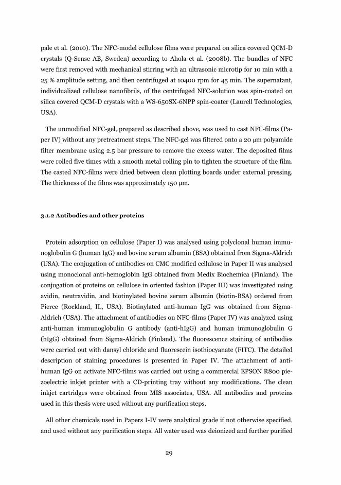
29
pale et al. (2010). The NFC-model cellulose films were prepared on silica covered QCM-D
crystals (Q-Sense AB, Sweden) according to Ahola et al. (2008b). The bundles of NFC
were first removed with mechanical stirring with an ultrasonic microtip for 10 min with a
25 % amplitude setting, and then centrifuged at 10400 rpm for 45 min. The supernatant,
individualized cellulose nanofibrils, of the centrifuged NFC-solution was spin-coated on
silica covered QCM-D crystals with a WS-650SX-6NPP spin-coater (Laurell Technologies,
USA).
The unmodified NFC-gel, prepared as described above, was used to cast NFC-films (Pa-
per IV) without any pretreatment steps. The NFC-gel was filtered onto a 20 μm polyamide
filter membrane using 2.5 bar pressure to remove the excess water. The deposited films
were rolled five times with a smooth metal rolling pin to tighten the structure of the film.
The casted NFC-films were dried between clean plotting boards under external pressing.
The thickness of the films was approximately 150 µm.
3.1.2 Antibodies and other proteins
Protein adsorption on cellulose (Paper I) was analysed using polyclonal human immu-
noglobulin G (human IgG) and bovine serum albumin (BSA) obtained from Sigma-Aldrich
(USA). The conjugation of antibodies on CMC modified cellulose in Paper II was analysed
using monoclonal anti-hemoglobin IgG obtained from Medix Biochemica (Finland). The
conjugation of proteins on cellulose in oriented fashion (Paper III) was investigated using
avidin, neutravidin, and biotinylated bovine serum albumin (biotin-BSA) ordered from
Pierce (Rockland, IL, USA). Biotinylated anti-human IgG was obtained from Sigma-
Aldrich (USA). The attachment of antibodies on NFC-films (Paper IV) was analyzed using
anti-human immunoglobulin G antibody (anti-hIgG) and human immunoglobulin G
(hIgG) obtained from Sigma-Aldrich (Finland). The fluorescence staining of antibodies
were carried out with dansyl chloride and fluorescein isothiocyanate (FITC). The detailed
description of staining procedures is presented in Paper IV. The attachment of anti-
human IgG on activate NFC-films was carried out using a commercial EPSON R800 pie-
zoelectric inkjet printer with a CD-printing tray without any modifications. The clean
inkjet cartridges were obtained from MIS associates, USA. All antibodies and proteins
used in this thesis were used without any purification steps.
All other chemicals used in Papers I-IV were analytical grade if not otherwise specified,
and used without any purification steps. All water used was deionized and further purified
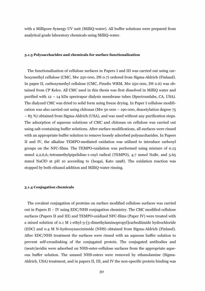
30
with a Millipore Synergy UV unit (MilliQ-water). All buffer solutions were prepared from
analytical grade laboratory chemicals using MilliQ-water.
3.1.3 Polysaccharides and chemicals for surface functionalization
The functionalization of cellulose surfaces in Papers I and III was carried out using car-
boxymethyl cellulose (CMC, Mw 250 000, DS 0.7) ordered from Sigma-Aldrich (Finland).
In paper II, carboxymethyl cellulose (CMC, Finnfix WRM, Mw 250 000, DS 0.6) was ob-
tained from CP Kelco. All CMC used in this thesis was first dissolved in MilliQ water and
purified with 12 – 14 kDa spectrapor dialysis membrane tubes (Spectrumlabs, CA, USA).
The dialyzed CMC was dried to solid form using freeze drying. In Paper I cellulose modifi-
cation was also carried out using chitosan (Mw 50 000 – 190 000, deacetylation degree 75
– 85 %) obtained from Sigma-Aldrich (USA), and was used without any purification steps.
The adsorption of aqueous solutions of CMC and chitosan on cellulose was carried out
using salt-containing buffer solutions. After surface modifications, all surfaces were rinsed
with an appropriate buffer solution to remove loosely adsorbed polysaccharides. In Papers
II and IV, the alkaline TEMPO-mediated oxidation was utilized to introduce carboxyl
groups on the NFC-films. The TEMPO-oxidation was performed using mixture of 0.13
mmol 2,2,6,6,-tetramethylpipelidine-1-oxyl radical (TEMPO), 4.7 mmol NaBr, and 5.65
mmol NaClO at pH 10 according to (Isogai, Kato 1998). The oxidation reaction was
stopped by both ethanol addition and MilliQ-water rinsing.
3.1.4 Conjugation chemicals
The covalent conjugation of proteins on surface modified cellulose surfaces was carried
out in Papers II – IV using EDC/NHS conjugation chemistry. The CMC modified cellulose
surfaces (Papers II and III) and TEMPO-oxidized NFC-films (Paper IV) were treated with
a mixed solution of 0.1 M 1-ethyl-3-[3-dimethylaminopropyl]carbodiimide hydrochloride
(EDC) and 0.4 M N-hydroxysuccinimide (NHS) obtained from Sigma-Aldrich (Finland).
After EDC/NHS treatment the surfaces were rinsed with an aqueous buffer solution to
prevent self-crosslinking of the conjugated protein. The conjugated antibodies and
(neutr)avidin were adsorbed on NHS-ester-cellulose surfaces from the appropriate aque-
ous buffer solution. The unused NHS-esters were removed by ethanolamine (Sigma-
Aldrich, USA) treatment, and in papers II, III, and IV the non-specific protein binding was
31
blocked using Superblock blocking agent obtained from Pierce (Rockland, IL, USA). Su-
perblock is a protein solution that fills the free spaces between the antibodies thus pre-
venting the non-specific binding of antigens on the surface of a biointerface. More detailed
description of the conjugation conditions and activation times are described in Papers II-
IV.
3.2 Methods
The main experimental methods used in this thesis were QCM-D, SPR, AFM, and XPS.
The main studies were carried out using cellulose model surfaces prepared with the
Langmuir-Schaefer technique or the spin coating technique. The detailed principles of
these methods are described here. Details of other methods are presented in Papers I–IV
and references presented here.
3.2.1 Model cellulose film preparation techniques
Langmuir-Schaefer deposition (LS) was used to deposit regenerated cellulose II surfaces
on QCM-D and SPR wafers (Papers I and II). The fundamental studies of the properties of
a water insoluble monolayer at a water-air interface were carried out by Langmuir (1917).
A water insoluble material is spread on a water surface, and by using external barriers, an
evenly spread monolayer can be produced. The transferring of a monolayer from a water-
air interface onto a solid substrate was first demonstrated by Blodgett (1935), but there,
the transferring of monolayer layers was carried out by sinking the substrate in a bath,
causing deposition on both sides of a substrate. Later, the horizontal dipping procedure,
Langmuir-Schaefer deposition (LS), was developed to attach monolayers selectively only
one surface of a substrate (Langmuir, Schaefer 1938). This LS-deposition method was
used in this thesis. Trimethylsilyl cellulose dissolved in chloroform was added to a water
surface (Figure 15), which after evaporation creates a uniform TMSC-layer. The properties
of the TMSC-monolayer were controlled by keeping the surface pressure of the bath con-
stant at 15 mN/m during the dipping cycles. The hydrophobic TMSC was transferred onto
polystyrene covered QCM-D and SPR wafers. Theoretically, a bilayer of TMSC is trans-
ferred on each dipping cycle. The uniformity of the TMSC-layer on the wafer was insured
by dipping 30 TMSC-monolayers onto each wafer surface. The TMSC on a wafer was con-
32
verted to cellulose form using an HCl-vapor treatment according to Kontturi, Thune &
Niemantsverdriet (2003). The description of preparation of cellulose model surfaces using
the LS-method has been presented in detail in Tammelin et al. (2006).
Figure 15. Schematic representation of Langmuir-Schaefer deposition to transfer a TMSC-layer
onto a hydrophobic surface. The horizontal arrows indicate the movement of the barriers. The
grey ovals represent TMSC molecules, and the black spots represent the hydrophobic part of
TMSC and a solid surface. (Adapted from (Tammelin et al. 2006)).
The spin-coating technique was employed in this work to prepare ultrathin cellulose II
and NFC-cellulose surfaces on QCM-D crystal surfaces. The spin-coating technique is
based on the spread of dissolved or dispersed materials in volatile solvents on solid surfac-
es by high speed spinning (Figure 16). The properties of the ultra-thin films are controlled
by several parameters e.g. spinning acceleration, spinning speed, solvent properties, and
concentration of the liquid (Hall, Underhill & Torkelson 1998). In the spin-coating tech-
nique used to prepared regenerated cellulose II model surfaces (Paper III), TMSC was first
dissolved in toluene and applied on a silica covered QCM-D crystal. The solvent was then
evaporated by spinning the crystal with a spinning speed of 3000 rpm according to
Kontturi, Thune & Niemantsverdriet (2003). The TMSC layer on the crystal surface was
then converted to cellulose form with an HCl-vapor treatment. In Paper IV the spin-
coating technique was utilized for preparing nanofibrillar cellulose (NFC) model surfaces.
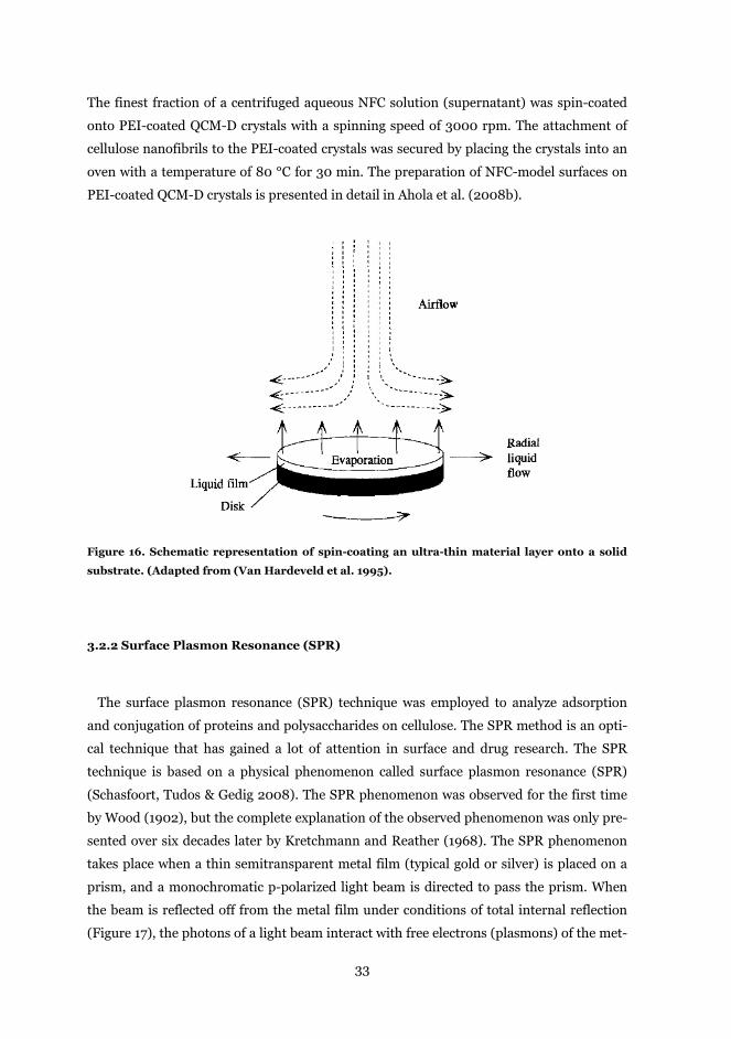
33
The finest fraction of a centrifuged aqueous NFC solution (supernatant) was spin-coated
onto PEI-coated QCM-D crystals with a spinning speed of 3000 rpm. The attachment of
cellulose nanofibrils to the PEI-coated crystals was secured by placing the crystals into an
oven with a temperature of 80 °C for 30 min. The preparation of NFC-model surfaces on
PEI-coated QCM-D crystals is presented in detail in Ahola et al. (2008b).
Figure 16. Schematic representation of spin-coating an ultra-thin material layer onto a solid
substrate. (Adapted from (Van Hardeveld et al. 1995).
3.2.2 Surface Plasmon Resonance (SPR)
The surface plasmon resonance (SPR) technique was employed to analyze adsorption
and conjugation of proteins and polysaccharides on cellulose. The SPR method is an opti-
cal technique that has gained a lot of attention in surface and drug research. The SPR
technique is based on a physical phenomenon called surface plasmon resonance (SPR)
(Schasfoort, Tudos & Gedig 2008). The SPR phenomenon was observed for the first time
by Wood (1902), but the complete explanation of the observed phenomenon was only pre-
sented over six decades later by Kretchmann and Reather (1968). The SPR phenomenon
takes place when a thin semitransparent metal film (typical gold or silver) is placed on a
prism, and a monochromatic p-polarized light beam is directed to pass the prism. When
the beam is reflected off from the metal film under conditions of total internal reflection
(Figure 17), the photons of a light beam interact with free electrons (plasmons) of the met-
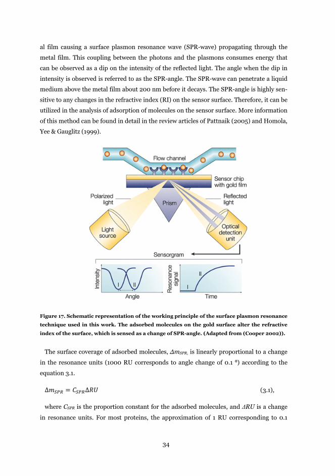
34
al film causing a surface plasmon resonance wave (SPR-wave) propagating through the
metal film. This coupling between the photons and the plasmons consumes energy that
can be observed as a dip on the intensity of the reflected light. The angle when the dip in
intensity is observed is referred to as the SPR-angle. The SPR-wave can penetrate a liquid
medium above the metal film about 200 nm before it decays. The SPR-angle is highly sen-
sitive to any changes in the refractive index (RI) on the sensor surface. Therefore, it can be
utilized in the analysis of adsorption of molecules on the sensor surface. More information
of this method can be found in detail in the review articles of Pattnaik (2005) and Homola,
Yee & Gauglitz (1999).
Figure 17. Schematic representation of the working principle of the surface plasmon resonance
technique used in this work. The adsorbed molecules on the gold surface alter the refractive
index of the surface, which is sensed as a change of SPR-angle. (Adapted from (Cooper 2002)).
The surface coverage of adsorbed molecules, ΔmSPR, is linearly proportional to a change
in the resonance units (1000 RU corresponds to angle change of 0.1 º) according to the
equation 3.1.
∆𝑚𝑆𝑃𝑅 = 𝐶𝑆𝑃𝑅∆𝑅𝑈 (3.1),
where CSPR is the proportion constant for the adsorbed molecules, and ΔRU is a change
in resonance units. For most proteins, the approximation of 1 RU corresponding to 0.1
35
ng/mm2 can be used (Stenberg et al. 1991). For polysaccharides, only a limited number of
proportion constants have been presented in the literature. However, another model for
the estimation of surface coverage on the sensor surface has been presented by Jung et al.
(1998). The surface coverage can be calculated by first calculating the thickness of the ad-
sorbed layer, d, according to equation 3.2.
𝑑 = 𝑙𝑑2
∆𝑆𝑃𝑅𝑚(𝑛𝑎−𝑛0)
(3.2),
where ld is a characteristic evanescent electromagnetic field decay length and can be es-
timated to be 0.37 of the wavelength of the laser, ΔSPR is a change in the SPR angle, m is a
sensitivity factor for the sensor obtained by calibrating the SPR, n0 is the refractive index
of the bulk medium (1.334 RIU for buffers), and na is the refractive index of the adsorbed
molecules. Then, the surface coverage, ΔmSPR, can easily be calculated according to the
equation 3.3.
∆𝑚𝑆𝑃𝑅 = 𝑑 ∗ 𝜌 (3.3),
where d is the thickness of the adsorbed layer (calculated by equation (3.2)), and ρ is the
specific volume of the adsorbed molecules. The refractive indices and the specific volumes
for various substances can be found in the literature.
The SPR measurements were carried out with a SPR Model Navi 200 (Oy BioNavis Ltd,
Finland) in Paper I, and a Biacore 1000 instrument (GE Healthcare, Sweden) in paper II.
Before experiments, the cellulose model films were stabilized by driving a buffer solution
through the measurement chambers until a stable baseline was acquired. All experiments
were duplicated at least once to get accurate results. A mass sensitivity of a SPR used in
this work was 0.15 mdeg that corresponds to a mass change of 0.1 ng/cm2.The details of
the experiments and the modeling parameters with the equations used can be found in the
Papers I and II.
3.2.3 Quartz Crystal Microbalance with Dissipation (QCM-D)
The QCM-D technique was used to characterize adsorption of proteins and polysaccha-
rides on cellulose model surfaces. This method is widely used in surface research because
of its unique property to follow mass adsorption on solid surfaces while simultaneously
obtained information on the viscoelastic properties of the adsorbed layer. The QCM-D
uses an oscillating quartz crystal, on which changes in frequency and dissipation are
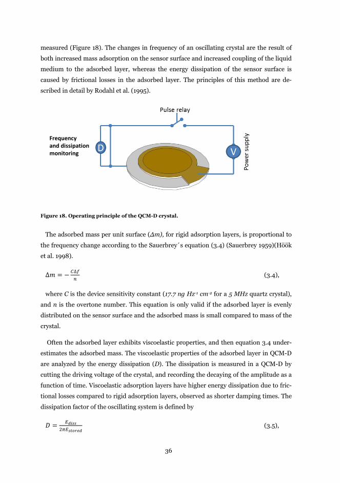
36
measured (Figure 18). The changes in frequency of an oscillating crystal are the result of
both increased mass adsorption on the sensor surface and increased coupling of the liquid
medium to the adsorbed layer, whereas the energy dissipation of the sensor surface is
caused by frictional losses in the adsorbed layer. The principles of this method are de-
scribed in detail by Rodahl et al. (1995).
Figure 18. Operating principle of the QCM-D crystal.
The adsorbed mass per unit surface (Δm), for rigid adsorption layers, is proportional to
the frequency change according to the Sauerbrey´s equation (3.4) (Sauerbrey 1959)(Höök
et al. 1998).
∆𝑚 = −𝐶∆𝑓𝑛
(3.4),
where C is the device sensitivity constant (17.7 ng Hz-1 cm-2 for a 5 MHz quartz crystal),
and n is the overtone number. This equation is only valid if the adsorbed layer is evenly
distributed on the sensor surface and the adsorbed mass is small compared to mass of the
crystal.
Often the adsorbed layer exhibits viscoelastic properties, and then equation 3.4 under-
estimates the adsorbed mass. The viscoelastic properties of the adsorbed layer in QCM-D
are analyzed by the energy dissipation (D). The dissipation is measured in a QCM-D by
cutting the driving voltage of the crystal, and recording the decaying of the amplitude as a
function of time. Viscoelastic adsorption layers have higher energy dissipation due to fric-
tional losses compared to rigid adsorption layers, observed as shorter damping times. The
dissipation factor of the oscillating system is defined by
𝐷 = 𝐸𝑑𝑖𝑠𝑠2𝜋𝐸𝑠𝑡𝑜𝑟𝑒𝑑
(3.5),
Frequency and dissipation monitoring
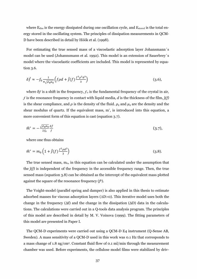
37
where Ediss is the energy dissipated during one oscillation cycle, and Estored is the total en-
ergy stored in the oscillating system. The principles of dissipation measurements in QCM-
D have been described in detail by Höök et al. (1998).
For estimating the true sensed mass of a viscoelastic adsorption layer Johansmann´s
model can be used (Johannsmann et al. 1992). This model is an extension of Sauerbrey´s
model where the viscoelastic coefficients are included. This model is represented by equa-
tion 3.6.
𝛿𝑓 ≈ −𝑓01
𝜋�𝜌𝑞𝜇𝑞�𝑓𝜌𝑑 + 𝐽(𝑓) 𝑓
3𝜌3𝑑3
3� (3.6),
where δƒ is a shift in the frequency, ƒ0 is the fundamental frequency of the crystal in air,
ƒ is the resonance frequency in contact with liquid media, d is the thickness of the film, ĵ(f)
is the shear compliance, and ρ is the density of the fluid. ρq and µq are the density and the
shear modulus of quartz. If the equivalent mass, m*, is introduced into this equation, a
more convenient form of this equation is cast (equation 3.7).
𝑚�∗ = −�𝜌𝑞𝜇𝑞2𝑓0
𝛿�̂�𝑓
(3.7),
where one thus obtains
𝑚�∗ = 𝑚0 �1 + 𝐽(𝑓) 𝑓2𝜌𝑑2
3� (3.8).
The true sensed mass, m0, in this equation can be calculated under the assumption that
the ĵ(f) is independent of the frequency in the accessible frequency range. Then, the true
sensed mass (equation 3.8) can be obtained as the intercept of the equivalent mass plotted
against the square of the resonance frequency (f2).
The Voight-model (parallel spring and damper) is also applied in this thesis to estimate
adsorbed masses for viscous adsorption layers (ΔD>0). This iterative model uses both the
change in the frequency (Δf) and the change in the dissipation (ΔD) data in the calcula-
tions. The calculations were carried out in a Q-tools data analysis program. The principles
of this model are described in detail by M. V. Voinova (1999). The fitting parameters of
this model are presented in Paper I.
The QCM-D experiments were carried out using a QCM-D E4 instrument (Q-Sense AB,
Sweden). A mass sensitivity of a QCM-D used in this work was 0.1 Hz that corresponds to
a mass change of 1.8 ng/cm2. Constant fluid flow of 0.1 ml/min through the measurement
chamber was used. Before experiments, the cellulose model films were stabilized by driv-
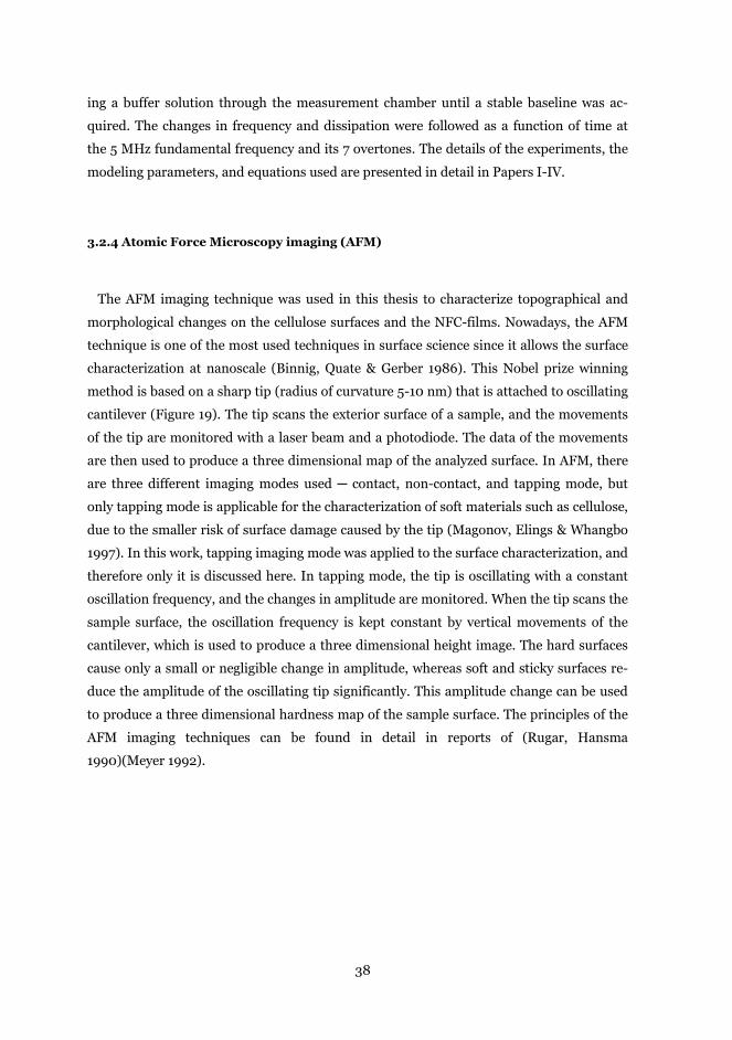
38
ing a buffer solution through the measurement chamber until a stable baseline was ac-
quired. The changes in frequency and dissipation were followed as a function of time at
the 5 MHz fundamental frequency and its 7 overtones. The details of the experiments, the
modeling parameters, and equations used are presented in detail in Papers I-IV.
3.2.4 Atomic Force Microscopy imaging (AFM)
The AFM imaging technique was used in this thesis to characterize topographical and
morphological changes on the cellulose surfaces and the NFC-films. Nowadays, the AFM
technique is one of the most used techniques in surface science since it allows the surface
characterization at nanoscale (Binnig, Quate & Gerber 1986). This Nobel prize winning
method is based on a sharp tip (radius of curvature 5-10 nm) that is attached to oscillating
cantilever (Figure 19). The tip scans the exterior surface of a sample, and the movements
of the tip are monitored with a laser beam and a photodiode. The data of the movements
are then used to produce a three dimensional map of the analyzed surface. In AFM, there
are three different imaging modes used ─ contact, non-contact, and tapping mode, but
only tapping mode is applicable for the characterization of soft materials such as cellulose,
due to the smaller risk of surface damage caused by the tip (Magonov, Elings & Whangbo
1997). In this work, tapping imaging mode was applied to the surface characterization, and
therefore only it is discussed here. In tapping mode, the tip is oscillating with a constant
oscillation frequency, and the changes in amplitude are monitored. When the tip scans the
sample surface, the oscillation frequency is kept constant by vertical movements of the
cantilever, which is used to produce a three dimensional height image. The hard surfaces
cause only a small or negligible change in amplitude, whereas soft and sticky surfaces re-
duce the amplitude of the oscillating tip significantly. This amplitude change can be used
to produce a three dimensional hardness map of the sample surface. The principles of the
AFM imaging techniques can be found in detail in reports of (Rugar, Hansma
1990)(Meyer 1992).
39
Figure 19. Working principle of the AFM technique. A sharp tip scans the sample surface. The
tip movement on the surface is monitored by a laser beam that is directed to top of the cantile-
ver, and the reflected beam is directed to a photodiode. The three dimensional map on the
sample surface is obtained by recording the vertical movements of the cantilever.
The AFM experiments were carried out in this work by using a Nanoscope IIIa Multi-
mode scanning probe microscope (Digital Instruments Inc., USA). All images were
scanned in tapping mode in air using silicon cantilevers (NSC15/AIBS from MicroMash,
Estonia, radius of curvature less than 10 nm, height of the tip approximately 25 nm). The
AFM images scan sizes were 1 x 1 µm2 and 5 x 5 µm2, and no any image processing was
done for the images. The experimental conditions in detail can be found in Papers I-IV.
3.2.5 Additional techniques
X-ray photoelectron spectroscopy (XPS). The changes in chemical composition of the
cellulose model surfaces (Papers I-IV) and the NFC-films (Paper IV) were characterized by
XPS. In this work, a Kratos analytical AXIS 165 electron spectrometer with a monochro-
matic Al Kα X-Ray source was used. The XPS experiments were performed on dry samples,
together with an in-situ reference sample (S&S 5893 Blue ribbon 6 ashless, 100% cellulose)
(Johansson, Campbell 2004), and the spectra were collected normal to the surface. The
sampling area was less than one mm in diameter, and several locations on each sample
was analyzed. The relative amounts of carbon, oxygen, nitrogen (protein marker), and
silicon were determined from C 1s, O 1s, N 1s, and Si 2p signals from low-resolution scans.
The high resolution C1s spectrum was curve fitted for further chemical analysis using pa-
40
rameters defined for cellulosic materials as described by Johansson, Campbell (2004).
More information about to the XPS technique can be found in Moulder, Chastain (1992).
Contact angle measurements (CAM). The introduction of carboxyl groups on the NFC-
films after TEMPO-mediated oxidation (Paper IV) were analyzed using a contact angle
contact angle goniometer CAM 200 (KSV instruments Ltd, Helsinki, Finland). Measure-
ments were performed at room temperature with water as a probe liquid. The droplet vol-
ume used was 6.5 μl, and the recording time was 120 sec for measuring the time depend-
ency in changes of the contact angle. Contact angles were measured on three different
spots on each sample. More information about to contact angle measurements on solid
surfaces can be found in the excellent review paper of Kwok, Neumann (1999).
Conductometric titration. The increase in the charged groups (carboxyls) on the NFC-
films after TEMPO-mediated oxidation (Paper IV) was analyzed using a conductometric
titrator 751 GPD Titrino (Metrohm AG, Herisau, Switzerland). Dried, acid washed NFC-
films (size 2.5 x 2.5 cm2) were disintegrated in MilliQ-water with a blade type homogeniz-
er, the Polytron PT 2000 (Kinematica Inc., NY, USA). The conductometric titration was
performed by adding 0.02 ml of 0.1 M NaOH at 30 sec intervals. The calculation of the
amount of weak acids (carboxyls) was performed as described in the standard SCAN-CM
65:02.
Confocal laser scanning microscopy (CLSM). Macro scale topography of the NFC-films
and printing of fluorescein stained anti-human IgG on the NFC-films was characterized
with a Leica TCS SP2 confocal laser scanning microscope (Leica microsystems CMS
GmbH, Manheim, Germany. The image was achieved using reflection image and fluores-
cence modes with 488 nm laser wavelengths, and image sizes of 750 x 750 μm2. The detec-
tion wavelength ranges were 488 ± 10 nm and approximately 490-550 nm. The 3D-image
was rendered from the 60 optical sections of the NFC-film using topography mode. No any
sample pretreatment was done except placing the sample between two clean microscopy
glasses. The roughness profile of the NFC-film was calculated using a rendered topograph-
ical 3-D image with the Leica confocal microscope software. Additional information about
to CLSM technique can be found in Paddock (1999).
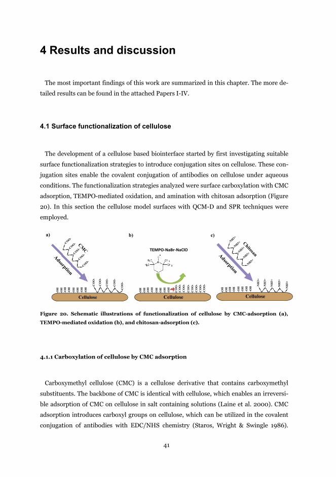
41
4 Results and discussion
The most important findings of this work are summarized in this chapter. The more de-
tailed results can be found in the attached Papers I-IV.
4.1 Surface functionalization of cellulose
The development of a cellulose based biointerface started by first investigating suitable
surface functionalization strategies to introduce conjugation sites on cellulose. These con-
jugation sites enable the covalent conjugation of antibodies on cellulose under aqueous
conditions. The functionalization strategies analyzed were surface carboxylation with CMC
adsorption, TEMPO-mediated oxidation, and amination with chitosan adsorption (Figure
20). In this section the cellulose model surfaces with QCM-D and SPR techniques were
employed.
Figure 20. Schematic illustrations of functionalization of cellulose by CMC-adsorption (a),
TEMPO-mediated oxidation (b), and chitosan-adsorption (c).
4.1.1 Carboxylation of cellulose by CMC adsorption
Carboxymethyl cellulose (CMC) is a cellulose derivative that contains carboxymethyl
substituents. The backbone of CMC is identical with cellulose, which enables an irreversi-
ble adsorption of CMC on cellulose in salt containing solutions (Laine et al. 2000). CMC
adsorption introduces carboxyl groups on cellulose, which can be utilized in the covalent
conjugation of antibodies with EDC/NHS chemistry (Staros, Wright & Swingle 1986).
TEMPO-NaBr-NaClO
-NH3+
-NH3+
-NH3+
-NH3+
-NH3+
Adsorption
Chitosan
-NH
3+
-NH
3+
-NH
3+
-NH
3+
-NH
3+
-OH
-OH
-OH
-OH
-OH
-OH
-OH
-OH
c)
Cellulose
-COO-
-COO-
-COO-
-COO-
-COO-
Adsorption
CMC
-CO
O-
-CO
O-
-CO
O-
-CO
O-
-CO
O-
-OH
-OH
-OH
-OH
-OH
-OH
-OH
-OH
a)
-OH
-OH
-OH
-OH
-OH
-OH
-OH
-OH
b)
Cellulose Cellulose
-CO
O-
-CO
O-
-CO
O-
-CO
O-
-CO
O-
-CO
O-
-CO
O-
-CO
O-
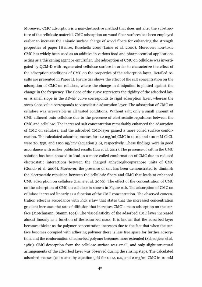
42
Moreover, CMC adsorption is a non-destructive method that does not alter the substruc-
ture of the cellulosic material. CMC adsorption on wood fiber surfaces has been employed
earlier to increase the anionic surface charge of wood fibers for enhancing the strength
properties of paper (Heinze, Koschella 2005)(Laine et al. 2000). Moreover, non-toxic
CMC has widely been used as an additive in various food and pharmaceutical applications
acting as a thickening agent or emulsifier. The adsorption of CMC on cellulose was investi-
gated by QCM-D with regenerated cellulose surface in order to characterize the effect of
the adsorption conditions of CMC on the properties of the adsorption layer. Detailed re-
sults are presented in Paper II. Figure 21a shows the effect of the salt concentration on the
adsorption of CMC on cellulose, where the change in dissipation is plotted against the
change in the frequency. The slope of the curve represents the rigidity of the adsorbed lay-
er. A small slope in the ΔD-ΔF curve corresponds to rigid adsorption layer, whereas the
steep slope value corresponds to viscoelastic adsorption layer. The adsorption of CMC on
cellulose was irreversible in all tested conditions. Without salt, only a small amount of
CMC adhered onto cellulose due to the presence of electrostatic repulsions between the
CMC and cellulose. The increased salt concentration remarkably enhanced the adsorption
of CMC on cellulose, and the adsorbed CMC-layer gained a more coiled surface confor-
mation. The calculated adsorbed masses for 0.2 mg/ml CMC in 0, 10, and 100 mM CaCl2
were 20, 530, and 1100 ng/cm2 (equation 3.6), respectively. These findings were in good
accordance with earlier published results (Liu et al. 2011). The presence of salt in the CMC
solution has been showed to lead to a more coiled conformation of CMC due to reduced
electrostatic interactions between the charged anhydroglucopyranose units of CMC
(Gondo et al. 2006). Moreover, the presence of salt has been demonstrated to diminish
the electrostatic repulsion between the cellulosic fibers and CMC that leads to enhanced
CMC adsorption on cellulose (Laine et al. 2000). The effect of the concentration of CMC
on the adsorption of CMC on cellulose is shown in Figure 21b. The adsorption of CMC on
cellulose increased linearly as a function of the CMC concentration. The observed concen-
tration effect is accordance with Fick´s law that states that the increased concentration
gradient increases the rate of diffusion that increases CMC´s mass adsorption on the sur-
face (Motchmann, Stamm 1991). The viscoelasticity of the adsorbed CMC layer increased
almost linearly as a function of the adsorbed mass. It is known that the adsorbed layer
becomes thicker as the polymer concentration increases due to the fact that when the sur-
face becomes occupied with adhering polymer there is less free space for further adsorp-
tion, and the conformation of adsorbed polymer becomes more extended (Scheutjens et al.
1980). CMC desorption from the cellulose surface was small, and only slight structural
arrangements of the adsorbed layer was observed during the rinsing steps. The calculated
adsorbed masses (calculated by equation 3.6) for 0.02, 0.2, and 2 mg/ml CMC in 10 mM
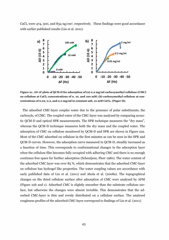
43
CaCl2 were 474, 500, and 834 ng/cm2, respectively. These findings were good accordance
with earlier published results (Liu et al. 2011).
Figure 21. ΔD-ΔF plots of QCM-D for adsorption of (a) 0.2 mg/ml carboxymethyl cellulose (CMC)
on cellulose at CaCl2 concentrations of 0, 10, and 100 mM; (b) carboxymethyl cellulose at con-
centrations of 0.02, 0.2, and 2.0 mg/ml in constant salt, 10 mM CaCl2. (Paper II).
The adsorbed CMC-layer couples water due to the presence of polar substituents, the
carboxyls, of CMC. The coupled water of the CMC-layer was analyzed by comparing acous-
tic QCM-D and optical SPR measurements. The SPR technique measures the “dry mass”,
whereas the QCM-D technique measures both the dry mass and the coupled water. The
adsorption of CMC on cellulose monitored by QCM-D and SPR are shown in Figure 22a.
Most of the CMC adsorbed on cellulose in the first minutes as can be seen in the SPR and
QCM-D curves. However, the adsorption curve measured in QCM-D, steadily increased as
a function of time. This corresponds to conformational changes in the adsorption layer
when the cellulose film becomes fully occupied with adhering CMC and there is no enough
continues free space for further adsorption (Scheutjens, Fleer 1980). The water content of
the adsorbed CMC layer was over 85 %, which demonstrates that the adsorbed CMC-layer
on cellulose has hydrogel like properties. The water coupling values are accordance with
early published data of Liu et al. (2011) and Ahola et al. (2008a). The topographical
changes on the dried cellulose surface after adsorption of CMC were analysed by AFM
(Figure 22b and c). Adsorbed CMC is slightly smoother than the substrate cellulose sur-
face, but otherwise the changes were almost invisible. This demonstrates that the ad-
sorbed CMC-layer is thin and evenly distributed on a cellulose surface. The analysed
roughness profiles of the adsorbed CMC-layer correspond to findings of Liu et al. (2011).
012345678
-50-40-30-20-100
∆D (1
E-6)
∆F (Hz)
a)
012345678
-50-40-30-20-100
∆D (1
E-6)
∆F (Hz)
b)100 mM
10 mM
0 mM
2 mg/ml
0.2 mg/ml
0.02 mg/ml
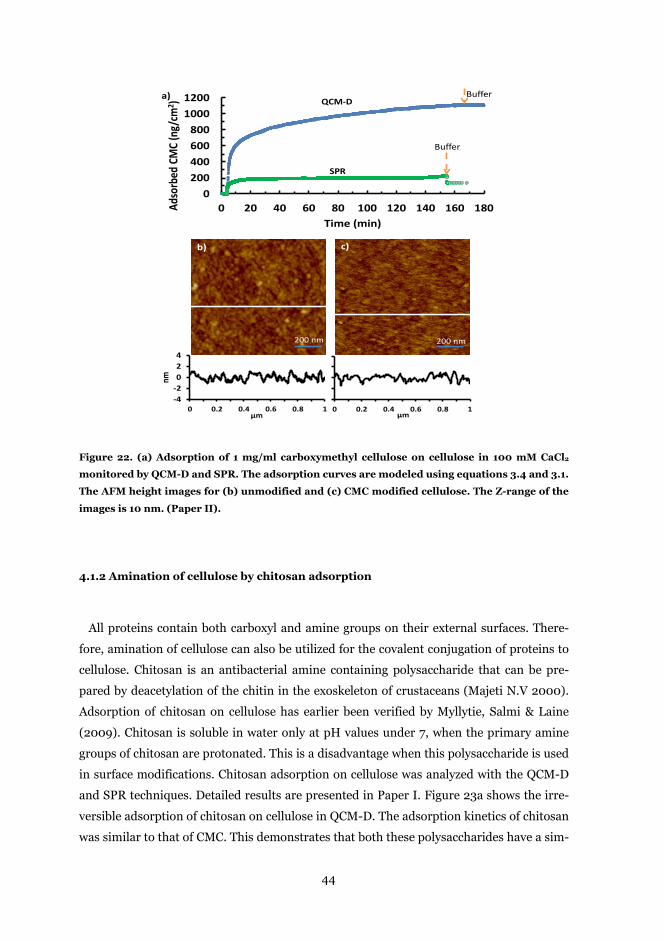
44
Figure 22. (a) Adsorption of 1 mg/ml carboxymethyl cellulose on cellulose in 100 mM CaCl2
monitored by QCM-D and SPR. The adsorption curves are modeled using equations 3.4 and 3.1.
The AFM height images for (b) unmodified and (c) CMC modified cellulose. The Z-range of the
images is 10 nm. (Paper II).
4.1.2 Amination of cellulose by chitosan adsorption
All proteins contain both carboxyl and amine groups on their external surfaces. There-
fore, amination of cellulose can also be utilized for the covalent conjugation of proteins to
cellulose. Chitosan is an antibacterial amine containing polysaccharide that can be pre-
pared by deacetylation of the chitin in the exoskeleton of crustaceans (Majeti N.V 2000).
Adsorption of chitosan on cellulose has earlier been verified by Myllytie, Salmi & Laine
(2009). Chitosan is soluble in water only at pH values under 7, when the primary amine
groups of chitosan are protonated. This is a disadvantage when this polysaccharide is used
in surface modifications. Chitosan adsorption on cellulose was analyzed with the QCM-D
and SPR techniques. Detailed results are presented in Paper I. Figure 23a shows the irre-
versible adsorption of chitosan on cellulose in QCM-D. The adsorption kinetics of chitosan
was similar to that of CMC. This demonstrates that both these polysaccharides have a sim-
0200400600800
10001200
0 20 40 60 80 100 120 140 160 180Adso
rbed
CMC
(ng/
cm2 )
Time (min)
Buffer
Buffer
QCM-D
SPR
0 0.2 0.4 0.6 0.8 1μm
-4-2024
0 0.2 0.4 0.6 0.8 1
nm
μm
b)
200 nm
c)
200 nm
a)
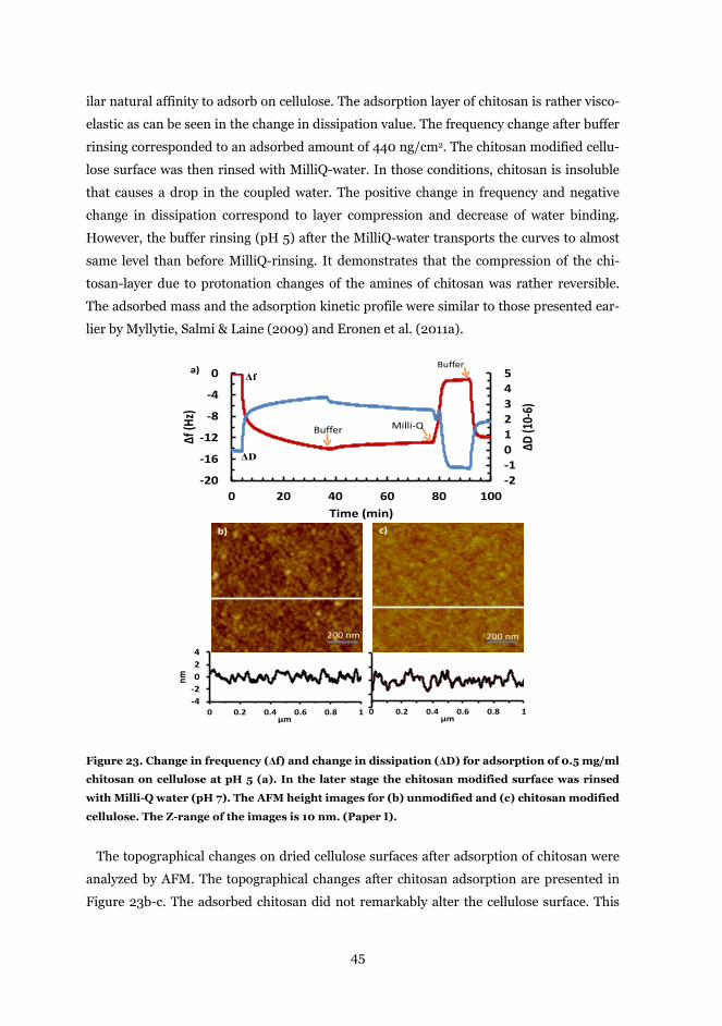
45
ilar natural affinity to adsorb on cellulose. The adsorption layer of chitosan is rather visco-
elastic as can be seen in the change in dissipation value. The frequency change after buffer
rinsing corresponded to an adsorbed amount of 440 ng/cm2. The chitosan modified cellu-
lose surface was then rinsed with MilliQ-water. In those conditions, chitosan is insoluble
that causes a drop in the coupled water. The positive change in frequency and negative
change in dissipation correspond to layer compression and decrease of water binding.
However, the buffer rinsing (pH 5) after the MilliQ-water transports the curves to almost
same level than before MilliQ-rinsing. It demonstrates that the compression of the chi-
tosan-layer due to protonation changes of the amines of chitosan was rather reversible.
The adsorbed mass and the adsorption kinetic profile were similar to those presented ear-
lier by Myllytie, Salmi & Laine (2009) and Eronen et al. (2011a).
Figure 23. Change in frequency (Δf) and change in dissipation (ΔD) for adsorption of 0.5 mg/ml
chitosan on cellulose at pH 5 (a). In the later stage the chitosan modified surface was rinsed
with Milli-Q water (pH 7). The AFM height images for (b) unmodified and (c) chitosan modified
cellulose. The Z-range of the images is 10 nm. (Paper I).
The topographical changes on dried cellulose surfaces after adsorption of chitosan were
analyzed by AFM. The topographical changes after chitosan adsorption are presented in
Figure 23b-c. The adsorbed chitosan did not remarkably alter the cellulose surface. This
-2-1012345
-20
-16
-12
-8
-4
0
0 20 40 60 80 100
ΔD (1
0-6)
Δf (H
z)
Time (min)
Buffer Milli-Q
BufferΔf
ΔD
-4-2024
0 0.2 0.4 0.6 0.8 1
nm
μm
b)
200 nm
c)
200 nm
0 0.2 0.4 0.6 0.8 1μm
a)
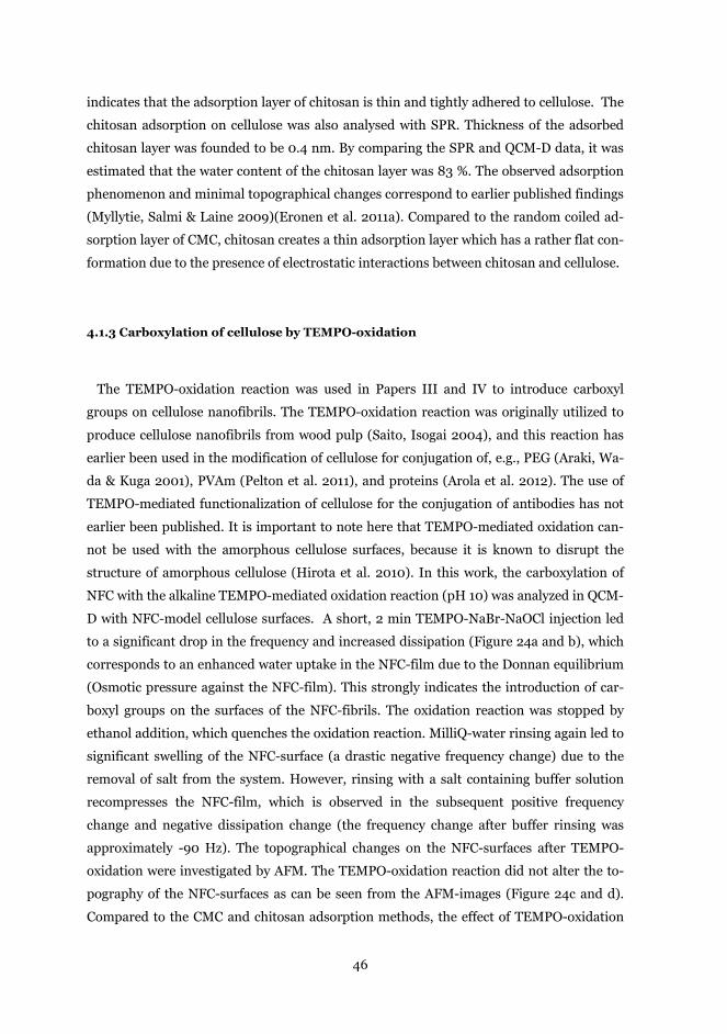
46
indicates that the adsorption layer of chitosan is thin and tightly adhered to cellulose. The
chitosan adsorption on cellulose was also analysed with SPR. Thickness of the adsorbed
chitosan layer was founded to be 0.4 nm. By comparing the SPR and QCM-D data, it was
estimated that the water content of the chitosan layer was 83 %. The observed adsorption
phenomenon and minimal topographical changes correspond to earlier published findings
(Myllytie, Salmi & Laine 2009)(Eronen et al. 2011a). Compared to the random coiled ad-
sorption layer of CMC, chitosan creates a thin adsorption layer which has a rather flat con-
formation due to the presence of electrostatic interactions between chitosan and cellulose.
4.1.3 Carboxylation of cellulose by TEMPO-oxidation
The TEMPO-oxidation reaction was used in Papers III and IV to introduce carboxyl
groups on cellulose nanofibrils. The TEMPO-oxidation reaction was originally utilized to
produce cellulose nanofibrils from wood pulp (Saito, Isogai 2004), and this reaction has
earlier been used in the modification of cellulose for conjugation of, e.g., PEG (Araki, Wa-
da & Kuga 2001), PVAm (Pelton et al. 2011), and proteins (Arola et al. 2012). The use of
TEMPO-mediated functionalization of cellulose for the conjugation of antibodies has not
earlier been published. It is important to note here that TEMPO-mediated oxidation can-
not be used with the amorphous cellulose surfaces, because it is known to disrupt the
structure of amorphous cellulose (Hirota et al. 2010). In this work, the carboxylation of
NFC with the alkaline TEMPO-mediated oxidation reaction (pH 10) was analyzed in QCM-
D with NFC-model cellulose surfaces. A short, 2 min TEMPO-NaBr-NaOCl injection led
to a significant drop in the frequency and increased dissipation (Figure 24a and b), which
corresponds to an enhanced water uptake in the NFC-film due to the Donnan equilibrium
(Osmotic pressure against the NFC-film). This strongly indicates the introduction of car-
boxyl groups on the surfaces of the NFC-fibrils. The oxidation reaction was stopped by
ethanol addition, which quenches the oxidation reaction. MilliQ-water rinsing again led to
significant swelling of the NFC-surface (a drastic negative frequency change) due to the
removal of salt from the system. However, rinsing with a salt containing buffer solution
recompresses the NFC-film, which is observed in the subsequent positive frequency
change and negative dissipation change (the frequency change after buffer rinsing was
approximately -90 Hz). The topographical changes on the NFC-surfaces after TEMPO-
oxidation were investigated by AFM. The TEMPO-oxidation reaction did not alter the to-
pography of the NFC-surfaces as can be seen from the AFM-images (Figure 24c and d).
Compared to the CMC and chitosan adsorption methods, the effect of TEMPO-oxidation
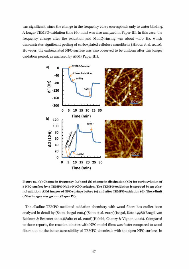
47
was significant, since the change in the frequency curve corresponds only to water binding.
A longer TEMPO-oxidation time (60 min) was also analyzed in Paper III. In this case, the
frequency change after the oxidation and MilliQ-rinsing was about +170 Hz, which
demonstrates significant peeling of carboxylated cellulose nanofibrils (Hirota et al. 2010).
However, the carboxylated NFC-surface was also observed to be uniform after this longer
oxidation period, as analysed by AFM (Paper III).
Figure 24. (a) Change in frequency (ΔF) and (b) change in dissipation (ΔD) for carboxylation of
a NFC-surface by a TEMPO-NaBr-NaClO solution. The TEMPO-oxidation is stopped by an etha-
nol addition. AFM images of NFC-surface before (c) and after TEMPO-oxidation (d). The z-limit
of the images was 30 nm. (Paper IV).
The alkaline TEMPO-mediated oxidation chemistry with wood fibers has earlier been
analyzed in detail by (Saito, Isogai 2004)(Saito et al. 2007)(Isogai, Kato 1998)(Bragd, van
Bekkum & Besemer 2004)(Saito et al. 2006)(Habibi, Chanzy & Vignon 2006). Compared
to those reports, the reaction kinetics with NFC model films was faster compared to wood
fibers due to the better accessibility of TEMPO-chemicals with the open NFC-surface. In
-200
-160
-120
-80
-40
0
0 5 10 15 20 25 30
ΔF (H
z)
Time (min)
0
20
40
60
80
100
120
0 5 10 15 20 25 30
ΔD (1
0-6)
Time (min)
TEMPO-Solution
Ethanol addition
MilliQ
Buffer
Buffer
MilliQ
a)
b)
c)
d)
200 nm
200 nm
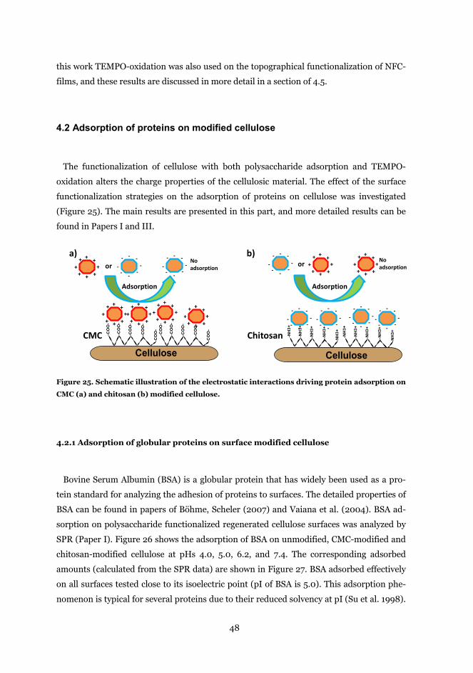
48
this work TEMPO-oxidation was also used on the topographical functionalization of NFC-
films, and these results are discussed in more detail in a section of 4.5.
4.2 Adsorption of proteins on modified cellulose
The functionalization of cellulose with both polysaccharide adsorption and TEMPO-
oxidation alters the charge properties of the cellulosic material. The effect of the surface
functionalization strategies on the adsorption of proteins on cellulose was investigated
(Figure 25). The main results are presented in this part, and more detailed results can be
found in Papers I and III.
Figure 25. Schematic illustration of the electrostatic interactions driving protein adsorption on
CMC (a) and chitosan (b) modified cellulose.
4.2.1 Adsorption of globular proteins on surface modified cellulose
Bovine Serum Albumin (BSA) is a globular protein that has widely been used as a pro-
tein standard for analyzing the adhesion of proteins to surfaces. The detailed properties of
BSA can be found in papers of Böhme, Scheler (2007) and Vaiana et al. (2004). BSA ad-
sorption on polysaccharide functionalized regenerated cellulose surfaces was analyzed by
SPR (Paper I). Figure 26 shows the adsorption of BSA on unmodified, CMC-modified and
chitosan-modified cellulose at pHs 4.0, 5.0, 6.2, and 7.4. The corresponding adsorbed
amounts (calculated from the SPR data) are shown in Figure 27. BSA adsorbed effectively
on all surfaces tested close to its isoelectric point (pI of BSA is 5.0). This adsorption phe-
nomenon is typical for several proteins due to their reduced solvency at pI (Su et al. 1998).
Cellulose
-CO
O-
-CO
O-
-CO
O-
-CO
O-
-CO
O-
-CO
O-
-CO
O-
-CO
O-
-CO
O-
-CO
O-
-NH
3+
-NH
3+
-NH
3+
-NH
3+
-NH
3+
Cellulose
-NH
3+
-NH
3+
-NH
3+
-NH
3+
-NH
3+
++ +
+ + ++
+ --
----
- -
++ +
+ + ++
+
--
----
- -
++ +
+ + ++
+
--
----
- ---
----
- - --
----
- --
----
-- -
++ +
+ + ++
+
++ +
+ + ++
++
+ +
+ + ++
+
CMC Chitosan
a) b)--
----
- -+
+ +
+ + ++
+ No adsorptionor or
Adsorption Adsorption
No adsorption
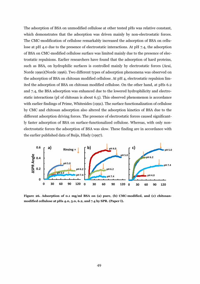
49
The adsorption of BSA on unmodified cellulose at other tested pHs was relative constant,
which demonstrates that the adsorption was driven mainly by non-electrostatic forces.
The CMC-modification of cellulose remarkably increased the adsorption of BSA on cellu-
lose at pH 4.0 due to the presence of electrostatic interactions. At pH 7.4, the adsorption
of BSA on CMC-modified cellulose surface was limited mainly due to the presence of elec-
trostatic repulsions. Earlier researchers have found that the adsorption of hard proteins,
such as BSA, on hydrophilic surfaces is controlled mainly by electrostatic forces (Arai,
Norde 1990)(Norde 1996). Two different types of adsorption phenomena was observed on
the adsorption of BSA on chitosan modified cellulose. At pH 4, electrostatic repulsion lim-
ited the adsorption of BSA on chitosan modified cellulose. On the other hand, at pHs 6.2
and 7.4, the BSA adsorption was enhanced due to the lowered hydrophilicity and electro-
static interactions (pI of chitosan is about 6.5). This observed phenomenon is accordance
with earlier findings of Prime, Whitesides (1991). The surface functionalization of cellulose
by CMC and chitosan adsorption also altered the adsorption kinetics of BSA due to the
different adsorption driving forces. The presence of electrostatic forces caused significant-
ly faster adsorption of BSA on surface-functionalized cellulose. Whereas, with only non-
electrostatic forces the adsorption of BSA was slow. These finding are in accordance with
the earlier published data of Buijs, Hlady (1997).
Figure 26. Adsorption of 0.1 mg/ml BSA on (a) pure, (b) CMC-modified, and (c) chitosan-
modified cellulose at pHs 4.0, 5.0, 6.2, and 7.4 by SPR. (Paper I).
0
0.2
0.4
0.6
0 30 60 90 120
ΔSPR
Ang
le
0 30 60 90 120 0 30 60 90 120
a) b) c)Rinsing =
pH 4.0
pH 5.0
pH 6.2
pH 7.4
pH 4.0
pH 5.0
pH 6.2
pH 7.4 pH 4.0
pH 5.0
pH 6.2
pH 7.4
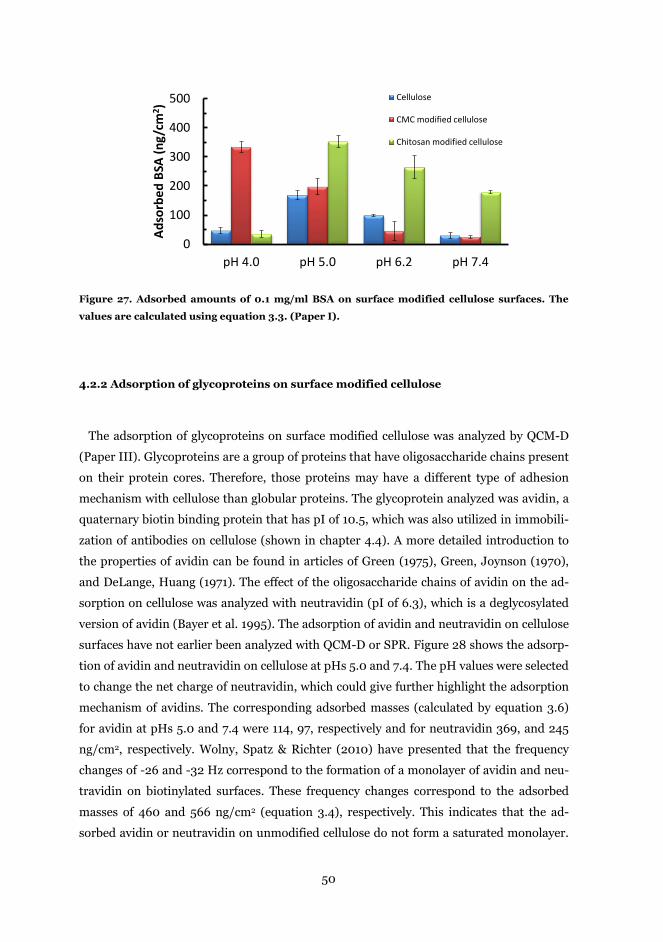
50
Figure 27. Adsorbed amounts of 0.1 mg/ml BSA on surface modified cellulose surfaces. The
values are calculated using equation 3.3. (Paper I).
4.2.2 Adsorption of glycoproteins on surface modified cellulose
The adsorption of glycoproteins on surface modified cellulose was analyzed by QCM-D
(Paper III). Glycoproteins are a group of proteins that have oligosaccharide chains present
on their protein cores. Therefore, those proteins may have a different type of adhesion
mechanism with cellulose than globular proteins. The glycoprotein analyzed was avidin, a
quaternary biotin binding protein that has pI of 10.5, which was also utilized in immobili-
zation of antibodies on cellulose (shown in chapter 4.4). A more detailed introduction to
the properties of avidin can be found in articles of Green (1975), Green, Joynson (1970),
and DeLange, Huang (1971). The effect of the oligosaccharide chains of avidin on the ad-
sorption on cellulose was analyzed with neutravidin (pI of 6.3), which is a deglycosylated
version of avidin (Bayer et al. 1995). The adsorption of avidin and neutravidin on cellulose
surfaces have not earlier been analyzed with QCM-D or SPR. Figure 28 shows the adsorp-
tion of avidin and neutravidin on cellulose at pHs 5.0 and 7.4. The pH values were selected
to change the net charge of neutravidin, which could give further highlight the adsorption
mechanism of avidins. The corresponding adsorbed masses (calculated by equation 3.6)
for avidin at pHs 5.0 and 7.4 were 114, 97, respectively and for neutravidin 369, and 245
ng/cm2, respectively. Wolny, Spatz & Richter (2010) have presented that the frequency
changes of -26 and -32 Hz correspond to the formation of a monolayer of avidin and neu-
travidin on biotinylated surfaces. These frequency changes correspond to the adsorbed
masses of 460 and 566 ng/cm2 (equation 3.4), respectively. This indicates that the ad-
sorbed avidin or neutravidin on unmodified cellulose do not form a saturated monolayer.
0
100
200
300
400
500
pH 4.0 pH 5.0 pH 6.2 pH 7.4
Adso
rbed
BSA
(ng/
cm2 )
Cellulose
CMC modified cellulose
Chitosan modified cellulose
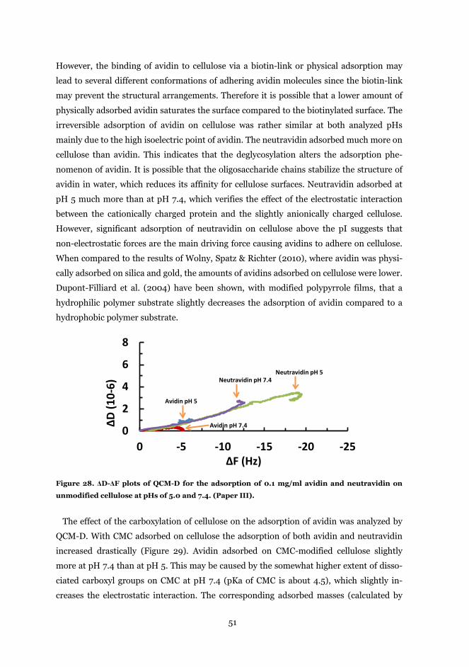
51
However, the binding of avidin to cellulose via a biotin-link or physical adsorption may
lead to several different conformations of adhering avidin molecules since the biotin-link
may prevent the structural arrangements. Therefore it is possible that a lower amount of
physically adsorbed avidin saturates the surface compared to the biotinylated surface. The
irreversible adsorption of avidin on cellulose was rather similar at both analyzed pHs
mainly due to the high isoelectric point of avidin. The neutravidin adsorbed much more on
cellulose than avidin. This indicates that the deglycosylation alters the adsorption phe-
nomenon of avidin. It is possible that the oligosaccharide chains stabilize the structure of
avidin in water, which reduces its affinity for cellulose surfaces. Neutravidin adsorbed at
pH 5 much more than at pH 7.4, which verifies the effect of the electrostatic interaction
between the cationically charged protein and the slightly anionically charged cellulose.
However, significant adsorption of neutravidin on cellulose above the pI suggests that
non-electrostatic forces are the main driving force causing avidins to adhere on cellulose.
When compared to the results of Wolny, Spatz & Richter (2010), where avidin was physi-
cally adsorbed on silica and gold, the amounts of avidins adsorbed on cellulose were lower.
Dupont-Filliard et al. (2004) have been shown, with modified polypyrrole films, that a
hydrophilic polymer substrate slightly decreases the adsorption of avidin compared to a
hydrophobic polymer substrate.
Figure 28. ΔD-ΔF plots of QCM-D for the adsorption of 0.1 mg/ml avidin and neutravidin on
unmodified cellulose at pHs of 5.0 and 7.4. (Paper III).
The effect of the carboxylation of cellulose on the adsorption of avidin was analyzed by
QCM-D. With CMC adsorbed on cellulose the adsorption of both avidin and neutravidin
increased drastically (Figure 29). Avidin adsorbed on CMC-modified cellulose slightly
more at pH 7.4 than at pH 5. This may be caused by the somewhat higher extent of disso-
ciated carboxyl groups on CMC at pH 7.4 (pKa of CMC is about 4.5), which slightly in-
creases the electrostatic interaction. The corresponding adsorbed masses (calculated by
0
2
4
6
8
-25-20-15-10-50
ΔD (1
0-6)
ΔF (Hz)
Avidin pH 5
Avidin pH 7.4
Neutravidin pH 5Neutravidin pH 7.4
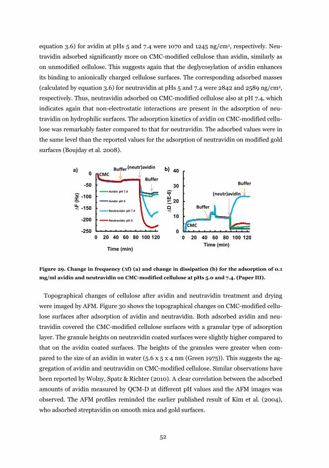
52
equation 3.6) for avidin at pHs 5 and 7.4 were 1070 and 1245 ng/cm2, respectively. Neu-
travidin adsorbed significantly more on CMC-modified cellulose than avidin, similarly as
on unmodified cellulose. This suggests again that the deglycosylation of avidin enhances
its binding to anionically charged cellulose surfaces. The corresponding adsorbed masses
(calculated by equation 3.6) for neutravidin at pHs 5 and 7.4 were 2842 and 2589 ng/cm2,
respectively. Thus, neutravidin adsorbed on CMC-modified cellulose also at pH 7.4, which
indicates again that non-electrostatic interactions are present in the adsorption of neu-
travidin on hydrophilic surfaces. The adsorption kinetics of avidin on CMC-modified cellu-
lose was remarkably faster compared to that for neutravidin. The adsorbed values were in
the same level than the reported values for the adsorption of neutravidin on modified gold
surfaces (Boujday et al. 2008).
Figure 29. Change in frequency (Δf) (a) and change in dissipation (b) for the adsorption of 0.1
mg/ml avidin and neutravidin on CMC-modified cellulose at pHs 5.0 and 7.4. (Paper III).
Topographical changes of cellulose after avidin and neutravidin treatment and drying
were imaged by AFM. Figure 30 shows the topographical changes on CMC-modified cellu-
lose surfaces after adsorption of avidin and neutravidin. Both adsorbed avidin and neu-
travidin covered the CMC-modified cellulose surfaces with a granular type of adsorption
layer. The granule heights on neutravidin coated surfaces were slightly higher compared to
that on the avidin coated surfaces. The heights of the granules were greater when com-
pared to the size of an avidin in water (5.6 x 5 x 4 nm (Green 1975)). This suggests the ag-
gregation of avidin and neutravidin on CMC-modified cellulose. Similar observations have
been reported by Wolny, Spatz & Richter (2010). A clear correlation between the adsorbed
amounts of avidin measured by QCM-D at different pH values and the AFM images was
observed. The AFM profiles reminded the earlier published result of Kim et al. (2004),
who adsorbed streptavidin on smooth mica and gold surfaces.
-250
-200
-150
-100
-50
0
0 20 40 60 80 100 120
∆F (H
z)
Time (min)
Avidin pH 7.4
Avidin pH 5
Neutravidin pH 7.4
Neutravidin pH 5
CMCBuffer (neutr)avidin
Buffer
0
10
20
30
40
0 20 40 60 80 100 120
∆D (1
E-6)
Time (min)
a)
CMC
Buffer
(neutr)avidin
Buffer
b)
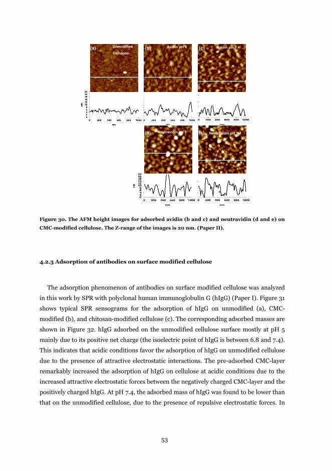
53
Figure 30. The AFM height images for adsorbed avidin (b and c) and neutravidin (d and e) on
CMC-modified cellulose. The Z-range of the images is 20 nm. (Paper II).
4.2.3 Adsorption of antibodies on surface modified cellulose
The adsorption phenomenon of antibodies on surface modified cellulose was analyzed
in this work by SPR with polyclonal human immunoglobulin G (hIgG) (Paper I). Figure 31
shows typical SPR sensograms for the adsorption of hIgG on unmodified (a), CMC-
modified (b), and chitosan-modified cellulose (c). The corresponding adsorbed masses are
shown in Figure 32. hIgG adsorbed on the unmodified cellulose surface mostly at pH 5
mainly due to its positive net charge (the isoelectric point of hIgG is between 6.8 and 7.4).
This indicates that acidic conditions favor the adsorption of hIgG on unmodified cellulose
due to the presence of attractive electrostatic interactions. The pre-adsorbed CMC-layer
remarkably increased the adsorption of hIgG on cellulose at acidic conditions due to the
increased attractive electrostatic forces between the negatively charged CMC-layer and the
positively charged hIgG. At pH 7.4, the adsorbed mass of hIgG was found to be lower than
that on the unmodified cellulose, due to the presence of repulsive electrostatic forces. In
1 x 1 μm
1 x 1 μm
(c)Unmodified
CelluloseAvidin pH 5 Avidin pH 7.4
Neutravidin pH 5 Neutravidin pH 7.4
(a) (b) (c)
(d) (e)
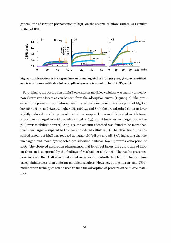
54
general, the adsorption phenomenon of hIgG on the anionic cellulose surface was similar
to that of BSA.
Figure 31. Adsorption of 0.1 mg/ml human immunoglobulin G on (a) pure, (b) CMC-modified,
and (c) chitosan-modified cellulose at pHs of 4.0, 5.0, 6.2, and 7.4 by SPR. (Paper I).
Surprisingly, the adsorption of hIgG on chitosan modified cellulose was mainly driven by
non-electrostatic forces as can be seen from the adsorption curves (Figure 31c). The pres-
ence of the pre-adsorbed chitosan layer dramatically increased the adsorption of hIgG at
low pH (pH 5.0 and 6.2). At higher pHs (pH 7.4 and 8.0), the pre-adsorbed chitosan layer
slightly reduced the adsorption of hIgG when compared to unmodified cellulose. Chitosan
is positively charged in acidic conditions (pI of 6.5), and it becomes uncharged above the
pI (lower solubility in water). At pH 5, the amount adsorbed was found to be more than
five times larger compared to that on unmodified cellulose. On the other hand, the ad-
sorbed amount of hIgG was reduced at higher pH (pH 7.4 and pH 8.0), indicating that the
uncharged and more hydrophobic pre-adsorbed chitosan layer prevents adsorption of
hIgG. The observed adsorption phenomenon that lower pH favors the adsorption of hIgG
on chitosan is supported by the findings of Machado et al. (2006). The results presented
here indicate that CMC-modified cellulose is more controllable platform for cellulose
based biointerfaces than chitosan-modified cellulose. However, both chitosan- and CMC-
modification techniques can be used to tune the adsorption of proteins on cellulosic mate-
rials.
0.0
0.4
0.8
1.2
1.6
0 20 40 60
ΔSPR
ang
le
0 20 40 60 0 30 60 90 120
a) b) c)
pH 8.0
pH 5.0pH 6.2pH 7.4
Rinsing =
pH 8.0
pH 5.0pH 6.2
pH 7.4
pH 8.0
pH 5.0
pH 6.2
pH 7.4O
rder
min
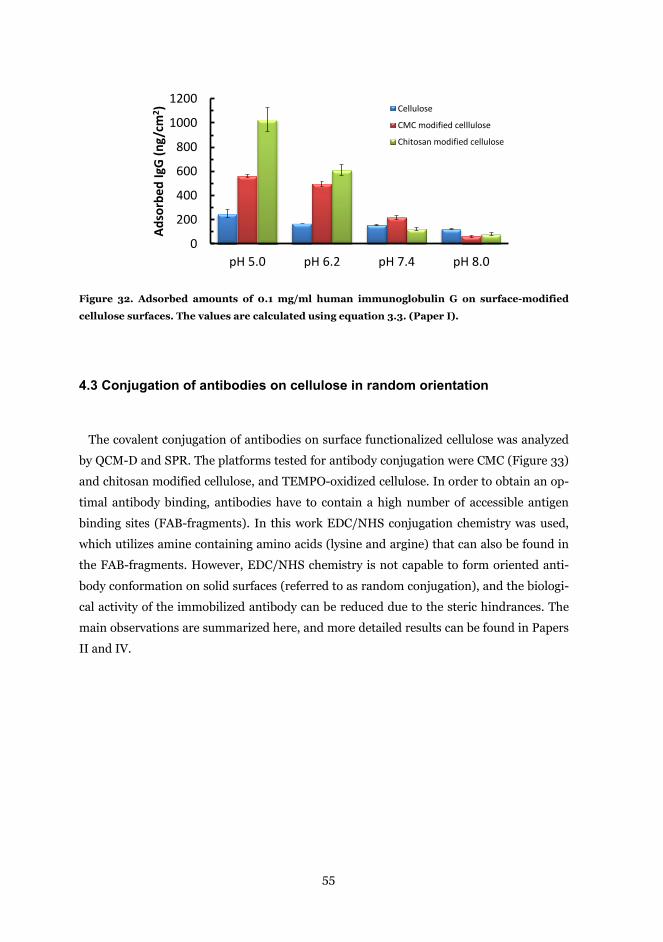
55
Figure 32. Adsorbed amounts of 0.1 mg/ml human immunoglobulin G on surface-modified
cellulose surfaces. The values are calculated using equation 3.3. (Paper I).
4.3 Conjugation of antibodies on cellulose in random orientation
The covalent conjugation of antibodies on surface functionalized cellulose was analyzed
by QCM-D and SPR. The platforms tested for antibody conjugation were CMC (Figure 33)
and chitosan modified cellulose, and TEMPO-oxidized cellulose. In order to obtain an op-
timal antibody binding, antibodies have to contain a high number of accessible antigen
binding sites (FAB-fragments). In this work EDC/NHS conjugation chemistry was used,
which utilizes amine containing amino acids (lysine and argine) that can also be found in
the FAB-fragments. However, EDC/NHS chemistry is not capable to form oriented anti-
body conformation on solid surfaces (referred to as random conjugation), and the biologi-
cal activity of the immobilized antibody can be reduced due to the steric hindrances. The
main observations are summarized here, and more detailed results can be found in Papers
II and IV.
0
200
400
600
800
1000
1200
pH 5.0 pH 6.2 pH 7.4 pH 8.0
Adso
rbed
IgG
(ng/
cm2 ) Cellulose
CMC modified celllulose
Chitosan modified cellulose
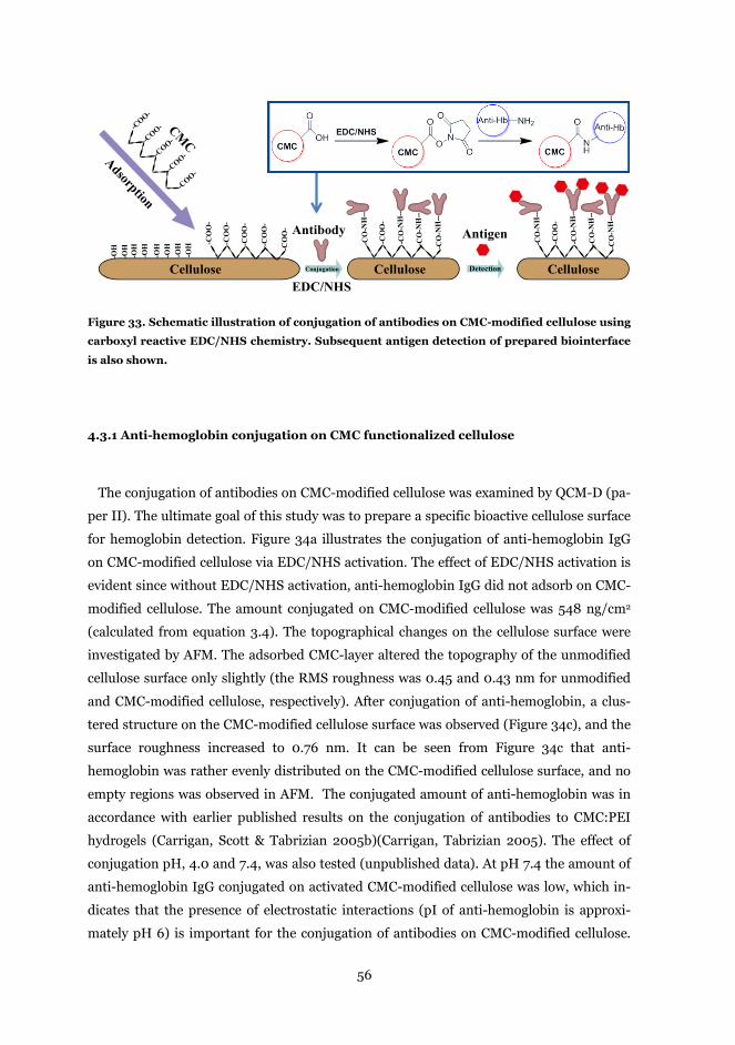
56
Figure 33. Schematic illustration of conjugation of antibodies on CMC-modified cellulose using
carboxyl reactive EDC/NHS chemistry. Subsequent antigen detection of prepared biointerface
is also shown.
4.3.1 Anti-hemoglobin conjugation on CMC functionalized cellulose
The conjugation of antibodies on CMC-modified cellulose was examined by QCM-D (pa-
per II). The ultimate goal of this study was to prepare a specific bioactive cellulose surface
for hemoglobin detection. Figure 34a illustrates the conjugation of anti-hemoglobin IgG
on CMC-modified cellulose via EDC/NHS activation. The effect of EDC/NHS activation is
evident since without EDC/NHS activation, anti-hemoglobin IgG did not adsorb on CMC-
modified cellulose. The amount conjugated on CMC-modified cellulose was 548 ng/cm2
(calculated from equation 3.4). The topographical changes on the cellulose surface were
investigated by AFM. The adsorbed CMC-layer altered the topography of the unmodified
cellulose surface only slightly (the RMS roughness was 0.45 and 0.43 nm for unmodified
and CMC-modified cellulose, respectively). After conjugation of anti-hemoglobin, a clus-
tered structure on the CMC-modified cellulose surface was observed (Figure 34c), and the
surface roughness increased to 0.76 nm. It can be seen from Figure 34c that anti-
hemoglobin was rather evenly distributed on the CMC-modified cellulose surface, and no
empty regions was observed in AFM. The conjugated amount of anti-hemoglobin was in
accordance with earlier published results on the conjugation of antibodies to CMC:PEI
hydrogels (Carrigan, Scott & Tabrizian 2005b)(Carrigan, Tabrizian 2005). The effect of
conjugation pH, 4.0 and 7.4, was also tested (unpublished data). At pH 7.4 the amount of
anti-hemoglobin IgG conjugated on activated CMC-modified cellulose was low, which in-
dicates that the presence of electrostatic interactions (pI of anti-hemoglobin is approxi-
mately pH 6) is important for the conjugation of antibodies on CMC-modified cellulose.
Cellulose Cellulose
-COO-
-COO-
-COO-
-COO-
-COO-
Adsorption
CMC
Cellulose CelluloseCellulose
-CO
O-
-CO
O-
-CO
O-
-CO
O-
-CO
O-
-CO
-NH
--
-OH
-OH
-OH
-OH
-OH
-OH
-OH
-OH
EDC/NHSConjugation
Antibody Antigen
Detection
-CO
-NH
--
-CO
-NH
--
-CO
-NH
--
-CO
O-
-CO
-NH
--
-CO
-NH
--
-CO
-NH
--
-CO
-NH
--
-CO
O-
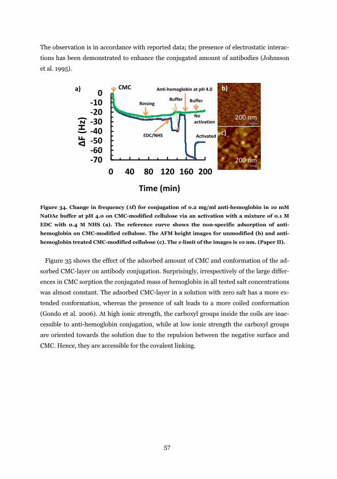
57
The observation is in accordance with reported data; the presence of electrostatic interac-
tions has been demonstrated to enhance the conjugated amount of antibodies (Johnsson
et al. 1995).
Figure 34. Change in frequency (Δf) for conjugation of 0.2 mg/ml anti-hemoglobin in 10 mM
NaOAc buffer at pH 4.0 on CMC-modified cellulose via an activation with a mixture of 0.1 M
EDC with 0.4 M NHS (a). The reference curve shows the non-specific adsorption of anti-
hemoglobin on CMC-modified cellulose. The AFM height images for unmodified (b) and anti-
hemoglobin treated CMC-modified cellulose (c). The z-limit of the images is 10 nm. (Paper II).
Figure 35 shows the effect of the adsorbed amount of CMC and conformation of the ad-
sorbed CMC-layer on antibody conjugation. Surprisingly, irrespectively of the large differ-
ences in CMC sorption the conjugated mass of hemoglobin in all tested salt concentrations
was almost constant. The adsorbed CMC-layer in a solution with zero salt has a more ex-
tended conformation, whereas the presence of salt leads to a more coiled conformation
(Gondo et al. 2006). At high ionic strength, the carboxyl groups inside the coils are inac-
cessible to anti-hemoglobin conjugation, while at low ionic strength the carboxyl groups
are oriented towards the solution due to the repulsion between the negative surface and
CMC. Hence, they are accessible for the covalent linking.
-70-60-50-40-30-20-10
0
0 40 80 120 160 200
∆F (H
z)
Time (min)
CMC
Rinsing
EDC/NHS
Anti-hemoglobin at pH 4.0
No activation
Activated
Buffer Buffer
200 nm
200 nm
a) b)
c)
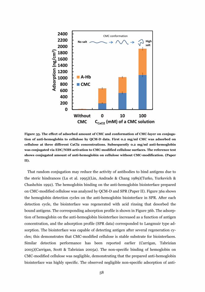
58
Figure 35. The effect of adsorbed amount of CMC and conformation of CMC-layer on conjuga-
tion of anti-hemoglobin to cellulose by QCM-D data. First 0.2 mg/ml CMC was adsorbed on
cellulose at three different CaCl2 concentrations. Subsequently 0.2 mg/ml anti-hemoglobin
was conjugated via EDC/NHS activation to CMC-modified cellulose surfaces. The reference test
shows conjugated amount of anti-hemoglobin on cellulose without CMC-modification. (Paper
II).
That random conjugation may reduce the activity of antibodies to bind antigens due to
the steric hindrances (Lu et al. 1995)(Lin, Andrade & Chang 1989)(Turko, Yurkevich &
Chashchin 1992). The hemoglobin binding on the anti-hemoglobin biointerface prepared
on CMC-modified cellulose was analyzed by QCM-D and SPR (Paper II). Figure 36a shows
the hemoglobin detection cycles on the anti-hemoglobin biointerface in SPR. After each
detection cycle, the biointerface was regenerated with acid rinsing that desorbed the
bound antigens. The corresponding adsorption profile is shown in Figure 36b. The adsorp-
tion of hemoglobin on the anti-hemoglobin biointerface increased as a function of antigen
concentration, and the adsorption profile (SPR data) corresponded to Langmuir type ad-
sorption. The biointerface was capable of detecting antigen after several regeneration cy-
cles; this demonstrates that CMC-modified cellulose is stable substrate for biointerfaces.
Similar detection performance has been reported earlier (Carrigan, Tabrizian
2005)(Carrigan, Scott & Tabrizian 2005a). The non-specific binding of hemoglobin on
CMC-modified cellulose was negligible, demonstrating that the prepared anti-hemoglobin
biointerface was highly specific. The observed negligible non-specific adsorption of anti-
0200400600800
10001200140016001800200022002400
WithoutCMC
0 10 100
Adso
rptio
n (n
g/cm
2 )
CCaCl2 (mM) of a CMC solution
A-HbCMC
CMC conformation
No salt High salt
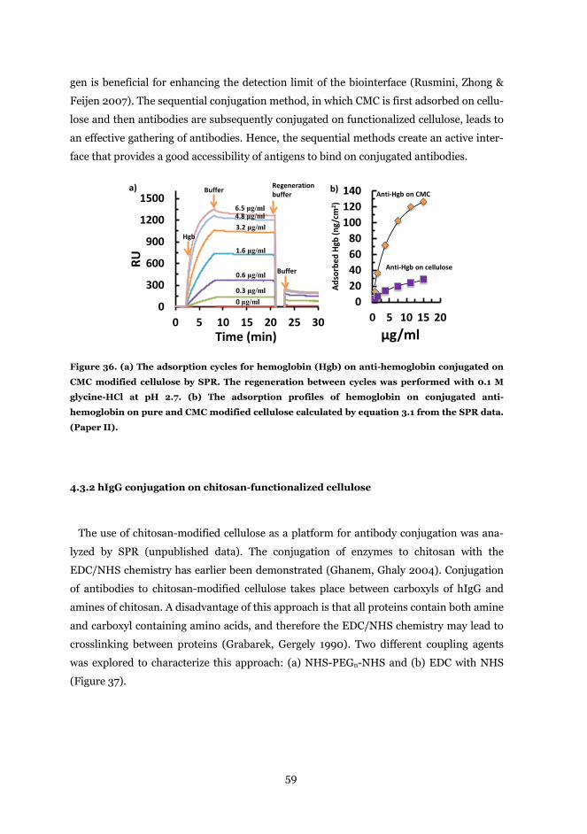
59
gen is beneficial for enhancing the detection limit of the biointerface (Rusmini, Zhong &
Feijen 2007). The sequential conjugation method, in which CMC is first adsorbed on cellu-
lose and then antibodies are subsequently conjugated on functionalized cellulose, leads to
an effective gathering of antibodies. Hence, the sequential methods create an active inter-
face that provides a good accessibility of antigens to bind on conjugated antibodies.
Figure 36. (a) The adsorption cycles for hemoglobin (Hgb) on anti-hemoglobin conjugated on
CMC modified cellulose by SPR. The regeneration between cycles was performed with 0.1 M
glycine-HCl at pH 2.7. (b) The adsorption profiles of hemoglobin on conjugated anti-
hemoglobin on pure and CMC modified cellulose calculated by equation 3.1 from the SPR data.
(Paper II).
4.3.2 hIgG conjugation on chitosan-functionalized cellulose
The use of chitosan-modified cellulose as a platform for antibody conjugation was ana-
lyzed by SPR (unpublished data). The conjugation of enzymes to chitosan with the
EDC/NHS chemistry has earlier been demonstrated (Ghanem, Ghaly 2004). Conjugation
of antibodies to chitosan-modified cellulose takes place between carboxyls of hIgG and
amines of chitosan. A disadvantage of this approach is that all proteins contain both amine
and carboxyl containing amino acids, and therefore the EDC/NHS chemistry may lead to
crosslinking between proteins (Grabarek, Gergely 1990). Two different coupling agents
was explored to characterize this approach: (a) NHS-PEGn-NHS and (b) EDC with NHS
(Figure 37).
0
300
600
900
1200
1500
0 5 10 15 20 25 30
RU
Time (min)
0 μg/ml0.3 μg/ml
0.6 μg/ml
1.6 μg/ml
3.2 μg/ml4.8 μg/ml6.5 μg/ml
Buffer
Buffer
Regeneration buffer
Hgb
020406080
100120140
0 5 10 15 20Ad
sorb
ed H
gb(n
g/cm
2 )µg/ml
Anti-Hgb on CMC
Anti-Hgb on cellulose
a) b)
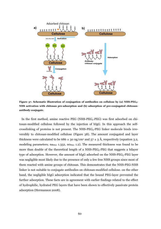
60
Figure 37. Schematic illustration of conjugation of antibodies on cellulose by (a) NHS-PEG5-
NHS activation with chitosan pre-adsorption and (b) adsorption of pre-conjugated chitosan-
antibody conjugate.
In the first method, amine reactive PEG (NHS-PEG5-PEG) was first adsorbed on chi-
tosan-modified cellulose followed by the injection of hIgG. In this approach the self-
crosslinking of proteins is not present. The NHS-PEG5-PEG linker molecule binds irre-
versibly to chitosan-modified cellulose (Figure 38). The amount conjugated and layer
thickness were calculated to be 686 ± 30 ng/cm2 and 57 ± 3 Å, respectively (equation 3.2,
modeling parameters; naPEG 1.352, n0PEG 1.2). The measured thickness was found to be
more than double of the theoretical length of a NHS-PEG5-PEG that suggests a bilayer
type of adsorption. However, the amount of hIgG adsorbed on the NHS-PEG5-PEG layer
was negligible most likely due to the presence of only a few free NHS groups since most of
them reacted with amine groups of chitosan. This demonstrates that the NHS-PEG-NHS
linker is not suitable to conjugate antibodies on chitosan-modified cellulose. on the other
hand, the negligible hIgG adsorption indicated that the bound PEG-layer prevented the
further adsorption. These facts are in agreement with earlier findings related to the effect
of hydrophilic, hydrated PEG layers that have been shown to effectively passivate protein
adsorption (Hermanson 2008).
Adsorbed chitosan
Cellulose
CelluloseCellulose
CelluloseCellulose
-NH
3+
-NH
3+
-NH
3+
-NH
3+
-NH
3+
-NH
-
-NH
2
-NH
-
-NH
2
-NH
-
-NH
-
-NH
2
-NH
-
-NH
2
--NH
-
-OH
-OH
-OH
-OH
-OH
-OH
-OH
-OH
Activation
Conjugation
-NH
--
-NH
--
-NH
--
-NH
--
-NH
--
Cellulose
Adsorption
-OH
-OH
-OH
-OH
-OH
-OH
-OH
-OH
Antibody-chitosan conjugate
a) b)
NHS-PEG-NHS
NHS NHS NHS
Antibody
-NH
--
-NH
--
-NH
--
-NH
--
-NH
--
Cellulose
-OH
-OH
-OH
-OH
-OH
-OH
-OH
-OH
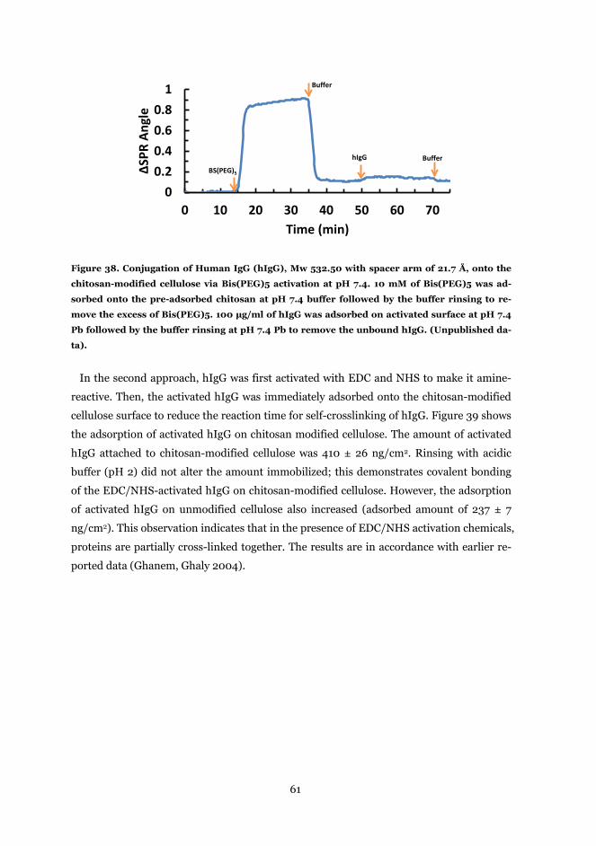
61
Figure 38. Conjugation of Human IgG (hIgG), Mw 532.50 with spacer arm of 21.7 Å, onto the
chitosan-modified cellulose via Bis(PEG)5 activation at pH 7.4. 10 mM of Bis(PEG)5 was ad-
sorbed onto the pre-adsorbed chitosan at pH 7.4 buffer followed by the buffer rinsing to re-
move the excess of Bis(PEG)5. 100 μg/ml of hIgG was adsorbed on activated surface at pH 7.4
Pb followed by the buffer rinsing at pH 7.4 Pb to remove the unbound hIgG. (Unpublished da-
ta).
In the second approach, hIgG was first activated with EDC and NHS to make it amine-
reactive. Then, the activated hIgG was immediately adsorbed onto the chitosan-modified
cellulose surface to reduce the reaction time for self-crosslinking of hIgG. Figure 39 shows
the adsorption of activated hIgG on chitosan modified cellulose. The amount of activated
hIgG attached to chitosan-modified cellulose was 410 ± 26 ng/cm2. Rinsing with acidic
buffer (pH 2) did not alter the amount immobilized; this demonstrates covalent bonding
of the EDC/NHS-activated hIgG on chitosan-modified cellulose. However, the adsorption
of activated hIgG on unmodified cellulose also increased (adsorbed amount of 237 ± 7
ng/cm2). This observation indicates that in the presence of EDC/NHS activation chemicals,
proteins are partially cross-linked together. The results are in accordance with earlier re-
ported data (Ghanem, Ghaly 2004).
00.20.40.60.8
1
0 10 20 30 40 50 60 70
∆SPR
Ang
le
Time (min)
BS(PEG)5
Buffer
BufferhIgG
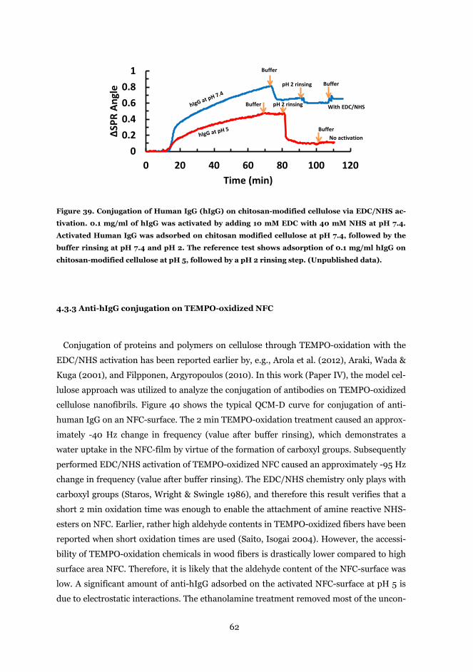
62
Figure 39. Conjugation of Human IgG (hIgG) on chitosan-modified cellulose via EDC/NHS ac-
tivation. 0.1 mg/ml of hIgG was activated by adding 10 mM EDC with 40 mM NHS at pH 7.4.
Activated Human IgG was adsorbed on chitosan modified cellulose at pH 7.4, followed by the
buffer rinsing at pH 7.4 and pH 2. The reference test shows adsorption of 0.1 mg/ml hIgG on
chitosan-modified cellulose at pH 5, followed by a pH 2 rinsing step. (Unpublished data).
4.3.3 Anti-hIgG conjugation on TEMPO-oxidized NFC
Conjugation of proteins and polymers on cellulose through TEMPO-oxidation with the
EDC/NHS activation has been reported earlier by, e.g., Arola et al. (2012), Araki, Wada &
Kuga (2001), and Filpponen, Argyropoulos (2010). In this work (Paper IV), the model cel-
lulose approach was utilized to analyze the conjugation of antibodies on TEMPO-oxidized
cellulose nanofibrils. Figure 40 shows the typical QCM-D curve for conjugation of anti-
human IgG on an NFC-surface. The 2 min TEMPO-oxidation treatment caused an approx-
imately -40 Hz change in frequency (value after buffer rinsing), which demonstrates a
water uptake in the NFC-film by virtue of the formation of carboxyl groups. Subsequently
performed EDC/NHS activation of TEMPO-oxidized NFC caused an approximately -95 Hz
change in frequency (value after buffer rinsing). The EDC/NHS chemistry only plays with
carboxyl groups (Staros, Wright & Swingle 1986), and therefore this result verifies that a
short 2 min oxidation time was enough to enable the attachment of amine reactive NHS-
esters on NFC. Earlier, rather high aldehyde contents in TEMPO-oxidized fibers have been
reported when short oxidation times are used (Saito, Isogai 2004). However, the accessi-
bility of TEMPO-oxidation chemicals in wood fibers is drastically lower compared to high
surface area NFC. Therefore, it is likely that the aldehyde content of the NFC-surface was
low. A significant amount of anti-hIgG adsorbed on the activated NFC-surface at pH 5 is
due to electrostatic interactions. The ethanolamine treatment removed most of the uncon-
00.20.40.60.8
1
0 20 40 60 80 100 120
∆SPR
Ang
le
Time (min)
Buffer
pH 2 rinsing Buffer
Buffer pH 2 rinsing
Buffer
With EDC/NHS
No activation
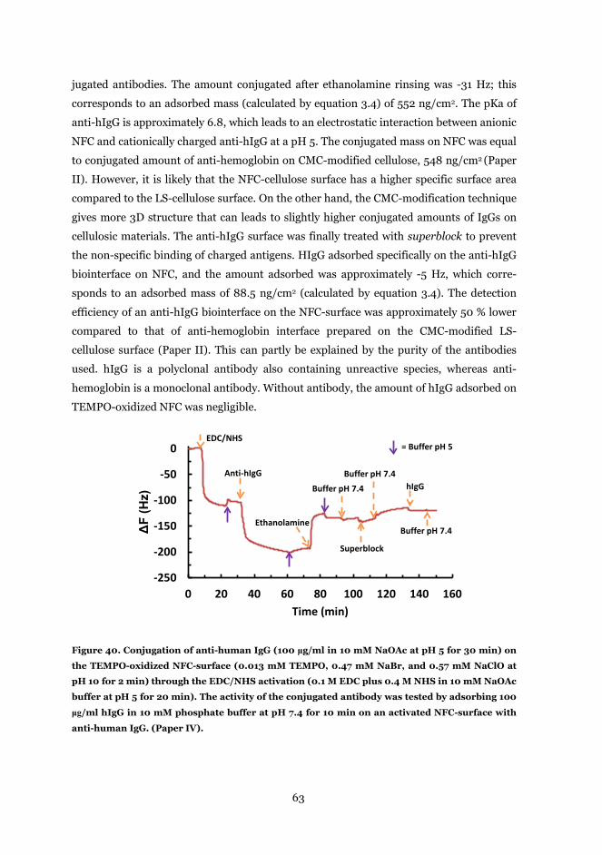
63
jugated antibodies. The amount conjugated after ethanolamine rinsing was -31 Hz; this
corresponds to an adsorbed mass (calculated by equation 3.4) of 552 ng/cm2. The pKa of
anti-hIgG is approximately 6.8, which leads to an electrostatic interaction between anionic
NFC and cationically charged anti-hIgG at a pH 5. The conjugated mass on NFC was equal
to conjugated amount of anti-hemoglobin on CMC-modified cellulose, 548 ng/cm2 (Paper
II). However, it is likely that the NFC-cellulose surface has a higher specific surface area
compared to the LS-cellulose surface. On the other hand, the CMC-modification technique
gives more 3D structure that can leads to slightly higher conjugated amounts of IgGs on
cellulosic materials. The anti-hIgG surface was finally treated with superblock to prevent
the non-specific binding of charged antigens. HIgG adsorbed specifically on the anti-hIgG
biointerface on NFC, and the amount adsorbed was approximately -5 Hz, which corre-
sponds to an adsorbed mass of 88.5 ng/cm2 (calculated by equation 3.4). The detection
efficiency of an anti-hIgG biointerface on the NFC-surface was approximately 50 % lower
compared to that of anti-hemoglobin interface prepared on the CMC-modified LS-
cellulose surface (Paper II). This can partly be explained by the purity of the antibodies
used. hIgG is a polyclonal antibody also containing unreactive species, whereas anti-
hemoglobin is a monoclonal antibody. Without antibody, the amount of hIgG adsorbed on
TEMPO-oxidized NFC was negligible.
Figure 40. Conjugation of anti-human IgG (100 μg/ml in 10 mM NaOAc at pH 5 for 30 min) on
the TEMPO-oxidized NFC-surface (0.013 mM TEMPO, 0.47 mM NaBr, and 0.57 mM NaClO at
pH 10 for 2 min) through the EDC/NHS activation (0.1 M EDC plus 0.4 M NHS in 10 mM NaOAc
buffer at pH 5 for 20 min). The activity of the conjugated antibody was tested by adsorbing 100
μg/ml hIgG in 10 mM phosphate buffer at pH 7.4 for 10 min on an activated NFC-surface with
anti-human IgG. (Paper IV).
-250
-200
-150
-100
-50
0
0 20 40 60 80 100 120 140 160
ΔF
(Hz)
Time (min)
EDC/NHS
Anti-hIgG
Ethanolamine
Superblock
Buffer pH 7.4hIgG
= Buffer pH 5
Buffer pH 7.4
Buffer pH 7.4
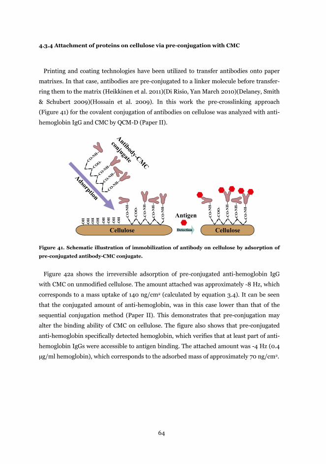
64
4.3.4 Attachment of proteins on cellulose via pre-conjugation with CMC
Printing and coating technologies have been utilized to transfer antibodies onto paper
matrixes. In that case, antibodies are pre-conjugated to a linker molecule before transfer-
ring them to the matrix (Heikkinen et al. 2011)(Di Risio, Yan March 2010)(Delaney, Smith
& Schubert 2009)(Hossain et al. 2009). In this work the pre-crosslinking approach
(Figure 41) for the covalent conjugation of antibodies on cellulose was analyzed with anti-
hemoglobin IgG and CMC by QCM-D (Paper II).
Figure 41. Schematic illustration of immobilization of antibody on cellulose by adsorption of
pre-conjugated antibody-CMC conjugate.
Figure 42a shows the irreversible adsorption of pre-conjugated anti-hemoglobin IgG
with CMC on unmodified cellulose. The amount attached was approximately -8 Hz, which
corresponds to a mass uptake of 140 ng/cm2 (calculated by equation 3.4). It can be seen
that the conjugated amount of anti-hemoglobin, was in this case lower than that of the
sequential conjugation method (Paper II). This demonstrates that pre-conjugation may
alter the binding ability of CMC on cellulose. The figure also shows that pre-conjugated
anti-hemoglobin specifically detected hemoglobin, which verifies that at least part of anti-
hemoglobin IgGs were accessible to antigen binding. The attached amount was -4 Hz (0.4
µg/ml hemoglobin), which corresponds to the adsorbed mass of approximately 70 ng/cm2.
Cellulose
AdsorptionCelluloseCellulose
-OH
-OH
-OH
-OH
-OH
-OH
-OH
-OH
Detection
Antibody-CMC
conjugate
Antigen
-CO-N
H--
-CO-N
H--
-CO-N
H--
-CO-N
H--
-COO-
-CO
-NH
--
-CO
-NH
--
-CO
-NH
--
-CO
-NH
--
-CO
O-
-CO
-NH
--
-CO
-NH
--
-CO
-NH
--
-CO
-NH
--
-CO
O-
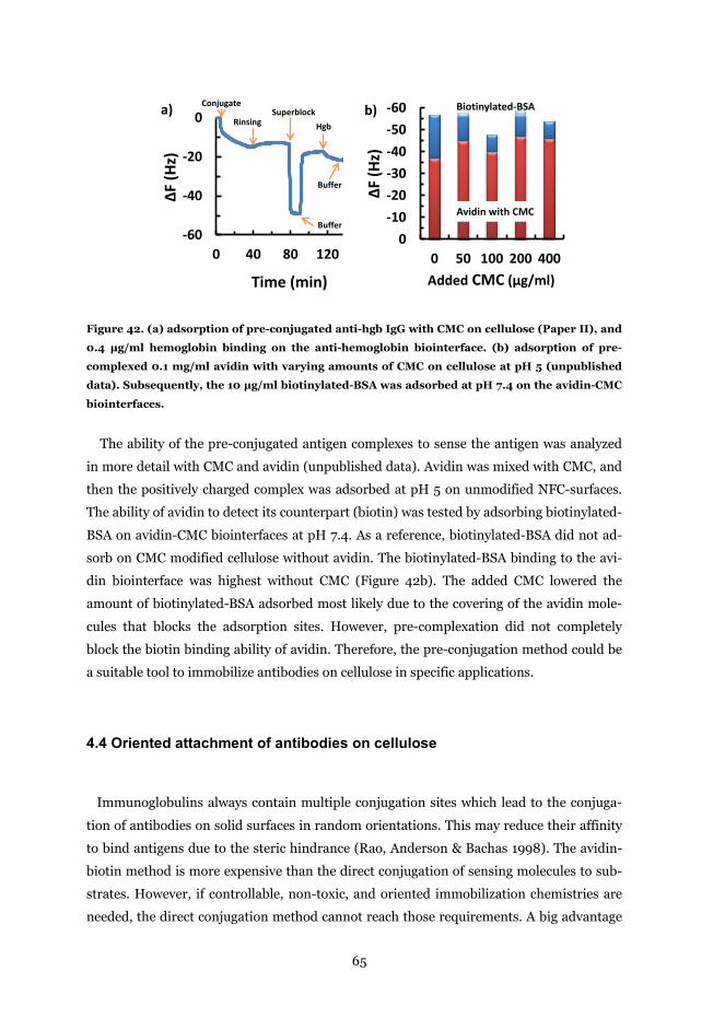
65
Figure 42. (a) adsorption of pre-conjugated anti-hgb IgG with CMC on cellulose (Paper II), and
0.4 µg/ml hemoglobin binding on the anti-hemoglobin biointerface. (b) adsorption of pre-
complexed 0.1 mg/ml avidin with varying amounts of CMC on cellulose at pH 5 (unpublished
data). Subsequently, the 10 µg/ml biotinylated-BSA was adsorbed at pH 7.4 on the avidin-CMC
biointerfaces.
The ability of the pre-conjugated antigen complexes to sense the antigen was analyzed
in more detail with CMC and avidin (unpublished data). Avidin was mixed with CMC, and
then the positively charged complex was adsorbed at pH 5 on unmodified NFC-surfaces.
The ability of avidin to detect its counterpart (biotin) was tested by adsorbing biotinylated-
BSA on avidin-CMC biointerfaces at pH 7.4. As a reference, biotinylated-BSA did not ad-
sorb on CMC modified cellulose without avidin. The biotinylated-BSA binding to the avi-
din biointerface was highest without CMC (Figure 42b). The added CMC lowered the
amount of biotinylated-BSA adsorbed most likely due to the covering of the avidin mole-
cules that blocks the adsorption sites. However, pre-complexation did not completely
block the biotin binding ability of avidin. Therefore, the pre-conjugation method could be
a suitable tool to immobilize antibodies on cellulose in specific applications.
4.4 Oriented attachment of antibodies on cellulose
Immunoglobulins always contain multiple conjugation sites which lead to the conjuga-
tion of antibodies on solid surfaces in random orientations. This may reduce their affinity
to bind antigens due to the steric hindrance (Rao, Anderson & Bachas 1998). The avidin-
biotin method is more expensive than the direct conjugation of sensing molecules to sub-
strates. However, if controllable, non-toxic, and oriented immobilization chemistries are
needed, the direct conjugation method cannot reach those requirements. A big advantage
-60
-40
-20
0
0 40 80 120
∆F (H
z)
Time (min)
Conjugate
RinsingSuperblock
Buffer
Hgb
-60-50-40-30-20-10
00 50 100 200 400
ΔF (H
z)
Added CMC (µg/ml)
a) b)
Avidin with CMC
Biotinylated-BSA
Buffer
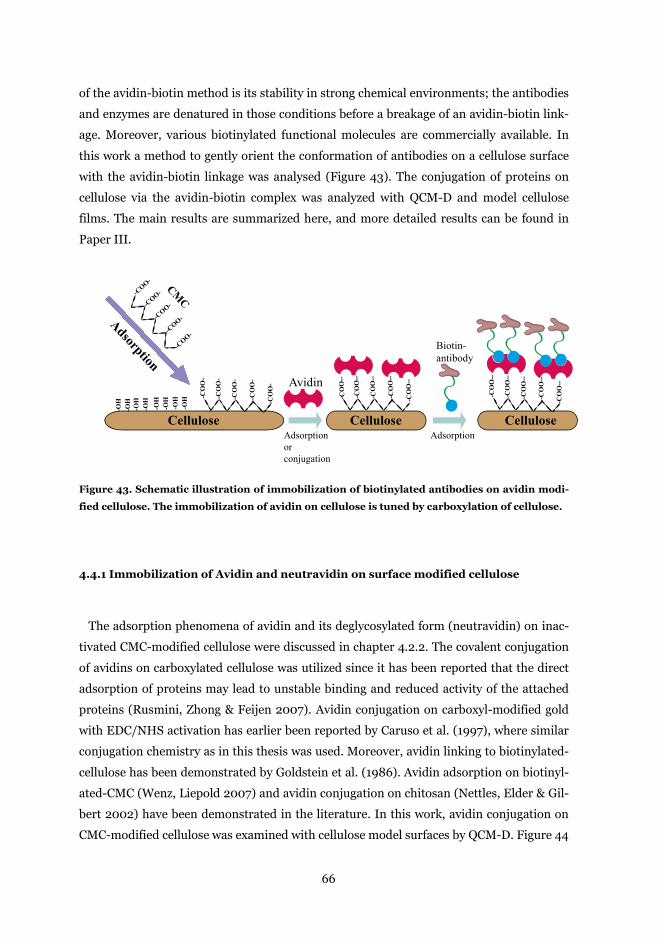
66
of the avidin-biotin method is its stability in strong chemical environments; the antibodies
and enzymes are denatured in those conditions before a breakage of an avidin-biotin link-
age. Moreover, various biotinylated functional molecules are commercially available. In
this work a method to gently orient the conformation of antibodies on a cellulose surface
with the avidin-biotin linkage was analysed (Figure 43). The conjugation of proteins on
cellulose via the avidin-biotin complex was analyzed with QCM-D and model cellulose
films. The main results are summarized here, and more detailed results can be found in
Paper III.
Figure 43. Schematic illustration of immobilization of biotinylated antibodies on avidin modi-
fied cellulose. The immobilization of avidin on cellulose is tuned by carboxylation of cellulose.
4.4.1 Immobilization of Avidin and neutravidin on surface modified cellulose
The adsorption phenomena of avidin and its deglycosylated form (neutravidin) on inac-
tivated CMC-modified cellulose were discussed in chapter 4.2.2. The covalent conjugation
of avidins on carboxylated cellulose was utilized since it has been reported that the direct
adsorption of proteins may lead to unstable binding and reduced activity of the attached
proteins (Rusmini, Zhong & Feijen 2007). Avidin conjugation on carboxyl-modified gold
with EDC/NHS activation has earlier been reported by Caruso et al. (1997), where similar
conjugation chemistry as in this thesis was used. Moreover, avidin linking to biotinylated-
cellulose has been demonstrated by Goldstein et al. (1986). Avidin adsorption on biotinyl-
ated-CMC (Wenz, Liepold 2007) and avidin conjugation on chitosan (Nettles, Elder & Gil-
bert 2002) have been demonstrated in the literature. In this work, avidin conjugation on
CMC-modified cellulose was examined with cellulose model surfaces by QCM-D. Figure 44
Cellulose Cellulose
-COO-
-COO-
-COO-
-COO-
-COO-
AdsorptionCM
C
Cellulose CelluloseCellulose
-CO
O-
-CO
O-
-CO
O-
-CO
O-
-CO
O-
-CO
O--
-CO
O--
-CO
O--
-CO
O--
-CO
O--
-CO
O--
-CO
O--
-CO
O--
-CO
O--
-CO
O--
-OH
-OH
-OH
-OH
-OH
-OH
-OH
-OH
Adsorptionorconjugation
Avidin
Adsorption
Biotin-antibody
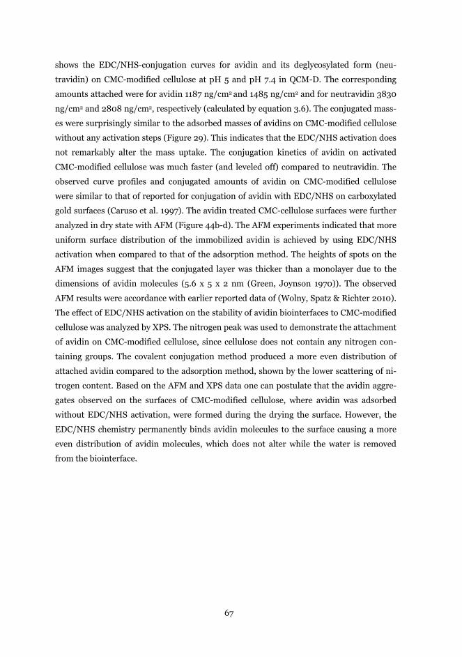
67
shows the EDC/NHS-conjugation curves for avidin and its deglycosylated form (neu-
travidin) on CMC-modified cellulose at pH 5 and pH 7.4 in QCM-D. The corresponding
amounts attached were for avidin 1187 ng/cm2 and 1485 ng/cm2 and for neutravidin 3830
ng/cm2 and 2808 ng/cm2, respectively (calculated by equation 3.6). The conjugated mass-
es were surprisingly similar to the adsorbed masses of avidins on CMC-modified cellulose
without any activation steps (Figure 29). This indicates that the EDC/NHS activation does
not remarkably alter the mass uptake. The conjugation kinetics of avidin on activated
CMC-modified cellulose was much faster (and leveled off) compared to neutravidin. The
observed curve profiles and conjugated amounts of avidin on CMC-modified cellulose
were similar to that of reported for conjugation of avidin with EDC/NHS on carboxylated
gold surfaces (Caruso et al. 1997). The avidin treated CMC-cellulose surfaces were further
analyzed in dry state with AFM (Figure 44b-d). The AFM experiments indicated that more
uniform surface distribution of the immobilized avidin is achieved by using EDC/NHS
activation when compared to that of the adsorption method. The heights of spots on the
AFM images suggest that the conjugated layer was thicker than a monolayer due to the
dimensions of avidin molecules (5.6 x 5 x 2 nm (Green, Joynson 1970)). The observed
AFM results were accordance with earlier reported data of (Wolny, Spatz & Richter 2010).
The effect of EDC/NHS activation on the stability of avidin biointerfaces to CMC-modified
cellulose was analyzed by XPS. The nitrogen peak was used to demonstrate the attachment
of avidin on CMC-modified cellulose, since cellulose does not contain any nitrogen con-
taining groups. The covalent conjugation method produced a more even distribution of
attached avidin compared to the adsorption method, shown by the lower scattering of ni-
trogen content. Based on the AFM and XPS data one can postulate that the avidin aggre-
gates observed on the surfaces of CMC-modified cellulose, where avidin was adsorbed
without EDC/NHS activation, were formed during the drying the surface. However, the
EDC/NHS chemistry permanently binds avidin molecules to the surface causing a more
even distribution of avidin molecules, which does not alter while the water is removed
from the biointerface.
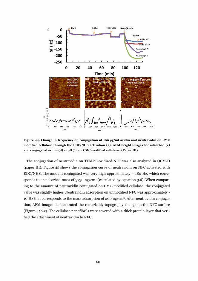
68
Figure 44. Change in frequency on conjugation of 100 μg/ml avidin and neutravidin on CMC
modified cellulose through the EDC/NHS activation (a). AFM height images for adsorbed (c)
and conjugated avidin (d) at pH 7.4 on CMC modified cellulose. (Paper III).
The conjugation of neutravidin on TEMPO-oxidized NFC was also analyzed in QCM-D
(paper III). Figure 45 shows the conjugation curve of neutravidin on NFC activated with
EDC/NHS. The amount conjugated was very high approximately – 180 Hz, which corre-
sponds to an adsorbed mass of 3730 ng/cm2 (calculated by equation 3.6). When compar-
ing to the amount of neutravidin conjugated on CMC-modified cellulose, the conjugated
value was slightly higher. Neutravidin adsorption on unmodified NFC was approximately -
10 Hz that corresponds to the mass adsorption of 200 ng/cm2. After neutravidin conjuga-
tion, AFM images demonstrated the remarkably topography change on the NFC surface
(Figure 45b-c). The cellulose nanofibrils were covered with a thick protein layer that veri-
fied the attachment of neutravidin to NFC.
Avidin pH 7.4 Conjugated on CMC-mod cellulose.
(d)(c)
Adsorbed on CMC-mod. cellulose
(c)
-250-200-150-100
-500
0 20 40 60 80 100 120
∆F (H
z)
Time (min)
Ne.avidin pH 5
Ne.avidin pH 7.4
Avidin pH 7.4
Avidin pH 5
CMC Buffer (Neutr)Avidin
Buffer
EDC/NHS
Ref. CMC-modified cellulose
(b)
a)
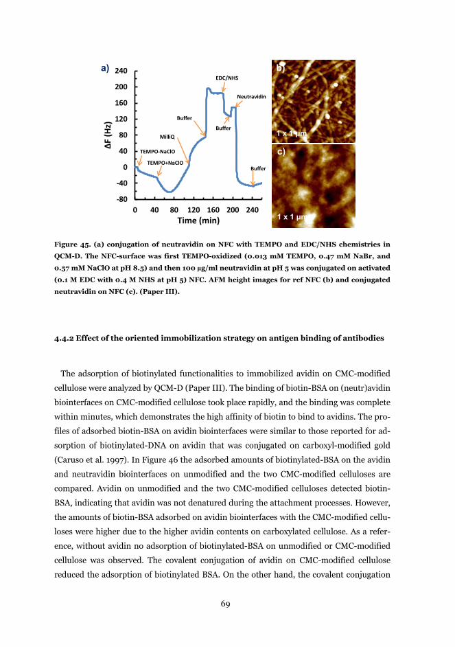
69
Figure 45. (a) conjugation of neutravidin on NFC with TEMPO and EDC/NHS chemistries in
QCM-D. The NFC-surface was first TEMPO-oxidized (0.013 mM TEMPO, 0.47 mM NaBr, and
0.57 mM NaClO at pH 8.5) and then 100 μg/ml neutravidin at pH 5 was conjugated on activated
(0.1 M EDC with 0.4 M NHS at pH 5) NFC. AFM height images for ref NFC (b) and conjugated
neutravidin on NFC (c). (Paper III).
4.4.2 Effect of the oriented immobilization strategy on antigen binding of antibodies
The adsorption of biotinylated functionalities to immobilized avidin on CMC-modified
cellulose were analyzed by QCM-D (Paper III). The binding of biotin-BSA on (neutr)avidin
biointerfaces on CMC-modified cellulose took place rapidly, and the binding was complete
within minutes, which demonstrates the high affinity of biotin to bind to avidins. The pro-
files of adsorbed biotin-BSA on avidin biointerfaces were similar to those reported for ad-
sorption of biotinylated-DNA on avidin that was conjugated on carboxyl-modified gold
(Caruso et al. 1997). In Figure 46 the adsorbed amounts of biotinylated-BSA on the avidin
and neutravidin biointerfaces on unmodified and the two CMC-modified celluloses are
compared. Avidin on unmodified and the two CMC-modified celluloses detected biotin-
BSA, indicating that avidin was not denatured during the attachment processes. However,
the amounts of biotin-BSA adsorbed on avidin biointerfaces with the CMC-modified cellu-
loses were higher due to the higher avidin contents on carboxylated cellulose. As a refer-
ence, without avidin no adsorption of biotinylated-BSA on unmodified or CMC-modified
cellulose was observed. The covalent conjugation of avidin on CMC-modified cellulose
reduced the adsorption of biotinylated BSA. On the other hand, the covalent conjugation
-80
-40
0
40
80
120
160
200
240
0 40 80 120 160 200 240
∆F (H
z)
Time (min)
b)
1 x 1 μm
c)
1 x 1 μm
a)
Buffer
Neutravidin
EDC/NHS
TEMPO-NaClO
TEMPO+NaClO
MilliQ
Buffer
Buffer
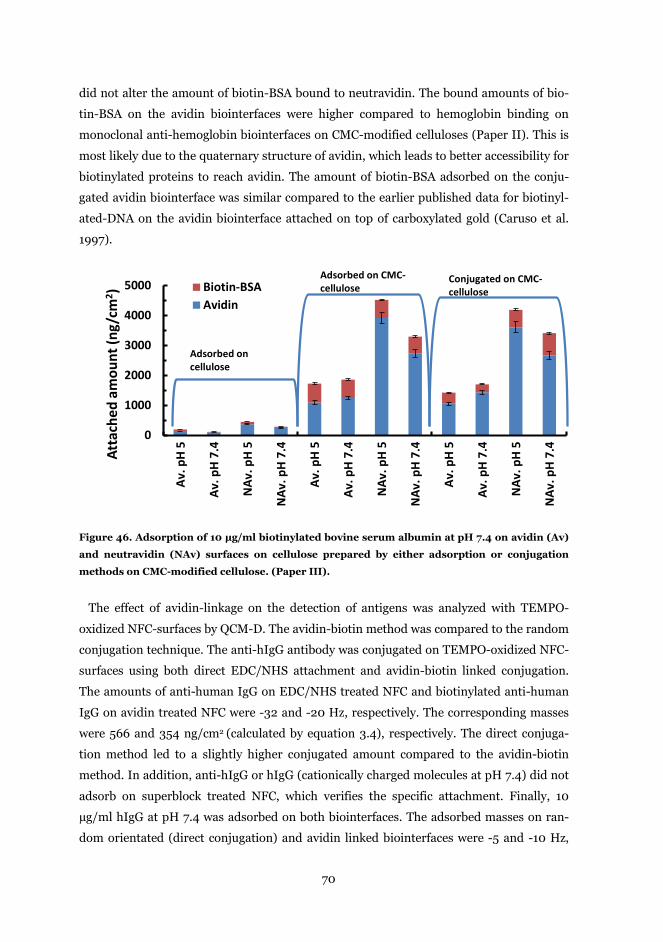
70
did not alter the amount of biotin-BSA bound to neutravidin. The bound amounts of bio-
tin-BSA on the avidin biointerfaces were higher compared to hemoglobin binding on
monoclonal anti-hemoglobin biointerfaces on CMC-modified celluloses (Paper II). This is
most likely due to the quaternary structure of avidin, which leads to better accessibility for
biotinylated proteins to reach avidin. The amount of biotin-BSA adsorbed on the conju-
gated avidin biointerface was similar compared to the earlier published data for biotinyl-
ated-DNA on the avidin biointerface attached on top of carboxylated gold (Caruso et al.
1997).
Figure 46. Adsorption of 10 µg/ml biotinylated bovine serum albumin at pH 7.4 on avidin (Av)
and neutravidin (NAv) surfaces on cellulose prepared by either adsorption or conjugation
methods on CMC-modified cellulose. (Paper III).
The effect of avidin-linkage on the detection of antigens was analyzed with TEMPO-
oxidized NFC-surfaces by QCM-D. The avidin-biotin method was compared to the random
conjugation technique. The anti-hIgG antibody was conjugated on TEMPO-oxidized NFC-
surfaces using both direct EDC/NHS attachment and avidin-biotin linked conjugation.
The amounts of anti-human IgG on EDC/NHS treated NFC and biotinylated anti-human
IgG on avidin treated NFC were -32 and -20 Hz, respectively. The corresponding masses
were 566 and 354 ng/cm2 (calculated by equation 3.4), respectively. The direct conjuga-
tion method led to a slightly higher conjugated amount compared to the avidin-biotin
method. In addition, anti-hIgG or hIgG (cationically charged molecules at pH 7.4) did not
adsorb on superblock treated NFC, which verifies the specific attachment. Finally, 10
µg/ml hIgG at pH 7.4 was adsorbed on both biointerfaces. The adsorbed masses on ran-
dom orientated (direct conjugation) and avidin linked biointerfaces were -5 and -10 Hz,
0
1000
2000
3000
4000
5000
Av. p
H 5
Av. p
H 7.
4
NAv
. pH
5
NAv
. pH
7.4
Av. p
H 5
Av. p
H 7.
4
NAv
. pH
5
NAv
. pH
7.4
Av. p
H 5
Av. p
H 7.
4
NAv
. pH
5
NAv
. pH
7.4
Atta
ched
am
ount
(ng/
cm2 ) Biotin-BSA
Avidin
Adsorbed on cellulose
Adsorbed on CMC-cellulose
Conjugated on CMC-cellulose
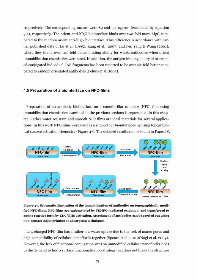
71
respectively. The corresponding masses were 89 and 177 ng/cm2 (calculated by equation
3.4), respectively. The orient anti-hIgG biointerface binds over two-fold more hIgG com-
pared to the random orient anti-hIgG biointerface. This difference is accordance with ear-
lier published data of Lu et al. (1995), Kang et al. (2007) and Pei, Yang & Wang (2001),
where they found over two-fold better binding ability for whole antibodies when orient
immobilization chemistries were used. In addition, the antigen binding ability of orientat-
ed conjugated individual FAB fragments has been reported to be over six-fold better com-
pared to random orientated antibodies (Peluso et al. 2003).
4.5 Preparation of a biointerface on NFC-films
Preparation of an antibody biointerface on a nanofibrillar cellulose (NFC) film using
immobilization chemistries examined in the previous sections is represented in this chap-
ter. Rather water resistant and smooth NFC films are ideal materials for several applica-
tions. In this work NFC-films were used as a support for biointerfaces by using topograph-
ical surface activation chemistry (Figure 47). The detailed results can be found in Paper IV.
Figure 47. Schematic illustration of the immobilization of antibodies on topographically modi-
fied NFC-films. NFC-films are carboxylated by TEMPO-mediated oxidation, and transferred to
amine reactive form by EDC/NHS activation. Attachment of antibodies can be carried out using
non-contact inkjet printing or adsorption techniques.
Low charged NFC-film has a rather low water uptake due to the lack of macro pores and
high compatibility of cellulose nanofibrils together (Spence et al. 2010)(Nogi et al. 2009).
However, the lack of functional conjugation sites on unmodified cellulose nanofibrils leads
to the demand to find a surface functionalization strategy that does not break the structure
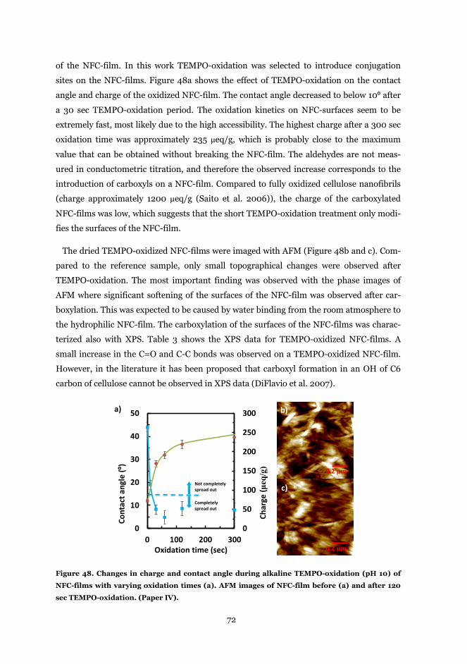
72
of the NFC-film. In this work TEMPO-oxidation was selected to introduce conjugation
sites on the NFC-films. Figure 48a shows the effect of TEMPO-oxidation on the contact
angle and charge of the oxidized NFC-film. The contact angle decreased to below 10º after
a 30 sec TEMPO-oxidation period. The oxidation kinetics on NFC-surfaces seem to be
extremely fast, most likely due to the high accessibility. The highest charge after a 300 sec
oxidation time was approximately 235 μeq/g, which is probably close to the maximum
value that can be obtained without breaking the NFC-film. The aldehydes are not meas-
ured in conductometric titration, and therefore the observed increase corresponds to the
introduction of carboxyls on a NFC-film. Compared to fully oxidized cellulose nanofibrils
(charge approximately 1200 μeq/g (Saito et al. 2006)), the charge of the carboxylated
NFC-films was low, which suggests that the short TEMPO-oxidation treatment only modi-
fies the surfaces of the NFC-film.
The dried TEMPO-oxidized NFC-films were imaged with AFM (Figure 48b and c). Com-
pared to the reference sample, only small topographical changes were observed after
TEMPO-oxidation. The most important finding was observed with the phase images of
AFM where significant softening of the surfaces of the NFC-film was observed after car-
boxylation. This was expected to be caused by water binding from the room atmosphere to
the hydrophilic NFC-film. The carboxylation of the surfaces of the NFC-films was charac-
terized also with XPS. Table 3 shows the XPS data for TEMPO-oxidized NFC-films. A
small increase in the C=O and C-C bonds was observed on a TEMPO-oxidized NFC-film.
However, in the literature it has been proposed that carboxyl formation in an OH of C6
carbon of cellulose cannot be observed in XPS data (DiFlavio et al. 2007).
Figure 48. Changes in charge and contact angle during alkaline TEMPO-oxidation (pH 10) of
NFC-films with varying oxidation times (a). AFM images of NFC-film before (a) and after 120
sec TEMPO-oxidation. (Paper IV).
0.2 μm
0.2 μm
b)
c)
a)
0
50
100
150
200
250
300
0
10
20
30
40
50
0 100 200 300
Char
ge (μ
eq/g
)
Cont
act a
ngle
(º)
Oxidation time (sec)
Completely spread out
Not completely spread out
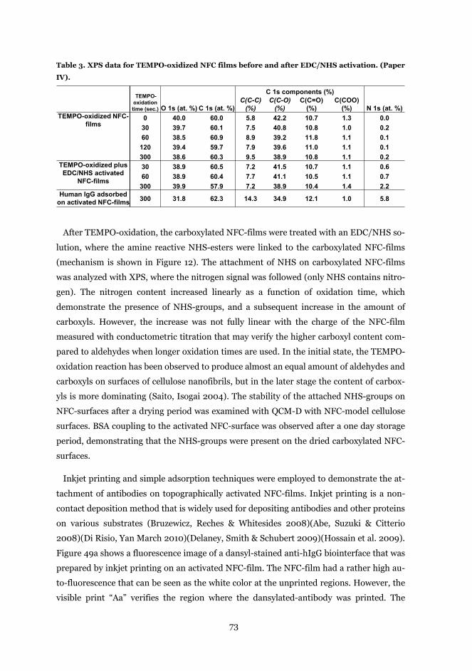
73
Table 3. XPS data for TEMPO-oxidized NFC films before and after EDC/NHS activation. (Paper
IV).
After TEMPO-oxidation, the carboxylated NFC-films were treated with an EDC/NHS so-
lution, where the amine reactive NHS-esters were linked to the carboxylated NFC-films
(mechanism is shown in Figure 12). The attachment of NHS on carboxylated NFC-films
was analyzed with XPS, where the nitrogen signal was followed (only NHS contains nitro-
gen). The nitrogen content increased linearly as a function of oxidation time, which
demonstrate the presence of NHS-groups, and a subsequent increase in the amount of
carboxyls. However, the increase was not fully linear with the charge of the NFC-film
measured with conductometric titration that may verify the higher carboxyl content com-
pared to aldehydes when longer oxidation times are used. In the initial state, the TEMPO-
oxidation reaction has been observed to produce almost an equal amount of aldehydes and
carboxyls on surfaces of cellulose nanofibrils, but in the later stage the content of carbox-
yls is more dominating (Saito, Isogai 2004). The stability of the attached NHS-groups on
NFC-surfaces after a drying period was examined with QCM-D with NFC-model cellulose
surfaces. BSA coupling to the activated NFC-surface was observed after a one day storage
period, demonstrating that the NHS-groups were present on the dried carboxylated NFC-
surfaces.
Inkjet printing and simple adsorption techniques were employed to demonstrate the at-
tachment of antibodies on topographically activated NFC-films. Inkjet printing is a non-
contact deposition method that is widely used for depositing antibodies and other proteins
on various substrates (Bruzewicz, Reches & Whitesides 2008)(Abe, Suzuki & Citterio
2008)(Di Risio, Yan March 2010)(Delaney, Smith & Schubert 2009)(Hossain et al. 2009).
Figure 49a shows a fluorescence image of a dansyl-stained anti-hIgG biointerface that was
prepared by inkjet printing on an activated NFC-film. The NFC-film had a rather high au-
to-fluorescence that can be seen as the white color at the unprinted regions. However, the
visible print “Aa” verifies the region where the dansylated-antibody was printed. The
TEMPO-oxidation time (sec.)
C 1s components (%)
N 1s (at. %)O 1s (at. %) C 1s (at. %)C(C-C)
(%)C(C-O)
(%)C(C=O)
(%)C(COO)
(%)TEMPO-oxidized NFC-
films0 40.0 60.0 5.8 42.2 10.7 1.3 0.030 39.7 60.1 7.5 40.8 10.8 1.0 0.260 38.5 60.9 8.9 39.2 11.8 1.1 0.1
120 39.4 59.7 7.9 39.6 11.0 1.1 0.1300 38.6 60.3 9.5 38.9 10.8 1.1 0.2
TEMPO-oxidized plus EDC/NHS activated
NFC-films
30 38.9 60.5 7.2 41.5 10.7 1.1 0.660 38.9 60.4 7.7 41.1 10.5 1.1 0.7
300 39.9 57.9 7.2 38.9 10.4 1.4 2.2Human IgG adsorbed
on activated NFC-films 300 31.8 62.3 14.3 34.9 12.1 1.0 5.8
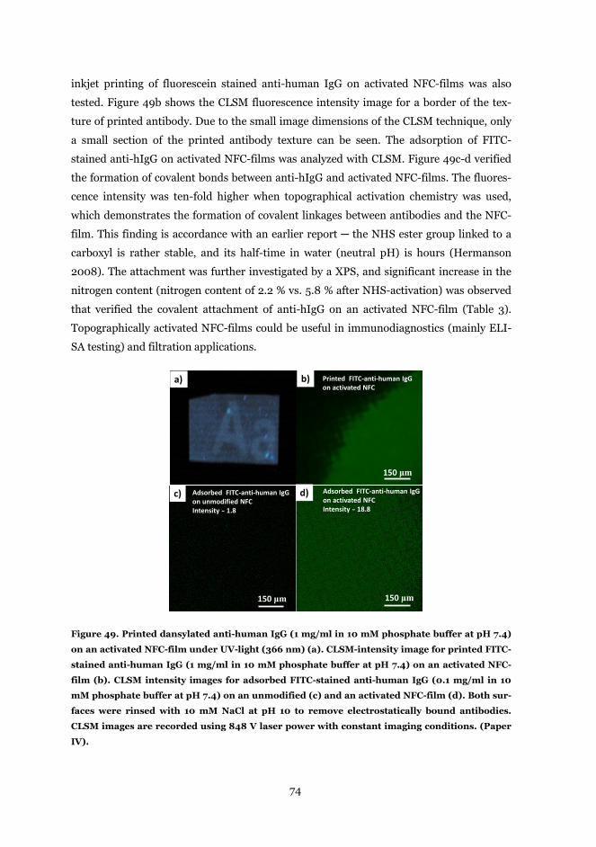
74
inkjet printing of fluorescein stained anti-human IgG on activated NFC-films was also
tested. Figure 49b shows the CLSM fluorescence intensity image for a border of the tex-
ture of printed antibody. Due to the small image dimensions of the CLSM technique, only
a small section of the printed antibody texture can be seen. The adsorption of FITC-
stained anti-hIgG on activated NFC-films was analyzed with CLSM. Figure 49c-d verified
the formation of covalent bonds between anti-hIgG and activated NFC-films. The fluores-
cence intensity was ten-fold higher when topographical activation chemistry was used,
which demonstrates the formation of covalent linkages between antibodies and the NFC-
film. This finding is accordance with an earlier report ─ the NHS ester group linked to a
carboxyl is rather stable, and its half-time in water (neutral pH) is hours (Hermanson
2008). The attachment was further investigated by a XPS, and significant increase in the
nitrogen content (nitrogen content of 2.2 % vs. 5.8 % after NHS-activation) was observed
that verified the covalent attachment of anti-hIgG on an activated NFC-film (Table 3).
Topographically activated NFC-films could be useful in immunodiagnostics (mainly ELI-
SA testing) and filtration applications.
Figure 49. Printed dansylated anti-human IgG (1 mg/ml in 10 mM phosphate buffer at pH 7.4)
on an activated NFC-film under UV-light (366 nm) (a). CLSM-intensity image for printed FITC-
stained anti-human IgG (1 mg/ml in 10 mM phosphate buffer at pH 7.4) on an activated NFC-
film (b). CLSM intensity images for adsorbed FITC-stained anti-human IgG (0.1 mg/ml in 10
mM phosphate buffer at pH 7.4) on an unmodified (c) and an activated NFC-film (d). Both sur-
faces were rinsed with 10 mM NaCl at pH 10 to remove electrostatically bound antibodies.
CLSM images are recorded using 848 V laser power with constant imaging conditions. (Paper
IV).
a) b)
c)
150 μm150 μm
Adsorbed FITC-anti-human IgG on unmodified NFCIntensity ̴ 1.8
Adsorbed FITC-anti-human IgGon activated NFCIntensity ̴ 18.8
d)
150 μm
Printed FITC-anti-human IgGon activated NFC
75
5 Concluding remarks
The immobilization of proteins on solid supports is the initial step in the development of
immunodiagnostic tests. In this thesis different methods to immobilize antibodies on cel-
lulosic supports were investigated. The first part relates to the interactions of proteins with
cellulose, and how protein adsorption can be controlled by using surface modification
chemistries. In the second part, immobilization strategies to covalently attach antibodies
on cellulose surfaces were developed. The third part of this thesis deals with the use of the
immobilization strategies developed to prepare an antibody biointerface on NFC-films.
First, surface plasmon resonance (SPR) studies were exploited in order to examine the
adsorption of proteins on modified cellulose surfaces. The findings suggest that the ad-
sorption of proteins on unmodified cellulose is driven mainly by non-electrostatic forces,
and adsorption is largely irreversible. On low charged cellulose surfaces, all proteins tested
adsorbed mostly at their isoelectric points (pI) due to their reduced solubility in water.
The increased surface charge that promotes the electrostatic interaction of globular pro-
teins lead to increased adsorption on the cellulose surface. The electrostatic interaction
introduced enhanced the absorption on cellulose surfaces for all tested proteins. The ad-
sorption phenomenon of glycoproteins is more complicated compared to globular proteins
since the adsorption is promoted by combinations of non-electrostatic and electrostatic
interactions. Moreover, the oligosaccharide chains of avidin were found to reduce the ad-
sorption of glycoproteins on cellulose surfaces.
In order to introduce functional groups on cellulosic surfaces for enabling covalent con-
jugation of antibodies on cellulose, CMC-adsorption, chitosan-adsorption, and TEMPO-
oxidation methods were explored by QCM-D. The studies showed that both CMC and chi-
tosan adsorbed irreversibly on cellulose surfaces but their adsorption phenomena varied
slightly. The adsorbed chitosan layer on cellulose has a less extended conformation com-
pared to the adsorbed CMC layer due to the presence of electrostatic attractive interac-
tions. Both polysaccharide layers had a hydrogel like structure and they coupled signifi-
cant amounts of water. The chemical modification method, TEMPO-oxidation created
carboxyl groups on cellulose that was observed to cause significant water uptake in the
cellulose film. The TEMPO-oxidation reaction was extremely fast, and seems to be a usa-
ble tool to functionalize cellulosic materials. However, the possible irreversible structural
changes it causes in the cellulosic material must be considered.
76
All tested functionalization strategies enabled covalent conjugation of antibodies to cel-
lulose. The CMC and TEMPO-oxidation methods were more usable compared to chitosan
modification due to the lack of self-crosslinking in antibodies. The CMC-modification
method is also a gentle method that does not alter the substructure of a cellulosic matrix.
Both the CMC-modification and the TEMPO-oxidation methods led to similar surface cov-
erages for immobilized antibodies. The biological activity of conjugated antibodies on
CMC-modified and TEMPO-oxidized cellulose surfaces were verified with specific counter
parts. The specific detection of antigens with high detection efficiency was found that sug-
gest the usability of the developed immobilization routes towards the development of im-
munodiagnostic applications.
The avidin-biotin chemistry was used to orient the conformation of antibodies on cellu-
lose surfaces for enhancing the detection sensitivity of the cellulose based biointerface.
The attachment of avidin on cellulose can be significantly enhanced by increasing the ani-
onic charge on cellulosic surfaces by CMC-adsorption and by TEMPO-oxidation. The im-
mobilized avidins bind biotinylated proteins specifically, which verifies the negligible
changes in their biological activity. The avidin-biotin system was compared to traditional
EDC/NHS conjugation, and an over two-fold better antigen detection was found com-
pared to the traditional conjugation chemistry.
Finally, NFC-films were utilized to act as a support for biointerfaces. The NFC-films
were first activated with TEMPO-oxidation. The studies shown that only a short TEMPO-
oxidation treatment was needed to create a high carboxyl content on the surfaces of a
NFC-film without remarkable damage to the substructure or water resistivity of the NFC-
film. The surface sensitive carboxylated NFC-films were transferred to amine reactive
form by EDC/NHS activation. The amine reactivity of the activated NFC-film was demon-
strated after a drying period, which showed that the reactivity of the film remains at least
short periods. The conjugation of antibodies on activated NFC-films was carried out using
both inkjet and adsorption methods. The covalent linking reaction was observed only
when the activation chemistry was utilized.
Taken together, understanding of the adsorption of proteins and their conjugation phe-
nomena on cellulosic materials give us valuable information on how cellulosic materials
can be utilized in advanced immunodiagnostic applications.
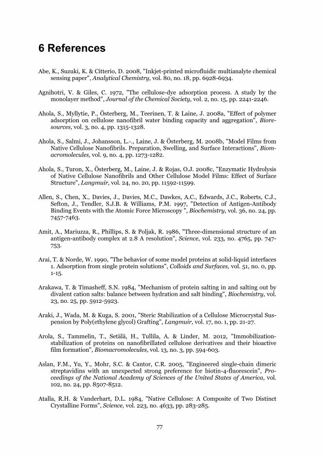
77
6 References
Abe, K., Suzuki, K. & Citterio, D. 2008, "Inkjet-printed microfluidic multianalyte chemical sensing paper", Analytical Chemistry, vol. 80, no. 18, pp. 6928-6934.
Agnihotri, V. & Giles, C. 1972, "The cellulose-dye adsorption process. A study by the monolayer method", Journal of the Chemical Society, vol. 2, no. 15, pp. 2241-2246.
Ahola, S., Myllytie, P., Österberg, M., Teerinen, T. & Laine, J. 2008a, "Effect of polymer adsorption on cellulose nanofibril water binding capacity and aggregation", Biore-sources, vol. 3, no. 4, pp. 1315-1328.
Ahola, S., Salmi, J., Johansson, L.-., Laine, J. & Österberg, M. 2008b, "Model Films from Native Cellulose Nanofibrils. Preparation, Swelling, and Surface Interactions", Biom-acromolecules, vol. 9, no. 4, pp. 1273-1282.
Ahola, S., Turon, X., Österberg, M., Laine, J. & Rojas, O.J. 2008c, "Enzymatic Hydrolysis of Native Cellulose Nanofibrils and Other Cellulose Model Films: Effect of Surface Structure", Langmuir, vol. 24, no. 20, pp. 11592-11599.
Allen, S., Chen, X., Davies, J., Davies, M.C., Dawkes, A.C., Edwards, J.C., Roberts, C.J., Sefton, J., Tendler, S.J.B. & Williams, P.M. 1997, "Detection of Antigen-Antibody Binding Events with the Atomic Force Microscopy ", Biochemistry, vol. 36, no. 24, pp. 7457-7463.
Amit, A., Mariuzza, R., Phillips, S. & Poljak, R. 1986, "Three-dimensional structure of an antigen-antibody complex at 2.8 A resolution", Science, vol. 233, no. 4765, pp. 747-753.
Arai, T. & Norde, W. 1990, "The behavior of some model proteins at solid-liquid interfaces 1. Adsorption from single protein solutions", Colloids and Surfaces, vol. 51, no. 0, pp. 1-15.
Arakawa, T. & Timasheff, S.N. 1984, "Mechanism of protein salting in and salting out by divalent cation salts: balance between hydration and salt binding", Biochemistry, vol. 23, no. 25, pp. 5912-5923.
Araki, J., Wada, M. & Kuga, S. 2001, "Steric Stabilization of a Cellulose Microcrystal Sus-pension by Poly(ethylene glycol) Grafting", Langmuir, vol. 17, no. 1, pp. 21-27.
Arola, S., Tammelin, T., Setälä, H., Tullila, A. & Linder, M. 2012, "Immobilization-stabilization of proteins on nanofibrillated cellulose derivatives and their bioactive film formation", Biomacromolecules, vol. 13, no. 3, pp. 594-603.
Aslan, F.M., Yu, Y., Mohr, S.C. & Cantor, C.R. 2005, "Engineered single-chain dimeric streptavidins with an unexpected strong preference for biotin-4-fluorescein", Pro-ceedings of the National Academy of Sciences of the United States of America, vol. 102, no. 24, pp. 8507-8512.
Atalla, R.H. & Vanderhart, D.L. 1984, "Native Cellulose: A Composite of Two Distinct Crystalline Forms", Science, vol. 223, no. 4633, pp. 283-285.
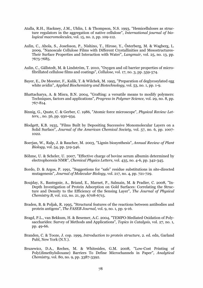
78
Atalla, R.H., Hackney, J.M., Uhlin, I. & Thompson, N.S. 1993, "Hemicelluloses as struc-ture regulators in the aggregation of native cellulose", International journal of bio-logical macromolecules, vol. 15, no. 2, pp. 109-112.
Aulin, C., Ahola, S., Josefsson, P., Nishino, T., Hirose, Y., Österberg, M. & Wagberg, L. 2009, "Nanoscale Cellulose Films with Different Crystallinities and Mesostructures-Their Surface Properties and Interaction with Water", Langmuir, vol. 25, no. 13, pp. 7675-7685.
Aulin, C., Gällstedt, M. & Lindström, T. 2010, "Oxygen and oil barrier properties of micro-fibrillated cellulose films and coatings", Cellulose, vol. 17, no. 3, pp. 559-574.
Bayer, E., De Meester, F., Kulik, T. & Wilchek, M. 1995, "Preparation of deglycosylated egg white avidin", Applied Biochemistry and Biotechnology, vol. 53, no. 1, pp. 1-9.
Bhattacharya, A. & Misra, B.N. 2004, "Grafting: a versatile means to modify polymers: Techniques, factors and applications", Progress in Polymer Science, vol. 29, no. 8, pp. 767-814.
Binnig, G., Quate, C. & Gerber, C. 1986, "Atomic force microscope", Physical Review Let-ters, , no. 56, pp. 930-934.
Blodgett, K.B. 1935, "Films Built by Depositing Successive Monomolecular Layers on a Solid Surface", Journal of the American Chemical Society, vol. 57, no. 6, pp. 1007-1022.
Boerjan, W., Ralp, J. & Baucher, M. 2003, "Lignin biosynthesis", Annual Review of Plant Biology, vol. 54, pp. 519-546.
Böhme, U. & Scheler, U. 2007, "Effective charge of bovine serum albumin determined by electrophoresis NMR", Chemical Physics Letters, vol. 435, no. 4-6, pp. 342-345.
Bordo, D. & Argos, P. 1991, "Suggestions for “safe” residue substitutions in site-directed mutagenesis", Journal of Molecular Biology, vol. 217, no. 4, pp. 721-729.
Boujday, S., Bantegnie, A., Briand, E., Marnet, P., Salmain, M. & Pradier, C. 2008, "In-Depth Investigation of Protein Adsorption on Gold Surfaces: Correlating the Struc-ture and Density to the Efficiency of the Sensing Layer", The Journal of Physical Chemistry B, vol. 112, no. 21, pp. 6708-6715.
Braden, B. & Poljak, R. 1995, "Structural features of the reactions between antibodies and protein antigens", The FASEB Journal, vol. 9, no. 1, pp. 9-16.
Bragd, P.L., van Bekkum, H. & Besemer, A.C. 2004, "TEMPO-Mediated Oxidation of Poly-saccharides: Survey of Methods and Applications", Topics in Catalysis, vol. 27, no. 1, pp. 49-66.
Branden, C. & Tooze, J. cop. 1999, Introduction to protein structure, 2. ed. edn, Garland Publ, New York (N.Y.).
Bruzewicz, D.A., Reches, M. & Whitesides, G.M. 2008, "Low-Cost Printing of Poly(dimethylsiloxane) Barriers To Define Microchannels in Paper", Analytical Chemistry, vol. 80, no. 9, pp. 3387-3392.
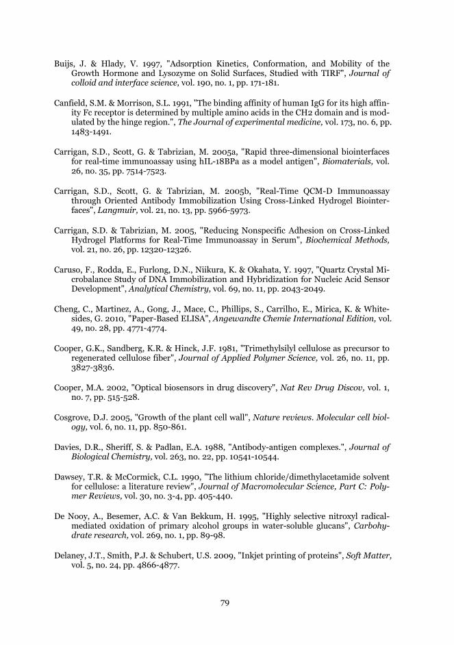
79
Buijs, J. & Hlady, V. 1997, "Adsorption Kinetics, Conformation, and Mobility of the Growth Hormone and Lysozyme on Solid Surfaces, Studied with TIRF", Journal of colloid and interface science, vol. 190, no. 1, pp. 171-181.
Canfield, S.M. & Morrison, S.L. 1991, "The binding affinity of human IgG for its high affin-ity Fc receptor is determined by multiple amino acids in the CH2 domain and is mod-ulated by the hinge region.", The Journal of experimental medicine, vol. 173, no. 6, pp. 1483-1491.
Carrigan, S.D., Scott, G. & Tabrizian, M. 2005a, "Rapid three-dimensional biointerfaces for real-time immunoassay using hIL-18BPa as a model antigen", Biomaterials, vol. 26, no. 35, pp. 7514-7523.
Carrigan, S.D., Scott, G. & Tabrizian, M. 2005b, "Real-Time QCM-D Immunoassay through Oriented Antibody Immobilization Using Cross-Linked Hydrogel Biointer-faces", Langmuir, vol. 21, no. 13, pp. 5966-5973.
Carrigan, S.D. & Tabrizian, M. 2005, "Reducing Nonspecific Adhesion on Cross-Linked Hydrogel Platforms for Real-Time Immunoassay in Serum", Biochemical Methods, vol. 21, no. 26, pp. 12320-12326.
Caruso, F., Rodda, E., Furlong, D.N., Niikura, K. & Okahata, Y. 1997, "Quartz Crystal Mi-crobalance Study of DNA Immobilization and Hybridization for Nucleic Acid Sensor Development", Analytical Chemistry, vol. 69, no. 11, pp. 2043-2049.
Cheng, C., Martinez, A., Gong, J., Mace, C., Phillips, S., Carrilho, E., Mirica, K. & White-sides, G. 2010, "Paper-Based ELISA", Angewandte Chemie International Edition, vol. 49, no. 28, pp. 4771-4774.
Cooper, G.K., Sandberg, K.R. & Hinck, J.F. 1981, "Trimethylsilyl cellulose as precursor to regenerated cellulose fiber", Journal of Applied Polymer Science, vol. 26, no. 11, pp. 3827-3836.
Cooper, M.A. 2002, "Optical biosensors in drug discovery", Nat Rev Drug Discov, vol. 1, no. 7, pp. 515-528.
Cosgrove, D.J. 2005, "Growth of the plant cell wall", Nature reviews. Molecular cell biol-ogy, vol. 6, no. 11, pp. 850-861.
Davies, D.R., Sheriff, S. & Padlan, E.A. 1988, "Antibody-antigen complexes.", Journal of Biological Chemistry, vol. 263, no. 22, pp. 10541-10544.
Dawsey, T.R. & McCormick, C.L. 1990, "The lithium chloride/dimethylacetamide solvent for cellulose: a literature review", Journal of Macromolecular Science, Part C: Poly-mer Reviews, vol. 30, no. 3-4, pp. 405-440.
De Nooy, A., Besemer, A.C. & Van Bekkum, H. 1995, "Highly selective nitroxyl radical-mediated oxidation of primary alcohol groups in water-soluble glucans", Carbohy-drate research, vol. 269, no. 1, pp. 89-98.
Delaney, J.T., Smith, P.J. & Schubert, U.S. 2009, "Inkjet printing of proteins", Soft Matter, vol. 5, no. 24, pp. 4866-4877.
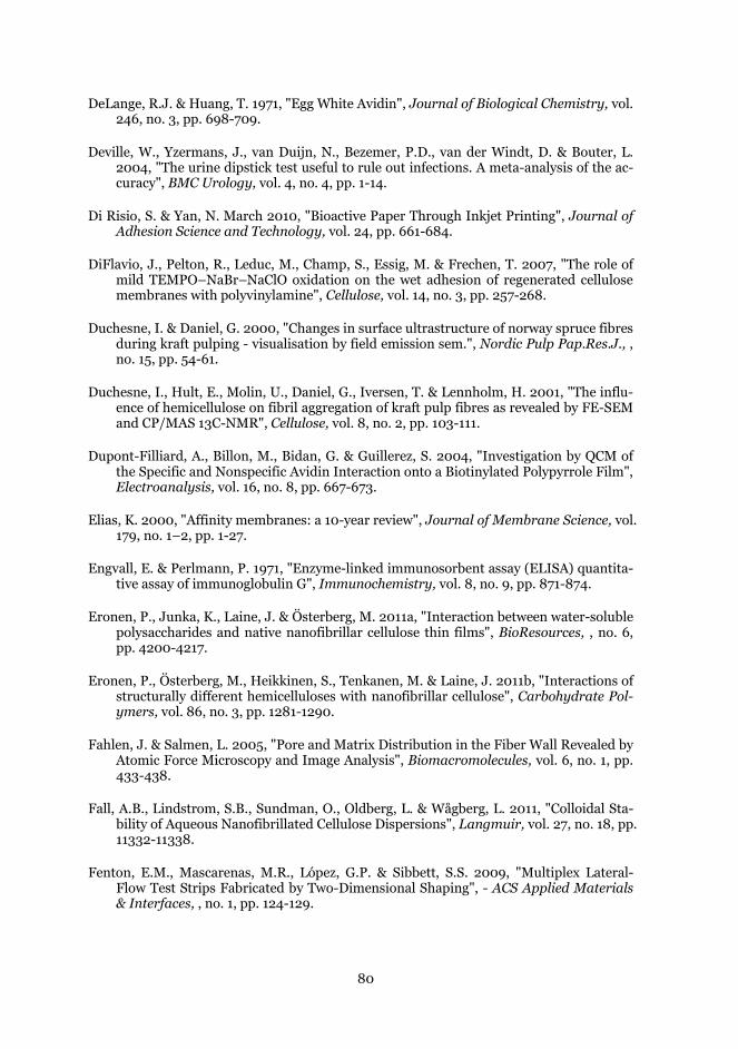
80
DeLange, R.J. & Huang, T. 1971, "Egg White Avidin", Journal of Biological Chemistry, vol. 246, no. 3, pp. 698-709.
Deville, W., Yzermans, J., van Duijn, N., Bezemer, P.D., van der Windt, D. & Bouter, L. 2004, "The urine dipstick test useful to rule out infections. A meta-analysis of the ac-curacy", BMC Urology, vol. 4, no. 4, pp. 1-14.
Di Risio, S. & Yan, N. March 2010, "Bioactive Paper Through Inkjet Printing", Journal of Adhesion Science and Technology, vol. 24, pp. 661-684.
DiFlavio, J., Pelton, R., Leduc, M., Champ, S., Essig, M. & Frechen, T. 2007, "The role of mild TEMPO–NaBr–NaClO oxidation on the wet adhesion of regenerated cellulose membranes with polyvinylamine", Cellulose, vol. 14, no. 3, pp. 257-268.
Duchesne, I. & Daniel, G. 2000, "Changes in surface ultrastructure of norway spruce fibres during kraft pulping - visualisation by field emission sem.", Nordic Pulp Pap.Res.J., , no. 15, pp. 54-61.
Duchesne, I., Hult, E., Molin, U., Daniel, G., Iversen, T. & Lennholm, H. 2001, "The influ-ence of hemicellulose on fibril aggregation of kraft pulp fibres as revealed by FE-SEM and CP/MAS 13C-NMR", Cellulose, vol. 8, no. 2, pp. 103-111.
Dupont-Filliard, A., Billon, M., Bidan, G. & Guillerez, S. 2004, "Investigation by QCM of the Specific and Nonspecific Avidin Interaction onto a Biotinylated Polypyrrole Film", Electroanalysis, vol. 16, no. 8, pp. 667-673.
Elias, K. 2000, "Affinity membranes: a 10-year review", Journal of Membrane Science, vol. 179, no. 1–2, pp. 1-27.
Engvall, E. & Perlmann, P. 1971, "Enzyme-linked immunosorbent assay (ELISA) quantita-tive assay of immunoglobulin G", Immunochemistry, vol. 8, no. 9, pp. 871-874.
Eronen, P., Junka, K., Laine, J. & Österberg, M. 2011a, "Interaction between water-soluble polysaccharides and native nanofibrillar cellulose thin films", BioResources, , no. 6, pp. 4200-4217.
Eronen, P., Österberg, M., Heikkinen, S., Tenkanen, M. & Laine, J. 2011b, "Interactions of structurally different hemicelluloses with nanofibrillar cellulose", Carbohydrate Pol-ymers, vol. 86, no. 3, pp. 1281-1290.
Fahlen, J. & Salmen, L. 2005, "Pore and Matrix Distribution in the Fiber Wall Revealed by Atomic Force Microscopy and Image Analysis", Biomacromolecules, vol. 6, no. 1, pp. 433-438.
Fall, A.B., Lindstrom, S.B., Sundman, O., Oldberg, L. & Wågberg, L. 2011, "Colloidal Sta-bility of Aqueous Nanofibrillated Cellulose Dispersions", Langmuir, vol. 27, no. 18, pp. 11332-11338.
Fenton, E.M., Mascarenas, M.R., López, G.P. & Sibbett, S.S. 2009, "Multiplex Lateral-Flow Test Strips Fabricated by Two-Dimensional Shaping", - ACS Applied Materials & Interfaces, , no. 1, pp. 124-129.
81
Fernandez-Lafuente, R., Rosell, C.M., Rodriguez, V., Santana, C., Soler, G., Bastida, A. & Guisán, J.M. 1993, "Preparation of activated supports containing low pK amino groups. A new tool for protein immobilization via the carboxyl coupling method", En-zyme and microbial technology, vol. 15, no. 7, pp. 546-550.
Filpponen, I. & Argyropoulos, D.S. 2010, "Regular Linking of Cellulose Nanocrystals via Click Chemistry: Synthesis and Formation of Cellulose Nanoplatelet Gels", Biom-acromolecules, vol. 11, no. 4, pp. 1060-1066.
Fink, H. & Philipp, B. 1985, "Models of cellulose physical structure from the viewpoint of the cellulose I ? II transition", Journal of Applied Polymer Science, vol. 30, no. 9, pp. 3779-3790.
Forsgren, A. & Sjöquist, J. 1966, "“Protein A” from S. Aureus", The Journal of Immunolo-gy, vol. 97, no. 6, pp. 822-827.
Free, A.H., Adams, E.C., Kercher, M.L., Free, H.M. & Cook, M.H. 1957, "Simple Specific Test for Urine Glucose", Clinical chemistry, vol. 3, no. 3, pp. 163-168.
Friguet, B., Chaffotte, A.F., Djavadi-Ohaniance, L. & Goldberg, M.E. 1985, "Measurements of the true affinity constant in solution of antigen-antibody complexes by enzyme-linked immunosorbent assay", Journal of immunological methods, vol. 77, no. 2, pp. 305-319.
Fujisawa, S., Okita, Y., Fukuzumi, H., Saito, T. & Isogai, A. 2011, "Preparation and charac-terization of TEMPO-oxidized cellulose nanofibril films with free carboxyl groups", Carbohydrate Polymers, vol. 84, no. 1, pp. 579-583.
Geffroy, C., Labeau, M.P., Wong, K., Cabane, B. & Cohen Stuart, M.A. 2000, "Kinetics of adsorption of polyvinylamine onto cellulose", Colloids and Surfaces A: Physicochem-ical and Engineering Aspects, vol. 172, no. 1-3, pp. 47-56.
Gersten, D.M. & Marchalonis, J.J. 1978, "A rapid, novel method for the solid-phase deri-vatization of IgG antibodies for immune-affinity chromatography", Journal of immu-nological methods, vol. 24, no. 3-4, pp. 305-309.
Ghanem, A. & Ghaly, A. 2004, "Immobilization of glucose oxidase in chitosan gel beads", Journal of Applied Polymer Science, vol. 91, no. 2, pp. 861-866.
Gilles, M.A., Hudson, A.Q. & Borders Jr., C.L. 1990, "Stability of water-soluble car-bodiimides in aqueous solution", Analytical Biochemistry, vol. 184, no. 2, pp. 244-248.
Goldstein, L., Yankofsky, S.A., Cohen, G. & Frank Chytil and Donald, B.M. 1986, "[13] Sol-id-phase assay for d-biotin and avidin on cellulose disks", Methods in Enzymology, vol. 122, pp. 72-82.
Gondo, T., Watanabe, M., Soma, H., Kitao, O., Yanagisawa, M. & Isogai, A. 2006, "SEC-MALS study on carboxymethyl cellulose (CMC): Relationship between conformation of CMC molecules and their adsorption behavior onto pulp fibers", Nordic Pulp & Pa-per Research Journal, vol. 21, no. 5, pp. 591-597.
82
Grabarek, Z. & Gergely, J. 1990, "Zero-length crosslinking procedure with the use of active esters", Analytical Biochemistry, vol. 185, no. 1, pp. 131-135.
Greber, G. & Paschinger, O. 1981, "Silylderivate der cellulose", Papier, , no. 35, pp. 547-554.
Green, N.M. 1975, "Avidin", Advances in protein chemistry, vol. Volume 29, no. 29, pp. 85-133.
Green, N.M. & Joynson, M.A. 1970, "A preliminary crystallographic investigation of avi-din", Biochemical Journal, vol. 118, no. 1, pp. 71-72.
Guesdon, J.L., Ternynck, T. & Avrameas, S. 1979, "The use of avidin-biotin interaction in immunoenzymatic techniques.", Journal of Histochemistry & Cytochemistry, vol. 27, no. 8, pp. 1131-1139.
Habibi, Y., Chanzy, H. & Vignon, M. 2006, "TEMPO-mediated surface oxidation of cellu-lose whiskers", Cellulose, vol. 13, no. 6, pp. 679-687.
Hage, D.S. 1999, "Affinity Chromatography: A Review of Clinical Applications", Clinical chemistry, vol. 45, no. 5, pp. 593-615.
Hall, D.B., Underhill, P. & Torkelson, J.M. 1998, "Spin coating of thin and ultrathin poly-mer films", Polymer Engineering & Science, vol. 38, no. 12, pp. 2039-2045.
Hamilton, R. 1987, "Human IgG subclass measurements in the clinical laboratory.", Clin Chem, , no. 33, pp. 1070-1075.
Hawkes, R., Niday, E. & Gordon, J. 1982, "A dot-immunobinding assay for monoclonal and other antibodies", Analytical Biochemistry, vol. 119, no. 1, pp. 142-147.
Haynes, C.A. & Norde, W. 1994, "Globular proteins at solid/liquid interfaces", Colloids and Surfaces B: Biointerfaces, vol. 2, no. 6, pp. 517-566.
Hearle, J.W.S. 1958, "A fringed fibril theory of structure in crystalline polymers", Journal of Polymer Science, vol. 28, no. 117, pp. 432-435.
Heikkinen, J.J., Kivimäki, L., Määttä, J.A.E., Mäkelä, I., Hakalahti, L., Takkinen, K., Ku-lomaa, M.S., Hytönen, V.P. & Hormi, O.E.O. 2011, "Versatile bio-ink for covalent im-mobilization of chimeric avidin on sol–gel substrates", Colloids and Surfaces B: Bio-interfaces, vol. 87, no. 2, pp. 409-414.
Heinze, T. & Koschella, A. 2005, "Carboxymethyl Ethers of Cellulose and Starch. A Re-view", Macromolecular Symposia, vol. 223, no. 1, pp. 13-40.
Henriksson, M., Berglund, L.A., Isaksson, P., Lindström, T. & Nishino, T. 2008, "Cellu-lose Nanopaper Structures of High Toughness", Biomacromolecules, vol. 9, no. 6, pp. 1579-1585.
Hermanson, G.T. 2008, Bioconjugate techniques, Academic Press, San Diego (CA).
83
Herrick, F., Casebier, R., Hamilton, J. & Sandberg, K. 1983, "Microfibrillated cellulose: Morphology and accessibility.", J. Appl. Polym. Sci.: Appl. Polym. Symp., vol. 2, no. 37, pp. 797-813.
Hiller, Y., Gershoni, J.M., Bayer, E.A. & Wilchek, M. 1987, "Biotin binding to avidin. Oli-gosaccharide side chain not required for ligand association", Biochem. J., vol. 248, no. 1, pp. 167-171.
Hirota, M., Furihata, K., Saito, T., Kawada, T. & Isogai, A. 2010, "Glucose/Glucuronic Acid Alternating Co-polysaccharides Prepared from TEMPO-Oxidized Native Celluloses by Surface Peeling", Angewandte Chemie, vol. 122, no. 42, pp. 7836-7838.
Homola, J., Yee, S.S. & Gauglitz, G. 1999, "Surface plasmon resonance sensors: review", Sensors and Actuators B: Chemical, vol. 54, no. 1-2, pp. 3-15.
Höök, F., Rodahl, M., Brzezinski, P. & Kasemo, B. 1998, "Energy dissipation kinetics for protein and antibody-antigen adsorption under shear oscillation on a quartz crystal microbalance", Langmuir, vol. 14, no. 4, pp. 729-734.
Hossain, S.M., Luckham, R.E., Smith, A.M., Lebert, J.M., Davies, L.M., Pelton, R.H., Filipe, C.D. & Brennan, J.D. 2009, "Development of a bioactive paper sensor for detection of neurotoxins using piezoelectric inkjet printing of sol-gel-derived bioinks", Analytical Chemistry, vol. 81, no. 13, pp. 5474-5483.
Ishimaru, Y. & Lindström, T. 1984, "Adsorption of water-soluble, nonionic polymers onto cellulosic fibers", Journal of Applied Polymer Science, vol. 29, no. 5, pp. 1675-1691.
Isogai, A. & Kato, Y. 1998, "Preparation of polyuronic acid from cellulose by TEMPO-mediated oxidation.", Cellulose, vol. 5, no. 5, pp. 153-164.
Jameson, D.M. & Wong, S.S. 2009, Chemistry of Protein Conjugation and Cross-Linking, Second Edition, Taylor and Francis.
Jefferis, R. 2009, "Glycosylation as a strategy to improve antibody-based therapeutics", Nat Rev Drug Discov, vol. 8, no. 3, pp. 226-234.
Johannsmann, D., Mathauer, K., Wegner, G. & Knoll, W. 1992, "Viscoelastic properties of thin films probed with a quartz-crystal resonator", Phys.Rev.B, vol. 46, no. 12, pp. 7808-7815.
Johansson, L. & Campbell, J.M. 2004, "Reproducible XPS on biopolymers: cellulose stud-ies", Surface and Interface Analysis, vol. 36, no. 8, pp. 1018-1022.
Johnsson, B., Löfås, S., Lindquist, G., Edström, Å., Hillgren, R.M. & Hansson, A. 1995, "Comparison of methods for immobilization to carboxymethyl dextran sensor surfac-es by analysis of the specific activity of monoclonal antibodies", Journal of Molecular Recognition, vol. 8, no. 1-2, pp. 125-131.
Jung, L.S., Campbell, C.T., Chinowsky, T.M., Mar, M.N. & Yee, S.S. 1998, "Quantitative Interpretation of the Response of Surface Plasmon Resonance Sensors to Adsorbed Films", Langmuir, vol. 14, no. 19, pp. 5636-5648.
84
Kang, J.H., Choi, H.J., Hwang, S.Y., Han, S.H., Jeon, J.Y. & Lee, E.K. 2007, "Improving immunobinding using oriented immobilization of an oxidized antibody", Journal of Chromatography A, vol. 1161, no. 1–2, pp. 9-14.
Kästner, U., Hoffmann, H., Dönges, R. & Hilbig, J. 1997, "Structure and solution proper-ties of sodium carboxymethyl cellulose", Colloids and Surfaces A: Physicochemical and Engineering Aspects, vol. 123-124, no. 0, pp. 307-328.
Kenna, J.G., Major, G.N. & Williams, R.S. 1985, "Methods for reducing non-specific anti-body binding in enzyme-linked immunosorbent assays", Journal of immunological methods, vol. 85, no. 2, pp. 409-419.
Kim, J., Yamasaki, R., Park, J., Jung, H., Lee, H. & KawaiI, T. 2004, "Highly Dense Pro-tein Layers Confirmed by Atomic Force Microscopy and Quartz Crystal Microbalance", Journal of bioscience and bioengineering., vol. 97, no. 2, pp. 138-140.
Klemm, D. 1998, Comprehensive cellulose chemistry. Volume 1, Fundamentals and ana-lytical methods, Wiley-VCH, Weinheim.
Klemm, D., Heinze, T., Philipp, B. & Wagenknecht, W. 1997, "New approaches to ad-vanced polymers by selective cellulose functionalization", Acta Polymerica, vol. 48, no. 8, pp. 277-297.
Klemm, D., Philipp, B. & Heinze, T. 1998., Comprehensive cellulose chemistry. Volume 2, Functionalization of cellulose, VCH, Weinheim.
Kontturi, E., Tammelin, T. & Österberg, M. 2006, "Cellulose-model films and the funda-mental approach", Chemical Society Reviews, vol. 35, no. 12, pp. 1287-1304.
Kontturi, E., Thune, P.C. & Niemantsverdriet, J.W. 2003, "Cellulose Model Surfaces: Sim-plified Preparation by Spin Coating and Characterization by X-ray Photoelectron Spectroscopy, Infrared Spectroscopy, and Atomic Force Microscopy", Langmuir, vol. 19, no. 14, pp. 5735-5741.
Krapp, S., Mimura, Y., Jefferis, R., Huber, R. & Sondermann, P. 2003, "Structural Analysis of Human IgG-Fc Glycoforms Reveals a Correlation Between Glycosylation and Struc-tural Integrity", Journal of Molecular Biology, vol. 325, no. 5, pp. 979-989.
Krässig, H.A. 1993., Cellulose : structure, accessibility and reactivity, Gordon & Breach, Lausanne.
Kretchmann, E. & Reather, H. 1968, "Radiative decay of non radiative surface plasmons excited by light (Surface plasma waves excitation by light and decay into photons ap-plied to nonradiative modes) ", Naturforsch., Teil A, , no. 23, pp. 2135-2136.
Krska, R. & Molinelli, A. 2009, "Rapid test strips for analysis of mycotoxins in food and feed", Analytical and Bioanalytical Chemistry, vol. 393, no. 1, pp. 67-71.
Kwok, D.Y. & Neumann, A.W. 1999, "Contact angle measurement and contact angle inter-pretation", Advances in Colloid and Interface Science, vol. 81, no. 3, pp. 167-249.
Laine, J., Lindstrom, T., Nordmark, G.G. & Risinger, G. 2000, "Studies on topochemical modification of cellulosic fibers. Part 1. Chemical conditions for the attachment of
85
carboxymethyl cellulose onto fibers", Nordic Pulp Pap.Res.J., vol. 15, no. 5, pp. 520-526.
Laine, J., Stenius, P. 1994, "Surface characterization of unbleached kraft pulps by means of ESCA", Cellulose, Vol 1, no. 2, pp. 145-160.
Laitinen, O.H., Nordlund, H.R., Hytönen, V.P., Uotila, S.T.H., Marttila, A.T., Savolainen, J., Airenne, K.J., Livnah, O., Bayer, E.A., Wilchek, M. & Kulomaa, M.S. 2003, "Ra-tional Design of an Active Avidin Monomer", Journal of Biological Chemistry, vol. 278, no. 6, pp. 4010-4014.
Langmuir, I. 1917, "The constitution and fundamental properties of solids and liquids. II liquids.", Journal of the American Chemical Society, vol. 39, no. 9, pp. 1848-1906.
Langmuir, I. & Schaefer, V.J. 1938, "Activities of Urease and Pepsin Monolayers", Journal of the American Chemical Society, vol. 60, no. 6, pp. 1351-1360.
Lappalainen, T., Teerinen, T., Vento, P., Hakalahti, L. & Erho, T. 2010, "Cellulose as a novel substrate for laminar flow assays.", Nordic Pulp & Paper Research Journal, vol. 25, no. 4, pp. 529-543.
Lee, R.G. & Kim, S.W. 1974, "Adsorption of proteins onto hydrophobic polymer surfaces: Adsorption isotherms and kinetics", Journal of Biomedical Materials Research, vol. 8, no. 5, pp. 251-259.
Leppiniemi, J., Maatta, J.A.E., Hammaren, H., Soikkeli, M., Laitaoja, M., Janis, J., Kulo-maa, M.S. & Hytonen, V.P. 2011, "Bifunctional Avidin with Covalently Modifiable Lig-and Binding Site", PLoS ONE, vol. 6, no. 1, pp. e16576.
Lequin, R.M. 2005, "Enzyme Immunoassay (EIA)/Enzyme-Linked Immunosorbent Assay (ELISA)", Clinical chemistry, vol. 51, no. 12, pp. 2415-2418.
Lin, J.N., Andrade, J.D. & Chang, I.-. 1989, "The influence of adsorption of native and modified antibodies on their activity", Journal of immunological methods, vol. 125, no. 1–2, pp. 67-77.
Lindstrom, T. & Wågberg, L. 1983, "Effect of pH and electrolyte concentration on the ad-sorption of cationic polyacrylamides on cellulose", Tappi, , no. 6, pp. 83-85.
Liu, Z., Choi, H., Gatenholm, P. & Esker, A.R. 2011, "Quartz Crystal Microbalance with Dissipation Monitoring and Surface Plasmon Resonance Studies of Carboxymethyl Cellulose Adsorption onto Regenerated Cellulose Surfaces", Langmuir, vol. 27, no. 14, pp. 8718-8728.
Lu, B., Smyth, M. & O'Kennedy, R. 1996, "Tutorial review. Oriented immobilization of an-tibodies and its applications in immunoassays and immunosensors", Analyst, vol. 121, no. 3, pp. 29R-32R.
Lu, B., Xie, J., Lu, C., Wu, C. & Wei, Y. 1995, "Oriented Immobilization of Fab' Fragments on Silica Surfaces", Analytical Chemistry, vol. 67, no. 1, pp. 83-87.
M V Voinova, e.a. 1999, Viscoelastic Acoustic Response of Layered Polymer Films at Flu-id-Solid Interfaces: Continuum Mechanics Approach.
86
Machado, R.L., de Arruda, E.J., Santana, C.C. & Bueno, S.M.A. 2006, "Evaluation of a chi-tosan membrane for removal of endotoxin from human IgG solutions", Process Bio-chemistry, vol. 41, no. 11, pp. 2252-2257.
Madigan, M.T., Martinko, J.M. & Brock, T.D. 2009, Brock biology of microorganisms, Pearson Prentice Hall, Upper Saddle River, NJ :.
Magonov, S.N., Elings, V. & Whangbo, M.-. 1997, "Phase imaging and stiffness in tapping-mode atomic force microscopy", Surface Science, vol. 375, no. 2–3, pp. L385-L391.
Majeti N.V, R.K. 2000, "A review of chitin and chitosan applications", Reactive and Func-tional Polymers, vol. 46, no. 1, pp. 1-27.
Maloney, T. & Paulapuro, H. 1999, "The formation of pores in the cell wall. ", Journal of pulp and paper science, vol. 25, pp. 430-436.
Manavalan, P. & Ponnuswamy, P. 1978, "Hydrophobic character of amino acid residues in globular proteins", Nature, vol. 275, no. 5681, pp. 673-674.
Martinez, A.W., Phillips, S.T., Whitesides, G.M. & Carrilho, E. 2010, "Diagnostics for the Developing World: Microfluidic Paper-Based Analytical Devices", Analytical Chemis-try, vol. 82, no. 1, pp. 3-10.
Meyer, E. 1992, "Atomic force microscopy", Progress in Surface Science, vol. 41, no. 1, pp. 3-49.
Mishima, T., Hisamatsu, M., York, W.S., Teranishi, K. & Yamada, T. 1998, "Adhesion of [beta]--glucans to cellulose", Carbohydrate Research, vol. 308, no. 3-4, pp. 389-395.
Mizuochi, T., Taniguchi, T., Shimizu, A. & Kobata, A. 1982, "Structural and numerical var-iations of the carbohydrate moiety of immunoglobulin G", The Journal of Immunolo-gy, vol. 129, no. 5, pp. 2016-2020.
Motchmann, H. & Stamm, M. 1991, Adsorption kinetics of block copolymers from a good solvent: a two-stage process, American Chemical Society.
Moulder, J.F. & Chastain, J. 1992, Handbook of x-ray photoelectron spectroscopy : a ref-erence book of standard spectra for identification and interpretation of XPS data, Physical Electronics Division, Perkin-Elmer Corp., Eden Prairie, Minn.
Müller, M., Czihak, C., Schober, H., Nishiyama, Y. & Vogl, G. 2000, "All Disordered Re-gions of Native Cellulose Show Common Low-Frequency Dynamics", Macromolecules, vol. 33, no. 5, pp. 1834-1840.
Myllytie, P., Salmi, J. & Laine, J. 2009, "The influence of PH on the adsorption and inter-action of chitosan with cellulose", Bioresources, vol. 4, no. 4, pp. 1647-1662.
Nakajima, N. & Ikada, Y. 1995, "Mechanism of Amide Formation by Carbodiimide for Bio-conjugation in Aqueous Media", Bioconjugate chemistry, vol. 6, no. 1, pp. 123-130.
Nakanishi, K., Sakiyama, T. & Imamura, K. 2001, "On the Adsorption of Proteins on Solid Surfaces, a Common but Very Complicated Phenomenon", The Society for Biotech-nology, Japan, vol. 91, no. 3, pp. 233-244.
87
Nehls, I., Wagenknecht, W., Philipp, B. & Stscherbina, D. 1994, "Characterization of cellu-lose and cellulose derivatives in solution by high resolution 13C-NMR spectroscopy", Progress in Polymer Science, vol. 19, no. 1, pp. 29-78.
Nettles, D., Elder, S. & Gilbert, J. 2002, "Potential use of chitosan as a cell scaffold materi-al for cartilage tissue engineering.", Tissue engineering, vol. 8, no. 6, pp. 1009-1016.
Nogi, M., Iwamoto, S., Nakagaito, A.N. & Yano, H. 2009, "Optically Transparent Nano-fiber Paper", Advanced Materials, vol. 21, no. 16, pp. 1595-1598.
Norde, W. 1996, "Driving forces for protein adsorption at solid surfaces", Macromolecular Symposia, vol. 103, no. 1, pp. 5-18.
Norde, W. & Zoungrana, T. 1998, "Surface-induced changes in the structure and activity of enzymes physically immobilized at solid/liquid interfaces", Biotechnology and ap-plied biochemistry, vol. 28, no. 2, pp. 133-143.
Nordgren, N., Eronen, P., Österberg, M., Laine, J. & Rutland, M.W. 2009, "Mediation of the Nanotribological Properties of Cellulose by Chitosan Adsorption", Biomacromole-cules, vol. 10, no. 3, pp. 645-650.
Nordlund, H.R., Laitinen, O.H., Hytönen, V.P., Uotila, S.T.H., Porkka, E. & Kulomaa, M.S. 2004, "Construction of a Dual Chain Pseudotetrameric Chicken Avidin by Combining Two Circularly Permuted Avidins", Journal of Biological Chemistry, vol. 279, no. 35, pp. 36715-36719.
Nordlund, H.R., Laitinen, O.H., Uotila, S.T.H., Nyholm, T., Hytönen, V.P., Slotte, J.P. & Kulomaa, M.S. 2003, "Enhancing the Thermal Stability of Avidin", Journal of Biolog-ical Chemistry, vol. 278, no. 4, pp. 2479-2483.
Osborn, J.L., Lutz, B., Fu, E., Kauffman, P., Stevens, D.Y. & Yager, P. 2010, "Microfluidics without pumps: reinventing the T-sensor and H-filter in paper networks", Lab on a Chip, vol. 10, no. 20, pp. 2659-2665.
Österberg, M. 2000, "The effect of a cationic polyelectrolyte on the forces between two cellulose surfaces and between on cellulose and one mineral surface", Journal of Col-loid and Interfacial Science, vol. 229, no. 2, pp. 1-8.
O'Shannessy, D.J. & Hoffman, W.L. 1987, Site-directed immobilization of glycoproteins on hydrazide-containing solid supports.
O'Sullivan, A. 1997, "Cellulose: the structure slowly unravels", Cellulose, vol. 4, no. 3, pp. 173-207.
Pääkkö, M., Ankerfors, M., Kosonen, H., Nykänen, A., Ahola, S., Österberg, M., Ruoko-lainen, J., Laine, J., Larsson, P.T., Ikkala, O. & Lindström, T. 2007, "Enzymatic Hy-drolysis Combined with Mechanical Shearing and High-Pressure Homogenization for Nanoscale Cellulose Fibrils and Strong Gels", Biomacromolecules, vol. 8, no. 6, pp. 1934-1941.
Paddock, S. 1999, "Confocal laser scanning microscopy", BioTechniques, , no. 27, pp. 992-1004.

88
Patrickios, C.S. & Yamasaki, E.N. 1995, "Polypeptide Amino Acid Composition and Isoe-lectric Point II. Comparison between Experiment and Theory", Analytical Biochemis-try, vol. 231, no. 1, pp. 82-91.
Pattnaik, P. 2005, "Surface plasmon resonance", Applied Biochemistry and Biotechnology, vol. 126, no. 2, pp. 79-92.
Payen, A. & Persoz, J.F. 1833, "Memoire sur la diastase, les principaux prodults de ses reactions et leur applications aux arts industries.", Ann. Chim. Phys., , no. 53, pp. 73-92.
Peakman, M., Vergani, D., Pyne, D.J. & Woodward, M. 2009, Basic and clinical immunol-ogy, Elsevier/Churchill Livingstone, Edinburgh; New York.
Pei, R., Yang, X. & Wang, E. 2001, "Enhanced surface plasmon resonance immunosensing using a streptavidin-biotinylated protein complex", Analyst, vol. 126, no. 1, pp. 4-6.
Pelton, R. 2009, "Bioactive paper provides a low-cost platform for diagnostics", TrAC Trends in Analytical Chemistry, vol. 28, no. 8, pp. 925-942.
Pelton, R., Ren, P., Liu, J. & Mijolovic, D. 2011, "Polyvinylamine-graft-TEMPO Adsorbs onto, Oxidizes, and Covalently Bonds to Wet Cellulose", Biomacromolecules, vol. 12, no. 4, pp. 942-948.
Peluso, P., Wilson, D.S., Do, D., Tran, H., Venkatasubbaiah, M., Quincy, D., Heidecker, B., Poindexter, K., Tolani, N., Phelan, M., Witte, K., Jung, L.S., Wagner, P. & Nock, S. 2003, "Optimizing antibody immobilization strategies for the construction of protein microarrays", Analytical Biochemistry, vol. 312, no. 2, pp. 113-124.
Peterson, E.A. & Sober, H.A. 1956, "Chromatography of Proteins. I. Cellulose Ion-exchange Adsorbents", Journal of the American Chemical Society, vol. 78, no. 4, pp. 751-755.
Postek, M., Vladár, A., Dagata, J., Farkas, N., Ming. B., Wagner, R., Raman, A., Moon, R.J., Sabo, R., Wegner, T. & Beecher, J. 2011, "Development of the metrology and imaging of cellulose nanocrystals", Measurement Science and Technology, vol. 22, no. 2, pp. 024005.
Posthuma-Trumpie, G., Korf, J. & van Amerongen, A. 2009, "Lateral flow (immuno)assay: its strengths, weaknesses, opportunities and threats. A literature survey", Analytical and Bioanalytical Chemistry, vol. 393, no. 2, pp. 569-582.
Prime, K.L. & Whitesides, G.M. 1993, "Adsorption of proteins onto surfaces containing end-attached oligo(ethylene oxide): a model system using self-assembled monolay-ers", Journal of the American Chemical Society, vol. 115, no. 23, pp. 10714-10721.
Prime, K. & Whitesides, G. 1991, "Self-assembled organic monolayers: model systems for studying adsorption of proteins at surfaces", Science, vol. 252, no. 5010, pp. 1164-1167.
Rao, S.V., Anderson, K.W. & Bachas, L.G. 1998, "Oriented immobilization of proteins", Microchimica Acta, vol. 128, no. 3, pp. 127-143.
89
Ricka, J. & Tanaka, T. 1984, "Swelling of ionic gels: quantitative performance of the Don-nan theory", Macromolecules, vol. 17, no. 12, pp. 2916-2921.
Riihimaki, T., Kukkurainen, S., Varjonen, S., Harha, J., Nyholm, T., Kulomaa, M. & Hytönen, V. 2011, "Construction of Chimeric Dual-Chain Avidin by Tandem Fusion of the Related Avidins", PLoS ONE, vol. 6, no. 5, pp. e20535.
Risén, J., Hultén, A.H. & Paulsson, M. 2004, "Surface Characterization of Softwood and Hardwood Kraft Pulp Fibers from Different Stages in a Bleaching Sequence", Journal of Wood Chemistry and Technology, vol. 24, no. 4, pp. 307-321.
Ritchie, C.D. 1972, "Nucleophilic reactivities toward cations", Accounts of Chemical Re-search, vol. 5, no. 10, pp. 348-354.
Rodahl, M., Hook, F., Krozer, A., Brzezinski, P. & Kasemo, B. 1995, "Quartz crystal micro-balance setup for frequency and Q-factor measurements in gaseous and liquid envi-ronments", Rev. Sci. Instrum., vol. 66, no. 7, pp. 3924-3931.
Rojas, R. & Apodaca, G. 2002, "Immunoglobulin transport across polarized epithelial cells", Nature reviews. Molecular cell biology, vol. 3, no. 12, pp. 944-956.
Rosenau, T., Potthast, A., Sixta, H. & Kosma, P. 2001, "The chemistry of side reactions and byproduct formation in the system NMMO/cellulose (Lyocell process)", Progress in Polymer Science, vol. 26, no. 9, pp. 1763-1837.
Roy, D., Semsarilar, M., Guthrie, J.T. & Perrier, S. 2009, "Cellulose modification by poly-mer grafting: a review", Chemical Society Reviews, vol. 38, no. 7, pp. 2046-2064.
Rugar, D. & Hansma, P. 1990, "Atomic Force Microscopy", Physics Today, vol. 43, no. 10, pp. 23-30.
Rusmini, F., Zhong, Z. & Feijen, J. 2007, "Protein Immobilization Strategies for Protein Biochips", Biomacromolecules, vol. 8, no. 6, pp. 1775-1789.
Saarinen, T., Orelma, H., Grönqvist, S., Andberg, M., Holappa, S. & Laine, J. 2009, "Ad-sorption of differenct laccases on cellulose and ligning surfaces.", BioResources, , no. 4, pp. 94-110.
Saito, T., Okita, Y., Nge, T.T., Sugiyama, J. & Isogai, A. 2006, "TEMPO-mediated oxida-tion of native cellulose: Microscopic analysis of fibrous fractions in the oxidized prod-ucts", Carbohydrate Polymers, vol. 65, no. 4, pp. 435-440.
Saito, T. & Isogai, A. 2004, "TEMPO-Mediated Oxidation of Native Cellulose. The Effect of Oxidation Conditions on Chemical and Crystal Structures of the Water-Insoluble Fractions", Biomacromolecules, vol. 5, no. 5, pp. 1983-1989.
Saito, T., Kimura, S., Nishiyama, Y. & Isogai, A. 2007, "Cellulose Nanofibers Prepared by TEMPO-Mediated Oxidation of Native Cellulose", Biomacromolecules, vol. 8, no. 8, pp. 2485-2491.
Saito, T., Nishiyama, Y., Putaux, J., Vignon, M. & Isogai, A. 2006, "Homogeneous Suspen-sions of Individualized Microfibrils from TEMPO-Catalyzed Oxidation of Native Cel-lulose", Biomacromolecules, vol. 7, no. 6, pp. 1687-1691.
90
Sauerbrey 1959, "Verwendung von Schwingquarzen zur Wägung dünner Schichten und zur Mikrowägung ", Zeitschrift fur Physik. C, Particles and fields, vol. 155, no. 2, pp. 206.
Schasfoort, R.B.M., Tudos, A.J. & Gedig, E.T. (eds) 2008, Handbook of Surface Plasmon Resonance, The Royal Society of Chemistry, Cambridge, UK.
Schaub, M., Wenz, G., Wegner, G., Stein, A. & Klemm, D. 1993, "Ultrathin films of cellu-lose on silicon wafers", Advanced Materials, vol. 5, no. 12, pp. 919-922.
Scheutjens, J.M.H.M. & Fleer, G.J. 1980, "Statistical theory of the adsorption of interact-ing chain molecules. 2. Train, loop, and tail size distribution", The Journal of physical chemistry, vol. 84, no. 2, pp. 178-190.
Sethuraman, A. & Belfort, G. 2005, "Protein Structural Perturbation and Aggregation on Homogeneous Surfaces", Biophysical journal, vol. 88, no. 2, pp. 1322-1333.
Siró, I. & Plackett, D. 2010, "Microfibrillated cellulose and new nanocomposite materials: a review", Cellulose, vol. 17, no. 3, pp. 459-494.
Sjöström, E. 1993., Wood chemistry : fundamentals and applications, 2nd, rev. ed. edn, Academic Press, San Diego (CA).
Smith, C.K., Withka, J.M. & Regan, L. 1994, "A Thermodynamic Scale for the .beta.-Sheet Forming Tendencies of the Amino Acids", Biochemistry, vol. 33, no. 18, pp. 5510-5517.
Smits, H.L., Eapen, C.K., Sugathan, S., Kuriakose, M., Gasem, M.H., Yersin, C., Sasaki, D., Pujianto, B., Vestering, M., Abdoel, T.H. & Gussenhoven, G.C. 2001, "Lateral-Flow Assay for Rapid Serodiagnosis of Human Leptospirosis", Clinical and diagnostic la-boratory immunology, vol. 8, no. 1, pp. 166-169.
Spence, K., Venditti, R., Rojas, O., Habibi, Y. & Pawlak, J. 2010, "The effect of chemical composition on microfibrillar cellulose films from wood pulps: water interactions and physical properties for packaging applications", Cellulose, vol. 17, no. 4, pp. 835-848.
Spinola, S.M. & Cannon, J.G. 1985, "Different blocking agents cause variation in the im-munologic detection of proteins transferred to nitrocellulose membranes", Journal of immunological methods, vol. 81, no. 1, pp. 161-165.
Staros, J.V., Wright, R.W. & Swingle, D.M. 1986, "Enhancement by N-hydroxysulfosuccinimide of water-soluble carbodiimide-mediated coupling reactions", Analytical Biochemistry, vol. 156, no. 1, pp. 220-222.
Stenberg, E., Persson, B., Roos, H. & Urbaniczky, C. 1991, "Quantitative determination of surface concentration of protein with surface plasmon resonance using radiolabeled proteins", Journal of Colloid and Interface Science, vol. 143, no. 2, pp. 513-526.
Stöllner, D., Scheller, F.W. & Warsinke, A. 2002, "Activation of Cellulose Membranes with 1,1′-Carbonyldiimidazole or 1-Cyano-4-dimethylaminopyridinium tetrafluoroborate as a Basis for the Development of Immunosensors", Analytical Biochemistry, vol. 304, no. 2, pp. 157-165.
91
Su, T.J., Lu, Thomas, R.K., Cui, Z.F. & Penfold, J. 1998, "The Conformational Structure of Bovine Serum Albumin Layers Adsorbed at the Silica−Water Interface", The Journal of Physical Chemistry B, vol. 102, no. 41, pp. 8100-8108.
Sverre-Henning, B. 1997, "Bovine serum albumin (BSA) as a reagent against non-specific immunogold labeling on LR-White and epoxy resin", Micron, vol. 28, no. 3, pp. 189-195.
Swerin, A., Ödberg, L. & lindström, T. 1990, "Deswelling of hardwood kraft pulp fibers by cationic polymers: the effect on wet pressing and sheet properties.", Nordic Pulp Pap.Res.J., , no. 5, pp. 188-196.
Syverud, K. & Stenius, P. 2009, "Strength and barrier properties of MFC films", Cellulose, vol. 16, no. 1, pp. 75-85.
Taipale, T., Österberg, M., Nykänen, A., Ruokolainen, J. & Laine, J. 2010, "Effect of micro-fibrillated cellulose and fines on the drainage of kraft pulp suspension and paper strength", Cellulose, vol. 17, no. 5, pp. 1005-1020.
Tammelin, T., Saarinen, T., Oesterberg, M. & Laine, J. 2006, "Preparation of Lang-muir/Blodgett-cellulose Surfaces by Using Horizontal Dipping Procedure. Applica-tion for Polyelectrolyte Adsorption Studies Performed with QCM-D", Section Title: Cellulose, Lignin, Paper, and Other Wood Products, vol. 13, no. 5, pp. 519-535.
Thanki, N., Thornton, J.M. & Goodfellow, J.M. 1988, "Distributions of water around ami-no acid residues in proteins", Journal of Molecular Biology, vol. 202, no. 3, pp. 637-657.
Turbak, A., Snyder, F. & Sandberg, K. 1983, "Microfibrillated cellulose, a new cellulose product: properties, uses, and commercial potential.", J Appl Polym Sci Appl Polym Symp, vol. 37, no. 37, pp. 815-827.
Turko, I.V., Yurkevich, I.S. & Chashchin, V.L. 1992, "Oriented immunoglobulin G layer onto the Langmuir-Blodgett films of protein A", Thin Solid Films, vol. 210–211, Part 2, no. 0, pp. 710-712.
Turon, X., Rojas, O.J. & Deinhammer, R.S. 2008, "Enzymatic Kinetics of Cellulose Hy-drolysis: A QCM-D Study", Langmuir, vol. 24, no. 8, pp. 3880-3887.
Uy, R. & Wold, F. 1977, "1,4-Butanediol diglycidyl ether coupling of carbohydrates to se-pharose: Affinity adsorbents for lectins and glycosidases", Analytical Biochemistry, vol. 81, no. 1, pp. 98-107.
Vaiana, S.M., Emanuele, A., Palma-Vittorelli, M.B. & Palma, M.U. 2004, "Irreversible formation of intermediate BSA oligomers requires and induces conformational changes", Proteins: Structure, Function, and Bioinformatics, vol. 55, no. 4, pp. 1053-1062.
Van Hardeveld, R.M., Gunter, P.L.J., van IJzendoorn, L.J., Wieldraaijer, W., Kuipers, E.W. & Niemantsverdriet, J.W. 1995, "Deposition of inorganic salts from solution on flat substrates by spin-coating: theory, quantification and application to model catalysts", Applied Surface Science, vol. 84, no. 4, pp. 339-346.
92
Van Leemputies, E. & Horisberger, M. 1974, "Immobilization of trypsin on partially oxi-dized cellulose", Biotechnology and bioengineering, vol. 16, no. 7, pp. 997-1003.
Wågberg, L. 2000, "Polyelectrolyte adsorption onto celluse fibries - A review", Nordic Pulp Pap.Res.J., , no. 5, pp. 586-597.
Wagberg, L., Decher, G., Norgren, M., Lindstrom, T., Ankerfors, M. & Axnas, K. 2008, "The Build-Up of Polyelectrolyte Multilayers of Microfibrillated Cellulose and Cation-ic Polyelectrolytes", Langmuir, vol. 24, no. 3, pp. 784-795.
Walecka, J. A. 1956, "An investigation of low degree of substitution of carboxymethylcellu-loses", Tappi journal, vol. 39, no. 7, pp. 458-463.
Wenz, G. & Liepold, P. 2007, "Self-assembly of biotin and thio-functionalized carboxyme-thyl celluloses on gold and molecular recognition of streptavidin detected by surface plasmon resonance", Cellulose, vol. 14, no. 2, pp. 89-98.
Wolny, P.M., Spatz, J.P. & Richter, R.P. 2010, "On the Adsorption Behavior of Biotin-Binding Proteins on Gold and Silica", Langmuir, vol. 26, no. 2, pp. 1029-1034.
Wood, R.W. 1902, "On a remarkable case of uneven distribution of light in a diffraction grating spectrum", Philosophical Magazine Series 6, vol. 4, no. 21, pp. 396-402.
Wu, S. & Wong, S. 2005, "Engineering Soluble Monomeric Streptavidin with Reversible Biotin Binding Capability", Journal of Biological Chemistry, vol. 280, no. 24, pp. 23225-23231.
Yager, P., Edwards, T., Fu, E., Helton, K., Nelson, K., Tam, M.R. & Weigl, B.H. 2006, "Mi-crofluidic diagnostic technologies for global public health", Nature, vol. 442, no. 7101, pp. 412-418.
Yalow, R.S. & Berson, S.A. 1959, "Assay of Plasma Insulin in Human Subjects by Immuno-logical Methods", Nature, vol. 184, no. 4699, pp. 1648-1649.
You, H.X., Lin, S. & Lowe, C.R. 1995, "A scanning tunnelling microscopic study of site-specifically immobilized immunoglobulin G on gold", Micron, vol. 26, no. 4, pp. 311-315.
Zuk, R.F., Ginsberg, V.K., Houts, T., Rabbie, J., Merrick, H., Ullman, E.F., Fischer, M.M., Sizto, C.C., Stiso, S.N. & Litman, D.J. 1985, "Enzyme immunochromatography--a quantitative immunoassay requiring no instrumentation.", Clinical chemistry, vol. 31, no. 7, pp. 1144-1150.


9HSTFMG*aeidfc+
ISBN 978-952-60-4835-2 ISBN 978-952-60-4836-9 (pdf) ISSN-L 1799-4934 ISSN 1799-4934 ISSN 1799-4942 (pdf) Aalto University School of Chemical Technology Department of Forest Products Technology www.aalto.fi
BUSINESS + ECONOMY ART + DESIGN + ARCHITECTURE SCIENCE + TECHNOLOGY CROSSOVER DOCTORAL DISSERTATIONS
Aalto-D
D 13
8/2
012
Hannes O
relma
Cellulose based bio-interfaces for im
munodiagnostic applications
Aalto
Unive
rsity
Department of Forest Products Technology
Cellulose based bio-interfaces for immunodiagnostic applications
Hannes Orelma
DOCTORAL DISSERTATIONS