cellular/molecular ... · cellular/molecular spatiallycoordinatedkinasesignalingregulateslocalaxon...
TRANSCRIPT

Cellular/Molecular
Spatially Coordinated Kinase Signaling Regulates Local AxonDegeneration
Mark Chen,1 Janice A. Maloney,1 Dara Y. Kallop,2 Jasvinder K. Atwal,1 Stephen J. Tam,1 Kristin Baer,4 Holger Kissel,4
Joshua S. Kaminker,3 Joseph W. Lewcock,1 Robby M. Weimer,1,2 and Ryan J. Watts1
Departments of 1Neuroscience, 2Biomedical Imaging, and 3Bioinformatics, Genentech, Inc., South San Francisco, California 94080, and 4TaconicArtemis,GmbH, 51063 Cologne, Germany
In addition to being a hallmark of neurodegenerative disease, axon degeneration is used during development of the nervous system toprune unwanted connections. In development, axon degeneration is tightly regulated both temporally and spatially. Here, we provideevidence that degeneration cues are transduced through various kinase pathways functioning in spatially distinct compartments toregulate axon degeneration. Intriguingly, glycogen synthase kinase-3 (GSK3) acts centrally, likely modulating gene expression in the cellbody to regulate distally restricted axon degeneration. Through a combination of genetic and pharmacological manipulations, includingthe generation of an analog-sensitive kinase allele mutant mouse for GSK3�, we show that the � isoform of GSK3, not the � isoform, isessential for developmental axon pruning in vitro and in vivo. Additionally, we identify the dleu2/mir15a/16-1 cluster, previously char-acterized as a regulator of B-cell proliferation, and the transcription factor tbx6, as likely downstream effectors of GSK3� in axondegeneration.
IntroductionNeurons are distinctly polarized cells that extend neurites longdistances to reach their targets. This unique spatial arrangementpresents an intricate biological system in which survival or de-struction of one cellular compartment, for example, axons, den-drites, or synapses, can be controlled independently from othercompartments. Indeed, localized axon destruction is a commoncellular mechanism for refinement of neuronal connections dur-ing development. A particularly robust example of spatial regu-lation of axon degeneration is the developmental pruning ofmouse retinal ganglion cell (RGC) projections in the superiorcolliculus (SC). In the embryonic stage, RGC axons initially growand extend beyond their targets. Shortly after birth, the axonportions that overshoot the termination zone are pruned throughlocalized degeneration (McLaughlin et al., 2003). It is also appar-ent that many neurodegenerative diseases begin with the loss ofsynapses and/or axons before complete cellular death is observed(Raff et al., 2002). The spatial control of specific compartmentaldegeneration, however, remains poorly understood.
The molecular mechanisms underlying naturally occur-ring, developmental axon pruning are beginning to be unrav-eled. Genetic disruption of the ubiquitin proteasome systemhas been shown to block � neuron pruning during Drosophilametamorphosis (Watts et al., 2003; Hoopfer et al., 2006).There is also evidence suggesting that developmental axondegeneration requires a transcriptional switch. For instance,the transcription factor Otx6 is required for pruning of corticalprojections (Weimann et al., 1999; Zhang et al., 2002) and ex-pression of the nuclear-localized ecdysone receptor is requiredfor all known forms of Drosophila axon pruning (Lee et al., 2000).More recently, DLK (dual leucine zipper kinase) signaling viac-Jun N-terminal kinase (JNK) has also been implicated in axondegeneration as a naturally occurring mechanism during devel-opment (Ghosh et al., 2011).
Significant effort has been devoted to understanding axondegeneration by studying Wallerian degeneration, a process inwhich axons separated from the cell body rapidly bleb and frag-ment in a manner analogous to developmental axon degenera-tion. In the Wallerian degeneration slow (Wlds) mutant mouse,severed axons lacking trophic support from the cell body remainmorphologically and physiologically intact up to 10-fold longerin mutants than in wild-type controls (Perry et al., 1990; Glass etal., 1993; Deckwerth and Johnson, 1994). Importantly, however,Wlds failed to block developmental axon degeneration, indicat-ing that axon pruning and degeneration following injury do notproceed entirely through the same mechanisms (Hoopfer et al.,2006).
We used nerve growth factor (NGF) withdrawal in dorsal rootganglion (DRG) neurons as a model system for investigating themolecular pathways regulating developmental axon degenera-tion. Using this system, we provide evidence that glycogen syn-
Received April 26, 2012; revised July 6, 2012; accepted July 28, 2012.Author contributions: M.C., J.W.L., R.M.W., and R.J.W. designed research; M.C., J.A.M., D.K., J.K.A., S.J.T., and
R.M.W. performed research; K.B. and H.K. contributed unpublished reagents/analytic tools; M.C., J.S.K., and R.J.W.analyzed data; M.C. and R.J.W. wrote the paper.
We thank Jeffrey Eastham Anderson, Meredith Sagolla, and Laszlo Kumovez for their assistance in microscopy.For helpful discussions and reagents, we thank Anatoly Nikolaev and Arundhati Senguptah-Ghosh. We also thankZora Modrusan for her assistance with microarray processing, and Martin Garcia for breeding of the mouse lines usedin this study.
K.B. and H.K. are employees of TaconicArtemis, GmbH. M.C., J.A.M., D.K., J.K.A., S.J.T., J.S.K., J.W.L., R.M.W., andR.J.W. are employees of Genentech, Inc.
Correspondence should be addressed to Ryan J. Watts, Department of Neuroscience, Genentech, Inc., 1 DNA Way,South San Francisco, CA 94080. E-mail: [email protected].
DOI:10.1523/JNEUROSCI.2039-12.2012Copyright © 2012 the authors 0270-6474/12/3213439-15$15.00/0
The Journal of Neuroscience, September 26, 2012 • 32(39):13439 –13453 • 13439

thase kinase-3 (GSK3) lies at the center of a concerted signalingprogram, functioning downstream of p38 mitogen-activatedprotein kinase (p38MAPK) and in parallel with JNK to fully ex-ecute axon degeneration. We also find that GSK3� acts largely inthe cell body to regulate distal axon degeneration, likely via atranscriptional cascade that includes the dleu2/mir15a/16-1 clus-ter, and the transcription factor tbx6.
Materials and MethodsImmunocytochemistryCells were fixed in 4% paraformaldehyde (PFA)/15% sucrose for 20 –30min and washed with PBS solution. Cells were then blocked and perme-abilized in 5% bovine serum albumin (BSA)/0.3% Triton X-100 for 1 h atroom temperature. Primary antibody was applied in block solution for2 h at room temperature or overnight at 4°C. After multiple PBS washes,secondary antibody was applied in block solution for up to 1 h at roomtemperature. After additional PBS washes, no. 1.5 coverslips (VWR)were applied with Fluoromount G (Electron Microscopy Sciences)mounting medium.
Small-molecule screenDRG explants were cultured from E13.5 CD-1 mouse (Charles River)embryos on poly-D-lysine (PDL)/laminin-coated glass chamber slides(BD Biosciences). Explants were cultured overnight in N3/F12 culturemedium (Tessier-Lavigne et al., 1988) with 25 ng/ml 2.5S NGF (BDBiosciences). Before NGF deprivation, cells were pretreated with small-molecule inhibitors for 2 h at either 10 or 100 �M (Tocris), with a finalconcentration of 1% dimethyl sulfoxide (DMSO) (Sigma-Aldrich). NGFneutralizing antibody (Genentech) was then added directly to each con-dition for a final concentration of 25 �g/ml. Cells were deprived of NGFfor 16 –20 h before 4% PFA/15% sucrose fixation, followed by immuno-cytochemistry with phalloidin conjugated to Alexa Fluor 568 (1:40; In-vitrogen), which labels actin, and anti-neuronal class III �-tubulin(Tuj1) (1:1000; Covance). Over 400 small molecules (Tocris) were man-ually evaluated on a scale from 1 (no protection) to 5 (complete protec-tion) with raters blinded to the identity of the inhibitors.
Compartment-specific inhibition in Campenotchamber systemCampenot chambers were assembled essentially as described previously(Nikolaev et al., 2009). Vacuum grease (Dow Corning) was applied toTeflon dividers (Tyler Research) and mounted to PDL-precoated 35 mmdishes (BD Biosciences) with additional laminin coating (5 �g/ml; Invit-rogen). Mouse CD-1 DRG neurons were dissociated after trypsin (Invit-rogen) digestion and resuspended in Neurobasal/B27 (Invitrogen) with50 ng/ml NGF and 3.5 g/L methylcellulose (Sigma-Aldrich). Approxi-mately 200,000 cells were plated per chamber. After 3 d in culture, theouter medium was changed to medium containing 25 ng/ml NGF. After5 d in culture, the medium in one axon compartment was replaced fourtimes with NGF-free medium, with the final wash containing 50 �g/mlanti-NGF. The medium for the outer compartment was also replacedand the medium inside the slot, the location of the cell bodies, wasreplaced three times. For small-molecule application to the axoncompartment, the inhibitor was included with the final wash. For cellbody inhibition, the inhibitor was included in the outer compartmentand the central slot washes. The final concentration of DMSO in bothcompartments was 0.5%. After 28 h of NGF deprivation, cells were fixedwith PFA/sucrose and processed for immunocytochemistry as describedabove using the Tuj1 antibody. For Campenot chamber/ASKA inhibitorexperiments, the culture medium was switched to methylcellulose-freemedium beginning on day 3.
Quantification of percentage axon degeneration in Campenotchambers or explantsFor explants from NGF withdrawal and axon lesion experiments, fluo-rescent images were taken in random locations, blind to treatment con-ditions and/or genotype. The images were cropped into 0.5-inch-wide
slits in Adobe Photoshop to better distinguish individual axons. The totalnumber of degenerated axons— defined as axons with fragmented,beaded, or rough surfaces—was determined for each image and dividedby the total number of axons for the percentage degeneration. Axonsfrom one to three images were pooled together for each condition. Forgenetic experiments, each n represents axons from a different animal.
For Campenot chambers, fluorescent images were taken from differ-ent “lanes” of the chamber. The percentage degeneration from six toeight images were pooled together for each chamber. Dividing the degen-erated axons by the total axons gave the percentage degeneration. Each nrepresents a separate Campenot chamber.
Axon lesion of cultured DRG explantsE13.5 CD-1 mouse DRG explants were cultured in one-well plasticchamber slides (Nunc) for 3 d in N3/F12 supplemented with 25 ng/mlNGF, and 50 �M 5-fluorodeoxyuridine (FudR) (MP Biomedicals)/50 �M
uridine (Sigma-Aldrich) to reduce contamination by non-neuronal cells.The culture medium was replaced 18 h before lesion, or immediatelyafter lesion with medium containing small-molecule inhibitors orDMSO control. Explants (cell bodies) were cut free from the axons usinga 15° ophthalmic scalpel (Feather Safety Razor). Twelve hours after axonlesion, axons were fixed and labeled for microtubules with the Tuj1antibody.
Phosphorylation of GSK3� and GSK3� in the cell bodycompartment after local NGF deprivationDissociated DRG neurons were cultured in Campenot chambers as de-scribed above in the presence of 50 �M FudR/uridine. Both axon com-partments were then deprived of NGF for varying periods of time. Toisolate cell body protein, the Teflon divider was removed, and the cellbodies cut away using a scalpel. The isolated cell bodies were lysed andprocessed for Western blotting using a trichloroacetic acid (TCA) pre-cipitation protocol, as previously described (Ghosh et al., 2011). Briefly,protein was precipitated with TCA in Tris–Triton X-100 lysis buffer (20mM Tris, pH 7.5, 150 mM NaCl, 1% Triton with protease and phospha-tase inhibitors; Roche). The pellet was washed in acetone and then sus-pended in 1� SDS NuPage loading buffer and reducing agent(Invitrogen). The primary antibodies targeting pGSK3�/� and totalGSK3�/� (both Cell Signaling), and actin (BD Biosciences), were de-tected and quantified using infrared secondary antibodies and the LI-COR Odyssey Imaging system. The relative abundance of pGSK3� andpGSK3� were determined relative to total GSK3� and GSK3� levels,respectively.
Generation of GSK3� (V110I, L132A, F175L) knock-in miceTargeting vectorsFirst targeting vector. The V110I mutation was introduced into exon 3 ofthe first targeting vector. An additional silent mutation was inserted inexon 3 to generate a restriction site (BsaWI) for analytical purposes. Thepositive selection marker [neomycin resistance (NeoR)] was flanked byFRT sites and inserted into intron 3. An additional loxP site was insertedinto the NeoR cassette for further characterization of clones with doubletargeting on the same chromosome. The distance between both target-ings was �14 kb.
Second targeting vector. The L132A and F175L mutations were intro-duced into exons 4 and 5 of the second targeting vector. In addition, exon5 was flanked by loxP sites to generate a conditional knock-out (KO)allele. The positive selection marker [puromycin resistance (PuroR)] wasflanked by F3 sites and was inserted into intron 4. Both targeting vectorswere generated using BAC clones from the C57BL/6J RPCIB-731 BAClibrary.
Generation and verification of targeted C57BL/6 NTac ES cellsThe C57BL/6NTac ES cell line was grown on a mitotically inactivatedfeeder layer comprised of mouse embryonic fibroblasts in DMEM high-glucose medium containing 20% FBS (PAN) and 1200 U/ml leukemiainhibitory factor (Millipore). The two targetings were performed with10 7 cells and with 20 �g of linearized DNA vector (Bio-Rad Gene Pulser;
13440 • J. Neurosci., September 26, 2012 • 32(39):13439 –13453 Chen et al. • Spatial Regulation of Axon Degeneration

240 V and 500 �F) each. Selection was used according to the targetingstrategy with 1 �g/ml puromycin and 200 �g/ml G418, respectively,starting on day 2 after electroporation. Counterselection with gancyclo-vir (2 �M) started on day 5 after electroporation. ES clones were isolatedon day 8 after electroporation and analyzed by Southern blot and PCR:heterozygous and double heterozygous-targeted C57BL/6 NTac ES cellclones were verified by Southern blot analysis. Correct homologous re-combination at the 5� and 3� sides and single integration were detectedusing external and internal probes, respectively. In addition, Southernblot analysis confirmed that both targetings occurred in cis- on the samechromosome. Insertion of all point mutations was verified by sequencingthe PCR amplification products.
Generation of knock-in miceGSK3� (V110I, L132A, F175L) mice were produced in the animalfacility at TaconicArtemis, GmbH, in microisolator cages (TecniplastSealsave). Blastocysts were isolated from superovulated BALB/c Ola/Hsd mice. A flat tip, piezo-actuated microinjection pipette with aninternal diameter of 12–15 �m was used to inject 10 –15 targetedC57BL/6NTac ES cells into each blastocyst. After recovery, eight in-jected blastocysts were transferred to each uterine horn of 2.5 dpcpseudopregnant NMRI females. Chimerism was determined accord-ing to coat color. Highly chimeric mice were bred with mice carryingthe Flpe recombinase transgene [C57BL/6-Tg(CAG-Flpe)2 Arte] toachieve germline transmission and to eliminate the selection marker.The resulting offspring, heterozygous for the analog-sensitive kinaseallele (ASKA) (C57BL/6-GSK3�tm2138(V110I,L132A,F175L)Arte), was in-tercrossed to eliminate the Flpe recombinase transgene and to obtainhomozygous animals.
Genotyping of mice by PCRGenomic DNA was extracted from 1- to 2-mm-long tail tips using theNucleoSpin Tissue kit (Macherey-Nagel). Sample analysis was per-formed using a Caliper LabChip GX device. Insertion of all three pointmutations was verified by sequencing of suitable PCR products.
Generation and in vitro characterization of GSK3� ASKAGatekeeper L132 of mouse GSK3� WT (NP_062801) was identified bysequence comparison. Because mutation of L132 to a smaller amino acidled to significant loss of activity, mutations at V110 and F175 were intro-duced to restore GSK3� L132A activity to �68% of WT. Mouse GSK3�WT, ASKA (V110I, L132A, F175L) and a kinase-deficient control (K85R)were immunoprecipitated from HEK cell lysate using a C-terminal V5-tag. A continuous fluorescent kinase assay (Omnia S/T peptide 13; Invit-rogen) was used for determining activity and shown to be linear for timeand kinase concentration. Inhibition by ASKA inhibitor 1-(tert-butyl)-3-(3-methylbenzyl)-1 H-pyrazolo[3,4-d]pyrimidin-4-amine [3MB-PP1(3MB)] was determined near Km ATP.
Genotyping of GSK3� and GSK3� knock-in animals fordegeneration experimentsREDExtract-N-Amp Tissue PCR Kit (Sigma-Aldrich) was used for DNApreparation and PCR. Primers for GSK3� were as follows: 32638_43, 5�-TACTTACAGGCCAGATCTGTGG-3�, and 2638_44, 5�-GCTATGTACTCAGGTTCTAGACAGG-3�. Band sizes were 276 bp for wild type and 378 bpfor knock-in. Primers for GSK3� were as follows: 23: 5�-CCATAGACCTGCGGCTTC-3�;24:5�-AAAGTACCGCAGCCTCACGATA-3�;4:5�-ACTTGGCTGAGCAGAGTAACAG-3�;andNeo3a:5�-GCAGCGCATCGCCTTCTATC-3�. Band sizes were 270 bp for wild-type and 552 bp for knock-out. PCRconditions were as follows: 94°C, 4 min; 94°C, 1 min; 60°C, 30 s; 72°C, 1 min;steps 2–4, 30 cycles; 72°C, 10 min; 4°C, �.
shRNA knockdown of GSK3� in retinal ganglion cellpopulations by in utero electroporationRGC populations were cotransfected with a CAGGs promoter-based expres-sion plasmid encoding EGFP combined with either a mCherry-shLacZ ormCherry-shGSK3� (target, 5�-GTCAGTTACAGACACGAAA-3�) expres-sion plasmid in utero via electroporation of RGC precursor cells as previouslydescribed (Garcia-Frigola et al., 2007). At E13.5, C56Bl7 time-pregnant fe-
males were anesthetized with 2% isoflurane and secured to a heating pad,and their uterine horns were exposed by incision through the abdominaldermis and body wall muscle. The right eye of individual embryos wereinjected through the uterine wall using a fine glass capillary needle(Humagen) connected to a Picospritzer pressure injector (Parker), cali-brated to deliver �0.5 �l of 1 �g/�l plasmid DNA, then electroporatedwith a series of five square electrical pulses (36 V, 50 ms, 500 ms interval)passed through 3-mm-diameter paddle electrodes (Tweezertrode; BTX).After electroporation, the uterine horns were placed back into the ab-dominal cavity, and the incision was closed by suture. Dams were treatedwith an analgesic (0.1 mg/kg buprenorphine) before recovery from an-esthesia. Approximately 6 d after surgery, electroporated pups were bornexpressing EGFP and mCherry in RGC populations located primarily inthe central region of the retina, with coexpression efficiency �90% (D.Kallop and R. Weimer, unpublished observation).
At postnatal day 6, electroporated pups were killed, and their brainswere fixed overnight in 4% PFA/PBS. After fixation, brains were im-mersed in PBS, stereotactically positioned under an objective lens (10�,NA 0.6 objective lens; Olympus) and a volume of �1 � 1 � 0.4 mm ofthe left superior colliculus was imaged en bloc by two-photon micros-copy: 910 nm Mai Tai DeepSee pulsed laser (Spectra Physics), 510/60 nmbandpass filter (Chroma), Ultima IV two-photon laser-scanning micro-scope with resolution of 1.1 �m/pixel (Prairie Technologies). The extentof axon pruning was estimated by counting the number of green fluores-cent protein (GFP)-expressing axons within 100 �m of the posterioredge of the imaged field of view, as previously described (Simon et al.,submitted). Termination zone size per animal was estimated by calculat-ing width at half-maximum corresponding to the distribution of EGFP-positive pixels along the anterior–posterior axis of the superiorcolliculus.
The specificity of shGSK3� knockdown was verified by qRT-PCR. E14mouse hippocampal/cortical neurons were nucleofected with mCherry-shRNA expression constructs targeting LacZ or GSK3� (10 6 cells/trans-fection; 600 ng DNA/transfection; program CU-110) and grown for 2 d(see culture and transfection protocols below). Two million cells wereplated per well in a six-well PDL/laminin plate (BD Biosciences). Wor-thington Papain Dissociation System was used to gently detach the cells,and mCherry-positive cells were sorted with a FACSAria system (BDBiosciences). RNA was extracted with the RNeasy Plus Mini kit (QIA-GEN) following sorting, and relative expression of GSK3� and GSK3�were determined relative to GAPDH.
Automated quantification of axon degenerationNinety-six-well plates were imaged with the ImageXpress Micro system(Molecular Devices) with the 4� objective using laser-based and image-based focusing and 2� binning. A single well consisted of nine imagesthat were stitched together in MetaXpress. Axon fragmentation wasquantified using the MetaXpress built-in angiogenesis module, whichdetects filamentous structures with the minimum diameter set to 1 �mand maximum to 5 �m. Intensity above background was set to 15 graylevels. As axons degenerate and fragment, the average “tube length perset” approaches 0 �m because axons break apart into smaller pieces. Theaverage tube length per set from all replicates of the degeneration condi-tion (anti-NGF/siControl or GSK3�S9A/siControl) was divided by themean tube length per set for each well to give a normalized relativedegeneration value.
qRT-PCR by SYBR GreenCampenot chambers were prepared as described above. Axon compart-ments were deprived of NGF, and inhibitors [trans-4-[4-(4-fluorophenyl)-5-(2-methoxy-4-pyrimidinyl)-1H-imidazol-1-yl]cyclohexanol (SB239063)and N-[(4-methoxyphenyl)methyl]-N�-(5-nitro-2-thiazolyl)urea (AR-A014418)] were added to the axon compartment or cell body compart-ments. At 6 and 12 h after NGF withdrawal, the Teflon divider was removedand the dish was washed with PBS. RNA was extracted and purified byadding 750 �l of Trizol, followed by on-column purification using theRNeasy Micro Kit according to Appendix C in the manufacturer’s hand-book. Relative expression was determined using the Quantitect SYBR Green
Chen et al. • Spatial Regulation of Axon Degeneration J. Neurosci., September 26, 2012 • 32(39):13439 –13453 • 13441

RT-PCR kit (QIAGEN) in a Stratagene MX3000P or an Applied Biosystems7500 Real-Time PCR System. Relative expression was calculated from��CT, relative to GAPDH, as described in the QIAGEN user manual.Primer assays for bdnf, c-jun, GAPDH, both isoforms of GSK3, and tbx6,were purchased from QIAGEN. Dleu2 forward primer was 5�-CCAGTTCTGGAAAACAACCAA-3�, and the reverse primer was 5�-CCTCCCACTTTCGTTTGTGT-3� (Klein et al., 2010).
siRNA target sequences and preparationsiRNA were all purchased from QIAGEN and resuspended in RNase-free water at a concentration of 300 ng/�l. siRNA target sequences areas follows: dleu2.1 (5�-ATGATAGGCGATTAAGGTTTA-3�); dleu2.2(5�-AACGGGAATCAAACAAGTCTA-3�); dleu2.3 (5�-CAGAAACACGATACTTCTTGA-3�); tbx6.1 (5�-CAGAAGAAACTACAACATGT
A-3�); tbx6.2 (5�-ACCCTGATTTGGATACTTCTA-3�); tbx6.3(5�-CCCTAGGATCACACAGCTGAA-3�).
Gene expression profiling of axon degenerationCampenot chambers were set as described in the text, with the addition of50 �M FudR/uridine to reduce contamination by non-neuronal cells. Onday 5, both axon compartments were washed three times with NGF-freemedium and, on the fourth wash, replaced with medium containingNGF (control) or NGF antibodies. The cell body compartment was re-placed with medium containing 30 �M GSK3 inhibitor AR-A014418(Tocris) or 0.3% DMSO (Sigma-Aldrich). RNA was prepared with Trizol(Invitrogen) followed by the RNeasy Micro Kit processing with DNase Itreatment (QIAGEN) as described in Appendix C of the RNeasy MicroKit manual.
Figure 1. Identification of chemical compounds that inhibit axon degeneration. Small-molecule inhibitors were applied to cultured E13.5 mouse DRG explants for 2 h before adding NGFneutralizing antibodies. After 20 h of NGF deprivation, cells were fixed and labeled for microtubules (Tuj1; green), which are enriched in the axon shaft, and actin (red), a major component of growthcones and an indicator of NGF signaling. Selected positive hits are shown, including ErbB inhibitor AG555 (10 �M), GSK3 inhibitor SB415286 (30 �M), JNK inhibitor SP600125 (20 �M), and p38MAPKinhibitor SB239063 (30 �M). Images are representative of results obtained from at least three independent experiments. Scale bar, 50 �m. High-magnification insets are 123 �m squares.
13442 • J. Neurosci., September 26, 2012 • 32(39):13439 –13453 Chen et al. • Spatial Regulation of Axon Degeneration

Agilent ratio data were collected as described. Expression ratios werewere log2 transformed. Probes were only included in subsequent analysisif expression values were reported for at least four samples. Comparisonof treatment groups was performed using the limma package (version3.6.9) in Bioconductor (version 2.12.1). For each time point, two com-parisons were made: control samples were compared with NGF-deprivedsamples, and NGF-deprived samples were compared with NGF-de-prived/GSK3 inhibitor-treated samples. Counts of probes described inthe text were generated using a cutoff of 0.5 logFC, where the logFCvalues were determined from the linear model fit in limma.
siRNA and NGF withdrawal axon degeneration assayE13.5 DRGs were dissociated following trypsin digestion. siRNA wasdelivered to cells using the 96-well Amaxa Nucleofector System (Lonza).Approximately 200,000 cells were nucleofected with 600 ng of siRNAusing the Mouse Basic Neuron kit according to the manufacturer’s pro-tocol (program DC-100). Cells were plated at a density of 25,000 cells perwell in a 96-well PDL-precoated plate (BD Biosciences) coated with ad-ditional laminin (5 �g/ml; Invitrogen). Cells were grown overnight inN3/F12 with 25 ng/ml NGF before 20 h NGF deprivation by addition ofNGF antibodies (25 �g/ml). Cells were fixed with PFA/sucrose and la-beled for tubulin as described above.
siRNA and GSK3S9A hippocampal/cortical axondegeneration assayHippocampal/cortical tissue was removed from E19.5 Sprague Dawleyrat embryos (Charles River) and dissociated after 0.1% trypsin digestion(30 min at 37°C). A total of 20,000 live cells in Nbactiv4 medium (Brain-Bits) was plated in each well of a 96-well PDL-precoated plate (BD Bio-sciences). After 5 d in culture, cells were transfected with vectorsencoding a constitutively active version of GSK3�S9A or dsRed (Con-trol), a GFP vector as a marker, and pooled siRNA. Cells were transfectedwith Neurofect (NF) (Genlantis) according to the manufacturer’s proto-col. Each well of a 96-well plate was transfected with 0.2 �l of NF, 160 ngof either Control or GSK3�S9A vector, 5 ng of GFP expression construct,and 80 ng of siRNA. After 1 and 3 d expression [6 – 8 d in vitro (DIV)],cells were fixed with 4% PFA/15% sucrose for 20 min, washed in PBS,and labeled with GFP primary antibody (1:500; Invitrogen) in 5% BSA/0.3% Triton X-100. Plates were then washed with PBS/0.1% Triton(PBST) and incubated with secondary antibody, Alexa Fluor 488 (1:800;Invitrogen). Cells were washed again in PBST, before switching to PBSand imaging.
Phosphorylation state of GSK3 and JNK afterNGF withdrawalDissociated DRG cultures were prepared from E13.5 CD-1 mice as de-scribed above and plated at 10 5 cells/well in a 24-well plate. Neurons werecultured for 5 DIV in N3/F12 media containing 25 ng/ml NGF. Cells werethen treated with fresh media containing 25 ng/ml NGF, 25 mg/ml anti-NGF, or 25 mg/ml anti-NGF in combination with different pharmaco-logical inhibitors as indicated. After 6 h treatment, medium was removedand cells were lysed with Tris lysis buffer. Lysates (�10 �g) were analyzedusing the Phospho (Ser9)/Total GSK3� or Phospho (Thr183/Tyr185)/Total JNK Multiplex Assays (Meso Scale Diagnostics), and percentagephosphoprotein levels were determined as described in the manufactur-er’s protocol. Data were pooled from three biological samples from in-dependent experiments, with duplicate measurements per sample.
Animal studiesRodents and their embryos were handled in accordance with GenentechInstitutional Animal Care and Use Committee guidelines. All animalprocedures at TaconicArtemis, GmbH, were run according to Germananimal welfare laws.
ResultsIdentification of candidate pathways regulating NGFwithdrawal-induced axon degenerationWithdrawal of NGF from cultured DRG neurons resulted instereotyped axon degeneration, characterized by growth cone
collapse within 40 min, a lag period of 8 –12 h, followed bywidespread blebbing and fragmentation of axons before en-gulfment of axonal debris by non-neuronal cells (data notshown). Nearly four hundred small-molecule modulators ofknown pathways were individually applied to neurons beforeNGF withdrawal. Of the small molecules tested, 20 small mol-ecules that are proposed to modulate nine unique pathwayspotently inhibited axon degeneration. Multiple inhibitors ofthe following kinase pathways were found to block axon de-generation: epidermal growth factor receptor/ErbB family(ErbB), p38MAPK (p38), and GSK3. Selected positive hits areshown in Figure 1 and summarized in Table 1.
Spatial differentiation of axon degeneration pathways intoaxon or cell body compartmentsWe were particularly interested in the mechanism of protectionvia inhibition of GSK3, a kinase often studied in the context ofAlzheimer’s disease and proposed to regulate microtubule stabil-ity through its interaction with microtubule-associated proteintau (Lovestone et al., 1996; Brion et al., 2001; Sang et al., 2001),and other pathways (Zhou et al., 2004; Zhou and Snider, 2005).Because disruption of the microtubule network is one of the ear-liest signs of axon degeneration in development and followinginjury (Watts et al., 2003; Zhai et al., 2003), we hypothesized thatGSK3 drives degeneration by destabilizing the microtubule net-work directly at the site of degeneration.
To test our GSK3-microtubule destabilization hypothesis andbegin to understand how other identified kinase pathways con-tribute to axon degeneration, we used the Campenot chambersystem (Campenot, 1977) (Fig. 2A). The separation of the distalaxon and cell body environments in the Campenot chamber per-mits selective manipulation of a single cellular compartment. Ax-ons locally deprived of NGF in the Campenot chamber showsigns of degeneration only at the site of NGF deprivation (Fig.2B), as reported by others (Campenot, 1982; Nikolaev et al.,2009). Although axons extend continuously through a grease/Teflon divider, the barrier has been shown to restrict the activityof small molecules to a single compartment (Kuruvilla et al.,
Table 1. Summary of chemical screen
Proposed target Compound
Adenylyl cyclase ForskolinNKH 477
p38MAPK SB 202190SB 203580SB 239063
GSK3� AR-A014418SB 415286TDZD-8TWS119�-4-Dibromoacetophenone
JNK SP 600125Ih cation channels ZD 7288Transcription Actinomycin DEGFR/ErbB AG 494
AG 555AG 556PD168393Tyrphostin B44
Bax, cytochrome c Bax channel blockerCaMKK STO-609 acetate
Shown is a summary of small molecules that inhibit axon degeneration following NGF withdrawal. A small-moleculelibrary was tested in a growth factor withdrawal assay. Compounds that reduced axon degeneration are listed alongwith their proposed targets. All compounds shown were effective within the range of 1–100 �M.
Chen et al. • Spatial Regulation of Axon Degeneration J. Neurosci., September 26, 2012 • 32(39):13439 –13453 • 13443

2000). By locally depriving axons of NGF, and then exposingeither the axon or cell body compartment to inhibitors, we hopedto find where these kinases functioned in the process of axondegeneration (Fig. 2C). The concentration used was determined
via dose titration until inhibition in either the axon or cell bodycompartment alone blocked axon degeneration.
At high concentrations, some of these small molecules inhib-ited local axon degeneration when applied to either the axon or
Figure 2. Compartment-specific roles for various pathways in axon degeneration. A, Diagram illustrating localized degeneration in the Campenot chamber. Dissociated DRGs neurons are plated in the “cellbody” compartment, and axons extend under a “grease/Teflon” barrier into the “axon compartment.” Axons locally deprived of NGF degenerate over a period of 28 h. B, Low-magnification image showingdegeneration of axons locally deprived of NGF. Proximal axons in the cell body compartment maintained in NGF and do not show signs of degeneration. C, Illustration of Campenot chamber assay used todetermine functional localization of pathways. Concurrent with local NGF deprivation from axons, inhibitors were applied to either the axon or the cell body compartment. After 28 h, neurons were fixed andlabeled for microtubules (Tuj1). D, Images of NGF-deprived axons with inhibitors applied to distal axons—at the site of degeneration— or in the cell body compartment. The p38MAPK and ErbB inhibitors arerequired at the site of degeneration, while GSK3 and transcription inhibitors are required at the cell body compartment to block axon degeneration. E, Quantification of axon degeneration in Campenot chambers(*p � 0.05, Student’s t test; n � 3–7). Values represent mean SEM. Scale bars: B, 100 �m; D, 25 �m. High-magnification insets in D are 62 �m squares.
13444 • J. Neurosci., September 26, 2012 • 32(39):13439 –13453 Chen et al. • Spatial Regulation of Axon Degeneration

the cell body compartment. However, as the concentration wasreduced, oftentimes these inhibitors showed efficacy only in asingle compartment. We found the ErbB [10 �M (E)-2-cyano-3-(3,4-dihydroxyphenyl)-N-(3-phenylpropyl)-2-propenamide(AG555)] and p38MAPK (30 �M SB239063) inhibitors to be ef-fective only when applied to the axon compartment and not thecell body compartment (Fig. 2D, top panel; E), suggesting thesekinases function directly at the site of degeneration. JNK inhibi-tor [20 �M anthra[1–9-cd]pyrazol-6(2H)-one (SP600125)] isslightly more effective in the axon compartment, although thedifference between axon inhibition and cell body inhibition is notas striking as with p38 and ErbB inhibitors (data not shown).
Unexpectedly, we found that the GSK3 inhibitor [30 �M 3-[(3-chloro-4-hydroxyphenyl)amino]-4-(2-nitrophenyl)-1H-pyrrole-2,5-dione (SB415286)] had to be applied to the cell body com-partment to block distal axon degeneration, exactly the reverse ofwhat we found with ErbB and p38 inhibitors, and in direct opposi-tion to our original hypothesis. These data suggest that GSK3 may beregulating developmental axon degenerating at a distance, possiblythrough transcriptional regulation. We then tested a transcriptioninhibitor (15 �M ActD) and, consistent with this idea, found that itblocked distal axon degeneration only when applied to the cell bodycompartment (a reasonable observation considering transcriptionoccurs in the cell body) (Fig. 2D, bottom panel; E). Although inhi-bition of either transcription or GSK3 effectively reduced degenera-tion and fragmentation of axons, the protected axons still formedlarge microtubule varicosities (Fig. 2D, bottom panel), a state rarelyobserved when degeneration was blocked with ErbB or p38MAPKinhibitors in the axon compartment (Fig. 2D, top panel). Evaluatingthe GSK3 inhibitor in the Campenot chamber system led us to con-sider whether GSK3 may be acting primarily through transcription,rather than directly on the axonal microtubule network to driveaxon degeneration.
Preincubation of GSK3 inhibitor protects cut axonsfrom degenerationTo determine whether the pathways we identified regulateboth Wallerian and developmental degeneration, we addedinhibitors immediately following axon transection. Lesionedaxons degenerate over a period of �12 h, at which point axonsshow extensive blebbing and fragmentation (Fig. 3A). ErbBand p38MAPK inhibitors have no effect on Wallerian degen-eration, whereas postlesion inhibition of GSK3 (30 �M
SB415286) reduces Wallerian degeneration, as reported byothers (Gerdts et al., 2011; Wakatsuki et al., 2011) (Fig. 3B).The level of protection was modest, however, as we detectedonly �20% less degeneration when GSK3 inhibitor was ap-plied after axon lesion.
Once separated, transcription occurring in the cell body can nolonger regulate axon health or axonal sensitivity to degenerativecues. If GSK3 regulates axon degeneration through a transcriptionalpathway, we reasoned that inhibitors would have to be added signif-icantly before lesion to be maximally protective. In contrast topostlesion inhibition of GSK3, prelesion inhibition of GSK3 for 18 hpotently inhibits Wallerian degeneration, to the same extent as ap-plying JNK inhibitor immediately after axon lesion (Fig. 3C,D). Sucha benefit after pretreatment is consistent with GSK3 regulating axondegeneration primarily through transcription. However, becausethe pathways regulating developmental axon degeneration and de-generation following injury do not fully overlap, we decided to focusour efforts on understanding the role of GSK3 in developmentallyrelevant axon degeneration.
GSK3�, not GSK3�, is the primary regulator of axondegeneration in vitroGSK3 exists in two isoforms, � and �, which are highly homolo-gous in the kinase domain (Woodgett, 1990). Despite their sim-ilarities, the biological functions of each GSK3 isoform are notfully redundant. For example, GSK3� knock-outs are embryoniclethal, showing extensive liver degeneration and hypersensitivityto TNF (tumor necrosis factor) toxicity (Hoeflich et al., 2000),while GSK3� knock-outs are fully viable and fertile (MacAulay etal., 2007). In Wnt/�-catenin signaling, however, GSK3� andGSK3� were shown to be redundant through gene deletion stud-ies (Doble et al., 2007). We could not attribute the axonal protec-tion observed via GSK3 inhibition to either GSK3� or GSK3�alone since most commercially available GSK3 inhibitors, includ-ing the ones used in this study, are not isoform specific (Meijer etal., 2004).
To address this, we first evaluated the localization and activa-tion of both isoforms in DRG neurons. Confocal imaging re-vealed a high concentration of GSK3�/� protein in the cell bodycompared with the axon or growth cone in cultured DRG neu-rons (Fig. 4A). To demonstrate that GSK3 was activated in the cellbody following NGF deprivation, we evaluated the phosphoryla-tion of serine 21 on GSK3� and serine 9 on GSK3�. The PI3K(phosphatidylinositol 3-kinase)/PKB (protein kinase B) pathwayis a key regulator of GSK3 phosphorylation and inactivation(Cross et al., 1995). We found that both isoforms were dephos-phorylated/activated in cell bodies after local NGF withdrawalfrom the axon compartment (Fig. 4B).
To evaluate whether a single isoform could regulate axondegeneration, we tested the GSK3� knock-out described byMacAulay et al. (2007). Genetic deletion of GSK3� had nodetectable effect on axon degeneration following global NGFdeprivation in DRG explants (Fig. 4C). Because we had previ-ously found the local NGF deprivation assay to be more sen-sitive for detecting small differences in axon degeneration, wealso evaluated the GSK3� knock-out in the Campenot chambersystem. Again, we were unable to find a significant difference indegeneration between wild type and GSK3� knock-outs followinglocal NGF deprivation, although we did find a trend toward in-creased axonal protection in GSK3� knock-outs (p � 0.07; n � 5–7;Fig. 4D).
Because systemic deletion of GSK3� is early embryonic lethal, wesought out alternative methods for genetic analysis of GSK3�.Shokat and colleagues (Bishop et al., 2000) have recently elucidatedpowerful chemical–genetic tools for investigating kinase function,referred to as kinase switch or ASKAs. Only kinases harboringASKA-specific mutations in their kinase domains—and not wild-type kinases—are sensitive to ASKA-specific inhibitors, allowing forhighly selective inhibition. Based on the GSK3� gatekeeper residue,L132, two candidate ASKA mutations were generated: V110I/L132Aand V110I/L132A/F175L. GSK3� V110I/L132A/F175L, fromhereon referred to as GSK3�-ASKA, was used to generate knock-inmice because it showed acceptable baseline activity (68% that ofwild-type GSK3�). In preliminary biochemical assays, GSK3�-ASKA was highly sensitive to the ASKA inhibitor 3MB-PP1, whereaswild-type GSK3� was unaffected (Fig. 4E).
We next investigated degeneration of DRG neurons harvestedfrom homozygous GSK3�-ASKA knock-in embryos. FollowingNGF deprivation, application of 3MB-PP1 inhibited axon degen-eration in knock-in DRGs but had no effect on degeneration ofwild-type DRGs (Fig. 4F,G). These data show that, unlikeGSK3�, GSK3� is essential for axon degeneration. Next, wewanted to verify that GSK3� activity is indeed required in the cell
Chen et al. • Spatial Regulation of Axon Degeneration J. Neurosci., September 26, 2012 • 32(39):13439 –13453 • 13445

body for distal axon degeneration. Using the compartmentalizedCampenot chamber system, we found 10 �M 3MB-PP1 inhibiteddistal axon degeneration when applied to the cell body compart-ment alone, but failed to block degeneration when applied to theaxon compartment alone. Addition of 3MB-PP1 had no effect ondegeneration of WT neurons in the compartmentalized culturesystem (Fig. 4H, I).
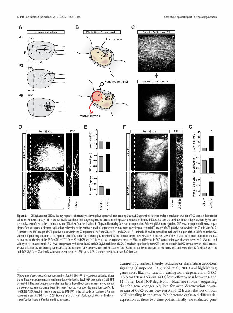
GSK3�, not GSK3�, is a key regulator of developmental axonpruning in vivoSpatially regulated pruning of RGC axons in the superior collicu-lus occurs during postnatal days 1 and 6 in the mouse and repre-sents an excellent model for testing spatially regulated axondegeneration during development (Fig. 5A). Pruning of RGCaxons during development was tracked by expressing GFP in theretinas of E13.5 embryos via in utero electroporation (Fig. 5B,C).As expected from our in vitro data, genetic deletion of GSK3� hadno effect on axon pruning (Fig. 5D,E).
To circumvent toxicity associated with systemic inhibition ofGSK3� during development, and to provide evidence of cell au-tonomy, we used an shRNA expression construct instead of the
ASKA knock-in animals. To knockdown GSK3� and simultane-ously label RGC projections, retinas of E13.5 embryos were co-electroporated with shRNA and GFP by in utero electroporation.Assessment of the selectivity of shRNA knockdown of GSK3�showed a �50% reduction in GSK3� mRNA as measured byRT-PCR (p � 0.05, Student’s t test; n � 3), while a modestinsignificant reduction in GSK3� expression was observed.Knockdown of GSK3� yielded robust RGC axon pruning defi-cits; compared with control shRNA, GSK3� knockdown pro-duced approximately fivefold more axons persisting at theposterior edge of the superior colliculus (Fig. 5F,G).
Identification of GSK3-regulated pro-degeneration genesdleu2 and tbx6As GSK3 regulates axon degeneration through activity in the cellbody, likely via regulation of transcription, we set out to identifypro-degeneration genes that are regulated in a GSK3-dependentfashion. To accomplish this, we used an unbiased approach usingmicroarrays to analyze RNA isolated from DRG neurons follow-ing NGF withdrawal in the presence or absence of GSK inhibi-tors. NGF deprivation was spatially limited to the axon using the
Figure 3. Long-term GSK3 inhibition reduces Wallerian degeneration. A, Diagram illustrating lesion experiment. DRG explants were cultured for 3 d, lesioned, and fixed after 12 h of degeneration.Lesioned axons show extensive degeneration at the microtubule level. B, Either DMSO, EGFR/ErbB inhibitor (10 �M AG555), GSK3 inhibitor (30 �M SB415286), or p38MAPK inhibitor (30 �M
SP239063) were applied immediately after axon lesion, and evaluated for microtubule degeneration 12 h later. Values represent mean SEM (*p � 0.05 compared with DMSO control, Student’st test; n � 3– 4). Of the tested inhibitors, only GSK3 inhibitor SB415286 reduces Wallerian degeneration. C, GSK3 inhibitor (30 �M SB415286) or JNK inhibitor (20 �M SP600125) were individuallyapplied after axon lesion and compared with 18 h of GSK3 inhibitor (30 �M SB415286) treatment before axon lesion. Long-term inhibition of GSK3 robustly inhibits axon degeneration followinglesion. D, Quantification of percentage degenerating axons, normalized to DMSO controls. Values represent mean SEM ( †p �0.05 compared with GSK3 inhibition postlesion, Student’s t test; n �3). Scale bars: A, C, top panels, 400 �m; C, bottom panels, 200 �m.
13446 • J. Neurosci., September 26, 2012 • 32(39):13439 –13453 Chen et al. • Spatial Regulation of Axon Degeneration

Figure 4. GSK3�, and not GSK3�, is a key regulator axon degeneration in vitro. A, Cultured DRG immunocytochemistry shows GSK3 enrichment in the soma. The yellow arrowheads point to neuronal cellbodies. B, Phosphorylation state of GSK3 isoforms in the cell body compartment following distal axon NGF deprivation. Over a period of 18 h, both isoforms of GSK3 are dephosphorylated, indicating elevatedkinase activity. Representative blot from three independent experiments is shown. Relative abundance of pGSK3�/total GSK3�and pGSK3�/total GSK3�are shown in the adjacent graph. Note the differencein total protein loaded for each lane. C, E13.5 DRG explants from GSK3�/, GSK3�/�, and GSK3��/�embryos were cultured overnight, and then deprived of NGF for 20 h. Genetic deletion of GSK3�hasno effect on axon degeneration following NGF deprivation. Values represent mean SEM (n � 3). D, Dissociated DRG neurons from GSK3�/, GSK3�/�, and GSK3��/� were cultured for 5 d inCampenot chambers and then deprived of NGF for 28 h. Genetic deletion of GSK3�does not significantly reduce local axon degeneration. Values represent meanSEM (n�5). E, HEK293 cells were transfectedwith wild-type GSK3� or GSK3� V110I/L132A/F175L. Kinase activities were determined in the presence of varying concentrations of 3MB-PP1 using anti-V5 immunoprecipitates and a continuous fluorescentkinase assay. 3MB-PP1 potently inhibits GSK3�-ASKA activity but not wild-type activity. Values represent meanSEM (*p�0.05, Wilcoxon’s test, 25�g of lysate, 20�M ATP, n�3). F, Images of DRG axonsafter20hNGFdeprivation,culturedfromASKAmutantsorwild-typeanimals. InthepresenceofASKAinhibitor3MB-PP1,ASKAmutantsshowsignificantlylessdegeneration.Wild-typeexplantsdegeneratewith3MB-PP1, indicating that GSK3� inhibition is specific. G, Quantification of reduced axon degeneration of GSK3�-ASKA knock-in DRGs in the presence of ASKA inhibitors. Values represent mean SEM (*p �0.05, Student’s t test; n � 9). H, Compartment-specific inhibition of GSK3� using chemical genetics. DRGs from GSK3�-ASKA knock-in embryos were cultured in (Figure legend continues.)
Chen et al. • Spatial Regulation of Axon Degeneration J. Neurosci., September 26, 2012 • 32(39):13439 –13453 • 13447
Campenot chamber, thereby reducing or eliminating apoptosissignaling (Campenot, 1982; Mok et al., 2009) and highlightinggenes most likely to function during axon degeneration. GSK3inhibitor (30 �M AR-A014418) loses effectiveness between 6 and12 h after local NGF deprivation (data not shown), suggestingthat the gene changes required for axon degeneration down-stream of GSK3 occur between 6 and 12 h after the loss of localNGF signaling in the axon. We therefore evaluated differentialexpression at these two time points. Finally, we evaluated gene
4
(Figure legend continued.) Campenot chambers for 5 d. 3MB-PP1 (10 �M) was added to eitherthe cell body or axon compartment immediately following local NGF deprivation. 3MB-PP1potently inhibits axon degeneration when applied to the cell body compartment alone, but notthe axon compartment alone. I, Quantification of reduced local axon degeneration, specificallyin GSK3�-ASKA knock-in neurons exposed to 3MB-PP1 in the cell body compartment. Valuesrepresent mean SEM (*p � 0.05, Student’s t test; n � 6). Scale bar: A, 60 �m. The high-magnification insets in F and H are 62 �m squares.
Figure 5. GSK3�, and not GSK3�, is a key regulator of naturally occurring developmental axon pruning in vivo. A, Diagram illustrating developmental axon pruning of RGC axons in the superiorcolliculus. At postnatal day 1 (P1), axons initially overshoot their target region and extend into the posterior superior colliculus (PSC). At P3, axons prune back through degeneration. By P6, axonterminals are confined to the termination zone (TZ), their final destination. B, Diagram illustrating in utero electroporation. Following DNA microinjection, DNA was electroporated by creating anelectric field with paddle electrodes placed on either side of the embryo’s head. C, Representative maximum intensity projection (MIP) images of GFP-positive axons within the SC at P1 and P6. D,Representative MIP images of GFP-positive axons within the SC at postnatal P6 from GSK3� / and GSK3� �/� animals. The white dotted line outlines the region of the SC defined as the PSC,shown in higher magnification to the right. E, Quantification of axon pruning as measured by the number of GFP-positive axons in the PSC, size of the TZ, and the number of axons in the PSCnormalized to the size of the TZ for GSK3� / (n � 5) and GSK3� �/� (n � 6). Values represent mean SEM. No difference in RGC axon pruning was observed between GSK3�-null andwild-type littermate controls. F, GFP was coexpressed with either shLacZ or shGSK3�. Knockdown of GSK3� results in significantly more GFP-positive axons in the PSC compared with shLacZ control.G, Quantification of axon pruning as measured by the number of GFP-positive axons in the PSC, size of the TZ, and the number of axons in the PSC normalized to the size of the TZ for shLacZ (n � 15)and shGSK3� (n � 9) animals. Values represent mean SEM (*p � 0.05, Student’s t test). Scale bar: B, C, 100 �m.
13448 • J. Neurosci., September 26, 2012 • 32(39):13439 –13453 Chen et al. • Spatial Regulation of Axon Degeneration

Figure 6. Time course microarray to identify candidate GSK3-driven axon degeneration genes. A, Expression profiling of genes following local NGF deprivation. RNA was isolated from neuronslocally deprived of NGF for 6 and 12 h, with and without GSK3 inhibition (30 �M AR-A014418) and compared with neurons globally maintained in NGF. Values represent mean SEM (n � 5). bdnfmRNA decreases and jun mRNA increases after local NGF deprivation, as expected. Dleu2 and tbx6 meet the criteria for candidate axon degeneration genes. They are upregulated after 12 h of localNGF deprivation in a GSK3-dependent manner. Results from the microarray experiment were verified by qRT-PCR. Samples from NGF control were compared with (Figure legend continues.)
Chen et al. • Spatial Regulation of Axon Degeneration J. Neurosci., September 26, 2012 • 32(39):13439 –13453 • 13449

changes following NGF withdrawal with and without GSK3 in-hibitor, further reducing the candidate list to gene changes thatare regulated by GSK3.
As expected, withdrawal of NGF from distal axons (mimick-ing loss of target-derived NGF) caused a decrease in the levels ofbrain-derived neurotrophic factor (bdnf) transcript (Deppmannet al., 2008) and an increase in jun transcript (Mok et al., 2009).We selected two genes identified in the microarray, transcriptionfactor t-box 6 (tbx6) and deleted in lymphocytic leukemia 2(dleu2), for further investigation. Probes for both of these genesrevealed upregulated expression when comparing control toNGF-deprived neurons, and they also revealed downregulatedexpression when comparing NGF-deprived to NGF-deprived/GSK3-inhibited neurons (Fig. 6A). Dleu2 is a long noncodingRNA and host gene for two micro-RNAs, mir15a and mir16-1(Liu et al., 1997; Migliazza et al., 2001), and tbx6 is involved inpatterning and somite formation (Chapman and Papaioannou,1998; Hadjantonakis et al., 2008). These results were verified byqRT-PCR, in which similar results were obtained except withbdnf, which showed similar trends but did not reach significance.
To show that upregulation of dleu2 and tbx6 plays a functionalrole in axon degeneration, siRNA targeting dleu2 or tbx6 werenucleofected into dissociated E13.5 DRG neurons before plating.After 1 d in culture, neurons were either maintained in NGF foranother 20 h, or deprived of NGF for 20 h with NGF neutralizingantibodies. Axon degeneration was then evaluated using an au-tomated analysis system, which was designed to evaluate frag-mentation of axons relative to the NGF deprivation/nontargetingsiRNA condition. Smaller values indicate less fragmentationand/or more continuous axons. In control DRG neurons main-tained in NGF, dleu2 and tbx6 siRNAs did not affect baseline axondegeneration. However, following NGF deprivation, siRNAs tar-geting either dleu2 or tbx6 significantly reduced axon degenera-tion (Fig. 6B,C). This effect was equivalent to �30% reduction indegenerating axons when determined manually (data notshown).
To more specifically evaluate the role of these genes in GSK3�-dependent degeneration, mixed hippocampal/cortical neurons weregrown 5 d to allow neurite outgrowth, and then transfected withsiRNA and a GSK3�S9A expression construct. GSK3�S9A is a mu-tant form of GSK3� that cannot be inactivated through phosphor-ylation at serine 9, thus representing a constitutively activated formof GSK3�. Cells were evaluated for neurite degeneration 3 d aftertransfection. GSK3�S9A overexpression resulted in a significant de-generation after 3 d, showing the GSK3 is not only necessary but also
sufficient to drive axon degeneration. When combined withGSK3�S9A, both dleu2 and tbx6 siRNA pools significantly reducedneurite degeneration downstream of GSK3�S9A (Fig. 6D,E). As acontrol, we cotransfected siRNA with a control vector and founddleu2 and tbx6 siRNA pools to have no detectable effect on baselineneurite degeneration 3 d after transfection (8 DIV) (Fig. 6D,E). De-generation was determined relative to GSK3�S9A/nontargetingsiRNA condition. CNS neurons were chosen in place of DRG neu-rons due to limitations with our transfection reagents. Nevertheless,these results suggest dleu2 and tbx6 may function in a similar man-ner across a variety of neuronal subtypes.
Concerted activity of pathways functioning in spatiallydistinct compartments regulates axon degenerationA number of pathways were identified in our small-moleculescreen. To understand how these pathways interact, if at all, weasked whether inhibition of any of the pathways we identifiedcould block GSK3 activation following NGF deprivation; such“initiator” pathways would lie upstream of a GSK3-mediatedtranscriptional axon degeneration program (Fig. 7A). NGF with-drawal drives GSK3� activation through dephosphorylation atserine 9 (Crowder and Freeman, 2000). We looked at variouspathway inhibitors and found that the p38MAPK inhibitor (30�M SB239063) and the CaMKK inhibitor [15 �M 7-oxo-7H-benzimidazo[2,1-a]benz[de]isoquinoline-3-carboxylic acid ace-tate (STO-609)] could prevent GSK3� dephosphorylationfollowing NGF withdrawal (Fig. 7B).
As described above, the proposed p38MAPK inhibitor,SB239063, is only effective when applied directly to axons under-going degeneration; we therefore reasoned that p38MAPK mightbe responsible for transducing the local trophic-deprivation sig-nal to the cell body. To test this hypothesis, axons were locallydeprived of NGF and either DMSO or SB239063 was applied tothe axon compartment. Local NGF deprivation causes upregula-tion of dleu2 and tbx6, and a downregulation of bdnf, consistentwith the microarray data. When local NGF deprivation is com-bined with local inhibition of p38MAPK with SB239063 in theaxon compartment, the upregulation of dleu2 and tbx6 are po-tently inhibited (reversal of bdnf downregulation did not reachsignificance) (Fig. 7C).
JNK inhibitors block axon degeneration, yet the JNK inhibitorAS60125 failed to block GSK3� dephosphorylation after NGFwithdrawal. This result might be explained by JNK acting down-stream or in parallel with GSK3 to regulate axon degeneration(Fig. 7D). To test these two possibilities, we looked at JNK acti-vation following 6 h NGF deprivation in the presence of small-molecule inhibitors. Phosphorylation at Thr183/Tyr185 servedas a readout for JNK activation (Fig. 7E). As in the case of GSK3,we found evidence that p38MAPK is upstream of JNK, withSB239063 partially reversing JNK phosphorylation followingNGF withdrawal. Interestingly, we found that GSK3 inhibitorSB415286 (30 �M) had no effect on JNK activation, suggestingJNK may act in parallel, as opposed to downstream of GSK3, toregulate axon degeneration.
DiscussionAxon degeneration requires signaling through various kinasepathways functioning in spatially distinct compartments. Work-ing in combination with other kinases, GSK3 likely functions as agatekeeper to a transcriptional axon degeneration program, driv-ing gene changes such as upregulation of dleu2 and tbx6 to regu-late distal axon degeneration.
4
(Figure legend continued.) 12 h NGF deprivation with and without GSK3 inhibition. Values rep-resent mean SEM (*p � 0.05 compared with NGF control; †p � 0.05 compared with 12 hNGF deprivation, Wilcoxon’s test; n � 8). B, DIV2 siRNA-transfected DRG neurons maintained inNGF (siRNA only) or following 20 h NGF deprivation (NGF deprivation). siRNA targeting dleu2and tbx6 do not affect baseline degeneration of cultured DRGs, but do reduce degenerationfollowing NGF deprivation. C, Automated quantification of degeneration in NGF control andafter 20 h NGF deprivation. Values are relative to the siControl/NGF deprivation condition andrepresent mean SEM (*p � 0.05 compared with siControl, Student’s t test; n � 3 wells from1 representative experiment). D, Cultured hippocampal/cortical neurons were transfected onDIV5 with siRNA and GFP, plus either Control vector or constitutively active GSK3 (GSK3�S9A orS9A). Neurons were fixed after 3 d of expression (8 DIV). The siRNA have no effect on baselineneurite degeneration when combined with Control vector. Both siDleu2 and siTbx6 reducerelative degeneration when cotransfected with constitutively active GSK3�. E, Automatedquantification of degeneration after cotransfection with dleu2 or tbx6 siRNA and either Controlvector or S9A (*p � 0.05, Student’s t test; n � 12 wells from a representative experiment).Values represent mean SEM. Scale bars: B, D, 100 �m.
13450 • J. Neurosci., September 26, 2012 • 32(39):13439 –13453 Chen et al. • Spatial Regulation of Axon Degeneration

Small-molecule screen identifies a number of pathwaysregulating axon degenerationMany of the pathways identified in our screen have been previ-ously investigated in the context of neurodegeneration, includingGSK3 (Kaytor and Orr, 2002), p38MAPK (Munoz and Ammit,2010), and JNK (Borsello and Forloni, 2007), while others such asCaMKK and EGFR/ErbB have limited prior evidence of playing arole in neurodegeneration.
It is difficult to validate these pathways through siRNA knock-down, as partial reduction in kinase levels may not be sufficient toblock the amplifying effect of kinase activation. Thus, further geneticvalidation of the pathways we characterize in these studies, with theexception of GSK3 and JNK, would provide additional evidence thatthe small molecules being used are acting via the proposed pathways.However, genetic validation also has its difficulties as the majority ofkinase pathways we describe have a number of isoforms or consist ofmultiple highly homologous genes. As an extreme example, validat-ing a role for ErbB signaling in axon degeneration would requireobtaining knock-outs for three of the four ErbB family members.Even when considering these important caveats, chemical inhibitorshave served as unique entry points for understanding the molecularmechanisms of axon degeneration.
Localization of axon degeneration pathways to distinctcellular compartmentsBy investigating these pathways in the Campenot chamber system,we found that the ErbB and p38MAPK inhibitors were protectiveonly at the site of degeneration. Unexpectedly, GSK3 inhibitors
blocked distal axon degeneration only when applied to the cellbody compartment, similar to a transcription inhibitor. Inmany instances in which axon degeneration was blocked witha GSK3 or transcription inhibitor, axons showed microtubulebulging/varicosities but were generally smooth contoured, incontrast to degenerating axons. This may result from blockingaxon degeneration downstream of these local changes, leavingaxons “frozen” in a degeneration-primed state. The overallspatial and morphological similarities observed between ax-ons protected with transcription and GSK3 inhibition led us toconsider whether GSK3 regulated axon degeneration acts viatranscription.
Examples of GSK3-regulated transcription are abundant; for ex-ample, GSK3 has been shown to regulate a large number of tran-scription factors, including �-catenin, c-Jun, and CREB (cAMPresponse element-binding protein), to name a few (Hur and Zhou,2010). Also, there are known processes in neurobiology in whichevents at a distal location require transcription for full execution. Forinstance, the immediate-early gene Arc/Arg3.1 is rapidly transcribedafter long-term potentiation-inducing stimuli and then transportedto the dendrite, where it is critical for the maintenance of synapticpotentiation (Bramham et al., 2008). Our data suggest GSK3 mayregulate axon degeneration in similar manner, inducing transcrip-tional changes in the cell body to prime axons for degeneration.
Consistent with a model in which GSK3� activation drives thetranscription of various genes to “prime” neurons for axon degen-eration (Fig. 7), we showed that p38MAPK inhibitors added to theaxon after local NGF deprivation blocked dleu2 and tbx6 transcrip-
Figure 7. Concerted activity of kinases functioning in spatially distinct compartments is necessary for developmental degeneration. A, Pathways that are required for GSK3-dependent “priming” aredescribed as “initiation” pathways. B, Dissociated DRG neurons were globally deprived of NGF for 6 h, with or without inhibitors of various pathways. Cell lysates were assayed for phospho-GSK3� (Ser9) and totalGSK3�. Only CaMKK inhibitor (15 �M STO-609) and p38MAPK inhibitor (30 �M SB239063) reduce dephosphorylation of GSK3� following NGF deprivation. Values represent mean SEM (* ,#p � 0.05compared with NGF deprivation/DMSO control, Student’s t test; n � 3). C, Local NGF deprivation/DMSO induces upregulation dleu2 and tbx6, and downregulation of bdnf. Applying the p38MAPK inhibitor (30�M SB239063) in the axon compartment with local NGF deprivation prevents upregulation of dleu2 and tbx6 but does not reverse the downregulation of bdnf (*p�0.05 compared with NGF control; †p�0.05compared with NGF deprivation/DMSO, Wilcoxon’s test; n �5). D, Does JNK act downstream of GSK3 or in parallel? E, Cell lysates from B were assayed for pJNK activity. GSK3 inhibitor (30 �M SB415286) doesnot prevent JNK phosphorylation after NGF withdrawal. Values represent mean SEM (* ,#p � 0.05 compared with NGF deprivation/DMSO, Student’s t test; n � 3).
Chen et al. • Spatial Regulation of Axon Degeneration J. Neurosci., September 26, 2012 • 32(39):13439 –13453 • 13451

tional upregulation, suggesting that p38MAPK and possibly otherpathways may function in the axon as an “initiators” upstream ofGSK3. Nevertheless, it remains to be determined whether p38MAPKor other pathways are sufficient to drive axon degeneration, similarto GSK3. Although phosphorylation of GSK3� at Ser9 is only onemeans of regulating activity, our data nevertheless provide a tenta-tive link between local degeneration signals acting in the axon andglobal transcriptional changes in the cell body that ultimately “exe-cute” axon degeneration. In summary, the requirement of multiplestress pathways acting in distinct cellular compartments may pro-vide spatial cues for restricting degeneration, as well as providing asafeguard against unwanted axon loss.
Although our data suggest GSK3 regulates developmentalaxon degeneration primarily through its activity in the cell body,it is important to note that this mechanism does not necessarilytranslate to Wallerian degeneration. GSK3 inhibition after axonlesion still reduces axon degeneration, indicating GSK3 can atleast partially regulate degeneration directly at the site of degen-eration in an injury paradigm. Additionally, our threshold fordetermining percentage degeneration was very low; any axonsthat showed rough edges at the microtubule level were deemed“degenerated.” It is possible that another measure axon degener-ation, such as percentage axon fragmentation, would reveal anaxon-specific role for GSK3 in developmental axon degeneration.
Evidence that dleu2 and tbx6 mediate axon degenerationdownstream of GSK3To provide additional evidence for this proposed transcrip-tional axon degeneration program, we set out to identify genesthat may function downstream of GSK3 to regulate axon de-generation. In our microarray experiment, two GSK3-regulated axon degeneration genes were identified: dleu2 andtbx6. Functional analysis of these two genes provides an addi-tional layer of evidence for our proposed GSK3-dependentaxon degeneration program and identifies dleu2 and tbx6 asnovel regulators of axon degeneration. Multiple siRNAs tar-geting either dleu2 or tbx6 modestly reduced axon degenera-tion, yielding �30% of the protection obtained with GSK3small-molecule inhibitors in the growth factor withdrawal as-say. These results were initially surprising as we suspected thatindividually modulating two genes from a genetic program,which may include the simultaneous upregulation and down-regulation of a large number of genes, would not necessarily elicitany phenotype. Nevertheless, we found a reproducible protectiveeffect with knockdown of either dleu2 or tbx6. Additionally, weused a constitutively active version of GSK3� (GSK3�S9A) toshow that GSK3 signaling is sufficient to induce axon degenera-tion and to provide evidence that dleu2 and tbx6 lie downstreamof GSK3-induced degeneration. Notably, we did not observe pro-tection of axons from degeneration with either dleu2 or tbx6knockdown after lesion-induced axon degeneration (data notshown). These data suggest that the ability of GSK to mediateaxon degeneration in development and after injury may diverge.It also remains to be determined whether knocking down bothgenes, or other genes identified from our screen, will have an evenmore robust effect in modulating axon degeneration.
GSK3�, not GSK3�, is the primary regulator ofdevelopmental axon degeneration in vitro and in vivoDespite using multiple, structurally unrelated inhibitors for each ofour pathways, we could not rule out “off-target” effects (Bain et al.,2003). To address this issue, and also provide isoform-specific anal-ysis of GSK3, we used gene deletion and chemical–genetics.
Through these tools, we found that GSK3� is the primary regulatorof developmental axon pruning, whereas GSK3� has little to no rolein axon degeneration. By using GSK3�-ASKA knock-in neuronscombined with ASKA-specific inhibitors, we were also able to verifythat GSK3� acts primarily in the cell body to regulate axon degener-ation following local trophic factor deprivation.
Pruning of RGC axons during development is a cell death-independent process that results in a substantial, spatially re-stricted loss of axonal material from P1 to P6. Consistent withour in vitro data, there was a pruning deficit with GSK3�knockdown but no detectable pruning phenotype with geneticdeletion of GSK3�. It has previously been reported that inhi-bition of GSK3� can promote axon growth and branching(Zhou et al., 2004; Kim et al., 2006; Bilimoria et al., 2010).Based on our time-lapse experiments, there was negligiblebranching or axon growth following NGF deprivation/GSK3inhibition (data not shown). We therefore speculate that theapparent RGC pruning deficit is indeed the result of blockingaxon degeneration, rather than promoting axon branching/growth. Determining whether branching/growth contributesto the increase in axons at the posterior superior colliculuswith GSK3� knockdown will require additional analysis ofRGC pruning at more time points or at higher resolution.
It may seem inconsistent that GSK3� is the primary regulator ofaxon degeneration, even though both GSK3 isoforms are activatedin the cell body to a similar extent following NGF deprivation, asdetermined by dephosphorylation at Ser21 or Ser9. However, regu-lation of GSK3 signaling is intricate. Formation of protein com-plexes, subcellular localization, and � isoform-specific inactivationby p38MAPK phosphorylation at Ser389 are examples of otherfactors that may regulate GSK3 isoform-specific activity (Hurand Zhou, 2010). Additionally, GSK3� is restricted from thenucleus by its N-terminal region, whereas GSK3� shuttlesfreely between the nucleus and cytoplasm (Azoulay-Alfaguteret al., 2011). Nuclear GSK3� is highly active, and its localiza-tion in the nucleus decreases following stimulation withgrowth factors and increases under apoptosis conditions (Bi-jur and Jope, 2001, 2003). These observations by others areconsistent with a model in which GSK3�, not GSK3�, is com-petent to mediate a transcriptional cascade to regulate axondegeneration.
ReferencesAzoulay-Alfaguter I, Yaffe Y, Licht-Murava A, Urbanska M, Jaworski J, Pi-
etrokovski S, Hirschberg K, Eldar-Finkelman H (2011) Distinct molec-ular regulation of glycogen synthase kinase-3alpha isozyme controlled byits N-terminal region: functional role in calcium/calpain signaling. J BiolChem 286:13470 –13480.
Bain J, McLauchlan H, Elliott M, Cohen P (2003) The specificities of proteinkinase inhibitors: an update. Biochem J 371:199 –204.
Bijur GN, Jope RS (2001) Proapoptotic stimuli induce nuclear accumula-tion of glycogen synthase kinase-3 beta. J Biol Chem 276:37436 –37442.
Bijur GN, Jope RS (2003) Glycogen synthase kinase-3 beta is highly acti-vated in nuclei and mitochondria. Neuroreport 14:2415–2419.
Bilimoria PM, de la Torre-Ubieta L, Ikeuchi Y, Becker EB, Reiner O, Bonni A(2010) A JIP3-regulated GSK3beta/DCX signaling pathway restrictsaxon branching. J Neurosci 30:16766 –16776.
Bishop AC, Ubersax JA, Petsch DT, Matheos DP, Gray NS, Blethrow J, Shi-mizu E, Tsien JZ, Schultz PG, Rose MD, Wood JL, Morgan DO, ShokatKM (2000) A chemical switch for inhibitor-sensitive alleles of any pro-tein kinase. Nature 407:395– 401.
Borsello T, Forloni G (2007) JNK signalling: a possible target to preventneurodegeneration. Curr Pharm Des 13:1875–1886.
Bramham CR, Worley PF, Moore MJ, Guzowski JF (2008) The immediateearly gene arc/arg3.1: regulation, mechanisms, and function. J Neurosci28:11760 –11767.
13452 • J. Neurosci., September 26, 2012 • 32(39):13439 –13453 Chen et al. • Spatial Regulation of Axon Degeneration

Brion JP, Anderton BH, Authelet M, Dayanandan R, Leroy K, Lovestone S,Octave JN, Pradier L, Touchet N, Tremp G (2001) Neurofibrillary tan-gles and tau phosphorylation. Biochem Soc Symp 2001:81– 88.
Campenot RB (1977) Local control of neurite development by nerve growthfactor. Proc Natl Acad Sci U S A 74:4516 – 4519.
Campenot RB (1982) Development of sympathetic neurons in compart-mentalized cultures. II. Local control of neurite survival by nerve growthfactor. Dev Biol 93:13–21.
Chapman DL, Papaioannou VE (1998) Three neural tubes in mouse embryoswith mutations in the T-box gene Tbx6. Nature 391:695– 697.
Cross DA, Alessi DR, Cohen P, Andjelkovich M, Hemmings BA (1995) In-hibition of glycogen synthase kinase-3 by insulin mediated by proteinkinase B. Nature 378:785–789.
Crowder RJ, Freeman RS (2000) Glycogen synthase kinase-3 beta activity iscritical for neuronal death caused by inhibiting phosphatidylinositol3-kinase or Akt but not for death caused by nerve growth factor with-drawal. J Biol Chem 275:34266 –34271.
Deckwerth TL, Johnson EM Jr (1994) Neurites can remain viable after de-struction of the neuronal soma by programmed cell death (apoptosis).Dev Biol 165:63–72.
Deppmann CD, Mihalas S, Sharma N, Lonze BE, Niebur E, Ginty DD (2008)A model for neuronal competition during development. Science320:369 –373.
Doble BW, Patel S, Wood GA, Kockeritz LK, Woodgett JR (2007) Func-tional redundancy of GSK-3alpha and GSK-3beta in Wnt/beta-cateninsignaling shown by using an allelic series of embryonic stem cell lines. DevCell 12:957–971.
Garcia-Frigola C, Carreres MI, Vegar C, Herrera E (2007) Gene deliveryinto mouse retinal ganglion cells by in utero electroporation. BMC DevBiol 7:103.
Gerdts J, Sasaki Y, Vohra B, Marasa J, Milbrandt J (2011) Image-basedscreening identifies novel roles for IkappaB kinase and glycogen synthasekinase 3 in axonal degeneration. J Biol Chem 286:28011–28018.
Ghosh AS, Wang B, Pozniak CD, Chen M, Watts RJ, Lewcock JW (2011)DLK induces developmental neuronal degeneration via selective regula-tion of proapoptotic JNK activity. J Cell Biol 194:751–764.
Glass JD, Brushart TM, George EB, Griffin JW (1993) Prolonged survival oftransected nerve fibres in C57BL/Ola mice is an intrinsic characteristic ofthe axon. J Neurocytol 22:311–321.
Hadjantonakis AK, Pisano E, Papaioannou VE (2008) Tbx6 regulates left/right patterning in mouse embryos through effects on nodal cilia andperinodal signaling. PLoS One 3:e2511.
Hoeflich KP, Luo J, Rubie EA, Tsao MS, Jin O, Woodgett JR (2000) Require-ment for glycogen synthase kinase-3beta in cell survival and NF-kappaBactivation. Nature 406:86 –90.
Hoopfer ED, McLaughlin T, Watts RJ, Schuldiner O, O’Leary DD, Luo L(2006) Wlds protection distinguishes axon degeneration following in-jury from naturally occurring developmental pruning. Neuron 50:883–895.
Hur EM, Zhou FQ (2010) GSK3 signalling in neural development. Nat RevNeurosci 11:539 –551.
Kaytor MD, Orr HT (2002) The GSK3 beta signaling cascade and neurode-generative disease. Curr Opin Neurobiol 12:275–278.
Kim WY, Zhou FQ, Zhou J, Yokota Y, Wang YM, Yoshimura T, Kaibuchi K,Woodgett JR, Anton ES, Snider WD (2006) Essential roles for GSK-3sand GSK-3-primed substrates in neurotrophin-induced and hippocam-pal axon growth. Neuron 52:981–996.
Klein U, Lia M, Crespo M, Siegel R, Shen Q, Mo T, Ambesi-Impiombato A,Califano A, Migliazza A, Bhagat G, Dalla-Favera R (2010) The DLEU2/miR-15a/16-1 cluster controls B cell proliferation and its deletion leads tochronic lymphocytic leukemia. Cancer Cell 17:28 – 40.
Kuruvilla R, Ye H, Ginty DD (2000) Spatially and functionally distinct rolesof the PI3-K effector pathway during NGF signaling in sympathetic neu-rons. Neuron 27:499 –512.
Lee T, Marticke S, Sung C, Robinow S, Luo L (2000) Cell-autonomous re-quirement of the USP/EcR-B ecdysone receptor for mushroom body neu-ronal remodeling in Drosophila. Neuron 28:807– 818.
Liu Y, Corcoran M, Rasool O, Ivanova G, Ibbotson R, Grander D, Iyengar A,Baranova A, Kashuba V, Merup M, Wu X, Gardiner A, Mullenbach R,Poltaraus A, Hultstrom AL, Juliusson G, Chapman R, Tiller M, Cotter F,Gahrton G, et al. (1997) Cloning of two candidate tumor suppressor geneswithin a 10 kb region on chromosome 13q14, frequently deleted inchronic lymphocytic leukemia. Oncogene 15:2463–2473.
Lovestone S, Hartley CL, Pearce J, Anderton BH (1996) Phosphorylation oftau by glycogen synthase kinase-3 beta in intact mammalian cells: theeffects on the organization and stability of microtubules. Neuroscience73:1145–1157.
MacAulay K, Doble BW, Patel S, Hansotia T, Sinclair EM, Drucker DJ, NagyA, Woodgett JR (2007) Glycogen synthase kinase 3alpha-specific regu-lation of murine hepatic glycogen metabolism. Cell Metab 6:329 –337.
McLaughlin T, Torborg CL, Feller MB, O’Leary DD (2003) Retinotopicmap refinement requires spontaneous retinal waves during a brief criticalperiod of development. Neuron 40:1147–1160.
Meijer L, Flajolet M, Greengard P (2004) Pharmacological inhibitors of gly-cogen synthase kinase 3. Trends Pharmacol Sci 25:471– 480.
Migliazza A, Bosch F, Komatsu H, Cayanis E, Martinotti S, Toniato E, Guc-cione E, Qu X, Chien M, Murty VV, Gaidano G, Inghirami G, Zhang P,Fischer S, Kalachikov SM, Russo J, Edelman I, Efstratiadis A, Dalla-FaveraR (2001) Nucleotide sequence, transcription map, and mutation analysisof the 13q14 chromosomal region deleted in B-cell chronic lymphocyticleukemia. Blood 97:2098 –2104.
Mok SA, Lund K, Campenot RB (2009) A retrograde apoptotic signal orig-inating in NGF-deprived distal axons of rat sympathetic neurons in com-partmented cultures. Cell Res 19:546 –560.
Munoz L, Ammit AJ (2010) Targeting p38 MAPK pathway for the treat-ment of Alzheimer’s disease. Neuropharmacology 58:561–568.
Nikolaev A, McLaughlin T, O’Leary DD, Tessier-Lavigne M (2009) APPbinds DR6 to trigger axon pruning and neuron death via distinct caspases.Nature 457:981–989.
Perry VH, Brown MC, Lunn ER, Tree P, Gordon S (1990) Evidence that veryslow Wallerian degeneration in C57BL/Ola mice is an intrinsic propertyof the peripheral nerve. Eur J Neurosci 2:802– 808.
Raff MC, Whitmore AV, Finn JT (2002) Axonal self-destruction and neu-rodegeneration. Science 296:868 – 871.
Sang H, Lu Z, Li Y, Ru B, Wang W, Chen J (2001) Phosphorylation of tau byglycogen synthase kinase 3beta in intact mammalian cells influences thestability of microtubules. Neurosci Lett 312:141–144.
Tessier-Lavigne M, Placzek M, Lumsden AG, Dodd J, Jessell TM (1988)Chemotropic guidance of developing axons in the mammalian centralnervous system. Nature 336:775–778.
Wakatsuki S, Saitoh F, Araki T (2011) ZNRF1 promotes Wallerian degen-eration by degrading AKT to induce GSK3B-dependent CRMP2 phos-phorylation. Nat Cell Biol 13:1415–1423.
Watts RJ, Hoopfer ED, Luo L (2003) Axon pruning during Drosophila meta-morphosis: evidence for local degeneration and requirement of theubiquitin-proteasome system. Neuron 38:871– 885.
Weimann JM, Zhang YA, Levin ME, Devine WP, Brulet P, McConnell SK(1999) Cortical neurons require Otx1 for the refinement of exuberantaxonal projections to subcortical targets. Neuron 24:819 – 831.
Woodgett JR (1990) Molecular cloning and expression of glycogen synthasekinase-3/factor A. EMBO J 9:2431–2438.
Zhai Q, Wang J, Kim A, Liu Q, Watts R, Hoopfer E, Mitchison T, Luo L, He Z(2003) Involvement of the ubiquitin-proteasome system in the earlystages of Wallerian degeneration. Neuron 39:217–225.
Zhang YA, Okada A, Lew CH, McConnell SK (2002) Regulated nuclear traf-ficking of the homeodomain protein otx1 in cortical neurons. Mol CellNeurosci 19:430 – 446.
Zhou FQ, Snider WD (2005) Cell biology. GSK-3beta and microtubule as-sembly in axons. Science 308:211–214.
Zhou FQ, Zhou J, Dedhar S, Wu YH, Snider WD (2004) NGF-induced axongrowth is mediated by localized inactivation of GSK-3beta and functionsof the microtubule plus end binding protein APC. Neuron 42:897–912.
Chen et al. • Spatial Regulation of Axon Degeneration J. Neurosci., September 26, 2012 • 32(39):13439 –13453 • 13453