cellular/molecular ph ... · cellular/molecular ph-dependentinhibitionofkainatereceptorsbyzinc...
TRANSCRIPT

Cellular/Molecular
pH-Dependent Inhibition of Kainate Receptors by Zinc
David D. Mott,1 Morris Benveniste,2 and Raymond J. Dingledine3
1Department of Pharmacology, Physiology, and Neuroscience, University of South Carolina School of Medicine, Columbia, South Carolina 29208,2Neuroscience Institute, Morehouse School of Medicine, Atlanta, Georgia 30310, and 3Department of Pharmacology, Emory University School of Medicine,Atlanta, Georgia 30322
Kainate receptors contribute to synaptic plasticity and rhythmic oscillatory firing of neurons in corticolimbic circuits including hip-pocampal area CA3. We use zinc chelators and mice deficient in zinc transporters to show that synaptically released zinc inhibitspostsynaptic kainate receptors at mossy fiber synapses and limits frequency facilitation of kainate, but not AMPA EPSCs during theta-pattern stimulation. Exogenous zinc also inhibits the facilitatory modulation of mossy fiber axon excitability by kainate but does notsuppress the depressive effect of kainate on CA3 axons. Recombinant kainate receptors are inhibited in a subunit-dependent manner byphysiologically relevant concentrations of zinc, with receptors containing the KA1 subunit being sensitive to submicromolar concentra-tions of zinc. Zinc inhibition does not alter receptor desensitization nor apparent agonist affinity and is only weakly voltage dependent,which points to an allosteric mechanism. Zinc inhibition is reduced at acidic pH. Thus, in the presence of zinc, a fall in pH potentiateskainate receptors by relieving zinc inhibition. Acidification of the extracellular space, as occurs during repetitive activity, may thereforeserve to unmask kainate receptor neurotransmission. We conclude that zinc modulation of kainate receptors serves an important role inshaping kainate neurotransmission in the CA3 region.
Key words: zinc; kainate receptor; CA3; glutamate receptor; mossy fiber; hippocampus; pH
IntroductionGlutamate mediates fast synaptic transmission through NMDA,AMPA, and kainate receptors (Dingledine et al., 1999). Kainatereceptor function has been studied at many synapses (Huettner,2003; Lerma, 2003), but has been best characterized at synapsesbetween mossy fibers (MFs) and CA3 pyramidal cells in the hip-pocampus (Castillo et al., 1997; Vignes and Collingridge, 1997).At mossy fiber synapses, postsynaptic kainate receptors produceexcitatory currents (Castillo et al., 1997) and contribute to tem-poral summation of synaptic input (Frerking and Ohliger-Frerking, 2002). Presynaptic kainate receptors contribute to fre-quency facilitation (Schmitz et al., 2001) and long-termpotentiation (Bortolotto et al., 1999). In addition, kainate recep-tors regulate excitability of mossy fiber axons (Kamiya andOzawa, 2000; Schmitz et al., 2000). Low kainate concentrationsenhance, whereas high concentrations depress mossy fiber excit-ability. Moreover, kainate receptors at these synapses contributeto seizure development (Mulle et al., 1998). The important role ofkainate receptors in mossy fiber synaptic transmission suggeststhat endogenous agents that regulate kainate receptor functionmay modulate excitability of CA3 neurons.
In this study, we examined regulation of kainate receptor
function by zinc. Zinc is concentrated in excitatory terminalsthroughout the brain and is particularly abundant in terminals ofhippocampal mossy fibers (Frederickson and Danscher, 1990;Frederickson et al., 2000). In these terminals, zinc is colocalizedwith glutamate in synaptic vesicles. Zinc is released in a calcium-dependent manner, after multiple (Assaf and Chung, 1984; How-ell et al., 1984) or single (Quinta-Ferreira et al. 2004) mossy fiberstimuli, then taken up by zinc transport processes (Cole et al.,1999) and refilled into synaptic vesicles (Sudhof, 1995; Clemens,1996). Previous estimates of the peak synaptic zinc concentrationduring neuronal activity are 10 –30 �M (Thompson et al., 2000; Liet al., 2001b; Ueno et al., 2002) (but see Kay, 2003). Resting zincconcentration is in the low nanomolar range because of high-affinity binding of zinc to extracellular matrix proteins.
Endogenous zinc inhibits postsynaptic NMDA and GABAA
receptors in hippocampus (Vogt et al., 2000; Ueno et al., 2002;Ruiz et al., 2004). NR2A containing NMDA receptors are mostsensitive to zinc, being inhibited by low nanomolar concentra-tions (Chen et al., 1997; Paoletti et al., 1997). AMPA receptors areunaffected or even potentiated by physiologically relevant con-centrations of zinc (Mayer et al., 1989; Dreixler and Leonard,1994; Lin et al., 2001). At native kainate receptors the effects ofzinc have not been studied, however zinc was reported to inhibitrecombinant glutamate receptor 6 (GluR6) (Hoo et al., 1994) andGluR6/KA2 kainate receptors (Fukushima et al., 2003).
We found that endogenous zinc inhibits postsynaptic kainatereceptors at mossy fiber synapses. This zinc inhibition limits fre-quency facilitation of kainate, but not AMPA receptors. Zincinhibition of kainate receptors is subunit dependent with KA1containing receptors being sensitive to submicromolar zinc con-centrations. Furthermore, protons relieve zinc inhibition, pro-
Received Aug. 6, 2007; revised Jan. 1, 2008; accepted Jan. 5, 2008.This work was supported by the Epilepsy Foundation (D.M.), National Alliance for Research on Schizophrenia and
Depression (D.M.), the University Research Council of Emory University (D.M.), National Institute of NeurologicalDisorders and Stroke (R.D.), and the South Carolina Research Foundation (D.M.). We thank S. Misra, N. Ciliax, S.Zhang, and R. Shaw for excellent technical assistance.
Correspondence should be addressed to Dr. David D. Mott, Department of Pharmacology, Physiology, and Neu-roscience, School of Medicine, University of South Carolina, Columbia, SC 29208. E-mail: [email protected].
DOI:10.1523/JNEUROSCI.3567-07.2008Copyright © 2008 Society for Neuroscience 0270-6474/08/281659-13$15.00/0
The Journal of Neuroscience, February 13, 2008 • 28(7):1659 –1671 • 1659

ducing an apparent potentiation of the response. Acidification ofthe extracellular space during strong activation of the synapsemay therefore unmask kainate neurotransmission that is toni-cally inhibited by released zinc.
Materials and MethodsPreparation of hippocampal slices. Transverse brain slices were preparedfrom isoflurane-anesthetized male Sprague Dawley rats or mocha mu-tant mice using a vibratome (VT1000S; Leica, Nussloch, Germany).Brain slices were 300 �m thick for whole-cell recording (16- to 27-d-oldrats) and 500 �m thick for field potential recording (21- to 45-d-old ratsor adult mice). Brain slices were incubated in room temperature ACSF(artificial CSF) containing (in mM) 120 NaCl, 3.3 KCl, 1.0 NaH2PO4, 25NaHCO3, 10 glucose, 0.5 CaCl2, and 5 MgCl2 and bubbled with a 95%O2/5% CO2 gas mixture at pH 7.4. Osmolarity was 301–308 mOsm.
Whole-cell recording. Slices were individually transferred to a recordingchamber maintained at 32–34°C and continuously perfused with oxy-genated ACSF, pH 7.4, containing 1.5 mM CaCl2 and 1.5 mM MgCl2. Forexperiments at pH 6.7, the ACSF contained 5 mM NaHCO3 and 140 mM
NaCl. Whole-cell recordings were obtained using borosilicate glass elec-trodes (3–10 M�) filled with an internal solution containing (in mM) 130Cs-gluconate, 5 CsCl, 10 HEPES, 0.5 CaCl2, 5 Cs-BAPTA, 2 MgCl2, 2MgATP, and 0.3 NaGTP, pH 7.3. Whole-cell patch-clamp recordingswere made from pyramidal cells in the CA3b region of the hippocampus,visually identified with infrared-differential interference contrast optics.Voltage-clamp recordings were made at a holding potential of �60 mV.Series resistance was 10 –25 M�, and recordings in which series resis-tance changed significantly were discarded.
Miniature synaptic currents were recorded in the presence of 1 �M
tetrodotoxin and 100 �M each of CaEDTA, bicuculline methochloride,and D-APV. Where indicated, synaptic currents were evoked by a glasselectrode filled with ACSF placed in stratum lucidum of CA3b. AMPAEPSCs were recorded as composite MF-EPSCs with ACSF containing 100�M each bicuculline methochloride, D-APV, and CaEDTA. MF-evokedEPSCs were identified by their large paired-pulse facilitation (�200% ata 50 ms interval) (Salin et al., 1996), rapid rise time, and short latency.These EPSCs were inhibited by the group II mGluR agonist (2S,2�R,3�R)-2-(2�,3�-dicarboxycyclopropyl)glycine (DCG-IV; 1 �M), which wasbath applied in each experiment. Kainate EPSCs were recorded fromCA3 pyramidal cells in the presence of 100 �M each of the AMPAantagonist 4-(8-methyl-9H-1,3-dioxolo[4,5-h][2,3]benzodiazepin-5-yl)-benzenamine (GYKI 52466), CaEDTA, bicuculline methochloride,and D-APV. Small kainate EPSCs that were obscured by stimulation ar-tifacts were resolved by the subtraction of a template stimulation tracethat was acquired after application of CNQX (100 �M) at the end of therecordings.
Responses were recorded using a Multiclamp 700A amplifier and fil-tered at 1 kHz. Responses were digitized by a Digidata 1320 analog-to-digital (A-D) board (Molecular Devices, Sunnyvale, CA) in a Windows-based computer using pClamp 9 software.
Field-potential recording. For recording, slices were placed into a smallsubmersion chamber maintained at 32–34°C and held in place by a bentpiece of platinum wire resting on the surface of the slice. Slices wereperfused with ACSF containing 1.5 mM CaCl2 and 1.5 mM MgCl2.
Stimuli were 0.1 ms, monophasic, cathodal, rectangular, constant-current pulses (10 –700 �A) delivered through a monopolar platinum-iridium electrode referenced to a bath ground. Mossy fibers were stimu-lated either in the granule cell layer or in stratum lucidum of CA3c.Associational fibers were stimulated in stratum radiatum of CA3b. Forextracellular recording, a glass pipette (resistance 5 M�) filled with re-cording ACSF was placed in stratum lucidum of CA3b to record mossyfiber field EPSPs (fEPSPs) or in the granule cell layer to record mossyfiber antidromic spikes. Responses were recorded using an Axopatch 200amplifier and filtered at 1 kHz. Responses were digitized by a Digidata1200 A-D board (Molecular Devices) in a Windows-based computerusing pClamp 9 software.
To examine the effect of zinc chelators, mossy fibers were stimulatedusing theta-pattern stimulation in which brief bursts of stimuli (100 Hz,
five pulses) were delivered to the mossy fibers every 200 ms for 1 s. Thisstimulation pattern was chosen as it simulates the discharge pattern ofgranule cells during behaviors associated with theta rhythm generation(Rose et al., 1983; Jung and McNaughton, 1993; Henze et al., 2002).
Oocyte preparation and injection. Xenopus oocytes were prepared andinjected as described previously (Mott et al., 2003). Briefly, stage V–VIoocytes were isolated from anesthetized frogs, enzymatically treated bygentle shaking with collagenase (type IV; Worthington Biochemical,Lakewood, NJ; 1.7 mg/ml for 45–120 min) in a calcium-free Barth’ssolution and then (in some cases) manually defolliculated. Cells wereinjected with up to 50 ng of mRNA transcribed from linearized con-structs in either a pGEM-HE, pSGEM, or Bluescript (Stratagene, La Jolla,CA) vector. For heteromeric receptors, mRNA was injected at either a10:1 ratio (GluR2:GluR3), a 1:6 ratio (GluR6:KA1 and GluR6:KA2), a 1:3ratio (GluR5:KA2; NR1:NR2), or a 1:1 ratio (GluR5Q:GluR6R). Injectedoocytes were maintained at 17°C in Barth’s solution containing genta-mycin (100 �g/ml), penicillin (10 U/ml), and streptomycin (10 �g/ml)for 3–10 d, after which two electrode voltage-clamp recordings weremade at room temperature (23–25°C) from cells continually perfused ina standard frog Ringer’s solution. This solution contained (in mM) 90NaCl, 1 KCl, 15 HEPES, 0.1 CaCl2, and 0.4 MgCl2. The contaminatingzinc concentration in this solution was measured by mass spectrometryto be 77 nM. Recording pipettes were filled with 3 M CsCl plus 0.4 M
EGTA, pH 7.5, to chelate Ca 2� and thereby minimize the activation ofcalcium-dependent chloride currents. GluR6/KA1, GluR6/KA2, andGluR5/KA2 receptors were activated with AMPA (30 –300 �M). AMPAselectively activates heteromeric kainate receptors containing GluR6 andKA1 or KA2 subunits, but not GluR6 homomeric receptors (Seeburg,1993). GluR5Q/GluR6R receptors were activated with 5-iodowillardiineand homomeric GluR5 and GluR6 receptors were activated by domoate(3–10 �M) or kainate (30 �M). To reduce desensitization when kainatewas used as the agonist, oocytes were pretreated with concanavalin-A(0.3 mg/ml for 3 min) and then washed for at least 10 min before use.NMDA receptors were activated using NMDA (100 �M) and glycine (10�M), and AMPA receptors were activated with kainate (300 �M). WhenNMDA receptors were studied, the MgCl2 in the Ringer’s solution wasreplaced with 0.4 mM BaCl2. Currents were elicited from a holding po-tential of �70 mV except where specified. Current signals were digitizedat 1 kHz using a Digidata 1200 analog-to-digital converter (MolecularDevices). Current–voltage curves during steady-state current responseswere generated using voltage ramps from �100 mV to �70 mV over aperiod of 1.3 s. Ramp currents were analyzed by subtracting the averageof the leak current before and after agonist application from the currentobtained in the presence of agonist. At least three ramps were recordedand averaged for each condition in each oocyte. To study the effect of pH,oocytes were perfused with Ringer’s solution at the desired pH for 30 – 60s or until a stable baseline had been reached before subsequent agonistapplication. Application of each of the agonists produced a stable, rapidlyrising and nondesensitizing or weakly desensitizing current in the major-ity of oocytes. Oocytes in which the current was not stable or in which thebaseline holding current drifted by �10% were discarded.
Data analysis. Analysis was performed using pClamp (Molecular De-vices), Origin (MicroCal, Northampton, MA) and Prism (GraphPad,San Diego, CA) software packages. Statistical comparisons were per-formed using the appropriate Student’s t test or ANOVA with post hoctests. Values are given as mean � SE. Concentration–inhibition relation-ships were fitted by nonlinear regression to a logistic equation of the form I�Imin � (100 � Imin)/(1 � ([A]/IC50)nH) where I is the current at a givenconcentration of agonist ([A]), Imin is the minimum current, IC50 is theantagonist concentration producing half-maximal inhibition of the current,and nH is the Hill coefficient. For most experiments, Imin was set to 0.
Miniature EPSCs (mEPSCs) were analyzed using MiniAnalysis 5.6(SynaptoSoft, Decatur, GA). For each event, the rise time (10 –90%),amplitude, and decay time constants were calculated. The detectionthreshold was set 2 pA above the baseline noise level (3–5 pA). In theabsence of AMPA antagonists, miniature AMPA, kainate, and mixedevents were distinguished based on their kinetics (Cossart et al., 2002).To do this, a 5 min window of data was obtained in each experimentalcondition in a single experiment. All events in this window were individ-
1660 • J. Neurosci., February 13, 2008 • 28(7):1659 –1671 Mott et al. • Zinc Modulation of Kainate Receptors

ually fitted using the MiniAnalysis program. Mixed events were identi-fied as those in which the decay was best fit by a double exponentialfunction. The SD of the fit given by the MiniAnalysis program was usedto determine whether one or two exponentials best fit the decays. Eventsbest described by a single exponential decay constant were then separatedbased on their decay time constant as described by Cossart et al. (2002).All monoexponential decay times were plotted against rise times for agiven experimental window. Using this graph, fast AMPA and slow kai-nate mEPSCs could be easily differentiated. The kinetic limit of the decaytime to separate fast AMPA and slow kainate mEPSCs was �25 ms.
The effect of zinc on cumulative charge transfer for kainate and AMPAreceptor-mediated mEPSCs was determined by comparing the total areaof the mEPSC current for 3 min windows of data obtained from control,zinc, and wash in each experiment. Igor Pro (WaveMetrics, Lake Os-wego, OR) with a custom-written macro was used to measure baselinesubtracted charge transfer in 1 s epochs during the 3 min window. Thesevalues were summed to yield cumulative charge transfer.
Buffered zinc solutions. For zinc concentration–response curves weused tricine to buffer zinc concentrations �1 �M. These solutions wereprepared according to the methods of Zheng et al. (1998) using theempirically determined binding constant of 10 �5
M (Paoletti et al., 1997).At pH 7.5, we added into 10 mM tricine (pKa 8.15) the following bulkconcentrations of ZnCl2: 0.16, 1.6, 16, 47, and 155 �M. The estimatedconcentrations of free zinc were calculated with WinMAXC (Patton etal., 2004) and BAD (Brooks and Storey, 1992) and were 1, 10, 100, 300,and 1000 nM.
At alkaline pH levels (�8.0), creation of zinc-hydroxide complexesdecreases the availability of free zinc. Because of this, the most alkalinepH used in this study was pH 8.3. For experiments at this pH, the zincconcentration was compensated by adding the following concentrationsof zinc: 1.7, 17, 51, 170, and 1701 �M. The corresponding estimatedconcentrations of free zinc were calculated with WinMAXC to be 1, 10,30, 100, and 1000 nM. It is important to note that we observed increasedinhibition by zinc at alkaline pH levels indicating that any loss of free zincfrom solution would cause us to underestimate our effect.
For experiments in brain slices that investigated the effects of zinc, weadded 100 �M CaEDTA to the ACSF to chelate any contaminating zinc inour ACSF or any zinc tonically present in the brain slice. We assumed thatzinc bound to CaEDTA in a 1:1 molar ratio and so increased the concen-tration of added zinc by 100 �M to achieve the desired final zincconcentration.
Materials. Mocha mutant mice (AP-3��/�) on a C57BL/6J C3Hbackground were a generous gift from Margit Burmeister at the Univer-sity of Michigan (Ann Arbor, MI) (Kantheti et al., 1998). GluR6(Q) andGluR6(R) in pGEM-HE and the pSGEM vector were a generous gift fromM. Mayer (National Institutes of Health, Bethesda, MD). GluR6(Q) inthe JG3.6 vector was generously provided by S. Heinemann (Salk Insti-tute, San Diego, CA), as were GluR5, KA1, and KA2 plasmids. NR1 andNR2 were generously provided by S. Nakanishi (Osaka Bioscience Insti-tute, Suita, Osaka, Japan). Kainate, CaEDTA, and glycine were purchasedfrom Sigma (St. Louis, MO). GYKI 52466, domoate, s-AMPA, D-APV,bicuculline methochloride, CNQX, TTX, DCG-IV, and NMDA werepurchased from Tocris Bioscience (Ellisville, MO). 5-Nitrobenzothiazolecoumarin (BTC-5N) was purchased from Invitrogen (Carlsbad, CA). Tominimize the concentration of contaminating zinc in our solutions weused only ultrapure salts (Sigma). All drugs were bath applied in theperfusion medium. Drugs were washed on until a steady-state effect wasobserved before any measurements were taken. CNQX was first dissolvedin dimethyl sulfoxide and then added to the ACSF. To insure that theaddition of 2.5 or 10 mM CaEDTA to the ACSF did not affect the overallCa 2� concentration in the ACSF, we measured the Ca 2� concentrationin the ACSF after addition of CaEDTA using a calcium electrode. CaCl2was then added until the desired Ca 2� concentration in the ACSF wasachieved.
ResultsZinc selectively inhibits synaptic kainate receptorsIn the presence of tetrodotoxin spontaneous mEPSCs can be re-corded from CA3 pyramidal cells. Pharmacological blockade of
GABAA and NMDA receptors reveals three populations of eventswith distinct kinetics. The first of these populations had rapid riseand decay time constants (Fig. 1A–C) and average amplitude of26 � 1.5 pA. The second population had a slower rise and decaytime and smaller average amplitude (15 � 0.9 pA). Finally thethird population of events had biphasic kinetics that reflected amix of the fast and slow events. Mixed mEPSCs had rise timessimilar to the first population (Fig. 1C) and two-component ex-ponential decays with one component comparable with the rapiddecay of the first population, and a second component similar tothe slow decay of the second population (Fig. 1C). CNQX (100�M) blocked all of these events indicating that they were medi-ated by AMPA or kainate receptors. GYKI 52466, at a concentra-tion selective for antagonism of AMPA receptors, blocked the fastevents, indicating that they were AMPA receptor mediated. Theslow events remaining in the presence of GYKI 52466 were sub-stantially reduced by (2S,4R)-4-methylglutamic acid (Sym 2081)(3 �M) (data not shown), consistent with kainate receptormediation.
We used these kainate and AMPA receptor-mediated mEP-SCs to test the zinc sensitivity of native synaptic kainate andAMPA receptors. Zinc (100 �M) abolished the slow kainate me-diated currents, preserving the population of fast AMPAreceptor-mediated mEPSCs (Fig. 1D). We quantified the inhib-itory effect of zinc on the kainate mEPSCs by pharmacologicallyisolating kainate mEPSCs with GYKI 52466 (100 �M) and mea-suring the cumulative charge transfer attributable to kainatemEPSCs during a 3 min window (Fig. 2). Zinc produced a 94%decrease in the cumulative charge transfer of miniature kainateEPSCs (Fig. 2B). CNQX (100 �M) blocked all charge transfer. Inthe absence of GYKI 52466 but in the presence of Sym 2081, zinchad no effect on cumulative charge transfer (Fig. 2C). Underthese conditions, AMPA receptor-mediated mEPSCs dominate.CNQX again blocked all charge transfer. Zinc had no effect on thefrequency of AMPA mEPSCs nor did zinc change the amplitudedistribution of AMPA mEPSCs (Fig. 2D,E), suggesting that zincdid not act presynaptically to alter glutamate release. These re-sults suggest that 100 �M zinc selectively blocks postsynaptic kai-nate receptors on CA3 pyramidal cells.
Zinc inhibits mossy-fiber-evoked kainate responsesThe ability of endogenous zinc to inhibit evoked kainate re-sponses in area CA3 was assessed using field potential recording.Mossy fiber- or commissural/associational (C/A)-evoked fEPSPswere studied in the presence of the NMDA receptor antagonistD-APV (100 �M) and the GABAA receptor antagonist bicuculline(100 �M). Mossy fibers were activated by stimulation in the den-tate granule cell layer whereas commissural/associational fiberswere stimulated in CA3 stratum radiatum (Fig. 3A). Mossy fiber-evoked fEPSPs arise predominantly (�90%) from activation ofAMPA receptors with a small kainate receptor component (Con-tractor et al., 2003). This kainate component could be isolated bythe addition of 100 �M GYKI 52466 to our antagonist mixture. Inthe presence of these antagonists, theta-pattern stimulation (fivepulses at 100 Hz every 200 ms for 1 s) to the mossy fibers evokeda kainate receptor-mediated fEPSP that became progressivelylarger in successive bursts of the train (Fig. 3A, bottom right). Incontrast, similar stimulation to the commissural/associationalpathway evoked no response (Fig. 3A, bottom left). A 76 � 5%suppression of the mossy-fiber-evoked fEPSP by 1 �M DCG-IV( p 0.01; n � 11) confirms specific mossy fiber activation (Fig.3B) (Kamiya et al., 1996). This suppression was reversed by thegroup II mGluR antagonist (2S)-2-amino-2-[(1S,2S)-2-carboxy-
Mott et al. • Zinc Modulation of Kainate Receptors J. Neurosci., February 13, 2008 • 28(7):1659 –1671 • 1661

cycloprop-1-yl]-3-(xanth-9-yl) propanoicacid (LY341495) (3 �M). C/A-evoked re-sponses were unaffected by DCG-IV (5 �10%; n � 4) (Fig. 3B).
CNQX blocks AMPA receptors withhigher affinity (IC50, 0.4 �M) than kainatereceptors [at GluR6, IC50 � 4 �M (Fletcherand Lodge, 1996); at GluR6/KA2, IC50 �78 �M (Alt et al., 2004)]. AMPA fEPSPsevoked by MF stimulation were 71 � 7%(n � 4) suppressed by 1.5 �M CNQX and85 � 5% (n � 3) suppressed by 100 �M
CNQX (Fig. 3C). In contrast, kainatereceptor-mediated fEPSPs were not af-fected by 1.5 �M CNQX (n � 3), but were92 � 5% suppressed by 100 �M CNQX(n � 3), further supporting the conclusionthat these responses were produced by kai-nate receptors (Fig. 3C). The high concen-tration (100 �M) of CNQX required toblock kainate responses in these experi-ments is consistent with a contribution ofGluR6/KA2 receptors to the synaptic re-sponse (Alt et al., 2004).
We tested the ability of exogenous zincto inhibit these fEPSPs in hippocampalslices from 21- to 27-d-old rats. KainatefEPSPs were evoked by MF stimulation(100 Hz, six pulses). Zinc (30 �M) revers-ibly inhibited the kainate fEPSP by 59 �8% ( p 0.01; n � 5) (Fig. 3D,F). Thirtymicromolars zinc had no effect on AMPAfEPSPs elicited by single stimuli deliveredto the mossy fibers in the presence of bicu-culline methochloride and D-APV (Fig.3F). Zinc inhibition was quantified bymeasuring EPSC peak amplitudes fromwhole-cell recordings of CA3 pyramidalcells in the presence of GYKI 52466,D-APV, bicuculline, and different concen-trations of zinc (16- to 17-d-old rats) (Fig.3E,G). The IC50 for zinc inhibition of kai-nate receptor-mediated EPSCs was 15 � 2�M. In the absence of GYKI 52466, singlestimuli to the mossy fibers evoked AMPAEPSCs. These EPSCs were not inhibited by100 �M zinc, indicating that at the concen-trations tested zinc selectively inhibitedsynaptic kainate receptors.
Endogenous zinc limits frequencyfacilitation of mossy-fiber-evokedkainate responsesZinc release is activity dependent (Fred-erickson et al., 2006a,b). Endogenously released zinc inhibits syn-aptic NMDA receptor currents (Vogt et al., 2000; Ueno et al.,2002) and regulates synaptic plasticity (Li et al., 2001a; Izumi etal., 2006). We used zinc chelators to test whether endogenouslyreleased zinc regulates kainate receptor-mediated fEPSPs atmossy fiber synapses. Zinc in mossy fiber boutons appears ap-proximately synchronously with the developmental appearanceof granule neurons (Frederickson et al., 2006a). According toFrederickson et al. (2006a), young rat pups (16 d old) have little
synaptic zinc. As granule neurons develop and mossy fiber axonsmature, increasing levels of zinc can be detected. By 23 d of age,zinc levels in the dentate hilus are 40 – 60% of adult levels and by30-d-old zinc levels have risen to 70 –90% of adult levels. There-fore, to examine the effect of zinc chelators on kainate fEPSPs, weused 30- to 45-d-old rats.
Kainate receptors were pharmacologically isolated and theta-pattern stimulus trains were delivered to the mossy fibers toevoke kainate receptor-mediated fEPSPs (Fig. 4A). The ampli-
Figure 1. Spontaneous miniature EPSCs mediated by kainate but not AMPA receptors are inhibited by zinc. A, Left, Sponta-neous miniature currents mediated by AMPA and kainate receptors were recorded from a CA3 pyramidal cell in the presence oftetrodotoxin (1 �M), bicuculline methochloride (100 �M), and D-APV (100 �M). Currents with fast (triangle), slow (circle), andmixed (square) kinetics were apparent. Middle, GYKI 52466 (100 �M) at a concentration selective for AMPA receptors blocked thefast and mixed currents leaving only the currents with slow kinetics. Right, CNQX (100 �M) blocked these slow currents, indicatingthat they were kainate receptor mediated. B, Left, Superimposition of representative currents with fast, slow, and mixed kinetics.Currents with mixed kinetics decayed with a combination of fast and slow kinetics. Middle, Scatterplot of current amplitude versusdecay time showing the separation of currents with fast (triangle), slow (circle), and mixed (square) kinetics into distinct groups.Right, GYKI 52466 blocked the currents with fast kinetics, leaving only the slower kainate currents. C, Average rise and decay timesfor currents mediated by AMPA and kainate receptors. Mixed current kinetics reflect a combination of both AMPA and kainatereceptor current kinetics. D, Spontaneous mEPSCs with fast (triangle), slow (circle), and mixed (square) kinetics are present. Zincblocks the slow kainate receptor-mediated mEPSCs, leaving only the fast AMPA mEPSCs.
1662 • J. Neurosci., February 13, 2008 • 28(7):1659 –1671 Mott et al. • Zinc Modulation of Kainate Receptors

tude of the fEPSP evoked by the last burst of the train was mea-sured. The average response time course of these kainatereceptor-mediated responses from four experiments is shown inFigure 4B. In these experiments, application of the low–affinityzinc chelator BTC-5N (100 �M) significantly ( p 0.05; n � 4)enhanced the amplitude of the kainate receptor-mediated fEPSPin a reversible manner. Subsequent application of the high-affinity zinc chelator CaEDTA (2.5 mM) mildly potentiated thefEPSP (112 � 6%; n � 4). Increasing the concentration of CaE-DTA to 10 mM increased the potentiation (140 � 17%; n � 4).These fEPSPs were produced by mossy fiber stimulation as theywere 80 � 12% inhibited by 1 �M DCG-IV (n � 4). These re-sponses were 100 � 2% blocked by 100 �M CNQX (n � 4)indicating that they were specifically mediated by kainate recep-tors because AMPA receptors had already been blocked by GYKI
52466. Overall, in hippocampal slices from a larger group of an-imals we found that both CaEDTA (10 mM; 134 � 11%; p 0.05;n � 7) and BTC-5N (100 �M; 145 � 15%; p 0.01; n � 8)significantly potentiated kainate fEPSPs. In contrast, BTC-5Nhad no effect on the amplitude of the AMPA receptor-mediatedfEPSP evoked by a 5 Hz stimulus train delivered to the mossyfibers (106 � 6%; n � 3). These results suggest that endogenouszinc inhibits kainate, but not AMPA receptors at mossy fibersynapses.
Theta-pattern stimulus trains to the mossy fibers producedstrong frequency facilitation of both the mossy-fiber-evokedfEPSP (data not shown) and its isolated kainate receptor fEPSPcomponent between the first and fifth stimulus trains (356 �40%; n � 8) (Fig. 4C). BTC-5N (100 �M) significantly enhancedthis frequency facilitation to 497 � 73% (n � 8) (Fig. 4A,C).Figure 4C indicates that the chelator had little effect on the kai-nate fEPSP during the first burst of the train, but produced asignificantly greater potentiation on the fifth burst ( p 0.01).Comparison of the amplitude of each burst during the train inBTC-5N to the corresponding burst in control revealed a signif-icant facilitation only after the second burst of the train (Fig. 4D).In contrast, during 5 Hz single pulse stimulation, the mossy fiber-evoked AMPA fEPSP exhibited frequency facilitation, but thisfacilitation was not affected by BTC-5N (Fig. 4D). Because theeffect of zinc chelation by BTC-5N increases with each stimulusburst, these results suggest that zinc is released and accumulatesin the synaptic cleft during the stimulus trains, limiting frequencyfacilitation of kainate, but not AMPA receptor-mediated fEPSPs.
To further examine the role of endogenous zinc, we used mo-cha mutant mice. These mice express a natural mutation thatresults in loss of the ZnT3 zinc transporter from synaptic vesiclesand so the mossy fibers fail to accumulate zinc. Other zinc storesare unchanged (Kantheti et al., 1998; Vogt et al., 2000). Usingthese mice we tested the ability of the zinc chelator, BTC-5N tofacilitate the kainate receptor-mediated fEPSP during a theta-pattern stimulus train to the mossy fibers. During the train themossy-fiber-evoked kainate receptor-mediated fEPSP was facili-tated 472 � 50% (n � 4) (Fig. 4E). However, in these animals,BTC-5N did not further facilitate the pharmacologically isolatedkainate receptor-mediated fEPSP in the mossy fiber pathway, nordid it alter frequency facilitation of the kainate fEPSP (Fig. 4E–G). Activation of the mossy fibers in these experiments was con-firmed by the sensitivity of the fEPSP to blockade by DCG-IV(70 � 8% inhibition; n � 4; p 0.01), and its reversal byLY341495. We confirmed that the response was generated bykainate receptor activation by demonstrating that it was inhibitedby 100 �M (91 � 4% inhibition; n � 4; p 0.01), but not 1.5 �M
CNQX (9 � 6% inhibition; n � 4). The inability of BTC-5N toenhance kainate responses in mocha mutant mice further sug-gests that endogenously released zinc can inhibit postsynaptickainate receptors.
Zinc suppresses the facilitatory but not inhibitory effect ofkainate on axon excitabilityLow nanomolar concentrations of kainate facilitate, whereashigher concentrations of kainate suppress, the excitability ofmossy fiber axons (Kamiya and Ozawa, 2000; Schmitz et al.,2000). Both ambient as well as synaptically released glutamate inthe extracellular space could activate these kainate receptors al-tering axonal excitability (Schmitz et al., 2000; Contractor et al.,2003). Kainate receptor modulation of axonal excitability wouldaffect neuronal transmission and synaptic plasticity at mossy fi-bers. Zinc could therefore affect the fidelity of network transmis-
Figure 2. Zinc blocks postsynaptic kainate but not AMPA receptor-mediated mEPSCs. A, Top,Current trace showing 3 min of spontaneous kainate receptor-mediated mEPSCs recorded in thepresence of tetrodotoxin (1 �M), bicuculline methochloride (Bic; 100 �M), D-APV (100 �M), andGYKI 52466 (100 �M). Downward deflections represent kainate receptor-mediated mEPSCs.Zinc reversibly blocks these kainate mEPSCs. Bottom, Instantaneous charge transfer measuredin 1 s epochs during the 3 min current trace shown above. Zinc reversibly blocked this chargetransfer. B, By summing the charge transfer in each 1 s epoch, we determined the cumulativecharge transfer. The cumulative charge transfer in each condition in each cell was calculated asa percentage of the cumulative charge transfer in control. Zinc (Zn; 100 �M) significantly andreversibly blocked this charge transfer (**p 0.01; n � 5). CNQX (100 �M) completely blockedthe mEPSCs ( p 0.01; n � 5). C, Cumulative charge transfer attributable to spontaneousAMPA receptor-mediated mEPSCs was calculated in the presence of tetrodotoxin (1 �M), bicu-culline methochloride (100 �M), D-APV (100 �M), and Sym 2081 (10 �M). Zinc (100 �M) did notreduce the cumulative charge transfer, whereas CNQX (100 �M) blocked it (**p 0.01; n � 4).The ability of zinc to reduce kainate, but not AMPA mEPSCs, suggests that zinc acts postsynap-tically on kainate receptors. D, E, Cumulative probability plots showing that zinc caused nochange in the frequency (D) or amplitude distribution (E) of AMPA receptor-mediated mEPSCs.These results further support a postsynaptic site for the action of zinc. Con, Control.
Mott et al. • Zinc Modulation of Kainate Receptors J. Neurosci., February 13, 2008 • 28(7):1659 –1671 • 1663

sion by inhibiting axonal kainate receptors.To examine this issue, we tested the effectof zinc on the action of a low concentrationof kainate at mossy fibers.
Granule cell axons were stimulated inCA3 stratum lucidum and the antidromicpopulation spike recorded in the dentategyrus granule cell layer. Kainate receptorswere pharmacologically isolated by appli-cation of 100 �M each of GYKI 52466,D-APV, and bicuculline methochloride.Kainate (500 nM) increased the amplitudeof the antidromic spike by 31 � 6% (n � 6,p 0.01). This increase in spike amplitudewas accompanied by a decrease in the la-tency of the spike, an increase in its risingslope and a decrease in its half width, indi-cating that kainate increased the synchronyof firing of mossy fibers (Fig. 5A–C). Be-cause peak amplitude of the spike is af-fected by firing synchrony, we assessed theeffect of kainate on the area of the anti-dromic spike. Kainate significantly in-creased the area of the MF antidromicspike ( p 0.01; n � 5) (Fig. 5D). Onehundred micromolars zinc blocked kai-nate induced potentiation in a reversiblemanner ( p 0.01; n � 5) (Fig. 5D). Theaveraged time course of the effect of kai-nate and zinc on the MF-evoked anti-dromic spike from four experiments isshown in Figure 5E. One hundred micro-molars zinc completely reversed kainate-evoked potentiation of the antidromicspike but did not depress the antidromicspike below baseline level. Washout of zincwas accelerated by addition of 400 �M
CaEDTA (total CaEDTA, 500 �M). Onehundred micromolars CNQX blockedkainate-induced potentiation of the anti-dromic spike. When applied in the absenceof kainate, 100 �M zinc had no significanteffect on the MF antidromic spike area(97 � 5%; n � 5). These observations in-dicate that kainate receptors on mossy fi-ber axons are sensitive to zinc.
We compared the zinc sensitivity of kai-nate receptors on MF axons with that ofkainate receptors on CA3 axons. Strong ac-tivation of kainate receptors on CA3 axonsdepresses fiber volleys recorded in areaCA1 (Frerking et al., 2001). In contrast,weak activation of kainate receptors withnanomolar concentrations (200 nM) of do-moate does not alter the amplitude of this fiber volley (Frerking etal., 2001). We stimulated CA3 axons in CA3 stratum radiatumand recorded the antidromic spike in the pyramidal cell layer.Kainate receptors were pharmacologically isolated as describedabove. In contrast to the lack of effect of 200 nM domoate on CA3fiber volleys previously reported (Frerking et al., 2001), we foundthat low nanomolar concentrations of kainate (20 –500 nM)strongly depressed CA3 antidromic spike area by decreasing therising slope of the antidromic spike and increasing the half width.
Kainate depressed the CA3 antidromic spike area with an IC50 of84 � 20 nM (n � 7) (Fig. 5H). This indicates that low concentra-tions of kainate may desynchronize action potential firing fromCA3 pyramidal cells (Fig. 5F,H). This effect of low concentra-tions of kainate is in stark contrast to the facilitation of mossyfibers produced by similar concentrations of kainate. Indeed, inseveral experiments we observed opposite effects of 50 nM kainateat CA3 antidromic spikes and mossy fiber volleys recorded at thesame location in the CA3 cell body layer. A moderate (50 nM)
Figure 3. Zinc blocks kainate responses evoked by mossy fiber stimulation. A, Paired stimulation to C/A- (left) or MF-evoked(right) fEPSPs (control). Application of a mixture of antagonists (Antags; 100 �M each of bicuculline methochloride, D-APV,CaEDTA, and GYKI 52466) designed to isolate the kainate fEPSP blocked the fEPSPs. Theta-pattern stimulation (bottom traces; 100Hz, 5 pulses every 200 ms for 1 s) restored the fEPSP in the MF but not C/A pathway. DCG-IV (1 �M) suppressed this fEPSP,indicating that it was produced by MF stimulation. CNQX (100 �M) blocked the response, indicating that it was kainate receptor-mediated. B, DCG-IV (1 �M) significantly reduced the MF- (**p 0.01; n � 11), but not C/A-evoked (n � 4) fEPSP. The groupII mGluR antagonist LY341495 (3 �M) reversed the effect of DCG-IV. C, A low concentration of CNQX (1.5 �M) suppressed theMF-evoked AMPA (n � 4), but not kainate fEPSP (n � 3), whereas a high concentration of CNQX (100 �M) blocked both fEPSPs(**p 0.01). D, E, In the antagonist mixture, a brief MF train (100 Hz) evoked a pharmacologically isolated kainate fEPSP (D) orEPSC recorded in whole-cell mode (E). Addition of 30 �M free zinc (130 �M total zinc added to ACSF that contained 100 �M
CaEDTA) suppressed the kainate response. CNQX (100 �M) blocked the response, indicating that it was kainate receptor-mediated. F, The bar graph shows the selective inhibition of the kainate ( p 0.05; n � 5), but not AMPA (n � 5) fEPSP by 30�M free zinc. G, Zinc inhibited the MF-evoked kainate EPSC with an IC50 of 15 � 2 �M (n � 11). In contrast, 100 �M zinc had noeffect on the AMPA EPSC.
1664 • J. Neurosci., February 13, 2008 • 28(7):1659 –1671 Mott et al. • Zinc Modulation of Kainate Receptors

concentration of kainate was used to test the effect of zinc on theCA3 antidromic spike. A 100 �M concentration of zinc did notblock the kainate-mediated depression (12 � 4% depression bykainate; 28 � 9% depression by kainate plus zinc; n � 7) (Fig. 5I).Therefore, kainate receptors on CA3 and MF axons not onlydiffer in their functional effect on axon excitability, they differ intheir sensitivity to zinc. Differences in the zinc sensitivity of kai-
nate receptors on MFs and CA3 axonscould be explained by a variety of mecha-nisms, including differences in the subunitcomposition of the expressed kainatereceptors.
Subunit-dependent modulation ofrecombinant kainate receptors by zincTo understand better the effects of zinc onkainate receptors of different subunit com-position, we examined zinc inhibition ofrecombinant kainate receptors expressedin Xenopus oocytes (Fig. 6). For each kai-nate receptor, steady-state currents wereevoked by a concentration of agonist thatyielded a maximal response, and the inhi-bition produced by increasing concentra-tions of zinc was measured (Fig. 6A). Zincinhibition of kainate receptors was com-pared with zinc inhibition of recombinantNMDA and AMPA receptors. Zinc inhib-ited all tested receptors and this inhibitionrapidly reversed after zinc washout. Zincinhibition of all tested kainate receptorswas monophasic and, except at GluR6R/KA1, was complete at the highest zinc con-centrations tested (Fig. 6B,C). At GluR6R/KA1 receptors, zinc inhibition wasincomplete reaching a maximum of 67 �4% at 1 mM zinc (Fig. 6C). Zinc inhibitedKA1- and KA2-containing heteromerickainate receptors with a higher apparentaffinity than it inhibited GluR5 and GluR6homomeric kainate receptors. In particu-lar, the IC50 for zinc inhibition of GluR6R/KA1 receptors (1.5 � 0.6 �M; n � 16) was�40-fold lower than that of GluR6R ho-momeric receptors (67 � 8 �M; n � 15).Zinc inhibited KA1- and KA2-containingkainate receptors with an apparent affinitythat was similar to that for zinc inhibitionof NR1a/NR2B NMDA receptors (2.1 �0.7 �M; n � 12). In contrast, NR2C-containing NMDA receptors as well asGluR2/GluR3 and GluR3 AMPA receptorswere markedly less sensitive to zinc thanany of the tested kainate receptors (Fig.6D). GluR5 and GluR6 subunits are sub-ject to RNA editing, which converts a singleamino acid residue in the channel porefrom a glutamine (Q) into an arginine (R).Editing at this Q/R site in GluR6 did notmarkedly affect zinc inhibition (Fig. 6D).
To determine whether zinc altered theapparent agonist affinity of the kainate re-ceptor, we tested the effect of 100 �M zinc
on the domoate concentration–response curve at GluR6R recep-tors (Fig. 6E). The EC50 for domoate at this receptor (EC50, 170 �16 nM) was not altered in the presence of zinc (EC50, 190 � 30nM), suggesting that zinc acts in a noncompetitive manner.
Zinc could influence the amplitude of steady-state current inoocytes by altering desensitization of the kainate receptor. To testthis possibility, concanavalin A (Con A; 0.3 mg/ml) was perfused
Figure 4. Endogenous zinc limits frequency facilitation of MF-evoked kainate fEPSPs. A, Kainate fEPSPs evoked by MF theta-pattern stimulation (arrows indicate stimulus bursts) in the presence of bicuculline methochloride (100 �M), D-APV (100 �M),and GYKI 52466 (100 �M) showed strong frequency facilitation. Application of the zinc chelator BTC-5N (100 �M) enhanced thisfacilitation. CNQX (100 �M) blocked the fEPSPs. B, Average time course showing potentiation of the last kainate fEPSP of thetheta-pattern stimulation train by 100 �M BTC-5N and 10 mM CaEDTA, but not by 2.5 mM CaEDTA (n � 4). DCG-IV (1 �M) andCNQX (100 �M) greatly suppressed the fEPSP. C, BTC-5N enhanced frequency facilitation of the kainate fEPSP during theta-pattern stimulus trains (n � 8). D, To measure potentiation on each burst of the train, kainate fEPSPs evoked by each stimulusburst in the presence of BTC-5N (100 �M) were compared with the corresponding burst in control. BTC-5N significantly potenti-ated the kainate fEPSP on the third, fourth, and fifth bursts of the train (*p 0.05, **p 0.01; n � 8). The pharmacologicallyisolated AMPA fEPSP was facilitated during a 5 Hz train of single pulses; however, BTC-5N did not increase this facilitation. E, Inmocha mutant mice, BTC-5N (100 �M) failed to enhance frequency facilitation of the isolated kainate fEPSP. F, Time course froma single experiment showing lack of facilitation of the last MF-evoked kainate fEPSP of the stimulus train by BTC-5N (100 �M). Thekainate fEPSP was suppressed by DCG-IV (1 �M) and this effect was antagonized by LY341495 (3 �M). CNQX at a concentration of100 �M, but not 1.5 �M, blocked the kainate fEPSP. G, Averaged responses showing the lack of effect of BTC-5N on the last kainatefEPSP of the theta-pattern stimulus train (n � 4). DCG-IV (1 �M) and CNQX (100 �M) significantly inhibited the kainate fEPSP(**p 0.01).
Mott et al. • Zinc Modulation of Kainate Receptors J. Neurosci., February 13, 2008 • 28(7):1659 –1671 • 1665

onto each oocyte for 3 min to reduce kai-nate receptor desensitization (Egebjerg etal., 1991). If zinc inhibition of kainate re-ceptors results from an enhancement ofdesensitization, then Con A should reducezinc inhibition. However, at both GluR6R(data not shown) and GluR6R/KA2 recep-tors (Fig. 6F), we found that Con A had noeffect on zinc inhibition. These results in-dicate that zinc does not inhibit kainate re-ceptors by enhancing desensitization.
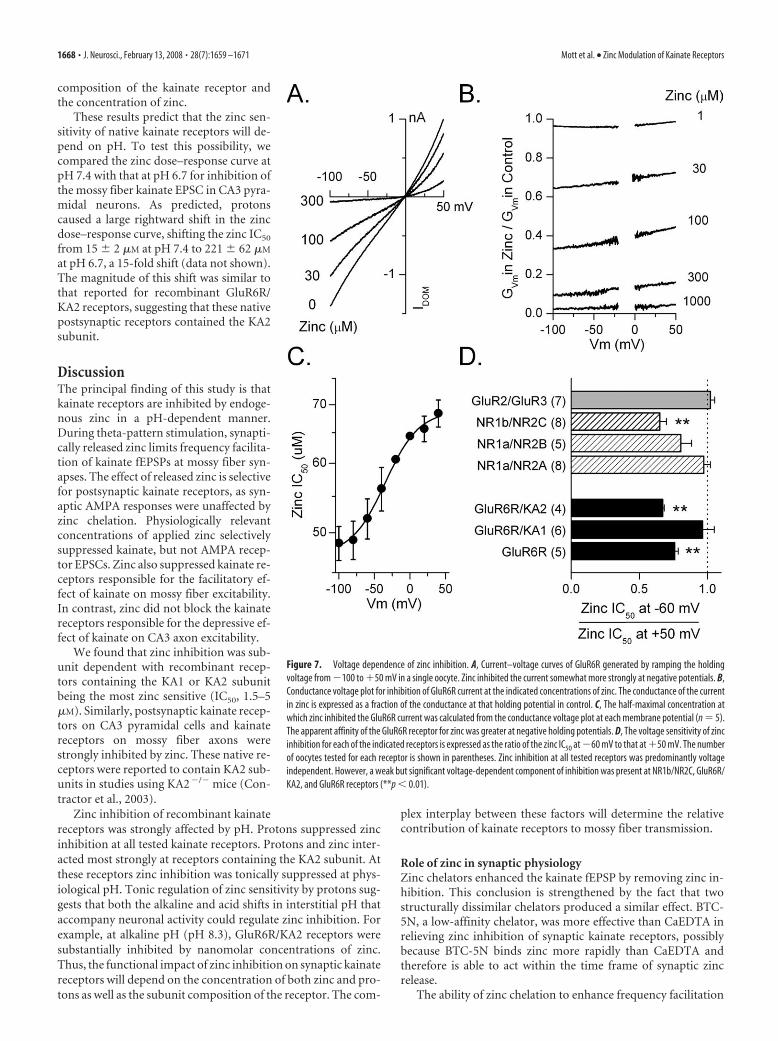
Voltage dependence of zinc inhibitionThe voltage sensitivity of zinc inhibitionwas examined to determine whether zincinhibits kainate receptors by acting at a sitewithin the transmembrane electric field.Agonist-evoked currents were recordedfrom oocytes as the holding potential wasramped from �100 to �50 mV. In the ab-sence of zinc, the current-voltage relation-ship from oocytes expressing GluR6R,GluR6R/KA1, or GluR6R/KA2 was linearor weakly outwardly rectifying and re-versed close to 0 mV (Fig. 7A).Conductance-voltage (G–V) relationshipsnormalized to responses at �100 mV re-vealed that the �50 mV/�60 mV rectifica-tion ratio for GluR6 (2.07 � 0.07; n � 8),GluR6R/KA1 (1.76 � 0.03; n � 6), andGluR6R/KA2 (1.39 � 0.04; n � 4) weresimilar to values reported previously(Bowie and Mayer, 1995; Kamboj et al.,1995; Pemberton et al., 1998; Cui andMayer, 1999). We evaluated the effect ofzinc by expressing the G–V relationship inthe presence of different concentrations ofzinc relative to that in the absence of zinc(Fig. 7B). The resulting curves were thenused to determine the IC50 for zinc at eachmembrane potential (Fig. 7C). We mea-sured the voltage dependence of zinc inhi-bition by comparing the zinc IC50 at �60mV with that at �50 mV (Fig. 7D). Allreceptor combinations tested showed lessthan a twofold change in zinc inhibition at�60 and �50 mV. At GluR6R andGluR6R/KA2 receptors, the change in zincinhibition was significantly different ( p 0.01) but small, indicating that the major-ity of zinc inhibition occurred through avoltage-independent mechanism. Theseobservations indicate little voltage depen-dence of zinc inhibition at the tested kai-nate receptors.
Zinc inhibition of other glutamate re-ceptors was similarly voltage independent.At all tested NMDA receptors (NR1a/NR2A, NR1a/NR2B, and NR1b/NR2C)zinc inhibition was primarily voltage independent over the rangeof zinc concentrations tested. This lack of voltage dependenceagrees with previous findings (Chen et al., 1997; Paoletti et al.,1997; Traynelis et al., 1998). Similarly, we found that the very
weak zinc inhibition (1–1000 �M) of GluR2/GluR3 AMPA receptorswas voltage independent. These results are consistent with a modu-latory effect of zinc on glutamate receptors at a site outside the chan-nel pore as has been previously suggested (Paoletti et al., 1997).
Figure 5. Zinc suppresses the facilitatory but not inhibitory effects of kainate on axon excitability. A, The MF antidromic spikewas facilitated by 500 nM kainate. Zinc (100 �M) reversed this facilitation. To isolate kainate receptors, the MF spike was recordedin the presence of 100 �M each of bicuculline methochloride, D-APV, CaEDTA, and GYKI 52466. B, C, Kainate (500 nM) potentiatedthe rising slope (B; 20 – 80%) and decreased the half-width (C) of the MF spike, suggesting that it potentiated the spike ampli-tude by enhancing the synchrony of the response (*p 0.05; n � 5). Zinc (100 �M) blocked these effects of kainate. In contrast,zinc applied alone influenced neither of these measures. D, Kainate potentiated the area of the MF antidromic spike. Zincsignificantly blocked this potentiation (**p 0.01; n � 5). E, Averaged time course of four experiments showing potentiationof the area of the MF antidromic spike by 500 nM kainate. Zinc (100 �M free zinc; 200 �M total zinc added) blocked thispotentiation. Increasing the concentration of CaEDTA to 500 �M reversed the effect of zinc. CNQX (100 �M) blocked the kainate-induced potentiation. Zinc in the absence of kainate (100 �M free zinc; 200 �M total zinc added) had no significant effect on thearea of the MF antidromic spike (n � 4). F, At CA3 axons, 50 nM kainate inhibited the antidromic spike recorded in the CA3 cellbody layer. In these experiments, kainate receptors were pharmacologically isolated, as described above. Kainate decreased therising slope of the antidromic spike and increased its half-width, indicating that it decreased firing synchrony. Zinc (100�M free zinc; 200�M total zinc added) slightly potentiated the depressant effect of kainate. G, Time course of an experiment showing the depression of thearea of the CA3 antidromic spike produced by kainate (50 nM) and the inability of zinc (100 �M free zinc) to block this depression. H,Concentration response curve for kainate on the CA3 antidromic spike (IC50, 84 � 20 nM). Even low concentrations of kainate, whichfacilitated MF spikes, depressed CA3 antidromic spikes. I, Kainate (50 nM) significantly depressed the area of the CA3 antidromic spike(*p 0.05; n � 7). Zinc (100 �M free zinc) did not reverse, but slightly potentiated, this depression.
1666 • J. Neurosci., February 13, 2008 • 28(7):1659 –1671 Mott et al. • Zinc Modulation of Kainate Receptors

Proton sensitivity of zinc inhibitionBrain pH is dynamic and can change markedly during neuronalactivity and in pathological conditions. Zinc inhibits NR1a/NR2A NMDA receptors partly by enhancing proton inhibition(Low et al., 2000). We have shown previously that protons toni-cally inhibit kainate receptors (Mott et al., 2003). Given the dy-namic nature of proton concentrations in the brain, an interac-tion between protons and zinc would be of importance inunderstanding the physiological effects of zinc. To evaluate thefunctional interaction between proton and zinc inhibition, wemeasured the zinc sensitivity of kainate receptors at different pH
levels (Fig. 8A). Zinc inhibition curveswere normalized and the effect of protonson zinc IC50 values compared. In contrastto NR1/NR2A receptors, we found thatGluR6R kainate receptors were less sensi-tive to zinc at acidic pH. Protons caused arightward shift in the zinc inhibition curve,shifting the IC50 for zinc inhibition of thisreceptor 2.1-fold from 67 � 8 �M at pH 7.5(n � 15) to 142 � 15 �M at pH 6.7 (n � 7).These results indicate that protons sup-press zinc inhibition of kainate receptors.
KA1 and KA2 subunits markedly en-hanced the zinc sensitivity of the kainatereceptor. We tested the effect of protons onkainate receptors containing these sub-units (Fig. 8B,C). At GluR6R/KA1 recep-tors, an increase in proton concentrationfrom pH 7.5 to pH 6.7 caused a 2.1-foldrightward shift in the IC50 for zinc (Fig.8B). The extent of this shift was similar tothat observed for GluR6 homomeric re-ceptors. Recalling that zinc inhibition forGluR6/KA1 receptor channels was incom-plete (Figs. 6C, 8B), we noticed that de-creasing the pH caused an increase in theamplitude of the residual current observedin 1 mM zinc, as would be expected if pro-tons suppressed zinc inhibition.
Protons produced a much greater in-crease in the IC50 for zinc inhibition ofGluR6R/KA2 receptors than for eitherGluR6R or GluR6R/KA1 receptors (Fig.8C). At these receptors, the IC50 for zincinhibition shifted ninefold between pH 7.5(IC50, 3 � 0.4 �M; n � 6) and pH 6.7 (IC50,27 � 2 �M; n � 5). The strong reduction inzinc sensitivity at low pH suggests that pro-ton concentration is an important deter-minant of the zinc sensitivity of these het-eromeric kainate receptors.
To better understand the interaction ofprotons and zinc, we evaluated the effect ofdifferent concentrations of zinc on protoninhibition of GluR6R/KA2 receptors (Fig.8D). In the absence of zinc, protons inhib-ited the receptor (Mott et al., 2003). In thepresence of zinc, current at GluR6R/KA2receptors was also inhibited. However,protons potentiated this current by reliev-ing zinc inhibition. The extent of the po-tentiation depended on both the proton
and zinc concentration. For example, as the zinc concentrationwas increased, greater concentrations of protons were required torelieve zinc inhibition. Thus, the half-maximal concentration ofprotons necessary to relieve zinc inhibition shifted from near pH8 at 1 �M zinc to about pH 6.8 at 100 �M zinc. These findingssuggest that at physiological pH, pH 7.3, zinc inhibition of kai-nate receptors would be tonically regulated by protons. Thus, inthe presence of zinc kainate receptor-mediated currents can bepotentiated as much as threefold with an acidic shift from pH 7.5to pH 6.7 (Fig. 8E). These experiments indicate that the effect ofa decrease in pH on kainate receptor current depends on both the
Figure 6. Zinc inhibition of recombinant kainate receptors is subunit dependent. A, Zinc inhibits currents evoked by AMPA (30�M) from an oocyte expressing GluR6R/KA2 receptors. Zinc was applied (black bar) at the concentration indicated above eachcurrent trace. B, Concentration–response curves for zinc inhibition of current from oocytes expressing each of the indicatedreceptors. Zinc inhibition of the kainate receptor-mediated current depended on the subunit composition of the kainate receptorwith GluR6R/KA2 receptors (n � 12) being more sensitive to zinc than GluR6R receptors (n � 15). GluR6R/KA2 receptors weresimilar to NR1a/NR2B receptors (n � 12) in their zinc sensitivity. C, Maximal zinc inhibition of current at GluR6R/KA2 receptorswas greater than that at GluR6R/KA1 receptors (n � 16). D, The half-maximal concentration at which zinc inhibited each of theindicated receptors is shown. The presence of the KA1 or KA2 subunit markedly increased the sensitivity of kainate receptors tozinc. The number of oocytes tested for each receptor is shown in parentheses. E, Zinc did not alter the apparent affinity of GluR6Rreceptors for domoate (control, n � 7; zinc, n � 8). Currents are normalized to the maximal current at 10 �M domoate in eachcondition. F, Reducing desensitization by adding con A (0.3 mg/ml) did not alter zinc inhibition of current at GluR6R/KA2receptors (control, n � 12; Con A, n � 6).
Mott et al. • Zinc Modulation of Kainate Receptors J. Neurosci., February 13, 2008 • 28(7):1659 –1671 • 1667
composition of the kainate receptor andthe concentration of zinc.
These results predict that the zinc sen-sitivity of native kainate receptors will de-pend on pH. To test this possibility, wecompared the zinc dose–response curve atpH 7.4 with that at pH 6.7 for inhibition ofthe mossy fiber kainate EPSC in CA3 pyra-midal neurons. As predicted, protonscaused a large rightward shift in the zincdose–response curve, shifting the zinc IC50
from 15 � 2 �M at pH 7.4 to 221 � 62 �M
at pH 6.7, a 15-fold shift (data not shown).The magnitude of this shift was similar tothat reported for recombinant GluR6R/KA2 receptors, suggesting that these nativepostsynaptic receptors contained the KA2subunit.
DiscussionThe principal finding of this study is thatkainate receptors are inhibited by endoge-nous zinc in a pH-dependent manner.During theta-pattern stimulation, synapti-cally released zinc limits frequency facilita-tion of kainate fEPSPs at mossy fiber syn-apses. The effect of released zinc is selectivefor postsynaptic kainate receptors, as syn-aptic AMPA responses were unaffected byzinc chelation. Physiologically relevantconcentrations of applied zinc selectivelysuppressed kainate, but not AMPA recep-tor EPSCs. Zinc also suppressed kainate re-ceptors responsible for the facilitatory ef-fect of kainate on mossy fiber excitability.In contrast, zinc did not block the kainatereceptors responsible for the depressive ef-fect of kainate on CA3 axon excitability.
We found that zinc inhibition was sub-unit dependent with recombinant recep-tors containing the KA1 or KA2 subunitbeing the most zinc sensitive (IC50, 1.5–5�M). Similarly, postsynaptic kainate recep-tors on CA3 pyramidal cells and kainatereceptors on mossy fiber axons werestrongly inhibited by zinc. These native re-ceptors were reported to contain KA2 sub-units in studies using KA2�/� mice (Con-tractor et al., 2003).
Zinc inhibition of recombinant kainatereceptors was strongly affected by pH. Protons suppressed zincinhibition at all tested kainate receptors. Protons and zinc inter-acted most strongly at receptors containing the KA2 subunit. Atthese receptors zinc inhibition was tonically suppressed at phys-iological pH. Tonic regulation of zinc sensitivity by protons sug-gests that both the alkaline and acid shifts in interstitial pH thataccompany neuronal activity could regulate zinc inhibition. Forexample, at alkaline pH (pH 8.3), GluR6R/KA2 receptors weresubstantially inhibited by nanomolar concentrations of zinc.Thus, the functional impact of zinc inhibition on synaptic kainatereceptors will depend on the concentration of both zinc and pro-tons as well as the subunit composition of the receptor. The com-
plex interplay between these factors will determine the relativecontribution of kainate receptors to mossy fiber transmission.
Role of zinc in synaptic physiologyZinc chelators enhanced the kainate fEPSP by removing zinc in-hibition. This conclusion is strengthened by the fact that twostructurally dissimilar chelators produced a similar effect. BTC-5N, a low-affinity chelator, was more effective than CaEDTA inrelieving zinc inhibition of synaptic kainate receptors, possiblybecause BTC-5N binds zinc more rapidly than CaEDTA andtherefore is able to act within the time frame of synaptic zincrelease.
The ability of zinc chelation to enhance frequency facilitation
Figure 7. Voltage dependence of zinc inhibition. A, Current–voltage curves of GluR6R generated by ramping the holdingvoltage from �100 to �50 mV in a single oocyte. Zinc inhibited the current somewhat more strongly at negative potentials. B,Conductance voltage plot for inhibition of GluR6R current at the indicated concentrations of zinc. The conductance of the currentin zinc is expressed as a fraction of the conductance at that holding potential in control. C, The half-maximal concentration atwhich zinc inhibited the GluR6R current was calculated from the conductance voltage plot at each membrane potential (n � 5).The apparent affinity of the GluR6R receptor for zinc was greater at negative holding potentials. D, The voltage sensitivity of zincinhibition for each of the indicated receptors is expressed as the ratio of the zinc IC50 at �60 mV to that at �50 mV. The numberof oocytes tested for each receptor is shown in parentheses. Zinc inhibition at all tested receptors was predominantly voltageindependent. However, a weak but significant voltage-dependent component of inhibition was present at NR1b/NR2C, GluR6R/KA2, and GluR6R receptors (**p 0.01).
1668 • J. Neurosci., February 13, 2008 • 28(7):1659 –1671 Mott et al. • Zinc Modulation of Kainate Receptors

of the kainate fEPSP suggests that during the theta-pattern train,zinc accumulates at mossy fiber synapses. The build-up of zincprogressively increases inhibition of kainate receptors. This con-clusion is in agreement with studies using a variety of techniques,including fluorescent zinc indicators that have found increasedzinc concentrations in the microenvironment of mossy fiber ter-minals after mossy fiber stimulation (Assaf and Chung, 1984;Howell et al., 1984; Li et al., 2001b; Ueno et al., 2002; Quinta-Ferreira et al., 2004; Frederickson et al., 2005, 2006a). Further-more, numerous studies have found effects of released zinc onsynaptic currents mediated by NMDA (Vogt et al., 2000; Ueno etal., 2002), GABA (Smart et al., 1994), and glycine (Bloomenthalet al., 1994; Laube et al., 1995; Hirzel et al., 2006). The ability of
zinc chelators to influence the function of these receptors pro-vides strong support for the activity dependent release of zinc atsynapses.
The ability of released zinc to regulate synaptic transmissionappears to be widespread across multiple strains and species ofanimal. Endogenously released zinc modulates mossy fiber re-sponses in mouse (Vogt et al., 2000; Dominguez et al., 2006),guinea pig (Ruiz et al., 2004), and rat (Molnar and Nadler, 2001;Ueno et al., 2002; Matias et al., 2006). We found that zinc chela-tors potentiate mossy-fiber-evoked kainate responses in rat. Toprovide additional support for a role of endogenous zinc in reg-ulating synaptic kainate responses, we examined the effect ofBTC-5N on kainate fEPSPs in mocha mutant mice. Mocha mu-tant mice express a natural mutation that results in loss of theZnT3 zinc transporter from synaptic vesicles, causing the mossyfibers to fail to accumulate zinc (Kantheti et al., 1998; Vogt et al.,2000). In these mocha mutants, BTC-5N did not enhance thekainate response during theta-pattern stimulus trains, suggestingthat the lack of mossy fiber zinc prevented potentiation by thezinc chelator. Although wild-type mice were not tested, thesefindings in mocha mutant mice are consistent with our sugges-tion that endogenously released zinc regulates synaptic kainateresponses.
Our data provide evidence that the release of zinc during burstactivation of mossy fiber terminals limits the activation of synap-tic kainate receptors. The slow time course of kainate EPSPs atmossy fiber synapses suggests that summation of these responsesduring sustained afferent activity may increase depolarization ofCA3 pyramidal cells, enhancing excitability in this network(Frerking and Ohliger-Frerking, 2002). Accordingly, kainate re-ceptors at mossy fiber synapses have been implicated in the de-velopment of seizures (Ben-Ari and Cossart, 2000). Zinc inhibi-tion could therefore act as a brake that prevents pathologicalactivation of mossy fiber kainate receptors that would otherwisepromote synchronized firing within the recurrent CA3 network.
A previous study (Lin et al., 2001) reported that zinc (50 –200�M) potentiated AMPA currents in about half of the neuronstested. At the concentrations used in our study we found no effectof zinc on synaptic AMPA responses in hippocampal slices. Atrecombinant receptors we found that high concentrations of zinc(�500 �M) were necessary to inhibit AMPA receptors. This find-ing agrees with an earlier study that reported AMPA receptorinhibition by high zinc concentrations (�500 �M) (Mayer et al.,1989). At low concentrations, zinc mildly potentiates AMPA re-sponses both at native (Mayer et al., 1989; Lin et al., 2001) andrecombinant receptors (Dreixler and Leonard, 1994). At recom-binant receptors, the potentiating effect of zinc was confined toGluR3 receptors and was only observed over a narrow range ofzinc concentrations (4 –7.5 �M), which were not explored here.
Kainate receptor modulationLike NMDA receptors, kainate receptors are regulated by severalendogenous substances in addition to zinc, such as protons andpolyamines (Mott et al., 2003). Kainate receptors containing KA1or KA2 subunits are markedly more sensitive to each of thesemodulators. Whereas KA1 and KA2 subunits enhance the sensi-tivity of kainate receptors to modulation, important differencesexist in the response of KA1 and KA2-containing receptors toeach of the endogenous regulators. For example, KA1 containingreceptors were potentiated by protons, whereas all other kainatereceptors were inhibited (Mott et al., 2003). In contrast, zincinhibition of KA2 containing kainate receptors was markedlymore proton sensitive than other kainate receptors. Thus, recep-
Figure 8. Protons relieve zinc inhibition of kainate receptors. A, Left, At GluR6R receptors,protons inhibited the current but relieved zinc inhibition as the pH was lowered from 7.5– 6.0(pH 7.5, n � 15; pH 6.7, n � 7; pH 6.0, n � 6). The inset shows zinc inhibition of GluR6Rcurrents at pH 7.5 and 6.0. The currents at pH 6.0 were scaled to those at pH 7.5 to betterdemonstrate the loss of zinc inhibition. Right, Here and in B and C the zinc inhibition curves ateach pH were normalized to the control current at that pH. Decreasing the pH from 7.5 to 6.0decreased the sensitivity of the receptor to zinc inhibition. B, At GluR6R/KA1 receptors, protonsincreased the zinc IC50 and decreased the maximal inhibition produced by zinc (pH 7.5, IC50,1.5 � 0.6 �M; n � 16; pH 6.7, IC50, 3.2 � 0.9 �M; n � 5). The shift in zinc sensitivity wassimilar to that at GluR6R receptors. C, At GluR6R/KA2 receptors, protons caused a much greatershift in zinc sensitivity than at GluR6R receptors (pH 8.3, IC50, 1.0 � 0.2 �M; n � 6; pH 7.5, IC50,3.0 � 0.4 �M; n � 6; pH 6.7, IC50, 27 � 2 �M; n � 5; pH 6.0, IC50, 610 � 210 �M; n � 6). D,In the absence of zinc, protons inhibited GluR6/KA2 receptors, but in the presence of zinc,protons potentiated the current by removing zinc inhibition. At each pH, current amplitude inthe presence of zinc is expressed as a percentage of the current at pH 9 in the absence of zinc. Atselected pHs, the current amplitude in the absence of zinc (open circles) and in the presence ofthe indicated zinc concentration (filled circles) is shown. E, In the presence of zinc, decreasing pHfrom 7.5 to 6.7 potentiated current at GluR6R/KA2 receptors (solid circles; n � 5) more thancurrent at GluR6 receptors (open circles; n � 7). The amplitude of the current at pH 6.7 isexpressed as a percentage of the current at pH 7.5 in each of the indicated concentrations of zinc.
Mott et al. • Zinc Modulation of Kainate Receptors J. Neurosci., February 13, 2008 • 28(7):1659 –1671 • 1669

tor subunit composition plays an important role in determiningthe response of these receptors to endogenous regulators. Giventhe specialized roles of kainate receptors of different subunitcomposition (Castillo et al., 1997; Mulle et al., 2000; Contractoret al., 2003), our results indicate that endogenous modulatoryagents will differentially regulate distinct kainate receptors andthereby exert selective influence over discrete aspects of kainateneurotransmission. These modulatory sites may also providenovel sites for the development of context-dependent andregion-selective neuroprotective agents (Mott et al., 1998).
Interaction between zinc and protonsThe pH dependence of zinc inhibition suggests an interactionbetween protons and zinc at the kainate receptor. Protonation ofa zinc-coordinating amino acid in the kainate receptor couldreduce the affinity of the zinc binding site. Alternatively, protonsand zinc could converge on the same allosteric mechanism forkainate receptor inhibition, with protons being less efficaciousthan zinc. The situation differs from NR1/NR2A NMDA recep-tors where zinc enhances proton inhibition (Choi and Lipton,1999; Low et al., 2000). However, at both kainate and NR1/NR2ANMDA receptors channel function is inhibited with sufficientprotons or zinc.
The interaction between protons and zinc has interesting im-plications for kainate receptor regulation. Protons reduced zincinhibition at each kainate receptor tested, with the effect beingstrongest for GluR6R/KA2 receptors. At GluR6R/KA2 receptorszinc sensitivity changed 600-fold over the tested pH range. Thus,at an acidic pH, GluR6R/KA2 receptors were less sensitive to zincthan GluR6 homomeric receptors, whereas at an alkaline pH,these receptors were sensitive to submicromolar zinc concentra-tions, much more zinc sensitive than GluR6 receptors. The pro-ton sensitivity of zinc inhibition falls within the physiological pHrange, indicating that zinc inhibition of these receptors is toni-cally regulated by protons.
Interestingly, the interaction between protons and zinc pro-duces functionally different effects on kainate and NMDA recep-tors. Thus, an alkaline shift in pH, as occurs during synaptictransmission, would be expected to enhance zinc inhibition ofpostsynaptic kainate receptors (Fig. 8C) but reduce inhibition ofNMDA receptors by zinc (Low et al., 2000). Alternatively, duringfocal acidic conditions that accompany seizures or ischemia,NMDA currents would be inhibited synergistically by protonsand zinc, whereas zinc inhibition of kainate receptors would berelieved. Thus, by differentially altering zinc inhibition, acidic pHwould increase the prominence of kainate signaling relative toNMDA signaling. The increase in kainate receptor-mediated cur-rent may contribute to network hyperexcitability during thesepathological situations.
ReferencesAlt A, Weiss B, Ogden AM, Knauss JL, Oler J, Ho K, Large TH, Bleakman D
(2004) Pharmacological characterization of glutamatergic agonists andantagonists at recombinant human homomeric and heteromeric kainatereceptors in vitro. Neuropharmacology 46:793– 806.
Assaf SY, Chung SH (1984) Release of endogenous Zn 2� from brain tissueduring activity. Nature 308:734 –736.
Ben-Ari Y, Cossart R (2000) Kainate, a double agent that generates seizures:two decades of progress. Trends Neurosci 23:580 –587.
Bloomenthal AB, Goldwater E, Pritchett DB, Harrison NL (1994) Biphasicmodulation of the strychnine-sensitive glycine receptor by zinc. MolPharmacol 46:1156 –1159.
Bortolotto ZA, Clarke VR, Delany CM, Parry MC, Smolders I, Vignes M, HoKH, Miu P, Brinton BT, Fantaske R, Ogden A, Gates M, Ornstein PL,
Lodge D, Bleakman D, Collingridge GL (1999) Kainate receptors areinvolved in synaptic plasticity. Nature 402:297–301.
Bowie D, Mayer ML (1995) Inward rectification of both AMPA and kainatesubtype glutamate receptors generated by polyamine-mediated ion chan-nel block. Neuron 15:453– 462.
Brooks SPJ, Storey KB (1992) Bound and determined: a computer programfor making buffers of defined ion concentrations. Anal. Biochem201:119 –126.
Castillo PE, Malenka RC, Nicoll RA (1997) Kainate receptors mediate a slowpostsynaptic current in hippocampal CA3 neurons. Nature 388:182–186.
Chen N, Moshaver A, Raymond LA (1997) Differential sensitivity of recom-binant N-methyl-D-aspartate receptor subtypes to zinc inhibition. MolPharmacol 51:1015–1023.
Choi YB, Lipton SA (1999) Identification and mechanism of action of twohistidine residues underlying high-affinity Zn 2� inhibition of the NMDAreceptor. Neuron 23:171–180.
Clemens JD (1996) Transmitter timecourse in the synaptic cleft: its role incentral synaptic function. Trends Neurosci 19:163–171.
Cole TB, Wenzel HJ, Kafer KE, Schwartzkroin PA, Palmiter RD (1999)Elimination of zinc from synaptic vesicles in the intact mouse brain bydisruption of the ZnT3 gene. Proc Natl Acad Sci USA 96:1716 –1721.
Contractor A, Sailer AW, Darstein M, Maron C, Xu J, Swanson GT, Heine-mann SF (2003) Loss of kainate receptor-mediated heterosynaptic facil-itation of mossy-fiber synapses in KA2�/� mice. J Neurosci 23:422– 429.
Cossart R, Epsztein J, Tyzio R, Becq H, Hirsch J, Ben-Ari Y, Crepel V (2002)Quantal release of glutamate generates pure kainate and mixed AMPA/kainate EPSCs in hippocampal neurons. Neuron 35:147–159.
Cui C, Mayer ML (1999) Heteromeric kainate receptors formed by the coas-sembly of GluR5, GluR6, and GluR7. J Neurosci 19:8281– 8291.
Dingledine R, Borges K, Bowie D, Traynelis SF (1999) The glutamate recep-tor ion channels. Pharmacol Rev 51:7– 62.
Dominguez MI, Blasco-Ibanez JM, Crespo C, Nacher J, Marques-Mari AI,Martinez-Guijarro FJ (2006) Neural overexcitation and implication ofNMDA and AMPA receptors in a mouse model of temporal lobe epilepsyimplying zinc chelation. Epilepsia 47:887– 899.
Dreixler JC, Leonard JP (1994) Subunit-specific enhancement of glutamatereceptor responses by zinc. Brain Res Mol Brain Res 22:144 –150.
Egebjerg J, Bettler B, Hermans-Borgmeyer I, Heinemann S (1991) Cloningof a cDNA for a glutamate receptor subunit activated by kainate but notAMPA. Nature 351:745–748.
Fletcher EJ, Lodge D (1996) New developments in the molecular pharma-cology of alpha-amino-3-hydroxy-5-methyl-4-isoxazole propionate andkainate receptors. Pharmacol Ther 70:65– 89.
Frederickson CJ, Danscher G (1990) Zinc-containing neurons in hip-pocampus and related CNS structures. Prog Brain Res 83:71– 84.
Frederickson CJ, Suh SW, Silva D, Frederickson CJ, Thompson RB (2000)Importance of zinc in the central nervous system: the zinc-containingneuron. J Nutr 130:1471S–1483S.
Frederickson CJ, Koh JY, Bush AI (2005) The neurobiology of zinc in healthand disease. Nat Rev Neurosci 6:449 – 462.
Frederickson CJ, Giblin III LJ, Balaji RV, Masalha R, Frederickson CJ, Zeng Y,Lopez EV, Koh JY, Chorin U, Besser L, Hershfinkel M, Li Y, ThompsonRB, Krezel A (2006a) Synaptic release of zinc from brain slices: factorsgoverning release, imaging, and accurate calculation of concentration.J Neurosci Methods 154:19 –29.
Frederickson CJ, Giblin LJ, Krezel A, McAdoo DJ, Muelle RN, Zeng Y, BalajiRV, Masalha R, Thompson RB, Fierke CA, Sarvey JM, de Valdenebro M,Prough DS, Zornow MH (2006b) Concentrations of extracellular freezinc (pZn)e in the central nervous system during simple anesthetization,ischemia, and reperfusion. Exp Neurol 198:285–293.
Frerking M, Ohliger-Frerking P (2002) AMPA receptors and kainate recep-tors encode different features of afferent activity. J Neurosci22:7434 –7443.
Frerking M, Schmitz D, Zhou Q, Johansen J, Nicoll RA (2001) Kainate re-ceptors depress excitatory synaptic transmission at CA33CA1 synapsesin the hippocampus via a direct presynaptic action. J Neurosci21:2958 –2966.
Fukushima T, Shingai R, Ogurusu T, Ichinose M (2003) Inhibition ofwillardiine-induced currents through rat GluR6/KA-2 kainate receptorchannels by zinc and other divalent cations. Neurosci Lett 349:107–110.
Henze DA, Wittner L, Buzsaki G (2002) Single granule cells reliably dis-
1670 • J. Neurosci., February 13, 2008 • 28(7):1659 –1671 Mott et al. • Zinc Modulation of Kainate Receptors

charge targets in the hippocampal CA3 network in vivo. Nat Neurosci5:790 –795.
Hirzel K, Muller U, Latal AT, Hulsmann S, Grudzinska J, Seeliger MW, BetzH, Laube B (2006) Hyperekplexia phenotype of glycine receptor alpha1subunit mutant mice identifies Zn( 2�) as an essential endogenous mod-ulator of glycinergic neurotransmission. Neuron 52:679 – 690.
Hoo KH, Nutt SL, Fletcher EJ, Elliott CE, Korczak B, Deverill RM, RampersadV, Fantaske RP, Kamboj RK (1994) Functional expression and pharma-cological characterization of the human EAA4 (GluR6) glutamate recep-tor: a kainate selective channel subunit. Receptors Channels 2:327–337.
Howell GA, Welch MG, Frederickson CJ (1984) Stimulation-induced up-take and release of zinc in hippocampal slices. Nature 308:736 –738.
Huettner JE (2003) Kainate receptors and synaptic transmission. Prog Neu-robiol 70:387– 407.
Izumi Y, Auberson YP, Zorumski CF (2006) Zinc modulates bidirectionalhippocampal plasticity by effects on NMDA receptors. J Neurosci26:7181–7188.
Jung MW, McNaughton BL (1993) Spatial selectivity of unit activity in thehippocampal granular layer. Hippocampus 3:165–182.
Kamboj SK, Swanson GT, Cull-Candy SG (1995) Intracellular spermineconfers rectification on rat calcium-permeable AMPA and kainate recep-tors. J Physiol (Lond) 486:297–303.
Kamiya H, Ozawa S (2000) Kainate receptor-mediated presynaptic inhibi-tion at the mouse hippocampal mossy fibre synapse. J Physiol (Lond)523:653– 665.
Kamiya H, Shinozaki H, Yamamoto C (1996) Activation of metabotropicglutamate receptor type 2/3 suppresses transmission at rat hippocampalmossy fibre synapses. J Physiol (Lond) 493:447– 455.
Kantheti P, Qiao X, Diaz ME, Peden AA, Meyer GE, Carskadon SL, Kapf-hamer D, Sufalko D, Robinson MS, Noebels JL, Burmeister M (1998)Mutation in AP-3 delta in the mocha mouse links endosomal transport tostorage deficiency in platelets, melanosomes, and synaptic vesicles. Neu-ron 21:111–122.
Kay AR (2003) Evidence for chelatable zinc in the extracellular space of thehippocampus, but little evidence for synaptic release of Zn. J Neurosci23:6847– 6855.
Laube B, Kuhse J, Rundstrom N, Kirsch J, Schmieden V, Betz H (1995)Modulation by zinc ions of native rat and recombinant human inhibitoryglycine receptors. J Physiol (Lond) 483:613– 619.
Lerma J (2003) Roles and rules of kainate receptors in synaptic transmis-sion. Nat Rev Neurosci 4:481– 495.
Li Y, Hough CJ, Frederickson CJ, Sarvey JM (2001a) Induction of mossyfiber3Ca3 long-term potentiation requires translocation of synapticallyreleased Zn 2�. J Neurosci 21:8015– 8025.
Li Y, Hough CJ, Suh SW, Sarvey JM, Frederickson CJ (2001b) Rapid trans-location of Zn 2� from presynaptic terminals into postsynaptic hip-pocampal neurons after physiological stimulation. J Neurophysiol86:2597–2604.
Lin DD, Cohen AS, Coulter DA (2001) Zinc-induced augmentation of ex-citatory synaptic currents and glutamate receptor responses in hippocam-pal CA3 neurons. J Neurophysiol 85:1185–1196.
Low CM, Zheng F, Lyuboslavsky P, Traynelis SF (2000) Molecular determi-nants of coordinated proton and zinc inhibition of N-methyl-D-aspartateNR1/NR2A receptors. Proc Natl Acad Sci USA 97:11062–11067.
Matias CM, Matos NC, Arif M, Dionisio JC, Quinta-Ferreira ME (2006)Effect of the zinc chelator N, N,N�,N�-tetrakis (2-pyridylmethyl)-ethylenediamine (TPEN) on hippocampal mossy fiber calcium signalsand on synaptic transmission. Biol Res 39:521–530.
Mayer ML, Vyklicky LJ, Westbrook GL (1989) Modulation of excitatoryamino acid receptors by group IIB metal cations in cultured mouse hip-pocampal neurones. J Physiol (Lond) 415:329 –350.
Molnar P, Nadler JV (2001) Synaptically-released zinc inhibits N-methyl-D-aspartate receptor activation at recurrent mossy fiber synapses. BrainRes 910:205–207.
Mott DD, Doherty JJ, Zhang S, Washburn MS, Fendley MJ, Lyuboslavsky P,Traynelis SF, Dingledine R (1998) Phenylethanolamines inhibit NMDAreceptors by enhancing proton inhibition. Nat Neurosci 1:659 – 667.
Mott DD, Washburn MS, Zhang S, Dingledine RJ (2003) Subunit-dependent modulation of kainate receptors by extracellular protons andpolyamines. J Neurosci 23:1179 –1188.
Mulle C, Sailer A, Perez-Otano I, Dickinson-Anson H, Castillo PE, Bureau I,Maron C, Gage FH, Mann JR, Bettler B, Heinemann SF (1998) Alteredsynaptic physiology and reduced susceptibility to kainate-induced sei-zures in GluR6-deficient mice. Nature 392:601– 605.
Mulle C, Sailer A, Swanson GT, Brana C, O’Gorman S, Bettler B, HeinemannSF (2000) Subunit composition of kainate receptors in hippocampal in-terneurons. Neuron 28:475– 484.
Paoletti P, Ascher P, Neyton J (1997) High-affinity zinc inhibition ofNMDA NR1-NR2A receptors. J Neurosci 17:5711–5725.
Patton C, Thompson S, Epel D (2004) Some precautions in using chelatorsto buffer metals in biological solutions. Cell Calcium 35:427– 431.
Pemberton KE, Belcher SM, Ripellino JA, Howe JR (1998) High-affinitykainate-type ion channels in rat cerebellar granule cells. J Physiol (Lond)510:401– 420.
Quinta-Ferreira ME, Matias CM, Arif M, Dionisio JC (2004) Measurementof presynaptic zinc changes in hippocampal mossy fibers. Brain Res1026:1–10.
Rose G, Diamond D, Lynch GS (1983) Dentate granule cells in the rat hip-pocampal formation have the behavioral characteristics of theta neurons.Brain Res 266:29 –37.
Ruiz A, Walker MC, Fabian-Fine R, Kullmann DM (2004) Endogenous zincinhibits GABAA receptors in a hippocampal pathway. J Neurophysiol91:1091–1096.
Salin PA, Scanziani M, Malenka RC, Nicoll RA (1996) Distinct short-termplasticity at two excitatory synapses in the hippocampus. Proc Natl AcadSci USA 93:13304 –13309.
Schmitz D, Frerking M, Nicoll RA (2000) Synaptic activation of presynaptickainate receptors on hippocampal mossy fiber synapses. Neuron27:327–338.
Schmitz D, Mellor J, Nicoll RA (2001) Presynaptic kainate receptor media-tion of frequency facilitation at hippocampal mossy fiber synapses. Sci-ence 291:1972–1976.
Seeburg PH (1993) The TiPS/Trends Neurosci lecture: the molecular biol-ogy of mammalian glutamate receptor channels. Trends Pharmacol Sci14:297–303.
Smart TG, Xie X, Krishek BJ (1994) Modulation of inhibitory and excitatoryamino acid receptor ion channels by zinc. Progr Neurobiol 42:393– 441.
Sudhof TC (1995) The synaptic vesicle cycle: a cascade of protein-proteininteractions. Nature 375:645– 653.
Thompson RB, Whetsell Jr WO, Maliwal BP, Fierke CA, Frederickson CJ(2000) Fluorescence microscopy of stimulated Zn(II) release from orga-notypic cultures of mammalian hippocampus using a carbonicanhydrase-based biosensor system. J Neurosci Methods 96:35– 45.
Traynelis SF, Burgess MF, Zheng F, Lyuboslavsky P, Powers JL (1998) Con-trol of Voltage-Independent Zinc Inhibition of NMDA Receptors by theNR1 Subunit. J Neurosci 18:6163– 6175.
Ueno S, Tsukamoto M, Hirano T, Kikuchi K, Yamada MK, Nishiyama N,Nagano T, Matsuki N, Ikegaya Y (2002) Mossy fiber Zn 2� spillovermodulates heterosynaptic N-methyl-D-aspartate receptor activity in hip-pocampal CA3 circuits. J Cell Biol 158:215–220.
Vignes M, Collingridge GL (1997) The synaptic activation of kainate recep-tors. Nature 388:179 –182.
Vogt K, Mellor J, Tong G, Nicoll R (2000) The actions of synaptically re-leased zinc at hippocampal mossy fiber synapses. Neuron 26:187–196.
Zheng F, Gingrich MB, Traynelis SF, Conn PJ (1998) Tyrosine kinase po-tentiates NMDA receptor currents by reducing tonic zinc inhibition. NatNeurosci 1:185–191.
Mott et al. • Zinc Modulation of Kainate Receptors J. Neurosci., February 13, 2008 • 28(7):1659 –1671 • 1671