cellular dynamical mechanisms for encoding the time and...
TRANSCRIPT
R
Cs
MC
a
ARRAA
KPMEHPGPHTS
C
0d
Behavioural Brain Research 215 (2010) 261–274
Contents lists available at ScienceDirect
Behavioural Brain Research
journa l homepage: www.e lsev ier .com/ locate /bbr
eview
ellular dynamical mechanisms for encoding the time and place of events alongpatiotemporal trajectories in episodic memory
ichael E. Hasselmo ∗, Lisa M. Giocomo, Mark P. Brandon, Motoharu Yoshidaenter for Memory and Brain, Department of Psychology and Program in Neuroscience, Boston University, 2 Cummington St., Boston, MA 02215, United States
r t i c l e i n f o
rticle history:eceived 16 August 2009eceived in revised form 5 December 2009ccepted 10 December 2009vailable online 16 December 2009
eywords:ersistent spikingembrane potential oscillations
a b s t r a c t
Understanding the mechanisms of episodic memory requires linking behavioral data and lesion effects todata on the dynamics of cellular membrane potentials and population interactions within brain regions.Linking behavior to specific membrane channels and neurochemicals has implications for therapeuticapplications. Lesions of the hippocampus, entorhinal cortex and subcortical nuclei impair episodic mem-ory function in humans and animals, and unit recording data from these regions in behaving animalsindicate episodic memory processes. Intracellular recording in these regions demonstrates specific cel-lular properties including resonance, membrane potential oscillations and bistable persistent spikingthat could underlie the encoding and retrieval of episodic trajectories. A model presented here shows
ntorhinal cortexippocampusostsubiculumrid cellslace cellsead direction cells
how intrinsic dynamical properties of neurons could mediate the encoding of episodic memories ascomplex spatiotemporal trajectories. The dynamics of neurons allow encoding and retrieval of uniqueepisodic trajectories in multiple continuous dimensions including temporal intervals, personal location,the spatial coordinates and sensory features of perceived objects and generated actions, and associationsbetween these elements. The model also addresses how cellular dynamics could underlie unit firing datasuggesting mechanisms for coding continuous dimensions of space, time, sensation and action.
rajectorypatial cognition
© 2010 Elsevier B.V. All rights reserved.
ontents
1. Introduction . . . . . . . . . . . . . . . . . . . . . . . . . . . . . . . . . . . . . . . . . . . . . . . . . . . . . . . . . . . . . . . . . . . . . . . . . . . . . . . . . . . . . . . . . . . . . . . . . . . . . . . . . . . . . . . . . . . . . . . . . . . . . . . . . . . . . . . . . . 2622. Anatomical circuits for episodic memory . . . . . . . . . . . . . . . . . . . . . . . . . . . . . . . . . . . . . . . . . . . . . . . . . . . . . . . . . . . . . . . . . . . . . . . . . . . . . . . . . . . . . . . . . . . . . . . . . . . . . . . . . . . 2623. Episodic memory in rats . . . . . . . . . . . . . . . . . . . . . . . . . . . . . . . . . . . . . . . . . . . . . . . . . . . . . . . . . . . . . . . . . . . . . . . . . . . . . . . . . . . . . . . . . . . . . . . . . . . . . . . . . . . . . . . . . . . . . . . . . . . . . 2634. Physiological data indicating episodic memory in animals . . . . . . . . . . . . . . . . . . . . . . . . . . . . . . . . . . . . . . . . . . . . . . . . . . . . . . . . . . . . . . . . . . . . . . . . . . . . . . . . . . . . . . . . . 2635. Synaptic modification and episodic memory function . . . . . . . . . . . . . . . . . . . . . . . . . . . . . . . . . . . . . . . . . . . . . . . . . . . . . . . . . . . . . . . . . . . . . . . . . . . . . . . . . . . . . . . . . . . . . . 2636. Possible intrinsic cellular mechanisms for episodic memory . . . . . . . . . . . . . . . . . . . . . . . . . . . . . . . . . . . . . . . . . . . . . . . . . . . . . . . . . . . . . . . . . . . . . . . . . . . . . . . . . . . . . . . 264
6.1. Membrane potential oscillations . . . . . . . . . . . . . . . . . . . . . . . . . . . . . . . . . . . . . . . . . . . . . . . . . . . . . . . . . . . . . . . . . . . . . . . . . . . . . . . . . . . . . . . . . . . . . . . . . . . . . . . . . . . . . 2646.2. Persistent spiking . . . . . . . . . . . . . . . . . . . . . . . . . . . . . . . . . . . . . . . . . . . . . . . . . . . . . . . . . . . . . . . . . . . . . . . . . . . . . . . . . . . . . . . . . . . . . . . . . . . . . . . . . . . . . . . . . . . . . . . . . . . . . 265
7. Modeling how cellular mechanisms could underlie episodic memory . . . . . . . . . . . . . . . . . . . . . . . . . . . . . . . . . . . . . . . . . . . . . . . . . . . . . . . . . . . . . . . . . . . . . . . . . . . . . 2658. Episodic memories as spatiotemporal trajectories through multiple sensory dimensions. . . . . . . . . . . . . . . . . . . . . . . . . . . . . . . . . . . . . . . . . . . . . . . . . . . . . . . . . 2669. Episodes can be encoded by associating states with actions . . . . . . . . . . . . . . . . . . . . . . . . . . . . . . . . . . . . . . . . . . . . . . . . . . . . . . . . . . . . . . . . . . . . . . . . . . . . . . . . . . . . . . . . 267
10. Review of a cellular model of episodic memory . . . . . . . . . . . . . . . . . . . . . . . . . . . . . . . . . . . . . . . . . . . . . . . . . . . . . . . . . . . . . . . . . . . . . . . . . . . . . . . . . . . . . . . . . . . . . . . . . . . . . 26811. Input determines coding of place, length or time . . . . . . . . . . . . . . . . . . . . . . . . . . . . . . . . . . . . . . . . . . . . . . . . . . . . . . . . . . . . . . . . . . . . . . . . . . . . . . . . . . . . . . . . . . . . . . . . . . . 26912. A general model of episodic memory . . . . . . . . . . . . . . . . . . . . . . . . . . . . . . . . . . . . . . . . . . . . . . . . . . . . . . . . . . . . . . . . . . . . . . . . . . . . . . . . . . . . . . . . . . . . . . . . . . . . . . . . . . . . . . . . 269
13. Network dynamics might enhance cellular phase code . . . . . . . . . . . . . . . . . . .14. Interaction of memory systems. . . . . . . . . . . . . . . . . . . . . . . . . . . . . . . . . . . . . . . . . . . .Acknowledgements . . . . . . . . . . . . . . . . . . . . . . . . . . . . . . . . . . . . . . . . . . . . . . . . . . . . . . . .References . . . . . . . . . . . . . . . . . . . . . . . . . . . . . . . . . . . . . . . . . . . . . . . . . . . . . . . . . . . . . . . . . .
∗ Corresponding author. Tel.: +1 617 353 1397; fax: +1 617 358 3296.E-mail address: [email protected] (M.E. Hasselmo).
166-4328/$ – see front matter © 2010 Elsevier B.V. All rights reserved.oi:10.1016/j.bbr.2009.12.010
. . . . . . . . . . . . . . . . . . . . . . . . . . . . . . . . . . . . . . . . . . . . . . . . . . . . . . . . . . . . . . . . . . . . . . . . . . 270
. . . . . . . . . . . . . . . . . . . . . . . . . . . . . . . . . . . . . . . . . . . . . . . . . . . . . . . . . . . . . . . . . . . . . . . . . . 270. . . . . . . . . . . . . . . . . . . . . . . . . . . . . . . . . . . . . . . . . . . . . . . . . . . . . . . . . . . . . . . . . . . . . . . . . . 271. . . . . . . . . . . . . . . . . . . . . . . . . . . . . . . . . . . . . . . . . . . . . . . . . . . . . . . . . . . . . . . . . . . . . . . . . . 271
2 al Brain Research 215 (2010) 261–274
1
nsiImitmvatgeottaf
moedeanptcmdiiaocaimr
2
dsiobe[oaoOptmm
sr
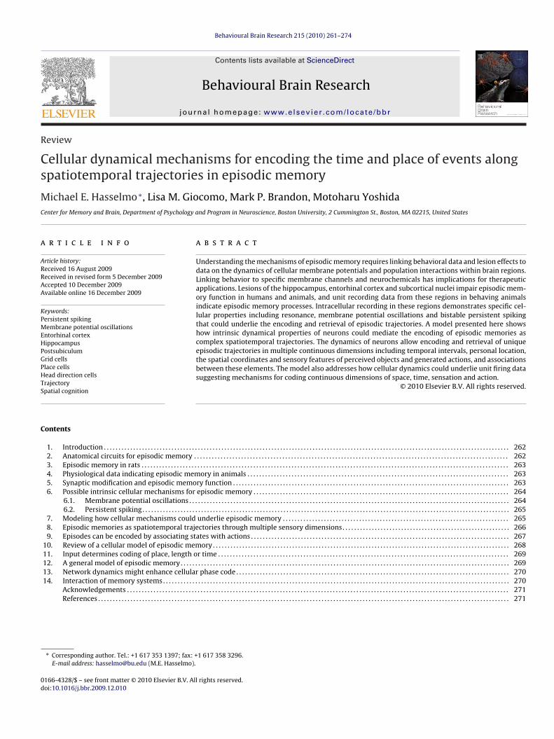
Fig. 1. (A) Schematic representation of the medial temporal lobe in humans, illus-trating structures that are associated with human episodic memory function. Theentorhinal cortex receives convergent input from a range of neocortical associa-tion cortices. Superficial layers of entorhinal cortex project into the hippocampalformation, which projects back to deep layers of entorhinal cortex. (B) Schematicrepresentation of analogous structures in the rat showing entorhinal cortex input tothe dentate gyrus (DG) and hippocampal regions CA3 and CA1, as well as connectionsbetween the hippocampus and the septum via the fornix (which also contains con-nections with the mammillary bodies and anterior thalamus). Output from regionCA1 reaches the postsubiculum (ps) which projects to entorhinal cortex. Cellular
62 M.E. Hasselmo et al. / Behaviour
. Introduction
As humans, most of us have personal experience of the phe-omenon of episodic memory. We have rich recollections ofequences of events from our recent or remote past that play outn our minds as if we were reliving the experience. For example,can remember going into the kitchen this morning to preparey breakfast, and sitting in the dining room eating it. I remember
ndividual movements involved in getting my cereal, and sitting athe table, with distinct memories of where I was facing and where
y family members were. Definitions of episodic memory by Tul-ing describe this experience as the capacity for mental time travelnd autonoetic awareness [42,176,177]. The process of mental timeravel goes beyond forming associations of single items with a sin-le, static behavioral context of location and time, and involvesncoding of a full spatiotemporal trajectory. This article will focusn models of the cellular dynamics of episodic memory involvinghe capacity to relive a sequence of events as a spatiotemporalrajectory with an explicit sense of position in continuous spacend duration in continuous time, and an explicit re-experience ofactors such as head direction and the direction of movements.
Some researchers have tried to argue that episodic memory andental time travel are a purely human capacity [165], whereas
thers argue that animals have this capacity [28,36]. The personalxperience of animals is beyond experimental test, but behavioralata provides a compelling argument that many of the capacities forpisodic memory shown in humans can be found in animals [36]. Inddition, electrophysiological recordings from animals show phe-omena that support the existence of mental time travel alongreviously experienced trajectories [93,109]. In addition, many ofhe qualitative anatomical and physiological features of neural cir-uits observed in human cortical structures are also found in otherammals [65,66]. Thus, it is reasonable to suppose that the cellular
ynamics mediating episodic memory in humans are also presentn animals, even if species differ in the quantity of data on the man-festations of episodic memory. This article will review some of thevailable behavioral and physiological data indicating the presencef episodic memory in animals, and describe a model of how spe-ific cellular dynamics may be involved in episodic memory. Therticle will focus primarily on the function of the oscillatory dynam-cs of membrane potentials and populations, with less focus on the
odification of synaptic connections that have been extensivelyeviewed in other work [12].
. Anatomical circuits for episodic memory
The first question is where we should look for the cellularynamics of episodic memory function? Human data providesome answers about the specific anatomical structures involvedn episodic memory (Fig. 1A). Considerable attention has focusedn the impairments of episodic memory caused in patient HMy the bilateral removal of the anterior hippocampus, the entirentorhinal cortex, and portions of other parahippocampal cortices30]. Patient HM showed striking deficits in quantitative mem-ry tests that test recall of discrete items from an episode, suchs the recall of information from paragraphs, or the free recallf words from a list, or cued recall of paired associates [29,147].ther patients with bilateral damage to the hippocampus andarahippocampal cortices also show impairments in these quanti-ative tests [61,134]. These lesions indicate the anatomical locus of
echanisms for episodic memory but do not provide physiologicalechanisms.Specific behavioral scoring methods have been developed to
how impairments of the richness of detail in human episodicecollection [106]. These techniques show significant reductions
dynamical mechanisms relevant to episodic memory function have been studiedextensively with cellular neurophysiological techniques in rats and non-humanprimates.
in the recall of internal details from an episodic memory afterlesions of the hippocampus and parahippocampal cortices [96,162].As shown in Fig. 1, the corresponding structures exist in therat, allowing detailed experiments on physiological dynamics ofneurons in these regions that could mediate episodic memoryfunction. Lesions of the medial temporal cortices also cause sig-nificant impairments in the description of future or imaginedepisodes [68,141]. In contrast to the impairment of episodic mem-ory, hippocampal and parahippocampal lesions have less effecton long-term semantic memory [162], or simple tests of work-ing memory for familiar items such as digit span [29,61]. Thesedata indicate the importance of neural circuits in the hippocam-pus and parahippocampal cortices for the performance of episodicmemory. Other lesion data in humans indicates impairments ofepisodic memory associated with lesions of the anterior thalamus,the mammillary nuclei and the medial prefrontal cortex [3].
Extensive animal research has addressed the memory functionof these structures, as reviewed in other articles in this volume. Aswith the human research, the early studies in non-human primatesfocused on memory for discrete items within episodes. Studiestesting memory for trial unique objects in delayed non-matchto sample tasks showed impairments after hippocampal lesions
[195,196], entorhinal lesions [105] and perirhinal and parahip-pocampal lesions [197]. Other studies in non-human primates havetested memory for associations between visual stimuli and specificspatial locations, indicating that lesions of the fornix impair the
l Brai
cf
3
ameetTeoma
ritmprbgclalpd
motatasafc[mTttPbsf
4a
lmamrnwes
M.E. Hasselmo et al. / Behavioura
onstruction of a snapshot memory for the spatial location of visualeatures [52] or associations with responses [53].
. Episodic memory in rats
Some tasks used in rats indicate a role for the hippocampusnd associated structures such as the entorhinal cortex in episodicemory for complex spatiotemporal trajectories (Fig. 1B). For
xample, the 8-arm radial maze task requires that rats visit 8 differ-nt arms without making an error by repeating an arm entry, andhe number of arm re-entries is increased by fornix lesions [87,131].he rat could avoid the error of repeating an arm entry by samplingach arm and testing for recall of a previous trajectory into that armn the same day, thereby using a strategy dependent on episodicemory. However, the task could also be performed by avoiding
rms with strong familiarity from the same day.Similarly, the delayed spatial alternation task requires that a
at alternates left and right arm responses, and rat performances impaired by fornix lesions [4] and entorhinal lesions [8]. Spa-ial alternation could be performed by episodic retrieval of the
ost recent trajectory at the choice point [193], but could also beerformed by persistent neural activity holding the most recentesponse in working memory [193]. The Morris water maze haseen used extensively to test rat spatial memory, both with a sin-le fixed platform location [119] and with a platform location thathanges between days [159]. Impairments of this task appear withesions of the dorsal entorhinal cortex [160], postsubiculum [171]nd lesions of the fornix [44]. A study comparing a fixed startingocation to changing starting locations [44] indicates that this taskuts demands on the capacity for planning a future trajectory fromifferent starting locations to a goal location.
Recent experiments in rats have focused on potential episodicemory function by testing the specific requirement for memory
f what, where and when. For example, rats have been tested forheir change in investigation time to objects that were presentedt different times and moved to new locations during a recogni-ion period [36]. This task effectively tests memory for what, wherend when, but could be performed based on retrieval of discreteingle time snapshots of object and location—a single element inn episodic memory. Another test in rats indicates the retrieval ofull trajectories. In this task, rats learn trajectories from a centralhoice point to two different hidden objects in an E-shaped maze39,40]. Then the rats are familiarized with one of the objects and
ust subsequently make a choice to visit the less familiar object.he trajectory followed from the choice point in this task appearso depend on actual episodic retrieval of a prior trajectory, ratherhan familiarity of cues or even a single snapshot memory [39,40].erformance in this task is impaired by fornix lesions. Thus, ratehavioral data support a role for the hippocampus and associatedtructures in the retrieval of episodic spatiotemporal trajectoriesor memory-guided behavior.
. Physiological data indicating episodic memory innimals
Another question is whether neural activity recorded at the cel-ular level in behaving animals indicates mechanisms of episodic
emory? In fact, rat physiological data provides a rich source ofdditional support for the existence of episodic memory in ani-als. In particular, unit recording data indicates the encoding and
etrieval of spatiotemporal trajectories. These data indicate that rateural circuits can selectively encode the timing of spatial locationsithin a sequence of events within a trial, and can also selectively
ncode and discriminate between the timing of events at differentpatial locations encountered on different trials.
n Research 215 (2010) 261–274 263
The strongest evidence for episodic retrieval involves replay ofspiking activity in region CA3 of the hippocampus during perfor-mance of a tone-cued alternation task [93]. In this task, rats heara tone that indicates the appropriate direction of response at alater choice point. In this task, hippocampal neurons show selec-tive spatial firing as place cells in different locations in the task,allowing statistical determination of the primary location codedby each neuron. At early stages of learning the task, when the ratis more hesitant at the choice point and turns between differentpossible response directions at the choice point, the neural activityshows sequential temporal reactivation of neurons coding spatiallocations along individual trajectories to the left or right [93]. Thisindicates the retrieval of these encoded trajectories, and indicatesthe precise temporal distinction between sequential places visitedon one trajectory, as well as indicating the separation of trajecto-ries encountered at longer temporal intervals (the left versus righttrajectories).
Other physiological studies analyzed the spiking activity of placecells in region CA1 that fire sequentially as a rat runs back and forthbetween reward locations at each end of a linear track [33,37,47].During the period of time when the rat is at the end of the track,hippocampal place cells show forward or reversed replay of thesequence of hippocampal place cells that spiked during a pre-ceding run along the linear track, further indicating the selectivespatiotemporal retrieval of encoded trajectories, and the temporalseparation of distinct episodes.
Other physiological support for episodic memory in rats comesfrom work on replay of episodes during sleep. Early studies showedreactivation in region CA1 of previously experienced neural ensem-bles during slow wave sleep [132,154,185]. Later studies showedthat this activity maintains the spatiotemporal structure of expe-rienced episodes. Hippocampal place cells sequentially activatedduring waking on a linear track appear to fire with the samesequential relationship during the ripple events in slow wave sleep[122,154]. Perhaps the most striking replay phenomena concernshippocampal spiking activity during long periods of waking behav-ior on a circular track that are replayed with a similar time scalein association with theta rhythm activity during REM sleep [109].This replay might be episodic, or could be based on a representationcreated over multiple learning experiences. This REM sleep replayis temporally structured, showing that the replay occurs at a timescale similar to waking, with time intervals of spiking activity aswell as theta rhythmicity that correspond to the time intervals thatthe rat spent in particular portions of the behavioral task [109]. Thisindicates that neural circuits in the rat not only encode the order ofevents, but the time interval of events in an episode.
5. Synaptic modification and episodic memory function
As summarized above, behavioral and physiological data sup-port the existence of episodic memory in animals, including rats.This raises the further question: what cellular processes in neuronsprovide the mechanisms for episodic memory? Most cellular workhas focused on mechanisms of synaptic modification referred toas long-term potentiation (LTP) and long-term depression (LTD) oras spike-timing dependent plasticity (STDP). In fact, the synapsesarising from entorhinal cortex and terminating in the dentate gyrusof the hippocampus (see Fig. 1B) were the focus of the first experi-mental studies of LTP [13]. Subsequently many studies of LTP havefocused on this pathway or the synapses of the Schaffer collater-
als from CA3 terminating in stratum radiatum of region CA1 [12].In particular, studies have shown that LTP in the hippocampushas Hebbian properties, depending on the temporal juxtapositionof presynaptic and postsynaptic activity. This was first shown inextracellular studies of the dentate gyrus [107,114] and then in
2 al Brain Research 215 (2010) 261–274
ipsb
sttbte
NrtBedlacnm
ebstsptl
fbdptfaStubfitpttdrTeetu
6m
acoc
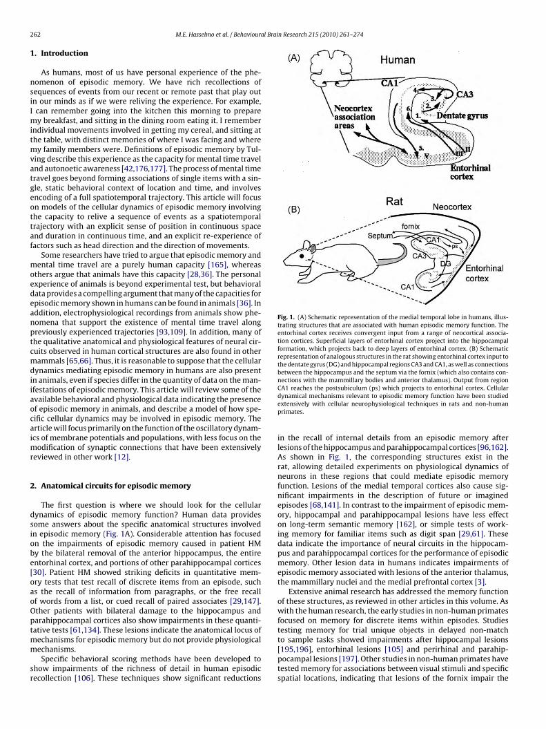
Fig. 2. (A) Whole cell patch recording in slice preparations from Giocomo andHasselmo [55] shows that layer II entorhinal stellate cells generate subthresholdmembrane potential oscillations in between the generation of action potentials.
64 M.E. Hasselmo et al. / Behaviour
ntracellular studies in CA1 [62,94,183]. Early models of hippocam-al memory function used Hebbian modification for encoding oftatic associations between features of items [111,115,130,174] oretween items and context [80,126].
Studies of the requirements for timing of pre- and postsynapticpikes show that presynaptic spikes should precede postsynap-ic spikes by less than 40 ms [10,83,84,108], as described with theerm spike-timing dependent synaptic plasticity (STDP). STDP haseen shown with intracellular recording in many cortical struc-ures [10,110], and its role in cortical function has been modeledxtensively [60,88].
The Hebbian properties of LTP are consistent with the role of theMDA receptor in mediating induction of LTP and STDP. The NMDA
eceptor blocker 2-amino-5-phosphonovaleric acid (APV) preventshe induction of Hebbian LTP and STDP in hippocampal regions [12].ehavioral studies have demonstrated that infusion of APV duringncoding periods slows the learning of a fixed location of a hid-en platform in the Morris water maze [119] and strongly impairs
earning of a new platform location on each day [120,159] as wells increasing errors in the 8-arm radial maze [26]. These data indi-ate that NMDA-dependent Hebbian synaptic modification may beecessary for encoding of spatiotemporal trajectories in episodicemory.However, is Hebbian STDP sufficient as a cellular mechanism for
pisodic memory function? Many models have shown how Heb-ian properties of STDP could allow the encoding and retrieval ofequences of discrete neural activity [90,92,115,120,175,180]. Inhese models, STDP can mediate chaining of associations betweenequentially activated discrete populations, potentially allowing aopulation of neurons activated at location A to activate a popula-ion activated at location B, that can then activate a population atocation C.
However, such a chaining mechanism is not sufficient to accountor memory of sequences in humans and animals, as a num-er of studies have shown that recall of sequences can occurespite omissions or transpositions of individual stimuli that wouldrevent a mechanism based on chaining [22,81,137,173]. In addi-ion, the very short timescale of STDP raises problems for theormation of sequential associations between behavioral items sep-rated by intervals many times longer than the time window forTDP [90,92,99,116]. In addition, humans can remember differentemporal intervals between events, but the chaining mechanismsing STDP cannot retrieve different time intervals between eventsecause STDP depends upon synaptic strengthening with a briefxed time window, and the retrieval interval depends upon synap-ic transmission with an even faster fixed time course. A furtherroblem concerns the issue of an episodic representation of a con-inuously varying dimension, such as movement through space orhe passage of time, or the expansion of a balloon or fading betweenifferent colors in a film. Continuous dimensions are difficult toepresent with synaptic links between discrete neural populations.hus, Hebbian synaptic modification is clearly important, but mod-ls based on Hebbian synaptic modification alone have difficultyncoding continuous dimensions such as time and space, and needo be supplemented by mechanisms for coding changes in contin-ous dimensions within an episode.
. Possible intrinsic cellular mechanisms for episodicemory
As described above, models based on synaptic modificationlone suffer the problems of chaining and from requiring a dis-rete and fixed representation of dimensions of time or spacer sensory features that are continuous in nature. This indi-ates a need for further cellular mechanisms that can mediate
Blowup focuses on subthreshold oscillations. (C) Whole cell patch recording fromYoshida et al. [188] in the presence of cholinergic or mGluR agonists shows thatlayer III pyramidal cells exhibit persistent spiking that is maintained after the initialinduction by a square pulse current injection.
encoding of continuous dimensions of time, space and sensory fea-tures.
Electrophysiological data from the entorhinal cortex indicatecellular mechanisms that could complement synaptic modificationfor encoding and retrieval of episodic trajectories. These intrinsiccellular mechanisms have been demonstrated using intracellularsharp electrode or whole cell patch recording in entorhinal cortexneurons. Fig. 2A and B illustrates important intrinsic properties ofentorhinal neurons that could contribute to the coding of episodicmemory.
6.1. Membrane potential oscillations
One intrinsic feature of neurons that could contribute to episodiccoding of continuous dimensions are the subthreshold membranepotential oscillations that appear when entorhinal layer II stellatecells are depolarized near firing threshold [6,7,56,57]. Fig. 2A showsan example of subthreshold oscillations [55] with an amplitudeof a few millivolts. These oscillations can influence the timing ofspikes [48,133,138] and may contribute to network theta frequencyoscillations in entorhinal cortex [1,5,118] and hippocampus [23,69]
The oscillations in superficial layers may be due to a hyper-polarization activated cation current or h-current [38]. Membranepotential oscillations show differences in frequency along the dor-sal to ventral axis of the medial entorhinal cortex [56,57] thatmay result from differences in the h-current time constant alongthe dorsal to ventral axis [55]. Membrane potential oscillationsappear less prominently in pyramidal cells of superficial layers[6], but are observed in layer V pyramidal cells, where they may
be caused by M-current [187]. The layer V membrane potentialoscillations also show a gradient in frequency from dorsal to ven-tral medial entorhinal cortex [54]. Membrane potential oscillationsare not as prominent in neurons of the lateral entorhinal cortex[166].
l Brain Research 215 (2010) 261–274 265
6
tptctiatwb
pstlmmfatGsb
nlfr[paa
7e
attdiorpct
dipfpfstn
oabT
Fig. 3. (A) Schematic representation of the coding of excitatory input by a shift inrelative phase of two persistent spiking neurons in model from Hasselmo [71]. Onthe left, the two persistent spiking neurons fire in phase with each other at the samebaseline frequency. In the center, an excitatory input drives the lower neuron to ahigher spiking frequency for a period of time, shifting its firing phase relative to thebaseline neuron. On the right, in the absence of further input, the relative phase offiring maintains a representation of the magnitude and duration of the excitatoryinput. (B) Coding of changes in two spatial dimensions. On the upper left, threeneurons fire in phase. Three different movements from this location have differenteffects. Movement to the right (in the x dimension) shifts the phase of one neuron
M.E. Hasselmo et al. / Behavioura
.2. Persistent spiking
The cellular property of persistent spiking may also contributeo the episodic coding of continuous dimensions. Even during theharmacological blockade of all excitatory and inhibitory synap-ic transmission, neurons in the entorhinal cortex demonstrate theapacity to display persistent spiking. Persistent spiking refers tohe capacity of neurons to show bistability, in which a neuron show-ng no spiking can transition to stable persistent spiking activityfter a transient depolarizing current injection or transient repeti-ive synaptic input [41,49,98,167,188]. In contrast, cortical neuronsithout persistent spiking will spike only during current injection
ut will stop after termination of the current injection.An example of persistent spiking is illustrated in Fig. 2B. Some
yramidal neurons in layer II of medial entorhinal cortex showtable persistent spiking whereas others show spiking that self-erminates over periods of many seconds [98]. Pyramidal cells inayer III show stable persistent spiking that can last for 2 min or
ore [188]. Pyramidal neurons in layer V of entorhinal cortex canaintain stable persistent spiking at different graded frequencies
or many minutes [41]. Both bistable and graded persistent spikingppear to be due to muscarinic or metabotropic glutamate activa-ion of a calcium-sensitive non-specific cation current [49,148,188].raded persistent firing could allow these neurons to integrateynaptic input over extended periods. Persistent firing has alsoeen shown in layer III of lateral entorhinal cortex [167].
The cellular phenomena described above appear in entorhinaleurons even in the presence of synaptic blockers. However, simi-
ar phenomena may arise due to circuit mechanisms, as describedurther below. The dynamics of interacting populations of neu-ons can result in network oscillations in the theta frequency range32,34] or in the gamma frequency range [15,27,182]. In addition,ersistent spiking can be obtained due to the effect of excitatorynd inhibitory synaptic feedback that drives neurons into stablettractor states [78,184].
. Modeling how cellular mechanisms could underliepisodic memory
The cellular mechanisms described here could provide a mech-nism for encoding changes in continuous dimension such asime, space and sensory features, and for episodic retrieval ofhese changes in continuous dimensions. The coding of continuousimensions for episodic memory could involve either a rate code,
n which the firing rate of a neuron varies in a continuous manner,r could involve a phase code, in which the firing time of a neuronelative to a baseline oscillation changes in a continuous manner. Ahase code has the advantage that the continuous representationan be coded by single spikes occurring at specific times, ratherhan requiring multiple spikes for rate coding.
The rhythmic cellular properties of entorhinal neuronsescribed above could allow continuous dimensions to be coded
n the form of phase. For example, a pair of neurons showingersistent spiking [41,167,188,189] might have a single baselinerequency, as shown in Fig. 3A. A synaptic input to one of theseersistent spiking neurons slightly increases the firing frequencyor a period of time, moving it progressively out of phase with thepiking phase of the neuron that stays at baseline frequency. Inhis manner, the relative phase of persistent spiking in the pair ofeurons can integrate the synaptic input to one of these neurons.
This framework can allow a group of neurons to encode continu-us dimensions based on differences in relative phase. For example,s shown in Fig. 3B, a two-dimensional environment can be codedy progressive shifts in phase of three persistent spiking neurons.he phase of the baseline neuron is illustrated by the spikes next
(x) relative to the baseline (b). Movement downward (in the y dimension) shifts thephase of another neuron (y) relative to baseline. Diagonal movement to the lowerright (in both dimensions) shifts the phase of both neurons relative to baseline.
to the letter “b”. The phase of the neuron coding the spatial dimen-sion ‘x’ is illustrated by the spikes next to the letter “x”, and thephase of the neuron coding the spatial dimension ‘y’ is illustratedby the spikes next to the letter “y”. Imagine that movement in the xdimension shifts the firing frequency of the x neuron. As shown inFig. 3B, this results in the x neuron coding a shift in location alongthe x dimension by a shift in phase relative to the baseline b. Sim-ilarly, movement in the y dimension shifts the frequency of the yneuron, resulting in a shift in the spiking phase of y relative to base-line. Diagonal movement shifts the relative phase of both neurons.Thus, the firing phase of these three neurons can code two dimen-sions. Addition of neurons with a smaller frequency change withinput and therefore a slower phase shift allows coding at a differ-ent spatial resolutions. These same mechanisms can be applied tothe relative phase of subthreshold membrane potential oscillations[54,57].
The mechanism of coding spatial state by relative phase wasinitially proposed by Burgess in a model of grid cell firing prop-erties [18,20,127]. The essential feature of this model is that thephase of neurons shifted by velocity determines their oscillatoryinterference, resulting in spiking when neurons are in phase andthe absence of spiking when cells are out of phase [18,20,77]. Thismodel was developed for oscillatory interactions, but has beenmodified by Hasselmo to use persistent spiking cells [71]. As shownin Fig. 4, different populations of persistent spiking neurons withthe same baseline firing frequency can drive the activity of a sim-ulated grid cell [71]. The persistent spiking model avoids some
problems of the membrane potential oscillation model [20,57,77].These models of grid cells require a velocity input thatdrives the shift in frequency. This is neurophysiologically realis-tic, as a velocity vector is coded by neurons responding to head

266 M.E. Hasselmo et al. / Behavioural Brai
Fig. 4. Mechanism for interaction of persistent firing cells to cause grid cell firing inmodel from Hasselmo [71]. (A) Spiking activity over time of three different groupsof persistent firing neurons. Here, each group consists of three persistent spikingcells firing with a baseline frequency of 3 Hz with different phases. Cells receiveinput from head direction (HD) cells with 0◦ preferred angle for Group 1, 120◦ anglefor Group 2, and 240◦ angle for Group 3. Grid cell firing arises from the convergentspiking of the three groups of persistent firing neurons. When all three persistentfiring groups fire in synchrony, the grid cell will fire (dots). (B) Grid cell spiking(re
dStatpavtpu
tdfcS
or transition that could be simple (e.g. I saw a sign—where the
dots) occurs only when all of the persistent firing neurons fire at the same phase,esulting in a typical grid cell firing pattern. Gray line indicates rat trajectory fromxperimental data (Hafting et al. [64]).
irection [150,169,170,172] and translational speed [128,152].elf-organization of phasic input would provide the heading selec-ivity. The models based on oscillatory interference account forn impressive range of cellular neurophysiological data, includinghe pattern of grid cell firing as well as the phenomenon of thetahase precession observed in hippocampal neurons [20,104,129]nd in entorhinal neurons [63]. The model predicted the dorsal toentral difference in frequency of membrane potential oscillationshat was shown experimentally in slices [57], and also correctlyredicted differences in intrinsic spiking frequency measured withnit recording in behaving rats [89].
The example presented here focused on the linear coding of spa-ial dimensions, but the same properties could be applied to other
imensions. For example, a change in brightness could drive firingrequency to cause a phase shift coding the state of brightness, or ahange in color could drive a phase shift to code the state of color.imilarly, cells responding to angular velocity [149,151] could driven Research 215 (2010) 261–274
a phase shift that codes the shift in visual angle of objects in thevisual field. Even complex actions such as expansion could be codedby a population of neurons. The population coding expansion woulddrive a change in firing frequency in neurons coding the width ofan object such as a balloon, causing a progressive change in relativephase coding the change in width of the balloon.
One question about phase coding concerns the capacity toencode dimensions beyond the scale coded by the phase of a sin-gle oscillatory cycle. However, this problem can be avoided byusing different rates of phase change in different oscillations, whichresults in different scales of interference. Neurophysiological dataon membrane potential oscillations indicates that neurons at dif-ferent positions along the dorsal to ventral axis of medial entorhinalcortex may respond with different magnitudes of frequency changein response to the same depolarizing signal [54,56,57]. In the gridcell model, this results in differences in the size and spacing ofgrid cell firing fields at different dorsal to ventral anatomical posi-tions consistent with neurophysiological data [17,57,64,140]. Theinteraction of coding at different spatial scales can effectively codevery large ranges according to the least common denominator ofinteractions [59,181] and could drive place cells with firing fields ofdifferent sizes in the hippocampus [72,97]. This raises the intriguingpossibility that anatomical differences in intrinsic frequencies inother structures such as prefrontal cortex and piriform cortex couldunderlie differences in the scale of coding for different behaviors[72].
The phase code mechanism presented here has useful features.One feature is that interactions can be positive or negative depen-dent not on the pattern of connectivity between neurons, butdependent upon their relative phase. This could provide importantcontext effects for cognitive processes. For example, initial con-ditions could set the phase of a population coding context to besynchronous with neurons coding current state, which would thenresult in firing of an output population. In contrast, if the initialconditions set a different phase in the population coding context,then the two populations will not drive output, and could evenprevent output based on feedback inhibition. Thus, a contextualcue can have a positive or negative influence on a gated output, bya changing relative phase rather than changing from excitatory toinhibitory synaptic connectivity.
The phase relationships of neurons could undergo complexinteractions, in which the influence of one neuron on anotherdepends upon dynamics analogous to the ‘mod’ function of theirrelative phase, such that only the difference in relative phaseinfluences the new spike time. This resembles the mod functioncommonly used in algorithms for random number generators, andcould potentially provide an oscillatory mechanism for the stochas-tic properties of neural firing observed in many systems.
8. Episodic memories as spatiotemporal trajectoriesthrough multiple sensory dimensions
As noted above, episodic memories can be described asspatiotemporal trajectories through multiple dimensions. The def-inition of episodic memory already includes a definition of what,where and when. Thus, an event is defined in terms of its spatialcoordinates (that can be defined by a two or three dimensional spa-tial state vector x) and its temporal coordinate (defined by a specifictime t). But the term episode or even event does not just mean astatic snapshot. The definition of an event includes some action
action is the movement of eyes or attention to the sign), or as com-plex as a conversation or purchase of an item. These events involvesome transition in state over time (described by the derivative ofthe state vector – dx/dt). The definition of episode usually includes

M.E. Hasselmo et al. / Behavioural Brain Research 215 (2010) 261–274 267
Fig. 5. Examples of the encoding and retrieval of episodic spatiotemporal trajectories by model in Hasselmo [74]. The trajectories experienced during encoding are shown aslight gray lines, and the events at different times and locations are shown as gray symbols. Black lines and symbols show the retrieval of trajectories and events generated byi ent ins e intc ent ia ocatio
ma
btttadapsttete
rTusHb
paa
nternal dynamics in the network model. (A) Spatial plot of an episode with movemhowing varying speeds in different segments during encoding resulting in long timoordinates in both space and time [74]. (C) Spatial plot of an episode with movemnd retrieval of the episode in (C) showing effective retrieval of both the time and l
ore than one event, and therefore usually includes more than onection.
A common type of episodic memory concerns a transitionetween locations over time, such as I drove to the store, or I wento see a film. These can be described as a spatiotemporal trajec-ory, as shown in the examples in Fig. 5, where the movementhrough space takes place over varying time intervals. In Fig. 5A,n agent moves at different speeds in a straight direction along oneimension (y) in a two-dimensional space and experiences eventst different locations and times. Fig. 5B shows the same episodelotted in both time and space as a gray line, with a black linehowing the retrieved spatiotemporal trajectory, indicating thathe model described here can retrieve both the spatial location andhe relative time intervals of events. Fig. 5C shows a more complexpisode involving movement in two dimensions and Fig. 5D showshe same episode plotted as a spatiotemporal trajectory duringncoding (gray) and during retrieval (black).
Current data does not yet describe the temporal and spatialesolution at which a human can encode and retrieve an episode.he autobiographical memory tests described above [96,106,162]se discrete details that could be extracted from a sequence ofnapshots equally as well as from a continuous movie-like replay.owever, episodes usually involve actions that mediate transitions
etween points in time and space.Episodic memories are not just defined in the spatial and tem-oral dimensions of an agent. The events within an episode couldlso occur with the agent in a single location. But in order to ben episode it requires some action, which requires some type of
the y dimension and five events. (B) Spatiotemporal plot of the same episode in (B)ervals between nearby events, and showing effective retrieval of events at correctn two spatial dimensions and five events. (D) Spatiotemporal plot of the encodingn of events despite the spatial overlap of the trajectory [74].
transition. This could be a transition over time by another agent(for example if you watch someone cook a meal, or ride a bike)resulting in an episodic memory in which the relevant dimensionspertain not to the location of the encoding agent, but the locationof an observed agent. The transition could also be more abstract,such as listening to a story (in which the actions are conceptual),or watching the sky grow dark (in which the action is a changein a non-spatial sensory dimension—the level of brightness). Allof these transitions can be described by a multidimensional statespace that can add dimensions dependent upon the relevant eventsbeing observed.
9. Episodes can be encoded by associating states withactions
What cellular mechanisms allow encoding and retrieval of acomplete episode? The above description is based on a circuitmodel that does not just use associations between discrete states,but uses continuous representations of the states in an episode andthe actions associated with individual events [73,74]. The states canbe represented by phase coding [20,57,71,127], as described above.This contrasts with an alternative not explored here in which thestate could be represented by other circuit mechanisms such as
attractor dynamics resulting in grid cells [50,113] or integrationdue to cyclical changes in firing rate [76].Episodes can be encoded by having the cellular mechanisms forrepresenting states and actions interact with synaptic mechanismsfor forming associations (e.g. spike-timing dependent plasticity).
2 al Brain Research 215 (2010) 261–274
Dcm(Diotidwdtsa
1
mibifiitcichsTsaio
stduTnc[ictapwttd
eedjortrtt
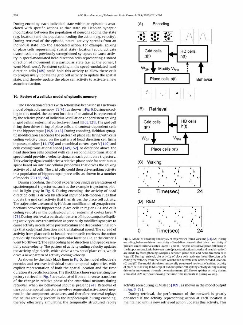
Fig. 6. Model of encoding and replay of trajectories from Hasselmo [73]. (A) Duringencoding, behavior drives the activity of head direction cells that drive the activity ofgrid cells in entorhinal cortex layers II and III. The grid cells drive place cell firing inthe hippocampus. Links between state (place) and action (speed and head direction)are made by strengthening synapses between place cells and head direction cellsWPH. (B) During retrieval, the activity of place cells activates head direction cellscoding the velocity from that state which then activates the next encoded location.
68 M.E. Hasselmo et al. / Behaviour
uring encoding, each individual state within an episode is asso-iated with specific actions at that state via Hebbian synapticodification between the population of neurons coding the state
e.g. location) and the population coding the action (e.g. velocity).uring retrieval of the episode, neural activity spreads from an
ndividual state into the associated action. For example, spikingf place cells representing spatial state (location) could activateransmission at previously strengthened synapses to cause activ-ty in speed-modulated head direction cells representing a storedirection of movement at a particular state (i.e. at the corner, Ient Northwest). Persistent spiking in the speed-modulated headirection cells [189] could hold this activity to allow these cellso progressively update the grid cell activity to update the spatialtate, and thereby update the place cell activity to activate a newssociated action.
0. Review of a cellular model of episodic memory
The association of states with actions has been used in a networkodel of episodic memory [73,74], as shown in Fig. 6. During encod-
ng in this model, the current location of an animal is representedy the relative phase of individual oscillations or persistent spiking
n grid cells in entorhinal cortex layer II and III [63,121]. The grid cellring then drives firing of place cells and context-dependent cells
n the hippocampus [19,51,113]. During encoding, Hebbian synap-ic modification associates the pattern of place cell firing with cellsoding velocity based on the pattern of head direction cell firingn postsubiculum [14,172] and entorhinal cortex layer V [140] andells coding translational speed [149,152]. As described above, theead direction cells coupled with cells responding to translationalpeed could provide a velocity signal at each point on a trajectory.his velocity signal could drive a relative phase code for continuouspace based on intrinsic cellular properties that drives the spikingctivity of grid cells. The grid cells could then drive spiking activityn a population of hippocampal place cells, as shown in a numberf models [73,136,156].
During encoding, the model experiences single presentations ofpatiotemporal trajectories, such as the example trajectories plot-ed in light gray in Fig. 5. During encoding, the activity of headirection cells is driven by afferent input of self-motion cues thatpdate the grid cell activity that then drives the place cell activity.he trajectories are stored by Hebbian modification of synaptic con-ections between hippocampal place cells in region CA1 and cellsoding velocity in the postsubiculum or entorhinal cortex layer V73]. During retrieval, a particular pattern of hippocampal cell spik-ng activity causes transmission at previously modified synapses toause activity in cells of the postsubiculum and deep entorhinal cor-ex that code head direction and translational speed. The spread ofctivity from place cells to head direction cells retrieves the actionreviously associated with a particular location (i.e. at the corner, Ient Northwest). The cells coding head direction and speed essen-
ially code velocity. The pattern of activity coding velocity updateshe activity of grid cells, which drive a new set of place cells, whichrive a new pattern of activity coding velocity.
As shown by the thick black lines in Fig. 5, the model effectivelyncodes and retrieves individual spatiotemporal trajectories, withxplicit representation of both the spatial location and the timeuration at specific locations. The thick black lines representing tra-
ectory retrieval in Fig. 5 are calculated from an inverse transformf the change in relative phase of the entorhinal neurons during
etrieval, when no behavioral input is present [74]. Retrieval ofhe spatiotemporal trajectory involves sequential activation of neu-ons in the component structures, and therefore retrieval replayshe neural activity present in the hippocampus during encoding,hereby effectively simulating the temporally structured replay(C) and (D) The model simulates temporally structured retrieval of spiking activityof place cells during REM sleep. (C) Shows place cell spiking activity during wakingdriven by movement through the environment. (D) Shows spiking activity duringsimulated REM retrieval showing the same time intervals as during waking.
activity seen during REM sleep [109], as shown in the model output
in Fig. 6 [73].During retrieval, the performance of the network is greatlyenhanced if the activity representing action at each location ismaintained until a new retrieved action updates this activity. This

l Brai
atweria[
eeritemilfiLpmTmrh
eermsimuler
1
ttcosiotato[iiitrttlvgt
M.E. Hasselmo et al. / Behavioura
voids the problem of the action representation being lost whenhere is no new update based on hippocampal neural activity, whichould cause the spatial and temporal intervals between different
vents to become distorted. The maintenance of spiking activity toepresent actions could be provided by the persistent spiking activ-ty shown in slice preparations of the postsubiculum [189], which isregion showing robust head direction activity in behaving animals
149,170,172].The head direction system plays a vital role in this model of
pisodic memory, providing a representation of both overall ori-ntation and direction of movement during both encoding andetrieval. Loss of components of this system could cause severempairments in the mechanisms of mental time travel along spa-iotemporal trajectories. This could underlie the impairments ofpisodic memory associated with lesions of the anterior thala-us and mammillary bodies [3]. Head direction cells are found
n the lateral mammillary nucleus [158] and the anterior tha-amus [58,157,190]. The anterior thalamus also shows selectivering during viewing of familiar stimuli in a recognition task [135].esions of the anterior thalamus abolish head direction responses inostsubiculum [58], and could thereby remove access to this infor-ation for encoding and retrieval of spatiotemporal trajectories.
he presence of theta rhythmic neurons in anterior thalamic andammillary nuclei close to head direction cells [178] suggests that
hythmic firing could code the distance and time intervals betweenead direction changes.
In the model described above, the spatiotemporal trajectory isncoded in entirety during one period of time, and retrieved inntirety during a separate time period. However, the encoding andetrieval of segments of the trajectory could occur in an interleavedanner [75] if there were changes in the influence on the post-
ubiculum alternating between external input (rat velocity) andnternal retrieval (hippocampal input to postsubiculum). Rhyth-
ic changes in hippocampal retrieval driving postsubiculum couldnderlie the place by head direction cells found in postsubicu-
um [25]. This would allow separate phases that alternate betweenncoding current trajectory input and retrieval of previously expe-ienced trajectory segments [75,125].
1. Input determines coding of place, length or time
The nature of the input regulating neural frequency determineshe information coded by relative phase in the model. In contrast tohe coding with velocity input described above, the input of speedould allow coding of length, and the representation of continu-us time intervals could arise from interference of oscillations atlightly different fixed frequencies. The mechanism using velocitynput can code Euclidean space, but has difficulty with the codingf overlapping trajectories, and with coding of the temporal dura-ion of stationary periods. These properties can be provided by thedditional role of cells in which the membrane potential oscilla-ions or persistent spiking do not depend on velocity, but respondnly to speed input, thereby coding the arc length of the trajectory70]. Alternately, the coding of time can be provided by oscillatorynterference of cells that keep the same frequency over time, caus-ng relative phase to directly code continuous temporal intervalsnstead of continuous space [77]. Both the arc length code and theemporal interval code can overcome the problem of spatiotempo-al trajectories that overlap in the same spatial location at differentimes. The integration of velocity cannot differentiate two visits
o the same spatial location, but the integration of speed gives arcength of the trajectory, which differs for an early visit versus a laterisit to the same location. Similarly, a fixed oscillation frequencyives a change in relative phase over time that provides a differentemporal code for an early visit versus a late visit to the same spa-n Research 215 (2010) 261–274 269
tial location. The fixed oscillation frequency will also allow relativephase to code the temporal duration spent stationary at a singlelocation [73,74,77]. Thus, depending on what input influences fre-quency, relative phase can encode Euclidean distance, arc length ortemporal intervals. The phase reset of temporal oscillations regu-lated by velocity could also provide context-dependent activity inthe grid cell model driven by velocity [71]. The continuous repre-sentation of time presented here resembles the oscillatory codesfor encoding word order in immediate serial recall models [16,21],or the temporal context model used to model conditional responseprobability in free recall [85,86]. In addition, this use of oscillationsresembles the use of oscillations for encoding temporal intervals inmodels of the timing of behavioral responses [112,117].
The simulated versions of the model above use sequential acti-vation of place cells or arc length cells and are still vulnerable to theproblem of chaining models. However, as noted above, the timingmechanisms can run concurrently in multiple persistent spikingcells to provide redundancy and avoid the chaining problems. Inthis framework, new items or features are proposed to activatea subpopulation of persistent spiking cells with stable frequencythat shift in spiking phase relative to other persistent spiking cells.Downstream cells could respond to these cells by spiking whenthe shifting phases are close in time, allowing generation of spik-ing that codes time intervals from the onset of persistent spiking.This allows redundant coding of the timing of items or events in asequence such that retrieval of each could depend on multiple prioronset cues.
12. A general model of episodic memory
A general model of episodic memory would include a widerange of possible dimensions, each of which could cue subsequentdimensions. In this framework, the initial sensory state of an organ-ism would involve a pattern of neural activity in a population ofneurons, potentially using phase coding to represent the initialdimensions. As the agent moves through the environment, velocityinput could update the phase code of location, and angular velocityinput could update the representation of head direction. When anew object is encountered, the match between the input patternand previously modified synaptic connectivity could drive a newpopulation of persistent spiking neurons over threshold. Once overthreshold, the newly activated population of neurons would showpersistent rhythmic spiking with specific phases relative to otherneurons that depend on the strength of a feature of the initial input,thereby representing the initial dimensions of the object, such asspatial location, object orientation, object size or object color. Anychanges in the features of an object would cause synaptic input thatwould drive shifts in frequency of the neurons coding the object,to alter the relative phase representing that feature of the object.For example, if the object were turned by 30◦ in orientation, thiscould cause synaptic input coding the rotation velocity of the objectthat would shift the phase of spiking representing orientation toa different phase of spiking relative to other spiking activity. Theassociations of events and items could involve interactions of spa-tial codes in medial entorhinal cortex with item representationsin lateral entorhinal cortex, which shows less spatial specificity[43,67] but shows object responses.
The time intervals of the episode could be encoded by neuronswith fixed frequency differences that progressively shift in phaserelative to each other, resulting in a precise code for relative time
intervals. For example, the population of neurons activated by thefirst object might include some neurons with slight frequency dif-ferences that result in a progressive change in relative phase suchthat different neurons are synchronized at different time points.The synchronized firing could drive other neurons at specific time
2 al Brai
itafiooirTwttap
baertldndaofifrmtpg
1
iiti[gnisgIoda
iioirotasrwct
70 M.E. Hasselmo et al. / Behaviour
ntervals, coding time since the first object appeared. Now imaginehat a second object appears. The neurons driven by synchronousctivity due to phase shifts coding the temporal interval since therst object will strengthen synapses with neurons coding this sec-nd object, and with later objects. In this framework, each newbject or dimension starts interference mechanisms that time thentervals to subsequent objects. All of these timing mechanismsun concurrently, so that any object can cue any subsequent object.his avoids the problem of the synaptic chaining framework inhich only one preceding object can cue the next object, and avoids
he problem of the fixed short time frame dependent on synapticransmission. This modeling could be formalized mathematically asnalogous to splines [102,143], in which the state representationrovides knots and the actions provide the knot vectors.
The state representation of space in a given environment wille shared between episodic trajectories. This might result in theppearance of place cells with stable spatial coordinates in a givennvironment [127] that could be combined with other state rep-esentations to be linked to multiple different spatiotemporalrajectories. In some cases, behavioral tasks with similar spatialocation in different behavioral context might functionally requireistinct context-dependent responses such as those that appear ineurophysiological data [45,103,155,186]. In these cases, context-ependent firing could be driven by oscillatory interference codingrc length [70] or by the interval since reset due to previous rewardr sensory stimulation [71]. In some cases, the context-dependentring of neurons appears to depend not on the actual trajectory
ollowed by the rat, but on the set of possible trajectories that aat could follow within a certain configuration of barriers [35]. Thisight be due to scaling of the contribution of individual head direc-
ion angles based on the possible movement along that dimension,ossibly analogous to the computation of all possible paths in aiven environment [46].
3. Network dynamics might enhance cellular phase code
A phase code could also arise from oscillatory dynamicsnvolving feedback interactions between excitatory neurons andnhibitory cortical interneurons. Numerous studies have shownhat circuits of excitatory neurons interacting with inhibitorynterneurons can cause oscillatory dynamics at gamma frequency27,182]. This could allow phase coding of memories relative toamma oscillations in neocortical structures [153]. More complexetwork level dynamical interactions can cause oscillatory dynam-
cs at theta frequency [31,32,34,101,133,139]. Circuits that generateynchronous rhythmic activity of neurons have the potential forenerating phasic firing of neurons at different phase relationships.f external depolarizing input causes a shift in frequency of onescillation, then this will cause shifts in relative phase of spiking inifferent groups of neurons, providing a phase code as describedbove.
One problem that confronts the models of grid cells based onntrinsic mechanisms concerns the effect of phase noise. As seenn Fig. 2, membrane potential oscillations show high variability inscillation period, and persistent spiking activity shows variabilityn spiking phase. Simulations with this level of variability show aapid loss of coding accuracy [54,181,191]. However, these effectsf noise could be reduced by network interactions. Experimen-al data shows that individual stellate cells receiving input from
dynamic clamp replicating excitatory interactions with other
tellate cells will synchronize [123,124]. Thus, stellate cells firinghythmically in response to external input will shift into phaseith each other due to recurrent excitatory coupling. This syn-hronization on the population level should be able to overcomehe independent variability of the intrinsic mechanisms for mem-
n Research 215 (2010) 261–274
brane potential oscillations or persistent spiking. Simulations byZilli have demonstrated that network dynamics can maintain syn-chrony despite noise within individual neurons [191].
A phase code could involve an interaction of persistent firingcells and cells showing membrane potential oscillations. Intrin-sic persistent spiking cells in medial entorhinal cortex [188] orpostsubiculum [189] could drive the stellate cells in layer II thatwould have weak excitatory interactions sufficient for synchroniza-tion but not strong enough to change the overall frequency of thecircuit [1,124,138]. A similar interaction could occur between per-sistent spiking cells in layer III of entorhinal cortex and local circuitsin region CA1 that generate synchronization through interactionsof pyramidal cells, and two types of interneurons: fast spikingcells (FS) and oriens-lacunosum-moleculare (OLM) cells [124,139].These CA1 circuits could interact with entorhinal circuits becauseof the topographic relationship between entorhinal projections toCA1 and the return projections from CA1 to deep layers of entorhi-nal cortex [168].
The cholinergic modulation of intrinsic properties could influ-ence the generation of oscillations. Cholinergic modulation hasbeen shown to enhance theta rhythm oscillations in the hippocam-pus [11,100]. On a single cell level, cholinergic modulation lowersthe resonance frequency of entorhinal stellate cells [82]. By reduc-ing neuronal intrinsic frequencies, acetylcholine could cause anincrease in the size and spacing of grid cell firing fields observed innovel environments [9]. Microdialysis shows increases in corticalacetylcholine levels in novel environments [2].
14. Interaction of memory systems
Previous modeling work demonstrates that tasks performedusing episodic retrieval of spatiotemporal trajectories would alsorequire a role for working memory or semantic memory. Work-ing memory could underlie the human capacity for immediaterecall of sequential verbal information using mechanisms thatmay depend upon phase codes or rate codes for temporal order[22,81,91]. This provides an important mechanism that could con-tribute to encoding of episodic memory. Working memory formultiple items based on persistent activity could be used to directlysolve behavioral tasks [193,194], or to provide input or output foran episodic store based on synaptic modification [192]. In supportof this, human imaging data shows that persistent activity in theabsence of a stimulus is correlated with the subsequent memoryfor that stimulus at a later time [144,145] and shows load effectsdependent on number of items held during a delay [146]. The mech-anisms of persistent spiking might play an important role in theneural activity present in the hippocampus and parahippocampalcortices during working memory for novel stimuli [79,164] andduring the encoding of novel information into long-term memory[95,163,179].
The new modeling framework presented here provides poten-tial mechanisms for simultaneously modeling the interaction ofmemory systems such as working memory and episodic memory.For example, the active maintenance of phase in multiple differentneurons can mediate working memory for the value of spatial loca-tions or features on many different dimensions. However, once thisworking memory for state causes activity to spread across previ-ously modified synapses to activate previously associated actions,then the working memory has cued retrieval of episodic memory.The new retrieved state would be held in working memory. Thus,
this framework uses an ongoing interaction of working memorywith episodic memory for memory-guided behavior. Mathemat-ical analysis shows how the interaction of memory systems candisambiguate individual states in behavioral tasks [192]. Reinforce-ment learning mechanisms can be used to guide the encoding and
l Brai
r[
rcntprcihw(cdpfnptuwplTraae
A
MC
R
M.E. Hasselmo et al. / Behavioura
etrieval of episodic spatiotemporal trajectories to guide behavior193].
In general, working memory for state can interact with synapticetrieval of actions guiding transitions to previously learned out-ome states. This same mechanism can be used to mentally projectovel trajectories through state and action space that allows mentalime travel through imaginary or future locations. Instead of tem-oral intervals due to interference or a recurrent loop driving theetrieval of a previous trajectory, the actions along the trajectoryould be determined by prefrontal input to the cells coding veloc-ty. For example, to imagine arbitrary movement through a familiarouse, semantic memory could activate memory of the front hall-ay, and prefrontal cortex could generate a representation of action
going forward). These cells could then drive the phase code of gridells to progressively update place cell populations representingifferent locations, and thereby activate associations with items atarticular locations. At the end of the imagined hallway, the pre-rontal cortex could generate an action to go left or go right. Thisew action would then update the phase code of grid cells to drivelace cells representing a location in a different room, and associa-ions with items in that room. This resembles the overall frameworksed in previous simulations of interactions of prefrontal cortexith medial temporal and parietal cortices [24], but the modelresented here focuses on understanding the role of specific cel-
ular intrinsic properties mediating coding of both time and space.he same cellular mechanisms described here may underlie theole of parahippocampal and hippocampal structures in encodingnd mental time travel during retrieval of episodic memory [161]s well as the mental time travel involving imagination of futurexperiences [142].
cknowledgements
Research supported by Silvio O. Conte Center grant NIMHH71702, NIMH R01 MH61492; NSF Sciences of Learning Center
ELEST SBE 0354378 and NIMH R01 60013.
eferences
[1] Acker CD, Kopell N, White JA. Synchronization of strongly coupled excita-tory neurons: relating network behavior to biophysics. J Comput Neurosci2003;15:71–90.
[2] Acquas E, Wilson C, Fibiger HC. Conditioned and unconditioned stimuliincrease frontal cortical and hippocampal acetylcholine release: effects ofnovelty, habituation, and fear. J Neurosci 1996;16:3089–96.
[3] Aggleton JP, Brown MW. Episodic memory, amnesia, and the hippocampal-anterior thalamic axis. Behav Brain Sci 1999;22:425–44 [discussion 444–89].
[4] Aggleton JP, Neave N, Nagle S, Hunt PR. A comparison of the effects of anteriorthalamic, mamillary body and fornix lesions on reinforced spatial alternation.Behav Brain Res 1995;68:91–101.
[5] Alonso A, Garcia-Austt E. Neuronal sources of theta rhythm in the entorhinalcortex of the rat. I. Laminar distribution of theta field potentials. Exp BrainRes 1987;67:493–501.
[6] Alonso A, Klink R. Differential electroresponsiveness of stellate andpyramidal-like cells of medial entorhinal cortex layer II. J Neurophysiol1993;70:128–43.
[7] Alonso A, Llinas RR. Subthreshold Na-dependent theta-like rhythmicity instellate cells of entorhinal cortex layer II. Nature 1989;342:175–7.
[8] Bannerman DM, Yee BK, Lemaire M, Wilbrecht L, Jarrard L, Iversen SD, et al.The role of the entorhinal cortex in two forms of spatial learning and memory.Exp Brain Res 2001;141:281–303.
[9] Barry C, Fleming SM, Jeewajee A, O’Keefe J, Burgess N. Effect of novelty on gridcell firing. Proc ICCNS 2008;12:35.
[10] Bi GQ, Poo MM. Synaptic modifications in cultured hippocampal neurons:dependence on spike timing, synaptic strength, and postsynaptic cell type. JNeurosci 1998;18:10464–72.
[11] Bland BH. The physiology and pharmacology of hippocampal-formation theta
rhythms. Progr Neurobiol 1986;26:1–54.[12] Bliss TV, Collingridge GL. A synaptic model of memory: long-term potentia-tion in the hippocampus. Nature 1993;361:31–9.
[13] Bliss TV, Lomo T. Long-lasting potentiation of synaptic transmission in thedentate area of the anaesthetized rabbit following stimulation of the perforantpath. J Physiol 1973;232:331–56.
n Research 215 (2010) 261–274 271
[14] Boccara CN, Sargolini F, Hult-Thoresen VM, Witter MP, Moser EI, Moser M-B. Laminar analysis of grid cells in presubiculum and parasubiculum. Soc.Neurosci. Abstr. 2008; 35: 94.9.
[15] Borgers C, Kopell N. Synchronization in networks of excitatory and inhibitoryneurons with sparse, random connectivity. Neural Comput 2003;15:509–38.
[16] Brown GD, Preece T, Hulme C. Oscillator-based memory for serial order. Psy-chol Rev 2000;107:127–81.
[17] Brun VH, Solstad T, Kjelstrup KB, Fyhn M, Witter MP, Moser EI, et al. Progres-sive increase in grid scale from dorsal to ventral medial entorhinal cortex.Hippocampus 2008;18:1200–12.
[18] Burgess N. Grid cells and theta as oscillatory interference: theory and predic-tions. Hippocampus 2008;18:1157–74.
[19] Burgess N, Barry C, Jeffery KJ, O’Keefe J. A grid and place cell model of path inte-gration utilizing phase precession versus theta. In: Computational cognitiveneuroscience meeting. 2005.
[20] Burgess N, Barry C, O’Keefe J. An oscillatory interference model of grid cellfiring. Hippocampus 2007;17:801–12.
[21] Burgess N, Hitch G. Memory for serial order: a network model of the phono-logical loop and its timing. Psychol Rev 1999;106:551–81.
[22] Burgess N, Hitch G. Computational models of working memory: putting long-term memory into context. Trends Cogn Sci 2005;9:535–41.
[23] Buzsaki G. Theta oscillations in the hippocampus. Neuron 2002;33:325–40.[24] Byrne P, Becker S, Burgess N. Remembering the past and imagining the
future: a neural model of spatial memory and imagery. Psychol Rev2007;114:340–75.
[25] Cacucci F, Lever C, Wills TJ, Burgess N, O’Keefe J. Theta-modulated place-by-direction cells in the hippocampal formation in the rat. J Neurosci2004;24:8265–77.
[26] Caramanos Z, Shapiro ML. Spatial memory and N-methyl-d-aspartate recep-tor antagonists APV and MK-801: memory impairments depend on familiaritywith the environment, drug dose, and training duration. Behav Neurosci1994;108:30–43.
[27] Chow CC, White JA, Ritt J, Kopell N. Frequency control in synchronized net-works of inhibitory neurons. J Comput Neurosci 1998;5:407–20.
[28] Clayton NS, Bussey TJ, Dickinson A. Can animals recall the past and plan forthe future? Nat Rev Neurosci 2003;4:685–91.
[29] Corkin S. Lasting consequences of bilateral medial temporal lobectomy: clin-ical course and experimental findings in H.M. Semin Neurol 1984;4:249–59.
[30] Corkin S, Amaral DG, Gonzalez RG, Johnson KA, Hyman BT. H.M.’s medialtemporal lobe lesion: findings from magnetic resonance imaging. J Neurosci1997;17:3964–79.
[31] Cutsuridis V, Cobb S, Graham BP. Encoding and retrieval in a CA1 microcircuitmodel of the hippocampus. In: Kurkova V, Neruda R, Koutnik J, editors. ICANN2008, LNCS. Berlin: Springer-Verlag; 2008. p. 238–47.
[32] Cutsuridis V, Cobb S, Graham BP. Encoding and retrieval in a model of thehippocampal CA1 microcircuit. Hippocampus 2009. doi:10.1002/hipo.20661.
[33] Davidson TJ, Kloosterman F, Wilson MA. Hippocampal replay of extendedexperience. Neuron 2009;63:497–507.
[34] Denham MJ, Borisyuk RM. A model of theta rhythm production in the septal-hippocampal system and its modulation by ascending brain stem pathways.Hippocampus 2000;10:698–716.
[35] Derdikman D, Whitlock JR, Tsao A, Fyhn M, Hafting T, Moser MB, et al.Fragmentation of grid cell maps in a multicompartment environment. NatNeurosci 2009;12:1325–32.
[36] Dere E, Kart-Teke E, Huston JP, De Souza Silva MA. The case for episodicmemory in animals. Neurosci Biobehav Rev 2006;30:1206–24.
[37] Diba K, Buzsaki G. Forward and reverse hippocampal place-cell sequencesduring ripples. Nat Neurosci 2007;10:1241–2.
[38] Dickson CT, Magistretti J, Shalinsky MH, Fransen E, Hasselmo ME, AlonsoA. Properties and role of I(h) in the pacing of subthreshold oscillations inentorhinal cortex layer II neurons. J Neurophysiol 2000;83:2562–79.
[39] Eacott MJ, Norman G. Integrated memory for object, place, and context in rats:a possible model of episodic-like memory? J Neurosci 2004;24:1948–53.
[40] Easton A, Zinkivskay A, Eacott MJ. Recollection is impaired, but famil-iarity remains intact in rats with lesions of the fornix. Hippocampus2009;19(9):837–43.
[41] Egorov AV, Hamam BN, Fransen E, Hasselmo ME, Alonso AA. Graded persistentactivity in entorhinal cortex neurons. Nature 2002;420:173–8.
[42] Eichenbaum H, Cohen NJ. From conditioning to conscious recollection: mem-ory systems of the brain. Oxford University Press, New York; 2001.
[43] Eichenbaum H, Lipton PA. Towards a functional organization of the medialtemporal lobe memory system: role of the parahippocampal and medialentorhinal cortical areas. Hippocampus 2008;18:1314–24.
[44] Eichenbaum H, Stewart C, Morris RG. Hippocampal representation in placelearning. J Neurosci 1990;10:3531–42.
[45] Ferbinteanu J, Shapiro ML. Prospective and retrospective memory coding inthe hippocampus. Neuron 2003;40:1227–39.
[46] Feynman RP, Hibbs AR. Quantum mechanics and path integrals. McGraw-Hill,New York; 1965.
[47] Foster DJ, Wilson MA. Reverse replay of behavioural sequences in hippocam-pal place cells during the awake state. Nature 2006;440:680–3.
[48] Fransen E, Alonso AA, Dickson CT, Magistretti J, Hasselmo ME. Ionicmechanisms in the generation of subthreshold oscillations and actionpotential clustering in entorhinal layer II stellate neurons. Hippocampus2004;14:368–84.

2 al Brai
72 M.E. Hasselmo et al. / Behaviour[49] Fransén E, Tahvildari B, Egorov AV, Hasselmo ME, Alonso AA. Mechanismof graded persistent cellular activity of entorhinal cortex layer v neurons.Neuron 2006;49:735–46.
[50] Fuhs MC, Touretzky DS. A spin glass model of path integration in rat medialentorhinal cortex. J Neurosci 2006;26:4266–76.
[51] Fyhn M, Hafting T, Treves A, Moser MB, Moser EI. Hippocampal remappingand grid realignment in entorhinal cortex. Nature 2007;446:190–4.
[52] Gaffan D, Harrison S. Place memory and scene memory: effects of fornixtransection in the monkey. Exp Brain Res 1989;74:202–12.
[53] Gaffan D, Saunders RC, Gaffan EA, Harrison S, Shields C, Owen MJ. Effectsof fornix transection upon associative memory in monkeys: role of the hip-pocampus in learned action. Q J Exp Psychol B 1984;36:173–221.
[54] Giocomo LM, Hasselmo ME. Computation by oscillations: implicationsof experimental data for theoretical models of grid cells. Hippocampus2008;18:1186–99.
[55] Giocomo LM, Hasselmo ME. Time constants of h current in layer II stellate cellsdiffer along the dorsal to ventral axis of medial entorhinal cortex. J Neurosci2008;28:9414–25.
[56] Giocomo LM, Hasselmo ME. Knock-out of HCN1 subunit flattens dorsal-ventral frequency gradient of medial entorhinal neurons in adult mice. JNeurosci 2009;29:7625–30.
[57] Giocomo LM, Zilli EA, Fransen E, Hasselmo ME. Temporal frequency of sub-threshold oscillations scales with entorhinal grid cell field spacing. Science2007;315:1719–22.
[58] Goodridge JP, Taube JS. Interaction between the postsubiculum and ante-rior thalamus in the generation of head direction cell activity. J Neurosci1997;17:9315–30.
[59] Gorchetchnikov A, Grossberg S. Space, time and learning in the hippocampus:how fine spatial and temporal scales are expanded into population codes forbehavioral control. Neural Netw 2007;20:182–93.
[60] Gorchetchnikov A, Versace M, Hasselmo ME. A model of STDP based on spa-tially and temporally local information: derivation and combination withgated decay. Neural Netw 2005;18:458–66.
[61] Graf P, Squire LR, Mandler G. The information that amnesic patients do notforget. J Exp Psychol Learn Mem Cogn 1984;10:164–78.
[62] Gustafsson B, Wigstrom H, Abraham WC, Huang YY. Long-term potentiationin the hippocampus using depolarizing current pulses as the condition-ing stimulus to single volley synaptic potentials. J Neurosci 1987;7:774–80.
[63] Hafting T, Fyhn M, Bonnevie T, Moser MB, Moser EI. Hippocampus-independent phase precession in entorhinal grid cells. Nature 2008;453:1248–52.
[64] Hafting T, Fyhn M, Molden S, Moser MB, Moser EI. Microstructure of a spatialmap in the entorhinal cortex. Nature 2005;436:801–6.
[65] Halliwell JV. M-current in human neocortical neurones. Neurosci Lett1986;67:1–6.
[66] Halliwell JV. Cholinergic responses in human neocortical neurones. In:Frotscher M, Misgeld U, editors. Central cholinergic synaptic transmission.Boston: Birkhauser; 1989.
[67] Hargreaves EL, Rao G, Lee I, Knierim JJ. Major dissociation between medial andlateral entorhinal input to dorsal hippocampus. Science 2005;308:1792–4.
[68] Hassabis D, Kumaran D, Vann SD, Maguire EA. Patients with hippocam-pal amnesia cannot imagine new experiences. Proc Natl Acad Sci USA2007;104:1726–31.
[69] Hasselmo ME. What is the function of hippocampal theta rhythm? Linkingbehavioral data to phasic properties of field potential and unit recording data.Hippocampus 2005;15:936–49.
[70] Hasselmo ME. Arc length coding by interference of theta frequency oscilla-tions may underlie context-dependent hippocampal unit data and episodicmemory function. Learn Mem 2007;14:782–94.
[71] Hasselmo ME. Grid cell mechanisms and function: contributions of entorhinalpersistent spiking and phase resetting. Hippocampus 2008;18:1213–29.
[72] Hasselmo ME. Neuroscience. The scale of experience. Science 2008;321:46–7.[73] Hasselmo ME. Temporally structured replay of neural activity in a model
of entorhinal cortex, hippocampus and postsubiculum. Eur J Neurosci2008;28:1301–15.
[74] Hasselmo ME. A model of episodic memory: mental time travel alongencoded trajectories using grid cells and phase coding. Neurobiol Learn Mem2009;92(4):559–73.
[75] Hasselmo ME, Bodelon C, Wyble BP. A proposed function for hippocampaltheta rhythm: separate phases of encoding and retrieval enhance reversal ofprior learning. Neural Comput 2002;14:793–817.
[76] Hasselmo ME, Brandon MP. Linking cellular mechanisms to behavior: entorhi-nal persistent spiking and membrane potential oscillations may underlie pathintegration, grid cell firing, and episodic memory. Neural Plast 2008:658323.
[77] Hasselmo ME, Giocomo LM, Zilli EA. Grid cell firing may arise from interfer-ence of theta frequency membrane potential oscillations in single neurons.Hippocampus 2007;17:1252–71.
[78] Hasselmo ME, Schnell E, Barkai E. Dynamics of learning and recall at excitatoryrecurrent synapses and cholinergic modulation in rat hippocampal region
CA3. J Neurosci 1995;15:5249–62.[79] Hasselmo ME, Stern CE. Mechanisms underlying working memory for novelinformation. Trends Cogn Sci 2006;10:487–93.
[80] Hasselmo ME, Wyble BP. Free recall and recognition in a network model of thehippocampus: simulating effects of scopolamine on human memory function.Behav Brain Res 1997;89:1–34.
n Research 215 (2010) 261–274
[81] Henson RN. Short-term memory for serial order: the start-end model. CognPsychol 1998;36:73–137.
[82] Heys JG, Giocomo LM, Hasselmo ME. Cholinergic modulation of the resonanceproperties of stellate cells in layer II of medial entorhinal cortex; submittedfor publication.
[83] Holmes WR, Levy WB. Insights into associative long-term potentiationfrom computational models of NMDA receptor-mediated calcium influx andintracellular calcium concentration changes. J Neurophysiol 1990;63:1148–68.
[84] Holmes WR, Levy WB. Quantifying the role of inhibition in associative long-term potentiation in dentate granule cells with computational models. JNeurophysiol 1997;78:103–16.
[85] Howard MW, Fotedar MS, Datey AV, Hasselmo ME. The temporal con-text model in spatial navigation and relational learning: toward a commonexplanation of medial temporal lobe function across domains. Psychol Rev2005;112:75–116.
[86] Howard MW, Kahana MJ. A distributed representation of temporal context. JMath Psychol 2002;46:269–99.
[87] Hudon C, Dore FY, Goulet S. Spatial memory and choice behavior in the radialarm maze after fornix transection. Prog Neuropsychopharmacol Biol Psychi-atry 2002;26:1113–23.
[88] Izhikevich EM. Solving the distal reward problem through linkage of STDPand dopamine signaling. Cereb Cortex 2007;17:2443–52.
[89] Jeewajee A, Barry C, O’Keefe J, Burgess N. Grid cells and theta as oscillatoryinterference: electrophysiological data from freely moving rats. Hippocam-pus 2008;18:1175–85.
[90] Jensen O, Idiart MA, Lisman JE. Physiologically realistic formation of autoas-sociative memory in networks with theta/gamma oscillations: role of fastNMDA channels. Learn Mem 1996;3:243–56.
[91] Jensen O, Lisman JE. An oscillatory short-term memory buffer model canaccount for data on the Sternberg task. J Neurosci 1998;18:10688–99.
[92] Jensen O, Lisman JE. Hippocampal sequence-encoding driven by a corticalmulti-item working memory buffer. Trends Neurosci 2005;28:67–72.
[93] Johnson A, Redish AD. Neural ensembles in CA3 transiently encode pathsforward of the animal at a decision point. J Neurosci 2007;27:12176–89.
[94] Kelso SR, Ganong AH, Brown TH. Hebbian synapses in the hippocampus. ProcNatl Acad Sci USA 1986;83:5326–30.
[95] Kirchhoff BA, Wagner AD, Maril A, Stern CE. Prefrontal-temporal circuitry forepisodic encoding and subsequent memory. J Neurosci 2000;20:6173–80.
[96] Kirwan CB, Bayley PJ, Galvan VV, Squire LR. Detailed recollection of remoteautobiographical memory after damage to the medial temporal lobe. ProcNatl Acad Sci USA 2008;105:2676–80.
[97] Kjelstrup KB, Solstad T, Brun VH, Hafting T, Leutgeb S, Witter MP, et al. Finitescale of spatial representation in the hippocampus. Science 2008;321:140–3.
[98] Klink R, Alonso A. Muscarinic modulation of the oscillatory and repeti-tive firing properties of entorhinal cortex layer II neurons. J Neurophysiol1997;77:1813–28.
[99] Koene RA, Gorchetchnikov A, Cannon RC, Hasselmo ME. Modeling goal-directed spatial navigation in the rat based on physiological data from thehippocampal formation. Neural Netw 2003;16:577–84.
[100] Konopacki J, MacIver MB, Bland BH, Roth SH. Carbachol-induced EEG ‘theta’activity in hippocampal brain slices. Brain Res 1987;405:196–8.
[101] Kunec S, Hasselmo ME, Kopell N. Encoding and retrieval in the CA3 regionof the hippocampus: a model of theta-phase separation. J Neurophysiol2005;94:70–82.
[102] Lee CH. A phase space spline smoother for fitting trajectories. IEEE Trans SystMan Cybern B Cybern 2004;34:346–56.
[103] Lee I, Griffin AL, Zilli EA, Eichenbaum H, Hasselmo ME. Gradual translocationof spatial correlates of neuronal firing in the hippocampus toward prospectivereward locations. Neuron 2006;51:639–50.
[104] Lengyel M, Szatmary Z, Erdi P. Dynamically detuned oscillations accountfor the coupled rate and temporal code of place cell firing. Hippocampus2003;13:700–14.
[105] Leonard BW, Amaral DG, Squire LR, Zola-Morgan S. Transient memory impair-ment in monkeys with bilateral lesions of the entorhinal cortex. J Neurosci1995;15:5637–59.
[106] Levine B, Svoboda E, Hay JF, Winocur G, Moscovitch M. Aging and autobi-ographical memory: dissociating episodic from semantic retrieval. PsycholAging 2002;17:677–89.
[107] Levy WB, Steward O. Synapses as associative memory elements in the hip-pocampal formation. Brain Res 1979;175:233–45.
[108] Levy WB, Steward O. Temporal contiguity requirements for long-termassociative potentiation/depression in the hippocampus. Neuroscience1983;8:791–7.
[109] Louie K, Wilson MA. Temporally structured replay of awake hip-pocampal ensemble activity during rapid eye movement sleep. Neuron2001;29:145–56.
[110] Markram H, Lubke J, Frotscher M, Sakmann B. Regulation of synaptic efficacyby coincidence of postsynaptic APs and EPSPs. Science 1997;275:213–5.
[111] Marr D. Simple memory: a theory for archicortex. Philos Trans R Soc B
1971;B262:23–81.[112] Matell MS, Meck WH. Cortico-striatal circuits and interval timing: coin-cidence detection of oscillatory processes. Brain Res Cogn Brain Res2004;21:139–70.
[113] McNaughton BL, Battaglia FP, Jensen O, Moser EI, Moser MB. Path integrationand the neural basis of the ‘cognitive map’. Nat Rev Neurosci 2006;7:663–78.

l Brai
[
[
[
[
[
[
[
[[
[
[
[
[
[
[
[
[
[
[
[
[
[
[
[
[
[
[
[
[
[
[
[
M.E. Hasselmo et al. / Behavioura
114] McNaughton BL, Douglas RM, Goddard GV. Synaptic enhancement in fasciadentata: cooperativity among coactive afferents. Brain Res 1978;157:277–93.
115] McNaughton BL, Morris RGM. Hippocampal synaptic enhancement andinformation storage within a distributed memory system. Trends Neurosci1987;10:408–15.
116] Mehta MR, Lee AK, Wilson MA. Role of experience and oscillations in trans-forming a rate code into a temporal code. Nature 2002;417:741–6.
117] Miall R. The storage of time intervals using oscillating neurons. Neural Comput1989;1:359–71.
118] Mitchell SJ, Ranck Jr JB. Generation of theta rhythm in medial entorhinalcortex of freely moving rats. Brain Res 1980;189:49–66.
119] Morris RG, Anderson E, Lynch GS, Baudry M. Selective impairment of learningand blockade of long-term potentiation by an N-methyl-d-aspartate receptorantagonist, AP5. Nature 1986;319:774–6.
120] Morris RG, Frey U. Hippocampal synaptic plasticity: role in spatial learningor the automatic recording of attended experience? Philos Trans R Soc LondB Biol Sci 1997;352:1489–503.
121] Moser EI, Moser MB. A metric for space. Hippocampus 2008;18:1142–56.122] Nadasdy Z, Hirase H, Czurko A, Csicsvari J, Buzsaki G. Replay and time
compression of recurring spike sequences in the hippocampus. J Neurosci1999;19:9497–507.
123] Netoff TI, Acker CD, Bettencourt JC, White JA. Beyond two-cell networks:experimental measurement of neuronal responses to multiple synapticinputs. J Comput Neurosci 2005;18:287–95.
124] Netoff TI, Banks MI, Dorval AD, Acker CD, Haas JS, Kopell N, et al. Syn-chronization in hybrid neuronal networks of the hippocampal formation. JNeurophysiol 2005;93:1197–208.
125] Norman KA, Newman E, Detre G, Polyn S. How inhibitory oscillations cantrain neural networks and punish competitors. Neural Comput 2006;18:1577–610.
126] Norman KA, O’Reilly RC. Modeling hippocampal and neocortical contribu-tions to recognition memory: a complementary-learning-systems approach.Psychol Rev 2003;110:611–46.
127] O’Keefe J, Burgess N. Dual phase and rate coding in hippocampal place cells:theoretical significance and relationship to entorhinal grid cells. Hippocam-pus 2005;15:853–66.
128] O’Keefe J, Burgess N, Donnett JG, Jeffery KJ, Maguire EA. Place cells, naviga-tional accuracy, and the human hippocampus. Philos Trans R Soc Lond B BiolSci 1998;353:1333–40.
129] O’Keefe J, Recce ML. Phase relationship between hippocampal place units andthe EEG theta rhythm. Hippocampus 1993;3:317–30.
130] O’Reilly RC, McClelland JL. Hippocampal conjunctive encoding, storage, andrecall: avoiding a trade-off. Hippocampus 1994;4:661–82.
131] Olton DS, Becker JT, Handelmann GE. Hippocampus, space and memory.Behav Brain Sci 1979;2:313–65.
132] Pavlides C, Winson J. Influences of hippocampal place cell firing in the awakestate on the activity of these cells during subsequent sleep episodes. J Neurosci1989;8:2907–18.
133] Pervouchine DD, Netoff TI, Rotstein HG, White JA, Cunningham MO, Whit-tington MA, et al. Low-dimensional maps encoding dynamics in entorhinalcortex and hippocampus. Neural Comput 2006;18:2617–50.
134] Rempel-Clower NL, Zola SM, Squire LR, Amaral DG. Three cases of endur-ing memory impairment after bilateral damage limited to the hippocampalformation. J Neurosci 1996;16:5233–55.
135] Rolls ET, Perrett DI, Caan AW, Wilson FA. Neuronal responses related to visualrecognition. Brain 1982;105(Pt 4):611–46.
136] Rolls ET, Stringer SM, Elliot T. Entorhinal cortex grid cells can map to hip-pocampal place cells by competitive learning. Network 2006;17:447–65.
137] Rosenbaum DA, Cohen RG, Jax SA, Weiss DJ, van der Wel R. The problem ofserial order in behavior: Lashley’s legacy. Hum Mov Sci 2007;26:525–54.
138] Rotstein HG, Oppermann T, White JA, Kopell N. The dynamic structureunderlying subthreshold oscillatory activity and the onset of spikes in amodel of medial entorhinal cortex stellate cells. J Comput Neurosci 2006;21:271–92.
139] Rotstein HG, Pervouchine DD, Acker CD, Gillies MJ, White JA, Buhl EH, et al.Slow and fast inhibition and an H-current interact to create a theta rhythmin a model of CA1 interneuron network. J Neurophysiol 2005;94:1509–18.
140] Sargolini F, Fyhn M, Hafting T, McNaughton BL, Witter MP, Moser MB, et al.Conjunctive representation of position, direction, and velocity in entorhinalcortex. Science 2006;312:758–62.
141] Schacter DL, Addis DR. The cognitive neuroscience of constructive memory:remembering the past and imagining the future. Philos Trans R Soc Lond BBiol Sci 2007;362:773–86.
142] Schacter DL, Addis DR, Buckner RL. Remembering the past to imagine thefuture: the prospective brain. Nat Rev Neurosci 2007;8:657–61.
143] Schoenberg IJ. Contributions to the problem of approximation of equidistantdata by analytic functions. Q Appl Math 1946;4:45–99.
144] Schon K, Atri A, Hasselmo ME, Tricarico MD, LoPresti ML, Stern CE. Scopo-lamine reduces persistent activity related to long-term encoding in theparahippocampal gyrus during delayed matching in humans. J Neurosci
2005;25:9112–23.145] Schon K, Hasselmo ME, Lopresti ML, Tricarico MD, Stern CE. Persistence ofparahippocampal representation in the absence of stimulus input enhanceslong-term encoding: a functional magnetic resonance imaging study ofsubsequent memory after a delayed match-to-sample task. J Neurosci2004;24:11088–97.
n Research 215 (2010) 261–274 273
[146] Schon K, Quiroz YT, Hasselmo ME, Stern CE. Greater working memoryload results in greater medial temporal activity at retrieval. Cereb Cortex2009;19(11):2561–71.
[147] Scoville WB, Milner B. Loss of recent memory after bilateral hippocampallesions. J Neurol Neurosurg Pychiatry 1957;20:11–21.
[148] Shalinsky MH, Magistretti J, Ma L, Alonso AA. Muscarinic activation of a cationcurrent and associated current noise in entorhinal-cortex layer-II neurons. JNeurophysiol 2002;88:1197–211.
[149] Sharp PE. Multiple spatial/behavioral correlates for cells in the rat postsubicu-lum: multiple regression analysis and comparison to other hippocampalareas. Cereb Cortex 1996;6:238–59.
[150] Sharp PE, Blair HT, Cho J. The anatomical and computational basisof the rat head-direction cell signal. Trends Neurosci 2001;24:289–94.
[151] Sharp PE, Tinkelman A, Cho J. Angular velocity and head direction signalsrecorded from the dorsal tegmental nucleus of gudden in the rat: implica-tions for path integration in the head direction cell circuit. Behav Neurosci2001;115:571–88.
[152] Sharp PE, Turner-Williams S, Tuttle S. Movement-related correlates of singlecell activity in the interpeduncular nucleus and habenula of the rat during apellet-chasing task. Behav Brain Res 2006;166:55–70.
[153] Siegel M, Warden MR, Miller EK. Phase-dependent neuronal coding ofobjects in short-term memory. Proc Natl Acad Sci USA 2009. Epubdoi:10.1073/pnas.0908193106.
[154] Skaggs WE, McNaughton BL. Replay of neuronal firing sequences inrat hippocampus during sleep following spatial experience. Science1996;271:1870–3.
[155] Smith DM, Mizumori SJ. Learning-related development of context-specificneuronal responses to places and events: the hippocampal role in contextprocessing. J Neurosci 2006;26:3154–63.
[156] Solstad T, Moser EI, Einevoll GT. From grid cells to place cells: a mathematicalmodel. Hippocampus 2006;16:1026–31.
[157] Stackman RW, Taube JS. Firing properties of head direction cells in therat anterior thalamic nucleus: dependence on vestibular input. J Neurosci1997;17:4349–58.
[158] Stackman RW, Taube JS. Firing properties of rat lateral mammillary sin-gle units: head direction, head pitch, and angular head velocity. J Neurosci1998;18:9020–37.
[159] Steele RJ, Morris RG. Delay-dependent impairment of a matching-to-placetask with chronic and intrahippocampal infusion of the NMDA-antagonistD-AP5. Hippocampus 1999;9:118–36.
[160] Steffenach HA, Witter M, Moser MB, Moser EI. Spatial memory in therat requires the dorsolateral band of the entorhinal cortex. Neuron2005;45:301–13.
[161] Steinvorth S, Corkin S, Halgren E. Ecphory of autobiographical memo-ries: an fMRI study of recent and remote memory retrieval. Neuroimage2006;30:285–98.
[162] Steinvorth S, Levine B, Corkin S. Medial temporal lobe structures are neededto re-experience remote autobiographical memories: evidence from H.M. andW.R. Neuropsychologia 2005;43:479–96.
[163] Stern CE, Corkin S, Gonzalez RG, Guimaraes AR, Baker JR, Jennings PJ, etal. The hippocampal formation participates in novel picture encoding: evi-dence from functional magnetic resonance imaging. Proc Natl Acad Sci USA1996;93:8660–5.
[164] Stern CE, Sherman SJ, Kirchhoff BA, Hasselmo ME. Medial temporal andprefrontal contributions to working memory tasks with novel and familiarstimuli. Hippocampus 2001;11:337–46.
[165] Suddendorf T, Corballis MC. Mental time travel and the evolution of thehuman mind. Genet Soc Gen Psychol Monogr 1997;123:133–67.
[166] Tahvildari B, Alonso A. Morphological and electrophysiological properties oflateral entorhinal cortex layers II and III principal neurons. J Comp Neurol2005;491:123–40.
[167] Tahvildari B, Fransen E, Alonso AA, Hasselmo ME. Switching between “On”and “Off” states of persistent activity in lateral entorhinal layer III neurons.Hippocampus 2007;17:257–63.
[168] Tamamaki N, Nojyo Y. Preservation of topography in the connections betweenthe subiculum, field CA1, and the entorhinal cortex in rats. J Comp Neurol1995;353:379–90.
[169] Taube JS. Head direction cells and the neurophysiological basis for a sense ofdirection. Prog Neurobiol 1998;55:225–56.
[170] Taube JS, Bassett JP. Persistent neural activity in head direction cells. CerebCortex 2003;13:1162–72.
[171] Taube JS, Kesslak JP, Cotman CW. Lesions of the rat postsubiculum impairperformance on spatial tasks. Behav Neural Biol 1992;57:131–43.
[172] Taube JS, Muller RU, Ranck Jr JB. Head-direction cells recorded from the post-subiculum in freely moving rats. I. Description and quantitative analysis. JNeurosci 1990;10:420–35.
[173] Terrace HS. The simultaneous chain: a new approach to serial learning. TrendsCogn Sci 2005;9:202–10.
[174] Treves A, Rolls ET. Computational analysis of the role of the hippocampus in
memory. Hippocampus 1994;4:374–91.[175] Tsodyks MV, Skaggs WE, Sejnowski TJ, McNaughton BL. Population dynamicsand theta rhythm phase precession of hippocampal place cell firing: a spikingneuron model. Hippocampus 1996;6:271–80.
[176] Tulving E. Elements of episodic memory. Oxford University Press, Oxford, UK;1983.

2 al Brai
74 M.E. Hasselmo et al. / Behaviour[177] Tulving E. Episodic memory: from mind to brain. Annu Rev Psychol2002;53:1–25.
[178] Vertes RP, Albo Z, Viana Di Prisco G. Theta-rhythmically firing neurons in theanterior thalamus: implications for mnemonic functions of Papez’s circuit.Neuroscience 2001;104:619–25.
[179] Wagner AD, Schacter DL, Rotte M, Koutstaal W, Maril A, Dale AM, et al. Buildingmemories: remembering and forgetting of verbal experiences as predicted bybrain activity. Science 1998;281:1188–91.
[180] Wallenstein GV, Hasselmo ME. GABAergic modulation of hippocampal pop-ulation activity: sequence learning, place field development, and the phaseprecession effect. J Neurophysiol 1997;78:393–408.
[181] Welinder PE, Burak Y, Fiete IR. Grid cells: the position code, neuralnetwork models of activity, and the problem of learning. Hippocampus2008;18:1283–300.
[182] White JA, Chow CC, Ritt J, Soto-Trevino C, Kopell N. Synchronization andoscillatory dynamics in heterogeneous, mutually inhibited neurons. J ComputNeurosci 1998;5:5–16.
[183] Wigstrom H, Gustafsson B, Huang Y-Y, Abraham WC. Hippocampal long-termpotentiation is induced by pairing single afferent volleys with intracellularlyinjected depolarizing current pulses. Acta Physiol Scand 1986;126:317–9.
[184] Wilson HR, Cowan JD. Excitatory and inhibitory interactions in localized pop-ulations of model neurons. Biophys J 1972;12:1–24.
[185] Wilson MA, McNaughton BL. Reactivation of hippocampal ensemble memo-ries during sleep. Science 1994;265:676–9.
[186] Wood ER, Dudchenko PA, Robitsek RJ, Eichenbaum H. Hippocampal neuronsencode information about different types of memory episodes occurring inthe same location. Neuron 2000;27:623–33.
[187] Yoshida M, Alonso A. Cell-type specific modulation of intrinsic firing prop-erties and subthreshold membrane oscillations by the m(kv7)-current inneurons of the entorhinal cortex. J Neurophysiol 2007;98:2779–94.
n Research 215 (2010) 261–274
[188] Yoshida M, Fransen E, Hasselmo ME. mGluR-dependent persistent firing inentorhinal cortex layer III neurons. Eur J Neurosci 2008;28:1116–26.
[189] Yoshida M, Hasselmo ME. Persistent firing supported by an intrinsic cel-lular mechanism in a component of the head direction system. J Neurosci2009;29:4945–52.
[190] Yu X, Yoganarasimha D, Knierim JJ. Backward shift of head direction tuningcurves of the anterior thalamus: comparison with CA1 place fields. Neuron2006;52:717–29.
[191] Zilli E, Yoshida M, Tahvildari B, Giocomo LM, Hasselmo ME. Evaluation of theoscillatory interference model of grid cell firing through analysis and mea-sured variability of some biological oscillators 2009; PLoS Comput Biol 5(11):e1000573.
[192] Zilli EA, Hasselmo ME. Analyses of Markov decision process structure regard-ing the possible strategic use of interacting memory systems. Front ComputNeurosci 2008;2:6.
[193] Zilli EA, Hasselmo ME. Modeling the role of working memory and episodicmemory in behavioral tasks. Hippocampus 2008;18:193–209.
[194] Zilli EA, Hasselmo ME. The influence of Markov decision process structureon the possible strategic use of working memory and episodic memory. PLoSONE 2008;3:e2756.
[195] Zola-Morgan S, Squire LR. Memory impairment in monkeys follow-ing lesions limited to the hippocampus. Behav Neurosci 1986;100:155–60.
[196] Zola-Morgan S, Squire LR, Amaral DG. Lesions of the hippocampal for-
mation but not lesions of the fornix or the mammillary nuclei producelong-lasting memory impairment in monkeys. J Neurosci 1989;9:898–913.[197] Zola-Morgan S, Squire LR, Clower RP, Rempel NL. Damage to the perirhinalcortex exacerbates memory impairment following lesions to the hippocampalformation. J Neurosci 1993;13:251–65.