cells - kean universityfosborne/b1000/cells.pdf · the objective lens is the lens nearest the...
TRANSCRIPT
CONCEPTUAL LIFE SCIENCE
CELLS
THE CELL THEORY
The word th~ory in science means "explanation." The ceU theory is the accepted explanation about cells. The cell theory was developed after many observations ofliving things by different biologists. The cell theory has two parts.
1. All living things are made ofcells. 2. All cells arise from pre-existing cens.
Exceptions to the cell theory.
1. Mitochondria and chloroplasts are self-replicating. 2. Viruses are not true cens.
There are two basic types ofcells. The cells that came first are called prokaryotic cens. The prefix pro means "before" and the bryo stem means "nucleus." These celIs came before the cells that have a nucleus. The celIs with a nucleus are called euluJryotic cells. The prefix eu means "tnie." So, eukaryotic celIs have a "true nucleus" me~g that the cellular nucleus has a membrane around it. .
The word ceU was used to describe these objects because the first ones seen in cork reminded the observer of the cells in a monastery. Cells are too small to see with the unaided eye so we use microscopes.
MlCROSCOPES
Simple microscope. The simple microscope consists ofonly one lens. The magnifying glass is an example ofa simple microscope.
Compound microscope. The compound microscope has two lenses working in series. The objective lens is the lens nearest the slide. It magnifies the object on the slide to produce a real image ofthe object inside the body ofthe microscope.
The ocUlar lens is the lens nearest the eye. It produces a virtual·image ofthe real image produced by the objective lens. The virtual image appears on the retina ofyom eye. You look into the ocular lens to see the magnified image ofthe specimen. When observing living material with the compound microscope it is possible to see the nucleus, the cytoplasm and, in plant cells, the chloroplasts. This is pretty much all that can be seen with optical microscopes. For further detail, much higher magnificatiOns are possible using the electron microscope.
4-1
4-2
Electron microscope
The electron microscope uses a beam ofelectrons instead oflight to magnify the specimen. Very high magnifications are possible using the electron microscope. These reveal the ultrastructure ofthe cell.
Ultrastructme and organelles
Ultrastructme is the fine detail observed with the electron microscope. Itis possible to see such structures as membranes which 8re not visible in the light microscope because they are too thin to see. The tiny structures observed in cells are known as organelles because they are small and they serve the functions ofcellular organs.
GENERALIZED CELLS AND CELL STRUCTURES
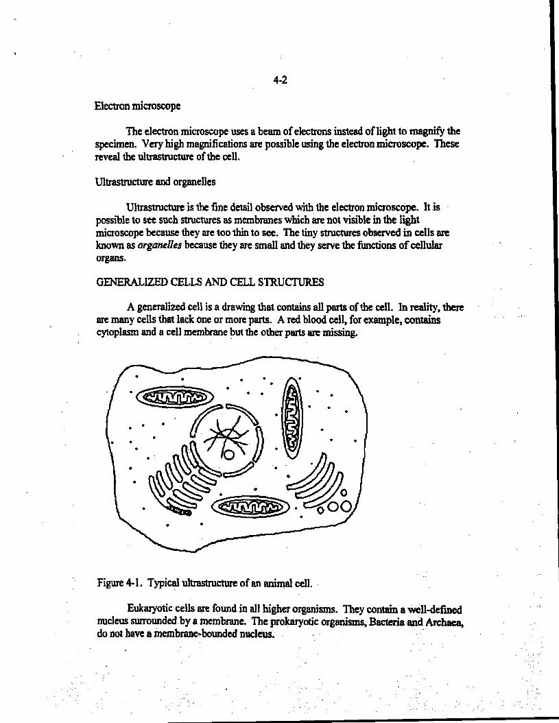
A generalized cell is a drawing that contains all parts ofthe cell. In reality, there are many cells that lack one or more parts. A red blood cell, for example, contains cytoplasm and a cell membrane ~ut the other parts are missing.
Figure 4-1. Typical ultrastructure of an animal cell. .
Eukaryotic cells are found in all higher organisms. They contain a Well-defmed nucleus surrounded by a membrane. The prokaryotic organisms, Baeteriaand An:haea, do not have a membrane-bounded nucleus.· .,.

I •
4-3
Cell membrane
The cell membrane is a structure that surrolDlds each cell. All living cells have a cell membrane. As shown in Figure 4-2, the cell membrane consists oftwo layers of lipid molecules with the polar heads toward the outside and the non-polar fatty acid tails in the center ofthe membrane. This prevents nearly all molecules from passing through the membrane. Only water and a few small neutral molecules can pass through the cell membrane directly. It is the bOlDldary between the living cell and its environment. It has the same function in both plant and animal cells.
The cell membrane has transport proteins to assist the passage of larger molecules or ionized molecUles. There are a variety ofother proteins embedded in the cell membrane. These are known as integralproteins. They help to maintain the structure ofthe cell membrane and other important functions. Sometimes the integral proteins have auxiliary proteins associated with them known as peripheralproteins. These are important for other cellular functions, such as tran~tting messages to parts of the cell.
Transport Protein
Twa I.,ers at lipidmolecUle.
Figure 4.2. Diagram ofcell membrane structure.
In the electron microscope, a single membrane, such as illustrated in Figure 4.3, appears as twoparaJlel lines at a distance of80 Angstrom lDlits apart. This is shown in Figure 4-3.
===::i1 IDA
Figure 4-3. Width ofmembrane.
4-4
The "fluid mosaic" model of the cell membrane
The proteins in the membrane can move around inside the two layers oflipicls and even protnJde through the SUlface. This gives the smface of the membrane the appearance of a mosaic, apiece of art made of little tiles glued together. The cell membrane therefore is referred to as a "fluid mosaic."
The Nucleus
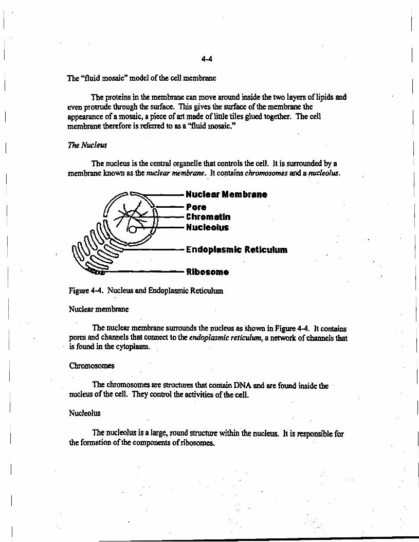
The nucleus is the central organelle that controls the cell. It is surrounded by a membrane known as the nuclear membrane. It coptains chromosomes and a-~cleolus.
r:~---Nuclear Membrene
r/*f,~ :~::matln ~» NucleolllB
~ ~)-------Endoplasmic Reticulum
~~;:!--------Ribosome
Figure 4-4. Nucleus and Endoplasmic Reticulum
Nuclear membrane
The nuclear membrane surrounds the nucleus as shown in Figure 4-4. It contains pores and channels that connect to the endoplasmic reticulum, a network ofchannels that is found· in the cytoplasm..
Chromosomes
The chromosomes are structures that contain DNA and are found inside the . nucleus of the cell. They control the activities ofthe cell.
Nucleolus
The nucleolus is a large, round structure within· the nucleus. It is responsible for the fonnation ofthe components ofribosomes. .
... '~.

, .
4-5
The Cytoplasm
The cytoplasm is the part ofthe cell that is found outside ofthe nucleus. It is bounded on the outside by the cell membrane. The cytoplasm contains many different organelles that perfonn various fimctions for the cell.
Mitochondria
Mitochondria are the structures that produce useful energy for the cell. They contain an internal membrane that is folded to form cristae. Each single crista is an individual fold ofthis inner membrane. See Figure 4-5.
IIl}!r---- Outer Membrane
H~I+--- Crist.
It.fli-----lnnerMembrane
Figuie 4-5. Struetme ofthe mitochondrion.
The folds ofthe cristae ofthe mitochondria contain enzymes that perfonn cellular respiration. Cellular respiration is the process by which useful energy is provided to the cell.
Endoplasmic reticulum
The endoplasmic reticulum (ER) is a series ofchannels in the cytopl8$ID~
. "Reticulum" means net or netWOrk. "Endo" means inside. So, "endoplasmic ~culum" means the network within the cytoplasm. It provides rapid molecular movement within . the cell. There are two kinds: rough ER and smooth ER.
Rough ER has ribosomeS attached to it. Its appearance in the electron microscope is that ofsandpaper. So it is known as rough because it looks rough. Smooth ER does not have ribosomes attached to its membranes.
. . Ribosomes
Ribosomes are the places where proteins are mad~. Each ribosome has a small .. sUbunit and a large subunit. When a protein is being synthesized, you find riboSomes
attached to a piece ofmessenger RNA. This structure is sometimes called a polyribosome orpalys0'r'e; .See Figure 4-6.
. ':.;: .'.
.',. '.".'

4-6
,..---Small Subunit
'6 6 6 6 ::Subunit
Figure 4-6. Apolysome.
Golgi bodies
Golgi bodies serve as packaging organelles for materials to be exported outside of the cell.. They start out as flat membranes that fill up as the material enters them. They become spherical and move toward the cell membrane. They are sometimes called dietyosomes. See Figme 4-7. .
rr--f- Flat membrane
~--- Filled membrane
__",,--Cellmembrane
.Figure 4-7. Golgi body.
Lysosomes
Lysosomes are structures that are membrane-bounded and contain digestive . . enzymes. The concept of lysis in biology implies breaking down, bursting, or otherWise being taken apart in some way. The digestive enzymes digest or breakdown chemically the particles that are taken into the lysosome. This process ofdigestion is the reverse of dehydration synthesis.
Vacuoles
Vacuoles are areas in the cytoplasm that have a membrane surrounding them. The term is derived from vacuum, which implies empty. Long ago it was thought that vacuoles were empty and contained no contents. It is now known that they are filled with cytoplasmic liquids or storage materials of various kinds.

. ...
4-7
The cytoskeleton
The cell has a netWork offlexible fibers within the cytoplasm. This network of fibers is called the cytoskeleton. The fibers are made ofpolymers ofproteins known as microtubules. These fibers have elastic properties that provide flexibility to the cell. They act as little muscles inside the cell.
Plant Cell Structures
A generalized plant cell is shown in Figure 4-8. There are some structures that are found only in plant cells. These include the cell wall and the plastids.
• • • 'O~'O 'O'O • •
.'O • .'O
~ • '.' • •
• ~ •
• I•
•. ~ .
Figure 4-8. Generalized plant celL .
Cell wall
The cell wall is made ofcellulose. It is considered non-living. The cellulose fibers fonn primary and secondary walls. The primary wall is usually thin. The secondary walls are thicker. Different types ofplant cells have different thicknesses-of cell wall. The cell wall pictured in Figure 4-8 is a primary cell wall. The cell membrane
. ofthe cell is pushed up against the inside ofthe cell wall by turgor pressure inside the Cell. .
Cell wall layers
The middle/amella is produced when a plant ceU1tivides. "Lamelli" means ... layer. The middle lamelJais the first layer fonned between. two plant cells. It is actuiJly .a membrane that the cells use to separate themselves at the end ofplant cell divisiC).L· .
;.... -..

4-8
Each cell produces aprimary wall on its side ofthe middle lamella. The primary wall is thin. All plant cells generally have a primary wall.
The secondary wall is generally much thicker than the primary wall. Secondary walls are found in plant cells that provide strength to structures in the plant. Not all plant cells produce secondary walls.
_ _ _ _ •_ Secondary Wall ••••• ~I. l... • .' z. -Middle Lamella'z:.: --:..: :.;: Primary Wan Z _ ._._ Z ,
Figure 4-9. Plant cell wall structure.
Plastids
Plastids are structures found only in plant cells. There are three kinds, which are chloroplasts, chromoplasts and leukoplasts.
Chloroplasts. ChloroplaSts are plant organelles that make food by performing the process ofphotosynthesis. Photosynthesis is the synthesis oforganic materials from inorganic raw materials. Using light energy the chloroplast converts carbon dioxide and water to glucose. The chloroplast contains layers ofmembranes called grana that contain chlorophyll. Chlorophyll is responsible for trapping light energy from the Sun.
@~j'5 g~=~Membran.
Figure 4-10. Chloroplast structure.
Chromoplasts. Chromoplasts are plastids that are colored ("chromo" means colored) but do not perfonn photosynthesis. Most contain carotenid pigments that are related to carotene. These pigments are various shades oforange or yellow. The pigments can be separated from leaves by means ofa technique known as chromatography.
Lellkoplasts. "Leuko" means lacking color or without color. Leukoplasts are plastids that do not have any color. They are used for storage ofmaterials. An example is the starch grain found within the cells ofpotatoes.

, .
4-9
Animal Cell Structures
Cilia and flagella
Cilia and flagella are organelles of locomotion. They contain contractile proteins.. The proteins allow the cilia and flagella to move. Flagella are much larger than cilia. Cilia are more numerous on a cell than flagella. A cell will have either cilia or flagella but not both.
Figure 4-11. Cross section ofa flagellum showing the 9+2 arrangemenl
The contractile proteins in a cilium or a flagelllDD have a characteristic arrangement. As shown in Figure 4-11, the protein fibers are in pairs. There are nine pairs around the outside with two fibers in the center. This structural configuration is
. known as the 9+2 arrangemenl~ .
~I-.--_-::::::brane ~ BasalBodJf ,-- Rootlet .
Figure 4-12. Flagellar attachment.
As shown in Figme 4-12, flageIJa are attached to the cell membrane. InSidethe .cell membrane is a basal body, which is anchored in the cytoplasm by rootlets.. The filament ofthe flagel1um, the partwith the 9+2 arrangement,.is located outside the cell membrane.
. .' ~'.::...
4-10
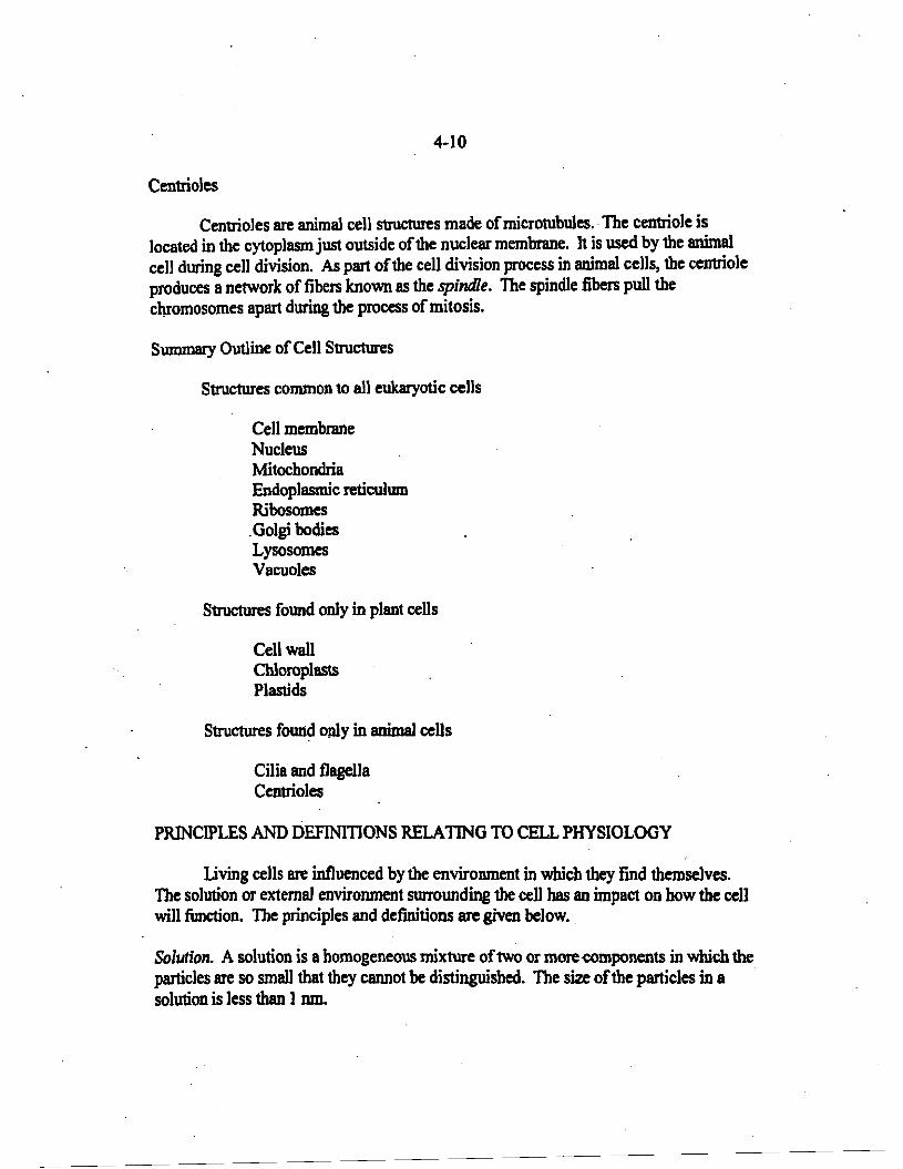
Centrioles
Centrioles are animal cell structures made ofmicrotubules.. The centriole is located in the cytoplasm just outside ofthe nuclear membrane. It is used by the animal cell during cell division. As part ofthe cell division process in animal cells, the centriole produces a network of fibers known as the spindle. The spindle fibers pull the chromosomes apart during the process ofmitosis.
Summary Outline ofCell Structures
Structures common to all eukaryotic cells
Cell membrane Nucleus Mitochondria Endoplasmic reticulmn Ribosomes .Golgi bodies Lysosomes Vacuoles
Structures found only in plant cells
Cell wall Chloroplasts Plastids
Structures foUli~ only in animal cells
Cilia and flagella Centrioles
PRINCIPLES AND DEFINITIONS RELATING TO CELL PHYSIOLOGY
Living cells are influenced by the environment in which they find themselves. The solution or external environment SUJTOunding the cell has an impact on how the cell will ftmction. The principles and definitions are given below..
Solution. A solution is a homogeneous mixture oftwo or more-components in which the particles are so small that they cannot be distinguished. The size ofthe particles in a solution is less than 1om.

4-11
Colloid. A colloid is a homogeneous dispersion ofparticles too large to be considered a solution, but which do not settle out appreciably on standing. The particle size in a conoid is approximately between 1 and 100 DID. A colloid exists either as a sol or a gel. The cytoplasm ofcens resembles a conoid.
Suspension. A suspension is a mixture ofparticles that settle out appreciably on standing. In a suspension, the particle size is greater than 100 nm.
Diffusion. Diffusion is the net movement of the particles ofa particular substance from an area ofhigher concentration ofthat subStance to an area oflower concentration ofthat substance, at constant temperatme and pressure,
Osmosis. Osmosis is the diffusion ofwater through a differentially (semi-) penneable membrane. In most biological systems, under normal conditions, water will move from the outside of the cell to the inside ofthe cen because the cen bas less water inside than there is outside. Osmosis is one ofthe most misunderstood biological concepts. Whatever is being seen, it has to involve water through a membrane. Ifnot, it is not osmosis.
. Outside
100% Water
Figure 4-13. Osmosis. Water enters the cen through the semi-penneable cell membrane..
In Figure 4-13, water enters the cell by omlosis because it is passing through the ceD membrane. This is because the concentration ofwater is higher outside the cell. If the cell were placed in salt water, water would leave the cell and pass through the membrane to the outside. This process is calledplasmolysis.
Active Transport. Active transport is the movement ofsubstance across the cell .-- -~ -- - -_. -- -------- membrane requiring the use ofenergy from the-celt-CelJular-eneriY isproduoed-by the
mitochondria and stored in the fann ofadenosine triphosphate (ATP).
Endocytosis. Endocytosis is the movement ofsubstances into the cell. If the substances are very large, such as bacteria, phagocytosis is used to allow them to enter the cen. The "phago" prefix refers to eating. So, phagocytosis is a process by which the cell eats. .

4-12
Pseudopod----"""" Food Vacuole
A B c
Figure 4-14. Phagocytosis. A. Cell approaches the food particle.B. Cell surroWltts the food particle using pseudopods~ C. Food particle is completely engulfed and is located inside a food vacuole.
In an organisni' such as the Amoeba, the cell uses pseudopods to surround the food particle. Pseudopods ("false feet'') are extensions ofthe cytoplasm ofthe cell. See Figure 4-14. Once the food particle is surrounded, it becomes part ofa food vacuole. The material in a food vacuole is digested by combination ofthe food vacuole with a lysosome. Certain white blood cells aiso can perform phasocytosis.
Another form ofendocytosis is pinoeyto$is. Pinocytosis is sometimes refmed to as "cell drinking." In pinocytosis, small droplets ofliquids or large molecules enter the cell via an invagination ofthe cell membrane. Pinocytosis is like phagocytosis only on a much smaller scale. Both processes require ATP as a source ofenergy.
Exocytosis. Exocytosis is the movement ofsubstances out ofthe cell. The substances to be exported are usually surrounded by a membranous vesicle, wch as that prodU<:ed by the Golgi bodies. When the membrane coalesces with the cell membrane, the material .inside the vesicle is exported outside the cell.
Regulation. The cell membrane cannot completely regulate the entrance or exit ofall materials. When a cell is placed in a hyperosmotic medium, it tends to. lose water into the medium outside the cell. This results in plasmolysis. When the cell is placed in a hypoosmotic medium, water enters the cell and it swells. Red blood cells burst open in water because water is a hypoosmotic medium to them. A cell in an isoosmotic medium will not appreciably gain or lose water.