cell trans
TRANSCRIPT

Cells
• Cells are the basic units of life
• All organisms are composed of cells.
• The activity of the body’s cells determines the both the structure and the function of the body.
• What we’ll be concentrating on is how things move in and out of cells.

Plasma Membrane• The PM separates the cell interior
(the cytoplasm) from the cell exterior (extracellular or interstitial fluid).
• Both the cytoplasm and the ECF are aqueous, i.e., they’re both water based.
• The PM is a bilayer (double layer) of phospholipids.
• A phospholipid is a molecule made of a glycerol backbone to which 2 fatty acids and one phosphorous-containing group are attached.
2 PM’s as seen w/ an electron microscope
Cartoon representation of the PM

In this molecule, we see the glycerol backbone (tan), the 2 fatty acids yellow), and the phosphorous-containing group (purple). The glycerol and fatty acids are nonpolar – they do not dissolve in water (hydrophobic). The phosphorus-containing group is polar – it does dissolve in water (hydrophilic). So, the phospholipid has a polar portion and a nonpolar portion (a portion that can dissolve in water and a portion that cannot). Because of this, we say that phospholipids are amphipathic.

Plasma Membrane• Since both the interior and
exterior of the cell are water-based, the phospholipids must form a 2-layered structure so that their nonpolar portion can be in the middle and not have any contact with the water-based ECF and ICF.
• This structure has important implications for how things travel thru this membrane.

• Notice that the PM is made up of more than just phospholipids.
• Proteins are found either embedded within the membrane itself (integral proteins), or weakly associated with either the interior or exterior face of the membrane (peripheral proteins).
• The PM also has molecules of cholesterol interspersed amongst the phospholipids. Cholesterol increases the temperature range over which the PM can function.

• Some integral proteins protrude into the ECF and have sugars attached. These are called glycoproteins.– Glycolipids often function as markers that label the cell as “self,” i.e., not
foreign.• Other integral proteins function as:
Enzymes Transport molecules Channels Structural supports

• The PM is a fluid structure. There is a lot of lateral movement of phospholipids and unanchored proteins.
• B/C of its gel-like nature and the fact that it’s made up of many different parts, we say the PM is a fluid mosaic.

What needs to cross the PM?
• Nutrients must get in and wastes must get out.
• Signaling molecules received by a cell may need to get in, while signaling molecules sent by a cell need to get out.
• Fluid must be able to get in and out.
• Certain ions must be able to get in and out.

Membrane Transport
• The transport of substances through the plasma membrane may be classified as:1. Passive Transport
• Depends solely on kinetic energy (KE).
– Every molecule has a certain amount of inherent energy that causes it to move about randomly
• No external energy is necessary2. Active transport
• KE does not suffice.• Additional energy
must be expended – usually via the hydrolysis of ATP.
1
2

Passive Transport
• Based on the concept of diffusion.– Random molecular movement
(due to KE) results in molecules moving from areas where their concentration is high to where their concentration is low.
• Note: Concentration is defined as how many molecules of a substance are present in a certain volume of liquid.
• The dissolved particles are the solutes while the fluid in which they’re dissolved is the solvent. Together, they create a solution. Note that in the above diagram,
molecules are moving down their concentration gradient!

Diffusion
• The rate at which molecules diffuse depends on:– Size of the concentration gradient
• Larger the gradient, the faster the diffusion. – The reason why is somewhat analogous to these 2 balls rolling
down these 2 hills. The red ball will get to the bottom first. Can you see why?

Diffusion• Diffusion also depends on:
– Size • Small molecules diffuse faster. • Diffusion depends on KE which
is dependent on temperature. So if 2 molecules are at the same temperature, they’ll have the same amount of KE.
• KE=(1/2)(mass)(velocity)2, so a larger molecule will have a reduced speed.
– Temperature • Diffusion is faster at higher
temperatures, because an increase in temperature causes an increase in kinetic energy.
THOUGHT PROBLEM: Ammonium hydroxide (weight=36.5) and hydrochloric acid (weight=35) can react as gases to form ammonium chloride, a smoky substance. In the tube below, we start with ammonium hydroxide at one end and hydrochloric acid at the other. At what point (A, B, or C) will we most likely see the smoky ammonium chloride? Why?
NH4OH HCl
A B C

Diffusion• Many molecules have the
capacity to diffuse right through the phospholipid bilayer.– Nonpolar molecules move thru
w/ ease b/c they do not mind the hydrophobic interior of the plasma membrane.
• Such molecules include O2, CO2, steroid hormones, fat soluble vitamins, and alcohol.
– What governs whether these molecules will move into or out of a cell?
• Its CONCENTRATION GRADIENT.
• Hydrophilic molecules must diffuse thru special protein channels in order to get thru the cell membrane.
• This type of transport through the plasma membrane is termed simple diffusion.

Diffusion• Some polar molecules enter cells via diffusion. They cannot go straight
through the nonpolar lipid bilayer, so they require the assistance of proteins that act as carrier molecules.
• These carriers bind the substance (glucose and other simple sugars are the best examples) on one side of the plasma membrane and then change their conformation and release the substance on the other side of the PM.
This is known as facilitated diffusion.

Diffusion
• Why are both simple diffusion and facilitated diffusion considered to be passive transport?
• Oxygen, water, and glucose are vitally important to cells. The fact that they are taken up with no energy expenditure is important to life in terms of how much energy we require.

Osmosis• Osmosis is the diffusion of water through a
semipermeable membrane.• A membrane that is semipermeable allows
some molecules to pass thru, but does not allow others.
• Water will diffuse from an area of high concentration to an area of low conc.
• It’s important that you understand the following:– An area that has a high [water] has a low
[dissolved particles].– An area that has a low [water] has a high
[dissolved particles].
Right: Water molecules (black dots) move to the right since they are high in conc. on the left and low on the right.

Osmosis• The total [solute] in a solution is the osmolarity of that solution.
• Water will diffuse from a solution with low osmolarity into a solution of high osmolarity if they are separated by a semipermeable membrane.– This is simply another
way of saying that water will flow down its concentration gradient.
(Permeable to water only)

Tonicity• Tonicity is defined as the ability of a solution to change
the shape of a cell immersed in it due to changes in the cell’s water volume.
• A solution with the same concentration of non-penetrating solutes as those found in cells are isotonic, i.e., “the same tonicity.”
• Cells exposed to such solution retain their normal shape and exhibit no net gain or loss of water.
• Most intravenous solutions are isotonic (e.g., 0.9% saline or 5% glucose).
– Why is this necessary?

Red Blood Cells inan Isotonic Solution

Tonicity• Suppose you placed a cell
in a solution, and the cell shrank (the technical term is crenated):– The cell must have lost
water which means that, the [non-penetrating solutes] was higher outside the cell.
– Thus, the solution was hypertonic (hyper means greater than usual).
Red blood cells in a hypotonic environment

Tonicity• Suppose you put a cell in solution and the cell burst (lysed):– The cell must have gained
water which means that the [non-penetrating solutes] was higher inside the cell.
– Thus the solution was hypotonic (hypo means less than usual).
• Do you think pure water is hypertonic to cells or hypotonic to cells?
RBC’s in a hypotonic environment


Dialysis• In renal failure (kidney failure), nitrogenous wastes can accumulate in the blood (azotemia), blood pH drops precipitously, and the ionic composition of the blood goes awry.
• If nothing is done, the condition can be fatal!
• To prevent any adverse consequences, the blood must be cleansed of metabolic wastes and its ionic composition restored.
• This can be accomplished by dialysis.

• One type of dialysis is hemodialysis, where the patient’s blood is passed through a membrane tubing that is permeable only to selected substances, and the tubing is set in a bath that is similar to normal plasma.
• As blood circulates through the tubing, substances such as nitrogenous wastes and K+ present in the blood (but not in the bath) diffuse out of the blood and into the bath.
• Meanwhile, buffering chemicals (which restore pH) and glucose (for malnourished patients) will diffuse from the bath into the blood.
Dialysis

Filtration• Passive process that forces water and solutes through a
membrane or capillary by fluid or hydrostatic pressure.• Based on a pressure gradient that pushes solute-
containing fluid from an area of higher pressure to an area of low pressure.
• When blood is pumped out of the heart, it is moving fast and is exerting a good deal of force on the walls of the blood vessels. As a result, fluid (+ solutes) is forced out of the capillaries and into the extracellular space.

Filtration• Just as fluid is pushed out of capillaries by hydrostatic pressure, fluid is pulled into capillaries because of osmotic pressure.
• The net result is that a small amount of fluid is forced out of cells. This is the interstitial fluid.
• Any excess interstitial fluid can be taken up into the lymphatic system and returned to the bloodstream.
• Filtration also plays a very large role in urine formation.

Active Transport• Involves the expenditure of energy to move solutes across the cell
membrane against their concentration gradient.• Active transport can be primary or secondary.
– In primary active transport, the direct hydrolysis of ATP provides the energy needed to transport the solute.
– In secondary active transport, the movement of a molecule down its concentration gradient is coupled to the movement of a molecule up its concentration gradient. ATP is not directly involved.

Primary Active Transport
• Transport may be solo (uniport) or coupled (symport and antiport):
1. Uniport:– The hydrolysis of ATP “pumps”
one molecule of one solute across a membrane against its gradient.
– Muscle cells contain Ca2+ uniporters. Lots of other examples.
2. Symport:– The hydrolysis of ATP pumps 2
molecules in the same direction against their concentration gradient

Primary Active Transport3. Antiport:
– Hydrolysis of ATP pumps one or more solute molecules against their gradient in one direction, and pumps one or more solute molecules against their gradient in the opposite direction.
– Best example is the Na+/K+ pump which pumps 3 Na+ out of the cell and 2 K+ into the cell for each ATP hydrolyzed.
– Probably the most important protein in your whole body. 40% of all the energy in your body goes towards powering this pump. We shall see it often!

Secondary Active Transport• When a molecule is transported
thru the PM down its concentration gradient, energy is released. This energy can be used to transport another molecule against its concentration gradient.
• This is secondary active transport.• The best example is the transport of
glucose in the digestive tract into the cells lining the small intestine.
• ATP is indirectly involved in this process. How might it be? (Look at the picture!)

Big Stuff?
• Protein pumps are adequate for bring small molecules into the cell or out of the cell.
• However, a different kind of active transport is necessary to move large things into/out of the cell – vesicular transport.
There are 2 types of vesicular transport: endocytosis and exocytosis

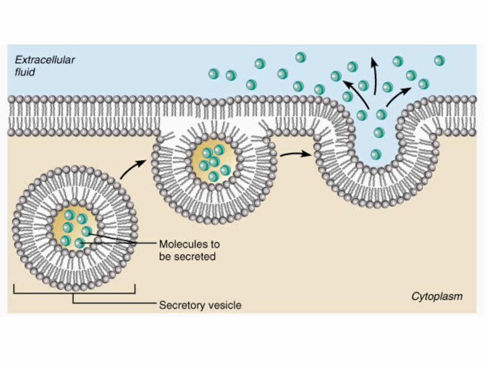
Exocytosis• Exocytosis literally means “out of the cell”• It accounts for hormone secretion, neurotransmitter release,
mucus secretion, and, sometimes, ejection of wastes.– Inside the cell, the substance to be exported is enclosed in a
membranous sac called a vesicle.– The vesicle will migrate to the PM fuse with it, and then rupture,
spilling the contents into the extracellular space.


Endocytosis
• Reverse of exocytosis. Allows macromolecules to enter cells.– The substance is progressively enclosed by an enfolding portion of the
plasma membrane.– This forms a vesicle which will pinch off the plasma membrane and
enter the cytosol where it is typically digested.• Types of endocytosis are:
– Phagocytosis– Pinocytosis (a.k.a. bulk-phase endocytosis)– Receptor-mediated endocytosis

Phagocytosis• Literally “cell-eating.”• Cytoplasmic extensions called
pseudopods “reach out and grab” large, solid material such as a clump of bacteria or cell debris, and then engulf it.
• The resulting vesicle is called a phagosome.
• Usually, the phagosome fuses with a lysosome, a membranous organelle that contains digestive enzymes, and its contents are digested.
• Macrophages and white blood cells are the most phagocytic cells in the body.

Pinocytosis• Literally means “cell-drinking.”• A bit of infolding plasma membrane surrounds a
droplet of extracellular fluid containing dissolved molecules. This creates a tiny membranous vesicle.
• Most cells routinely perform this.• Unlike phagocytosis, pinocytosis is unselective!


Receptor-Mediated Endocytosis
• Main mechanism for the specific uptake of macromolecules by most cells – remember, pinocytosis is non-selective and phagocytosis is typically only performed by macrophages and white blood cells.
• Molecules taken up by cells via RME include:– Enzymes– Hormones, e.g., insulin– Low-density lipoproteins (LDL), i.e., the “bad cholesterol”– Flu viruses and the diphtheria toxin also use RME to enter
cells

Receptor-Mediated Endocytosis• Receptors for the molecule to be ingested by a cell are on the PM.
Different cells have different receptors and thus take up different molecules.
• A macromolecule will bind with its particular receptor and then these receptor-macromolecule complexes cluster together, invaginate and are internalized.