cell to cell communication yasir waheed. the three largest classes of cell surface receptor proteins...
TRANSCRIPT

Cell to Cell Communication
Yasir Waheed

The Three Largest Classes of Cell Surface Receptor Proteins Are
1. Ion-Channel-linked
2. G-Protein-linked
3. Enzyme-linked Receptors

Figure 15-15. Three classes of cell-surface receptors. (A) Ion channel-linked receptors, (B) G-proteinlinked receptors, and (C) enzyme-linked receptors. Although many enzyme-linked receptors have intrinsic enzyme activity, as shown on the left, many others rely on associated enzymes, as shown on the right.
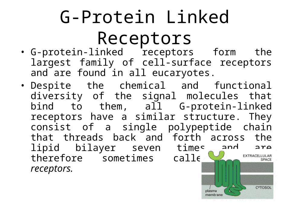
G-Protein Linked Receptors• G-protein-linked receptors form the largest family of cell-
surface receptors and are found in all eucaryotes.• Despite the chemical and functional diversity of the
signal molecules that bind to them, all G-protein-linked receptors have a similar structure. They consist of a single polypeptide chain that threads back and forth across the lipid bilayer seven times and are therefore sometimes called serpentine receptors.

Trimeric G Proteins Disassemble to Relay Signals from GProtein-linked Receptors
• When extracellular signaling molecules bind to serpentine receptors, the receptors undergo a conformational change that enables them to activate trimeric GTP-binding proteins (G proteins).
• G proteins are composed of three protein subunits alpha, beta, and gamma. In the unstimulated state, the subunits has GDP bound and the G protein is inactive
• When stimulated by an activated receptor, the a subunit releases its bound GDP, allowing GTP to bind in its place. This exchange causes the trimer to dissociate into two activated components an alpha subunit and a beta-gamma complex.

Figure 15-28. The disassembly of an activated G-protein into twosignaling components. (A) In the unstimulated state, the receptor and the G protein are both inactive. (B) Binding of an extracellular signal to the receptorchanges the conformation of the receptor, which in turn alters the conformation of the G protein that is bound to the receptor. (C) The alteration of the alpha subunit of the G protein allows it to exchange its GDP for GTP. This causes the G protein to break up into two active components an alpha subunit and a betagamma complex, both of which can regulate the activity of target proteins in the plasma membrane. The receptor stays active untill ligand molecule is bound to it.

Figure 15-29. The switching off of the G-protein a subunit by the hydrolysis of its bound GTP. After a G-protein a subunit activates its target protein, it shuts itself off by hydrolyzing its bound GTP to GDP. This inactivates the a subunit, which dissociates from the target protein and reassociates with a betagamma complex to re-form an inactive G protein. Binding to the target protein or to a membrane-bound RGS protein (not shown) usually stimulates the GTPase activity of the a subunit; this stimulation greatly speeds up the inactivation process shown here.

• RGS proteins (regulator of G protein signaling) act as a-subunit-specific GTPase activating proteins (GAPs), and they are thought to have a crucial role in shutting off G-protein mediated responses in all eucaryotes. There are about 25 RGS proteins encoded in the human genome, each of which is thought to interact with a particular set of G proteins.
• Cyclic AMP (cAMP) act as secondary messenger (ligand, primary messenger). Found in all procaryotic and animal cells that have been studied. The normal concentration of cyclic AMP inside the cell is about 10-7 M, but an extracellular signal can cause cyclic AMP levels to change by more than twentyfold in seconds . such a rapid response requires that a rapid synthesis of the molecule be balanced by its rapid breakdown or removal. In fact, cyclic AMP is synthesized from ATP by a plasma-membrane-bound enzyme adenylyl cyclase, and it is rapidly and continuously destroyed by one or more cyclic AMP phosphodiesterases that hydrolyze cyclic AMP to adenosine 5 -monophosphate (5 -AMP).

Figure 15-33. How gene transcription is activated by a rise in cyclic AMP concentration. The binding of an extracellular signal molecule to its Gprotein- linked receptor leads to the activation of adenylyl cyclase and a rise incyclic AMP concentration. The increase in cyclic AMP concentration activates PKA in the cytosol, and the released catalytic subunits then move into the nucleus, where they phosphorylate the CREB gene regulatory protein. Once phosphorylated, CREB recruits the coactivator CBP, which stimulates gene transcription. This signaling pathway controls many processes in cells, ranging from hormone synthesis in endocrine cells to the production of proteins required for long-term memory in the brain. We shall see later that some kinases that are activated by a rise in intracellular Ca2+ can also phosphorylate and thereby activate CREB.

Inositol Phospholipid pathway activated by G proteins

Some G Proteins Activate the Inositol PhospholipidSignaling Pathway by Activating Phospholipase C-beta
Phosphotidylinositol present on the cytoplasmic side of plasma membrane is phosphorylated into PI4-P which is then phosphorylated to PI 4,5 P.

Figure 15-35. The hydrolysis of PI(4,5)P2 by phospholipase C-b. Two intracellular mediators are produced when PI(4,5)P2 is hydrolyzed: inositol 1,4,5-trisphosphate (IP3), which diffuses through the cytosol and releases Ca2+ from the ER, and diacylglycerol, which remains in the membrane and helps to activate the enzyme protein kinase C.


Once activated, PKC phosphorylates target proteins that vary depending on the cell type. The principles are the same as discussed earlier for PKA, although most of the target proteins are different.

Signaling through Enzyme linked surface recrptors.
• Like G-protein-linked receptors, enzyme-linked receptors are transmembrane proteins with their ligand-binding domain on the outer surface of the plasma membrane, their cytosolic domain either has an intrinsic enzyme activity or associates directly with an enzyme.
• Six classes of enzyme-linked receptors have thus far been identified:1. Receptor tyrosine kinases phosphorylate specific tyrosines on a small set of
intracellular signaling proteins.2. Tyrosine-kinase-associated receptors associate with intracellular proteins that have
tyrosine kinase activity.3. Receptorlike tyrosine phosphatases remove phosphate groups from tyrosines of
specific intracellular signaling proteins. (They are called "receptorlike“ because the presumptive ligands have not yet been identified, and so their receptor function has not been directly demonstrated.)
4. Receptor serine/threonine kinases phosphorylate specific serines or threonines on associated latent gene regulatory proteins.
5. Receptor guanylyl cyclases directly catalyze the production of cyclic GMP in the cytosol.
6. Histidine-kinase-associated receptors activate a "two-component" signaling pathway in which the kinase phosphorylates itself on histidine and then immediately transfers the phosphate to a second intracellular signaling protein.

Figure 15-49. Seven subfamilies of receptor tyrosine kinases. Only one or two members of each subfamily are indicated. Note that the tyrosine kinase domain is interrupted by a "kinase insert region" in some of the subfamilies.
Structure of Immunoglobulin

Figure 15-50. Three ways in which signaling proteins can cross-link receptor chains. When the receptor chains are crosslinked, the kinase domains of adjacent receptors cross-phosphorylate each other, stimulating the kinase activity of the receptor and creating docking sites for intracellular proteins. (A) Platelet-derived growth factor (PDGF) is a covalently linked dimer with two receptor-binding sites, so it can directly cross-link adjacent receptors to initiate the intracellular signaling process. (B) Some monomeric ligands, such as fibroblast growth factors (FGFs), bind in clusters to proteoglycans, enabling the ligands to cross-link their receptors. (C) Membrane-bound signaling proteins, such as ephrins, can cross-link their receptors even though they are monomeric, because they cluster in the plasma membrane of the signaling cell.

Figure 15-52. The docking of intracellular signaling proteins on an activated receptor tyrosine kinase. The activated receptor and its bound signaling proteins form a signaling complex that can then broadcast signals along multiple signaling pathways.

Figure 15-55. The activation of Ras by an activated receptor tyrosine kinase. Most of the signaling proteins bound to the activated receptor are omitted for simplicity. The Grb-2 adaptor protein binds to a specific phosphotyrosine on the receptor and to the Ras guanine nucleotide exchange factor (GEF), which stimulates Ras to exchange its bound GDP for GTP. The activated Ras then activates several downstream signaling pathways, one of which is shown in Figure 15-56.

Figure 15-56. The MAP-kinase serine/threonine phosphorylation pathway activated by Ras. The pathway activated by Ras begins with a MAP-kinase-kinase-kinase called Raf, which activates the MAP-kinase-kinase Mek, which then activates the MAP-kinase called Erk. Erk in turn phosphorylates a variety of downstream proteins, including other kinases, as well as gene regulatory proteins in the nucleus. The resulting changes in gene expression and protein activity cause complex changes in cell behavior.

Figure 15-61. Five parallel intracellular signaling pathways activated by Gprotein- linked receptors, receptor tyrosine kinases, or both. In this schematic example, the five kinases (shaded yellow) at the end of each pathway phosphorylate target proteins (shaded red), some of which are phosphorylated by more than one of the kinases. The specific phospholipase C activated by the two types of receptors is different: G-protein-linked receptors activate PLC-beta, whereas receptor tyrosine kinases activate PLC-gamma (not shown).

THANKS