cell structure and function for stds ts
TRANSCRIPT
Tanveer Saeed
Assistant Professor
AKU-SONAM
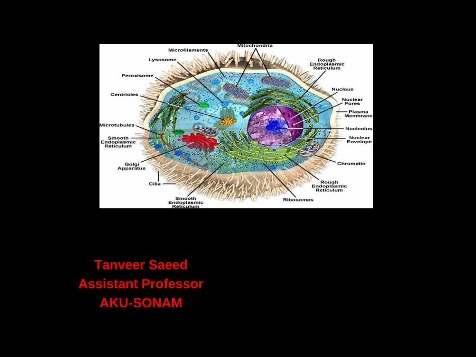
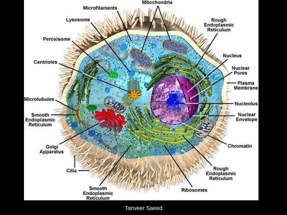
• Smallest basic unit of life.
• Body has about 100 trillion cells.
• All cells have DNA and cytoplasm.
Cell size and Shape
Human cell are microscopic in size they
are in different size and shapes.
• RBC diameter 7.5µm
• Egg cell size is 1000µm
Cell
Tanveer Saeed
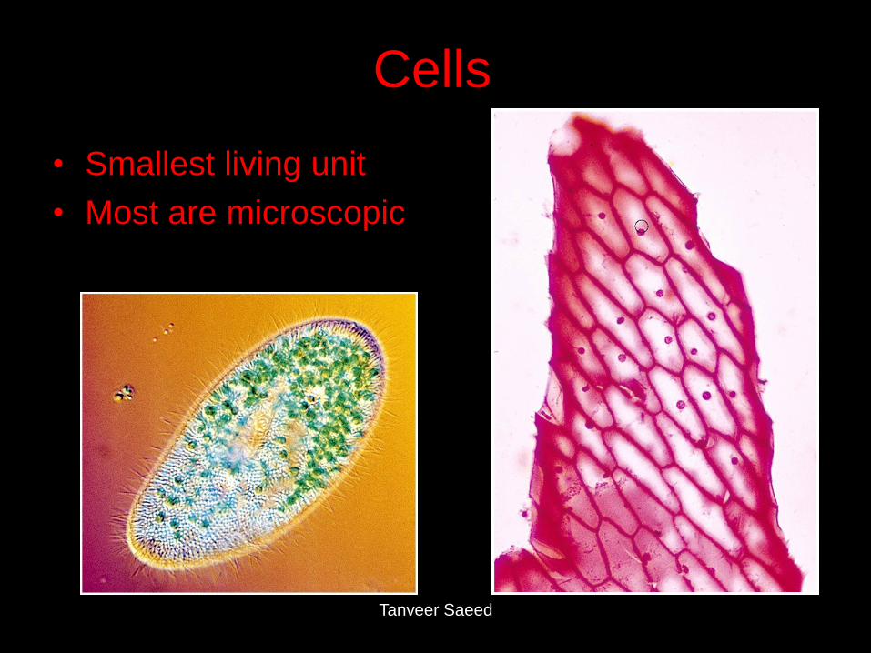
Cells
• Smallest living unit
• Most are microscopic
Tanveer Saeed
Tanveer Saeed
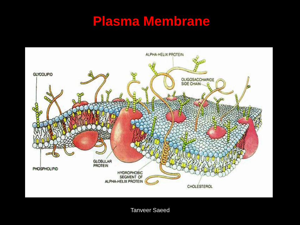
Plasma Membrane
Tanveer Saeed
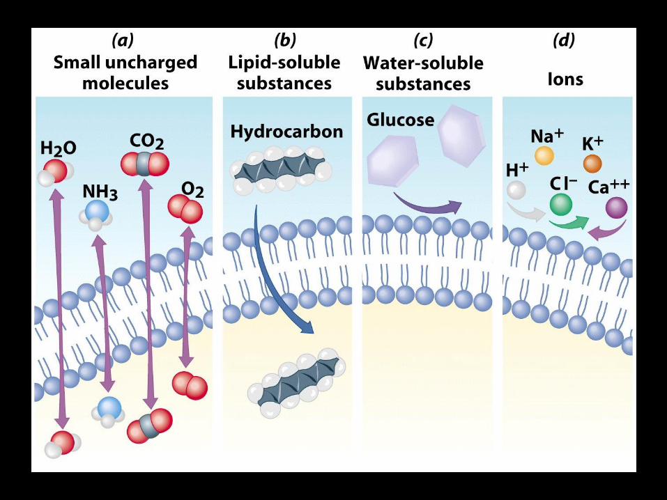
• Selectively permeable membrane that
surround the cell is called plasma
membrane
• Phosphobilipidc consists of two back to
back layer made up of three types of lipid
molecules.
Phospholipid 75% Glycolipid
5% Cholesterol 20%
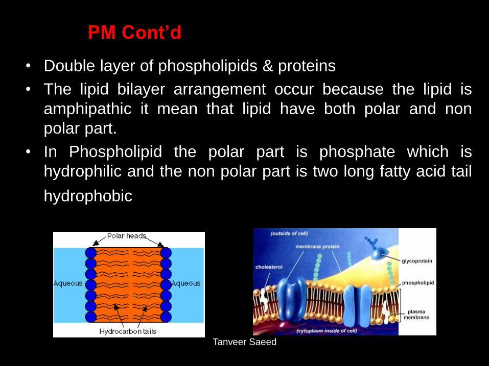
PM Cont’d
• Double layer of phospholipids & proteins
• The lipid bilayer arrangement occur because the lipid is
amphipathic it mean that lipid have both polar and non
polar part.
• In Phospholipid the polar part is phosphate which is
hydrophilic and the non polar part is two long fatty acid tail
hydrophobic
Tanveer Saeed
Tanveer Saeed
1. Plasma membrane give form to the cell and separate the
cell internal structure from the extra cellular environment
2. It provides selective transportation across the membrane
3. The membrane protein serve a variety of functions i.e.
• Provide structural support
• Form tiny channels through which certain substance flow
into or out of the cell
• Act as transporter or carrier of some substances
• Some integral membrane protein are called receptors
which serve as cellular recognition sites
• Membrane glycoprotein and Glycolipid often are cell
identity marker. It give the cell its immunologic identity
Function of Plasma Membrane

Tanveer Saeed
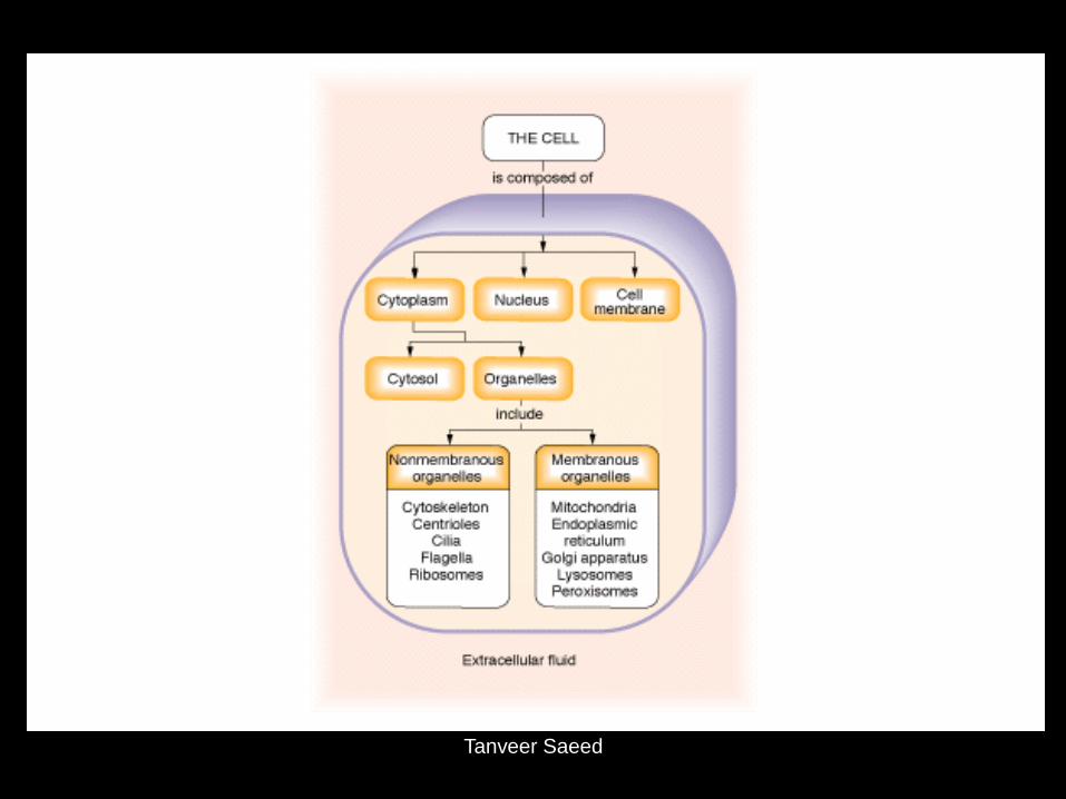
Cytoplasm Cytoplasm has 2 component
• Cytosol
• Organelles Viscous fluid containing organelles
• components of cytoplasm
– Interconnected filaments & fibers
– Fluid = cytosol
– Organelles (not nucleus) specialized structure that have characteristic shape and that perform specific function in cellular growth, maintenance and reproduction.
– storage substances
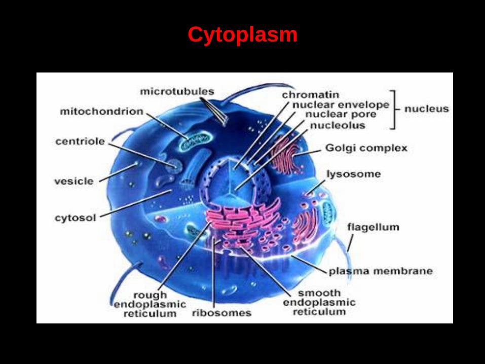
Cytoplasm
Tanveer Saeed
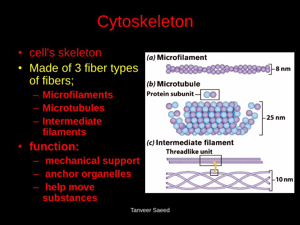
Cytoskeleton
• cell's skeleton
• Made of 3 fiber types of fibers; – Microfilaments
– Microtubules
– Intermediate filaments
• function: – mechanical support
– anchor organelles
– help move substances
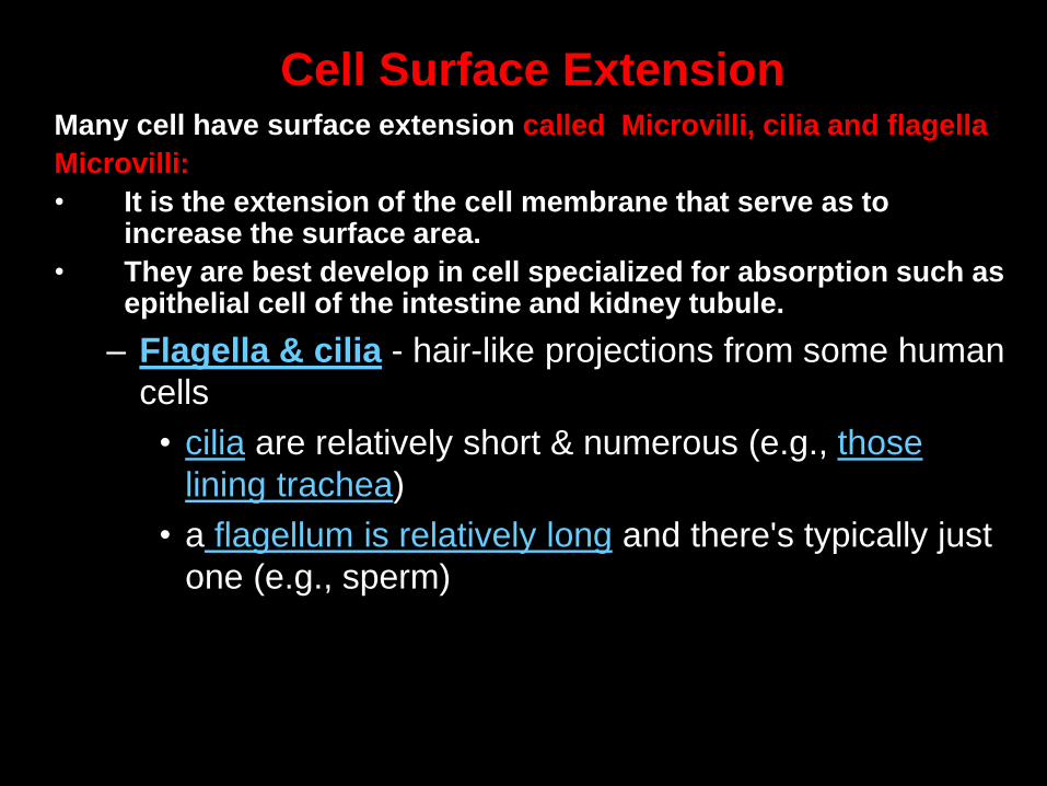
Many cell have surface extension called Microvilli, cilia and flagella
Microvilli:
• It is the extension of the cell membrane that serve as to increase the surface area.
• They are best develop in cell specialized for absorption such as epithelial cell of the intestine and kidney tubule.
– Flagella & cilia - hair-like projections from some human
cells
• cilia are relatively short & numerous (e.g., those
lining trachea)
• a flagellum is relatively long and there's typically just
one (e.g., sperm)
Cell Surface Extension
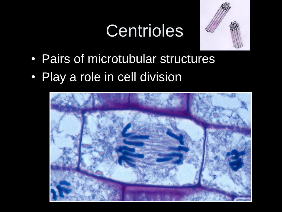
Centrioles
• Pairs of microtubular structures
• Play a role in cell division
Endoplasmic Reticulum
• Helps move substances within cells
• Network of interconnected membranes
• Two types
– Rough endoplasmic reticulum
– Smooth endoplasmic reticulum
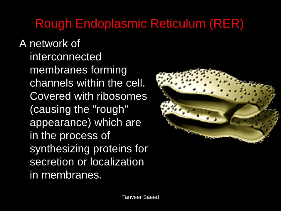
Rough Endoplasmic Reticulum (RER)
A network of
interconnected
membranes forming
channels within the cell.
Covered with ribosomes
(causing the "rough"
appearance) which are
in the process of
synthesizing proteins for
secretion or localization
in membranes.
Tanveer Saeed
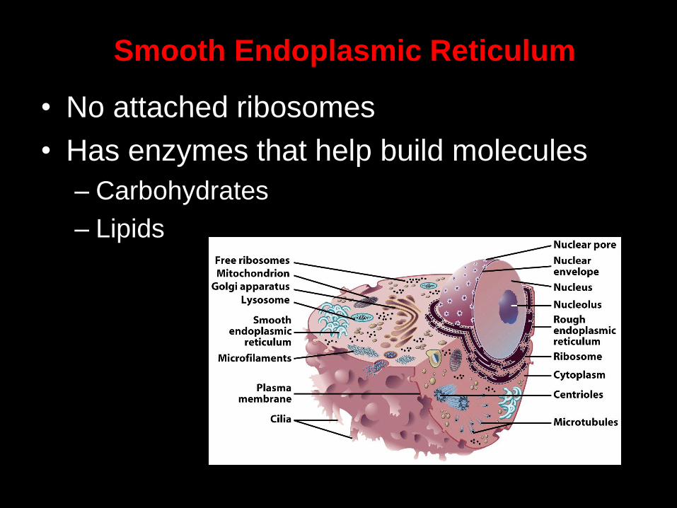
Smooth Endoplasmic Reticulum
• No attached ribosomes
• Has enzymes that help build molecules
– Carbohydrates
– Lipids
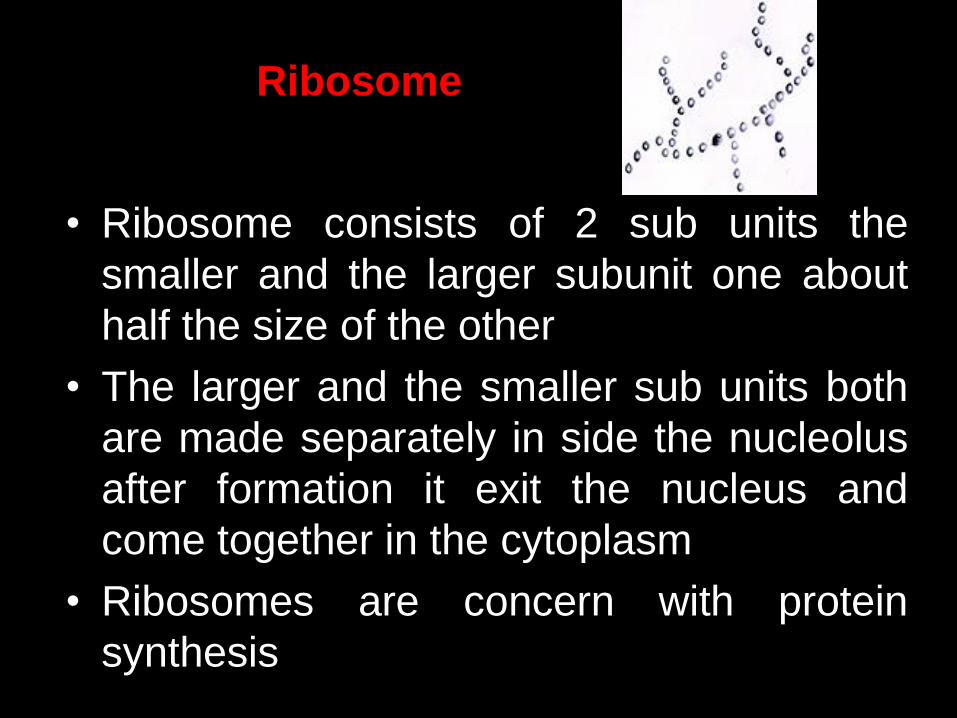
• Ribosome consists of 2 sub units the
smaller and the larger subunit one about
half the size of the other
• The larger and the smaller sub units both
are made separately in side the nucleolus
after formation it exit the nucleus and
come together in the cytoplasm
• Ribosomes are concern with protein
synthesis
Ribosome
Ribosomes
– composed of rRNA (ribosomal RNA) &
protein
• may be dispersed randomly
throughout the cytoplasm or attached
to surface of rough endoplasmic
reticulum
• often linked together in chains called
polyribosomes or polysomes
• primary function is to produce proteins
Tanveer Saeed
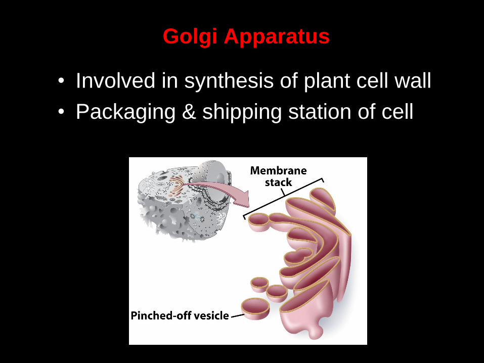
Golgi Apparatus
• Involved in synthesis of plant cell wall
• Packaging & shipping station of cell
Golgi apparatus
A series of stacked membranes. Vesicles
(small membrane surrounded bags) carry
materials from the RER to the Golgi
apparatus. Vesicles move between the
stacks while the proteins are "processed" to
a mature form. Vesicles then carry newly
formed membrane and secreted proteins to
their final destinations including secretion or
membrane localization.
Tanveer Saeed
Golgi Apparatus Function
1. Molecules come in vesicles
2. Vesicles fuse with Golgi membrane
3. Molecules may be modified by Golgi4. Molecules
pinched-off in separate vesicle
5. Vesicle leaves Golgi apparatus
6. Vesicles may combine with plasma membrane to
secrete contents
Tanveer Saeed
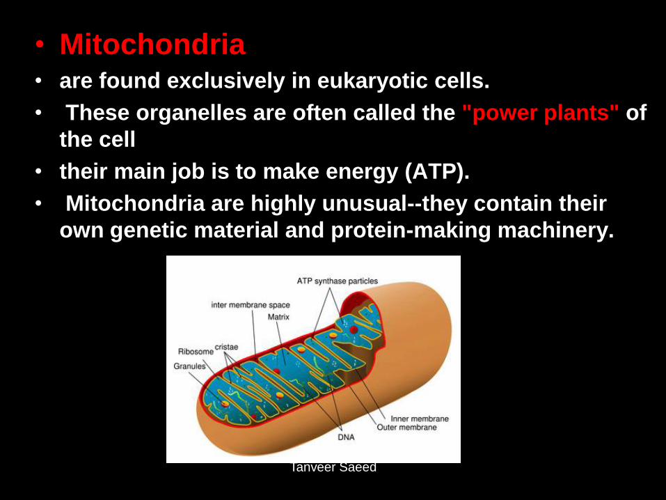
• Mitochondria • are found exclusively in eukaryotic cells.
• These organelles are often called the "power plants" of
the cell
• their main job is to make energy (ATP).
• Mitochondria are highly unusual--they contain their
own genetic material and protein-making machinery.
Tanveer Saeed
Mitochondria
• They have a double-membrane: outer
membrane & highly convoluted inner
membrane.
• inner membrane has folds or shelf-like
structures called cristae that contain
elementary particles; these particles
represent an enzyme important in ATP
production
Tanveer Saeed

– Centrioles -
• paired cylindrical structures located near the
nucleus
• play an important role in cell division
• Mitosis and the role of centrioles
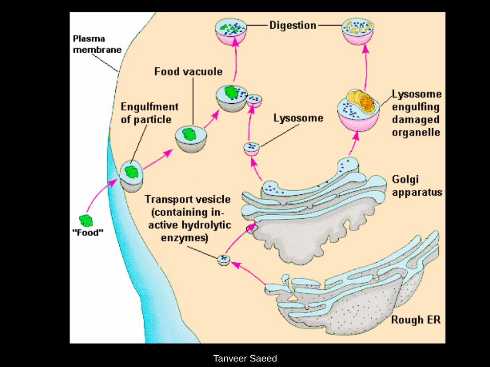
• Lysosomes: Contain digestive enzymes
• Functions
– Aid in cell renewal
– Break down old cell parts
– Digests invaders
Tanveer Saeed
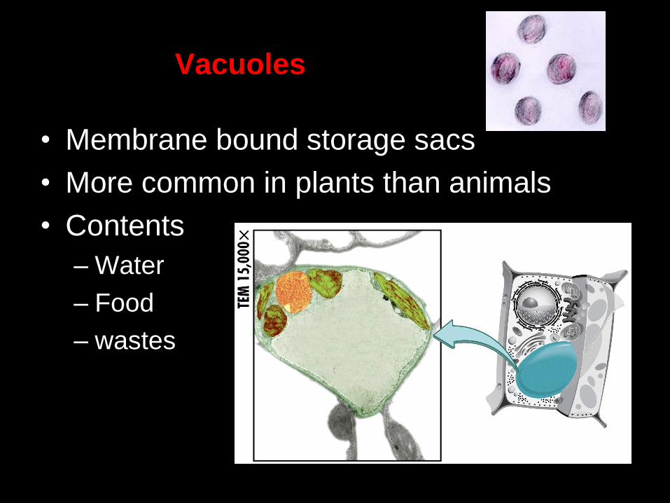
Vacuoles
• Membrane bound storage sacs
• More common in plants than animals
• Contents
– Water
– Food
– wastes

Tanveer Saeed
Molecule Movement & Cells
• Passive Transport
• Active Transport
• Endocytosis
(phagocytosis & pinocytosis)
• Exocytosis
Types of Passive Transport
1. Diffusion
2. Osmosis
3. Facilitated diffusion
Passive Transport
• No energy required
• Move due to gradient
– differences in concentration, pressure, charge
• Move to equalize gradient
– High moves toward low
Osmosis
• Special form of diffusion
• Fluid flows from lower solute concentration
• Often involves movement of water
– Into cell
– Out of cell