cell rearrangement and segmentation in xenopus:...
TRANSCRIPT

Development 105, 155-166 (1989)Printed in Great Britain © The Company of Biologists Limited 1989
155
Cell rearrangement and segmentation in Xenopus: direct observation of
cultured explants
PAUL A. WILSON1, GEORGE OSTER2 and RAY KELLER1
1 Department of Zoology, University of California, Berkeley, CA 94720, USA1 Departments of Biophysics, Entomology & Zoology, University of California, Berkeley, CA 94720, USA
Summary
We make use of a novel system of explant culture andhigh resolution video-film recording to analyse for thefirst time the cell behaviour underlying convergentextension and segmentation in the somitic mesoderm ofXenopus. We find that a sequence of activities sweepsthrough the somitic mesoderm from anterior to pos-terior during gastrulation and neurulation, beginningwith radial cell intercalation or thinning, continuingwith mediolateral intercalation and cell elongation, andculminating in segmentation and somite rotation. Radialintercalation at the posterior tip lengthens the tissue,while mediolateral intercalation farther anterior con-
verges it toward the midline. This extension of thesomitic mesoderm helps to elongate the dorsal side ofintact neurulae. By separating tissues, we demonstratethat cell rearrangement is independent of the notochord,but radial intercalation - and thus the bulk of extension- requires the presence of an epithelium, either endoder-mal or ectodermal. Segmentation, on the other hand,can proceed in somitic mesoderm isolated at the end ofgastrulation. Finally, we discuss the relationship be-tween cell rearrangement and segmentation.
Key words: somitogenesis, cell rearrangement, mesoderm.
Introduction
Early amphibian development is largely the develop-ment of the dorsal mesoderm. The notochord and thesomites of the vertebrate embryo are constructed di-rectly by this tissue, while the neural tube results fromits inductive influence. Spemann recognized the excep-tional pattern-forming properties of this region, demon-strating that it had the capacity to induce patterneddorsal structures in ventral ectoderm and mesoderm; hechristened it the 'organizer' (Spemann, 1938). In coop-eration with the prospective neural plate, the conver-gence and extension of the dorsal mesoderm also drivesmuch of gastrulation (Keller, 1986; Keller & Danilchik,1988), and elongates the dorsal side of the embryoduring neurulation (Jacobson, 1981; Keller & Danil-chik, 1988). Although the importance of these morpho-genetic movements has been known for a long time(Vogt, 1929), almost nothing is known about how cellsbring them about.
Finally, the appearance of somites in the dorsalmesoderm during neurulation is particularly interestingbecause the pattern of somites prefigures - and in factdirects - all subsequent segmental organization in thevertebrate embryo (Detwiler, 1934). Hamilton usedhistological sections and Youn and his co-workers usedSEM to describe various aspects of amphibian somito-genesis (Hamilton, 1969; Youn et al. 1980; Youn &
Malacinski, 1981a,b; Malacinski et al. 1981). Severalgroups also investigated the effect on somite pattern ofvarious microsurgical manipulations (Waddington &Deuchar, 1953; Deuchar & Burgess, 1967; Cooke,1975), and of brief heat shocks (Pearson et al. 1976;Cooke, 1978; Pearson & Elsdale, 1979; Elsdale &Davidson, 1983). Although much has been learnedfrom these studies, we still do not understand howamphibian cells group into segments, or what cellularactivities precede segmentation.
These processes have been difficult to study becausethe amphibian embryo is opaque - inner layers of theintact embryo are hidden from view. Thus until recentlyit was not possible to observe directly the patterns ofcell behaviour responsible for mesodermal morphogen-esis. In the last few years, however, Keller and Danil-chik have developed a method of culturing explants ofamphibian tissue that permits almost normal develop-ment and continuous observation of the mesoderm(Keller et al. 1985a,b). We have used this culturetechnique to observe, in vitro, morphogenesis anddifferentiation of the somitic mesoderm during theneurula stages, after its involution is complete. We havefilmed for the first time in the amphibian not only thesegmentation and subsequent morphogenesis of thedorsal mesoderm, but the cell rearrangement thatprecedes it and drives dorsal elongation. In this paper,we will analyse the progression of cell behaviours that

156 P. A. Wilson et al.
sweep from anterior to posterior in the axial mesoderm.Our focus will be on the two most dramatic processestaking place during this time: convergent extension andsegmentation. In a companion paper (Wilson & Keller,1989), we present our analysis of cell behaviour in thedorsal mesoderm during gastrulation. In a third paper,Keller and others (1989) use both this new culturetechnique and SEM to analyse notochord development.
Although we believe our work to be the first detailedstudy of axial cell behaviour in living amphibian tissue,it is worth noting that these events can be studied insome fish embryos, whose transparency allows indi-vidual cells to be followed with Nomarski optics. For ananalysis that in many ways confirms our own, seeThorogood & Wood (1987). Kimmel & Warga (1987)have followed labelled mesoderm cells during axialmorphogenesis in the zebrafish.
Materials and methods
(A) Explantation and cultureEggs were obtained from females previously injected withhuman chorionic gonadotropin, and fertilized with spermfrom testis stored at 6°C. Jelly coats were removed by brieftreatment in 0-2 M cysteine HC1 at pH7-9; vitelline envelopeswere stripped away with forceps immediately prior to oper-ation. Embryos were grown to neurula stages in one-thirdstrength modified Niu-Twitty solution (Keller, 1984), andstaged by reference to the tables of Nieuwkoop & Faber(1967).
Dissection was carried out using hairloops and knives madefrom eyebrow hairs, in 100% Danilchik's saline (see below).Incisions were made through the remaining yolk plug or slitblastopore and through the lateral walls of the archenteron
(Fig. 1A). The dorsal side of the embryo was then foldedaway from the rest while preserving a connection at theanterior end (Fig. IB). Next, the endoderm of the archen-teron roof was carefully peeled away, exposing the notochordand paraxial mesoderm. Finally, the dorsal piece was freed bya transverse cut at about the level of the anterior end of thenotochord, and the explant was trimmed laterally to theapproximate edge of the somitic mesoderm (Fig. 1C), whichis not yet distinct from the lateral mesoderm. Most explantswere made from stage-12-5 to stage-13 embryos; others weremade from older neurulae (stages 14-16). In order to observesomitogenesis from the dorsal side, several explants withoutneural plates were made. After the neural plates were peeledaway in 0-4mgml-1 collagenase (Cooper Biomedical),explants of dorsal mesoderm and underlying endoderm wereexcised, rinsed and cultured dorsal side up for filming.Cultures of somitic mesoderm isolated from all other tissueswere also studied. These were prepared by immersing anexplant of the type described above (consisting of axialmesoderm and neural plate ectoderm) in dilute collagenase(0-4mgml~') for one minute and then peeling the somiticmesoderm away from notochord and ectoderm in Danilchik'ssolution.
Explants were cultured in 35 mm plastic Petri dishes (Fal-con), in 100% modified Danilchik's solution. This salinesolution supports normal development of mesodermal cellsand retards the spread of epithelia, which in earlier attemptsat explant culture tended to heal over and obscure themesoderm. Its ionic composition resembles that of the blasto-coel fluid (Keller et al. 1985a, modified as follows: no sodiumisothionate, Na2CO3 concentration now 53 min, HEPESbuffer added to set pH). The explants were positioned in thedish with the mesoderm facing up and immobilized beneath asmall piece of no. 1-5 glass coverslip (Fig. ID). Vacuumgrease was used to hold the glass in place and support it abovethe bottom of the dish. The coverslips were pressed upon thetissue firmly enough to prevent healing and curling, but not
-4BP
•V
BDVacuum grease
Mesoderm Coverslip
Fig. 1. Explantation and culture. Using eyebrow knife and hairloop, the dorsal roof is folded back from the embryo and theendoderm peeled away to expose the mesoderm (A,B), and the explant is trimmed (C). Explants are then cultured undercoverslip fragments in Danilchik's solution (D) and filmed in time-lapse.

Mesoderm morphogenesis in Xenopus 157
enough to damage the cells. The dishes were then kept atroom temperature, or on an operating stage cooled to 17°C.
(B) Filming and analysisExplants were observed on a Zeiss standard 16 uprightmicroscope, using xlO, x20, and x40 lenses with low-anglefibre optic illumination. A video image was generated by aDage high-resolution camera (model 81, 1300x1050 lines)and a Dage MTI 2000 monitor. The video screen was thenfilmed in time lapse with an Arriflex 16 mm camera on Plus Xreversal film. This system, first described in Keller & Hardin(1987), is preferable to direct filming because it permits theelectronic enhancement of contrast; at the same time, filmingthe screen preserves resolution better than the videorecorders now available. The video image was also photo-graphed on 35 mm Plus X pan film, at 1/4 second exposures.The behaviour of explants and individual cells was analysed inthe films using a NAC analysis projector.
Results
(A) Convergent extensionExplants of dorsal mesoderm and neural plate changeshape dramatically, lengthening 35-50 % between theend of gastrulation and the early tailbud stage (Fig. 2).The rate of extension, which averages about 65 fxm h"1
at 23°C, is quite constant, at least through the end ofneurulation (Fig. 2C). Extension can be considerablyfaster than this: we have observed rates of up to115 /imh"1 in films. Explants converge toward themidline as they extend, the posterior part narrowing farmore than the anterior part.
Analysis of cell movements in low-power films ofextending explants reveals that all regions of the somiticmesoderm do not contribute equally to extension. AsFig. 3 illustrates, only the most posterior 20 or 25 % ofthe tissue extends. Cells in the rest of the somiticmesoderm move very little along the axis. Furthermore,as the explant extends, the zone of extension remainsrestricted to a small posterior region of the somiticmesoderm. Some cells that participated actively inextension between stage 12-5 and stage 16 are leftbehind by the growing tip of the explant, and move littleduring the second half of the film. In contrast, all partsof the notochord lengthen (see Keller et al. 1989). Cellsthroughout the notochord are displaced caudally as theexplant extends, but more posterior cells move farther.(The anterior end generally remains fixed with respectto the culture dish.) This implies that all regions of thenotochord are extending.
Although the notochord and somitic mesodermlengthen at the same rate overall (neither leaves theother behind), the very different regional patterns ofextension ensure that the two tissues must shear pastone another at their boundary. In fact, the films show adramatic posterior flow of notochord relative to themesoderm at its flanks in all but the most posteriorregions, where rapid cell rearrangement enables thesomitic mesoderm to keep pace (see below).
(B) Cell rearrangementThe considerable elongation of the somitic mesoderm
during the neurula stages is not accomplished by acorresponding change in cell shape, since the cellelongation that does eventually occur is transverse,rather than axial. Neither can growth be responsible,since the volume of the early amphibian embryo mustremain essentially constant. In any case, there is verylittle cell division in the somitic mesoderm at this stage.Therefore, extension of the tissue must occur solely bycell rearrangement. We have documented this directlyby following individual cells in films.
(1) Radial intercalationTwo kinds of rearrangement take place during explantelongation: radial intercalation (exchange of cells be-tween layers, and thus along the radius of the embryo),and mediolateral intercalation within the tissue layer.(Keep in mind that the surface we observe is the layer ofmesoderm in contact with the endoderm before explan-tation, not the surface of the embryo.)
Fig. 2. Convergent extension. Between stage 12-5 (A) andstage 23 (B), cultured explants elongate about 40% alongtheir axes. The posterior region converges toward themidline. Anterior is left. Bar, 200^m. The extension rate ofcultured explants is nearly constant during neurula stages(C). The curve represents the average of five explants fromthe same spawning.

158 P. A. Wilson et al.
Fig. 3. Local displacement during extension. Tracings ofindividual cells from film of an extending explant reveal thatonly the posterior 20-25 % of the somitic mesodermextends (thin arrows). In contrast, cells throughout thenotochord move caudally (thick arrows), causing shearalong the notochord-somite boundary. Broken portions ofarrows trace movement of cells between stage 12-5/13 andstage 16; solid portions movement from stage 16 to stage20. Anterior and posterior regions are from films ofdifferent explants. Bar, 50 jim.
Radial intercalation takes place at the posterior, orblastoporal, edge of the somitic mesoderm, where thegreatest extension occurs. Cells enter the surface layerfrom below, causing a rapid increase in the surface areaof the tissue. Fig. 4 charts the appearance of new cells inthe surface layer of an explant from stage 13-5 to stage14-5, and the rapid extension of the posterior tip of theexplant that results. These tracings also show that radialintercalation remains confined to a narrow band, per-haps only five or six cells wide, at the posterior edge ofthe tissue. As new area is added, some cells are pushedaway from the extending tip and leave the zone of radialintercalation. Former neighbours of these cells mayremain in the rapidly intercalating tip. Compare, forexample, the fates of cells 13 and 17 in Fig. 4. We stressthat the pattern of radial intercalation and cell separ-ation in the surface varies within and among explants.These are statistical rather than deterministic processes.
Fig. 4 also illustrates graphically a striking feature ofradial intercalation: newly arrived cells in the surfacelayer are not distributed at random within the interca-lating zone, but lie in groups. The films suggest that thisresults mostly from the sequential appearance of sev-eral cells in a small region of the tissue, rather than fromthe simultaneous addition of a group of cells. That is, itis quite common for cells to enter the surface layeralongside newly arrived cells. This observation suggeststhat the contacts of newly intercalated cells with theirneighbours in the surface layer are somehow weaker, ormore vulnerable to intrusion from below. Whatever thereason, radial intercalation is characterized by 'hotspots' of greater activity.
Radial intercalation increases the area of the somiticmesoderm and thins it. However, it is clear from Fig. 4that this expansion is not isotropic: the tissue lengthenswithout becoming wider. Analysis of individual inter-calation events suggests that the addition of new cells tothe surface may be biased, at least in gastrula explants,where a similar episode of radial intercalation takesplace (Wilson & Keller, 1989). Intercalating cells ap-pear to separate surface cells along the anterior-poster-ior axis more often than along the transverse axis. Thiscould help the tissue expand mostly along the anterior-posterior axis, but it cannot explain why it doesn'twiden at all, because transverse separations do occur.Thus radial intercalation must be accompanied byrearrangement within the surface layer (mediolateralintercalation), which works to further lengthen thetissue and to narrow it toward the notochord. At theposterior edge of the explant both kinds of cell intercal-
J
S20

Mesoderm morphogenesis in Xenopus 159
S14-5
Fig. 4. Radial intercalation in the posterior somiticmesoderm. A group of presomitic cells at the posterior edgeof an extending explant were followed individually through250 frames of a film. Shaded cells are new arrivals at thesurface layer; most recent arrivals are more darkly shaded.The notochord is just off-screen to the left in this film. Cellsintercalate rapidly between layers, expanding the surface ofthe somitic mesoderm while thinning it. The tissuelengthens along the anterior-posterior axis but does notwiden because cells rearrange within the surface as well asbetween layers. A, anterior; bar, 50jum.
ation apparently occur together, bringing about rapidextension without much change in width. Indeed, Fig. 4documents well the importance of both types of re-arrangement. As an example, consider again cells 17and 13, neighbours in the first drawing that are eventu-ally separated by three or four cell diameters. Of theseven cells that lie more or less in between 13 and 17 inthe last tracing, four (56, 19, 37 and 28) are new to thesurface layer. However, three others (1, 18 and 11) areformer medial or lateral neighbours of the separatedpair.
In one film, cells seem to appear at the very end of thesomitic mesoderm, as if the tissue were extending byinvolution (rolling around a lip from another layer),rather than by radial intercalation. Fate maps show thatnot all mesoderm has 'involuted' by the end of gastru-lation (Vogt, 1929; Keller, 1976), although the limits ofvital dye mapping make this result difficult to interpret.Thus continuing involution probably does contribute toextension in vivo. However, in the other explants thatwe have filmed cells are added individually or in smallgroups throughout an extending tip, as in Fig. 4.Furthermore, the extension of explants from which theneural plate - and presumably any pre-involution meso-derm - has been removed is not impaired (see SectionF). For these reasons, we believe that radial intercal-ation, or thinning, is the primary mechanism of exten-sion in the somitic mesoderm, both in explants and inthe embryo. In any case, the thick collar of mesodermthat borders the blastopore at the end of gastrulation isnot composed of well-defined layers, and in thesecircumstances radial intercalation and involution aredifficult to distinguish. We are currently working toclarify the roles of these two morphogenetic processes.
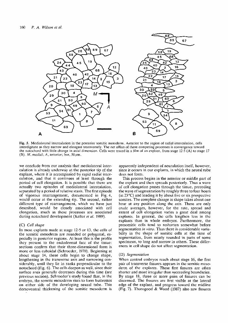
(2) Mediolateral intercalationIn the somitic mesoderm, mediolateral rearrangementcontinues anterior to the region of rapid radial intercal-ation, where it is eventually accompanied by cellelongation perpendicular to the notochord. Fig. 5 fol-lows a patch of cells through this process. Despite theopposing effect of changing cell shape, the patch as awhole extends slightly and converges. The area of thepatch decreases by 25 %, suggesting that cells lengthenin the third dimension (along the radius of the embryo),sections reveal that this is in fact the case (Schroeder,1970). In other explants, the region of the somiticmesoderm where rearrangement and cell shape changeare taking place together does not extend at all, andmay even shrink along the axis. Thus the narrowing ofindividual cells is sometimes more than enough to offsetthe effect of organized neighbour change. Althoughthese regions may or may not extend, they alwaysconverge toward the midline. The cells of the noto-chord also change shape and intercalate mediolaterallyduring this time (Keller et al. 1989). In the notochord,however, the net effect of these processes is always bothextension and convergence.
Since we must follow populations of cells for quitesome time to detect cell rearrangement, it is difficult todelimit precisely its beginning and end. Nonetheless,
160 P. A. Wilson et al.
B
Fig. 5. Mediolateral intercalation in the posterior somitic mesoderm. Anterior to the region of radial intercalation, cellsinterdigitate as they narrow and elongate transversely. The net effect of these competing processes is convergence towardthe notochord with little change in axial dimension. Cells were traced in a film of an explant, from stage 12-5 (A) to stage 17(B). M, medial; A, anterior; bar, 50^m.
we conclude from our analysis that mediolateral inter-calation is already underway at the posterior tip of theexplant, where it is accompanied by rapid radial inter-calation, and that it continues at least through theperiod of cell elongation. It is possible that there areactually two episodes of mediolateral intercalation,separated by a period of relative stasis. The first episodeof vigorous rearrangement, documented in Fig. 4,would occur at the extending tip. The second, ratherdifferent type of rearrangement, which we have justdescribed, would be closely associated with cellelongation, much as these processes are associatedduring notochord development (Keller et al. 1989).
(C) Cell shapeIn most explants made at stage 12-5 or 13, the cells ofthe somitic mesoderm are rounded or polygonal, es-pecially in posterior regions. At least this is the profilethey present to the endodermal face of the tissue:sections confirm that their three-dimensional form ismore or less cuboidal (Schroeder, 1970). Beginning atabout stage 14, these cells begin to change shape,lengthening in the transverse axis and narrowing con-siderably, until they lie in ranks perpendicular to thenotochord (Fig. 6). The cells deepen as well, since theirsurface area generally decreases during this time (seeprevious section). Schroeder's study found that, in theembryo, the somitic mesoderm rises to form buttresseson either side of the developing neural tube. Thisdorsoventral thickening of the somitic mesoderm is
apparently independent of neurulation itself, however,since it occurs in our explants, in which the neural tubedoes not form.
This process begins in the anterior or middle part ofthe explant and then spreads posteriorly. Thus a waveof cell elongation passes through the tissue, precedingthe wave of segmentation by roughly three to four hours(at 23 °C) and leading it by about five or six prospectivesomites. The complete change in shape takes about onehour at any position along the axis. These are onlycrude averages, however, for the rate, spread andextent of cell elongation varies a great deal amongexplants. In general, the cells lengthen less in theexplants than in whole embryos. Furthermore, thepresomitic cells tend to reshorten somewhat beforesegmentation in vitro. Thus there is considerable varia-bility in the shape of somitic cells at the time ofsegmentation, from nearly rounded in parts of somespecimens, to long and narrow in others. These differ-ences in cell shape do not affect segmentation.
(D) SegmentationWhen control embryos reach about stage 16, the firstpair of transverse fissures appears in the somitic meso-derm of the explants. These first fissures are oftenshorter and more irregular than succeeding boundaries.By stage 18, three or more pairs of fissures can bediscerned. The fissures are first visible at the lateraledge of the explant, and progress toward the midline(Fig. 7). Thorogood & Wood (1987) also saw fissures

Mesoderm morphogenesis in Xenopus 161
Fig. 6. Cell shape change. (A) Unelongated cells of the posterior somitic mesoderm at about stage 13. (B) Elongated cellsfrom the central part of an explant at about stage 16. This dramatic change in cell shape begins at the level of somite 3 or 4at about stage 14 and sweeps posteriorly several somite lengths ahead of the wave of segmentation. These and all otherphotographs are taken from the video screen during observation of living explants. Boundary with notochord is at bottom;arrows point anterior. Bar, 50 /MX\.
forms. Cells anterior to the prospective boundary movetoward the notochord; cells on the posterior side movelaterally. This shearing at the boundary is the firstmanifestation of the somite rotation described byHamilton (1969) and by Youn & Malacinski (1981a).We do not observe the line of blebbing or protrusiveactivity that sometimes precedes the notochord somiteboundary in gastrula explants (Keller & Wilson, 1989).New fissures succeed each other in anterior-posteriorprogression at approximately 45min intervals (at23 °C). By the equivalent of stage 21-22, there are 8 to10 fissures on each side of the notochord and segmen-tation of the explant is virtually complete (Fig. 8).
Although segmentation in the explants is remarkablyorderly, there are occasional irregularities. The mostcommon deviation from the standard pattern is vari-ation in the size of individual somites, on one side of thenotochord or both. This can lead to fissures appearingout of register on the two sides. Occasionally fissuresare sharply .angled. In a very few cases we have seenbranching fissures.
(E) Extension without the notochordIn a total of 9 explants fashioned in the usual way at theend of gastrulation, the notochord was separated fromthe somitic mesoderm on both sides. The three regionsof the mesoderm, each with its associated neural plate,were cultured separately until the end of neurulation.In every case, the central piece, consisting of notochordand notoplate, shortened and fell apart, while thepieces containing somitic mesoderm extended and seg-mented (Fig. 9A,B)- The average increase in length ofthe paraxial explants was 160 ̂ m, or about 15%, buttheir curvature makes extension difficult to measure. Asimple solution is to combine the left and right sides ofthe somitic mesoderm in a single notochordless explant.The two sides can be joined at their medial edges or, byswitching the left and right sides, at their lateral
Fig. 7. Segmentation in progress. The anterior region of anexplant at stage 19. Behind the completed segments, newfissures are working toward the notochord from the lateraledge (arrows). A segment boundary can begin to formbefore its predecessor is complete. Bar, 100^m.
form from lateral to medial in the teleost embryo. Thetime required for a fissure to reach the notochord variesconsiderably. Some boundaries cover the entire dis-tance almost at once, while others remain incompletefor an hour or longer. Thus it is not uncommon for aboundary to begin to form before its predecessor hasreached the notochord.
We observe two kinds of activity immediately beforethe formation of a fissure. First, two or more faint near-fissures, or unusually pronounced stretches of cellboundary, can sometimes be discerned in the regionwhere the next boundary will form. The definitivefissure emerges as one of these candidates straightensand intensifies. Second, the cells that will come to lie onopposite sides of the eventual boundary sometimesbegin to move past one another before the fissure

162 P. A. Wilson et al.
Fig. 8. Advanced segmentation. About stage 23. Note well-rotated anterior somites (left) and vacuolated notochord.Bar, 100 ̂ m.
seconds of separation from the rest of the dorsal tissue,suggesting that the medial tissue is under compressionrelative to the lateral region. Perhaps the part of theexplant closest to the midline seeks to extend morevigorously than the more lateral part, which thenrestrains elongation. Such an uneven distribution oflengthening activity, expressed during gastrulation,could lead to the pattern of stresses revealed when thetissue is cut at stage 12-5.
The smaller curve, which bends the posterior tip backtoward the former midline, arises during subsequentextension. The direction of curvature is surprising,since it seems to imply that the lateral part of theexplant lengthens fastest. This would contradict notonly the conclusions drawn from the response to cuts,but the simple notion that the capacity for autonomousextension should be greatest at the dorsal midline andfall off laterally. However, the same paradoxical bendtoward the midline is observed in left and right half-explants made at the start of gastrulation.
(F) Extension and segmentation without the neuralplateWith the aid of dilute collagenase, the neural plate canbe peeled away from the underlying dorsal mesodermat the end of gastrulation. The axial mesoderm can thenbe explanted, with or without the endoderm of thearchenteron roof, and cultured in the usual manner. Inthis way, one can test the role of the ectoderm in theneurula-stage development of the mesoderm.
The axial mesoderm develops in combination withthe endodermal epithelium very much as it does whenexplanted with the neural plate. It converges andextends: one group of six explants extended an averageof 50% from stage 12-5 to stage 20, somewhat morethan the average for explants of mesoderm with neuralplate. These explants also segment. Fissures appear atregular intervals, working their way from the lateraledge of the tissue toward the notochord (see Section B).Segmentation is preceded by cell rearrangement and
Fig. 9. Extension of separated notochord and somiticmesoderm. (A) Notochord and paraxial mesoderm at stage12-5. (B) The same part-explants at the end of neurulation.The somitic mesoderm has extended; the notochord hasnot. (C) Joined left and right somitic mesoderm at stage12-5. (D) After extension. Here the extension of somiticmesoderm without notochord is clear. Mesoderm is backedby neural plate in all explants and anterior is left. Bar,500 ̂ m.
borders. These symmetric recombinations also extendand segment, without bending. (See Fig. 9C,D.) Theaverage elongation of 3 specimens was 540 fim (about45%). This is indistinguishable from the extension ofwhole explants.
Isolated paraxial regions (somitic mesoderm plusneural plate) are invariably S-shaped after extending inculture. This shape is the result of two distinct phenom-ena. The larger anterior bend, bowing the somiticmesoderm away from the notochord, develops within

Mesoderm morphogenesis in Xenopus 163
elongation, and followed by the movements of somiterotation. In fact, the behaviour of the somitic meso-derm in films of these neural-plate-less explants appearsto differ from its behaviour in the standard explantsonly in that it is somewhat less regular. This is probablythe result of the collagenase treatment. Explantsstripped of both the neural plate and the endoderm alsosegment, sometimes quite normally. They extend con-siderably less than explants that include one of theepithelial tissues, however. The average of ten explantswas 75 /im (less than 10%).
Discussion
(A) Convergent extensionDuring Xenopus gastrulation, both the involuting mar-ginal zone (the prospective notochord and somites) andthe non-involuting marginal zone (the prospectivenotoplate region of the neural plate) narrow dramati-cally toward the dorsal midline (converge) and extendin the animal-vegetal axis. By culturing isolated mar-ginal zones, Keller and his co-workers showed that thisshape change is autonomous, and that extension isdriven primarily by active cell rearrangement (Keller etal. 1985a,6; Keller & Danilchik, 1988). By following theinterdigitation of fluorescently labelled grafts and un-labelled hosts Keller & Tibbetts (1989) demonstratedthat much of this cell intercalation takes place along themediolateral axis. Wilson & Keller (1989) have con-firmed this directly by filming gastrula explants, andshown that an initial burst of radial intercalation alsocontributes to extension. The results that we have justpresented reveal that, in explants of already involutedmesoderm, these movements continue throughout theneurula stages.
In neurula explants, as in gastrula explants, rapidradial intercalation precedes mediolateral intercalationin each region of the somitic mesoderm. Moreover, thissequence is linked to a well-defined spatial pattern ofcell behaviour that we do not see as clearly in ourgastrula-stage explants. When the dorsal mesoderm isfirst explanted at the end of gastrulation, cells inroughly the posterior half are rearranging within thesurface, while radial intercalation is restricted to anarrow band at the posterior edge. Cells in the anteriorhalf are already changing shape, and subsequentlyrearrange little. As the tissue extends, the zones ofradial and mediolateral intercalation move caudally, sothat cells in each region progress from one behaviour tothe next. At stage 16-17, the wave of segmentation andsomite rotation begins to advance through the tissuefrom anterior to posterior, following behind the wavesof cell rearrangement and cell shape change. Thispattern is summarized in Fig. 10.
Thus, a sequential pattern of cell activities spansgastrulation and neurulation in the somitic mesoderm.As each region of the mesoderm is awakened from itspregastrular quiescence, its cells begin the progressionfrom radial to mediolateral rearrangement, to shapechange and finally to segmentation and somite rotation.
Prospective anterior parts of the mesoderm begin thissequence early in gastrulation, and have virtually com-pleted rearrangement by the onset of neurulation.More posterior (and ventral) tissue begins the sequencelater, and continues to actively rearrange during neuru-lation, extending and converging toward the midline.The most ventral tissue, which finds itself in thethickened collar surrounding the blastopore at the endof gastrulation, may not begin to extend until thetailbud stage. Therefore, the same sequence of cellularbehaviours may drive the major morphogenetic move-ments of the mesoderm in both the gastrula and theneurula, despite their radically different geometries.
The sequence of dorsal mesodermal behaviours thatwe have described in Xenopus may be shared by asurprisingly wide range of vertebrates. Warga & Kim-mel have recently noted a progression from radial tomediolateral intercalation to segmentation in the pro-spective mesoderm of the zebrafish (Kimmel, personalcommunication). Convergence and extension occur inother anurans (Schechtman, 1942) and in urodeles(Vogt, 1929), although Shi and his co-workers (1987)report that in explants of Pleurodeles convergent exten-sion (and presumably cell rearrangement) do not getunder way until the late gastrula. This suggests thatextension by active cell rearrangement may be primar-ily a neurula-stage mechanism in this species, andperhaps other urodeles as well. Keller & Danilchik(1988) have suggested that the precocious onset of thisprocess in Xenopus, where it does much of the work ofgastrulation, may be related to the very rapid develop-ment of this frog.
Convergent extension in neurula explants differsfrom similar processes taking place in gastrula explantsin at least two ways. First, the anterior-posteriorsequence of cell behaviour is more regular in theneurula. This may be an artifact of explant culture,which disturbs morphogenesis to a greater extent atearlier stages. Alternatively, it may be that theanterior-posterior prepattern is not as well establishedin the mesoderm prior to involution. It is important toremember that the primordium of notochord and me-dial somitic mesoderm is only a few cells long at thestart of gastrulation (Keller, 1976), providing littlespace for extensive patterning. It may be that axialpattern becomes progressively more detailed as themesoderm lengthens during gastrulation, makinggreater resolution possible. This refinement of pos-itional specification may be linked to radial intercal-ation, which is responsible for much of the extension. Inthe embryo, these events would coincide approximatelywith involution.
Dorsal elongation in the neurula also differs fromextension in the gastrula in that the notochord and thesomitic mesoderm are now distinct. The cells of the twotissues are morphologically distinguishable by stage11-5 (Keller et al. 1989); the actual boundary betweenthem appears around stage 12 or 12-5 (Wilson & Keller,1989). Despite the subtle differences, the two tissues (ortheir primordia) seem to extend by essentially the samecellular mechanisms during most of gastrulation. In the

164 P. A. Wilson et al.
Stage 21
CEF
Fig. 10. Summary diagram. A sequence of cell behaviours moves through the somitic mesoderm from anterior to posterioras cells in each region pass from one activity to the next. The progression of behaviours across the explant is shown at theleft and details of the cell behaviour are illustrated at the right. In the cell motion diagrams, small arrows indicate cellmovements and large arrows show the resulting tissue movements. At the posterior edge, cells intercalate radially (darkshading) as they come into the surface from deeper layers, thinning and extending the tissue (lower right). Within the bandof radial intercalation and anterior to it, cells rearrange mediolaterally (light shading), driving further axial extension andconvergence toward the midline (middle right). After stage 14, mediolateral intercalation is accompanied by cell elongation,narrowing and thickening. We have labelled the cell elongation frontier (CEF) which forms the posterior limit of cell shapechange. Finally, segmentation begins around stage 16. Fissures appear at the lateral edge of the tissue and continue towardthe notochord, as stretches of connected cell boundary become straighter and more pronounced (upper right). By stage 21,about 10 somites have formed, and cell intercalation is restricted to the small region of unsegmented mesoderm at theposterior tip of the explant.
neurula explants, however, the two patterns of exten-sion and underlying cell behaviour are quite different,although cells intercalate and elongate in both tissues.In the notochord, mediolateral intercalation and cellshape change result in convergence and extension.These processes occur in the somitic mesoderm as well,but here their combined effect is to narrow and thickenthe tissue, rather than to lengthen it. Extension of thesomitic mesoderm derives instead primarily from radialintercalation at the posterior tip.
Which tissue drives the dorsal elongation of theneurula? Jacobson has attributed this leading role to thecombination of notochord and notoplate (the special-ized region of the neural plate along the midline)(Jacobson & Gordon, 1976; Jacobson, 1981). The
results reported in Section E demonstrate unequivo-cally that the somitic mesoderm, together with itsneural plate backing, is also capable of substantialautonomous extension in Xenopus. Although isolatedcombinations of notochord and notoplate do notlengthen, this does not imply that they are not active inthe embryo, or in whole explants. In fact, Jacobson andhis co-workers (1986) recently proposed that theelongation of this tissue requires a boundary withsomitic mesoderm and neural plate. Such a boundary isapparently not required for extension of the somiticmesoderm and neural plate. Furthermore, the somiticmesoderm can extend without the neural plate if theendodermal epithelium is present, strongly suggestingthat it is an active partner in dorsal elongation. The

Mesoderm morphogenesis in Xenopus 165
neural plate probably lengthens by its own efforts aswell, for Keller & Danilchik (1988) demonstrated thiscapacity in explants made at the start of gastrulation.
(B) Independence of the somitic mesodermOur results demonstrate that the somitic mesoderm atthe end of gastrulation needs no assistance from othertissues to carry out convergent extension, cellelongation and segmentation. All three take place inmaterial isolated at stage 13. Brustis (1976) reportedsegmentation of isolated material in Rana and Bufo, sothe same result in Xenopus is not surprising. Oneshould not infer from this that the pattern of segmentboundaries is already established at the time of explan-tation, only that the somitic mesoderm has acquired thecapacity to establish this pattern.
The dorsal mesoderm extends vigorously during theneurula stages in conjunction with either the endoder-mal epithelium or the neural plate, and weakly incomplete isolation. By comparison, explants made atthe beginning of gastrulation almost never extendwithout an accompanying epithelium, even when theneurula stage is reached (Keller et al. 1985a,b; Keller &Danilchik, 1988). Our current hypothesis, based onunpublished work on gastrula explants, is that radialintercalation requires the presence of an epithelium.Once a region of mesoderm has passed through thisphase of activity, however, it can undergo mediolateralintercalation without the epithelium. Since the bulk ofextension in the neurula explants is generated by radialintercalation, these explants extend much less when theepithelia are removed.
(C) Extension and segmentationExtensive cell rearrangement precedes segmentation,resulting in dramatic extension and convergence. Whatconsequences does this have for the mechanism ofsegmentation? First, extension implies that, beforerearrangement, the somite primordia are tightly com-pressed along the anterior-posterior axis. This is seenin fate maps of the somitic mesoderm made at the startof gastrulation, and more dramatically in Elsdale &Davidson's (1983) map of the tailbud in Rana. Thiscondensation of the fate map must set a limit on thetime at which individual somite identities are estab-lished, since more than one primordium eventuallyevolves from a single cell. For example, the involutingmarginal zone at the start of gastrulation is at most 12cells in prospective anterior-posterior length. Yet herelie the anlagen of at least the 6-8 somites that involutefully by stage 12-5-13. The condensation of prospectivesegments around the blastopore at the end of gastru-lation may be even greater. Furthermore, the cellrearrangement that drives extension itself arguesagainst early determination of the segments, since thedetailed arrangement of tiny primordia would have tobe preserved through all this mixing. We see noevidence that this is the case, for neighbouring cells canbe pushed several cell diameters apart, while othersremain together. (See Results, Section B.) These con-siderations lead us to believe that segmental primordia
are determined no earlier than the middle or end of theperiod of rapid cell intercalation. However, until wemanipulate the pattern of extension and segmentationexperimentally, this conclusion must be tentative.
Armstrong & Graveson (1988) reached conclusionssimilar to ours from a study of somitogenesis in theaxolotl. They distinguish two regions of the unseg-mented mesoderm: a 'cohesive zone' behind the last-formed somite - containing up to nine prospectivesomites - and a more loosely organized extending zone.Somites whose primordia lie in the posterior part of thecohesive zone are vulnerable to disruption by heatshock. Moreover, parts of the mesoderm posterior tothis can be deleted without loss of segments. From theseresults, Armstrong & Graveson conclude that determi-nation of primordia occurs within the cohesive zone andthus after cells cease extension. These authors do notdiscuss cell rearrangement, which they cannot observedirectly, and appear to imply that extension occurslargely by cell division. This is not the case in Xenopus(Results, Section B).
According to Elsdale & Davidson (1983) the situationin the tailbud of Rana is apparently quite different.They also divide the presomitic mesoderm into regions:a prepatterned zone, corresponding roughly to thecohesive zone of Armstrong & Graveson, a zone ofextension and a third, less well-defined, 'packing zone'.The anterior boundary of the extension zone coincideswith the transition from heat-shock sensitivity to insen-sitivity, suggesting to Elsdale & Davidson, as to Arm-strong & Graveson, the onset of segmental determi-nation. However, unlike in the axolotl, Elsdale &Davidson find that small lesions invariably result inmissing or damaged somites, even at the posterior endof the tailbud. This extraordinary lack of patternregulation leads them to conclude that, well before theonset of extension, the packing zone is a 'mosaic ofsmall groups of cells already differentiated from theirneighbors'. They do not propose that these cells havesegmental identities, but that they possess anterior-posterior positional values that will be converted intosomite identities much later, at the time of heat-shocksensitivity. We cannot explain the apparent contradic-tion between the results of these deletion experimentsand those of Armstrong & Graveson, except to notethat one set is performed on the tailbud of Rana, whilethe others concern the trunk somites of the axolotl.Finally, Elsdale & Davidson do not explain how somany somite primordia could be packed into such asmall region at the posterior end of the tailbud, andthey give no data on cell size. Nor do they speculate onthe mechanism of extension and the consequences ofthis for the precise anterior-posterior ordering thatthey postulate. We do not believe that such a detailedaxial pattern could exist in the posterior somitic meso-derm of Xenopus before its extension. There are toofew cells to permit sufficient resolution and, in any case,precise pattern could not survive the vigorous cellrearrangement that our analysis reveals.
This work was supported in part by NSF Grant no. DMS-

166 P. A. Wilson et al.
8618975 to P. Wilson and G. Oster, and by NIH Grant no. HD18979 to R. Keller. We would also like to thank Paul Tibbettsfor his assistance in preparing the plates.
ReferencesARMSTRONG, J. B. & GRAVESON, A. C. (1988). Progressive
patterning precedes somite segmentation in the Mexican axolotl(Ambystoma mexicanum). Devi Biol. 126, 1-6.
BRUSTIS, J.-J. (1976). Organisation et differenciation du mesodermesomitique cultive en presence des tissus adjacents (corde, tubenerveux, ectoderme, endoderme) chez les Anoures. C. r. hebdAcad. Sci. Paris 283 Serie D, 379-382.
COOKE, J. (1975). Control of somite number during morphogenesisof a vertebrate, Xenopus laevis. Nature, Lond. 254, 196-199.
COOKE, J. (1978). Somite abnormalities caused by short heat shocksto pre-rieurula stages of Xenopus laevis. J. Embryol. exp. Morph.45, 283-294.
DETWILER, S. R. (1934). An experimental study of spinal nervesegmentation in Ambystoma with reference to the plurisegmentalcontribution to the brachial plexus. J. exp. Zool. 67, 395-441.
DEUCHAR, E. & BURGESS, A. M. C. (1967). Somite segmentation inamphibian embryos: is there a transmitted control mechanism? J.Embryol. exp. Morph. 17, 349-358.
ELSDALE, T. & DAVIDSON, D. (1983). Somitogenesis in amphibianembryos: IV. The dynamics of tail development. J. Embryol.exp. Morph. 76, 157-176.
HAMILTON, L. (1969). The formation of somites in Xenopus laevis.J. Embryol. exp. Morph. 22, 253-264.
JACOBSON, A. (1981). Morphogenesis of the neural plate and tube.Morphogenesis and Pattern Formation (ed. T. G. Connelly, L. •Brinkley & B. Carlson), pp. 233-263. New York: John Wiley &Sons.
JACOBSON, A. & GORDON, R. (1976). Changes in the shape of thedeveloping vertebrate nervous system analysed experimentally,mathematically, and by computer simulation. J. exp. Zool. 197,191-246.
JACOBSON, A., OSTER, G., ODELL, G. & CHENG, L. (1986).Neurulation and the cortical tractor model for epithelial folding.J. Embryol. exp. Morph. 96, 19-49.
KELLER, R. E. (1976). Vital dye mapping of the gastrula andneurula of Xenopus laevis. II. Prospective areas andmorphogenetic movements of the deep layer. Devi Biol. 51,118-137.
KELLER, R. E. (1984). The cellular basis of gastrulation in Xenopuslaevis: active post-involution convergence and extension bymedio-lateral interdigitation. Am. Zool. 24, 589-603.
KELLER, R. E. (1986). The cellular basis of amphibian gastrulation.In Developmental Biology: A Comprehensive Synthesis. Vol. 2.The Cellular Basis of Morphogenesis (ed. L. Browder), pp.241-327.
KELLER, R. E., COOPER, M. S., DANILCHIK, M., TIBBETTS, P. &WILSON, P. A. (1989). Cell intercalation during notochorddevelopment in Xenopus. Submitted to J. exp. Zool.
KELLER, R. E. & DANILCHIK, M. (1988). Regional expression,pattern and timing of convergence and extension duringgastrulation of Xenopus laevis. Development 103, 193-210.
KELLER, R. E., DANILCHIK, M., GIMLICH, R. & SHIH, J. (1985a).Convergent extension by cell intercalation during gastrulation ofXenopus laevis. In Molecular Determinants of Animal Form (ed.G. M. Edelman). UCLA Symp. Mol. Cell. Biol., vol. 31, pp.111-141. New York: Alan R. Liss, Inc.
KELLER, R. E., DANILCHIK, M., GIMLICH, R. & SHIH, J. (1985ft).The function of convergent extension during gastrulation ofXenopus laevis. J. Embryol. exp. Morph. 89 Suppl. 185-209.
KELLER, R. E. & HARDIN, J. (1987). Cell behavior during activecell rearrangement: evidence and speculation. J. Cell Sci. Suppl.8, 369-393.
KELLER, R. E. & TIBBETTS, P. (1989). Mediolateral cellintercalation is a property of the dorsal, axial mesoderm ofXenopus laevis. Developmental Biology (in press).
KIMMEL, C. B. & WARGA, R. M. (1987). Cell lineages generatingaxial muscle in the zebrafish embryo. Nature, Lond. 327,234-237.
MALACINSKI, G. M., YOUN, B. W. & JURAND, A. (1981). Tissueinteractions during axial structure pattern formation inAmphibia. Scanning Electron Microscopy 1981: II, 307-318.
NIEUWKOOP, P. D. & FABER, J. (1967). Normal Table o/Xenopuslaevis (Daudin). Second edition. Amsterdam: North HollandPublishing Company.
PEARSON, M. & ELSDALE, T. (1979). Somitogenesis in amphibianembryos: I. Experimental evidence for and interaction betweentwo temporal factors in the specification of somite pattern. J.Embryol. exp. Morph. 51, 27-50.
PEARSON, M., ELSDALE, T. & WHITEHEAD, M. (1976).Abnormalities in somite segmentation following heat shock toXenopus embryos. J. Embryol. exp. Morph. 35, 625-635.
SCHECHTMAN, A. M. (1942). The mechanics of amphibiangastrulation. I. Gastrulation-producing interactions betweenvarious regions of an anuran egg (Hyla regilia). Univ. Calif.Publ. Zool. 51, 1-39.
SCHROEDER, T. E. (1970). Neurulation in Xenopus laevis: ananalysis and model based upon light and electron microscopy. J.Embryol. exp. Morph. 23, 427-462.
SHI, D.-L., DELARUE, M., DARRIBERE, T., RIOU, J.-F. & BOUCAUT,J.-C. (1987). Experimental analysis of the extension of the dorsalmarginal zone in Pleurodeles waltl gastrula. Development 100,147-161.
SPEMANN, H. (1938). Embryonic Development and Induction. NewYork: Yale University Press. Reprinted 1962. Hafner PublishingCompany, Inc.
THOROGOOD, P. & WOOD, A. (1987). Analysis of in vivo cellmovement using transparent tissue systems. J. Cell Sci. Suppl. 8,395-413.
VOGT, W. (1929). Gestaltanalyse am Amphibienkein mit ortlicherVitalfarbung. II. Teil. Gastrulation und Mesodermbildung beiUrodelen und Anuren. Wilhelm Roux Arch. EntwMech. Org.120, 384-706.
WADDINGTON, C. H. & DEUCHAR, E. (1953). Studies on themechanism of meristic segmentation. I. The dimensions ofsomites. J. Embryol. exp. Morph. 1, 349-356.
WILSON, P. A. & KELLER, R. E. (1989). Pattern and function ofcell intercalation in Xenopus gastrulation: direct observation incultured explants. In preparation.
YOUN, B. W., KELLER, R. E. & MALACINSKI, G. M. (1980). Anatlas of notochord and somite morphogenesis in several anuranand urodelean amphibians. J. Embryol. exp. Morph. 59,223-247.
YOUN, B. W. & MALACINSKI, G. M. (1981a). Somitogenesis in theamphibian Xenopus laevis: scanning electron microscopic analysisof intrasomitic cellular arrangements during somite rotation. J.Embryol. exp. Morph. 64, 23-43.
YOUN, B. W. & MALACINSKI, G. M. (19816). Comparative analysisof amphibian somite morphogenesis: cell rearrangement patternsduring rosette formation and myoblast fusion. J. Embryol. exp.Morph. 66, 1-26.
(Accepted 30 September 1988)