cell cycle-dependent regulation of tiap/m-survivin expression
TRANSCRIPT

Promoter paper
Cell cycle-dependent regulation of TIAP/m-survivin expression
Masayuki Otaki a;b, Masahiko Hatano a, Koichi Kobayashi a, Takeshi Ogasawara a,Takayuki Kuriyama b, Takeshi Tokuhisa a;*
a Department of Developmental Genetics, Chiba University Graduate School of Medicine, Chiba 260-8670, Japanb Department of Chest Medicine, Chiba University School of Medicine, Chiba 260-8670, Japan
Received 3 April 2000; received in revised form 16 May 2000; accepted 16 May 2000
Abstract
TIAP, a murine homologue of human survivin, is a member of the inhibitor of apoptosis (IAP) family and is specifically expressed at G2/M phase of the cell cycle. To elucidate regulatory mechanisms of the cycle-dependent expression, we have analyzed the promoter region ofTIAP/mouse survivin (m-survivin). The 5P-flanking region of the TIAP/m-survivin gene contained a TATA-less promoter, two AP2 sites,three NF-kB sites, one Sp1 site, many cell cycle-dependent elements (CDEs) and one cell cycle gene homology region (CHR). Primerextension and 5P-rapid amplification of cDNA ends identified one transcription start site at position 3100 upstream of the ATG start site(+1). TIAP/m-survivin promoter^luciferase analysis identified a minimal promoter region within the most proximal 3271 bp upstream of theATG start site, and the region between 3410 and 3272 was critical for the enhancer activity. The combination between the CHR at 351and the CDE at 357 is also essential for the cell cycle-dependent expression. Mutation of the CDE/CHR element and the enhancer elementsmay cause disordered expression of TIAP/m-survivin to affect cell survival and oncogenesis. ß 2000 Elsevier Science B.V. All rightsreserved.
Keywords: TIAP/m-survivin; Promoter; CDE/CHR; Cell cycle ; Inhibitor of apoptosis ; G2/M
The inhibitor of apoptosis (IAP) proteins comprise ahighly conserved gene family that prevents cell death inresponse to a variety of stimuli. IAP was ¢rst identi¢ed inbaculovirus genes that can complement the loss of thecaspase inhibitor, p35, in mutant viruses [1]. Cellular ho-mologues of IAPs have also been noted in mammals.NAIP [2], c-IAP1 (HIAP2/MIHB), c-IAP2 (HIAP1/MIHC/hITA) [3^5], XIAP (hILP) [4,5], BRUCE [6] andsurvivin [7] have been isolated. All of the IAP genes iso-lated from di¡erent species have the common structuretermed the baculovirus IAP repeat (BIR). Recent studiesshowed that four mammalian IAPs, XIAP, c-IAP1, c-IAP2 and survivin, bind to speci¢c cell death proteases,caspase-3 and caspase-7, and inhibit their proteolytic ac-tivity in vitro [8,9]. We have described a newly de¢nedmurine IAP, designated TIAP, that proved to be a murinehomologue of human survivin [10]. TIAP/mouse survivin
(m-survivin) has one BIR, and interacts with the processedform of caspase-3 and inhibits caspase-induced cell death.Histological examinations revealed that TIAP is expressedin growing tissues such as thymus, testis and intestine ofadult mice, and many tissues of embryos. Furthermore,expression of TIAP is up-regulated in synchronized cellsat S to G2/M phase of the cell cycle. Survivin is also ex-pressed in G2/M phase of the cell cycle in a cycle-regulatedmanner, and associates with microtubules of the mitoticspindle in a speci¢c and saturable reaction that is regu-lated by microtubule dynamics [11]. TIAP/m-survivin is,therefore, a newly identi¢ed member of the growing IAPgene family encoding caspase inhibitors and may be in-volved in molecular mechanisms of apoptosis during cellproliferation.
The S/G2-speci¢c transcription of the human cdc25Cgene is due to the periodic occupation of repressor ele-ments (cell cycle-dependent element (CDE) and cell cyclegenes homology region (CHR)) located in the basal pro-moter region [12,13]. Recent study identi¢ed a factor,CDF-1, which interacts with the cdc25C CDE/CHR mod-ule [14,15]. CDF-1 binds to the CDE (consensus sequence:5P-G/CGC/TGG/C-3P) in the major groove and to theCHR (consensus sequence: 5P-TTGAA-3P) in the minor
0167-4781 / 00 / $ ^ see front matter ß 2000 Elsevier Science B.V. All rights reserved.PII: S 0 1 6 7 - 4 7 8 1 ( 0 0 ) 0 0 1 4 2 - 1
Abbreviations: BIR, baculovirus IAP repeat; CDE, cell cycle-depen-dent elements; CHR, cell cycle gene homology region; IAP, inhibitor ofapoptosis ; Luc, the ¢re£y luciferase gene; PCR, polymerase chain reac-tion
* Corresponding author. Fax: +81-43-226-2183;E-mail : [email protected]
BBAEXP 91435 28-8-00
Biochimica et Biophysica Acta 1493 (2000) 188^194
www.elsevier.com/locate/bba

groove in a cooperative fashion. Since DNA binding byCDF-1 in nuclear extracts is down-regulated during cellcycle progression, CDF-1 in G0 and G1 phases of the cellcycle results in a phase-speci¢c repression of activatedtranscription. CDE/CHR-mediated repression is also themajor principle underlying the periodic transcription ofthe human cyclin A [13], cdc2 [13], PLK [16], cyclin B[17] and B-myb [18] genes. In addition, these genes sharethe CDE/CHR element with a ¢ve bases distance in theirpromoter region.
Li and Altieri [19] have recently characterized the m-survivin (TIAP) locus. Analysis of the 5P-£anking regionof the m-survivin gene revealed a TATA-less promotercontaining two CDE elements and one CHR element. Aminimal promoter region was identi¢ed within the mostproximal 174 bp upstream of the ¢rst ATG. Mutagenesisof the CDE/CHR element in the promoter region abol-ished cell cycle periodicity in G2/M-synchronized cells.Thus, cell cycle expression of m-survivin requires integra-tion of typical CDE/CHR G1 repressor elements. Here weanalyzed the TIAP/m-survivin locus including the 5P-£ank-ing region. Although we also identi¢ed a CDE/CHR ele-ment upstream of the ¢rst ATG, the position of CDE/CHR is di¡erent from that they identi¢ed. Furthermore,we detected some di¡erences from the data previously pre-sented [19], especially in a genomic map, a transcriptionstart site, and a presence of enhancer region upstream ofthe minimal promoter region.
A mouse genomic DNA library was screened by hybrid-
ization with the 112 probe that was the 5P side sequence ofthe TIAP/m-survivin cDNA. A clone that includes a 12.5kb fragment (Sv11-2) was isolated (Fig. 1). Since Sv11-2contained the 5P side and the 3P side of the TIAP/m-survi-vin cDNA determined by Southern hybridization, Sv11-2was further digested with several restriction enzymes, andeach fragment was subcloned. The BamHI/BglII region(10 kb) of Sv11-2 was sequenced to identify the intron/exon boundary of the TIAP/m-survivin gene. Sv11-2 con-tained four exons and three introns of the TIAP/m-survivingene. When the initiating A of ATG start codon numbersas +1, sizes of intron 1 (+112 to +403), intron 2 (+514 to+3310) and intron 3 (+3429 to +5870) were 0.28 kb, 2.80kb and 2.44 kb, respectively. To isolate further 5P side ofSv11-2, the genomic library was hybridized with the 396probe that was the DNA sequence of the 5P side of Sv11-2.A clone that contains a 8.5 kb fragment (Sv321) was iso-lated. Since a 5.5 kb region at the 3P side of Sv321 isoverlapped with the 5P side of Sv11-2, we isolated total15.5 kb genomic DNA which includes the TIAP/m-survivingene.
We determined the transcription initiation site of thegene by primer extension analysis and by the 5P-rapidampli¢cation of cDNA ends (RACE) analysis withmRNA from WEHI231 cells. Fig. 2 shows that one ini-tiation site was identi¢ed at 100 bp upstream from theATG (+1) by Tep1 (338 to 38) primer. However, Tep3primer that is complementary to the 3269 to 3250 ge-nomic sequence did not amplify any band (data not
Fig. 1. Genomic organization of the TIAP/m-survivin gene. Map of overlapping mouse genomic clones, Sv11-2 and Sv321. Organization of the exonsand introns of the TIAP/m-survivin gene. The closed boxes indicate exons. The number of bases indicates positions of the exon/intron boundary identi-¢ed by the sequence analysis. The positions of the probes (112 and 396 probes) used for isolation of these clones and that of sequenced region are indi-cated. The 112 probe was made from the exon 1 of TIAP/m-survivin cDNA as a template by polymerase chain reaction (PCR) using primers (5P-GCGATTTGAATCCTGCGTTTG-3P and 5P-CGATGCGGTAGTTCTTGAGGTACAG-3P). The 396 probe (32828/32433) was made from the 5P sideof the Sv11-2 DNA sequence as a template by PCR using primers (5P-AATTCGGATCTGATGCCCTCTTCTG-3P and 5P-CAGATGGTTATGAACT-CAGGACCTCTGG-3P). Bg, BglII ; E, EcoRI; H, HindIII; S, SacI ; Sm, SmaI; X, XbaI.
BBAEXP 91435 28-8-00
M. Otaki et al. / Biochimica et Biophysica Acta 1493 (2000) 188^194 189

shown). Comparison of the sequence of the 5P-RACEproducts to the genomic sequence con¢rmed that no in-tron existed on the 5P-£anking region from the ATG trans-lation start codon (data not shown).
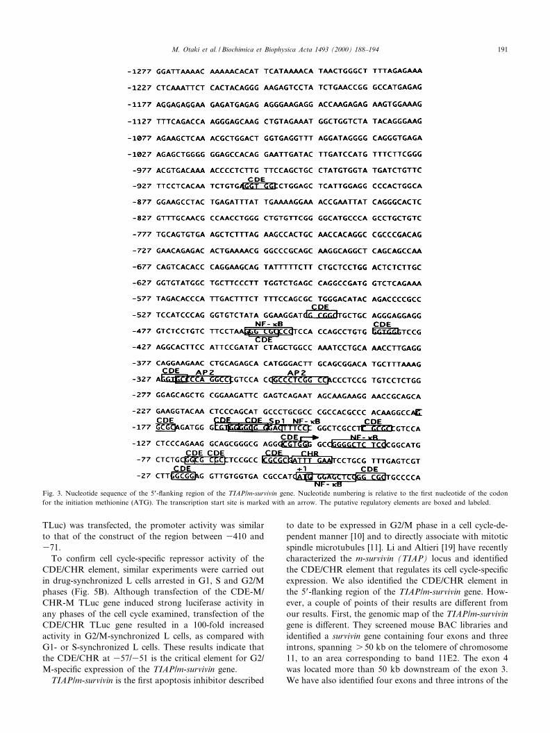
We compared the 5P-£anking sequence of the ATG startcodon with established consensus sequences in a transcrip-tion factors data base (Fig. 3). This regulatory region con-tained two putative binding sites of AP2, four sites of NF-kB, and one site of Sp1 between 31277 and +23. Further-more, one site of CHR and 15 sites of CDEs, which aretypically found in S/G2 expression genes, were identi¢ed inthe same DNA region. However, the region lacked theconventional TATA box and the CAAT box sequence.
We next analyzed a promoter activity in the 5P-£ankingregion of the TIAP/m-survivin gene by transient transfec-tion of the promoter^reporter fusion genes into L mouse¢broblast cells. Various lengths of the 5P-£anking region
of the TIAP/m-survivin gene from the position at 371without the putative CHR at 351 were fused to the ¢re£yluciferase gene (Luc). Those chimeric constructs weretransfected into L cells to examine the promoter activity(Fig. 4A). A signi¢cant increase (s 50-fold) of the lucifer-ase activity was observed when the 897TLuc was trans-fected. Transfection of the 897TLuc, the 1857TLuc, the2824TLuc and the 6000TLuc constructs demonstrated al-most the same activity, suggesting the presence of a min-imal promoter element within the ¢rst 897 bp of this 5P-£anking region.
To further characterize the regulatory region of theTIAP/m-survivin gene, the region between 3897 and 371was digested and fused to the luciferase reporter gene.Those constructs were transfected into L cells (Fig. 4B).Although the 897TLuc, the 581TLuc and the 410TLucconstructs showed the similar promoter activity, activityof the 271TLuc was about 25% of the 897TLuc. These¢ndings suggest that the region between 3271 and 371contains a minimal promoter element and that the regionbetween 3410 and 3272 plays a role in determining en-hancer activity of the TIAP/m-survivin gene. Since thisenhancer region contains two putative AP2 binding se-quences, the enhancer activity of AP2 in the region wasexamined by the 410TLuc with deletion of the region be-tween 3323 and 3296 (410TLuc-AP2). However, the de-letion reduced promoter activity by 60% of that of the410TLuc. Thus, AP2 may explain a part of the enhanceractivity.
There are one CHR and nine CDEs in the 5P-£ankingregion of the ATG start codon between 3178 and 31 ofthe TIAP/m-survivin gene. To analyze the regulation of thecell cycle-speci¢c expression of the TIAP/m-survivin gene,a systematic mutation analysis of the cell cycle-regulatedelements was performed (Fig. 5A). When the luciferaseconstruct (CDE/CHR/CDE TLuc) of the region between3410 and 31 that includes one CHR and 10 CDEs wastransfected in asynchronously growing L cells, the pro-moter activity was completely reduced to the backgroundlevel. The construct (CDE/CHR TLuc) of the region be-tween 3410 and 338 that deleted one CDE at 324 stillexpressed the low activity. However, the luciferase con-struct (410TLuc) of the region between 3410 and 371without one CHR at 351 and four CDEs at 371, 369,357 and 324 expressed strong promoter activity, suggest-ing that CHR at 351 and one of three CDEs at 371, 369and 357 are the critical for the cell cycle-speci¢c expres-sion of the TIAP/m-survivin gene. To con¢rm the regula-tion, we made a series of deletion or mutation clones.Since the distance between CDE and CHR of S/G2-regu-lated genes is ¢ve bases [12,16,18] and there is one CHR at351, we made the 410TLuc with a mutation or a deletionof CHR at 351 and/or CDE at 357. Mutation or deletionof one of them partially diminished the reduction of pro-moter activity. When the construct that has mutations ofboth CHR at 351 and CDE at 357 (CDE-M/CHR-M
Fig. 2. Primer extension analysis on the 5P end of the TIAP/m-survivingene. Primer extension was performed by the method described else-where [21]. Two oligonucleotide primers, Tep1 (5P-TCACCA-CAACCTCCGCCAAGACGACTCAAAC-3P ; complementary to nucleo-tides between 38 and 338 of the TIAP/m-survivin genomic sequence)and Tep3 (5P-TCTGACTCGAATCTTCCGCA-3P ; complementary tonucleotides between 3250 and 3269 of the TIAP/m-survivin genomic se-quence), were used. (A) Schematic diagram of primers used in primerextension (PE) and the position of PE product. (B) The autoradiographshows a primer extension on TIAP/m-survivin mRNA isolated fromWEHI231 cells (WEHI231). Transfer RNA (T) was used as a negativecontrol. The extended product is indicated by an arrowhead. T, yeasttRNA; M, size marker.
BBAEXP 91435 28-8-00
M. Otaki et al. / Biochimica et Biophysica Acta 1493 (2000) 188^194190
TLuc) was transfected, the promoter activity was similarto that of the construct of the region between 3410 and371.
To con¢rm cell cycle-speci¢c repressor activity of theCDE/CHR element, similar experiments were carried outin drug-synchronized L cells arrested in G1, S and G2/Mphases (Fig. 5B). Although transfection of the CDE-M/CHR-M TLuc gene induced strong luciferase activity inany phases of the cell cycle examined, transfection of theCDE/CHR TLuc gene resulted in a 100-fold increasedactivity in G2/M-synchronized L cells, as compared withG1- or S-synchronized L cells. These results indicate thatthe CDE/CHR at 357/351 is the critical element for G2/M-speci¢c expression of the TIAP/m-survivin gene.
TIAP/m-survivin is the ¢rst apoptosis inhibitor described
to date to be expressed in G2/M phase in a cell cycle-de-pendent manner [10] and to directly associate with mitoticspindle microtubules [11]. Li and Altieri [19] have recentlycharacterized the m-survivin (TIAP) locus and identi¢edthe CDE/CHR element that regulates its cell cycle-speci¢cexpression. We also identi¢ed the CDE/CHR element inthe 5P-£anking region of the TIAP/m-survivin gene. How-ever, a couple of points of their results are di¡erent fromour results. First, the genomic map of the TIAP/m-survivingene is di¡erent. They screened mouse BAC libraries andidenti¢ed a survivin gene containing four exons and threeintrons, spanning s 50 kb on the telomere of chromosome11, to an area corresponding to band 11E2. The exon 4was located more than 50 kb downstream of the exon 3.We have also identi¢ed four exons and three introns of the
Fig. 3. Nucleotide sequence of the 5P-£anking region of the TIAP/m-survivin gene. Nucleotide numbering is relative to the ¢rst nucleotide of the codonfor the initiation methionine (ATG). The transcription start site is marked with an arrow. The putative regulatory elements are boxed and labeled.
BBAEXP 91435 28-8-00
M. Otaki et al. / Biochimica et Biophysica Acta 1493 (2000) 188^194 191

TIAP/m-survivin gene. However, we identi¢ed the fourexons within 6.4 kb of a single phage clone from the ge-nomic library. Southern blot analysis of murine genomicDNA supported our results since a 7 kb, a 6 20 kb or a6 15 kb fragment of genomic DNA digested with BglII,BamHI or NheI was hybridized with two probes speci¢cfor the exon 3 or the exon 4, respectively (data notshown). Thus, the TIAP/m-survivin gene in the BAC clonethey examined may be the fusion gene.
Second, the transcription start site of the TIAP/m-survi-vin gene is di¡erent. They demonstrated that primer ex-tension and S1 nuclease mapping identi¢ed three tran-scription start sites at positions 332, 336 and 340 fromthe initiating ATG. Our analysis indicated that the tran-scription start site is at position 3100 from the ATG byprimer extension and 5P-RACE analysis. When we exam-ined cDNA library, the longest cDNA clone that we iso-lated indicated the 100 bp of 5P UT [10], supporting ourresults. Furthermore, the luciferase constructs with theregion upstream of 370 expressed enzyme activity in asyn-chronously growing cells, indicating that there is at least
one transcription start site upstream of 371. This di¡er-ence may come from di¡erence of source of mRNA usedfor the study. Since we used the DNA sequence at theposition between 338 and 38 as a primer (Tep1), wecould not detect the start sites (332, 336 and 340) theyde¢ned. Thus, our results cannot deny the presence ofthree more transcription start sites. Even though theTIAP/m-survivin gene has three transcription start sites,there must be at least one more transcription start siteat position 3100.
Third, the position of CDE/CHR element of the TIAP/m-survivin gene is di¡erent. Analysis of the 5P-£ankingregion of the m-survivin gene revealed a TATA-less pro-moter containing a canonical CpG island, Sp1 sites, sev-eral CDEs and one CHR. They analyzed the importantCDE/CHR element for cell cycle-regulated expression withmutated genes, and CHR at 351 and CDE at 324 withintwo CDEs were critical for the regulation in asynchro-nously growing cells. However, when we examined theregion by computer, we identi¢ed nine CDEs in thesame region as they examined. They missed seven CDEs
Fig. 4. TIAP/m-survivin promoter^reporter constructs and their relative luciferase activity. (A) Left, schematic representation of the reporter gene con-structs, 6000TLuc (XbaI^BssHII fragment), 2824TLuc (KpnI^BssHII fragment), 1857TLuc (NheI^BssHII fragment), 897TLuc (SacI^BssHII fragment)and Luc, obtained by deletions of the 5P region of the TIAP/m-survivin gene and SVLuc as a positive control. Right, luciferase activity observed in Lcells transfected with the di¡erent reporter genes. The luciferase reporter plasmid (2.5 Wg) and the sea pansy luciferase expression vector, pRL-SV40 (10ng) (Toyo Ink, Tokyo, Japan) as a transfection e¤ciency control, were transiently co-transfected into those cells using Lipofectamine (Life Technolo-gies). The position of transcription start site is indicated (arrow). (B) Left, schematic representation of the reporter gene constructs, 897TLuc, 581TLuc(BstXI^BssHII fragment), 410TLuc (EcoRV^BssHII fragment), 410TLuc-AP2 (deleted the AP2 binding sequence), 271TLuc (PvuII^BssHII fragment),Luc. Right, luciferase activity observed in L cells. The values were the means þ S.E.M. of triplicate experiments from three independent studies. All val-ues were as a percent activity of the construct 897TLuc (100%). The position of transcription start site is indicated (arrow).
BBAEXP 91435 28-8-00
M. Otaki et al. / Biochimica et Biophysica Acta 1493 (2000) 188^194192

in the region between 3178 and 31. Our deletion analysisconcludes that CHR at 351 and CDE at 357 are criticalfor regulation of the expression in asynchronously grow-ing cells. Since the distance of core elements between CDEand CHR is ¢ve bases in S/G2-speci¢c genes such ascdc25C, cdc2, cyclin A, B-myb and PLK [13,16,18], thedistance in our study is three bases instead of 20 basesof the CDE and the CHR identi¢ed by them (Fig. 6).Furthermore, the CDE/CHR TLuc with deletion of
CDE at 324 still expressed strong regulatory activity inasynchronously growing cells. Therefore, CHR at 351and CDE at 357 are important for the cell cycle-speci¢cexpression of the TIAP/m-survivin gene.
Finally, the presence of enhancer region in the 5P-£ank-ing of the TIAP/m-survivin gene is di¡erent. We demon-strated that transfection of TIAP/m-survivin promoter^lu-ciferase constructs identi¢ed a minimal promoter regionwithin the most proximal 271 bp upstream of the ¢rst
Fig. 5. TIAP/m-survivin promoter^reporter constructs with deletion of a part of the CDE (357) and/or CHR (351) and their relative luciferase activity.(A)Left, schematic representation of the TIAP promoter region and the reporter gene constructs. The positions of putative repressor elements (CDE/CHR), that of transcription start site (arrow) and that of translation start site (+1) are indicated. The TIAP wild-type or mutant (deletion or point mu-tation) promoter constructs were generated by PCR and inserted in the pGL2. The CDE at 357 and CHR at 351 mutated constructs: the DNA re-gion between 3410 and 342 was ampli¢ed by PCR with the 5P side primer (5P-CAATGGTACCGATATCTAGCTGGCCAAATC-3P) and the CDE,CHR mutated 3P side primers. The CDE/CHR TLuc: 5P-GGTTAAGCTTGGATTCAAATCGCGCGGGC-3P, the CDE-M/CHR-M TLuc: 5P-ATG-GAAGCTTGGATTAAAATCGCGAGGGC-3P, the CDE-M/CHR TLuc: 5P-ATGGAAGCTTGGATTCAAATCGCGAGGGC-3P, the CDE/CHR-MTLuc: 5P-ATGGAAGCTTGGATTAAAATCGCGCGGGC-3P, the CHR TLuc: 5P-AAGCTTGGATTCAAATCGGCGGAGGCGCG-3P, the CDETLuc: 5P-AAGCTTGGAATCGCGCGGGCGGAGGC-3P. Right, relative luciferase activity of the constructs expressed as the means þ S.E.M. of tripli-cate experiments from three independent studies is shown on the right. All values were as a percent activity of the construct 410TLuc (100%). (B) TIAPpromoter activity in drug-synchronized L cells treated with 400 WM mimosine (G1 phase) [22], 2 mM thymidine (S phase) or 0.1 Wg/ml nocodazole (G2/M phase) [20]. Comparison of luciferase activity between TIAP wild-type (CDE/CHR TLuc) and CDE/CHR double point mutation^luciferase (CDE-M/CHR-M TLuc) constructs. All values were as a percent activity of the CDE-M/CHR-M TLuc (100%).
Fig. 6. Sequence homology of the CDE/CHR element between TIAP/m-survivin and S/G2 regulated genes. Alignments of cdc25C, cdc2, cyclin A,B-myb, PLK, cyclin B, TIAP/m-survivin and TIAP/m-survivin with mutations in the region of the CDE and CHR elements. Core sequences are under-lined and point-mutated bases of TIAP/m-survivin are double underlined. Degrees of the repressor activity by TIAP/m-survivin or TIAP/m-survivin withmutations are indicated.
BBAEXP 91435 28-8-00
M. Otaki et al. / Biochimica et Biophysica Acta 1493 (2000) 188^194 193

ATG. They also identi¢ed a minimal promoter regionwithin the most proximal 174 bp upstream of the ¢rstATG. Mutagenesis of the Sp1 sites in this region, aloneor in combination, reduced transcriptional activity by 40^60% in asynchronously growing cells. Although we dem-onstrated that the region between 3410 and 3272 playsan important role in determining enhancer activity of theTIAP/m-survivin gene, they demonstrated no enhancer re-gion upstream of the minimal promoter region. This dif-ference might come from the cells used for the transfectionof the reporter genes. The enhancer region contains twoputative AP2 binding sequences. However, the deletion ofAP2 sites reduced enhancer activity by 60% in asynchro-nously growing cells. Thus, two AP2 sites in the region areone of enhancer binding elements of the TIAP/m-survivingene.
TIAP/m-survivin is expressed in G2/M phase of the cellcycle in a cycle-regulated manner [10] and associates withmicrotubules of the mitotic spindle in a speci¢c and satu-rable reaction that is regulated by microtubule dynamics[11]. Disruption of survivin^microtubule interactions byinhibiting expression of endogenous survivin with anti-sense DNA results in loss of survivin's anti-apoptosisfunction and increased caspase-3 activity, a mechanisminvolved in cell death, during mitosis [20]. Thus, the over-expression of survivin in cancer may overcome this apop-totic checkpoint and favor aberrant progression of trans-formed cells through mitosis. Therefore, disruption of theCDE/CHR element in the 5P-£anking region induces dis-ordered expression of TIAP/m-survivin to favor aberrantprogression of transformed cells. Furthermore, disruptionof the transcriptional enhancer elements may provide analternative strategy to block the overexpression of survivinin cancer.
This work was supported by a Grant-in-Aid for Scien-ti¢c Research on Priority Areas from the Ministry of Ed-ucation, Science, Sports and Culture, Japan.
References
[1] N.E. Crook, R.J. Clem, L.K. Miller, An apoptosis-inhibiting bacu-lovirus gene with a zinc ¢nger-like motif, J. Virol. 67 (1993) 2168^2174.
[2] N. Roy, M.S. Mahadevan, M. McLean, G. Shutler, Z. Yaraghi, R.Farahani, S. Baird, A. Besner-Johnston, C. Lefebvre, X. Kang, M.Salih, H. Aubry, K. Tamai, X. Guan, P. Ioannou, T.O. Crawford,P.J. deJong, L. Surh, J. Ikeda, R.G. Korneluk, A. MacKenzie, Thegene for neuronal apoptosis inhibitory protein is partially deleted inindividuals with spinal muscular atrophy, Cell 80 (1995) 167^178.
[3] M. Rothe, M.G. Pan, W.J. Henzel, T.M. Ayres, D.V. Goeddel, TheTNFR2-TRAF signaling complex contains two novel proteins relatedto baculoviral inhibitor of apoptosis proteins, Cell 83 (1995) 1243^1252.
[4] C.S. Duckett, V.E. Nava, R.W. Gedrich, R.J. Clem, J.L. VanDon-gen, M.C. Gil¢llan, H. Shiels, J.M. Hardwick, C.B. Thompson, Aconserved family of cellular genes related to the baculovirus iap geneand encoding apoptosis inhibitors, EMBO J. 15 (1996) 2685^2694.
[5] P. Liston, N. Roy, K. Tamai, C. Lefebvre, S. Baird, G. Cherton-Horvat, R. Farahani, M. McLean, J.E. Ikeda, A. MacKenzie, R.G.Korneluk, Suppression of apoptosis in mammalian cells by NAIPand a related family of IAP genes, Nature 379 (1996) 349^353.
[6] H.P. Hauser, M. Bardro¡, G. Pyrowolakis, S. Jentsch, A giant ubiq-uitin-conjugating enzyme related to IAP apoptosis inhibitors, J. Cell.Biol. 141 (1998) 1415^1422.
[7] G. Ambrosini, C. Adida, D.C. Altieri, A novel anti-apoptosis gene,survivin, expressed in cancer and lymphoma, Nat. Med. 3 (1997) 917^921.
[8] Q.L. Deveraux, R. Takahashi, G.S. Salvesen, J.C. Reed, X-linkedIAP is a direct inhibitor of cell-death proteases, Nature 388 (1997)300^304.
[9] N. Roy, Q.L. Deveraux, R. Takahashi, G.S. Salvesen, J.C. Reed, Thec-IAP-1 and c-IAP-2 proteins are direct inhibitors of speci¢c cas-pases, EMBO J. 16 (1997) 6914^6925.
[10] K. Kobayashi, M. Hatano, M. Otaki, T. Ogasawara, T. Tokuhisa,Expression of a murine homologue of the inhibitor of apoptosisprotein is related to cell proliferation, Proc. Natl. Acad. Sci. USA96 (1999) 1457^1462.
[11] F. Li, G. Ambrosini, E.Y. Chu, J. Plescia, S. Tognin, P.C. Marchisio,D.C. Altieri, Control of apoptosis and mitotic spindle checkpoint bysurvivin, Nature 396 (1998) 580^584.
[12] J. Zwicker, C. Gross, F.C. Lucibello, M. Truss, F. Ehlert, K. Enge-land, R. Mu«ller, Cell cycle regulation of cdc25C transcription ismediated by the periodic repression of the glutamine-rich activatorsNF-Y and Sp1, Nucleic Acids Res. 23 (1995) 3822^3830.
[13] J. Zwicker, F.C. Lucibello, L.A. Wolfraim, C. Gross, M. Truss, K.Engeland, R. Mu«ller, Cell cycle regulation of the cyclin A, cdc25Cand cdc2 genes is based on a common mechanism of transcriptionalrepression, EMBO J. 14 (1995) 4514^4522.
[14] N. Liu, F.C. Lucibello, K. Korner, L.A. Wolfraim, J. Zwicker, R.Mu«ller, CDF-1, a novel E2F-unrelated factor, interacts with cellcycle-regulated repressor elements in multiple promoters, NucleicAcids Res. 25 (1997) 4915^4920.
[15] F.C. Lucibello, N. Liu, J. Zwicker, C. Gross, R. Mu«ller, The di¡er-ential binding of E2F and CDF repressor complexes contributes tothe timing of cell cycle-regulated transcription, Nucleic Acids Res. 25(1997) 4921^4925.
[16] T. Uchiumi, D.L. Longo, D.K. Ferris, Cell cycle regulation of thehuman polo-like kinase (PLK) promoter, J. Biol. Chem. 272 (1997)9166^9174.
[17] J.P. Cogswell, M.M. Godlevski, M. Bonham, J. Bisi, L. Babiss, Up-stream stimulatory factor regulates expression of the cell cycle-depen-dent cyclin B1 gene promoter, Mol. Cell. Biol. 15 (1995) 2782^2790.
[18] N. Liu, F.C. Lucibello, J. Zwicker, K. Engeland, R. Mu«ller, Cellcycle-regulated repression of B-myb transcription: cooperation ofan E2F site with a contiguous corepressor element, Nucleic AcidsRes. 24 (1996) 2905^2910.
[19] F. Li, D.C. Altieri, The cancer antiapoptosis mouse survivin gene:characterization of locus and transcriptional requirements of basaland cell cycle-dependent expression, Cancer Res. 59 (1999) 3143^3151.
[20] G. Ambrosini, C. Adida, G. Sirugo, D.C. Altieri, Induction of apop-tosis and inhibition of cell proliferation by survivin gene targeting,J. Biol. Chem. 273 (1998) 11177^11182.
[21] Y. Iitsuka, H. Shimizu, M.M. Kang, K. Sasagawa, S. Sekiya, T.Tokuhisa, M. Hatano, An enhancer element for expression of theNcx (Enx, Hox11L1) gene in neural crest-derived cells, J. Biol.Chem. 274 (1999) 24401^24407.
[22] T. Krude, Mimosine arrests proliferating human cells before onset ofDNA replication in a dose-dependent manner, Exp. Cell. Res. 247(1999) 148^159.
BBAEXP 91435 28-8-00
M. Otaki et al. / Biochimica et Biophysica Acta 1493 (2000) 188^194194