cell communication. cellular messaging cell-to-cell communication is essential for both...
TRANSCRIPT
Cell Communication
Cellular Messaging
• Cell-to-cell communication is essential for both multicellular and unicellular organisms
• Biologists have discovered some universal mechanisms of cellular regulation
• Cells most often communicate with each other via chemical signals
© 2011 Pearson Education, Inc.
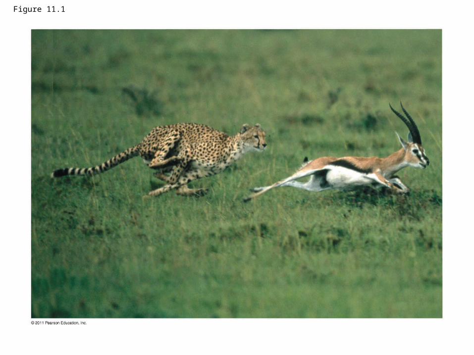
Figure 11.1
External Signals
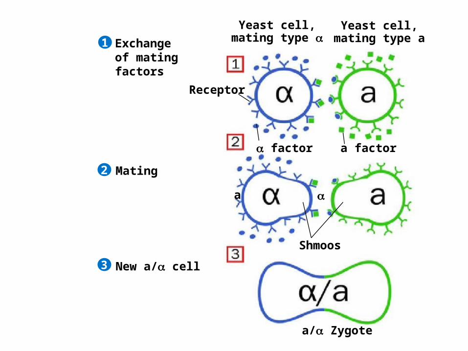
• The yeast, Saccharomyces cerevisiae, has two mating types, a and
• Cells of different mating types locate each other via secreted factors specific to each type
© 2011 Pearson Education, Inc.

Exchange of mating factors
Receptor
factor a factor
Yeast cell,mating type a
Yeast cell,mating type
Mating
New a/ cell
1
2
3
a
a/ Zygote
Shmoos
Evolution of Cell Signaling
• A signal transduction pathway is a series of steps by which a signal on a cell’s surface is converted into a specific cellular response
• Signal transduction pathways convert signals on a cell’s surface into cellular responses
© 2011 Pearson Education, Inc.
Exchange of mating factors
Receptor
factor a factor
Yeast cell,mating type a
Yeast cell,mating type
Mating
New a/ cell
1
2
3
a
a/ Zygote
Shmoos
• Pathway similarities suggest that ancestral signaling molecules evolved in prokaryotes and were modified later in eukaryotes
• The concentration of signaling molecules allows bacteria to sense local population density
© 2011 Pearson Education, Inc.
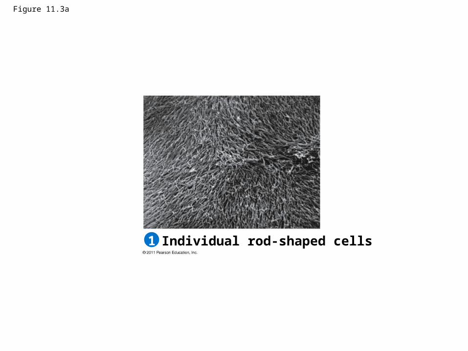
Figure 11.3a
Individual rod-shaped cells1

Figure 11.3b
Aggregation in progress2
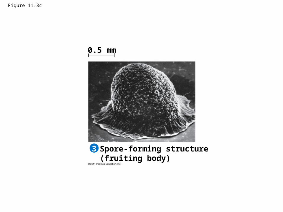
Figure 11.3c
Spore-forming structure(fruiting body)
0.5 mm
3
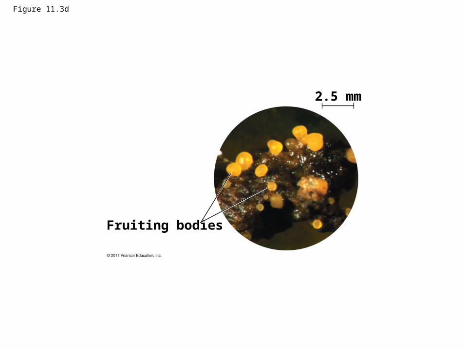
Figure 11.3d
Fruiting bodies
2.5 mm
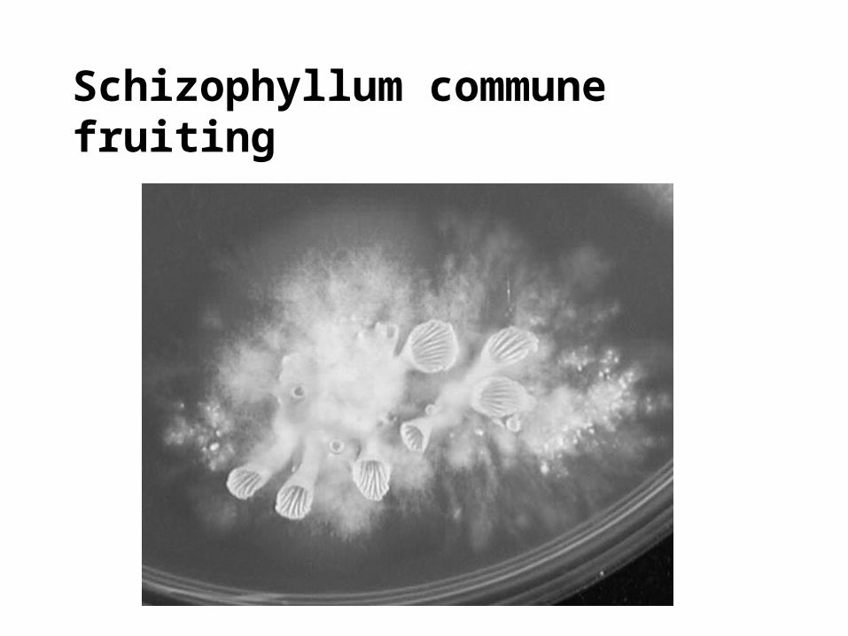
Schizophyllum commune fruiting

Local and Long-Distance Signaling
• Animal and plant cells have cell junctions that directly connect the cytoplasm of adjacent cells.
• A cell cannot respond to a signal if it lacks a receptor specific to that signal.
© 2011 Pearson Education, Inc.
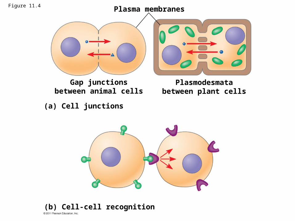
Figure 11.4Plasma membranes
Gap junctionsbetween animal cells
Plasmodesmatabetween plant cells
(a) Cell junctions
(b) Cell-cell recognition
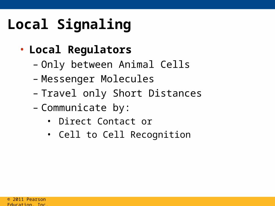
• Local Regulators– Only between Animal Cells
– Messenger Molecules
– Travel only Short Distances
– Communicate by:• Direct Contact or • Cell to Cell Recognition
© 2011 Pearson Education, Inc.
Local Signaling
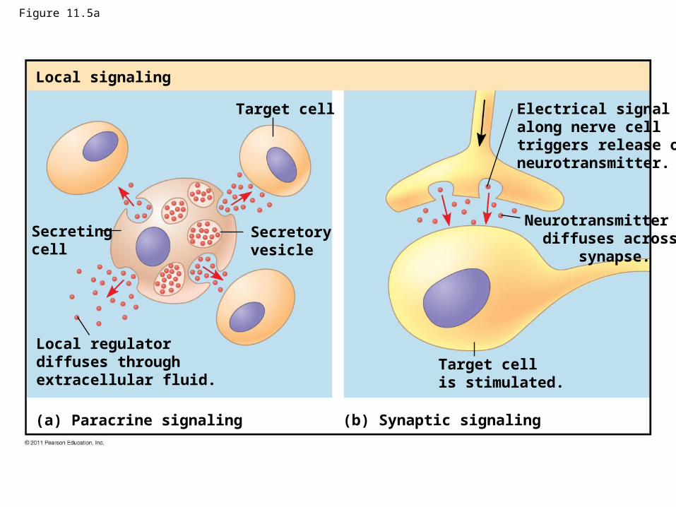
Figure 11.5a
Local signaling
Target cell
Secretingcell
Secretoryvesicle
Local regulatordiffuses throughextracellular fluid.
(a) Paracrine signaling (b) Synaptic signaling
Electrical signalalong nerve celltriggers release ofneurotransmitter.
Neurotransmitter diffuses across synapse.
Target cellis stimulated.
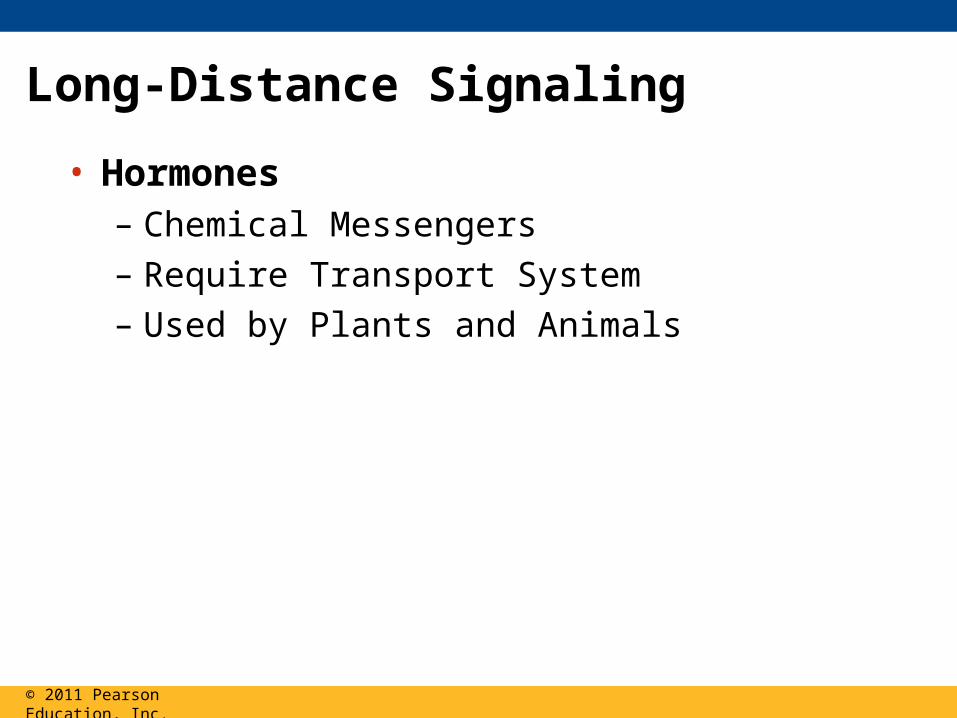
• Hormones– Chemical Messengers
– Require Transport System
– Used by Plants and Animals
© 2011 Pearson Education, Inc.
Long-Distance Signaling
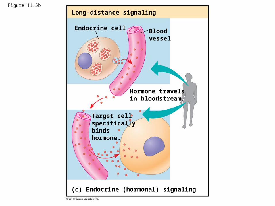
Figure 11.5b
Long-distance signaling
Endocrine cell Bloodvessel
Hormone travelsin bloodstream.
Target cellspecificallybinds hormone.
(c) Endocrine (hormonal) signaling
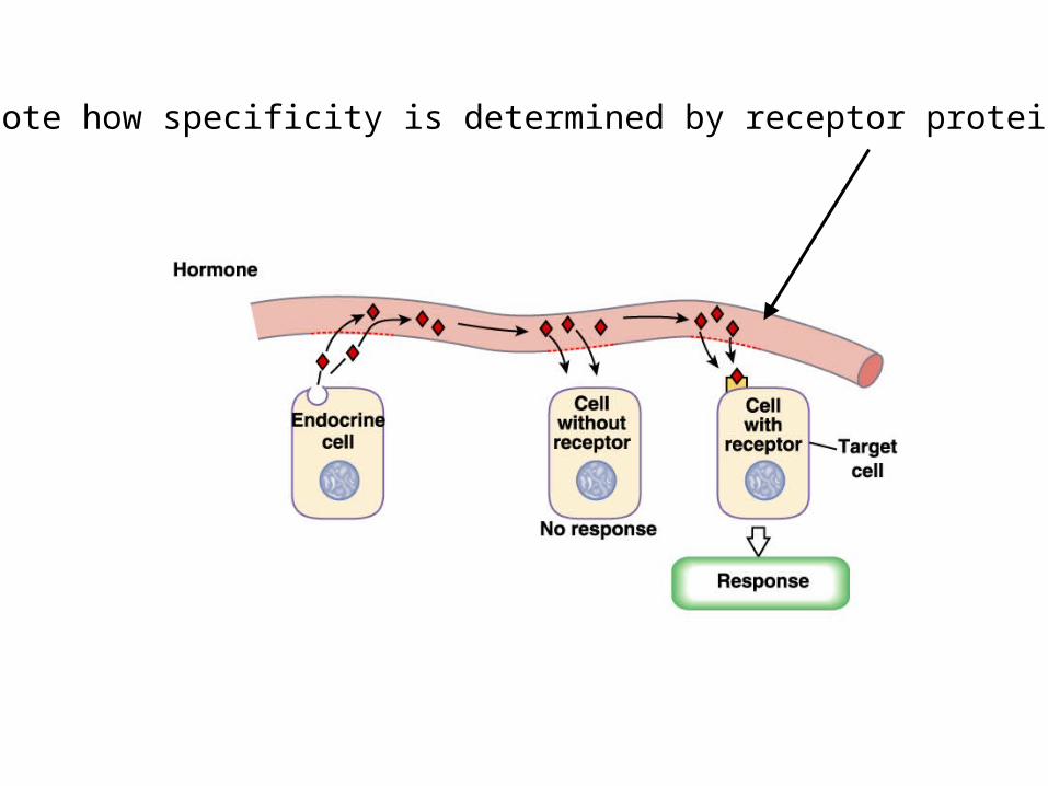
Note how specificity is determined by receptor protein
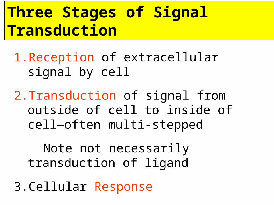
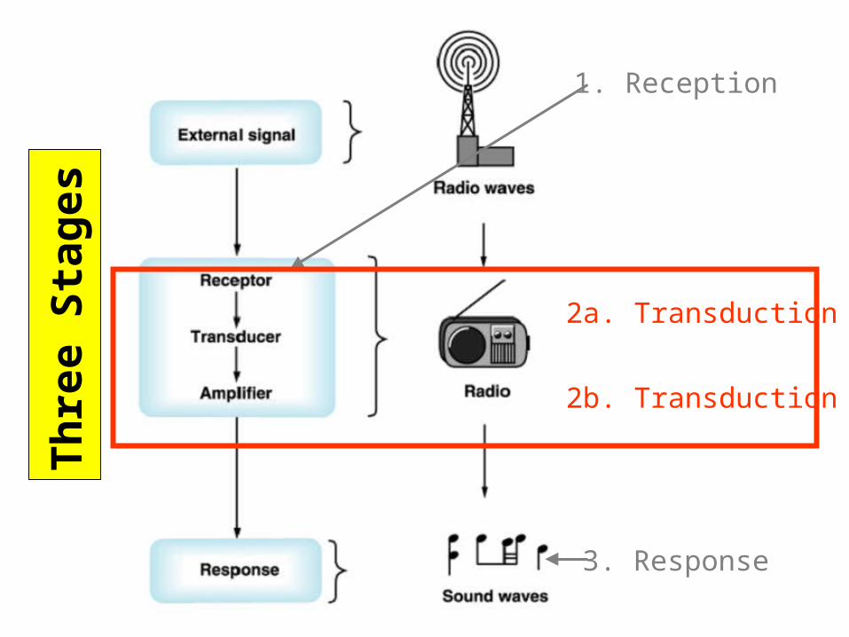
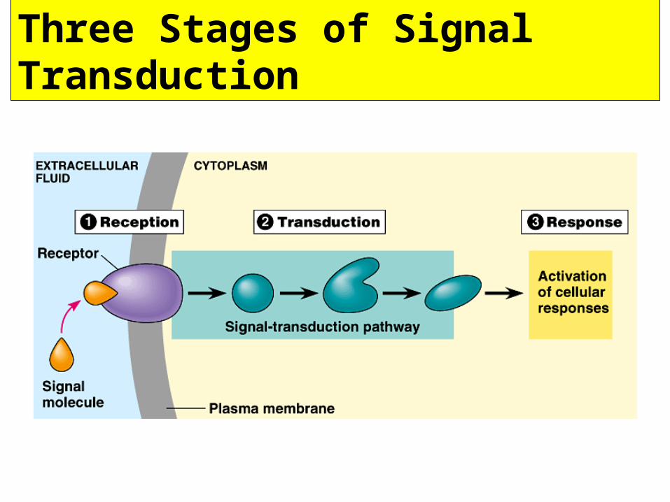
Three Stages of Signal Transduction
1. Reception of extracellular signal by cell
2. Transduction of signal from outside of cell to inside of cell—often multi-stepped
Note not necessarily transduction of ligand
3. Cellular Response
Response is inititiated and/or occurs entirely within receiving cell
Th
ree
Sta
ges
2a. Transduction
2b. Transduction
1. Reception
3. Response
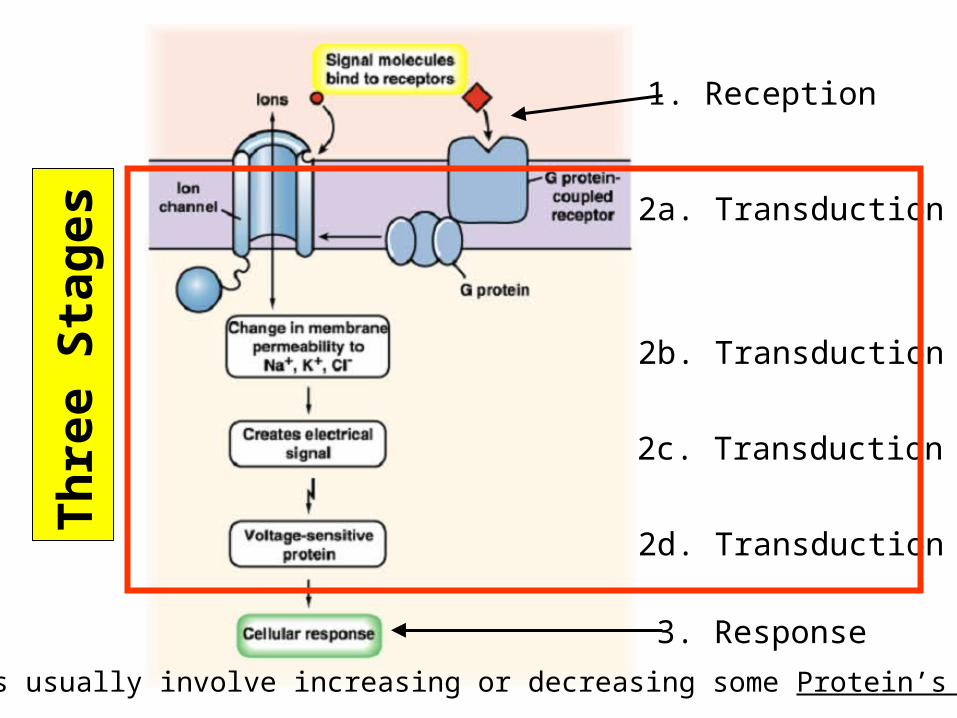
Three Stages of Signal Transduction
Th
ree
Sta
ges
2a. Transduction
2b. Transduction
2c. Transduction
2d. Transduction
1. Reception
3. Response
Responses usually involve increasing or decreasing some Protein’s Function
Var
iou
s R
esp
on
ses
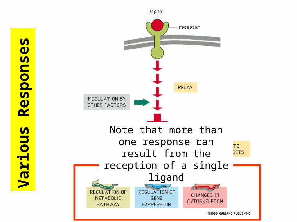
Note that more than one response can result from the reception of a single ligand
Exa
mp
les
of
Su
rfac
e R
ecep
tors
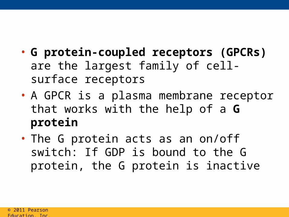
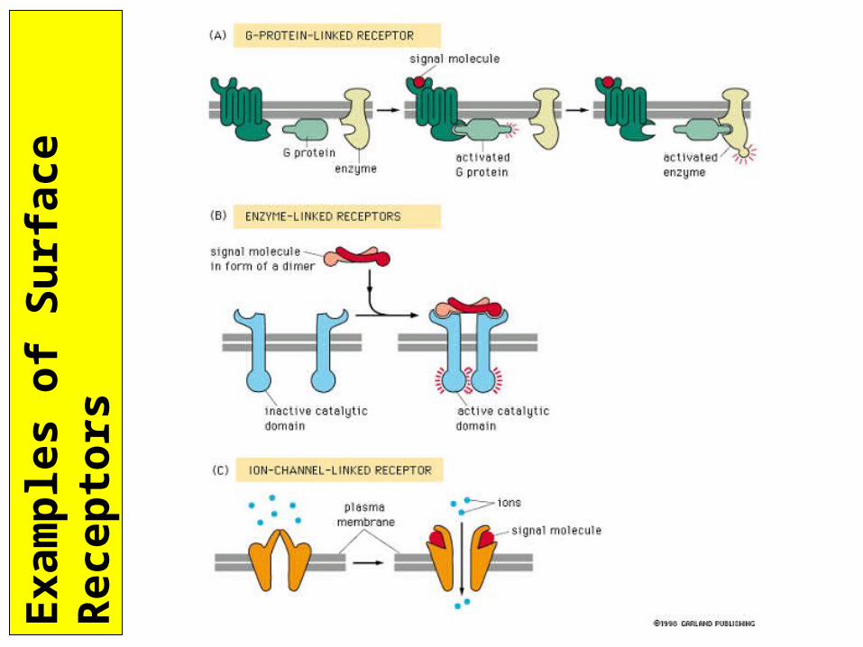
• G protein-coupled receptors (GPCRs) are the largest family of cell-surface receptors
• A GPCR is a plasma membrane receptor that works with the help of a G protein
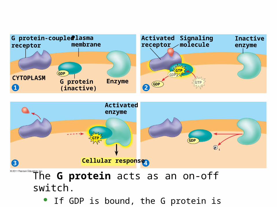
• The G protein acts as an on/off switch: If GDP is bound to the G protein, the G protein is inactive
© 2011 Pearson Education, Inc.
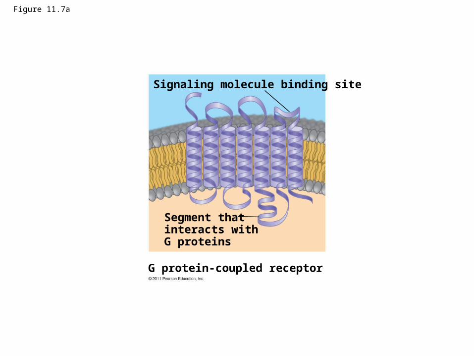
Figure 11.7a
G protein-coupled receptor
Signaling molecule binding site
Segment thatinteracts with G proteins
G protein-coupledreceptor
21
3 4
Plasmamembrane
G protein(inactive)
CYTOPLASM Enzyme
Activatedreceptor
Signalingmolecule
Inactiveenzyme
Activatedenzyme
Cellular response
GDPGTP
GDPGTP
GTP
P i
GDP
GDP
The G protein acts as an on-off switch. If GDP is bound, the G protein is inactive. If GTP is bound, the G protein is active.
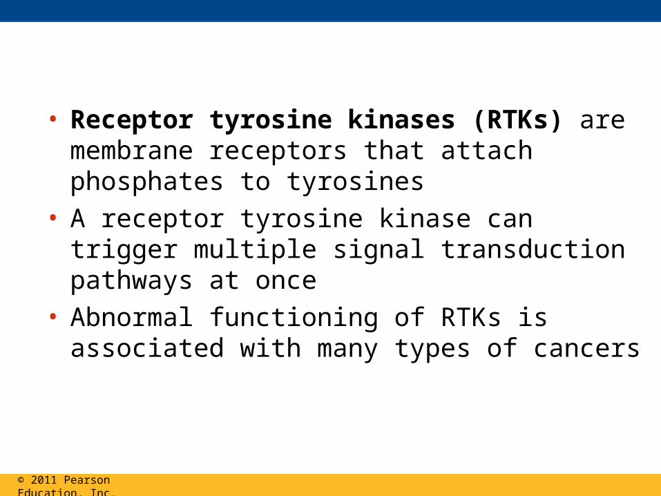
• Receptor tyrosine kinases (RTKs) are membrane receptors that attach phosphates to tyrosines
• A receptor tyrosine kinase can trigger multiple signal transduction pathways at once
• Abnormal functioning of RTKs is associated with many types of cancers
© 2011 Pearson Education, Inc.
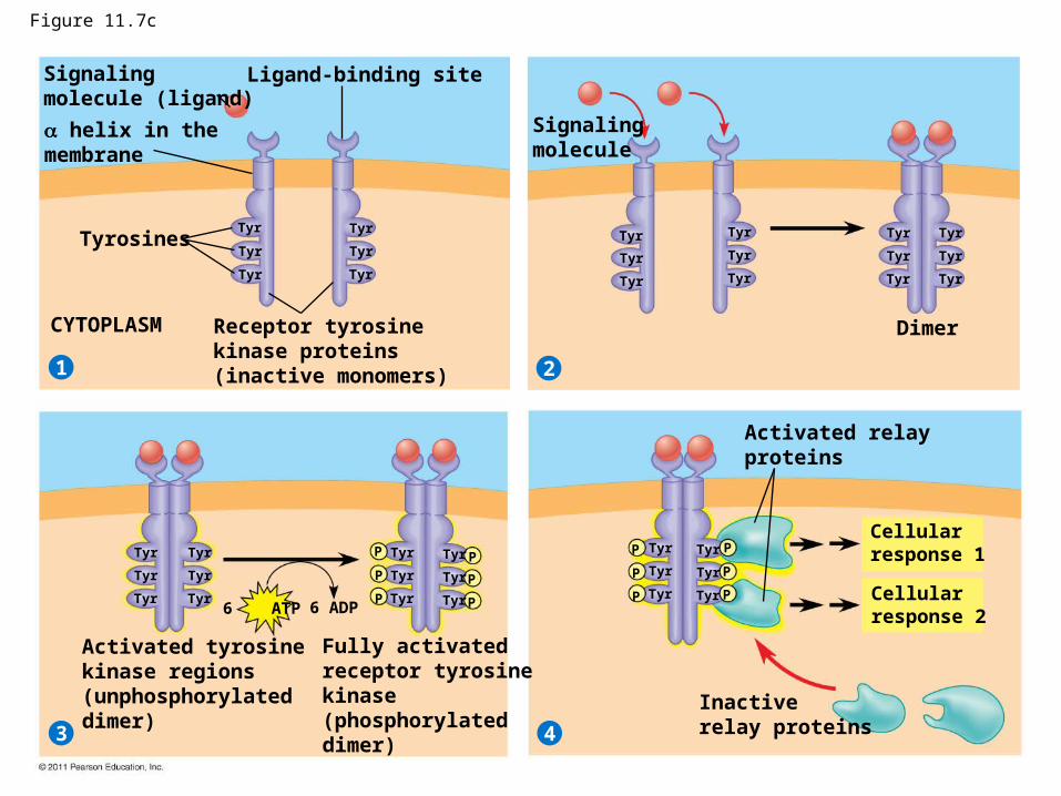
Figure 11.7c
Signalingmolecule (ligand)
21
3 4
Ligand-binding site
helix in themembrane
Tyrosines
CYTOPLASM Receptor tyrosinekinase proteins(inactive monomers)
Signalingmolecule
Dimer
Tyr
Tyr
Tyr
Tyr
Tyr
Tyr
Tyr
Tyr
Tyr
Tyr
Tyr
Tyr
Tyr
Tyr
Tyr
Tyr
Tyr
Tyr
Tyr
Tyr
Tyr
Tyr
Tyr
Tyr
Tyr
Tyr
Tyr
Tyr
Tyr
Tyr
Tyr
Tyr
Tyr
Tyr
Tyr
Tyr
P
P
P
P
P
P
P
P
P
P
P
P
Activated tyrosinekinase regions(unphosphorylateddimer)
Fully activatedreceptor tyrosinekinase(phosphorylateddimer)
Activated relayproteins
Cellularresponse 1
Cellularresponse 2
Inactiverelay proteins
6 ATP 6 ADP
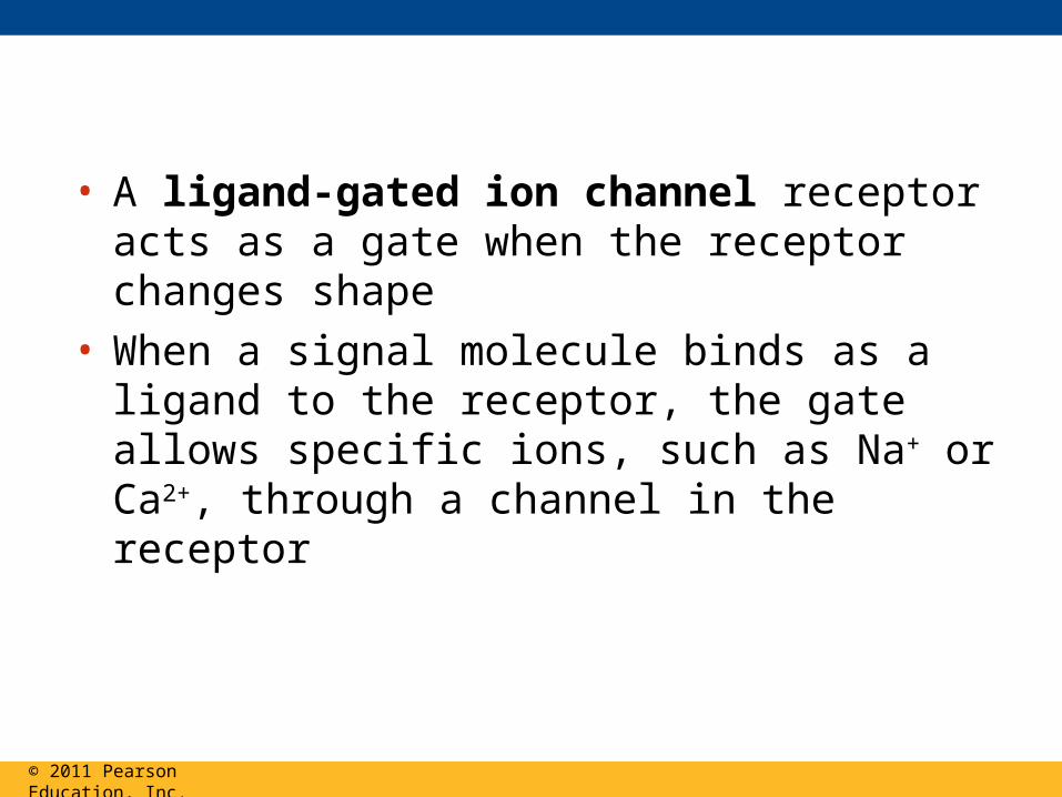
• A ligand-gated ion channel receptor acts as a gate when the receptor changes shape
• When a signal molecule binds as a ligand to the receptor, the gate allows specific ions, such as Na+ or Ca2+, through a channel in the receptor
© 2011 Pearson Education, Inc.

Figure 11.7d
Signalingmolecule (ligand)
21 3
Gate closed Ions
Ligand-gatedion channel receptor
Plasmamembrane
Gate open
Cellularresponse
Gate closed

Intracellular Receptors
• Intracellular receptor proteins are found in the cytosol or nucleus of target cells
• Small or hydrophobic chemical messengers can readily cross the membrane and activate receptors
• Examples of hydrophobic messengers are the steroid and thyroid hormones of animals
• An activated hormone-receptor complex can act as a transcription factor, turning on specific genes
© 2011 Pearson Education, Inc.
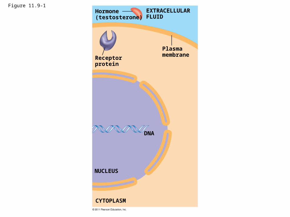
Figure 11.9-1Hormone(testosterone)
Receptorprotein
Plasmamembrane
DNA
NUCLEUS
CYTOPLASM
EXTRACELLULARFLUID
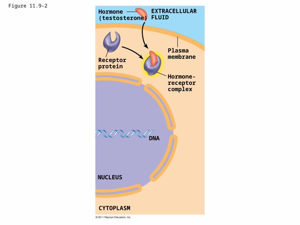
Figure 11.9-2Hormone(testosterone)
Receptorprotein
Plasmamembrane
Hormone-receptorcomplex
DNA
NUCLEUS
CYTOPLASM
EXTRACELLULARFLUID
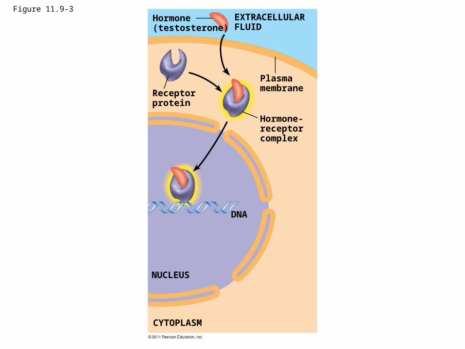
Figure 11.9-3Hormone(testosterone)
Receptorprotein
Plasmamembrane
Hormone-receptorcomplex
DNA
NUCLEUS
CYTOPLASM
EXTRACELLULARFLUID
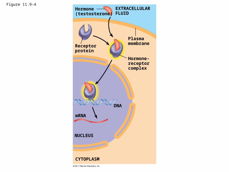
Figure 11.9-4Hormone(testosterone)
Receptorprotein
Plasmamembrane
Hormone-receptorcomplex
DNA
mRNA
NUCLEUS
CYTOPLASM
EXTRACELLULARFLUID
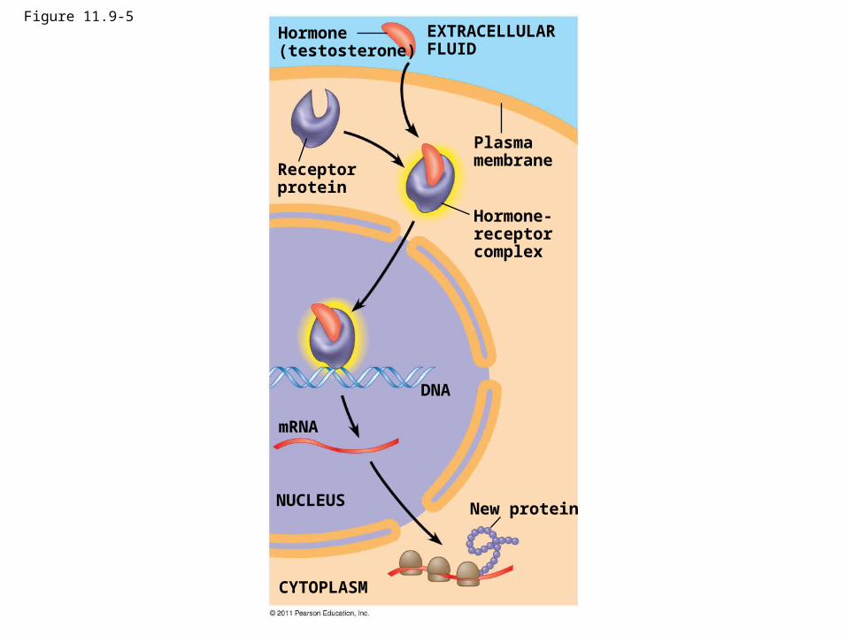
Figure 11.9-5Hormone(testosterone)
Receptorprotein
Plasmamembrane
EXTRACELLULARFLUID
Hormone-receptorcomplex
DNA
mRNA
NUCLEUS
CYTOPLASM
New protein
Transduction: Cascades of molecular interactions relay signals from receptors to target molecules in the cell
• Signal transduction usually involves multiple steps• Multistep pathways can amplify a signal: A few
molecules can produce a large cellular response• Multistep pathways provide more opportunities for
coordination and regulation of the cellular response
© 2011 Pearson Education, Inc.
Signal Transduction Pathways
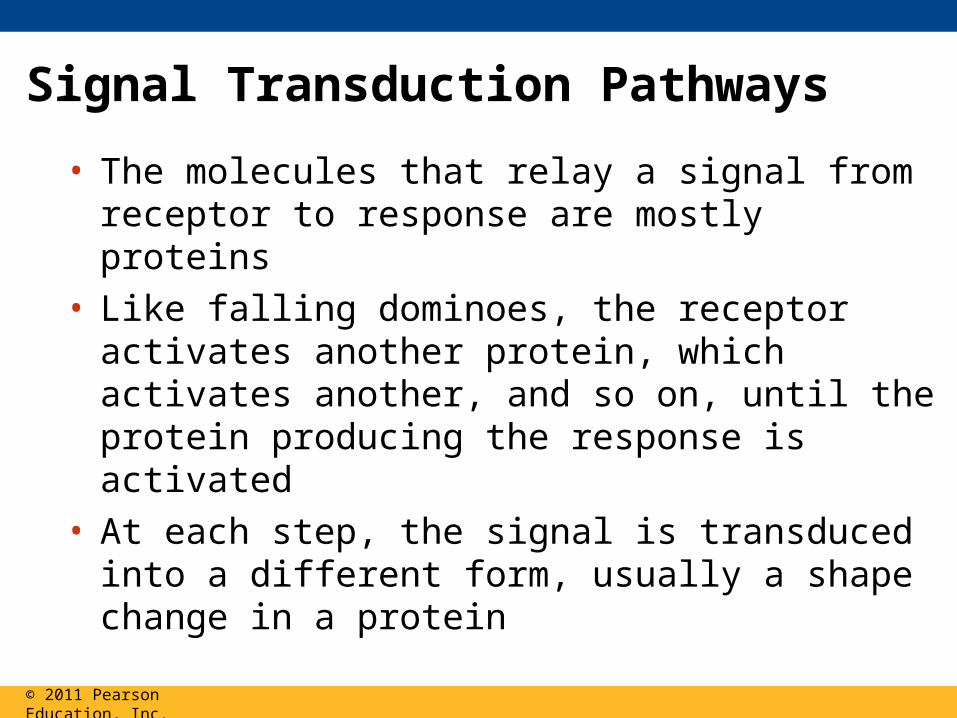
• The molecules that relay a signal from receptor to response are mostly proteins
• Like falling dominoes, the receptor activates another protein, which activates another, and so on, until the protein producing the response is activated
• At each step, the signal is transduced into a different form, usually a shape change in a protein
© 2011 Pearson Education, Inc.
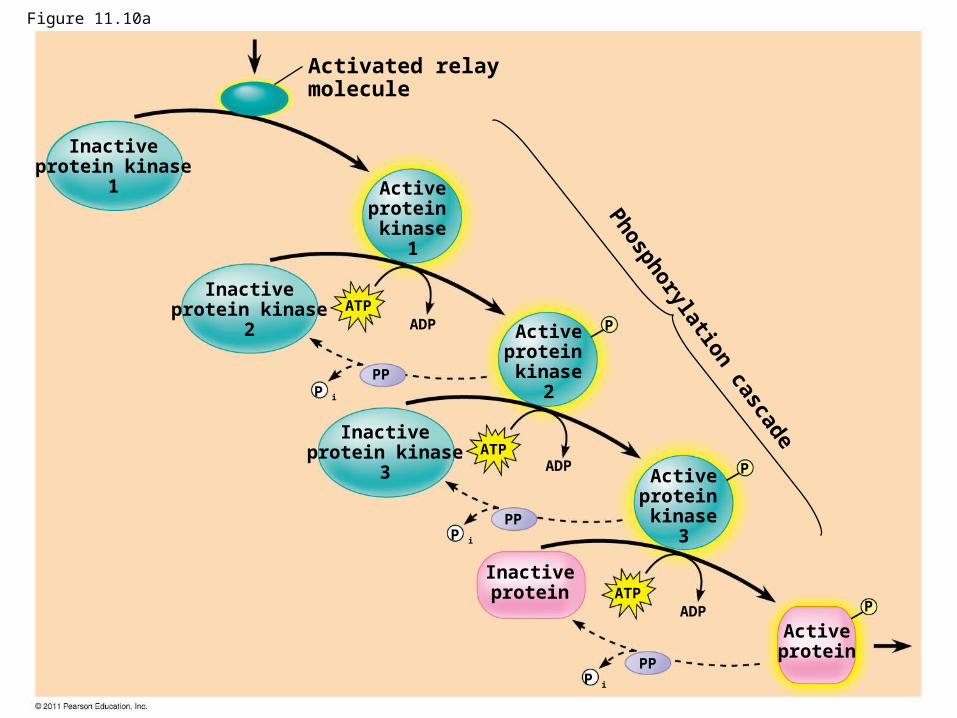
Protein Phosphorylation and Dephosphorylation
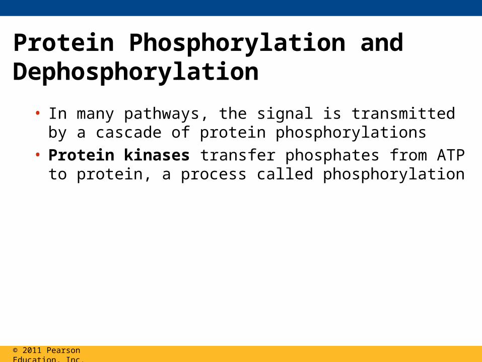
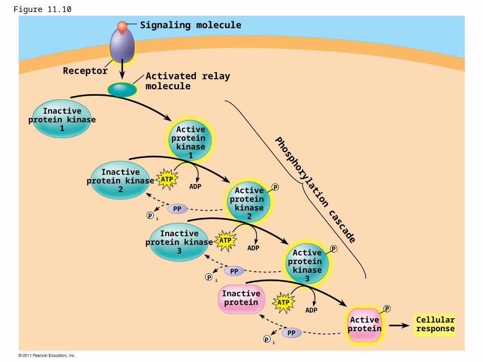
• In many pathways, the signal is transmitted by a cascade of protein phosphorylations
• Protein kinases transfer phosphates from ATP to protein, a process called phosphorylation
© 2011 Pearson Education, Inc.
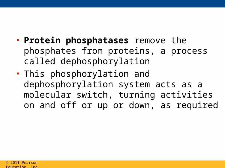
• Protein phosphatases remove the phosphates from proteins, a process called dephosphorylation
• This phosphorylation and dephosphorylation system acts as a molecular switch, turning activities on and off or up or down, as required
© 2011 Pearson Education, Inc.
Receptor
Signaling molecule
Activated relaymolecule
Phosphorylation cascade
Inactiveprotein kinase
1 Activeprotein kinase
1
Activeprotein kinase
2
Activeprotein kinase
3
Inactiveprotein kinase
2
Inactiveprotein kinase
3
Inactiveprotein
Activeprotein
Cellularresponse
ATPADP
ATPADP
ATPADP
PP
PP
PP
P
P
P
P i
P i
P i
Figure 11.10
Activated relaymolecule
Phosphorylation cascade
Inactiveprotein kinase
1 Activeprotein kinase
1
Activeprotein kinase
2
Activeprotein kinase
3
Inactiveprotein kinase
2
Inactiveprotein kinase
3
Inactiveprotein
Activeprotein
ATPADP
ATPADP
ATPADP
PP
PP
PP
P
P
P i
P i
P i
P
Figure 11.10a

Small Molecules and Ions as Second Messengers
• The extracellular signal molecule (ligand) that binds to the receptor is a pathway’s “first messenger”
• Second messengers are small, nonprotein, water-soluble molecules or ions that spread throughout a cell by diffusion
• Second messengers participate in pathways initiated by GPCRs and RTKs
• Cyclic AMP and calcium ions are common second messengers
© 2011 Pearson Education, Inc.
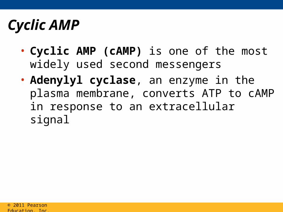
Cyclic AMP
• Cyclic AMP (cAMP) is one of the most widely used second messengers
• Adenylyl cyclase, an enzyme in the plasma membrane, converts ATP to cAMP in response to an extracellular signal
© 2011 Pearson Education, Inc.

Figure 11.11
Adenylyl cyclase Phosphodiesterase
Pyrophosphate
AMP
H2O
ATP
P iP
cAMP
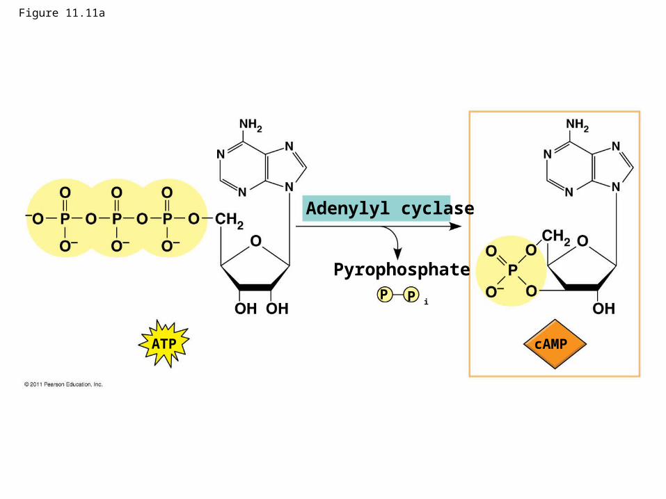
Figure 11.11a
Adenylyl cyclase
Pyrophosphate
ATP
P iP
cAMP
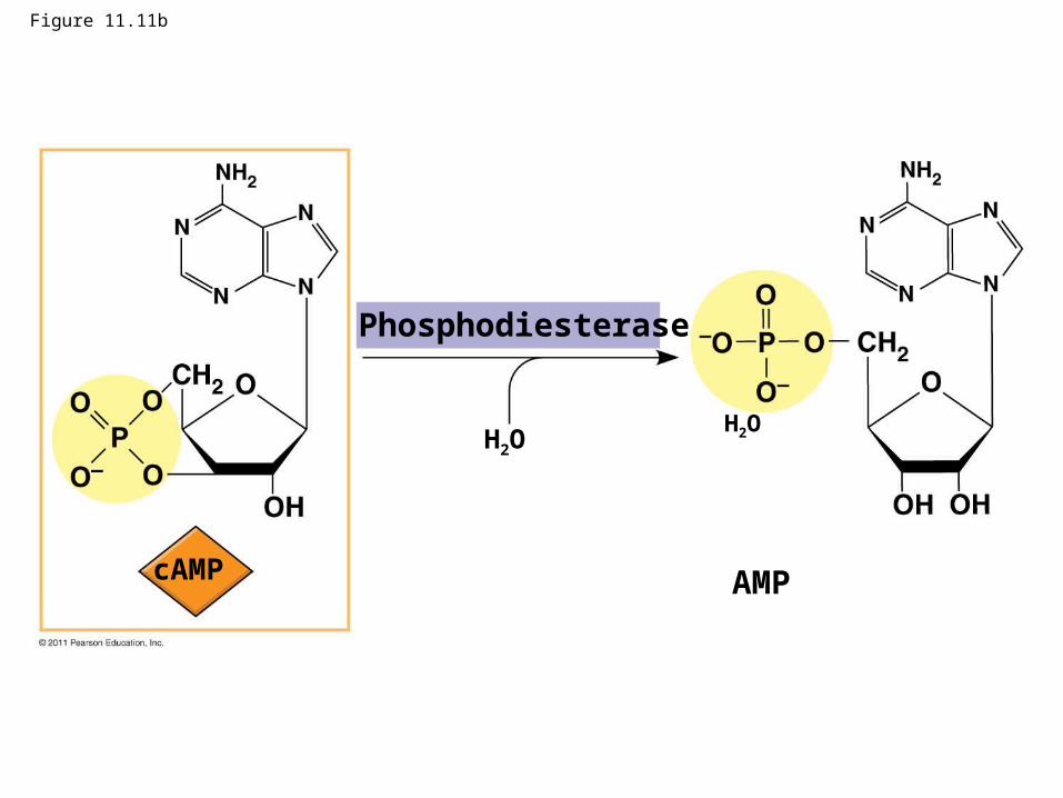
Figure 11.11b
Phosphodiesterase
AMP
H2O
cAMP
H2O
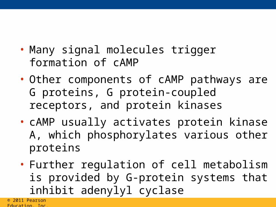
• Many signal molecules trigger formation of cAMP
• Other components of cAMP pathways are G proteins, G protein-coupled receptors, and protein kinases
• cAMP usually activates protein kinase A, which phosphorylates various other proteins
• Further regulation of cell metabolism is provided by G-protein systems that inhibit adenylyl cyclase
© 2011 Pearson Education, Inc.
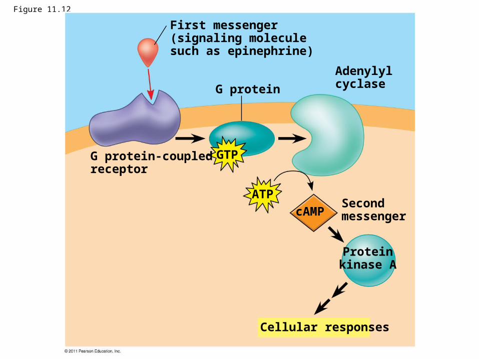
Figure 11.12
G protein
First messenger(signaling moleculesuch as epinephrine)
G protein-coupledreceptor
Adenylylcyclase
Second messenger
Cellular responses
Proteinkinase A
GTP
ATP
cAMP
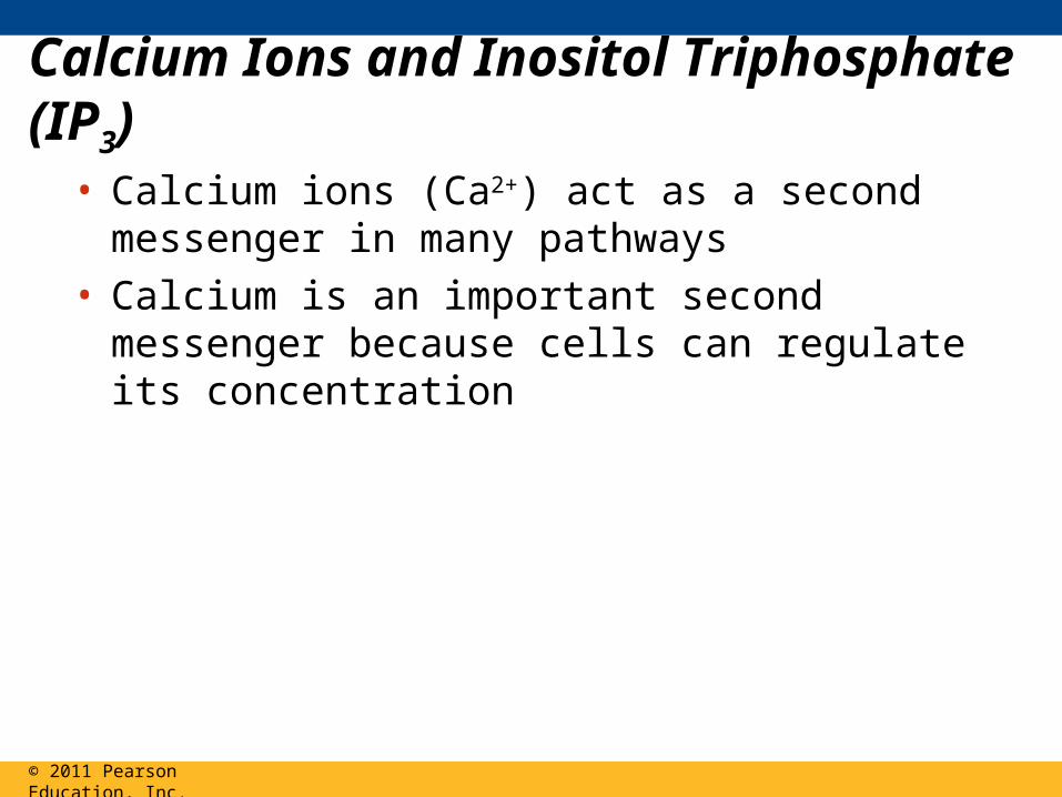
Calcium Ions and Inositol Triphosphate (IP3)
• Calcium ions (Ca2+) act as a second messenger in many pathways
• Calcium is an important second messenger because cells can regulate its concentration
© 2011 Pearson Education, Inc.
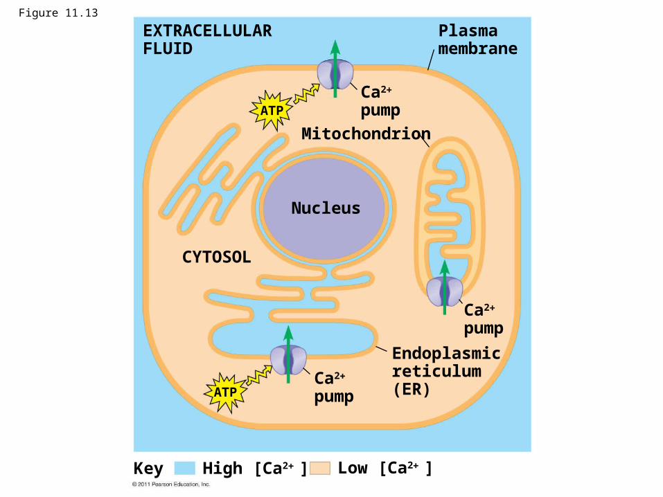
Figure 11.13
Mitochondrion
EXTRACELLULARFLUID
Plasmamembrane
Ca2
pump
Nucleus
CYTOSOL
Ca2
pump
Ca2
pump
Endoplasmicreticulum(ER)
ATP
ATP
Low [Ca2 ]High [Ca2 ]Key
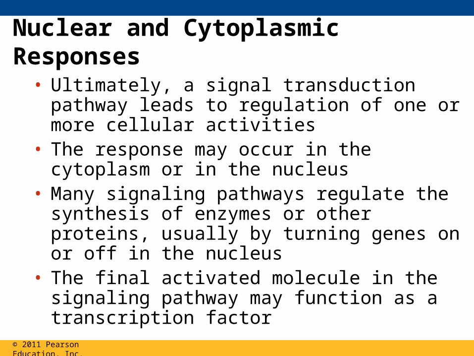
Nuclear and Cytoplasmic Responses
• Ultimately, a signal transduction pathway leads to regulation of one or more cellular activities
• The response may occur in the cytoplasm or in the nucleus
• Many signaling pathways regulate the synthesis of enzymes or other proteins, usually by turning genes on or off in the nucleus
• The final activated molecule in the signaling pathway may function as a transcription factor
© 2011 Pearson Education, Inc.
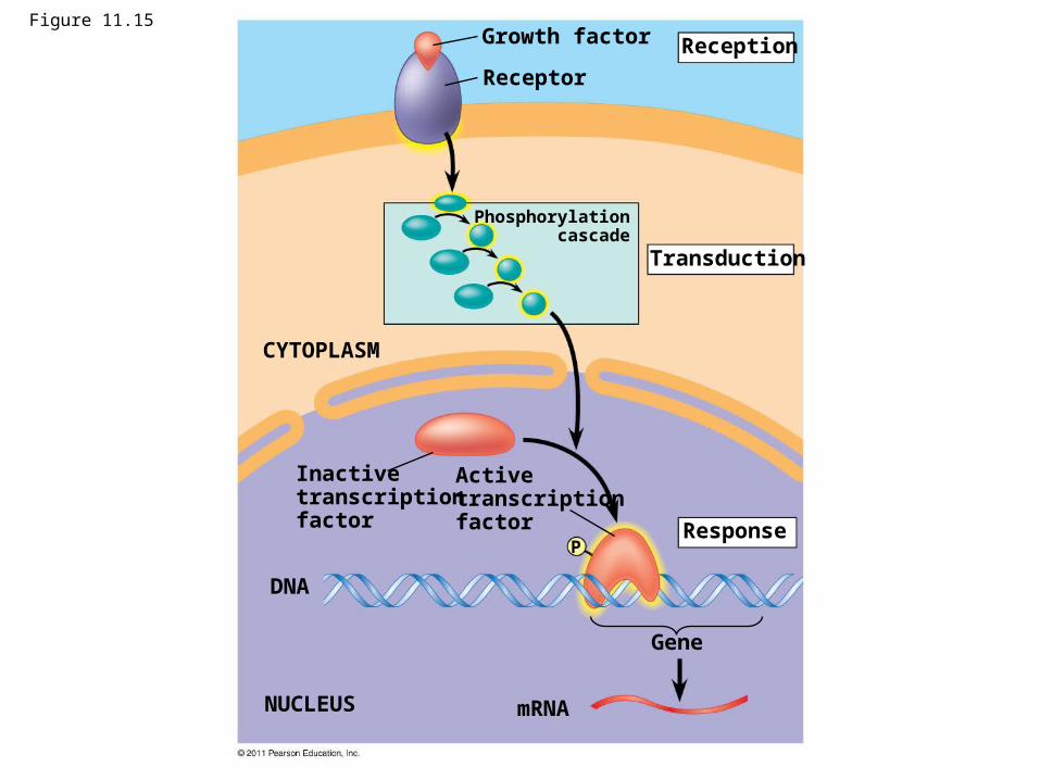
Figure 11.15Growth factor
Receptor
Reception
Transduction
CYTOPLASM
Response
Inactivetranscriptionfactor
Activetranscriptionfactor
DNA
NUCLEUS mRNA
Gene
Phosphorylationcascade
P
• Other pathways regulate the activity of enzymes rather than their synthesis
© 2011 Pearson Education, Inc.
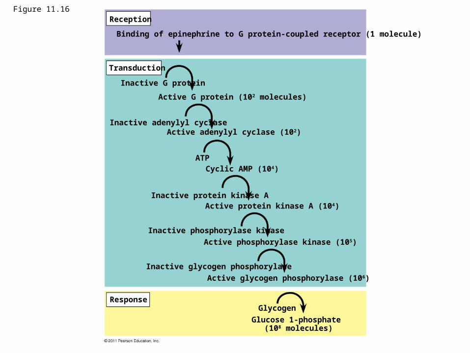
Figure 11.16Reception
Transduction
Response
Binding of epinephrine to G protein-coupled receptor (1 molecule)
Inactive G protein
Active G protein (102 molecules)
Inactive adenylyl cyclaseActive adenylyl cyclase (102)
ATPCyclic AMP (104)
Inactive protein kinase AActive protein kinase A (104)
Inactive phosphorylase kinase
Active phosphorylase kinase (105)
Inactive glycogen phosphorylase
Active glycogen phosphorylase (106)
Glycogen
Glucose 1-phosphate (108 molecules)

• Signaling pathways can also affect the overall behavior of a cell, for example, changes in cell shape
© 2011 Pearson Education, Inc.

Wild type (with shmoos) Fus3 formin
Matingfactoractivatesreceptor.
Matingfactor G protein-coupled
receptor
Shmoo projectionforming
Formin
G protein binds GTPand becomes activated.
2
1
3
4
5
P
P
P
PForminFormin
Fus3
Fus3Fus3
GDPGTP
Phosphory- lation cascade
Microfilament
Actinsubunit
Phosphorylation cascadeactivates Fus3, which movesto plasma membrane.
Fus3 phos-phorylatesformin,activating it.
Formin initiates growth ofmicrofilaments that formthe shmoo projections.
RESULTS
CONCLUSION
Figure 11.17
Apoptosis integrates multiple cell-signaling pathways
• Apoptosis is programmed or controlled cell suicide
• Components of the cell are chopped up and packaged into vesicles that are digested by scavenger cells
• Apoptosis prevents enzymes from leaking out of a dying cell and damaging neighboring cells
© 2011 Pearson Education, Inc.
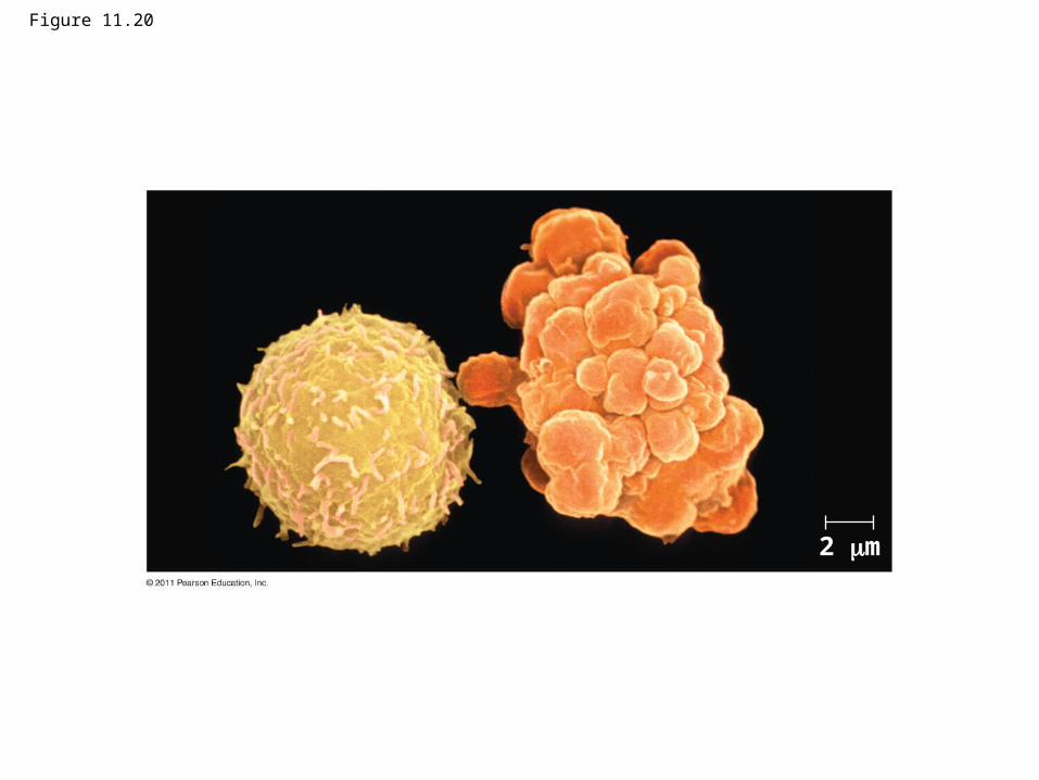
Figure 11.20
2 m
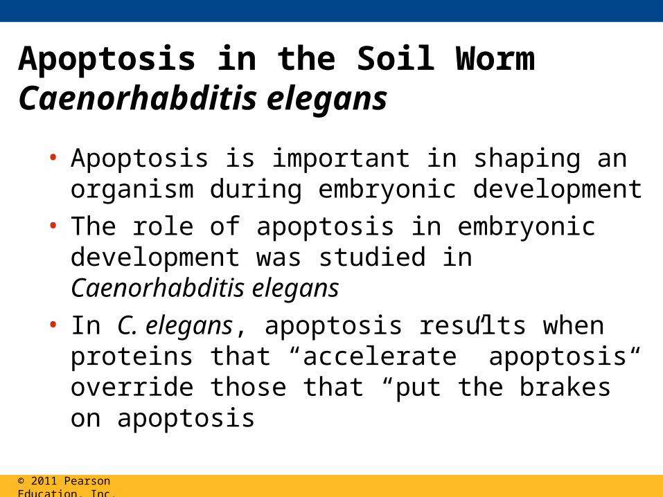
Apoptosis in the Soil Worm Caenorhabditis elegans
• Apoptosis is important in shaping an organism during embryonic development
• The role of apoptosis in embryonic development was studied in Caenorhabditis elegans
• In C. elegans, apoptosis results when proteins that “accelerate” apoptosis override those that “put the brakes” on apoptosis
© 2011 Pearson Education, Inc.
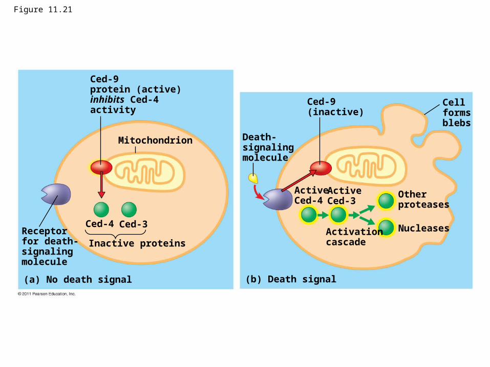
Figure 11.21
Mitochondrion
Ced-9protein (active)inhibits Ced-4activity
Receptorfor death-signalingmolecule
Ced-4 Ced-3
Inactive proteins
(a) No death signal
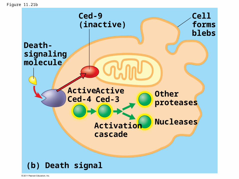
Death-signalingmolecule
Ced-9(inactive)
Cellformsblebs
ActiveCed-4
ActiveCed-3
Otherproteases
NucleasesActivationcascade
(b) Death signal
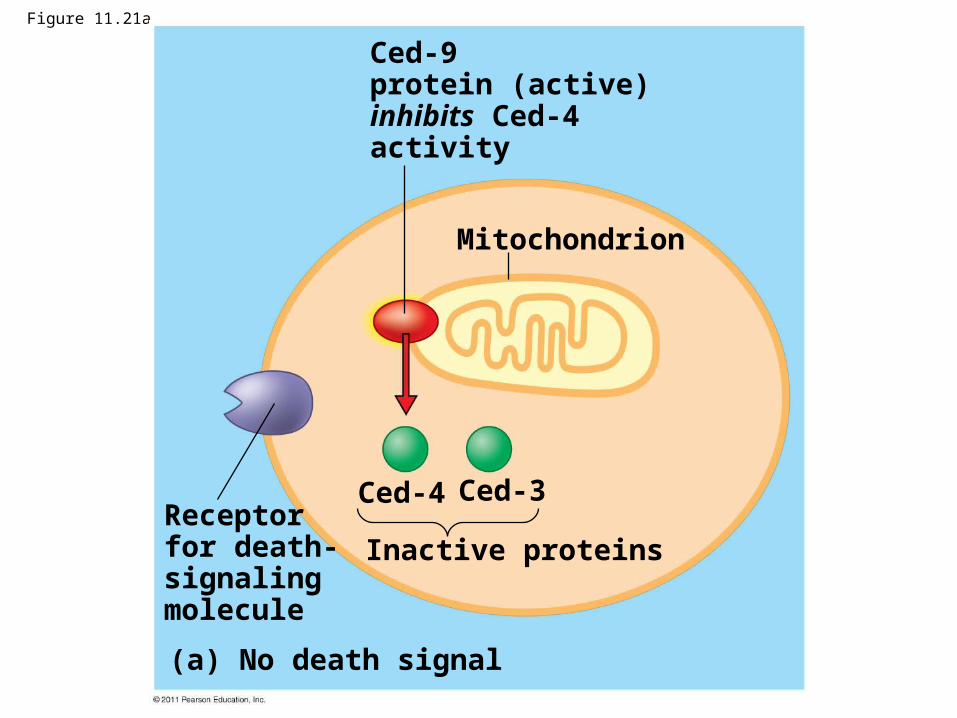
Figure 11.21a
Mitochondrion
Ced-9protein (active)inhibits Ced-4activity
Receptorfor death-signalingmolecule
Ced-4 Ced-3
Inactive proteins
(a) No death signal
Death-signalingmolecule
Ced-9(inactive)
Cellformsblebs
ActiveCed-4
ActiveCed-3
Otherproteases
NucleasesActivationcascade
(b) Death signal
Figure 11.21b
Apoptotic Pathways and the Signals That Trigger Them
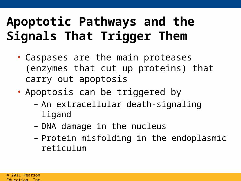
• Caspases are the main proteases (enzymes that cut up proteins) that carry out apoptosis
• Apoptosis can be triggered by– An extracellular death-signaling ligand – DNA damage in the nucleus– Protein misfolding in the endoplasmic reticulum
© 2011 Pearson Education, Inc.
• Apoptosis evolved early in animal evolution and is essential for the development and maintenance of all animals
• Apoptosis may be involved in some diseases (for example, Parkinson’s and Alzheimer’s); interference with apoptosis may contribute to some cancers
© 2011 Pearson Education, Inc.
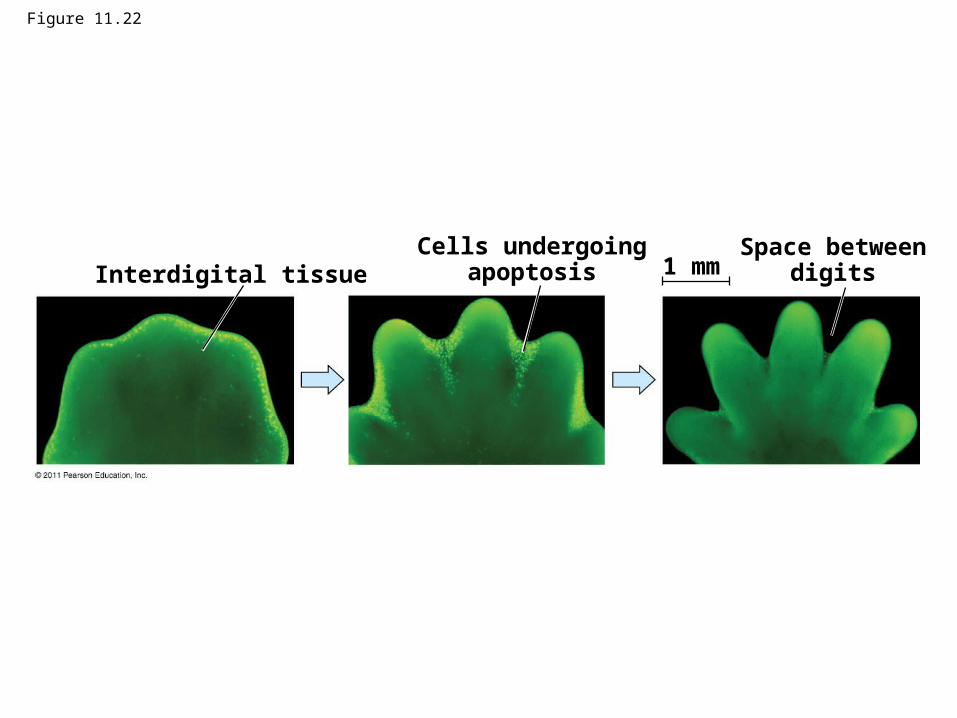
Figure 11.22
Interdigital tissueCells undergoing
apoptosisSpace between
digits1 mm
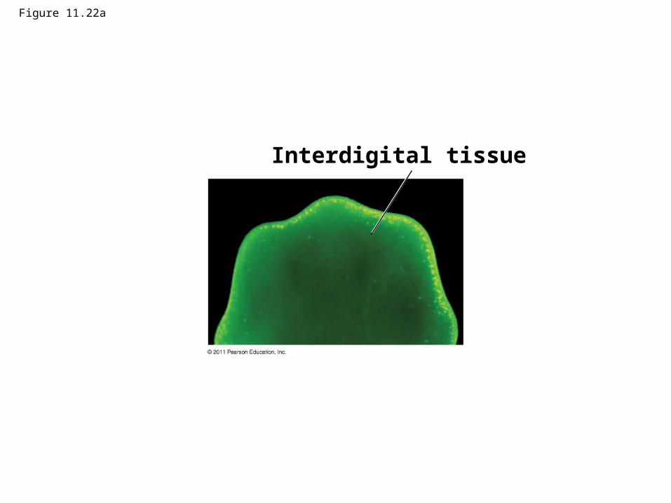
Figure 11.22a
Interdigital tissue
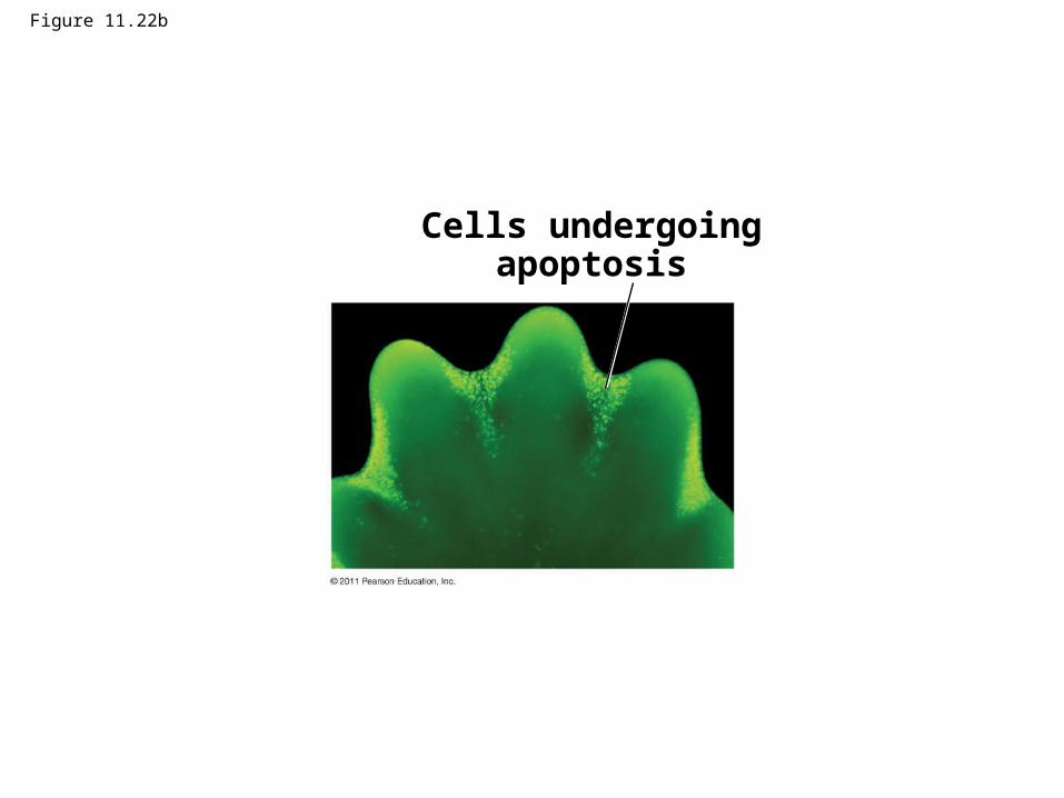
Figure 11.22b
Cells undergoingapoptosis
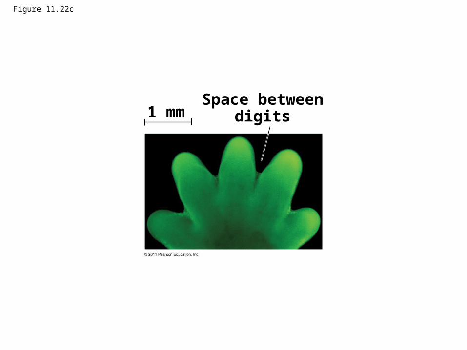
Figure 11.22c
Space betweendigits1 mm

Figure 11.UN01
Reception1 2 3Transduction Response
Receptor
Signalingmolecule
Relay molecules
Activation of cellularresponse