cell biology: sh2b1 and irsp53 proteins promote the...
TRANSCRIPT

Hui-Ching Wang and Linyi ChenChang, Shao-Jing Hong, Tian-Neng Li, Lily Chien-Jen Chen, Chien-Hung Shih, Yu-Jung BranchesFormation of Dendrites and Dendritic SH2B1 and IRSp53 Proteins Promote theCell Biology:
doi: 10.1074/jbc.M114.603795 originally published online January 13, 20152015, 290:6010-6021.J. Biol. Chem.
10.1074/jbc.M114.603795Access the most updated version of this article at doi:
.JBC Affinity SitesFind articles, minireviews, Reflections and Classics on similar topics on the
Alerts:
When a correction for this article is posted•
When this article is cited•
to choose from all of JBC's e-mail alertsClick here
http://www.jbc.org/content/290/10/6010.full.html#ref-list-1
This article cites 36 references, 11 of which can be accessed free at
at National T
sing-Hua U
niversity on March 25, 2015
http://ww
w.jbc.org/
Dow
nloaded from
at National T
sing-Hua U
niversity on March 25, 2015
http://ww
w.jbc.org/
Dow
nloaded from

SH2B1 and IRSp53 Proteins Promote the Formation ofDendrites and Dendritic Branches*
Received for publication, August 18, 2014, and in revised form, January 2, 2015 Published, JBC Papers in Press, January 13, 2015, DOI 10.1074/jbc.M114.603795
Chien-Jen Chen‡1, Chien-Hung Shih‡1, Yu-Jung Chang‡1, Shao-Jing Hong‡, Tian-Neng Li§, Lily Hui-Ching Wang§¶,and Linyi Chen‡¶2
From the ‡Institute of Molecular Medicine, §Institute of Molecular and Cellular Biology, ¶Department of Medical Science, NationalTsing Hua University, Hsinchu, Taiwan 30013, China
Background: Filopodium formation is a prerequisite for neurite initiation.Results: SH2B1 interacts with IRSp53 and promotes neurite outgrowth of hippocampal neurons.Conclusion: SH2B1-IRSp53 complexes promote filopodium formation, neurite initiation, and neurite branching.Significance: These results suggest that SH2B1 may regulate the formation of filopodia required for many cellular functions.
SH2B1 is an adaptor protein known to enhance neurite out-growth. In this study, we provide evidence suggesting that theSH2B1 level is increased during in vitro culture of hippocampalneurons, and the � isoform (SH2B1�) is the predominant iso-form. The fact that formation of filopodia is prerequisite forneurite initiation suggests that SH2B1 may regulate filopodiumformation and thus neurite initiation. To investigate whetherSH2B1 may regulate filopodium formation, the effect of SH2B1and a membrane and actin regulator, IRSp53 (insulin receptortyrosine kinase substrate p53), is investigated. Overexpressingboth SH2B1� and IRSp53 significantly enhances filopodiumformation, neurite outgrowth, and branching. Both in vivo andin vitro data show that SH2B1 interacts with IRSp53 in hip-pocampal neurons. This interaction depends on the N-terminalproline-rich domains of SH2B1. In addition, SH2B1 and IRSp53co-localize at the plasma membrane, and their levels increase inthe Triton X-100-insoluble fraction of developing neurons.These findings suggest that SH2B1-IRSp53 complexes promotethe formation of filopodia, neurite initiation, and neuronalbranching.
During development, neural stem cells undergo a morpho-genetic series to become mature neurons in response to extrin-sic as well as intrinsic factors. One prominent feature of neuro-genesis is neurite outgrowth. Although an array of studies haveidentified the growth factors, receptors, and signaling eventsthat are responsible for neurite outgrowth and guidance, intrin-sic regulation for neurite initiation is less clear. For hippocam-pal and cortical neurons cultured in vitro, the formation oflamellipodia containing filopodia is required for navigation ofneurites. Thus, the coordination of actin and microtubuleassembly becomes a major determinant of neurite outgrowth.As several studies have implicated the importance of filopodia
in neurite formation, a recent report clearly demonstrates theformation of filopodia preceding neurite initiation (1). Dent etal. (1) show that extracellular matrix laminin promotes the for-mation of actin-rich protrusions (filopodium-like) and is able torescue neuritogenesis in vasodilator-stimulated phosphopro-tein (VASP)3-deficient neurons. This study also reveals theimportance of regulators of filopodium formation during neu-rite outgrowth.
Filopodia are actin-rich membrane protrusions involve incell migration, neurite initiation, axon guidance in neuronalgrowth cones, endocytosis, and wound healing (1–3). Filopodiaconsist of unbranched F-actin filament bundles that are regu-lated by many actin-binding proteins such as IRSp53 (insulinreceptor tyrosine kinase substrate p53), fascin, Mena/VASP,and formins (3–5). IRSp53 belongs to Inverse Bin-Amphiphy-sin-Rvs 167 (I-BAR), also known as IMD (IRSp53-missing inmetastasis homology domain), domain-containing superfamilyof proteins and is known to drive membrane deformation, thesubsequent plasma membrane protrusions, and thus filopo-dium formation (3, 6 – 8). They are retracted by retrograde flowof F-actin and capping protein activity. The dynamic balance ofbarbed-end actin polymerization and retraction determines theinitiation, maintenance, and stability of filopodia. The molecu-lar mechanisms for controlling the initiation of dendritic filop-odia are not clear.
IRSp53 contains IMD, CRIB (Cdc42/Rac-interactive bind-ing), Src homology 3 (SH3), WW domains, and PDZ domainbinding sites (3). The IMD domain allows IRSp53 targeting toplasma membrane by binding to lipid molecules and triggersmembrane protrusion (3, 8). The SH3 domain of IRSp53 hasbeen shown to interact with regulatory proteins of actin, allow-ing IRSp53 to regulate actin cytoskeleton-associated proteinsand thus filopodium formation (8). The polymerization state ofactin is important in affecting IMD-lipid interaction. Mono-meric actin partially disrupts the binding between IMD andlipid, whereas assembled actin filament stabilizes the IRSp53-lipid interaction (9).
* This work was supported by National Science Council of Taiwan GrantNSC101-2311-B-007-012-MY3 and National Health Research InstitutesGrant NHRI-EX104-10206NI.
1 These authors contributed equally to this work.2 To whom correspondence should be addressed: Institute of Molecular Med-
icine and Dept. of Medical Science, National Tsing Hua University, 101,Section 2, Kuang-Fu Rd., Hsinchu, Taiwan 30013, China. Tel.: 886-3-5742775; Fax: 886-3-5715934; E-mail: [email protected].
3 The abbreviations used are: VASP, vasodilator-stimulated phosphoprotein;DIV, day in vitro; PLA, proximity ligation assay; SH, Src homology.
THE JOURNAL OF BIOLOGICAL CHEMISTRY VOL. 290, NO. 10, pp. 6010 –6021, March 6, 2015© 2015 by The American Society for Biochemistry and Molecular Biology, Inc. Published in the U.S.A.
6010 JOURNAL OF BIOLOGICAL CHEMISTRY VOLUME 290 • NUMBER 10 • MARCH 6, 2015
at National T
sing-Hua U
niversity on March 25, 2015
http://ww
w.jbc.org/
Dow
nloaded from

SH2B1 belongs to the SH2B adaptor proteins family, includ-ing SH2B1 (SH2-B), SH2B2 (APS), and SH2B3 (Lnk) (10 –13).Four SH2B1 splice variants identified so far, �, �, �, and �, differonly in their C termini (11, 14). SH2B1 contains two proline-rich domains, two actin-binding regions, a pleckstrin homologydomain, and an SH2 domain. SH2B1 also has a nuclear local-ization sequence and a nuclear export sequence, which affect itscellular distribution and thus differentiation genes (15–19).Human subjects with SH2B1 mutations display behavioralabnormalities, including social isolation and aggression (20 –22). Overexpressing SH2B1� has previously been shown toenhance neurite outgrowth of neuronal PC12 cells and corticaland hippocampal neurons (18, 19, 23–26). However, exactlyhow SH2B1 promotes neurite initiation remains unclear. Usingthe hippocampal and cortical neuron culture, we tested thehypothesis that SH2B1 promotes filopodium formation andthus neurite initiation by interacting with IRSp53.
MATERIALS AND METHODS
Animal Handling and Ethics Statement—All experimentswere conducted in accordance with the guidelines of the Labo-ratory Animal Center of National Tsing Hua University. Ani-mal use protocols were reviewed and approved by the NationalTsing Hua University Institutional Animal Care and Use Com-mittee (approval number 10126).
Antibodies and Reagents—Polyclonal antibody to rat SH2B1was raised against a glutathione S-transferase fusion proteincontaining amino acids 527– 670 of SH2B1�, as described pre-viously (27), and was used at a dilution of 1:1000 for Westernblotting. Anti-IRSp53, used at a dilution of 1:5000 for Westernblotting and 1:200 for immunoprecipitation, and anti-Mycantibodies, used at a dilution of 1:1000 for Western blotting and1:200 for immunoprecipitation, were purchased from Millipore(Billerica, MA). Anti-MAP2 (used at a dilution of 1:200 forimmunostaining), anti-Tau-1 (used at a dilution of 1:200 forimmunostaining), anti-�-tubulin antibodies (used at a dilutionof 1:1000 for Western blotting), and protein G-agarose beads werepurchased from Santa Cruz Biotechnology (Santa Cruz, CA).Anti-�III tubulin antibody was purchased from COVANCE(Berkeley, CA) and used at a dilution of 1:1000 for immuno-staining. Anti-GFP (used at a dilution of 1:1000 for Westernblotting and 1:200 for immunoprecipitation) and anti-GAP43antibodies were purchased from GeneTex (San Antonio, TX)and used at a dilution of 1:1000 for Western blotting. Anti-actinantibody was purchased from Sigma and used at a dilution of1:200 for immunostaining. IRDye800CW-labeled anti-rabbitand IRDye680CW-labeled anti-mouse secondary antibodieswere purchased from LI-COR Bioscience (Lincoln, NE). AlexaFluor fluorescence-conjugated secondary antibodies (AF350,AF488, AF555, and AF647), minimum essential media, neuro-basal medium, and B-27 serum-free supplement were pur-chased from Invitrogen. Dulbecco’s modified Eagle’s medium,horse serum, fetal bovine serum (FBS), L-glutamine (Gln), anti-biotic-antimycotic, rhodamine phalloidin, 4�,6-diamidino-2-phenylindole (DAPI), and Lipofectamine 2000 were purchasedfrom Invitrogen. Protease inhibitors, including aprotinin andleupeptin, were purchased from Roche Applied Science.Enhanced chemiluminescence (ECL) reagent was purchased
from PerkinElmer Life Sciences. Mammalian transfection sys-tem-calcium phosphate reagent was purchased from Promega(Madison, WI).
Plasmids—pEGFP-C1, GFP-SH2B1�(1–670), GFP-SH2B1�-(270–670), GFP-SH2B1�(397–670), GFP-SH2B1�(505–670),GFP-SH2B1�(1–150), GFP-SH2B1�(1–260), and myc-SH2B1�were generous gifts from Dr. Christin Carter-Su at University ofMichigan. IRSp53/pCMV-SPORT6 was purchased from ThermoFisher Scientific (Waltham, MA). Full-length IRSp53(1–521)cDNAs were both subcloned into pRK5-myc via BamHI-EcoRIsites.
Primary Neuronal Culture and 293T Cell Culture—Sprague-Dawley rats were purchased from BioLASCO Taiwan Co., Ltd.Cortical or hippocampal neurons were dissociated from hip-pocampus dissected from rat embryos (18th day of gestation;E18) by treatment with papain solution (10 units/ml). The iso-lated primary neurons (1–2 � 105/ml) were seeded on a poly-L-lysine (30 ng/ml)-coated dish or coverslip. On day in vitro(DIV) 0, primary neurons were cultured in minimum essentialmedia/high glucose medium supplemented with 5% FBS and5% horse serum under 5% CO2 conditions. On DIV 1, cells werecultured in neurobasal medium with B27 (containing addi-tional glutamine (Gln) and 0.025 mM glutamate). On DIV 2,cells were treated with 5 �M cytosine-1-�-D-arabinofuranosideto inhibit the growth of glial cells. On DIV 3, cells were culturedin neurobasal and Gln medium (neurobasal medium with B27containing additional Gln), and then half of the neurobasal andGln medium was replaced by fresh medium every 3 days. Lipo-fectamine 2000 or calcium phosphate reagents were used totransfect primary neurons according to the manufacturer’sinstruction. 1.5–3 h after transfection, culture medium wasreplaced with fresh medium. 293T cells were grown in DMEMcontaining 10% FBS, 1% L-Gln, and 1% antibiotic/antimycoticunder 5% CO2 conditions.
Reverse Transcription-Polymerase Chain Reaction (RT-PCR)—TRIzol reagent was used to isolate total RNA from neuronsaccording to the manufacturer’s instructions. For reverse tran-scription, 2 mg of total RNA was converted to cDNA using thereverse transcription kit (Applied Biosystems). SH2B1 isoformprimer pairs are as follows: forward 5�-TTCGATATGCTTG-AGCACTTCCGG-3� and reverse 5�-GCCTCTTCTGCCCC-AGGATGT-3�. Glyceraldehyde-3-phosphate dehydrogenase(GAPDH) primer pairs are as follows: forward 5�-ACCACAG-TCCATGCCATGCCATCAC-3� and reverse 5�-TCCACCAC-CCTGTTGCTGTA-3�. The mRNA levels of SH2B1 isoformfrom RT-PCR were normalized to that of GAPDH.
Knockdown of Endogenous SH2B1—The pLKO.1 lentiviralvector that contains oligonucleotides targeting specific genesequence pLKO.1-shSH2B1 (clone ID TRCN0000247808(number 1), 0000247810 (number 2), 0000247809 (number 3),0000247811 (number 4), 0000247807 (number 5), 0000217475(number 6), and 0000196146 (number 7)) and pLKO.1-shLacZ(clone ID TRCN0000072236, 0000231717) were purchased fromthe National Core Facility, located at the Institute of MolecularBiology/Genomic Research Center, Academic Sinica, Taiwan.
Western Blotting, Immunoprecipitation, and Immunofluores-cence Staining—Cells were lysed with radioimmunoprecipita-tion assay buffer, containing 1 mM Na3VO4, 1 mM phenylmeth-
SH2B1 and IRSp53 Promote Dendritic Branches
MARCH 6, 2015 • VOLUME 290 • NUMBER 10 JOURNAL OF BIOLOGICAL CHEMISTRY 6011
at National T
sing-Hua U
niversity on March 25, 2015
http://ww
w.jbc.org/
Dow
nloaded from
ylsulfonyl fluoride (PMSF), 10 ng/ml aprotinin, and 10 ng/mlleupeptin. Equal amounts of proteins were separated by SDS-PAGE and analyzed by Western blotting using the indicatedantibodies followed by incubation with IRDye-conjugated sec-ondary antibody. Protein signal was detected using Odysseyinfrared imaging system (Odyssey Imager). For immunopre-cipitation, cell lysates were incubated with the specific antibodyat 4 °C overnight followed by incubation with protein A- orG-agarose beads and rotated at 4 °C for 1 h to pull down specificantibodies. Samples were centrifuged at 2000 rpm at 4 °C for 1min. The beads were washed three times with radioimmuno-precipitation assay lysis buffer to remove nonspecific binding.The precipitates were dissolved in 1� sample buffer, boiled at95 °C for 10 min, and analyzed by Western blotting. For immu-nofluorescence staining, cells were fixed by 4% paraformalde-hyde, incubated with the indicated primary antibodies, fol-lowed by Alexa Fluor-conjugated secondary antibody. DAPIstaining was used to mark the location of the nucleus. Cellswere then mounted with Prolong Gold reagent, and the fluo-rescent images were taken using Carl Zeiss Observer Z1 micro-scope or Carl Zeiss LSM 510 meta confocal microscope.
GST Pulldown Assay—GST-SH2B1� constructs wereexpressed in a BL21 strain, and lysates were harvested. GST-SH2B1� proteins were pulled down via glutathione-Sepharose4B (GE Healthcare). Sepharose beads conjugated with GST-SH2B1� were then incubated with 293T cell lysates for 2 h at4 °C. Beads were washed and resuspended in SDS sample bufferfor Western blotting.
Neurite Outgrowth of PC12 Cells—For nerve growth factor(NGF)-induced neurite outgrowth, PC12-shLacZ or PC12-shSH2B1 cells were split onto collagen I-coated plates at about20 –30% confluency in low serum differentiation medium(DMEM containing 2% horse serum, 1% FBS, 1% antibiotic/antimycotic, and 1% L-Gln). The average length of the longestneurite of cells was determined with ImageJ software. Differen-tiation of PC12 cells is defined as the length of the neuritesbeing at least twice the diameter of the cell body.
Triton X-100 Cytoskeleton Extraction—Cells were lysed withextraction buffer (0.5% Triton X-100, 100 mM NaF, 50 mM KCl, 2mM MgCl2, 1 mM EGTA, 10 mM KPO4, pH 7.5, 0.5 M sucrose)supplemented with protease inhibitors containing 1 mM Na3VO4,1 mM PMSF, 10 ng/ml aprotinin, and 10 ng/ml leupeptin and thencentrifuged at 13,000 rpm at 4 °C for 10 min. The supernatant(detergent-soluble fraction containing the G-actin fraction) wastaken for immunoblotting. Cell pellets containing F-actin fractionswere scraped into the same extraction buffer of the supernatant,and both fractions were solubilized with the same volume of 5�sample buffer. Equal volumes of proteins from each fraction wereresolved by SDS-PAGE and detected by Western blotting analysisusing the indicated antibodies.
Subcellular Fractionation—Cells were lysed by fractionationbuffer (10 mM Tris-HCl, pH 7.9, 10 mM KCl, 0.1 mM EDTA, 0.1mM EGTA, 1 mM DTT) containing 1 mM Na3VO4, 1 mM PMSF,10 ng/ml aprotinin and leupeptin. Lysates were passed througha 27-gauge needle 50 times and then centrifuged at 2300 rpm(500 � g) at 4 °C for 5 min. After centrifugation, the supernatantwas removed and transferred to freshly labeled tubes (Thick-wall Polycarbonate) and then centrifuged by an ultracentrifuge
(Hitachi, CS150NX, Rotor-S80AT3) at 45,000 rpm (100,000 �g) for 1 h. After ultracentrifugation, the supernatant was desig-nated as the cytosolic fraction. The pellet, designated as themembrane fraction, was washed with fractionation buffer anddissolved by lysis buffer. The remaining pellet, designated as thenuclear fraction (nuclei and nucleus-associated structures), waswashed twice by fractionation buffer with 0.5% Nonidet P-40and dissolved by lysis buffer (1% Triton X-100, 1% sodiumdeoxycholate, 50 mM Tris-HCl, 150 mM NaCl, 1 mM EDTA, 0.1mM EGTA) containing 1 mM Na3VO4, 1 mM PMSF, 10 ng/mlaprotinin and leupeptin.
Measurement of Attachment, End Points, Axon Length, Pear-son’s Correlation Coefficient, and Co-localization Analysis—The number of attachment and end points was quantified usingthe built-in cell-counter in ImageJ software (rsb.info.nih.gov,National Institutes of Health, Bethesda). The attachmentpoints are defined as the location where neurite and filopodiaconnect to the cell body and end point as the location at the tipof filopodia and neurites (28). The average length of the axonwas measured using the Simple Neurite Tracer of Fiji software,a plugin in ImageJ software performing semi-automatic tracingof neurons. The mean pixel value of axon length was measuredand converted pixels into micrometers (�m). The Pearson’scorrelation coefficient (r) was used to measure the relationshipbetween two variables. Images were analyzed using Meta-Morph software (Molecular Devices, Sunnyvale, CA). To exam-ine the correlation between IRSp53 and SH2B1� in hippocam-pal neurons, cells expressing GFP-SH2B1� were fixed, then
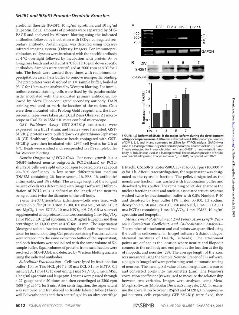
FIGURE 1. � isoform of SH2B1 is the major isoform during the developmentof hippocampal neurons. A, RNA was extracted from E18 hippocampal neuronsof DIV 1, 3, 8, and 14 and converted to cDNAs for RT-PCR analysis. GAPDH wasused as a loading control. B, lysates from hippocampal neurons of DIV 1, 3, 5, and7 were extracted for immunoblotting with anti-SH2B1 or anti-�-tubulin anti-body. �-Tubulin was used as a loading control. The relative expression of SH2B1was quantified by using ImageJ software. *, p � 0.05, compared with DIV 1.
SH2B1 and IRSp53 Promote Dendritic Branches
6012 JOURNAL OF BIOLOGICAL CHEMISTRY VOLUME 290 • NUMBER 10 • MARCH 6, 2015
at National T
sing-Hua U
niversity on March 25, 2015
http://ww
w.jbc.org/
Dow
nloaded from

stained with IRSp53, and imaged by using a confocal micro-scope (Zeiss LSM 510). Both the raw single plane graph ofIRSp53 and SH2B1� were quantified by “Co-localizationFinder” and “Co-localization Colormap” plugins in ImageJ soft-ware. To reach the top 2% overlapping pixels, images shouldrestrain selection to pixels with 85%. Correlation between pair ofpixels from IRSp53 and SH2B1� is displayed as distribution ofnormalized mean deviation product values and visualized with acolor scale. Negative indexes are represented by cold colors.Indexes above 0 are represented by hot colors, indicating co-local-ization. The correlations of IRSp53 and SH2B1� are representedas a color map that is calculated based on Equation 1,
nMDPx,y ��Ia � Ia��Ib � Ib�
�Iamax � Ia��Ibmax � Ib�(Eq. 1)
Ia indicates intensity for the given pixel in image a; Ia indicatesaverage intensity of image a; Iamax indicates highest pixel inten-sity in image a; Ib indicates intensity for the given pixel in imageb; Ib indicates average intensity of image b; and Ibmax indicateshighest pixel intensity in image b.
Duolink in Situ Proximity Ligation Assay (PLA)—TheDuolink in situ PLA assay kit was purchased from Olink Biosci-ence (Uppsala, Sweden) and was performed according to themanufacturer’s instruction. Hippocampal neurons were fixedby 4% paraformaldehyde for 15 min, permeabilized by 0.1%Triton X-100 for 10 min, and incubated with 1% bovine serumalbumin in PBS. Neurons were incubated with rabbit anti-SH2B1 antibody and mouse anti-IRSp53 antibodies at 4 °Covernight, followed by incubation with Duolink PLA rabbitPLUS and mouse MINUS probes. After incubation, ligation and
FIGURE 2. SH2B1� increases branches of neurite in development of hippocampal neurons. A, hippocampal neurons were transiently transfected with GFP-SH2B1� on DIV 3 and images of neuronal morphology were visualized on DIV 7. Attachment points and end points are shown as red and green dots, respectively. B,hippocampal neurons were transiently transfected with either GFP or GFP-SH2B1� on DIV 4, and images of neuronal morphology on days 1–3 after transfection werevisualized. Enlarged images of the neurite branches are shown on the bottom panels. Scale bar, 40 �m. A total of 12 hippocampal neurons were counted per conditionfrom three independent experiments. Values are mean � S.E. from three independent experiments and statistically compared by Student’s t test (*, p � 0.05,overexpressing GFP-SH2B1� compared with GFP). The number of attachment points and end points were measured using ImageJ software.
SH2B1 and IRSp53 Promote Dendritic Branches
MARCH 6, 2015 • VOLUME 290 • NUMBER 10 JOURNAL OF BIOLOGICAL CHEMISTRY 6013
at National T
sing-Hua U
niversity on March 25, 2015
http://ww
w.jbc.org/
Dow
nloaded from

amplification were followed by using Duolink DetectionReagents Red. Nuclei were stained with DAPI, and cells weremounted using Prolong Gold reagent (Invitrogen). Alexa Fluor488-phalloidin incubation was used to detect actin structure.Fluorescent signals were obtained by the Carl Zeiss ObserverZ1 microscope with a �100 oil objective.
RESULTS
� Isoform of SH2B1 Is the Major Isoform during in Vitro Cul-ture of Hippocampal Neurons—To study the role of SH2B1 dur-ing the development of the central nervous system, we first deter-mined whether the splice variants of SH2B1 are differentiallyexpressed during the development of neurites. Hippocampal neu-
rons were isolated from embryonic day 18 (E18) of rat brain andcultured in vitro. RNA levels of the four splice variants of SH2B1from DIV 1, 3, 8, and 14 hippocampal neurons were compared viaRT-PCR. As shown in Fig. 1A, mRNA level of � isoform increasedon DIV 3, remained high through DIV 8, and decreased by DIV 14.Protein levels of SH2B1 were also up-regulated during early devel-opment of hippocampal neurons and a more than 2-fold increaseon DIV 7 (Fig. 1B). These results indicate that the expression ofSH2B1 is increased, and SH2B1� is the predominant isoform dur-ing neural differentiation.
SH2B1� Increases Neurite Branches of HippocampalNeurons—To provide better analysis of the effect of SH2B1 onmorphogenesis of hippocampal neurons, E18 hippocampal
FIGURE 3. Knockdown of SH2B1 expression reduces neurite outgrowth for hippocampal neurons and PC12 cells. A, hippocampal neurons were transientlytransfected with either shLacZ or shSH2B1#3, #4, #5, #6, or #7 together with the pEGFP vector on DIV 4, and live cell images of neuronal morphology were taken. Thenumber of end points was counted from 12 to 18 hippocampal neurons per condition. PC12 cells stably expressing shLacZ or shSH2B1 constructs (shSH2B1#3, #4, #5,#6, and #7) were treated with 100 ng/ml NGF for 1 day. Length of the longest neurite of each cell was calculated from three independent experiments. *, p � 0.05 bypaired Student’s t test compared with shLacZ control. Lysates from PC12-shLacZ and PC12-shSH2B1 cells were collected and immunoblotted with anti-SH2B1 andanti-�-tubulin antibodies. B, hippocampal neurons were transiently transfected with either shLacZ�mCherry vector or shSH2B1#3�mCherry vector, together withGFP or GFP-SH2B1� on DIV 4, and live cell images were taken. The number of end points was counted from a total of 10–13 hippocampal neurons per condition.PC12-shLacZ or PC12-shSH2B1(#3, #4, #5, #6, and #7) cells were transiently transfected with GFP or GFP-SH2B1� constructs. Transfected cells were then treated withNGF for 1 day, and the length of the longest neurite of each cell was measured from three independent experiments. *, p � 0.05 by paired Student’s t test comparedwith PC12-shLacZ�GFP control or with its respective control. C, hippocampal neurons were transiently transfected with either shLacZ or shSH2B1 together withpEGFP vector on DIV 4, and images of neuronal morphology on days 1–3 after transfection were taken. The number of attachment points or end points on days 1–3after transfection was measured. Enlarged images of the neurite branches are shown on the right panels. Scale bar, 40 �m. A total of 12 hippocampal neurons werecounted per condition from three independent experiments. Values are mean � S.E. from three independent experiments and statistically compared by Student’s ttest (*, p � 0.05, shSH2B1 compared with shLacZ). D, hippocampal neurons were transfected with either shLacZ or shSH2B1 together with pEGFP vector on DIV 4, andthe images of neuronal morphology of were visualized on DIV 7. Immunofluorescence staining was performed with MAP2 (red), GFP (green), and DAPI (blue) antibod-ies. Cell lysates from cultured hippocampal neurons transiently transfected with shLacZ or shSH2B1(#3) were collected and analyzed via Western blotting withanti-SH2B1 or anti-�III tubulin antibody.
SH2B1 and IRSp53 Promote Dendritic Branches
6014 JOURNAL OF BIOLOGICAL CHEMISTRY VOLUME 290 • NUMBER 10 • MARCH 6, 2015
at National T
sing-Hua U
niversity on March 25, 2015
http://ww
w.jbc.org/
Dow
nloaded from

neurons were transiently transfected with GFP or GFP-SH2B1� on DIV 4, and morphology of neurons was imaged onDIV 5–7. Quantification of the attachment points and endpoints, symbolic of neurites and neurite branches, respectively(28), is shown in Fig. 2A. Overexpression of SH2B1� increasedthe numbers of attachment points and end points (Fig. 2B). IfSH2B1 is required for neurite outgrowth, knocking downSH2B1 would reduce neurite outgrowth. Five shSH2B1 con-structs were tested for the efficiency of knocking down SH2B1.Hippocampal neurons were transfected with GFP together witheither shLacZ or shSH2B1(numbers 3–7). Endogenous SH2B1was differentially reduced by these shSH2B1, and the numbersof end points were reduced. The effect of shSH2B1 on neuritelength of PC12 cells were also reduced (Fig. 3A). The relativereduction of endogenous SH2B1 protein by each shSH2B1 con-struct is shown (Fig. 3A, bottom panel). In the following exper-iments, we used shSH2B1#3 for morphogenesis analysis. To
verify that the reduced end points and neurite length resultedspecifically from knockdown of SH2B1, rescue experimentswere performed. Hippocampal neurons were transientlytransfected with shLacZ or shSH2B1 plus mCherry vector tomark the transfected neurons, together with GFP or GFP-SH2B1� overexpression to examine the rescue effect. Endpoints were counted and compared. As shown in Fig. 3B,shSH2B1 reduced the number of end points, and expressingGFP-SH2B1� rescued the neurite branching back to the levelof shLacZ control. Similar experiments were performedusing PC12 cells and putting back SH2B1� also rescued thereduced neurite length by shSH2B1 (Fig. 3B). By comparingthe attachment and end points, knockdown of SH2B1 sig-nificantly reduced the number of end points most dramati-cally, with 75% reduction 3 days after transfection comparedwith shLacZ control (Fig. 3C). Moreover, knocking downshSH2B1 led to the reduced numbers of primary dendrites as
FIGURE 4. SH2B1� enhances F-actin assembly by interacting with IRSp53 in neuronal differentiation. A, cell lysates from cultured hippocampal neuronson DIV 2, 4, 7, and 10 were collected and analyzed via SDS-PAGE and immunoblotted with anti-SH2B1, IRSp53, GAP-43, and �-tubulin antibodies. SH2B1 andIRSp53 expression levels were normalized to �-tubulin (*, p � 0.05, Student’s t test, compared with DIV 2). B, cell lysates from cortical neurons on DIV 10 werecollected and subjected to immunoprecipitation (IP) using anti-IgG or anti-IRSp53 antibodies followed by immunoblotting with anti-IRSp53 and anti-SH2B1antibodies. C, hippocampal neurons were transiently transfected with GFP-SH2B1� on DIV 4 and subjected to immunofluorescent staining using anti-IRSp53(red) antibody together with DAPI (blue). Scale bar, 40 �m. Enlarged images of the neurite branches (panels i and ii) are shown on the bottom and right panels.Panel iii, top 2% of overlapped pixels of GFP-SH2B1� and IRSp53 on the neurite branches (panel ii) was superimposed on IRSp53 images as displayed in white.Correlation between paired pixels of IRSp53 and SH2B1� was also displayed as normalized mean deviation product color map. Indexes above 0 are representedin red, indicating co-localization. Scale bar, 40 �m. D, whole cell lysates and membrane fractionation levels of SH2B1 and IRSp53 on DIV 2, 4, 7, and 10 werequantified (*, p � 0.05, Student’s t test, compared with DIV 2). E, cell lysates from hippocampal neurons of DIV 2, 4, and 6 were collected. Triton X-100 detergentof soluble G-actin and insoluble F-actin fractions was equally subjected to immunoblotting with anti-SH2B1, IRSp53, and actin. The relative expression levelswere quantified on DIV 2, 4, and 6 in soluble and insoluble fractions (*, p � 0.05, Student’s t test, compared with DIV 2).
SH2B1 and IRSp53 Promote Dendritic Branches
MARCH 6, 2015 • VOLUME 290 • NUMBER 10 JOURNAL OF BIOLOGICAL CHEMISTRY 6015
at National T
sing-Hua U
niversity on March 25, 2015
http://ww
w.jbc.org/
Dow
nloaded from
demonstrated by the staining of the dendritic marker, micro-tubule-associated protein 2 (MAP2) (Fig. 3D). These find-ings suggest that SH2B1 is required for the growth of den-drites and their branches.
SH2B1 Interacts with IRSp53—The formation of actin-richfilopodia precedes the formation of neurites (1). To determinewhether SH2B1 may promote dendrite formation by enhancingfilopodium formation, we set out to examine the expression of aknown filopodium-regulating protein, IRSp53. For hippocam-pal neurons cultured in vitro, the expressions of endogenousIRSp53 increased. Similarly, the expressions of SH2B1 and thedifferentiation marker growth-associated protein 43 (GAP43)were increased over time (Fig. 4A). IRSp53 is an IMD domain-containing protein known to promote membrane protrusion.To examine whether SH2B1 is in the IRSp53-containing com-plexes, endogenous IRSp53 was immunoprecipitated from the
lysate of cortical neurons, and the presence of endogenousSH2B1 was examined. As demonstrated in Fig. 4B, SH2B1existed in the IRSp53-containing complexes.
Whether SH2B1 actively participates in the IRSp53-depen-dent membrane protrusion, they should co-localize at theplasma membrane fraction. Hippocampal neurons were tran-siently transfected with GFP-SH2B1�, and immunofluores-cence staining of IRSp53 was performed to visualize the poten-tial co-localization of SH2B1 and IRSp53. As revealed in Fig. 4C,GFP-SH2B1� and IRSp53 co-localize mostly at the branchpoints as well as the tips of neurites. The co-localizationbetween SH2B1 and IRSp53 was also analyzed by Pearson’s cor-relation coefficient ( 0.8) and showed a high level of co-local-ization. The top 2% overlapped pixels of GFP-SH2B1� andIRSp53 on the neurite branches are shown in white (Fig. 4C,panel iii). To complement the single cell assays, fractionation
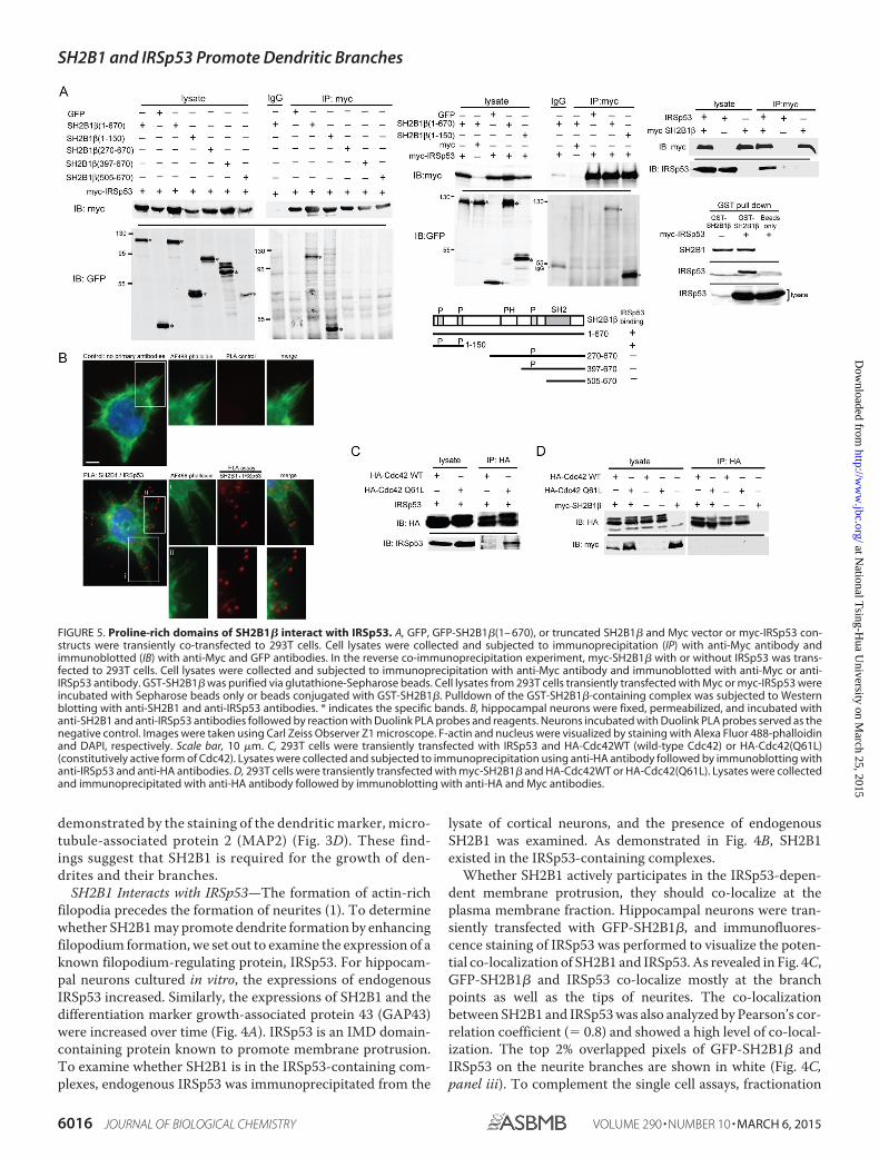
FIGURE 5. Proline-rich domains of SH2B1� interact with IRSp53. A, GFP, GFP-SH2B1�(1– 670), or truncated SH2B1� and Myc vector or myc-IRSp53 con-structs were transiently co-transfected to 293T cells. Cell lysates were collected and subjected to immunoprecipitation (IP) with anti-Myc antibody andimmunoblotted (IB) with anti-Myc and GFP antibodies. In the reverse co-immunoprecipitation experiment, myc-SH2B1� with or without IRSp53 was trans-fected to 293T cells. Cell lysates were collected and subjected to immunoprecipitation with anti-Myc antibody and immunoblotted with anti-Myc or anti-IRSp53 antibody. GST-SH2B1� was purified via glutathione-Sepharose beads. Cell lysates from 293T cells transiently transfected with Myc or myc-IRSp53 wereincubated with Sepharose beads only or beads conjugated with GST-SH2B1�. Pulldown of the GST-SH2B1�-containing complex was subjected to Westernblotting with anti-SH2B1 and anti-IRSp53 antibodies. * indicates the specific bands. B, hippocampal neurons were fixed, permeabilized, and incubated withanti-SH2B1 and anti-IRSp53 antibodies followed by reaction with Duolink PLA probes and reagents. Neurons incubated with Duolink PLA probes served as thenegative control. Images were taken using Carl Zeiss Observer Z1 microscope. F-actin and nucleus were visualized by staining with Alexa Fluor 488-phalloidinand DAPI, respectively. Scale bar, 10 �m. C, 293T cells were transiently transfected with IRSp53 and HA-Cdc42WT (wild-type Cdc42) or HA-Cdc42(Q61L)(constitutively active form of Cdc42). Lysates were collected and subjected to immunoprecipitation using anti-HA antibody followed by immunoblotting withanti-IRSp53 and anti-HA antibodies. D, 293T cells were transiently transfected with myc-SH2B1� and HA-Cdc42WT or HA-Cdc42(Q61L). Lysates were collectedand immunoprecipitated with anti-HA antibody followed by immunoblotting with anti-HA and Myc antibodies.
SH2B1 and IRSp53 Promote Dendritic Branches
6016 JOURNAL OF BIOLOGICAL CHEMISTRY VOLUME 290 • NUMBER 10 • MARCH 6, 2015
at National T
sing-Hua U
niversity on March 25, 2015
http://ww
w.jbc.org/
Dow
nloaded from
experiments were performed to examine the subcellular distri-bution of endogenous SH2B1 and IRSp53 in the plasma mem-brane fraction. As the expression of the endogenous SH2B1 andIRSp53 increased during the formation of the neurite networkof hippocampal neurons (Fig. 4A), SH2B1 and IRSp53 increased2-fold at the plasma membrane fraction on DIV 7 and DIV 10(Fig. 4D, right panel). The initiation and formation of the pro-spective neurites require the assembly of G-actin to F-actin. Todelineate whether SH2B1-IRSp53 complexes may reside with
polymerized F-actin, cell lysates of hippocampal neurons fromDIV 2 to 6 were collected for fractionation to separate deter-gent-soluble and -insoluble fractions. The endogenous SH2B1and IRSp53 were obviously in the detergent-insoluble fractioncontaining polymerized F-actin on DIV 4 and DIV 6 comparedwith DIV 2 (Fig. 4E).
The fact that SH2B1� and IRSp53 interact and co-localize,we next examined which region(s) of SH2B1� is required fortheir interaction. To this end, SH2B1 or various truncation
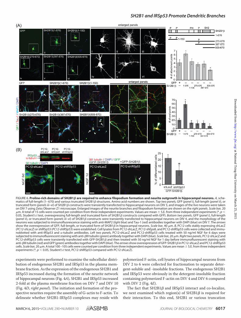
FIGURE 6. Proline-rich domains of SH2B1� are required to enhance filopodium formation and neurite outgrowth in hippocampal neurons. A, sche-matics of full-length (1– 670) and various truncated SH2B1� structures. Amino acid numbers are shown. Top two panels, GFP (panel i), full-length (panel ii), ortruncated form (panels iii–vi) of SH2B1� constructs were transiently transfected to hippocampal neurons on DIV 3, and images of the live neurons were takenon DIV 7 using Zeiss Observer Z1 microscope. Enlarged images of the neurite branches and filopodium formation are shown on the right panels. Scale bar, 20�m. A total of 15 cells were counted per condition from three independent experiments. Values are mean � S.E. from three independent experiments (*, p �0.05, Student’s t test, overexpressing full-length and truncated form of SH2B1� constructs compared with GFP). Bottom two panels, GFP (panel i), full-length(panel ii), or truncated form (panels iii–vi) of SH2B1� constructs were transiently transfected to hippocampal neurons on DIV 4, and the morphology of theneurons was subjected to immunofluorescence staining with anti-MAP2 (light blue) and Tau-1 (red) antibodies together with DAPI (blue) on DIV 7. The arrowsshow the overexpression of GFP, full-length, or truncated form of SH2B1� in hippocampal neurons. Scale bar, 40 �m. B, PC12 cells stably expressing shLacZ(PC12-shLacZ) or shIRSp53 (PC12-shIRSp53) were established. Cell lysates from PC12-shLacZ, PC12-shEps8, and PC12-shIRSp53 cells were collected and immu-noblotted with anti-IRSp53 and �-tubulin antibodies. Left two panels, PC12-shLacZ and PC12-shIRSp53 cells treated with 50 ng/ml NGF for 6 days weresubjected to immunofluorescent staining with anti-�III tubulin (green) antibody together with DAPI (blue). Scale bar, 20 �m. Right two panels, PC12-shLacZ andPC12-shIRSp53 cells were transiently transfected with GFP-SH2B1� and then treated with 50 ng/ml NGF for 1 day before immunofluorescent staining withanti-�III tubulin (red) and GFP (green) antibodies together with DAPI (blue). The arrows show overexpression of GFP-SH2B1� in PC12-shLacZ and PC12-shIRSp53cells. Scale bar, 20 �m. A total 100 –105 cells were counted per condition from three independent experiments. Values are mean � S.E. from three independentexperiments (*, p � 0.05, Student’s t test, PC12-shIRSp53 compared with PC12-shLacZ).
SH2B1 and IRSp53 Promote Dendritic Branches
MARCH 6, 2015 • VOLUME 290 • NUMBER 10 JOURNAL OF BIOLOGICAL CHEMISTRY 6017
at National T
sing-Hua U
niversity on March 25, 2015
http://ww
w.jbc.org/
Dow
nloaded from

SH2B1 and IRSp53 Promote Dendritic Branches
6018 JOURNAL OF BIOLOGICAL CHEMISTRY VOLUME 290 • NUMBER 10 • MARCH 6, 2015
at National T
sing-Hua U
niversity on March 25, 2015
http://ww
w.jbc.org/
Dow
nloaded from

mutants of SH2B1�, including SH2B1�(1–150), SH2B1�(270 –670), SH2B1�(397– 670), and SH2B1�(505– 670), were used totransfect 293T cells together with myc-IRSp53. IRSp53 wasimmunoprecipitated, and the presence of SH2B1� or itsmutants was examined. As in Fig. 5A, SH2B1� and SH2B1�(1–150) interacted with IRSp53. SH2B1�(270–670), SH2B1�-(397– 670), and SH2B1�(505– 670) did not. This result suggeststhat SH2B1� likely interacts with IRSp53 through its N-termi-nal proline-rich domains. Reverse co-immunoprecipitationwas also performed through immunoprecipitating myc-SH2B1�and re-probed with anti-IRSp53. In vitro GST-SH2B1� pull-down assay also detected IRSp53 in the same complex (Fig. 5A,right panels). To confirm the in vitro interaction results,Duolink in situ PLA assays were used to examine the in vivoassociation between endogenous SH2B1� and IRSp53 in hip-pocampal neurons. Hippocampal neurons were incubated withanti-SH2B1 and anti-IRSp53 antibodies followed by reactionwith in situ PLA assay probes and reagents. The interactionbetween SH2B1 and IRSp53 was detected by red fluorescentpuncta. In addition, actin structure was visualized by AlexaFluor 488-phalloidin staining. As shown Fig. 5B, the SH2B1-IRSp53 complexes existed at the plasma membrane, actin, andfilopodium tips. During the elongation of actin filaments,IRSp53 recruits VASP to the site of action, and this recruitmentis regulated by Cdc42 (29, 30). To examine whether IRSp53interacts with Cdc42, 293T cells were transfected with IRSp53together with HA-Cdc42 or the constitutive active form ofCdc42, HA-Cdc42(Q61L). When immunoprecipitating withanti-HA antibody, IRSp53 was in the same complex withHA-Cdc42(Q61L) (Fig. 5C). This result raised the possibilitythat SH2B1 may interact with Cdc42 or VASP instead ofdirectly with IRSp53. Nonetheless, SH2B1� does not interactwith VASP (31). Thus, we examined the possible interaction ofSH2B1� and Cdc42. As shown in Fig. 5D, SH2B1� did notinteract with either wild-type Cdc42 or the constitutive activeform, Cdc42(Q61L).
Proline-rich Domains of SH2B1� Are Required to EnhanceFilopodium Formation and Neurite Outgrowth in HippocampalNeurons—To determine which region(s) of SH2B1� is requiredfor enhancing filopodium formation and neurite outgrowth,various truncation mutants of GFP-SH2B1� were transfectedto hippocampal neurons, and the numbers of attachmentpoints and end points were quantified. As shown in Fig. 6A,overexpressing full-length GFP-SH2B1� significantly enhancedthe numbers of attachment points and end points in hippocam-
pal neurons compared with control GFP cells. SH2B1� trunca-tion mutants cannot increase the attachment points. In con-trast, overexpressing these mutants significantly reduced thenumber of end points of hippocampal neurons compared withGFP-expressing cells. It is possible that SH2B1�(1–150) titratesout the limiting amount of endogenous IRSp53, whereasSH2B1�(270 – 670) and SH2B1�(397– 670) may bind to andsequester other filopodia regulating proteins. Overexpressionof SH2B1�(505– 670), lacking all three N- and C-terminal pro-line-rich domains, inhibited both attachment and end points.This finding could result from the SH2 domain only mutant,SH2B1�(505– 670), sequestering the neurotrophin receptorsrequired for neurite outgrowth. Neuronal markers of dendritesand axons were also determined. Fixed neurons were subjectedto immunofluorescence staining with anti-MAP2 (dendriticmarker), Tau-1 (axonal marker), and DAPI. Overexpressingtruncated mutants of SH2B1� significantly reduced the den-dritic or axonal filopodia and neurite branching compared withoverexpressing full-length GFP-SH2B1� cells. Interestingly,overexpressing GFP-SH2B1� lacking both N- and C-terminalproline-rich domains strongly blocked the filopodium forma-tion and neurite outgrowth. These results suggest that the N-and C-terminal proline-rich domains of SH2B1� are requiredfor enhancing the formation of filopodia and neurite branchingduring neural development. To determine whether SH2B1�-mediated neurite outgrowth depends on IRSp53, the stable cellline PC12-shLacZ and PC12-shIRSp53 were established. Asshown in the immunoblot, the protein level of IRSp53 was sig-nificantly reduced (Fig. 6B). Neurite outgrowth was inhibited inPC12-shIRSp53 cells and cannot be rescued by putting backGFP-SH2B1�. These results suggest that IRSp53 is requiredfor SH2B1�-enhanced filopodium formation and neuriteoutgrowth.
N-terminal Proline-rich Domains of SH2B1� Enhance IRSp53-induced Neurite Outgrowth—To examine whether interactionbetween SH2B1 and IRSp53 is required to enhance IRSp53-induced neurite outgrowth, hippocampal neurons were tran-siently transfected with myc-IRSp53 together with GFP,GFP-SH2B1�, GFP-SH2B1�(1–150), GFP-SH2B1�(1–260),GFP-SH2B1�(270 – 670), or GFP-SH2B1�(397– 670). Themorphology of neurons was examined, and the numbers ofattachment points as well as end points were quantified. Theco-expression of SH2B1 and IRSp53 is shown in Fig. 7A. Over-expressing SH2B1� and IRSp53 increased both attachment(1.7-fold) and end (2.5-fold) points, whereas overexpression of
FIGURE 7. SH2B1� and IRSp53 synergistically regulate the formation of dendritic and axonal filopodia during neurite outgrowth. A, GFP (panel i),full-length (panel ii) or truncated form (panels iii–vi) of SH2B1� constructs were transiently co-transfected with myc-IRSp53 to hippocampal neurons on DIV 4,and the images of neuronal morphology were taken on DIV 7. Neurons were subjected to immunofluorescent staining with anti-Myc (red) and GFP (green).Enlarged images of the neurite branches and filopodium formation for GFP channel are shown in the right panels. Scale bar, 40 �m. A total of 15 cells werecounted per condition from three independent experiments. Values are mean � S.E. from three independent experiments (*, p � 0.05, Student’s t test). PC12cells stably expressing GFP or GFP-SH2B1�(1–150) were immunoprecipitated using anti-IgG or anti-GFP antibodies. Immunoprecipitated complexes wereresolved and immunoblotted using antibodies against anti-GFP and anti-SH2B1 antibodies. B, GFP (panel i), full-length (panel ii), or the truncated form (panelsiii–vi) of SH2B1� constructs were transiently co-transfected with myc-IRSp53 to hippocampal neurons on DIV 4, and the morphology of the neurons wasvisualized on DIV 7. Enlarged images of the dendritic filopodia and branches are shown in the right panels. Neurons were subjected to immunofluorescencestaining with anti-MAP2 (Cy5) and GFP (green) antibodies and F-actin (red). The end points of MAP2� or MAP2-/F-actin� signal were quantified to analyze thefilopodial and dendritic branches. MAP2 was highlighted in light blue for observation. C, GFP (panel i), full-length (panel ii), or truncated from (panels iii–vi) ofSH2B1� constructs were transiently co-transfected with myc-IRSp53 to hippocampal neurons on DIV 4, and the morphology of the neurons was visualized onDIV 7. Enlarged images of the axonal filopodia are shown in the right panels. Neurons were subjected with immunofluorescence staining with anti-GFP (green)and Tau-1 (red) antibodies together with DAPI (blue). Scale bar, 40 �m. Panel vii, axonal filopodia were quantified per 100 �m. Total 12 cells were counted percondition from three independent experiments. Values are mean � S.E. from three independent experiments (*, p � 0.05, Student’s t test).
SH2B1 and IRSp53 Promote Dendritic Branches
MARCH 6, 2015 • VOLUME 290 • NUMBER 10 JOURNAL OF BIOLOGICAL CHEMISTRY 6019
at National T
sing-Hua U
niversity on March 25, 2015
http://ww
w.jbc.org/
Dow
nloaded from
IRSp53 together with SH2B1�(1–150) or SH2B1�(1–260)increased attachment points 1.4-fold and end points 2-foldcompared with IRSp53 and GFP-expressing neurons. Overex-pression of IRSp53 and SH2B1�(270 – 670) or SH2B1�(397–670), however, did not enhance attachment or end points(Fig. 7A). The fact that overexpressing SH2B1�(1–150) orSH2B1�(1–260) together with IRSp53 did not exhibit domi-nant negative or loss-of-function phenotype was likely due tothe interaction of SH2B1�(1–150) and endogenous SH2B1(Fig. 7A, bottom right panel). Both SH2B1�(1–150) andSH2B1�(1–260) contain the dimerization domain and canbring together full-length SH2B1 to stimulate IRSp53-inducedfilopodium formation and neurite outgrowth.
To investigate whether the increased dendritic branches bySH2B1� and IRSp53 result from increased actin-rich filopodia,we quantified the relative actin-positive and MAP2-negativeend points, representative of filopodia/immature dendrites.Similar to the results from Fig. 7A, the relative numbers of filo-podia and dendritic branches were increased in neurons over-expressing IRSp53 together with SH2B1�, SH2B1�(1–150), orSH2B1�(1–260). Overexpression of IRSp53 together withSH2B1�(270 – 670) or SH2B1�(397– 670) did not increase den-dritic branches compared with control neurons (Fig. 7B). Bystaining with Tau-1, the numbers of filopodia along the axonalso increased in neurons overexpressing IRSp53 plusSH2B1�, SH2B1�(1–150), or SH2B1�(1–260) but not inneurons overexpressing IRSp53 plus SH2B1�(270 – 670) orSH2B1�(397– 670) (Fig. 7C). These results point to theimportance of the N-terminal proline-rich domains ofSH2B1� in promoting IRSp53-dependent filopodium for-mation and neurite outgrowth.
DISCUSSION
Filopodia are generally known to sense environmental cuesand to regulate tissue morphogenesis and regeneration. How-ever, exactly how filopodia are initiated at a specific localeremains unclear. IRSp53 has been well documented in promot-ing membrane protrusion and filopodium formation. In thisstudy, we demonstrate that SH2B1 interacts with IRSp53 andpromotes neurite outgrowth and neuronal branching. Based onthe known interaction between SH2B1 and neurotrophinreceptors at the cell surface during neuronal differentiation (18,19, 23–25), these results raise the possibility that SH2B1-IRSp53 complexes specify locations for filopodium formationthrough SH2B1-mediated signaling events. In line with thispossibility, overexpression of SH2 domain only mutant,SH2B1�(505– 670), inhibits neurite outgrowth (Fig. 6A) poten-tially through sequestering neurotrophin receptors.
The process from membrane protrusion (actin-free) to theformation filopodia (actin-rich) is very dynamic (32). Stabiliz-ing the structure of filopodia is essential for neurite initiation.The fact that overexpression of SH2B1� and IRSp53 promotesthe formation of actin-positive (filopodia) and MAP2-negative(immature dendrites) end points suggests that SH2B1� may berecruited or targeted to the plasma membrane to participate inIRSp53-induced membrane protrusion process. Amino acids150 –260 of SH2B1� contain membrane targeting and actin-binding regions (33, 34). Nonetheless, SH2B1�(1–150) and
SH2B1�(1–260) showed a similar effect on dendritic andaxonal branches (Fig. 7, B and C). Thus, it is likely that SH2B1is recruited to the plasma membrane by mechanisms other thanusing its own membrane targeting region. We show in thisstudy that SH2B1�(1–150), containing two proline-richregions as well as the dimerization domain, brings together theendogenous SH2B1 to action (Fig. 7A).
During actin elongation, IRSp53 is known to recruit VASP tothe site of action, and this recruitment is regulated by Cdc42(29, 30). Nonetheless, SH2B1� does not interact with eitherVASP (31) or Cdc42 (Fig. 5D). An IRSp53-interacting protein isEps8 (epidermal growth factor receptor kinase substrate 8).Eps8 can cap barbed ends of actin filaments when binding toAbi-1, whereas the association of Eps8 with IRSp53 inducesfilopodium formation by cross-linking actin filaments (30, 35,36). These findings suggest that Eps8 can induce or inhibit thefilopodium formation depending on its binding proteins.Although published reports suggest the involvement of Eps8mainly in spine formation, it would be of interest to investigatewhether Eps8 is in the complex of SH2B1-IRSp53 to promotefilopodium formation and thus spine formation.
Acknowledgments—We thank Dr. Eric Huang from the NationalChiao Tung University in Taiwan and Dr. H. Benjamin Peng fromHong Kong University of Science and Technology for the insightfuldiscussion concerning this project. We are also grateful for the techni-cal advice on neural culture from Dr. Yu-Chia Jenny Chou at theNational Yang-Ming University and Dr. Yen-Chung Chang at theNational Tsing Hua University, Taiwan.
REFERENCES1. Dent, E. W., Kwiatkowski, A. V., Mebane, L. M., Philippar, U., Barzik, M.,
Rubinson, D. A., Gupton, S., Van Veen, J. E., Furman, C., Zhang, J., Alberts,A. S., Mori, S., and Gertler, F. B. (2007) Filopodia are required for corticalneurite initiation. Nat. Cell Biol. 9, 1347–1359
2. Kwiatkowski, A. V., Rubinson, D. A., Dent, E. W., Edward van Veen, J.,Leslie, J. D., Zhang, J., Mebane, L. M., Philippar, U., Pinheiro, E. M., Burds,A. A., Bronson, R. T., Mori, S., Fassler, R., and Gertler, F. B. (2007) Ena/VASP is required for neuritogenesis in the developing cortex. Neuron 56,441– 455
3. Ahmed, S., Goh, W. I., and Bu, W. (2010) I-BAR domains, IRSp53, andfilopodium formation. Semin. Cell Dev. Biol. 21, 350 –356
4. Schirenbeck, A., Arasada, R., Bretschneider, T., Schleicher, M., and Faix, J.(2005) Formins and VASPs may co-operate in the formation of filopodia.Biochem. Soc. Trans. 33, 1256 –1259
5. Vignjevic, D., Kojima, S., Aratyn, Y., Danciu, O., Svitkina, T., and Borisy,G. G. (2006) Role of fascin in filopodial protrusion. J. Cell Biol. 174,863– 875
6. Mattila, P. K., Pykalainen, A., Saarikangas, J., Paavilainen, V. O., Vihinen,H., Jokitalo, E., and Lappalainen, P. (2007) Missing-in-metastasis andIRSp53 deform PI(4,5)P2-rich membranes by an inverse BAR domain-likemechanism. J. Cell Biol. 176, 953–964
7. Scita, G., Confalonieri, S., Lappalainen, P., and Suetsugu, S. (2008) IRSp53:crossing the road of membrane and actin dynamics in the formation ofmembrane protrusions. Trends Cell Biol. 18, 52– 60
8. Zhao, H., Pykalainen, A., and Lappalainen, P. (2011) I-BAR domain pro-teins: linking actin and plasma membrane dynamics. Curr. Opin. Cell Biol.23, 14 –21
9. Futo, K., Bodis, E., Machesky, L. M., Nyitrai, M., and Visegrady, B. (2013)Membrane binding properties of IRSp53-missing in metastasis domain(IMD) protein. Biochim. Biophys. Acta 1831, 1651–1655
10. Yokouchi, M., Suzuki, R., Masuhara, M., Komiya, S., Inoue, A., and Yo-
SH2B1 and IRSp53 Promote Dendritic Branches
6020 JOURNAL OF BIOLOGICAL CHEMISTRY VOLUME 290 • NUMBER 10 • MARCH 6, 2015
at National T
sing-Hua U
niversity on March 25, 2015
http://ww
w.jbc.org/
Dow
nloaded from
shimura, A. (1997) Cloning and characterization of APS, an adaptor mol-ecule containing PH and SH2 domains that is tyrosine phosphorylatedupon B-cell receptor stimulation. Oncogene 15, 7–15
11. Yousaf, N., Deng, Y., Kang, Y., and Riedel, H. (2001) Four PSM/SH2-Balternative splice variants and their differential roles in mitogenesis. J. Biol.Chem. 276, 40940 – 40948
12. Iseki, M., Takaki, S., and Takatsu, K. (2000) Molecular cloning of themouse APS as a member of the Lnk family adaptor proteins. Biochem.Biophys. Res. Commun. 272, 45–54
13. Huang, X., Li, Y., Tanaka, K., Moore, K. G., and Hayashi, J. I. (1995) Clon-ing and characterization of Lnk, a signal transduction protein that linksT-cell receptor activation signal to phospholipase C�1, Grb2, and phos-phatidylinositol 3-kinase. Proc. Natl. Acad. Sci. U.S.A. 92, 11618 –11622
14. Nelms, K., O’Neill, T. J., Li, S., Hubbard, S. R., Gustafson, T. A., and Paul,W. E. (1999) Alternative splicing, gene localization, and binding of SH2-Bto the insulin receptor kinase domain. Mamm. Genome 10, 1160 –1167
15. Chen, L., and Carter-Su, C. (2004) Adaptor protein SH2-B� undergoesnucleocytoplasmic shuttling: implications for nerve growth factor induc-tion of neuronal differentiation. Mol. Cell. Biol. 24, 3633–3647
16. Chen, L., Maures, T. J., Jin, H., Huo, J. S., Rabbani, S. A., Schwartz, J., andCarter-Su, C. (2008) SH2B1� (SH2-B�) enhances expression of a subset ofnerve growth factor-regulated genes important for neuronal differentia-tion including genes encoding urokinase plasminogen activator receptorand matrix metalloproteinase 3/10. Mol. Endocrinol. 22, 454 – 476
17. Maures, T. J., Chen, L., and Carter-Su, C. (2009) Nucleocytoplasmic shut-tling of the adaptor protein SH2B1� (SH2-B�) is required for nervegrowth factor (NGF)-dependent neurite outgrowth and enhancement ofexpression of a subset of NGF-responsive genes. Mol. Endocrinol. 23,1077–1091
18. Lin, W. F., Chen, C. J., Chang, Y. J., Chen, S. L., Chiu, I. M., and Chen, L.(2009) SH2B1� enhances fibroblast growth factor 1 (FGF1)-induced neu-rite outgrowth through MEK-ERK1/2-STAT3-Egr1 pathway. Cell. Signal.21, 1060 –1072
19. Chang, Y. J., Chen, K. W., Chen, C. J., Lin, M. H., Sun, Y. J., Lee, J. L., Chiu,I. M., and Chen, L. (2014) SH2B1� interacts with STAT3 and enhancesfibroblast growth factor 1-induced gene expression during neuronal dif-ferentiation. Mol. Cell. Biol. 34, 1003–1019
20. Doche, M. E., Bochukova, E. G., Su, H. W., Pearce, L. R., Keogh, J. M.,Henning, E., Cline, J. M., Saeed, S., Dale, A., Cheetham, T., Barroso, I.,Argetsinger, L. S., O’Rahilly, S., Rui, L., Carter-Su, C., and Farooqi, I. S.(2012) Human SH2B1 mutations are associated with maladaptive behav-iors and obesity. J. Clin. Invest. 122, 4732– 4736
21. Volckmar, A. L., Bolze, F., Jarick, I., Knoll, N., Scherag, A., Reinehr, T., Illig,T., Grallert, H., Wichmann, H. E., Wiegand, S., Biebermann, H., Krude, H.,Fischer-Posovszky, P., Rief, W., Wabitsch, M., et al. (2012) Mutationscreen in the GWAS derived obesity gene SH2B1 including functionalanalyses of detected variants. BMC Med. Genomics 5, 65
22. Sandholt, C. H., Vestmar, M. A., Bille, D. S., Borglykke, A., Almind, K.,Hansen, L., Sandbæk, A., Lauritzen, T., Witte, D., Jørgensen, T., Pedersen,O., and Hansen, T. (2011) Studies of metabolic phenotypic correlates of 15
obesity associated gene variants. PLoS One 6, e2353123. Rui, L., and Carter-Su, C. (1999) Identification of SH2-b� as a potent
cytoplasmic activator of the tyrosine kinase Janus kinase 2. Proc. Natl.Acad. Sci. U.S.A. 96, 7172–7177
24. Qian, X., Riccio, A., Zhang, Y., and Ginty, D. D. (1998) Identification andcharacterization of novel substrates of Trk receptors in developing neu-rons. Neuron 21, 1017–1029
25. Shih, C. H., Chen, C. J., and Chen, L. (2013) New function of the adaptorprotein SH2B1 in brain-derived neurotrophic factor-induced neurite out-growth. PLoS One 8, e79619
26. Wang, T. C., Chiu, H., Chang, Y. J., Hsu, T. Y., Chiu, I. M., and Chen, L.(2011) The adaptor protein SH2B3 (Lnk) negatively regulates neurite out-growth of PC12 cells and cortical neurons. PLoS One 6, e26433
27. Rui, L., Mathews, L. S., Hotta, K., Gustafson, T. A., and Carter-Su, C.(1997) Identification of SH2-B� as a substrate of the tyrosine kinase JAK2involved in growth hormone signaling. Mol. Cell. Biol. 17, 6633– 6644
28. Ho, S. Y., Chao, C. Y., Huang, H. L., Chiu, T. W., Charoenkwan, P., andHwang, E. (2011) NeurphologyJ: an automatic neuronal morphologyquantification method and its application in pharmacological discovery.BMC Bioinformatics 12, 230
29. Bisi, S., Disanza, A., Malinverno, C., Frittoli, E., Palamidessi, A., and Scita,G. (2013) Membrane and actin dynamics interplay at lamellipodia leadingedge. Curr. Opin. Cell Biol. 25, 565–573
30. Disanza, A., Mantoani, S., Hertzog, M., Gerboth, S., Frittoli, E., Steffen, A.,Berhoerster, K., Kreienkamp, H. J., Milanesi, F., Di Fiore, P. P., Ciliberto,A., Stradal, T. E., and Scita, G. (2006) Regulation of cell shape by Cdc42 ismediated by the synergic actin-bundling activity of the Eps8-IRSp53 com-plex. Nat. Cell Biol. 8, 1337–1347
31. Diakonova, M., Helfer, E., Seveau, S., Swanson, J. A., Kocks, C., Rui, L.,Carlier, M. F., and Carter-Su, C. (2007) Adaptor protein SH2-B� stimu-lates actin-based motility of Listeria monocytogenes in a vasodilator-stim-ulated phosphoprotein (VASP)-dependent fashion. Infect. Immun. 75,3581–3593
32. Yang, C., Hoelzle, M., Disanza, A., Scita, G., and Svitkina, T. (2009) Coor-dination of membrane and actin cytoskeleton dynamics during filopodiaprotrusion. PLoS One 4, e5678
33. Maures, T. J., Su, H. W., Argetsinger, L. S., Grinstein, S., and Carter-Su, C.(2011) Phosphorylation controls a dual-function polybasic nuclear local-ization sequence in the adaptor protein SH2B1� to regulate its cellularfunction and distribution. J. Cell Sci. 124, 1542–1552
34. Rider, L., Tao, J., Snyder, S., Brinley, B., Lu, J., and Diakonova, M. (2009)Adaptor protein SH2B1� cross-links actin filaments and regulates actincytoskeleton. Mol. Endocrinol. 23, 1065–1076
35. Vaggi, F., Disanza, A., Milanesi, F., Di Fiore, P. P., Menna, E., Matteoli, M.,Gov, N. S., Scita, G., and Ciliberto, A. (2011) The Eps8/IRSp53/VASPnetwork differentially controls actin capping and bundling in filopodiaformation. PLoS Comput. Biol. 7, e1002088
36. Funato, Y., Terabayashi, T., Suenaga, N., Seiki, M., Takenawa, T., andMiki, H. (2004) IRSp53/Eps8 complex is important for positive regulationof Rac and cancer cell motility/invasiveness. Cancer Res. 64, 5237–5244
SH2B1 and IRSp53 Promote Dendritic Branches
MARCH 6, 2015 • VOLUME 290 • NUMBER 10 JOURNAL OF BIOLOGICAL CHEMISTRY 6021
at National T
sing-Hua U
niversity on March 25, 2015
http://ww
w.jbc.org/
Dow
nloaded from