cell-based assays (4 mb) - sigma-aldrich
TRANSCRIPT
Volume 5, Number 8Biofiles
Assays and Reagents for Measuring Cytotoxicity, Proliferation, and Viability
Apoptosis
Autophagy
Reactive Oxygen Species
LOPAC® Library
Cell and Organelle Labeling
Cell Culture
Cell Culture Labware
Cell-Based Assays

Biofilescontents
Introduction 3
Assays and Reagents for Measuring Cytotoxicity, Proliferation,and Viability 5
Apoptosis 7
Autophagy 10
Reactive Oxygen Species 14
LOPAC® Library 17
Cell and Organelle Labeling 18
Cell Culture 23
Cell Culture Labware 26
Cover: Image titled "Cells" by Sebastian Kaulitzki. The image emphasizes the simplicity of life's most fundametal unit, the cell.
BiofilesonlineYour gateway to Biochemicals and Reagents
for Life Science Research
Biofiles Online allows you to:
Easily navigate the content of •the current Biofiles issue
Access any issue of Biofiles •Subscribe for email notifications •
of future eBiofiles issues
Register today for upcoming issues and eBiofiles announcements at
sigma.com/biofiles
Highlights from this issue: Cell-Based Assays
This is the first Biofiles to touch on the broad subject of cell-based assays. Within
this issue we have assembled a broad offering of reagents, kits and labware and
have added over 1,500 ECACC® cell lines to our U.S. portfolio. Given the growing
understanding and importance of autophagy, we have compiled a selection of products for
induction, inhibition and monitoring of autophagy in cells.
Coming Next Issue: Drug Metabolism
The next issue of Biofiles will feature our drug metabolism platform developed over
more than forty years. Of special interest will be cellular drug transport including
proteins and antibodies. "Next generation" cytochrome P450 enzymes are being
introduced along with new antibodies, additional Phase I oxidases and P450 modulators. Also included will be
the most extensive portfolio of enzymes and analytical tools for drug metabolite analysis in biological fluids.
Technical content: Linda Stephenson PhD. and Jeremy Benedick M.Sc.

Order sigma.com/order Technical service sigma.com/techinfo sigma.com/lifescience 3
The challenge of understanding the biology of living organisms, including disease processes, is in its incredible complexity. Scientists early on realized the value in utilizing life's most fundamental unit, the cell. Many aspects of living organisms are analyzed based on studies of living cells from cell signaling, proliferation, apoptosis, cellular genetics/morphology, neurobiology, cancer and more.
In both the pharma industry and academic research, the term cell-based assay is commonly used to refer to any assay based on some measurement of a living cell.
The pharmaceutical industry's need to efficiently commercialize drugs is a driving force in cell-based assay innovation. The industry is hoping to utilize cell-based assays to help reverse the increasing trend of costly late-stage drug failures. Having an Investigational New Drug (IND) candidate fail in phase III clinical trials can result in a loss of several hundred million dollars for that company. It is reported that with the current level of late-stage failures the return on investment for pharmaceutical companies could drop as low as 5%, which is an unacceptable level for most companies.
A lot of scientific resources are being poured into cell-based methodologies. The promise these technologies need to fulfill is daunting and the phrase “fail early” is an over-simplification. There is a need for rapid cell-based methodologies to predict with a high degree of accuracy which IND will be successful through to commercialization.
Drugs are failing for a number of reasons. While animal models are commonly used for toxicity and metabolism studies of INDs, they are both expensive and low-throughput
which usually limits their use to late stages of preclinical testing. Additionally, animal models may not always replicate human metabolism which results in late-stage failures due to unforeseen toxicity issues. Adverse drug reactions (ADRs) due to idiosyncratic differences in humans are another cause of late-stage drug failures. For example, Troglitazone, a type II diabetes drug, was taken off the market 3 years after release due to a small number of cases of drug-induced hepatitis. ADRs due to idiosyncratic differences tend to be rare events and even large clinical phase III trials could fail to detect the low rate (about 1 in 10,000) of idiosyncratic liver failure typical of this type of safety issue.
Many cell-based methodologies are being considered but so far no single technology or group of technologies has been reported to improve the late-stage failure rate. One important consideration is the cell type used for the metabolic and toxicity testing. Primary cells are often preferred over long-term established cell lines because they are thought to more closely replicate “real cells”. However, primary cells are variable and often can only be cultured for a few passages before they reach senescence or undergo undesirable phenotypic changes. Established cells have the benefit of phenotypic stability and ease of culture and are used extensively for cell-based assays. For example, cell lines like HepG2 and NIH/3T3 are ideal for testing mitochondrial toxicity because of their anaerobic metabolism. However, in regards to liver toxicity studies, established hepatocytic lines are less than ideal because they only weakly express cytochrome P450 enzymes.
Current research is investigating the use of induced pluripotent stem cells (IPSCs)
IntroductionDon FinleyMarket Segment Manager, Cell [email protected]

4
for creating better in vitro models for drug studies. For example, differentiated stem cells may be better models for studying drug-induced liver injury than primary hepatocytes as they maintain function longer than primary cells. ADRs due to idiosyncratic events are often due to patient-specific susceptibility factors such as genetic variations. One proposal to address this issue is to create large libraries of IPSCs that can be differentiated to hepatocytes as the basis for an automated assay.
Another limitation to traditional cell-based assays is a geometric issue. Most cell culture assays are performed in two dimensions while the real world of an in vivo cell is three-dimensional. 3-D cell culture can better create or mimic the extracellular environment (geometric shapes, scaffolding, forces placed on cells) so that cells will react to stimuli, toxins etc. more like they would in vivo.
A broad offering of products from Sigma® Life Science are used in cell-based assays that are suitable for both pharma and academic applications. We have broadened our U.S. offering of established cell lines and hybridomas to include over 1500 cell lines from the European Collection of Cell Cultures (ECACC®). ECACC cell lines are guaranteed to be authenticated and mycoplasma free.
We have the best selection of reagents and biologically active compounds in the industry. Our LOPAC® library makes accessing these compounds far easier than ever with thousands of pre-dispensed compounds on 96-well plates. In addition, we offer an exhaustive collection of Prestige Antibodies® for detection of a wide variety of cellular organelles and gene products that serve the basis for a broad variety of cell-based assay applications.
Sigma has applied the revolutionary CompoZr® Zinc Finger Nuclease technology to create an unparalleled range of genetically modified mammalian cell lines for use in areas such as basic research, target validation, drug discovery and drug development. With targeted and heritable gene deletions, integrations, or modifications our isogenic cell lines give you the tools to take your research to new heights. In the coming months you can expect a number of cell lines that will enable cell-based assay innovation never before possible.References:
(1) Tsaioun, K., Bottlaender, M., Mabondzo, A. ADDME - Avoiding Drug Development Mistakes Early: central nervous system drug discovery perspective. BMC Neurol. 9, S1–11 (2009).
(2) Gilbert, J., Henske, P., Singh, A. Rebuilding Big Pharma's Business Model. The Business & Medicine Report. 21 (2003).
(3) Shaw, R. Industrializing Stem Cell Production. BioPro-cess Int. 8, 10–15 (2010).
Nancy-520A non-toxic and ultrasensitive stain for DNA detection.
• Higher sensitivity compared to Ethidium bromide and SYBR Green I
• Fast and reliable staining (1 h)
• Lower mutagenicity than Ethidium bromide and SYBR® Green I according to Ames test
• For the detection of dsDNA on Agarose Gel Electrophoresis and for the determination of DNA in solutions
• Product Number 01494
biohighlight
sigma.com/nancy520
SYBR® Green is a registered trademark of Molecular Probes, Inc.

Order sigma.com/order Technical service sigma.com/techinfo sigma.com/lifescience 5
Assays to measure proliferation, viability and cytotoxicity are commonly used to monitor the response and health of cells in culture after treatment with various stimuli. The proper choice of an assay method depends on the number and type of cells used as well as the expected outcome. Assays for cell proliferation may monitor the number of cells over time, the number of cellular divisions, metabolic activity or DNA synthesis. Cell counting using viability dyes such as trypan blue or calcein-AM can provide both the rate of proliferation as well as the percentage of viable cells.
5(6)-Carboxyfluorescein diacetate N-succinimidyl ester (CFSE) is a popular choice for measuring the number of cellular divisions a population has undergone. Upon entering the cell, CFSE is cleaved by intracellular esterases to form the fluorescent compound and the succinimidyl ester group covalently reacts with primary amines on intracellular proteins. Upon division, the fluorescence intensity of each daughter cell is halved which allows for the simple detection of the number of cell divisions by flow cytometry.
Assays that measure metabolic activity are suitable for analyzing proliferation, viability, and cytotoxicity. The reduction of tetrazolium salts such as MTT and XTT to colored formazan compounds or the bioreduction of resazurin only occurs in metabolically active cells. Actively proliferating cells increase their metabolic activity while cells exposed to toxins will have decreased activity.
KitsIn Vitro Toxicology Assay Kit, MTT based
Conversion of MTT to a water-insoluble colored formazan derivative which is then solubilized in acidic isopropanol.
TOX1-1KT 1 kit
In Vitro Toxicology Assay Kit, XTT based
Conversion of XTT to a water-soluble colored formazan derivative.
TOX2-1KT 1 kit
In Vitro Toxicology Assay Kit, Neutral Red based
Neutral red is taken up by viable cells and stored in the lysosomes. The dye is extracted and the uptake is quantitated by spectroscopy.
TOX4-1KT 1 kit
In Vitro Toxicology Assay Kit,Sulforhodamine B based
Dye binds to cellular protein and is then solubilized in base.
TOX6-1KT 1 kit
In Vitro Toxicology Assay Kit, LacticDehydrogenase based
LDH reduces NAD+, which then converts a tetrazolium dye to a soluble, colored formazan derivative.
TOX7-1KT 1 kit
In Vitro Toxicology Assay Kit, Resazurin based
Bioreduction of the dye reduces the amount of its oxidized form (blue) and concomitantly increases the fluorescent intermediate (red).
TOX8-1KT 1 kit
Assays and Reagents for Measuring Cytotoxicity, Proliferation and Viability
Proliferation and Viability
Senescence Cells HistochemicalStaining Kit
Replicative senescence is a growth-arrest state associated with loss of division potential, changes in cell morphology, shape and physical appearance, and the pattern of gene expression in cells.
Histochemical staining of β-galactosidase activity is performed at pH 6.0. Under these conditions, β-galactosidase is a biomarker specific for senescent cells, but is not found in quiescent, immortal, or tumor cells.
Human Foreskin Fibroblasts passage 28 (senescent cells)
Human Foreskin Fibroblasts passage 5 (control)
Detection of senescent cells. Primary Human Foreskin Fibroblasts (HFF) at early and late passages (5 and 28 pas-sages, respectively) were stained using the Senescent Cell Staining Kit (Prod. No. CS0030). The HFF cells at passage 28 show a blue staining indicating that these cells are se-nescent, whereas at passage 5, senescent cells are absent.
CS0030-1KT 1 kit
6
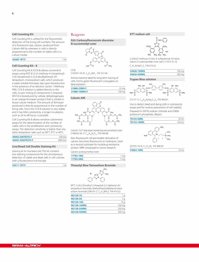
Cell Counting Kit
Cell Counting Kit is utilized for the fluorometric detection of the living cell numbers. The amount of a fluorescent dye, calcein, produced from Calcein-AM by esterases in cells is directly proportional to the number of viable cells in a culture media.
03285-1KT-F 1 kit
Cell Counting Kit - 8
Cell Counting Kit-8 (CCK-8) allows convenient assays using WST-8 (2-(2-methoxy-4-nitrophenyl)-3-(4-nitrophenyl)-5-(2,4-disulfophenyl)-2H-tetrazolium, monosodium salt), which produces a water-soluble formazan dye upon bioreduction in the presence of an electron carrier, 1-Methoxy PMS. CCK-8 solution is added directly to the cells, no pre-mixing of components is required. WST-8 is bioreduced by cellular dehydrogenases to an orange formazan product that is soluble in tissue culture medium. The amount of formazan produced is directly proportional to the number of living cells. Since the CCK-8 solution is very stable and it has little cytotoxicity, a longer incubation, such as 24 to 48 hours, is possible.
Cell Counting Kit-8 allows sensitive colorimetric assays for the determination of the number of viable cells in the proliferation and cytotoxicity assays. The detection sensitivity is higher than any other tetrazolium salts such as MTT, XTT or MTS.
96992-500TESTS-F 500 test96992-3000TESTS-F 3000 test
Live/Dead Cell Double Staining Kit
Staining kit for live/dead cells The kit contains two staining components for the simultaneous detection of viable and dead cells in cell cultures with a fluorescence microscope
04511-1KT-F 1 kit
Reagents5(6)-Carboxy fluor escein di acetateN-succini midyl ester
O
OO O
O
OOH3C CH3
O
ON
O
O
CFSE
[150347-59-4] C29H19NO11 FW 557.46
Amine-reactive label for long term tracing of cells. Forms green fluorescent conjugates on deacetylation.21888-25MG-F 25 mg21888-100MG-F 100 mg
Calcein-AM
O
O
O
O CH3OH3C
O O
NOO
NO O
OOH3C O O CH3
OO O O
O
H3C
O
CH3
OO
Calcein O,O'-di acetate tetra kis(acetoxy methyl) ester
[148504-34-1] C46H46N2O23 FW 994.86
Non-fluorescent cell permeable derivative of calcein, becomes fluorescent on hydrolysis. Used as a neutral substrate for multidrug resistance protein, MRP; employed in tumor research.
Calcein acetoxy methyl ester 17783-1MG 1 mg17783-5MG 5 mg
Thia zolyl Blue Tetra zolium Bro mide
NN
NN
N
S
CH3
CH3Br
MTT; 3-(4,5-Dimethyl-2-thia zolyl)-2,5-diphenyl-2H-tetra zolium bro mide; Methyl thia zolyl diphenyl-tetra-zolium bro mide [298-93-1] C18H16BrN5S FW 414.32
M2128-1G 1 gM2128-5G 5 gM2128-10G 10 gM2128-100MG 100 mgM2128-250MG 250 mgM2128-500MG 500 mg
XTT sodium salt
NN N
NCH3O
NO2 SO3 SO3- NO2
OCH3
C NHO
Na+
2,3-Bis(2-methoxy-4-nitro-5-sulfo phenyl)-2H-tetra-zolium-5-carbox ani lide inner salt [111072-31-2]
C22H16N7NaO13S2 FW 673.52
X4626-100MG 100 mgX4626-500MG 500 mg
Trypan Blue solution
N NN NOHNH2 OH NH2
SO
OONaSNaO
O
OS ONaO
OSNaOO
O
H3C CH3
[72-57-1] C34H24N6Na4O14S4 FW 960.81 .
Use to detect dead and dying cells in cytotoxicity assays and for routine assessment of cell viability.Prepared in 0.81% sodium chloride and 0.06% potassium phosphate, dibasic.
T8154-20ML 20 mLT8154-100ML 100 mL
NH2N
NH2
N CH3
CH3
CH3I I
[25535-16-4] C27H34I2N4 FW 668.39
P4864-10ML 10 mL

Order sigma.com/order Technical service sigma.com/techinfo sigma.com/lifescience 7
Apoptosis is a highly regulated program of cell death that is critical for normal homeostasis, development, wound repair and the removal of pathogen-infected cells. Abnormalities in apoptosis contribute to pathologies in disorders as diverse as Amyotrophic Lateral Sclerosis (ALS), AIDS, cancer, and ischemia. The ability to circumvent the normal induction of apoptosis is often utilized by tumor cells and viruses as a method of increased survival to normal host defenses. Conversely, excessive levels of apoptosis underlie the depletion of T-helper CD4+ cells during HIV progression and neurons in Parkinson's and Alzheimer's disease.
The molecular mechanisms of apoptosis have been extensively characterized and involve multiple energy-dependent steps which culminate in DNA fragmentation, protein cleavage, alternations in membrane asymmetry, organelle degradation, and the breakdown of the cell into vesicles called apoptotic bodies. At least three major pathways are known to initiate apoptosis, the extrinsic (death receptor) pathway, the intrinsic (mitochondrial) pathway, and the perforin-granzyme pathway. However, the majority of apoptosis-initiating processes converge at the execution pathway initiated by caspase-3 cleavage.
The choice of a kit or reagent depends on multiple parameters including which pathway induces the apoptotic cascade, the type and number of cells used, and the desired method of analysis. Sigma offers a wide variety of kits and reagents for the study of and measurement of apoptosis including assays for caspase activation, DNA fragmentation, loss of mitochondrial
integrity, and the induction of membrane asymmetry. Our collection of products also includes the most complete selection of inhibitors and inducers of apoptosis in the industry.
Apoptosis Detecting Kits
Flow Cytometry Kit for Apoptosis
Br-dUTP is incorporated more readily into the DNA fragments than deoxyuridine triphosphate labeled with larger dyes such as FITC, biotin or digoxigenin. One of the biological characteristics that defines apoptosis is the degradation of genomic DNA into fragments of 180-200 bp, commonly called "DNA laddering". The fragmentation creates a large number of 3'-hydroxyl sites at the DNA breaks. This property is used in the APO-BRDU kit to identify apoptotic cells by labeling the 3'-hydroxyl sites with bromodeoxyuridine triphosphate (Br-dUTP). Br-dUTP is enzymatically attached to the 3-hydroxyl sites of double- or single-stranded DNA by terminal transferase (TdT). Non-apoptotic cells do not incorporate Br-dUTP due to the lack of available 3-hydroxyl sites.
APOBRDU-1KT 1 kit
Annexin V-Cy3 Apoptosis Detection Kit
Sigma's Annexin V-Cy3 kit allows detection of annexin V bound to apoptotic cells by fluorescence microscopy. The Annexin V-Cy3 kit uses the dye Cy3.18 as the fluorochrome conjugated with annexin V. By microscopy, Cy3.18 fluoresces more brightly than the FITC conjugate. The kit includes the non-fluorescent compound 6-carboxyfluorescein diacetate (6-CFDA), which enters the cell and is hydrolyzed by the esterases present in living cells to the fluorescent compound 6-carboxyfluorescein, indicating that the cells are viable. This combination allows the differentiation among early apoptotic cells (annexin V positive, 6-CFDA positive), necrotic cells (annexin V positive, 6-CFDA negative), and viable cells (annexin V negative, 6-CFDA positive).
Apoptosis
Apoptosis
Double staining of control cells and apoptotic cells with Annexin V-Cy3 Apoptosis Detection Kit. Apoptosis was induced in Jurkat cells by 1 μg/ml staurosporine for 2 hours. The cells were washed with PBS and suspended at a concentration of 0.5-1x10⁶ cells/ml. The cells were stained with annexin V-Cy3.18 (1 μg/ml) and carboxyfluorescein diacetate (100 μM) in binding buffer for 10 minutes at room temperature. After staining, the cells were washed with binding buffer and observed by fluorescence microscopy. Figure A: Non-induced control cells stained with CDFA. Figure B: Cells induced with staurosporine and stained with CDFA. Figure C: Non-induced control cells from Figure A after annexin V-Cy3.18 staining. Note the absence of apoptotic cells. Figure D: Cells induced with staurosporine from Figure B stained with annexin V-Cy3.18. Note that apoptotic cells are stained both with CDFA and annexin V-Cy3.18, while necrotic cells are stained with annexin V-Cy3, but not by CDFA.
APOAC-1KT 1 kit
Annexin V-FITC Apoptosis Detection Kit
Annexin V-FITC kit allows fluorescent detection of annexin V bound to apoptotic cells and quantitative determination by flow cytometry. The Annexin V-FITC kit uses annexin V conjugated with fluorescein isothiocyanate (FITC) to label phosphatidylserine sites on the membrane surface. The kit includes propidium iodide (PI) to label the cellular DNA in necrotic cells where the cell membrane has been totally compromised. This combination allows the differentiation among early apoptotic cells (annexin V positive, PI negative), necrotic cells (annexin V positive, PI positive), and viable cells (annexin V negative, PI negative).
APOAF-20TST 20 testAPOAF-50TST 50 test

8
Caspase 3 Assay Kit, Colorimetric
Based on the hydrolysis of acetyl-Asp-Glu-Val-Asp p-nitroanilide (Ac-DEVD-pNA) by caspase 3, resulting in the release of the p-nitroaniline (pNA) moiety.
Caspase-3 Activity inApoptotic Jurkat Cells
Caspase 3 activity in apoptotic Jurkat cells. Apoptosis was induced in Jurkat human cells with 1 μg/ml stauro-sporine for 0, 1, 2, and 3 hours. The cells were lysed in 1X lysis buffer at a ratio of 100 μl per 107 cells. The caspase 3 activity in the lysate was determined using the Caspase 3 Colorimetric Assay Kit (CASP-3-C). Ten μl of each lysate was tested both with and without caspase 3 inhibitor, in a total reaction volume of 100 μl using 96 well multiwell plates. The substrate (Ac-DEVD-pNA) concentration was 200 μM, and the inhibitor (Ac-DEVD-CHO) concentration was 0.05 μM. The assay was performed at pH 7.4 at 37 °C for 42 minutes.
CASP3C-1KT 1 kit
Caspase 3 Assay Kit, Fluorimetric
The Caspase 3 Fluorimetric Assay Kit is based on the hydrolysis of acetyl Asp-Glu-Val-Asp 7-amido-4-methylcoumarin (Ac-DEVD-AMC) by caspase 3, resulting in the release of the fluorescent 7-amino-4-methylcoumarin (AMC).
CASP3F-1KT 1 kit
Caspase 8 Assay Kit, Colorimetric
The Caspase 8 Colorimetric Assay Kit is based on the hydrolysis of the peptide substrate Acetyl-Ile-Glu-Thr-Asp-p-Nitroaniline (Ac-IETD-pNA) by caspase-8 that results in the release of a p-Nitroaniline (p-NA) moiety. The p-NA is read at 405 nm. The concentration of the p-NA released can be calculated from a calibration curve prepared from p-NA standards (included with the kit).
The kit detects caspase 8 activity in crude and purified preparations of caspase 8.
Complete kit contains:
purified caspase 8 for a positive control•caspase 8 inhibitor•p-NA fluorescent standard•
CASP8C-1KT 1 kit
Caspase 8 Assay Kit, Fluorimetric
The Caspase 8 Fluorimetric Assay Kit is based on the hydrolysis of the peptide substrate Acetyl-Ile-Glu-Thr-Asp-7-amino-4-methyl coumarin (Ac-IETD-AMC) by caspase-8 that results in the release of a 7-amino-4-methyl coumarin (AMC) moiety. The excitation and emission wavelengths of AMC are 360 nm and 460 nm, respectively. The concentration of the AMC released can be calculated from a calibration curve prepared from AMC standards (included with the kit).
The kit detects caspase 8 activity in crude and purified preparations of caspase 8.
Complete kit contains:
purified caspase 8 for a positive control•caspase 8 inhibitor•AMC fluorescent standard•
CASP8F-1KT 1 kit
Cathepsin D Assay Kit
Cathepsin D, a ubiquitous aspartic protease belonging to the A1 peptidase family, is found intracellularly in lysosomes. The enzyme has been associated with various biological processes such as: apoptotic events (e.g., the release of cytochrome c from mitochondria and the loss of the transmembrane potential (ΔΨ), aging, Alzheimer's disease, and breast cancer.
CS0800-1KT 1 kit
Reagents for Monitoring Mitochondrial Function
Mitochondria Staining Kit
In normal cells, the JC-1 dye concentrates in the mitochondrial matrix where it forms red fluorescent aggregates. Any event that dissipates the mitochondrial membrane potential (e.g. apoptosis) prevents the accumulation of the JC-1 dye in the mitochondria and thus, the dye is dispersed throughout the entire cell leading to a shift from red (JC-1-aggregates) to green fluorescence (JC-1 monomers). The fluorescence of the cells stained with this kit may be observed by fluorescence microscopy or measured by fluorimetric and flow cytometry analysis.
CS0390-1KT 1 kit
Citrate Synthase Assay Kit
Contains all the required reagents (including a positive control enzyme) for a fast and simple measurement of citrate synthase activity in a whole cell extract or in isolated mitochondria; also enables testing of the intactness of the mitochondrial inner membrane.
CS0720-1KT 1 kit
Cytochrome c Oxidase Assay Kit
soluble and membrane bound mitochondriaCytochrome c oxidase [EC 1.9.3.1] is located on the inner mitochondrial membrane dividing the mitochondrial matrix from the intermembrane space, and has traditionally been used as a marker for this membrane. It is also located in the cytoplasmic membrane of bacteria. Cytochrome c oxidase provides energy for the cell by coupling electron transport through the cytochrome chain with the process of oxidative phosphorylation.
CYTOCOX1-1KT 1 kit
JC-1
N
N
N
N
CH3 H3C
H3CCH3
Cl
Cl
Cl
ClI
CBIC2(3) [3520-43-2] C25H27Cl4IN4 FW 652.23
T4069-5MG 5 mg
Reagents for Monitoring Apoptosis
bis Benz imide H 33258
N N
NH
N
NH
N
OH
CH3
• 3HCl
• xH2O
HOE 33258; 2-[2-(4-Hydroxy phenyl)-6-benz-imidazoyl]-6-(1-methyl-4-piperazyl)benz imid-azole trihydrochloride; 2′-(4-Hydroxy phenyl)-5-(4-methyl-1-piperazinyl)-2,5′-bi(1H-benz imid-azole) trihydrochloride; BBIH; Hoechst 33258
[23491-45-4] C25H24N6O · 3HCl FW 533.88Membrane-permeable, fluorescent DNA stain with low cytotoxicity that intercalate in A-T regions of DNA.B1155-25MG 25 mgB1155-100MG 100 mg

Order sigma.com/order Technical service sigma.com/techinfo sigma.com/lifescience 9
7-Amino actinomycin D
ProD-Val Thr
OMeVal
SarPro
D-Val ThrOMeVal
Sar
O
N
CH3 CH3
O
NH2
CO C O
NH2
7-AAD [7240-37-1] C62H87N13O16 FW 1270.43Fluorescent DNA stainA9400-1MG 1 mgA9400-5MG 5 mg
Propidium Iodide Solution
NH2N
NH2
N CH3
CH3
CH3I I
[25535-16-4] C27H34I2N4 FW 668.39
P4864-10ML 10 mL
Annexin V Cy3.18 Conjugate fromhuman placenta
For detection of apoptotic cells by fluorescence microscopy.
A4963-10UG 10 μg
Annexin V FITC Conjugate fromhuman placenta
For detection of apoptotic cells by flow cytometry or fluorescence microscopy.
Package size based on protein content
A9210-10UG 10 μg
Product Name Effective Concentration Mechanism of Action Cat. No.
BI-6C9 2 micromolar BI-6C9 is a tBid inhibitor and antiapoptotic B0186
BAX Inhibiting Peptide V5 ~ 50–200 micromolar Interact with Bax in a Ku70 - competitive manner and prevent its conformational change and mitochondrial translocation
B1436
Bongkrekic acid solution 10–50 micromolar Bongkrekic acid is an inhibitory ligand of the mitochondrial adenine nucleotide translocase. It inhibits mitochondrial permeability transition pore opening.
B6179
MDL 28170 20 micromolar Selective inhibitor of calpain and cathepsin B M6690
NSCI A selective inhibitor of nonpeptide caspase 3 N1413
NS3694 50 micromolar Specifically prevents the active ~700-kDa apoptosome complex formation triggered by cytochrome c release, thus blocking apoptosome-mediated caspase activation and cell death (50 Μm completely blocks TNF-Α-induced death in MCF-casp3 cells).
N7787
Pifithrin-Μ 50 micromolar Pifithrin-Μ is an inhibitor of p53 binding and anti-apoptotic P0122
Pifithrin-Α 0.5–1 micromolar Pifithrin-Α is a reversible inhibitor of p53-mediated apoptosis P4359
S-15176 difumarate salt Inhibits mitochondrial permeability transition, prevents the early step in apoptosis by preventing collapse of the electrochemical gradient across the mitochondrial membrane
S5944
Apoptosis-Inhibiting Compounds
Apoptosis-Inducing CompoundsProduct Name Effective Concentration Mechanism of Action Cat. No.
2-Amino-N-quinolin-8-yl-benzenesulfonamide 10–60 micromolar Inhibits cell cycle at G2 phase A3105
17-(Allylamino)-17-demethoxygeldanamycin 0.010–10 micromolar Inhibits chaperone function of Hsp-90 A8476
Bendamustine hydrochloride hydrate Activates the DNA damage stress response B5437
(S)-(+)-Camptothecin 5–25 micrograms/mL C9911
Etoposide 25–100 micromolar Induces double-stranded DNA damage E1383
Staurosporine from Streptomyces sp. 0.01 to 10 micromolar Mechanism not clear but may be through Protein Kinase C inhibition.
S4400

10
Autophagy is an evolutionarily conserved degradation process that is required for multiple cellular roles including survival during starvation, the clearance of intracellular components, development, and immunity. Decreases in autophagy occur during aging and may be associated with many age-related pathologies such as dementia and cancer. The vital dyes Acridine Orange and Dansylcadaverine (MDC) are commonly used to study autophagy. Acridine Orange is a lysotropic dye that accumulates in acidic organelles in a pH-dependent manner. At neutral pHs, Acridine Orange is a hydrophobic green fluorescent molecule. Within acidic vesicles, Acridine Orange becomes protonated and trapped within the organelle. Protonated Acridine Orange forms aggregates that emit bright red fluorescence. MDC is another popular autofluorescent marker that preferentially accumulates in autophagic vacuoles. While Acridine Orange staining in lysosomes is primarily due to ion trapping, MDC accumulation in auotphagic
vacuoles is due to a combination of ion trapping and specific interactions with vacuole membrane lipids. In addition to these vital dyes, Sigma® Life Science also offers the largest selection of small molecule inhibitors and activators of autophagy and an ever expanding collection of autophagy-related antibodies.
Reagents For Analyzing Autophagy
Dansyl cada verine
SO
O HN
NH3C CH3
NH2
N-(Dimethyl amino naph tha lene sul fonyl)-1,5-pentane-
di amine; Mono dansyl cada verine; N-(5-Amino-pentyl)-5-di methyl amino naph tha lene-1-sulfo n amide
[10121-91-2] C17H25N3O2S FW 335.46
30432-100MG 100 mg30432-1G 1 g
Autophagy
Autophagy
▼ Acridine Orange
[65-61-2] C17H19N3 · HCl FW 301.81
N NCH3N
H3C
CH3CH3
• xH2O
• HCl
A rigorously purified zinc-free material suitable for use in flow cytometry.
318337-1G 1 g318337-5G 5 g
Acri dine Orange hydrochloride solution3,6-Bis(dimethyl amino)acri dine hydrochloride
A8097-10ML 10 mL
Acridine Orange ▲
Product Name Effective Concentration Mechanism of Action Cat. No.
3-Methyladenine 5 millimolar Inhibitor of type III Phosphatidylinositol 3-kinases M9281
E64d 1–200 micromolar Inhibits lysosomal enzymes, typically used with Pepstatin A and Leupeptin E8640
Leupeptin 1–100 micromolar Inhibits lysosomal enzymes, typically used with E64d and Pepstatin A L5793
Pepstatin A 1–10 micromolar Inhibits lysosomal enzymes, typically used with E64d and Leupeptin P5318
Bafilomycin A1 0.1–1 micromolar Inhibitor of vacuolar type H+-ATPase (V-ATPase) B1793
L-Asparagine 10 millimolar Blocks autophagic-lysosomal delivery A4159
LY-294,002 10–100 micromolar Phosphatidylinositol 3-kinase inhibitor L9908
Wortmannin 0.1-10 micromolar Phosphatidylinositol 3-kinase inhibitor W1628
Chloroquine diphosphate salt 5–40 micromolar Raises lysosomal pH leading to an inhbition of lysosomal autophagosome fusion C6628
Autophagy Inhibitors

Order sigma.com/order Technical service sigma.com/techinfo sigma.com/lifescience 11
Product Name Effective Concentration Mechanism of Action Cat. No.
Amiodarone Hydrochloride 10-50 micromolar Inhibitor of mTORC1 A8423
Niclosamide 1-10 micromolar Inhibitor of mTORC1 N3510
Rottlerin 0.3-3.0 micromolar Inhibitor of mTORC1 R5648
GF 109203X 0.02-4 micromolar PKC inhibitor G2911
STF-62247 0.5-20 micromolar Disrupts ER-Golgi trafficking S7448
Rapamycin 10-500 nanomolar Inhibitor of mammalian target of rapamycin (mTOR) R0395
Autophagy Activators
biocells
Bioascend.Achieve stellar results with
cell lines shaped for your research.
Get your hands on disease model, reporter orcustom-engineered cell lines, created using CompoZr® ZFN technology from Sigma® Life Science, and launch your research into extraordinary new heights.
wherebiobegins.com/biocells
Sigma, CompoZr and MISSION are registered trademarks of Sigma-Aldrich and its affiliate Sigma-Aldrich Biotechnology, L.P.
12
Product Name
Host
Form
Reacts With
Species Reactivity
Cat. No.
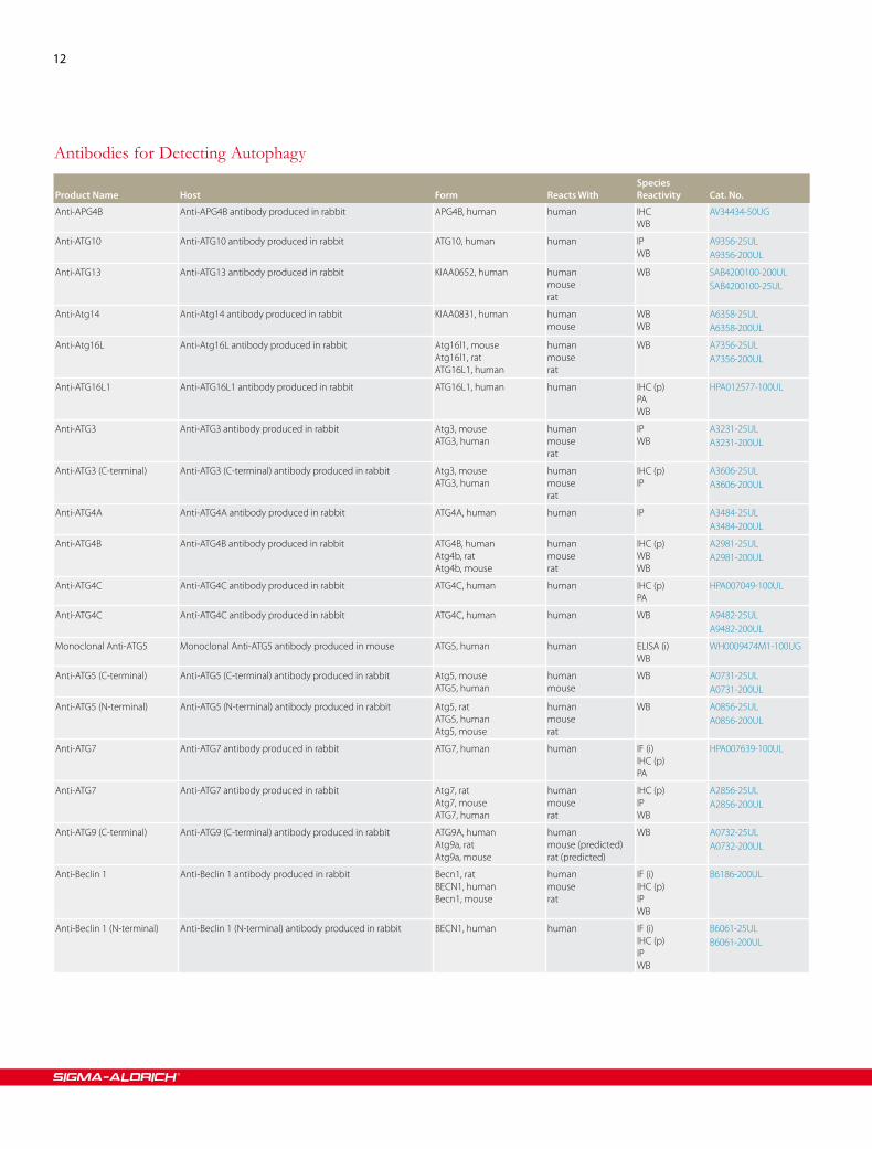
Anti-APG4B Anti-APG4B antibody produced in rabbit APG4B, human human IHCWB
AV34434-50UG
Anti-ATG10 Anti-ATG10 antibody produced in rabbit ATG10, human human IPWB
A9356-25ULA9356-200UL
Anti-ATG13 Anti-ATG13 antibody produced in rabbit KIAA0652, human humanmouserat
WB SAB4200100-200ULSAB4200100-25UL
Anti-Atg14 Anti-Atg14 antibody produced in rabbit KIAA0831, human humanmouse
WBWB
A6358-25ULA6358-200UL
Anti-Atg16L Anti-Atg16L antibody produced in rabbit Atg16l1, mouseAtg16l1, ratATG16L1, human
humanmouserat
WB A7356-25ULA7356-200UL
Anti-ATG16L1 Anti-ATG16L1 antibody produced in rabbit ATG16L1, human human IHC (p)PAWB
HPA012577-100UL
Anti-ATG3 Anti-ATG3 antibody produced in rabbit Atg3, mouseATG3, human
humanmouserat
IPWB
A3231-25ULA3231-200UL
Anti-ATG3 (C-terminal) Anti-ATG3 (C-terminal) antibody produced in rabbit Atg3, mouseATG3, human
humanmouserat
IHC (p)IP
A3606-25ULA3606-200UL
Anti-ATG4A Anti-ATG4A antibody produced in rabbit ATG4A, human human IP A3484-25ULA3484-200UL
Anti-ATG4B Anti-ATG4B antibody produced in rabbit ATG4B, humanAtg4b, ratAtg4b, mouse
humanmouserat
IHC (p)WBWB
A2981-25ULA2981-200UL
Anti-ATG4C Anti-ATG4C antibody produced in rabbit ATG4C, human human IHC (p)PA
HPA007049-100UL
Anti-ATG4C Anti-ATG4C antibody produced in rabbit ATG4C, human human WB A9482-25ULA9482-200UL
Monoclonal Anti-ATG5 Monoclonal Anti-ATG5 antibody produced in mouse ATG5, human human ELISA (i)WB
WH0009474M1-100UG
Anti-ATG5 (C-terminal) Anti-ATG5 (C-terminal) antibody produced in rabbit Atg5, mouseATG5, human
humanmouse
WB A0731-25ULA0731-200UL
Anti-ATG5 (N-terminal) Anti-ATG5 (N-terminal) antibody produced in rabbit Atg5, ratATG5, humanAtg5, mouse
humanmouserat
WB A0856-25ULA0856-200UL
Anti-ATG7 Anti-ATG7 antibody produced in rabbit ATG7, human human IF (i)IHC (p)PA
HPA007639-100UL
Anti-ATG7 Anti-ATG7 antibody produced in rabbit Atg7, ratAtg7, mouseATG7, human
humanmouserat
IHC (p)IPWB
A2856-25ULA2856-200UL
Anti-ATG9 (C-terminal) Anti-ATG9 (C-terminal) antibody produced in rabbit ATG9A, humanAtg9a, ratAtg9a, mouse
humanmouse (predicted)rat (predicted)
WB A0732-25ULA0732-200UL
Anti-Beclin 1 Anti-Beclin 1 antibody produced in rabbit Becn1, ratBECN1, humanBecn1, mouse
humanmouserat
IF (i)IHC (p)IPWB
B6186-200UL
Anti-Beclin 1 (N-terminal) Anti-Beclin 1 (N-terminal) antibody produced in rabbit BECN1, human human IF (i)IHC (p)IPWB
B6061-25ULB6061-200UL
Antibodies for Detecting Autophagy

Order sigma.com/order Technical service sigma.com/techinfo sigma.com/lifescience 13
Product Name
Host
Form
Reacts With
Species Reactivity
Cat. No.
Anti-LC3 Anti-LC3 antibody produced in rabbit Map1lc3b, mouseMAP1LC3B, humanMAP1LC3A, humanMap1lc3a, ratMap1lc3b, ratMap1lc3a, mouse
humanmouserat
IPWB
L8918-25ULL8918-200UL
Anti-LC3A Anti-LC3A antibody produced in rabbit MAP1LC3A, humanMap1lc3a, ratMap1lc3a, mouse
humanmouserat
IPWB
L8793-25ULL8793-200UL
Anti-LC3B Anti-LC3B antibody produced in rabbit Map1lc3b, ratMAP1LC3B, humanMap1lc3b, mouse
humanmouserat
IF (i)WB
L7543-25ULL7543-60ULL7543-200UL
Anti-MAP1LC3A Anti-MAP1LC3A antibody produced in rabbit MAP1LC3A, human human WB SAB2101425-50UG
Anti-MAP1LC3A Anti-MAP1LC3A antibody produced in rabbit MAP1LC3A, human human IHC (p)PA
HPA007649-100UL
Anti-p62/SQSTM1 Anti-p62/SQSTM1 antibody produced in rabbit Sqstm1, ratSQSTM1, humanSqstm1, mouse
humanmouserat
IPWBWB
P0068-25ULP0068-200UL
Anti-PIK3C3 (841-853) Anti-PIK3C3 (841-853) antibody produced in rabbit PIK3C3, human human WB K3141-200UL
Monoclonal Anti-ULK2 Monoclonal Anti-ULK2 antibody produced in mouse ULK2, human human ELISA (i)WB
WH0009706M1-100UG
Anti-UVRAG Anti-UVRAG antibody produced in rabbit UVRAG, human human IHC (p)PAWB
HPA016932-100UL
Anti-UVRAG Anti-UVRAG antibody produced in rabbit UVRAG, humanUvrag, mouse
humanmouse
WB U7508-25ULU7508-200UL
Monoclonal Anti-UVRAG Monoclonal Anti-UVRAG antibody produced in mouse Uvrag, mouseUVRAG, human
humanmouse
IPWB
SAB4200005-25ULSAB4200005-200UL
Anti-VPS34 Anti-VPS34 antibody produced in rabbit PIK3C3, humanPik3c3, mousePik3c3, rat
humanmouserat
IF (i)WB
V9764-25ULV9764-200UL
Anti-VPS4 Anti-VPS4 antibody produced in rabbit Vps4a, mouseVPS4A, humanVps4a, rat
humanmouserat
IPWB
SAB4200025-25ULSAB4200025-200UL
Anti-VPS4A Anti-VPS4A antibody produced in rabbit VPS4A, humanVps4a, mouseVps4a, rat
humanrat
IPWB
SAB4200022-25ULSAB4200022-200UL
Anti-VPS4B Anti-VPS4B antibody produced in rabbit VPS4B, human humanmouse
IPWB
SAB4200023-25ULSAB4200023-200UL
Anti-WIPI1 Anti-WIPI1 antibody produced in rabbit WIPI1, human human IHC (p)PAWB
HPA007493-100UL
Monoclonal Anti-WIPI1 Monoclonal Anti-WIPI1 antibody produced in mouse WIPI1, human humanmouserat
ELISA (i)WB
WH0055062M2-100UG
Antibodies For Detecting AutophagyImmunoblottingWhole extracts of human HEK-293T cells (Lane A) or HEK-293T cells pre-treated with tunicamycin for 48 h (Lane B) were separated on SDS-PAGE and blotted.
Upper panel: Anti-Actin (Cat. No. A3853). Lower panel: 0.5 μg/mL Rabbit Anti-LC3B (Cat. No. L7543). The antibody was developed with Goat Anti-Rabbit IgG, Peroxidase conjugate and a chemiluminescent substrate.
DMSO Tun
Actin
LC3I
LC3II
43 —
26 —
17 —
14
Oxidative Stress EnzymesName Description Cat No.
Catalase from bovine liver Activates the decomposition of hydrogen peroxide, Natural antioxidant used to study roles of reactive oxygen species in gene expression and apoptosis.
C100-50MGC100-500MGC100-1G
Cytochrome c Oxidase from bovine heart Cytochrome c oxidase is the principal terminal oxidase of high oxygen affinity in the aerobic metabolism of all animals, plants, yeasts and some bacteria.
C5499-250UG
Glutathione Peroxidase from bovine erythrocytes
- G6137-100UNG6137-200UNG6137-500UN
Myeloperoxidase from human leukocytes Catalyzes oxidations by hydrogen peroxide, including cytotoxic actions on bacteria, fungi, and tumor cells. Also catalyzes cross-linking of immune complexes and inactivates chemotactic factors.
M6908-5UN
Methionine Sulfoxide Reductase A, histidine-tagged human
MsrA reduces S-MetO and MsrB reduces R-MetO, which may prevent irreversible oxidative protein damage and thus extend an organism's life span. There is evidence showing a connection between MsrA and Alzheimer's disease.
M8698-100UG
Nitrate Reductase (cytochrome) from Escherichia coli
- N0519-.5UN
Peroxidase from horseradish - P6782-5MGP6782-10MGP6782-25MGP6782-50MGP6782-100MG
Peroxiredoxin 1 human Peroxiredoxins are a novel defined family of peroxidases of approximately 25 kDa that reduce H2O
2 and alkyl
hydroperoxides and use mainly the thioredoxin (Trx) system (Trx, thioredoxin reductase and NADPH) as electron donor. The peroxiredoxin family includes more than 30 proteins from organisms of all kingdoms. Peroxiredoxin I belongs to the Type I Peroxiredoxin family. This family consists of human natural killer cell enhancing factor (NKEFA), human proliferation associated gene (PAG), mouse macrophage stress induced protein (MSP23), mouse osteoblast specific factor (OSF-3), and rat heme-binding protein (HBP23). These proteins share about 95% homology.
P8986-250UG
Superoxide Dismutase from bovine erythrocytes
Catalyzes the dismutation of superoxide radicals to hydrogen peroxide and molecular oxygen. Plays a critical role in the defense of cells against the toxic effects of oxygen radicals. Competes with nitric oxide (NO) for superoxide anion (which reacts with NO to form peroxynitrite), thereby SOD promotes the activity of NO. SOD has also been shown to suppress apoptosis in cultured rat ovarian follicles, neural cell lines, and transgenic mice.
S7571-15KUS7571-30KUS7571-75KUS7571-300KU
Superoxide Dismutase from human erythrocytes
Catalyzes the dismutation of superoxide radicals to hydrogen peroxide and molecular oxygen. Plays a critical role in the defense of cells against the toxic effects of oxygen radicals. Competes with nitric oxide (NO) for superoxide anion (which reacts with NO to form peroxynitrite), thereby SOD promotes the activity of NO. SOD has also been shown to suppress apoptosis in cultured rat ovarian follicles, neural cell lines, and transgenic mice.
S9636-1KUS9636-3KUS9636-15KUS9636-30KU
Thioredoxin Reductase from Escherichia coli
Thioredoxin reductase (TrxR) is an NADPH-dependent oxidoreductase containing one FAD per subunit that reduces the active site disulfide in oxidised thioredoxin (Trx). The molecular weight of the isozymes from mammalian sources vary between 55-67 kDa as compared with 35 kDa in prokaryotes, plants or yeast. The substrate specificity of the mammalian enzyme is much broader than the prokaryotic enzyme reducing both mammalian and E. coli thioredoxins as well as well as non-disulfide substrates such selenite, lipoic acids, lipid hydroperoxides and hydrogen peroxide. FAD-containing enzyme involved in transfer of hydrogen from E. coli thioredoxin to other proteins.
T7915-250UG
Thioredoxin Reductase from rat liver Thioredoxin reductase (TrxR) is a NADPH-dependent oxidoreductase containing one FAD per subunit that reduces the active site disulfide in oxidized thioredoxin (Trx).
T9698-50UG
Reactive Oxygen Species
Reactive Oxygen Species

Order sigma.com/order Technical service sigma.com/techinfo sigma.com/lifescience 15
Kits For ROS AnalysisName Description Cat. No. Qty
Catalase Assay Kit, determination of catalase activity in tissues and cells
Catalase is a ubiquitous antioxidant enzyme which catalyses the decomposition of hydrogen peroxide (H
2O
2) to water and oxygen. Hydrogen peroxide is formed in the eukaryotic cell as a by-
product of various oxidases and superoxide dismutases. Hydrogen peroxide accumulation in cells causes oxidation of cellular targets such as DNA, proteins, and lipids leading to mutagenesis and cell death. Removal of the H
2O
2 from the cell by catalase provides protection against oxidative damage
to living cells and its role in oxidative stress related diseases has been widely studied.
CAT100-1KT 1 kit
Thioredoxin Reductase Assay Kit - CS0170-1KT 1 kit
Glutathione Assay Kit Reduced glutathione (GSH), a tripeptide (g-glutamyl-cysteinylglycine), is the major free thiol in most living cells and is involved in many biological processes such as detoxification of xenobiotics, removal of hydroperoxides, and maintenance of the oxidation state of protein sulfhydryls. It is the key antioxidant in animal tissues, Reduced glutathione (GSH), a tripeptide (g-glutamyl-cysteinylglycine), is the major free thiol in most living cells and is involved in detoxification of xenobiotics, removal of hydroperoxides, and maintenance of the oxidation state.
CS0260-1KT 1 kit
Antioxidant Assay Kit - CS0790-1KT 1 kit
Superoxide Anion Assay Kit The superoxide anion (O2
-) is a short-lived radical generated by the addition of an electron to oxygen. It is formed in response to environmental factors such as UV light, cigarette smoke, environmental pollutants, and γ-radiation, or by oxidases like xanthine oxidase or NADPH oxidase. Once formed, O
2-
attacks cellular components causing damage to lipids, proteins, and DNA. This can initiate numerous diseases, including cancer, atherosclerosis, rheumatoid arthritis, diabetes, liver damage, and central nervous system disorders.
CS1000-1KT 1 kit
Glutathione Assay Kit, Fluorimetric Reduced glutathione (GSH), a tripeptide (γ-glutamyl-cysteinylglycine), is the major free thiol in most living cells and is involved in many biological processes such as detoxification of xenobiotics, removal of hydroperoxides, and maintenance of the oxidation state of protein sulfhydryls. It is the key antioxidant in animal tissues. Glutathione is present inside cells primarily in the reduced form (90-95% of the total glutathione). The remainder is present in the oxidized form (glutathione disulfide, GSSG). Intracellular GSH status appears to be a sensitive indicator of the overall health of a cell and its ability to resist toxic challenge. High levels of GSH in the cell may indicate pathological changes., Reduced glutathione (GSH), a tripeptide (γ-glutamyl-cysteinylglycine), is the major free thiol in most living cells and is involved in many biological processes such as detoxification of xenobiotics, removal of hydroperoxides, and maintenance of the oxidation state of protein sulfhydryls. It is the key antioxidant in animal tissues.
CS1020-1KT 1 kit
Nitric Oxide Synthase Detection System, Fluorimetric
Simple and specific assay for the measurement of free nitric oxide (NO) and nitric oxide synthase (NOS) activity in living cells under physiological conditions. Nitric oxide, formed from the amino acid L-arginine via the action of NOS, is a unique cell signaling molecule that functions as both an intracellular and an extracellular messenger. NO has been implicated in vasodilation, neurotransmission, cytotoxicity, and inflammation.
The standard assay for NOS activity is for solubilized cell-free enzyme preparations such as cell lysates, crude tissue extracts or purified enzymes. It measures the production of radio labeled citrulline from radiolabeled arginine substrate (Sigma Product No. NOS-1). In contrast, the Fluorometric Cell-Associated NOS Detection System measures intracellular production of NO by a non-radiometric method. Radioisotopes are replaced by a cell-permeable diacetate derivative of 4,5-diaminofluorescein (DAF-2 DA). DAF-2 DA penetrates cells rapidly where it is hydrolyzed by intracellular esterase to DAF-2 that, in turn, reacts with NO produced by NOS to form a fluorescent triazolo-fluorescein. The fluorescent product can be quantitated using an excitation filter at 492 nm and an emission filter at 515 nm.
FCANOS1-1KT 1 kit
Glutathione Reductase Assay Kit Glutathione reductase (EC 1.6.4.2) (GR) is a ubiquitous enzyme that catalyzes the reduction of oxidized glutathione (GSSG) to glutathione (GSH). Glutathione reductase is essential for the glutathione redox cycle that maintains adequate levels of reduced cellular GSH, which serves as an antioxidant reacting with free radicals and organic peroxides. Glutathione is also a substrate for the glutathione peroxidases and glutathione S-transferases in the detoxification of organic peroxides and the metabolism of xenobiotics. This kit contains reagents for the spectrophotometric determination of glutathione reductase activity either by following the decrease in absorbance caused by the oxidation of NADPH at 340 nm (UV assay) or the increase in absorption caused by the reduction of dithiobis(2-nitrobenzoic acid) (DTNB) at 412 nm (colorimetric assay). This kit provides reagents for a spectrophotometric assay for measuring the activity of glutathione reductase either by following the decrease in A
340 caused by the oxidation of NADPH or the increase in A
412 caused by the reduction of
dithiobis(2-nitrobenzoic acid)., Spectrophotometric assay for measuring the activity of glutathione reductase either by following the decrease in A
340 caused by the oxidation of NADPH or the increase
in A412
caused by the reduction of dithiobis(2-nitrobenzoic acid).
GRSA-1KT 1 kit

16
Reagents
Erastin2-[1-[4-[2-(4-Chloro phen oxy)acetyl]-1-piperazinyl]ethyl]-3-(2-ethoxy phenyl)-4(3H)-Quin a zolin-one [571203-78-6] C30H31ClN4O4
FW 547.04
N
O
NN
O
CH3
N
O O
Cl
CH3
E7781-1MG 1 mgE7781-5MG 5 mg
l-Gluta thio ne reduced
γ-L-Glut amyl-L-cysteinyl-gly cine; GSH γ-Glu-Cys-Gly
[70-18-8] H2NCH(CO2H)CH2CH2CONHCH(CH2SH)CONHCH2CO2H
FW 307.32
HO NH
HN
OH
O O
NH2
O
O
SH
Endogenous antioxidant that plays a major role in reducing reactive oxygen species formed during cellular metabolism and the respiratory burst. Glutathione-S-transferase catalyzes the formation of glutathione thioethers with xenobiotics, leukotrienes, and other molecules that have an electrophilic center. Glutathione also forms disulfide bonds with cysteine residues in proteins. Via these mechanisms, it can have the paradoxical effect of reducing the efficacy of anti-cancer agents.
G4251-10MG 10 mgG4251-300MG 300 mgG4251-1G 1 gG4251-5G 5 gG4251-10G 10 gG4251-25G 25 gG4251-50G 50 gG4251-100G 100 gG4251-250G 250 gG4251-500G 500 g
Dihydro ethi dium
2,7-Di amino-10-ethyl-9-phenyl-9,10-dihydro phenan thridine; 3,8-Di amino-5,6-dihydro-5-ethyl-6-phenyl phenan thridine; Hydro-ethi dine [104821-25-2] C21H21N3
FW 315.41
N CH3H2N
NH2
Redox indicator. Blue fluorescence until oxidized to ethidium.1
Lit cited: 1. Vytautas, P., J. Neurosci. 16, 1324 (1996)
Packaged under Argon.
D7008-10MG 10 mg
N-Acetyl-l-cys teine
NAC; LNAC [616-91-1] HSCH2CH(NHCOCH3)CO2H FW 163.19 OHHS
O
HN
O
CH3
Antioxidant and mucolytic agent. Increases cellular pools of free radical scavengers. Reported to prevent apoptosis in neuronal cells but induce apoptosis in smooth muscle cells. Inhibits HIV replication. May serve as a substrate for microsomal glutathione transferase.
A9165-5G 5 gA9165-25G 25 gA9165-100G 100 g
2,3-Dimethoxy-1,4-naph tho quin oneDMNQ [6956-96-3] C12H10O4 FW 218.21Used to study the role of ROS in cell toxicity, apoptosis, and necrosis.
D5439-5MG 5 mgD5439-25MG 25 mg
2′,7′-Dichloro fluor escin di acetate
2′,7′-Dichloro dihydro fluor-escein di acetate [4091-99-0] C24H16Cl2O7 FW 487.29
O OO
OO
ClCl
CH3CH3
COOH
A cell-permeable non-fluorescent probe. It is de-esterified intracellularly and turns to highly fluorescent 2′,7′-dichlorofluorescein upon oxidation.
Applications: sensitive and rapid quantitation of oxygen-reactive species in response to oxidative metabolism; microplate assay for detecting oxidative products in phagocytic cells, and quantitative multiwell myeloid differentiation assay.
D6883-50MG 50 mgD6883-250MG 250 mg
Hydrogen peroxide solution
[7722-84-1] H2O2 FW 34.01 H2O2
contains stabilizerUsed to prepare solutions for induction of apoptosis. ~60% of HL60 cells were apoptotic after exposure to 15 μM H2O2 for 8 hr.1
Lit cited: 1. Lennon, S.V., et al., Dose-dependent induction of apoptosis in human tumour cell lines by widely diverging stimuli. Cell Prolif. 24, 203-214 (1991)
H1009-5ML 5 mLH1009-100ML 100 mLH1009-500ML 500 mL
3-Nitro-l-tyro sine
[621-44-3] O2NC6H3-4-(OH)CH2CH(NH2)CO2H FW 226.19 OH
NH2
OO2N
HO
Oxidant and cytotoxic agent.
Marker for peroxynitrite.
N7389-5G 5 gN7389-10G 10 gN7389-25G 25 g
PyocyaninPyo cyanine; 5-Methyl-1(5H)-phenazinone; Sanazin; Sanasin
C13H10N2O FW 210.23 N
N
O
CH3
P0046-5MG 5 mgP0046-25MG 25 mg
Order sigma.com/order Technical service sigma.com/techinfo sigma.com/lifescience 17
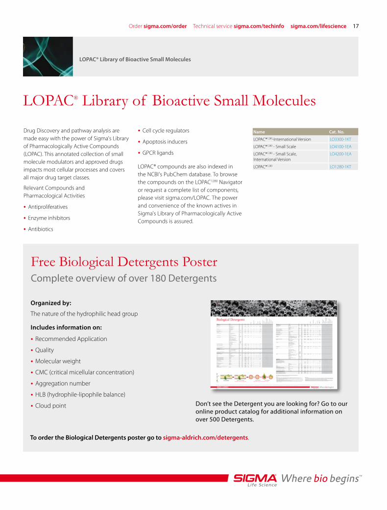
Drug Discovery and pathway analysis are made easy with the power of Sigma's Library of Pharmacologically Active Compounds (LOPAC). This annotated collection of small molecule modulators and approved drugs impacts most cellular processes and covers all major drug target classes.
Relevant Compounds and Pharmacological Activities
Antiproliferatives•Enzyme inhibitors•Antibiotics•
Name Cat. No.
LOPAC®1280-International Version LO3300-1KT
LOPAC®1280 - Small Scale LO4100-1EA
LOPAC®1280 - Small Scale, International Version
LO4200-1EA
LOPAC®1280 LO1280-1KT
Cell cycle regulators•Apoptosis inducers•GPCR ligands•
LOPAC® compounds are also indexed in the NCBI's PubChem database. To browse the compounds on the LOPAC1280 Navigator or request a complete list of components, please visit sigma.com/LOPAC. The power and convenience of the known actives in Sigma's Library of Pharmacologically Active Compounds is assured.
LOPAC® Library of Bioactive Small Molecules
LOPAC® Library of Bioactive Small Molecules
Free Biological Detergents PosterComplete overview of over 180 Detergents
Organized by:
The nature of the hydrophilic head group
Includes information on:
Recommended Application•Quality•Molecular weight•CMC (critical micellular concentration)•Aggregation number•HLB (hydrophile-lipophile balance)•Cloud point• Don’t see the Detergent you are looking for? Go to our
online product catalog for additional information on over 500 Detergents.
To order the Biological Detergents poster go to sigma-aldrich.com/detergents.
+ + +Detergent
Lipid
Protein
CM
C (
mM
)
Ag
gre
gat
ion
Nu
mb
er
HLB
Clo
ud
Po
int
(°C
)
Ave
rag
e M
icel
lar
Wei
gh
t
Dia
gn
ost
ic A
pp
licat
ion
s
Mo
lecu
lar
Bio
log
y
Cel
l Cu
ltu
re
Elec
tro
ph
ore
sis/
Ch
rom
ato
gra
ph
y
Mem
bra
ne
Pro
tein
So
lub
iliza
tio
n
Enzy
mo
log
y
An
tig
en/V
acci
ne
Prep
arat
ion
Dru
g D
eliv
ery/
Lip
oso
mes
Mas
s Sp
ectr
osc
op
y
Mic
rob
iolo
gy
Name Description Brand Cat. No. CAS MW
Anionic Detergents
Chenodeoxycholic acid 95% Sigma C9377 474-25-9 392.57 3 * * * * x x x
Choleate sodium salt crude ox bile extract Sigma S9875 * * * * *
Cholic acid 99.0% T Fluka 27010 81-25-4 408.57 9-15 2-3 18 * 900-1300 x x x x x x x x
ox or sheep bile, 98% Sigma C1129
Cholic acid sodium salt Bioreagent, suitable for cell culture Sigma C9282 206986-87-0 430.55 9-15 2-3 18 * 900-1300 x x x x x x x x
97.0% NT dried material Sigma 27029
BioXtra, 99% Sigma C6445
ox or sheep bile, 99% Sigma C1254
Deoxycholic acid BioXtra, 99% TLC and titration Sigma D4297 83-44-3 392.57 2-6 3-12 16 * 1200-5000 x x x x x x x x
99% TLC and titration Sigma D2510
Deoxycholic acid sodium salt 98.0% NT dry matter Sigma 30970 302-95-4 414.55 2-6 3-12 16 * 1200-5000 x x x x x x x x
97% titration Sigma-Aldrich D6750
Glycocholic acid hydrate 97% TLC Sigma G2878 475-31-0 465.62 7.1 2.1 * * 1000 x x
Glycocholic acid sodium salt hydrate 97% TLC Sigma G7132 863-57-0 487.6 7.1 2.1 * * 1000 x x
Lauroylsarcosine 95% Sigma L5000 97-78-9 271.4 14.6 2 * * 600 x x x x
98.0% GC Sigma-Aldrich 61739
Lauroylsarcosine sodium salt 30% aqueous solution, 97.0% Sigma 61747 137-16-6 293.38 14.6 2 * * 600 x x x x
BioUltra, for molecular biology, 99.0% Sigma 61743
BioXtra, 97% TLC Sigma L5777
94% Sigma L5125
BioReagent, for molecular biology, 94% Sigma L9150
Lithium dodecyl sulfate BioReagent, for molecular biology, ~ 99% GC Sigma L9781 2044-56-6 272.33 7-10 * * * * x x x x x
98.5% GC Sigma-Aldrich L4632
BioXtra, 98.5% GC Sigma-Aldrich L5901
SDS, Sodium dodecyl sulfate 98.0% GC, dust-free pellets Sigma 75746 151-21-3 288.38 7-10 62 40 >100 18000 x x x x x x x x x
BioUltra, for molecular biology, 10% water Sigma 71736
98.0% GC Sigma 71729
puriss. p.a., for ion pair chromatography, 99.0 Fluka 71726
BioUltra, for molecular biology, 99.0% Sigma 71725
BioUltra, for molecular biology, 20% water Fluka 05030
BioReagent, for molecular biology, 98.5% GC Sigma L4390
BioReagent, suitable for electrophoresis, 98.5% GC Sigma L3771
90% Sigma-Aldrich 62862
ACS reagent, 99.0% Sigma-Aldrich 436143
92.5-100.5% based on total alkyl sulfate content Sigma-Aldrich L5750
ReagentPlus®, 98.5% GC Sigma-Aldrich L4509
BioXtra, 99.0% GC Sigma-Aldrich L6026
Sodium deoxycholate monohydrate BioUltra, 99.0% NT Sigma 30968 145224-92-6 432.57 2-6 3-12 16 * 1200-5000 x x x x x x x x
BioXtra, 99% titration Sigma D5670
Taurochenodeoxycholic acid sodium salt 95% Sigma T6260 6009-98-9 521.69 4 * 20.1 * * x x
Taurocholic acid sodium salt hydrate BioXtra, 95% TLC Sigma T9034 345909-26-4 537.68 3-11 4 * * 2100 x x x
97.0% TLC Sigma 86339
95% TLC Sigma T4009
Taurodeoxycholic acid sodium salt monohydrate BioXtra, 97% TLC Sigma T0557 207737-97-1 521.69 1-4 6 * * 3100 x x x x
95% TLC Sigma T0875
Tauroursodeoxycholate sodium salt ~ 90% Sigma T0266 14605-22-2 521.69 4 * 20.1 * * x
Ursodeoxycholic acid 99% Sigma U5127 128-13-2 392.57 7 * * * * x
Cationic Detergents
CTAB, Hexadecyltrimethyl ammonium bromide puriss. p.a., for ion pair chromatography, 99.0 Fluka 52367 57-09-0 364.45 092 170 10 * 62000 x x x x x
BioUltra, for molecular biology, 99.0% Sigma 52365
BioReagent, for molecular biology, 99% Sigma H6269
BioXtra, 99% Sigma H9151
98%, powder Sigma H5882
TTAB ,Trimethyl-tetradecylammonium bromide puriss. p.a., for ion pair chromatography, 99.0 Fluka 87208 1119-97-7 336.39 4-5 80 * * 27000 x
99% Sigma T4762
Non-ionic Detergents
APO-10, Dimethyldecylphosphine oxide purum, 98.0% GC Fluka 40108 2190-95-6 218.32 4.6 131 * * 28597 x x
Big CHAP Deoxy, deoxy-BigCHAP 90% HPLC Sigma 14840 86303-23-3 862.06 1.1-1.4 8-16 * * 10500 x
> 65% TLC Sigma D9414
Brij® 35 30% (w/v) solution Sigma-Aldrich B4184 9002-92-0 1200 0.09 40 16.9 >100 48000 x x x x x x
Stein-Moore chromatography suitable Sigma-Aldrich P1254
Brij 35 P ~ 10% water solution Sigma 16012 9002-92-0 1200 0.09 40 16.9 >100 48000 x x x x x x
main component: tricosaethylene Sigma 16005
Brij 58 average Mn ~ 1124 Sigma P5884 9004-95-9 ~1124 (avg.) 0.08 70 15.7 >100 79000 x x x x x x
CYMAL-1®, Cyclohexylmethyl-β-D-maltoside 99.0% TLC Fluka 29467 260804-64-6 438.47 340 * * * * x x
CYMAL-2®, Cyclohexylmethyl-β-D-maltoside 99.0% TLC Sigma 29395 260804-65-7 452.49 120 * * * * x x
CYMAL-5®, Cyclohexylmethyl-β-D-maltoside 98.0% TLC Sigma 96193 250692-65-0 494.57 2.4-5 66 * * 32600 x x
CYMAL-6®, Cyclohexylmethyl-β-D-maltoside 99.0% TLC Sigma 29396 228579-27-9 508.6 0.56 63 * * 32000 x x
Decanoylsucrose, Sucrose monodecanoate Sigma S1266 31835-06-0 496.55 2.5 * * * * x x
Decyl β-D-glucopyranoside ~ 98% GC Sigma D5394 58846-77-8 320.42 2.2 * * * * x
Decyl β-D-maltopyranoside 10 mM solution Fluka 07509 82494-09-5 482.56 1.6-1.8 * * * * x
98% GC Sigma D7658
Decyl β-D-1-thiomaltopyranoside 99.0% TLC Sigma 30727 148565-56-4 498.63 0.9 * * * * x x
Digitonin Sigma D141 11024-24-1 1229.31 0.2-0.73 60 * * 74000 x x x x x x
~ 50% TLC Sigma D5628
Dodecanoylsucrose, Sucrose monolaurate 97.0% TLC Sigma 84110 25339-99-5 524.6 0.3 * * * * x x
Dodecyl β-D-glucopyranoside 98% GC Sigma D8035 59122-55-3 348.47 0.13 * * * 70000 x
Dodecyl β-D-maltoside BioXtra, 98% GC Sigma D5172 69227-93-6 510.62 0.15-0.19 98 * * 50000 x x x x x x x
98% GC Sigma D4641
Genapol® C-100 Sigma 61028 9002-92-0 627 0.075 * * * *
Genapol X-080 Sigma 48750 9043-30-5 553 0.06-0.15 * * * *
Genapol X-100 Sigma G6923 9043-30-5 641 0.15 88 * * 56000
HECAMEG, Methyl 6-O-(N-heptylcarbamoyl)-β-D-glucopyranoside 97.0% TLC Sigma 67328 115457-83-5 335.39 19.5 * * * *
Heptyl β-D-glucopyranoside 98.0% TLC Sigma 51980 78617-12-6 278.34 79 * * * *
Detergents are water-soluble, surface-active agents composed of a hydrophilic head group and a hydrophobic or lipophilic tail group. Due to their amphiphilic character, detergent molecules aggregate in solution to form micelles. They can also align at aqueous/non-aqueous interfaces, reducing surface tension, increasing miscibility, and stabilizing emulsions.
Categories In order to help you choose a detergent for a particular application, detergents are arranged into four categories, based on the nature of the hydrophilic head group:
Gentle detergents used for solubilizing proteins while maintaining native subunit structure, enzymatic activity, or other functions.Strong detergents that often completely disrupt cell membranes and fully denature proteins. They are sensitive to pH, ionic strength, and the nature of the
counter-ion and can interfere with charge-based analytical methods.Strong detergents with properties similar to those for anionic detergents. These are used in DNA purification, as surfactants in drug/vaccine delivery systems,
and in cleaning and disinfecting applications. Electrically neutral detergents that not only protect the native state of proteins but also prevent non-specific aggregation. They are often useful alternatives
to nonionic detergents in ion-exchange chromatography, electrophoresis, and isoelectric focusing. Although not detergents, these reagents possess hydrophilic groups similar to those of zwitterionic detergents but with much shorter
hydrophobic chains. They may improve the yield of membrane proteins when used with detergents and reportedly prevent aggregation of denatured proteins. Sulfobetaines do not form micells.
Physical Properties
Critical Micellular Concentration is the concentration at which micelles begin to form (i.e. the maximum monomer concentration). It should be noted that micelles cannot form, even above this concentration, if the temperature is too low. The minimum temperature for self-aggregation is called the critical micellar temperature (CMT). CMC values are a guide to detergent hydrophobic binding strength and stability. The lower the CMC, the more stable the micelle is, and the more slowly molecules are incorporated into or removed from the micelle. The higher the CMC, the weaker the binding and the easier the removal of the detergent, such as by dialysis.
is the average number of monomers in a micelle. A low aggregation number and high CMC favor removal by dialysis. is the hydrophile-lipophile balance. It defines the hydrophilic character of the detergent. A low HLB favors removal of the detergent by reverse-phase chromatography.
temperature is the temperature at which a detergent solution begins to look cloudy due to aggregation into larger structures that scatter light. The “cloud point” phenomenon interferes with applications that require optical clarity, but can be used to advantage in removing a detergent from aqueous solution.
CM
C (
mM
)
Ag
gre
gat
ion
Nu
mb
er
HLB
Clo
ud
Po
int
(°C
)
Ave
rag
e M
icel
lar
Wei
gh
t
Dia
gn
ost
ic A
pp
licat
ion
s
Mo
lecu
lar
Bio
log
y
Cel
l Cu
ltu
re
Elec
tro
ph
ore
sis/
Ch
rom
ato
gra
ph
y
Mem
bra
ne
Pro
tein
So
lub
iliza
tio
n
Enzy
mo
log
y
An
tig
en/V
acci
ne
Prep
arat
ion
Dru
g D
eliv
ery/
Lip
oso
mes
Mas
s Sp
ectr
osc
op
y
Mic
rob
iolo
gy
Name Description Brand Cat. No. CAS MW Non-ionic DetergentsHeptyl -D-thioglucopyranoside 99% GC Sigma H3264 85618-20-8 294.41 30 * * * * x Hexadecyl -D-maltoside 99.0% TLC Sigma 52318 98064-96-1 566.72 0.0006 * * * * x x Hexyl -D-glucopyranoside 98.0% TLC Sigma 53180 59080-45-4 264.32 250 * * * * Igepal® CA-630 BioReagent, suitable for electrophoresis, 2-D electrophoresis Sigma I7771 9036-19-5 603 (avg.) 0.08 * 13 63-69 * x x x x x x x x CMC 0.083 mM Fluka 56741 BioReagent, for molecular biology Sigma I8896 viscous liquid Sigma-Aldrich I3021 MEGA-10, Decanoyl-methylglucamine 98% GC Sigma D6277 85261-20-7 349.46 6-7 * * * * x x x x x x MEGA-8, Octanoyl-methylglucamine 500 mM solution Sigma 39490 85316-98-9 321.41 58 * * * * x x x x ~ 98% Sigma O3129 MEGA-9, Nonanoyl-methylglucamine ~ 98% Sigma N1138 85261-19-4 335.44 19-25 * * * * x x x x x Nonaethylene glycol monododecyl ether Sigma P9641 3055-99-0 582.81 0.08 110 * * 64000 x x Nonidet™ P 40 Substitute ampule, ~ 10% water solution Sigma 74388 9016-45-9 680 0.059 ~132 * 45-50 ~90000 x x x x x x x x mixture of 15 homologues Sigma 74385 Nonyl -D-glucopyranoside ~ 98% GC Sigma N7507 69984-73-2 306.4 6.5 * * * * Nonyl -D-1-thiomaltoside 98.0% TLC Sigma 74436 148565-55-3 484.6 3.2 * * * * Octaethylene glycol monododecyl ether 98% GC Sigma P8925 3055-98-9 538.75 0.11 123 * * 66000 x x x 98.0% GC Sigma 74680 Octyl -D-1-thioglucopyranoside 98% GC Sigma O6004 85618-21-9 308.43 9 * * * * x x x Octyl -D-maltoside 99.0% HPLC Sigma 19181 82494-08-4 454.51 23.4 84 * * 38000 Octyl- -D-glucopyranoside BioReagent, suitable for electrophoresis, 50% (w/v) aq. solution Sigma O3757 29836-26-8 292.37 20-25 84 * >100 24500-25001 x x x x x x x 98% GC Sigma-Aldrich O8001 BioXtra, > 98% GC Sigma-Aldrich O9882 Pluronic® F-127 BioReagent, suitable for cell culture Sigma P2443 9003-11-6 12500 (avg.) 4-11 * * * * Pluronic F-68 10%, solution, sterile-filtered, BioReagent, for insect Sigma P5556 9003-11-6 8350 (avg.) 0.04 * 29 >100 * x x x x cell culture solid, BioReagent, suitable for cell culture Sigma P1300 tested, insect cell culture tested solid, BioReagent, Sigma P7061 suitable for cell culture Polysorbate® 20 (see also Tween® 20) Ph Eur Fluka 44112 9005-64-5 1228 (avg.) 0.06 * 16.7 76 * x x x x x x x Polysorbate 80 (see also Tween® 80) Ph Eur Fluka 59924 9005-65-6 1310 (avg.) 0.012 60 15 65 79000 x x x x x x Saponin BioReagent, for molecular biology Sigma 47036 8047-15-2 1000-2000 0.001-0.01% * * * * x x x x x Sapogenin content 20-35% Sigma S4521 Sapogenin content 10% Sigma S7900 Sigma 84510 Thesit® BioXtra, for membrane research, 10% water soution Sigma 88317 9002-92-0 0.1 * * * * x x x x x for membrane research Sigma 88315 Triton® X-100 ampule, ~ 10% water solution Sigma 93427 9002-93-1 ~628 (avg.) 0.2-0.9 100-155 13.5 65 80000 x x x x x x x BioReagent, suitable for electrophoresis Sigma T8532 BioReagent, for molecular biology Sigma 93426 BioUltra, for molecular biology, ~ 10% in water Sigma 93443 BioReagent, for molecular biology Sigma T8787 laboratory grade Sigma-Aldrich X100 peroxide- and carbonyl-free Sigma-Aldrich X100PC BioXtra Sigma-Aldrich T9284 Triton X-100 reduced Sigma X100RS 92046-34-9 ~628 (avg.) 0.2-0.9 100-155 13.5 65 80000 x x x x x x x Triton X-114 ampule, ~ 10% water solution Sigma 93428 9036-19-5 ~537 (avg.) 0.2 * 12.4 23 * x x Sigma 93422 laboratory grade Sigma-Aldrich X114 Tween® 20 Fluka 65698 9005-64-5 1228 (avg.) 0.06 * 16.7 76 * x x x x x x x Sigma T2700 viscous liquid, cell culture tested Sigma P2287 BioReagent, suitable for electrophoresis, Sigma P5927 BioReagent, for molecular biology, viscous liquid Sigma P9416 250-450mPa.s 25°C Sigma 93773 average Mn ~ 1,228 Sigma-Aldrich 274348 Low-peroxide; Low-carbonyls, BHT as antioxidant Sigma-Aldrich P6585 Low-peroxide; Low-carbonyls Sigma-Aldrich P8341 BioXtra Sigma-Aldrich P7949 viscous liquid Sigma-Aldrich P1379 Tween 40 viscous liquid Sigma P1504 9005-66-7 1283 (avg.) 0.027 * * * * x x x x x x Tween 80 BioReagent, suitable for insect cell culture, viscous liquid Sigma P4675 9005-65-6 1310 (avg.) 0.012 60 15 65 79000 x x x x x x BioReagent, for molecular biology, syrup Sigma P5188 BioReagent, suitable forcell culture, viscous liquid Sigma P4780 375-480mPa.s 25°C Sigma 93781 BioXtra Sigma-Aldrich P8074 10%, Low peroxide Sigma-Aldrich P8192 viscous liquid Sigma-Aldrich P1754 viscous liquid, Low Peroxide Sigma-Aldrich P6349 viscous liquid, Preservative Free, Low-peroxide; Low carbonyls Sigma-Aldrich P6474 Undecyl -D-maltoside 99.0% TLC Sigma 94206 170552-39-3 496.59 0.59 * * * * x x
ASB 14-4 Sigma 77644 448.7 * * * * * x ASB C7BzO, 3-(4-Heptyl) phenyl 3-hydroxy propyl) dimethylammonio propane sulfonate Sigma C0856 399.6 * <25 * * <10000 x x x ASB-14, Amidosulfobetaine-14, 3-[N,N-Dimethyl (3-myristoylaminopropyl)ammonio]propanesulfonate Sigma A1346 216667-08-2 434.68 * >23 * * >10000 x x x ASB-C8Ø, 3-{N,N-Dimethyl-N-[3-(4-octylbenzoylamino) propyl]ammonio}propanesulfonate Sigma 49845 216667-49-1 4406 * * * * * x x CHAPS 98% TLC Sigma-Aldrich C3023 75621-03-3 614.88 6 10 * >100 6150 x x x x x x x 100 mM solution Sigma 19899 BioReagent, suitable for electrophoresis, 98% TLC Sigma C9426 BioXtra, 98% TLC Sigma-Aldrich C5070 CHAPSO BioXtra Sigma C4695 82473-24-3 630.88 8 11 * 90 7000 x x x x 98% Sigma C3649 DDMAB 97.0% Sigma 00413 15163-30-1 299.49 4.3 * * * * x EMPIGEN® BB detergent active substance ~ 35% water Sigma 45165 66455-29-6 272 1.6-2.1 * * * * x x x SB3-8, Dimethyloctylammonio propanesulfonate 98% Sigma O6626 15178-76-4 279.44 330 * * * * x x SB3-10, Decyldimethylammonio propanesulfonate 98% Sigma D4266 15163-36-7 307.49 25-40 41 * * 12600 x x SB3-12, Dodecyl-N,dimethyl-3-ammonio-1-propanesulfonate BioXtra Sigma D0431 14933-08-5 335.55 2-4 55 * * 18500 x x x x x x 97.0% CHN dried material Sigma 40232 SB3-14, Dimethylmyristylammonio propanesulfonate BioXtra Sigma T0807 14933-09-6 363.6 0.1-0.4 83 * * 30200 x x x x x 99% TLC Sigma T7763 purum, 98.0% T Fluka 40772 SB3-16, Dimethylpalmitylammonio propanesulfonate Sigma H6883 2281-11-0 391.65 0.01-0.06 155 * * 60700 x x x x SB3-18, Dimethyloctadecylammonio propanesulfonate purum, 99.0% TLC Aldrich 41570 13177-41-8 419.71 * * * * * x x
NDSB 195, Dimethylethylammoniumpropane sulfonate Sigma D0195 160255-06-1 195.3 n/a * * * * x x NDSB 211, Dimethyl-(2-hydroxyethyl)-(3-sulfopropyl)ammonium Sigma N0665 38880-58-9 211.3 n/a * * * * x x NDSB 221, 3-(1-Methylpyridinium)-1-propane sulfonate Sigma N0790 1670788-56-7 221.3 n/a * * * * x x NDSB 256-4T, 3-(4-tert-Butyl-1-pyridinio)-1-propanesulfonate Sigma 53487 257.4 n/a * * * * x x
Cell Lipid Membrane with Surface Proteins
Membrane with Detergents
Low Detergents Concentration (Below CMC)
Detergents Micelles Protein-detergents
High Detergents Concentration (At or greater than CMC)
Biological Detergents
©2010 Sigma-Aldrich Co. All rights reserved. SIGMA, SAFC, SIGMA-ALDRICH, ALDRICH, FLUKA, and SUPELCO are trademarks belonging to Sigma-Aldrich Co. and its affiliate Sigma-Aldrich Biotechnology, L.P. EMPIGEN is a registered trademark of Albright & Wilson UK Ltd. Tween is a registered trademark of Croda International PLC. Thesit is a registered trademark of Desitin Arzneimittel GmbH. Pluronic is a registered trademark of BASF. SE Igepal is a registered trademark of Rhodia Operations. Brij is a registered trademark of Uniqema Americas LLC. Genapol is a registered trademark of Clariant Produkte GmbH. CYMAL is a registered trademark of Affymetrix Inc.
bioreagents
MWT74561-5103221080

18
Prestige Antibodies® powered by Atlas Antibodies for Organelle LabelingThe Human Protein Atlas Program has carefully selected three different human cell lines, A-431 epidermoid carcinoma, U-251 MG glioblastoma and U-205 osteosarcoma, for organelle mapping of the proteome. As Prestige Antibodies are studied by
Cell and Organelle Labeling
Cell and Organelle Labeling
immunofluorescence (IF) staining, three well-characterized organelle markers for nuclei, microtubules and endoplasmic reticulum are used correspondingly as specific probes. The high-resolution confocal images are annotated based on sub-cellular localization. In addition, staining intensity and characteristics are also part of an overall assessment as well as comparison to literature (if available). All these factors
contribute to a final reliability score publicly available on line. Among the Prestige Antibodies tested in immunofluorescence (IF), a set of specific antibodies is classified as organelle markers. These probes have been subjected to the well-established quality control by the HPA program, and are thus very well suited for sub-cellular localization studies by immunofluorescence (IF.)
Cytoskeleton-Intermediate Filaments
Product Name
Host
Form
Reacts With
SpeciesReactivity
Cat. No.
Anti-VIM Anti-VIM antibody produced in rabbit
VIM, human human IF (i)IHC (p)PAWB
HPA001762-100UL
Anti-NES Anti-NES antibody produced in rabbit
NES, human human IF (i)IHC (p)PAWB
HPA007007-100UL
Cytoskeleton-Actin Filaments
Product Name
Host
Form
Reacts With
SpeciesReactivity
Cat. No.
Anti-FLNA Anti-FLNA antibody produced in rabbit
FLNA, human human IF (i)IHC (p)PAWB
HPA002925-100UL
Anti-ACTN1 Anti-ACTN1 antibody produced in rabbit
ACTN1, human human IF (i)IHC (p)PAWB
HPA006035-100UL
Cytoskeleton-Microtubules
Product Name
Host
Form
Reacts With
SpeciesReactivity
Cat. No.
Anti-TGFB1I1 Anti-TGFB1I1 antibody produced in rabbit
TGFB1I1, human human IHC (p)PAWB
HPA006376-100UL
ImmunofluorescenceAnti-NES: Cat. No. HPA007007: Immunofluorescent staining of human cell line U-2 OS shows posi-tivity in cytoskeleton.
ImmunofluorescenceAnti-ACTN1: Cat. No. HPA006035: Immunofluorescent staining of human cell line A-431 shows posi-tivity in cytoskeleton.
ImmunofluorescenceAnti-TGFB1I1: Cat. No. HPA006376: Immunofluorescent staining of human cell line U-2 OS shows posi-tivity in cytoskeleton.

Order sigma.com/order Technical service sigma.com/techinfo sigma.com/lifescience 19
Centrosomes Product Name
Host
Form
Reacts With
SpeciesReactivity
Cat. No.
Anti-DNAL4 Anti-DNAL4 antibody produced in rabbit
DNAL4, human human IF (i)IHC (p)PAWB
HPA003647-100UL
Endoplasmic Reticulum Product Name
Host
Form
Reacts With
SpeciesReactivity
Cat. No.
Anti-KTN1 Anti-KTN1 antibody produced in rabbit
KTN1, human human IF (i)IHC (p)PAWB
HPA003178-100UL
Anti-PDIA3 Anti-PDIA3 antibody produced in rabbit
PDIA3, human human IF (i)IHC (p)PAWB
HPA003230-100UL
Anti-BCAP31 Anti-BCAP31 antibody produced in rabbit
BCAP31, human human IF (i)IHC (p)PAWB
HPA003906-100UL
Plasma Membrane Product Name
Host
Form
Reacts With
SpeciesReactivity
Cat. No.
Anti-SLC9A3R2 Anti-SLC9A3R2 antibody produced in rabbit
SLC9A3R2, human human IF (i)IHC (p)PAWB
HPA001672-100UL
Anti-NF2 Anti-NF2 antibody produced in rabbit
NF2, human human IF (i)IHC (p)PAWB
HPA003097-100UL
Focal Adhesions Product Name
Host
Form
Reacts With
SpeciesReactivity
Cat. No.
Anti-MYH6 Anti-MYH6 antibody produced in rabbit
MYH7, humanMYH6, human
human IF (i)IHC (p)PA
HPA001349-100UL
Anti-ZYX Anti-ZYX antibody produced in rabbit
ZYX, human human IF (i)IHC (p)PA
HPA004835-100UL
Anti-VASP Anti-VASP antibody produced in rabbit
VASP, human human IF (i)IHC (p)PAWB
HPA005724-100UL
ImmunofluorescenceAnti-DNAL4: Cat. No. HPA003647: Immunofluorescent staining of human cell line U-251MG shows positivity in centrosome.
ImmunofluorescenceAnti-PDIA3: Cat. No. HPA003230: Immunofluorescent staining of human cell line U-2 OS shows posi-tivity in endoplasmic reticulum.
ImmunofluorescenceAnti-NF2: Cat. No. HPA003097: Immunofluorescent staining of human cell line U-2 OS shows positivity in cytoplasm & plasma membrane.
ImmunofluorescenceAnti-ZYX: Cat. No. HPA004835: Immunofluorescent staining of human cell line A-431 shows posi-tivity in focal adhesions.

20
Cytoplasm Product Name
Host
Form
Reacts With
SpeciesReactivity
Cat. No.
Anti-MTHFD1 Anti-MTHFD1 antibody produced in rabbit
MTHFD1, human human IF (i)IHC (p)PAWB
HPA001290-100UL
Anti-DDX3X Anti-DDX3X antibody produced in rabbit
DDX3X, human human IF (i)IHC (p)PAWB
HPA001648-100UL
Anti-USP10 Anti-USP10 antibody produced in rabbit
USP10, human human IF (i)IHC (p)PA
HPA006731-100UL
Peroxisomes
Product Name
Host
Form
Reacts With
SpeciesReactivity
Cat. No.
Anti-ACAA1 Anti-ACAA1 antibody produced in rabbit
ACAA1, human human IF (i)IHC (p)PAWB
HPA007244-100UL
Mitochondria
Product Name
Host
Form
Reacts With
SpeciesReactivity
Cat. No.
Anti-SYNJ2BP Anti-SYNJ2BP antibody produced in rabbit
COX16, human human IF (i)IHC (p)PAWB
HPA000866-100UL
Anti-HSPA9 Anti-HSPA9 antibody produced in rabbit
HSPA9, human human IF (i)IHC (p)PAWB
HPA000898-100UL
Anti-TIMM9 Anti-TIMM9 antibody produced in rabbit
TIMM9, human human IF (i)IHC (p)PAWB
HPA002932-100UL
Anti-MRPL40 Anti-MRPL40 antibody produced in rabbit
MRPL40, human human IF (i)IHC (p)PAWB
HPA006181-100UL
Golgi
Product Name
Host
Form
Reacts With
SpeciesReactivity
Cat. No.
Anti-GOLGA5 Anti-GOLGA5 antibody produced in rabbit
GOLGA5, human human IF (i)IHC (p)PAWB
HPA000992-100UL
Anti-GOLIM4 Anti-GOLIM4 antibody produced in rabbit
GOLIM4, human human IF (i)IHC (p)PA
HPA001677-100UL
ImmunofluorescenceAnti-DDX3X: Cat. No. HPA001648: Immunofluorescent staining of human cell line U-251MG shows positivity in cytoplasm.
ImmunofluorescenceAnti-ACAA1: Cat. No. HPA007244: Immunofluorescent staining of human cell line U-251MG shows positivity in peroxisomes.
ImmunofluorescenceAnti-HSPA9: Cat. No. HPA000898: Immunofluorescent staining of human cell line U-2 OS shows posi-tivity in mitochondria.
ImmunofluorescenceAnti-GOLIM4: Cat. No. HPA001677: Immunofluorescent staining of human cell line U-2 OS shows positivity in golgi.

Order sigma.com/order Technical service sigma.com/techinfo sigma.com/lifescience 21
Nuclear Envelope
Product Name
Host
Form
Reacts With
SpeciesReactivity
Cat. No.
Anti-UNC84B Anti-UNC84B antibody produced in rabbit
UNC84B, human human IF (i)IHC (p)PAWB
HPA001209-100UL
Anti-UNC84A Anti-UNC84A antibody produced in rabbit
UNC84A, human human IF (i)IHC (p)PAWB
HPA008461-100UL
Nucleus without Nucleoli
Product Name
Host
Form
Reacts With
SpeciesReactivity
Cat. No.
Anti-AKAP8 Anti-AKAP8 antibody produced in rabbit
AKAP8, human human IF (i)IHC (p)PAWB
HPA004776-100UL
Anti-FUBP1 Anti-FUBP1 antibody produced in rabbit
FUBP1, human human IF (i)IHC (p)PAWB
HPA006149-100UL
Anti-FUS Anti-FUS antibody produced in rabbit
FUS, human human IF (i)IHC (p)PA
HPA008784-100UL
Nucleoli
Product Name
Host
Form
Reacts With
SpeciesReactivity
Cat. No.
Anti-VSX2 Anti-VSX2 antibody produced in rabbit
VSX2, human human IF (i)IHC (p)PA
HPA003436-100UL
Anti-ZSCAN1 Anti-ZSCAN1 antibody produced in rabbit
ZSCAN1, human human IF (i)IHC (p)PAWB
HPA007938-100UL
Nucleus
Product Name
Host
Form
Reacts With
SpeciesReactivity
Cat. No.
Anti-MECP2 Anti-MECP2 antibody produced in rabbit
MECP2, human human IF (i)IHC (p)PAWB
HPA000593-100UL
Anti-GPKOW Anti-GPKOW antibody produced in rabbit
GPKOW, human human IF (i)IHC (p)PAWB
HPA001894-100UL
Anti-FBXO18 Anti-FBXO18 antibody produced in rabbit
FBXO18, human human IF (i)IHC (p)PA
HPA002844-100UL
Anti-MYST4 Anti-MYST4 antibody produced in rabbit
MYST4, human human IF (i)IHC (p)PA
HPA006104-100UL
Anti-XRCC4 Anti-XRCC4 antibody produced in rabbit
XRCC4, human human IF (i)IHC (p)PA
HPA006801-100UL
ImmunofluorescenceAnti-UNC84A: Cat. No. HPA008461: Immunofluorescent staining of human cell line A-431 shows positiv-ity in nuclear membrane & vesicles.
ImmunofluorescenceAnti-FUS: Cat. No. HPA008784: Immunofluorescent staining of human cell line U-251MG shows positivity in nuclei but not nucleoli.
ImmunofluorescenceAnti-VSX2: Cat. No. HPA003436: Immunofluorescent staining of human cell line A-431 shows positivity in nucleoli.
ImmunofluorescenceAnti-FBXO18: Cat. No. HPA002844: Immunofluorescent staining of human cell line U-251MG shows positivity in nuclei.
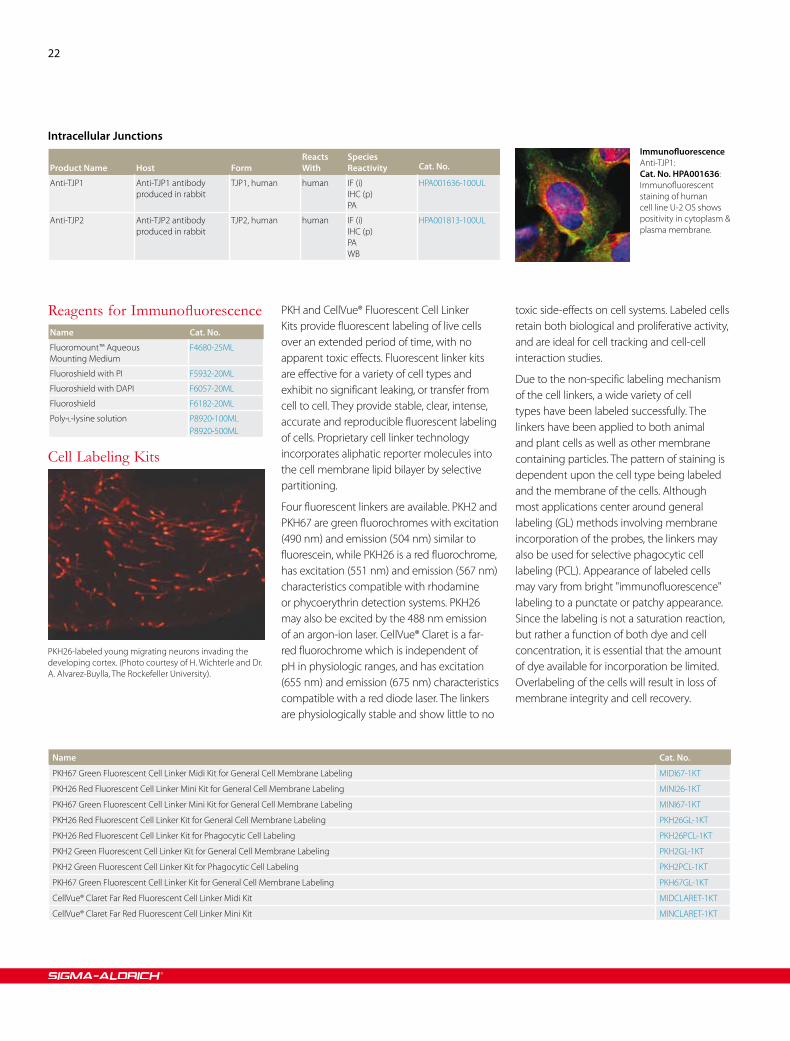
22
Intracellular Junctions
Product Name
Host
Form
Reacts With
SpeciesReactivity
Cat. No.
Anti-TJP1 Anti-TJP1 antibody produced in rabbit
TJP1, human human IF (i)IHC (p)PA
HPA001636-100UL
Anti-TJP2 Anti-TJP2 antibody produced in rabbit
TJP2, human human IF (i)IHC (p)PAWB
HPA001813-100UL
Reagents for ImmunofluorescenceName Cat. No.
Fluoromount™ Aqueous Mounting Medium
F4680-25ML
Fluoroshield with PI F5932-20ML
Fluoroshield with DAPI F6057-20ML
Fluoroshield F6182-20ML
Poly-L-lysine solution P8920-100MLP8920-500ML
ImmunofluorescenceAnti-TJP1: Cat. No. HPA001636: Immunofluorescent staining of human cell line U-2 OS shows positivity in cytoplasm & plasma membrane.
Cell Labeling Kits
PKH26-labeled young migrating neurons invading the developing cortex. (Photo courtesy of H. Wichterle and Dr. A. Alvarez-Buylla, The Rockefeller University).
PKH and CellVue® Fluorescent Cell Linker Kits provide fluorescent labeling of live cells over an extended period of time, with no apparent toxic effects. Fluorescent linker kits are effective for a variety of cell types and exhibit no significant leaking, or transfer from cell to cell. They provide stable, clear, intense, accurate and reproducible fluorescent labeling of cells. Proprietary cell linker technology incorporates aliphatic reporter molecules into the cell membrane lipid bilayer by selective partitioning.
Four fluorescent linkers are available. PKH2 and PKH67 are green fluorochromes with excitation (490 nm) and emission (504 nm) similar to fluorescein, while PKH26 is a red fluorochrome, has excitation (551 nm) and emission (567 nm) characteristics compatible with rhodamine or phycoerythrin detection systems. PKH26 may also be excited by the 488 nm emission of an argon-ion laser. CellVue® Claret is a far-red fluorochrome which is independent of pH in physiologic ranges, and has excitation (655 nm) and emission (675 nm) characteristics compatible with a red diode laser. The linkers are physiologically stable and show little to no
toxic side-effects on cell systems. Labeled cells retain both biological and proliferative activity, and are ideal for cell tracking and cell-cell interaction studies.
Due to the non-specific labeling mechanism of the cell linkers, a wide variety of cell types have been labeled successfully. The linkers have been applied to both animal and plant cells as well as other membrane containing particles. The pattern of staining is dependent upon the cell type being labeled and the membrane of the cells. Although most applications center around general labeling (GL) methods involving membrane incorporation of the probes, the linkers may also be used for selective phagocytic cell labeling (PCL). Appearance of labeled cells may vary from bright "immunofluorescence" labeling to a punctate or patchy appearance. Since the labeling is not a saturation reaction, but rather a function of both dye and cell concentration, it is essential that the amount of dye available for incorporation be limited. Overlabeling of the cells will result in loss of membrane integrity and cell recovery.
Name Cat. No.
PKH67 Green Fluorescent Cell Linker Midi Kit for General Cell Membrane Labeling MIDI67-1KT
PKH26 Red Fluorescent Cell Linker Mini Kit for General Cell Membrane Labeling MINI26-1KT
PKH67 Green Fluorescent Cell Linker Mini Kit for General Cell Membrane Labeling MINI67-1KT
PKH26 Red Fluorescent Cell Linker Kit for General Cell Membrane Labeling PKH26GL-1KT
PKH26 Red Fluorescent Cell Linker Kit for Phagocytic Cell Labeling PKH26PCL-1KT
PKH2 Green Fluorescent Cell Linker Kit for General Cell Membrane Labeling PKH2GL-1KT
PKH2 Green Fluorescent Cell Linker Kit for Phagocytic Cell Labeling PKH2PCL-1KT
PKH67 Green Fluorescent Cell Linker Kit for General Cell Membrane Labeling PKH67GL-1KT
CellVue® Claret Far Red Fluorescent Cell Linker Midi Kit MIDCLARET-1KT
CellVue® Claret Far Red Fluorescent Cell Linker Mini Kit MINCLARET-1KT

Order sigma.com/order Technical service sigma.com/techinfo sigma.com/lifescience 23
Cell LinesWe have recently added over 1500 new ECACC® cell lines to our portfolio in the U.S. Many hard-to-get cell lines are included so that the time and effort required for
Cell Culture
1500 New ECACC® Cell Lines
procurement will be greatly reduced. All cell lines are authenticated and mycoplasma tested so you can use them in your applications with confidence. The cell lines can be used in a variety of applications
including, cancer research, neurobiology, and toxicology. For a more extensive listing of cell lines, visit sigma.com/ecacc. Check the site often as we are continually increasing our offering.
Name Description Biological Source Cat. No.
CHO Protein-Free Cell Line Chinese hamster ovary from Chinese hamster ovary cells 00102307-1VL
1301 Cell Line T-cell leukemia human 01051619-1VL
MDCK-SIAT1 Cell Line Canine Cocker Spaniel Kidney Sialic Acid Over Expression canine 05071502-1VL
MRC-5 PD 19 Cell Line Human fetal lung human 05072101-1VL
MRC-5 Cell Line Fetal lung fibroblast-like human 84101801-1VL
MDCK Cell Line Normal female kidney canine 84121903-1VL
L929 Cell Line mouse C3H/An connective tissue from mouse 85011425-1VL
J774.2 Cell Line BALB/C monocyte macrophage from mouse 85011428-1VL
Hep G2 Cell Line hepatocyte carcinoma human 85011430-1VL
BHK 21 (Clone 13) kidney from hamster 85011433-1VL
U937 Cell Line Lymphoblast from lung human 85011440-1VL
IMR-90 Cell Line Human Caucasian fetal lung fibroblast human 85020204-1VL
Hep-2C Cell Line human cervix carcinoma (HeLa derivative) human 85020207-1VL
CHO Cell Line Chinese ovary from hamster 85050302-1VL
CHO-K1 Cell Line ovary from hamster 85051005-1VL
HT55 Cell Line Human colon carcinoma human 85061105-1VL
SP2/O-Ag14 Cell Line Non-producing hybridoma murine 85072401-1VL
A431 Cell Line squamous carcinoma human 85090402-1VL
OAW28 Cell Line ovarian tumor epithelial human 85101601-1VL
293 Cell Line embryonic kidney human 85120602-1VL
CACO-2 Cell Line Caucasian colon adenocarcinoma human 86010202-1VL
A549 Cell Line human lung carcinoma human 86012804-1VL
1321N1 Cell Line Glial cells from brain astrocytoma human 86030402-1VL
3T3 L1 Cell Line embryo from mouse 86052701-1VL
PC-12 Cell Line adrenal gland phaeochromocytoma from rat 88022401-1VL
Jurkat E6.1 Cell Line leukemic T cell lymphoblast human 88042803-1VL
THP 1 Cell Line Leukemic monocyte human 88081201-1VL
Fao Cell Line rat hepatoma from rat 89042701-1VL
Sf9 Cell Line For Baculovirus vectors from Spodoptera frugiperda pupal ovarian tissue 89070101-1VL
K562 Cell line chronic myelogenous leukemia human 89121407-1VL
Nthy-ori 3-1 Cell Line thyroid follicular epithelial human 90011609-1VL
RAW 264.7 Cell Line Macrophage from blood murine 91062702-1VL
MDA-MB-231 Cell Line human breast adenocarcinoma human 92020424-1VL
ND7/23 Cell Line Mouse neuroblastoma X Rat neuron hybrid - 92090903-1VL

24
Name Description Biological Source Cat. No.
HeLa Cell Line epitheloid cervix carcinoma human 93021013-1VL
TF1 Cell Line Erythroleukemic cell line from blood. human 93022307-1VL
NIH 3T3 Cell Line Embryonic fibroblast murine 93061524-1VL
A2780cis Cell Line ovarian carcinoma human 93112517-1VL
A2780 Cell Line ovarian carcinoma human 93112519-1VL
SH-SY5Y Cell Line Neuroblast from neural tissue. human 94030304-1VL
FTC-133 Cell Line Human follicular thyroid carcinoma human 94060901-1VL
PNT2 Cell Line Normal prostate epithelium immortalized with SV40. human 95012613-1VL
RPMI 8866 Cell Line Lymphoblastoid from blood human 95041316-1VL
NCI-H322 Cell Line Human Caucasian bronchioalveolar carcinoma human 95111734-1VL
OE21 Cell Line Caucasian oesophageal squamous cell carcinoma human 96062201-1VL
OE33 Cell Line Caucasian esophageal carcinoma human 96070808-1VL
OE19 Cell Line - human 96071721-1VL
B9 Cell Line Mouse B cell hybridoma from mouse 96080128-1VL
tsa201 Cell Line embryonal kidney, SV40 transformed human 96121229-1VL
Nb2-11 Cell Line Thymus/lymph node, lymphoblast morphology from rat 97041101-1VL
Ishikawa Cell Line endometrial adenocarcinoma human 99040201-1VL
Reagents for Detecting and Eliminating Mycoplasma ContaminationName Cat. No.
LookOut® mycoplasma qPCR detection kit MP0040-1KT
VenorGeM® Mycoplasma Detection Kit, PCR-based MP0025-1KT
LookOut® Mycoplasma PCR Detection Kit MP0035-1KT
LookOut® Mycoplasma Elimination Kit MP0030-1KT
LookOut® Mycoplasma Erase, Mycoplasma erase spray L1420-500ML
Reagents for 3-D Cell CultureName Cat. No.
HydroMatrix™ Peptide Cell Culture Scaffold, mixture A6982-1MLA6982-5MLA6982-10ML
MaxGel™ ECM , mixture E0282-100ULE0282-1ML
HydroMatrix™ Peptide Cell Culture Scaffold, 96 well plate H4165-1EA
HydroMatrix™ Peptide Cell Culture Scaffold, 24 well plate H4040-1EA
HydroMatrix™ Peptide Cell Culture Scaffold, 6 well plate H3915-1EA
HyStem™ Cell Culture Scaffold Kit, Trial Kit, For 2.5 mL of hydrogel scaffold solution HYS010-1KT
HyStem™ Cell Culture Scaffold Kit, For 7.5 mL of hydrogel scaffold solution HYS020-1KT
HyStem™-C Cell Culture Scaffold Kit, For 7.5 mL of hydrogel scaffold solution HYSC020-1KT
HyStem™-C Cell Culture Scaffold Kit, Trial Kit, For 2.5 mL of hydrogel scaffold solution HYSC010-1KT
HyStem™-HP Cell Culture Scaffold Kit, For 7.5 mL of hydrogel scaffold solution HYSHP020-1KT
HyStem™-HP Cell Culture Scaffold Kit, Trial Kit, For 2.5 mLof hydrogel scaffold solution HYSHP010-1KT
MaxGel™ ECM , 96 well plate M1073-1EA
Order sigma.com/order Technical service sigma.com/techinfo sigma.com/lifescience 25
Reagents for Stem Cell CultureStem cell biology offers astonishing research potential. Sigma® Life Science has the innovations you need to discover the promise it holds. Access a world of RNAi with the MISSION® RNAi Library, efficiently edit genes of interest using advanced CompoZr® ZFN technology, and characterize your
stem cells with our Prestige Antibodies®, powered by Atlas Antibodies. Optimized formulations and proven, consistent results have earned Sigma's Stemline Media a reputation of quality and excellence. Ideal for expansion and maintenance of neural, hematopoietic, and other adult stem cells, Stemline media provide optimal, consistent cellular environments. Produced in a GMP
state-of-the-art facility with an available Device Master File (DMF), Stemline Media are clearly an excellent choice for your stem cell applications.
The applications are endless—and so are the possibilities.
Name Cat. No.
Stemline® Hematopoietic Stem Cell Expansion Medium, Serum-free, Without L-glutamine S0189-500MLS0189-6X500ML
Stemline® II Hematopoietic Stem Cell Expansion Medium, Serum-free, contains L-glutamine S0192-500MLS0192-6X500ML
Stemline® Keratinocyte Medium II S0196-500ML
Stemline® Mesenchymal Stem Cell Expansion Medium, Without L-glutamine S1569-1L
Stemline® T Cell Expansion Medium, Serum-free, Without L-glutamine S1694-1L
Stemline® Neural Stem Cell Expansion Medium, without L-glutamine, growth factors, and antibiotics S3194-500ML
Stemline® Dendritic Cell Maturation Medium, Serum-free, Without L-glutamine S3444
Stemline® Keratinocyte Growth Supplement, For use with S0196, Stemline® Keratinocyte Medium II S9945-1VL
Sigma-Aldrich® Offers a Full Line of MicroscopesJenco™ Microscopes
Jenco International, Inc. specializes in the design, assembly, service and distribution of high-quality, reproducible optical instruments. Simply stated, there products deliver brilliant clarity, parfocality, depth of field and durability previously thought impossible at an affordable price range.
Standard Compound•Inverted Compound•Stereoscope•
As a leading supplier of optical instruments in several market areas, which include the Educational/Research market, High Technology/Medical Device industry and
Biotechnology/Pharmaceutical market, Jenco has a proven track record of supplying the best quality for price solutions.
Features
Affordable•Versatile•Durable•Compliant•
To find out more, visit sigma-aldrich.com/microscopes

26
Equipment
For a complete selection of these products please visit sigma.com/labware
Cell Culture Labware
Cell Culture Labware
Name Cat. No.
Jenco™ inverted compound microscopes, 30W halogen bulb Z723975-1EA
Jenco™ inverted compound microscopes, 30W halogen bulb Z724084-1EA
Hypoxia Chamber Glove Box, CO2 range: 0 - 20% C, sensor accuracy: 0.2%, O
2 range: 0 - 25% with
sensor accuracy of +/- 0.25%Z736848-1EA
Specialty Surfaces and Formats
For a complete selection of these products please visit sigma.com/labware
Name Cat. No.
Corning® CellBIND® cell culture plates, 6 wells CLS3335-5EACLS3335-50EA
Corning® CellBIND® cell culture plates, 12 wells CLS3336-50EA
Corning® CellBIND® cell culture plates, 24 wells CLS3337-5EACLS3337-50EA
Corning® CellBIND® cell culture plates, 48 wells CLS3338-50EA
Corning® Ultra-low attachment multiwell plates, 6 wells CLS3471-24EA
Corning® Ultra-low attachment multiwell plates CLS3473-24EA
Corning® Ultra-low attachment multiwell plates, 96 wells CLS3474-24EA
Corning® Costar® cell culture plates, 6 well, flat bottom CLS3506-100EA
Corning® Costar® cell culture plates, 6 well, flat bottom (Individually wrapped) CLS3516-1EACLS3516-10EACLS3516-50EA
Corning® Costar® cell culture plates, 12 well, flat bottom CLS3512-100EA
Corning® Costar® cell culture plates, 12 well, flat bottom CLS3513-50EA
Corning® Costar® cell culture plates, 24 well, flat bottom CLS3527-100EA
Corning® Costar® cell culture plates, 24 well, flat bottom CLS3526-1EACLS3526-50EA
Corning® Costar® cell culture plates, 24 well, flat bottom CLS3524-100EA
Corning® Costar® cell culture plates, 48 well, flat bottom CLS3548-100EA
Corning® Costar® cell culture plates, 96 well, flat bottom CLS3595-1EACLS3595-10EACLS3595-50EA
Corning® Costar® cell culture plates, 96 well, flat bottom, with special low evaporation lid CLS3585-50EA
Corning® Costar® cell culture plates, 96 well, flat bottom CLS3596-50EA
Corning® Costar® cell culture plates, 96 well, flat bottom CLS3997-10EACLS3997-50EA
Corning® Costar® cell culture plates, growth area 0.32 cm2 CLS3598-100EA
Corning® Costar® cell culture plates, 96 well, flat bottom CLS3599-100EA
Corning® Costar® cell culture plates, 96 well, round bottom CLS3799-1EACLS3799-50EA

Order sigma.com/order Technical service sigma.com/techinfo sigma.com/lifescience 27
Multiwell Plate Inserts
For a complete selection of these products please visit sigma.com/labware
Name Cat. No.
Corning® Costar® cell culture plates, 8 well strip plate, assembled 12 strips per 96 well plate CLS9102-50EA
Nunclon™ D Multidishes, 4 well round D6789-1CS
Nunclon™ D Multidishes, 12 well round D6315-1CS
Nunclon™ D Multidishes, 24 well round D7039-1CS
Nunclon™ D Multidishes, 48 well round D6440-1CS
Nunc® MicroWell™ mini tray, 60 well M0815-100EA
Nunc® MicroWell™ mini tray, 72 well M0690-100EA
Name Cat. No.
Corning® HTS Transwell® 96 well permeable support CLS3381-1EA
Corning® HTS Transwell® 96 well permeable support CLS3391-5EA
Corning® HTS Transwell® 96 well permeable support CLS3380-1EA
Corning® HTS Transwell® 96 well permeable support CLS3392-5EA
Corning® HTS Transwell® 96 well permeable support CLS3374-2EA
Corning® HTS Transwell® 96 well permeable support CLS3384-8EA
Corning® HTS Transwell®-24 well permeable support CLS3396-2EA
Corning® HTS Transwell®-24 well permeable support CLS3398-2EA
Corning® HTS Transwell®-24 well permeable support CLS3397-12EA
Corning® HTS Transwell®-24 well permeable support CLS3399-12EA
Corning® Transwell® polycarbonate membrane inserts CLS3401-48EA
Corning® Transwell® polycarbonate membrane inserts CLS3402-48EA
Corning® Snapwells™ inserts CLS3407-24EA
Corning® Transwell® polycarbonate membrane inserts CLS3413-48EA
Corning® Transwell® polycarbonate membrane inserts CLS3412-24EA
Corning® Transwell® polycarbonate membrane inserts CLS3419-12EA
Corning® Transwell® polycarbonate membrane inserts CLS3415-48EA
Corning® Transwell® polycarbonate membrane inserts CLS3414-24EA
Corning® Transwell® polycarbonate membrane inserts CLS3420-12EA
Corning® Transwell® polycarbonate membrane inserts CLS3421-48EA
Corning® Transwell® polycarbonate membrane inserts CLS3422-48EA
Corning® Transwell® polycarbonate membrane inserts CLS3428-24EA
Corning® Transwell® polyester membrane inserts CLS3470-48EA
Corning® Transwell® polyester membrane inserts CLS3460-48EA
Corning® Transwell® polyester membrane inserts CLS3450-24EA
Corning® Transwell® polyester membrane inserts CLS3472-48EA
Corning® Transwell® polyester membrane inserts CLS3462-48EA
Corning® Transwell® polyester membrane inserts CLS3452-24EA
Corning® Netwells® inserts CLS3477-48EA
Corning® Netwells® inserts CLS3478-48EA
Corning® Netwells® inserts CLS3479-48EA
Corning® Netwells® inserts CLS3480-48EA
Corning® Transwell®-COL collagen-coated membrane inserts CLS3491-24EA
Corning® Transwell®-COL collagen-coated membrane inserts CLS3492-24EA
Corning® Transwell®-COL collagen-coated membrane inserts CLS3493-24EA
Corning® Transwell®-COL collagen-coated membrane inserts CLS3494-24EA
Corning® Transwell®-COL collagen-coated membrane inserts CLS3495-24EA
Corning® Transwell®-COL collagen-coated membrane inserts CLS3496-24EA

28
Name Cat. No.
Corning® 1536 well plate, solid black, tissue-culture treated plate CLS3726-10EACLS3726-50EA
Corning® 1536 well plate, solid white, tissue-culture treated plate with lid CLS3727-50EA
Corning® 1536 well plate, solid black, NBS surface plate CLS3728-10EACLS3728-50EA
Corning® low volume 2 μL 1536 well multiwell plates CLS3855-100EA
Greiner® high and medium binding 1536 well plates M7686-60EA
Greiner® high and medium binding 1536 well plates M7561-60EA
1536-Well Plates
For a complete selection of these products please visit sigma.com/labware
Name Cat. No.
Corning® 384 Well Plates CLS3822-10EACLS3822-50EA
Corning® 384 Well Plates CLS3571-10EACLS3571-50EA
Corning® 384 Well Plates CLS3542-10EACLS3542-50EA
Corning® 384 Well Plates CLS3985-100EA
Corning® 384 Well Plates CLS3712-20EACLS3712-100EA
Corning® 384 Well Plates CLS3570-10EACLS3570-50EA
Corning® 384 Well Plates CLS3663-100EA
Corning® 384 Well Plates CLS3707-20EACLS3707-100EA
Corning® 384 Well Plates CLS3680-100EA
Corning® 384 Well Plates CLS3701-20EACLS3701-100EA
Corning® 384 Well Plates CLS3662-100EA
Corning® CellBIND® 384 Well Plates CLS3683-50EA
Nunc® polystyrene 384 well plates P5991-1CS
Name Cat. No.
Well cap for Nunc® 96 DeepWell plates C1111-50EA
Corning® Universal-BIND™ Plate, 96 well CLS2503-50EA
Corning® Sulfhydryl-BIND™ 96 well plate, 96 well CLS2509-50EA
Corning® 96 Well Plates, non-treated CLS2595-100EA
Corning® 96 Well Plates, non-treated CLS2797-100EA
Corning® 96 Well Plates, non-treated CLS2897-100EA
Corning® Robolids 96 well, 96 well CLS3090-50EA
Corning® CellBIND® 96 Well Plates CLS3300-50EA
Corning® CellBIND® 96 Well Plates CLS3340-50EA
Corning® 96 Well Plates, non-treated CLS3370-100EA
Corning® 96 Well Plates, non-treated CLS3367-50EA
Corning® 96 Well Plates, tissues culture treated CLS3358-100EA
Corning® 96 Well Plates, tissue culture treated CLS3360-100EA
Corning® HTS Transwell® 96 well permeable support CLS3381-1EA
Corning® HTS Transwell® 96 well permeable support CLS3380-1EA
Corning® HTS Transwell® 96 well permeable support CLS3374-2EA
Corning® HTS Transwell®-96 well accessory plate, receiver plate CLS3382-10EA
Corning® HTS Transwell® 96 well permeable support CLS3391-5EA
384-Well Plates
For a complete selection of these products please visit sigma.com/labware
96-Well Plates
For a complete selection of these products please visit sigma.com/labware

Order sigma.com/order Technical service sigma.com/techinfo sigma.com/lifescience 29
Name Cat. No.
Corning® HTS Transwell® 96 well permeable support CLS3392-5EA
Corning® HTS Transwell® 96 well permeable support CLS3385-2EA
Corning® HTS Transwell® 96 well permeable support CLS3386-8EA
Corning® HTS Transwell® 96 well permeable support CLS3384-8EA
Corning® HTS Transwell® 96 well permeable support CLS3387-8EA
Corning® HTS Transwell® 96 well permeable support CLS3388-2EA
Corning® HTS Transwell®-96 well accessory plate, receiver plate CLS3583-10EA
Corning® volume adapter for 96 well filter plates CLS3584-50EA
Corning® NBS™ 96 well plates, without lids, SPA CLS3600-100EA
Corning® 96 Well Plates, Clear Bottom, high binding CLS3601-100EA
Corning® 96 Well Plates, Clear Bottom, tissue culture treated CLS3603-48EA
Corning® NBS™ 96 well plates, without lids, SPA CLS3604-100EA
Corning® NBS™ 96 well plates, without lids, SPA CLS3605-100EA
Corning® 96 Well Plates, Clear Bottom, tissue culture treated CLS3610-48EA
Corning® 96 well special optics plates, tissue culture treated CLS3614-100EA
Corning® 96 well special optics plates, non-treated CLS3615-100EA
Corning® 96 Well Plates, tissue culture treated CLS3628-100EA
Corning® 96 Well Plates, Clear Bottom, non-treated CLS3631-100EA
Corning® NBS™ 96 well plates, without lids, fluorescence CLS3650-100EA
Corning® NBS™ 96 well plates, without lids, fluorescence CLS3651-100EA
Corning® NBS™ 96 well plates, without lids CLS3641-100EA
Corning® 96 well special optics plates, tissue culture treated CLS3720-25EA
Corning® 96 Well Plates, non-treated CLS3795-100EA
Corning® 96 Well Plates, non-treated CLS3896-48EA
Corning® 96 Well Plates, non-treated CLS3897-100EA
Corning® 96 Well Plates, non-treated CLS3898-100EA
Corning® 96 Well Plates, not-treated CLS3798-100EA
Corning® 96 Well Plates, Clear Bottom, tissue culture treated CLS3904-100EA
Corning® 96 Well Plates, Clear Bottom, tissue culture treated CLS3903-100EA
Corning® 96 Well Plates, NBS™ CLS3990-25EA
Nunc-Immuno™ plates, 96-well plate, MaxiSorp M9410-1CS
Nunc-Immuno™ plates, 96-well plate, MaxiSorp, with certificate M5785-1CS
Nunc-Immuno™ plates, 96-well plate, PolySorp M0661-1CS
Nunc® MicroWell™ 96 well polystyrene plates P4241-50EA
Nunc® amplification plates, 96 well with mini skirt P4741-50EA
Nunc® MicroWell™ 96 well polystyrene plates P5366-50EA
Nunc® MicroWell™ 96 well polypropylene plates P6991-1CS
Nunc® MicroWell™ 96 well polystyrene plates P7491-1CS
Nunc® 96 deepwell plates P8241-50EA
Nunc® 96 deepwell plates P8116-50EA
Nunc® FluoroNunc™ 96 well plates P8616-50EA
Nunc® FluoroNunc™ 96 well plates P8741-50EA
Nunc® MicroWell™ 96 well optical bottom plates P8866-30EA
Nunc® MicroWell™ 96 well optical bottom plates P8991-30EA
Nunc® amplification plates, 96 well Silicon spacer plate S6315-1EA
Nunc® 96 deepwell plates Z717266-60EA
Nunc® 96 deepwell plates Z717274-60EA

30
Name Cat. No.
Corning® Universal-BIND™ Plate, 1 × 8 Stripwell plate CLS2504-50EA
Greiner® multiwell plates for culture scale up, 6 wells (TC treated with lid) M8562-100EA
Greiner® multiwell plates for culture scale up, 6 wells (suspension culture with lid) M9062-100EA
Greiner® multiwell plates for culture scale up, 12 wells (TC treated with lid) M8687-100EA
Greiner® multiwell plates for culture scale up, 12 wells (suspension culture with lid) M9187-100EA
Greiner® multiwell plates for culture scale up, 24 wells (TC treated with lid) M8812-100EA
Greiner® multiwell plates for culture scale up, 24 wells (suspension culture with lid) M9312-100EA
Greiner® multiwell plates for culture scale up, 48 wells (TC treated with lid) M8937-100EA
Greiner® multiwell plates for culture scale up, 48 wells (suspension culture with lid) M9437-100EA
Greiner® Multiwell Strips , medium binding S6688-400EA
Greiner® Multiwell Strips , high binding S7063-400EA
Greiner® Multiwell Strips , high binding S6063-400EA
Greiner® Multiwell Strips , high binding S6563-300EA
Name Cat. No.
AeraSeal™ film A9224-50EA
SealPlate® film Z369667-100EA
SealPlate® MiniStrips™ film Z707392
ThinSeal™film Z707414-100EA
EZ-Pierce™ films Z721581-100EA
EZ-Pierce™ films Z721603-50EA
EZ-Pierce™ ColorTabs™, printed with color dots Z721638-50EA
Zone-Free™ Films Z721654-50EA
X-Pierce™ film Z722529-50EA
AbsorbMax™ film Z722545-50EA
SealMate™ System for Adhesive Microplate-Sealing Films, SealPlate® starter kit, includes SealMate™ dispenser + 2 rolls of SealPlate® film
Z724858-1EA
SealMate™ System for Adhesive Microplate-Sealing Films, AeraSeal™ starter kit, includes SealMate™ dispenser + 2 rolls of AeraSeal™ film
Z725072-1EA
SealMate™ System for Adhesive Microplate-Sealing Films, SealMate™ dispenser only Z725404-4EAZ725404-1EA
SealMate™ System for Adhesive Microplate-Sealing Films, SealPlate® rolls Z725854-8EAZ725854-2EA
SealMate™ System for Adhesive Microplate-Sealing Films, AeraSeal™ rolls Z726281-8EAZ726281-2EA
Name Cat. No.
Culturewell™ coverslip system, 2 wells C6610-10EA
Culturewell™ coverslip system, 2 wells C6735-10EA
Culturewell™ coverslip system, 4 wells C6860-10EA
Culturewell™ coverslip system, 50 wells C7110-10EA
Culturewell™ coverslip system, coverslips only (5 pouches of 4 coverslips) C8110-1PAK
Small Format Plates
For a complete selection of these products please visit sigma.com/labware
Multiwell Plate Sealing Films
For a complete selection of these products please visit sigma.com/labware
Grace Bio-Labs Coverslip System
For a complete selection of these products please visit sigma.com/labware

Order sigma.com/order Technical service sigma.com/techinfo sigma.com/lifescience 31
Name Cat. No.
CollagenPRO™ COAT cultureware, culture dish, 35 mm Z380512-20EA
CollagenPRO™ COAT cultureware, culture flask, T-25 Z380520-10EA
CollagenPRO™ COAT cultureware, 6 well plate Z380539-5EA
CollagenPRO™ COAT cultureware, 24 well plate Z380490-5EA
CollagenPRO™ COAT cultureware, 96 well plate Z380504-5EA
Name Cat. No.
CoverWell™ perfusion chambers, 1 chamber per slide Z379034-40EA
CoverWell™ perfusion chambers, 1 chamber per slide Z379042-40EA
CoverWell™ perfusion chambers, 1 chamber per slide Z379050-20EA
CoverWell™ perfusion chambers, 1 chamber per slide Z379069-20EA
CoverWell™ perfusion chambers, 8 chambers per slide Z379204-20EA
CoverWell™ perfusion chambers, 8 chambers per slide Z379212-20EA
CoverWell™ perfusion chambers, 8 chambers per slide Z379220-10EA
CoverWell™ perfusion chambers, 8 chambers per slide Z379239-10EA
CoverWell™ perfusion chambers, 1 chamber per slide Z379077-40EA
CoverWell™ perfusion chambers, 1 chamber per slide Z379085-40EA
CoverWell™ perfusion chambers, 1 chamber per slide Z379093-20EA
CoverWell™ perfusion chambers, 1 chamber per slide Z379107-20EA
CoverWell™ perfusion chambers, 3 chambers per slide Z379115-40EA
CoverWell™ perfusion chambers, 3 chambers per slide Z379123-40EA
CoverWell™ perfusion chambers, 3 chambers per slide Z379131-20EA
CoverWell™ perfusion chambers, 3 chambers per slide Z379158-20EA
CoverWell™ perfusion chambers, 4 chambers per slide Z379166-40EA
CoverWell™ perfusion chambers, 4 chambers per slide Z379174-40EA
CoverWell™ perfusion chambers, 4 chambers per slide Z379182-20EA
CoverWell™ perfusion chambers, 4 chambers per slide Z379190-20EA
Name Cat. No.
CoverWell™ imaging chambers, Adhesive backed C9599-40EA
CoverWell™ imaging chambers, Adhesive backed C9724-40EA
CoverWell™ imaging chambers, Adhesive backed C9849-20EA
CoverWell™ imaging chambers, Adhesive backed C9974-20EA
CoverWell™ imaging chambers Z365858-40EA
CoverWell™ imaging chambers Z365866-40EA
CoverWell™ imaging chambers Z365874-20EA
CoverWell™ imaging chambers Z365882-20EA
Coated Plates
For a complete selection of these products please visit sigma.com/labware
Grace Bio-Labs Perfusion Chambers
For a complete selection of these products please visit sigma.com/labware
Grace Bio-Labs Imaging Chambers
For a complete selection of these products please visit sigma.com/labware

Order/Customer Service (800) 325-3010 • Fax (800) 325-5052 Technical Service (800) 325-5832 • sigma-aldrich.com/techservice Develop.m.ent/Custom Manufacturing Inquiries (800) 244-1173 Safety-related Information sigma-aldrich.com/safetycenter
Sigma-Aldrich® Worldwide Offices
NBZ74983-510221
1110
©2010 Sigma-Aldrich Co. All rights reserved. SIGMA, SAFC, SIGMA-ALDRICH, ALDRICH, FLUKA, and SUPELCO are trademarks belonging to Sigma-Aldrich Co. and its affiliate Sigma-Aldrich Biotechnology, L.P. Sigma-Aldrich, Aldrich, SAFC, CompoZr, LookOut, LOPAC, Mission, Prestige Antibodies, and Stemline are registered trademarks of Sigma-Aldrich Biotechnology L.P., Sigma-Aldrich Biotechnology Holding Company, Inc., and Sigma-Aldrich Biotechnology Investment LLC. Fluka is a registered trademark of Fluka GmbH. HydroMatrix and MaxGel are trademarks of Sigma-Aldrich Biotechnology LP and Sigma-Aldrich Co. CollegenPRO COAT is a trademark of Novagen, Inc. CoverWell and CultureWell are trademarks of Grace Bio-Labs, Inc. HyStem is a trademark of Glycosan BioSystems, Inc. AbsorbMax, AeraSeal, EZ-Pierce, MiniStrips, SealMate, ThinSeal and X-Pierce are trademarks of Excel Scientific, Inc. Chamber Slide and Nunclon are trademarks of Nalge Nunc International. Jenco is a trademark of Jenco International, Inc. Universal-BIND is a trademark of Corning, Inc. HydroMatrix and MaxGel are trademarks of Sigma-Aldrich Biotechnology L.P. and Sigma-Aldrich Co. Lab-Tek and Permanox are registered trademarks of Miles Laboratories, Inc. CompoZr, LookOut, LOPAC, Mission, Prestige Antibodies and Stemline are registered trademarks of Sigma-Aldrich Biotechnology L.P. and Sigma-Aldrich Co. CellVue is a registered trademark of PTI Research, Inc. Corning, CellBIND, Costar, Netwells, Snapwells and Transwell are registered trademarks of Corning, Inc. Nunc and MicroWell are registered trademarks of Nalge Nunc International. Greiner is a registered trademark of Greiner Bio-One GmbH. SealPlate is a registered trademark of Excel Scientific, Inc. VenorGeM is a registered trademark of Minerva Biolabs GmbH. ECACC is a registered trademark of the Health Protection Agency in the U.K.
World Headquarters 3050 Spruce St.
St. Louis, MO 63103 (314) 771-5765
sigma-aldrich.com
ArgentinaFree Tel: 0810 888 7446 Tel: (+54) 11 4556 1472 Fax: (+54) 11 4552 1698
AustraliaFree Tel: 1800 800 097 Free Fax: 1800 800 096 Tel: (+61) 2 9841 0555 Fax: (+61) 2 9841 0500
AustriaTel: (+43) 1 605 81 10 Fax: (+43) 1 605 81 20
BelgiumFree Tel: 0800 14747 Free Fax: 0800 14745 Tel: (+32) 3 899 13 01 Fax: (+32) 3 899 13 11
BrazilFree Tel: 0800 701 7425 Tel: (+55) 11 3732 3100 Fax: (+55) 11 5522 9895
CanadaFree Tel: 1800 565 1400 Free Fax: 1800 265 3858 Tel: (+1) 905 829 9500 Fax: (+1) 905 829 9292
ChileTel: (+56) 2 495 7395 Fax: (+56) 2 495 7396
ChinaFree Tel: 800 819 3336 Tel: (+86) 21 6141 5566 Fax: (+86) 21 6141 5567
Czech RepublicTel: (+420) 246 003 200 Fax: (+420) 246 003 291
DenmarkTel: (+45) 43 56 59 00 Fax: (+45) 43 56 59 05
FinlandTel: (+358) 9 350 9250 Fax: (+358) 9 350 92555
FranceFree Tel: 0800 211 408 Free Fax: 0800 031 052 Tel: (+33) 474 82 28 88 Fax: (+33) 474 95 68 08
GermanyFree Tel: 0800 51 55 000 Free Fax: 0800 64 90 000 Tel: (+49) 89 6513 0 Fax: (+49) 89 6513 1160
HungaryIngyenes telefonszám: 06 80 355 355 Ingyenes fax szám: 06 80 344 344 Tel: (+36) 1 235 9063 Fax: (+36) 1 269 6470
IndiaTelephone Bangalore: (+91) 80 6621 9400 New Delhi: (+91) 11 4358 8000 Mumbai: (+91) 22 2570 2364 Hyderabad: (+91) 40 4015 5488 Kolkata: (+91) 33 4013 8003 Fax Bangalore: (+91) 80 6621 9550 New Delhi: (+91) 11 4358 8001 Mumbai: (+91) 22 4087 2364 Hyderabad: (+91) 40 4015 5488 Kolkata: (+91) 33 4013 8000
IrelandFree Tel: 1800 200 888 Free Fax: 1800 600 222 Tel: (+353) 402 20370 Fax: (+ 353) 402 20375
IsraelFree Tel: 1 800 70 2222 Tel: (+972) 8 948 4100 Fax: (+972) 8 948 4200
ItalyFree Tel: 800 827 018 Tel: (+39) 02 3341 7310 Fax: (+39) 02 3801 0737
JapanTel: (+81) 3 5796 7300 Fax: (+81) 3 5796 7315
Korea Free Tel: (+82) 80 023 7111 Free Fax: (+82) 80 023 8111 Tel: (+82) 31 329 9000 Fax: (+82) 31 329 9090
MalaysiaTel: (+60) 3 5635 3321 Fax: (+60) 3 5635 4116
MexicoFree Tel: 01 800 007 5300 Free Fax: 01 800 712 9920 Tel: (+52) 722 276 1600 Fax: (+52) 722 276 1601
The NetherlandsFree Tel: 0800 022 9088 Free Fax: 0800 022 9089 Tel: (+31) 78 620 5411 Fax: (+31) 78 620 5421
New ZealandFree Tel: 0800 936 666 Free Fax: 0800 937 777 Tel: (+61) 2 9841 0555 Fax: (+61) 2 9841 0500
NorwayTel: (+47) 23 17 60 00 Fax: (+47) 23 17 60 10
PolandTel: (+48) 61 829 01 00 Fax: (+48) 61 829 01 20
PortugalFree Tel: 800 202 180 Free Fax: 800 202 178 Tel: (+351) 21 924 2555 Fax: (+351) 21 924 2610
RussiaTel: (+7) 495 621 5828 Fax: (+7) 495 621 6037
SingaporeTel: (+65) 6779 1200 Fax: (+65) 6779 1822
SlovakiaTel: (+421) 255 571 562 Fax: (+421) 255 571 564
South AfricaFree Tel: 0800 1100 75 Free Fax: 0800 1100 79 Tel: (+27) 11 979 1188 Fax: (+27) 11 979 1119
SpainFree Tel: 900 101 376 Free Fax: 900 102 028 Tel: (+34) 91 661 99 77 Fax: (+34) 91 661 96 42
SwedenTel: (+46) 8 742 4200 Fax: (+46) 8 742 4243
SwitzerlandFree Tel: 0800 80 00 80 Free Fax: 0800 80 00 81 Tel: (+41) 81 755 2828 Fax: (+41) 81 755 2815
United KingdomFree Tel: 0800 717 181 Free Fax: 0800 378 785 Tel: (+44) 1747 833 000 Fax: (+44) 1747 833 313
United StatesToll-Free: 800 325 3010 Toll-Free Fax: 800 325 5052 Tel: (+1) 314 771 5765 Fax: (+1) 314 771 5757
VietnamTel: (+84) 3516 2810 Fax: (+84) 6258 4238
Internet sigma-aldrich.com
Enabling Science to Improve the Quality of Life