ceftaroline restores daptomycin activity against daptomycin...
TRANSCRIPT
Ceftaroline Restores Daptomycin Activity against Daptomycin-Nonsusceptible Vancomycin-Resistant Enterococcus faecium
George Sakoulas,a Warren Rose,b Poochit Nonejuie,a Joshua Olson,a Joseph Pogliano,a Romney Humphries,c Victor Nizeta
University of California San Diego School of Medicine, La Jolla, California, USAa; University of Wisconsin-Madison School of Pharmacy, Pharmacy Practice Division,Madison, Wisconsin, USAb; David Geffen School of Medicine at the University of California, Los Angeles, Los Angeles, California, USAc
Daptomycin-nonsusceptible vancomycin-resistant Enterococcus faecium (VRE) strains are a formidable emerging threat to pa-tients with comorbidities, leaving few therapeutic options in cases of severe invasive infections. Using a previously characterizedisogenic pair of VRE strains from the same patient differing in their daptomycin susceptibilities (Etest MICs of 0.38 mg/liter and10 mg/liter), we examined the effect of ceftaroline, ceftriaxone, and ampicillin on membrane fluidity and susceptibility of VRE tosurface binding and killing by daptomycin and human cathelicidin antimicrobial peptide LL37. Synergy was noted in vitro be-tween daptomycin, ampicillin, and ceftaroline for the daptomycin-susceptible VRE strain, but only ceftaroline showed synergyagainst the daptomycin-nonsusceptible VRE strain (�2 log10 CFU reduction at 24 h). Ceftaroline cotreatment increased dapto-mycin surface binding with an associated increase in membrane fluidity and an increase in the net negative surface charge of thebacteria as evidenced by increased poly-L-lysine binding. Consistent with the observed biophysical changes, ceftaroline resultedin increased binding and killing of daptomycin-nonsusceptible VRE by human cathelicidin LL37. Using a pair of daptomycin-susceptible/nonsusceptible VRE strains, we noted that VRE is ceftaroline resistant, yet ceftaroline confers significant effects ongrowth rate as well as biophysical changes on the cell surface of VRE that can potentiate the activity of daptomycin and innatecationic host defense peptides, such as cathelicidin. Although limited to just 2 strains, these finding suggest that additional invivo and in vitro studies need to be done to explore the possibility of using ceftaroline as adjunctive anti-VRE therapy.
Loss of susceptibility to daptomycin is an increasing concernamong vancomycin-resistant Enterococcus faecium (VRE) (1).
When faced with invasive infections by daptomycin-nonsuscep-tible VRE, clinicians have limited therapeutic options. Of greatconcern are the lack of a bactericidal agent, antibiotic-associatedside effects such as linezolid-induced thrombocytopenia and qui-nupristin-dalfopristin (QD)-associated myalgias, and drug-druginteractions such as microsomal P450 effects of QD and serotoninsyndrome concerns with linezolid and concomitant serotonin re-uptake inhibitors. Therefore, a great need exists for infectious-disease physicians practicing in tertiary medical centers with pa-tients at high risk for VRE infections, e.g., bone marrow and livertransplant recipients, to develop innovative pharmacotherapies totreat such patients (2, 3). The clinical dilemma faced by physicianstreating these patients is further compounded by the facts thatnovel therapeutics targeting VRE are lacking and that single orcombination antibiotics with in vitro activity against VRE have notbeen clinically validated by appropriate trials (4).
Our group has previously shown a perhaps counterintuitive effectof ampicillin in converting daptomycin from a bacteriostatic to abactericidal antibiotic against an ampicillin-resistant bloodstreamVRE, with successful clearance of refractory bacteremia by this organ-ism using daptomycin plus ampicillin combination therapy (5). Inthis study, ampicillin also conferred increased susceptibility of VRE tokilling by innate cationic host defense peptides (HDPs) such as cathe-licidin LL37. To further explore the possible benefit of beta-lactamsagainst VRE, we examined ampicillin and ceftaroline each in combi-nation with daptomycin against a previously characterized daptomy-cin-nonsusceptible VRE strain and its isogenic parent strain (6).
MATERIALS AND METHODSBacterial strains and antimicrobial susceptibility testing. Daptomycin-susceptible VRE strain 8019 and the isogenic daptomycin-nonsusceptible
strain 5938 later isolated from the same patient were previously charac-terized phenotypically and genotypically (6). VRE strain 5938 was previ-ously reported as having a daptomycin MIC of 192 mg/liter by Etest.However, upon retrieval of this isolate from the freezer for these studies,the daptomycin MIC by Etest was decreased to 10 mg/liter, consistentwith the instability of daptomycin nonsusceptibility previously reported(7). Antimicrobial susceptibility testing was performed by using CLSImethods or the Epsilometer test according to the manufacturer’s instruc-tions (bioMérieux, Durham, NC) (8). Time-kill assays were performed induplicate using an initial bacterial inoculum of 106 CFU/ml in Luria-Bertani broth (LB) supplemented with 50 mg/liter calcium, with antibi-otic concentrations chosen to encompass readily achievable serum levelsof each agent during clinical treatment regimens (9–13). Quantitativebacterial counts were determined at 0, 6, and 24 h of incubation at 37°C.LB was chosen because it yielded more consistent results than when usingMueller-Hinton broth.
Cell surface characterization studies. The cell envelopes of the 8019and 5938 strains were evaluated morphologically and structurally follow-ing overnight cultures with study strains in the presence and absence ofantibiotics, ampicillin at 50 mg/liter or ceftaroline at 1 mg/liter. Transmis-sion electron microscopy (TEM) was used to determine cell wall thicknessand septation, and membrane fluidity was analyzed by fluorescence po-larization using 6-diphenyl-1,3,5-hexatriene as previously described (6).Fluorescein isothiocyanate (FITC)-labeled poly-L-lysine (PLL) binding
Received 19 October 2013 Returned for modification 13 November 2013Accepted 14 December 2013
Published ahead of print 23 December 2013
Address correspondence to George Sakoulas, [email protected].
Supplemental material for this article may be found at http://dx.doi.org/10.1128/AAC.02274-13.
Copyright © 2014, American Society for Microbiology. All Rights Reserved.
doi:10.1128/AAC.02274-13
1494 aac.asm.org Antimicrobial Agents and Chemotherapy p. 1494 –1500 March 2014 Volume 58 Number 3
on February 19, 2014 by U
NIV
OF
CA
LIF S
AN
DIE
GO
http://aac.asm.org/
Dow
nloaded from

assays were performed using flow cytometry methods previously de-scribed (5). PLL is a polycationic molecule used to study the interactionsof cationic peptides with charged bacterial envelopes. In this analysis, theextent of bacterial-bound FITC-labeled PLL reflects the relative net neg-ative surface charge. A total of 10,000 events were counted and analyzedusing a BD FACSCalibur system (Becton, Dickinson Labware, San Jose,CA). Data are expressed as mean relative fluorescent units � standarddeviation (SD). At least two independent experiments of triplicate sam-ples were performed.
Daptomycin binding assays. To determine if various beta-lactam an-tibiotics were able to impact the ability of daptomycin to bind to the VREmembrane, the organism was grown in LB at 37°C to an optical density at600 nm (OD600) of 0.6 in the presence or absence of ampicillin at 40mg/liter, ceftaroline at 5 mg/liter, or ceftriaxone at 20 mg/liter and thenincubated for 20 min with Bodipy-fluorescein-labeled daptomycin(Bo-DAP; supplied by Cubist Pharmaceuticals, Lexington, MA). Concen-trations of Bo-DAP used were 8 mg/liter for staining of the daptomycin-susceptible VRE and 16 mg/liter for staining of the daptomycin-non-susceptible VRE. These concentrations of labeled daptomycin were estab-lished by pilot studies as optimal for fluorescence microscopy (data notshown). The activity of Bo-DAP has consistently shown an approximately2-fold increased MIC compared to that of the unlabeled daptomycin mol-ecule (MIC of 1 mg/liter for strain 8019 and 16 to 32 mg/liter for strain5938). Excess unincorporated label was removed by washing the cellsthree times in LB. The cells were counterstained with 2 mg/liter 4=,6-diamidino-2-phenylindole (DAPI) in the final LB wash to visualize thenucleoid and then imaged using a Delta Vision deconvolution microscope(Applied Precision, Inc., Issaquah, WA) as previously described (5).
Human cathelicidin LL37 killing assays. Human cathelicidin LL37(net charge � 6 at pH 7.5) was purchased from AnaSpec, Inc. (Fremont,CA). The VRE strain was grown to stationary phase in LB either in theabsence or presence of the different antibiotics, pelleted and washed inphosphate-buffered saline (PBS), and resuspended to an OD600 of 0.5 inPBS. Bacteria were diluted to 103 CFU/ml in RPMI and 5% LB containing1� MICs of LL37 (2 �M for strain 8019 and 4 �M for strain 5938) andincubated at 37°C. Aliquots (10 �l) were plated on blood agar after 2 h ofincubation, and colonies were enumerated after 24 h to quantify the per-centage of surviving bacteria (�SD). Results represent three separate ex-periments performed in duplicate.
LL37 binding assays. The VRE strains were grown in the presence orabsence of antibiotics as in the Bo-DAP preparation steps describedabove, diluted 1:1 in phenol red-free RPMI 1640 medium (Invitrogen,Grand Island, NY) to a final volume of 200 �l with 6-carboxytetrameth-ylrhodamine (TAMRA)-labeled LL37 (American Peptide, Sunnyvale,CA) to a final concentration of 4 �M, and incubated for 45 min at 37°Cwith shaking. Bacteria were pelleted, washed 3 times with RPMI, counter-stained with 2 mg/liter DAPI in the final wash, and visualized using aDeltaVision deconvolution microscope (Applied Precision, Inc., Is-saquah, WA).
Statistics. Statistical evaluations of the differences in survival in thepresence of various cationic peptides and differences in PLL binding wereperformed via Mann-Whitney U test (GraphPad Prism 5.0; GraphPadSoftware, Inc., San Diego, CA). Differences in cell wall thickness, mem-brane fluidity, and Bo-DAP or LL37 binding studies were evaluated bynonpaired t test. P values of �0.05 were considered statistically signifi-cant.
RESULTSDaptomycin, vancomycin, and LL37 MIC activity against VRE,in combination with ceftaroline. Daptomycin and vancomycinMICs were determined for VRE strains 8019 (daptomycin suscep-tible) and 5938 (daptomycin nonsusceptible) in the presence ofincreasing concentrations of ampicillin or ceftaroline, as well asselected concentrations of other beta-lactams. Both strains hadMICs (mg/liter) in Mueller-Hinton broth as follows: ceftaroline,
�32; ampicillin, �64; piperacillin, �64; ceftriaxone, �64; cefa-zolin, �64; linezolid, 2. Of interest, however, was that ceftarolinehad appreciable effects on growth on both VRE strains, most no-table of which was a dose-dependent decrease in maximal bacte-rial density (see Fig. S1 in the supplemental material). Morpho-logically, bacteria grown in the presence of ceftaroline also showedincreased chain length (data not shown). Results in Table 1 showa decreased daptomycin MIC of VRE strain 5938 in the presenceof ceftaroline across the concentration range expected in vivo afterdosing 600 mg every 12 h (q12h). Minimal effects were seen forother beta-lactam agents at much higher concentrations.
MICs to cathelicidin LL37 in RPMI-5% LB for strains 8019 and5938 were 2 �M and 4 �M, respectively, consistent with trackingof the susceptibility of daptomycin to cationic antimicrobial pep-tides (14). No change in LL37 MIC was observed for strain 8019 inthe presence of ceftaroline at 2 mg/liter or ampicillin at 50 mg/literor for strain 5938 in ampicillin at 50 mg/liter. In ceftaroline at 2mg/liter, the LL37 MIC decreased consistently (n 4 experi-ments) to 2 �M for strain 5938.
Ceftaroline enhanced daptomycin killing of both daptomy-cin-susceptible and -nonsusceptible VRE. In vitro kill curve stud-ies were performed for VRE strains 8019 (Fig. 1, left) and 5938(Fig. 1, right) by using daptomycin alone or in combination withceftriaxone, ampicillin, or ceftaroline. The concentration of dap-tomycin utilized was below the MIC, whereas the concentration ofthe beta-lactam antibiotic was chosen to reflect concentrationsachievable with standard dosing. Mean linezolid concentrationsof 8 mg/liter expected with standard doses and 4� MIC wereperformed in parallel for comparison (13). At the concentrationschosen, growth was seen for daptomycin alone. Synergy was seenwith ampicillin and ceftaroline against daptomycin-susceptibleVRE 8019. However, for daptomycin-nonsusceptible strain 5938,
TABLE 1 Daptomycin and vancomycin MICs of strains 8019(daptomycin susceptible) and 5938 (daptomycin resistant) in Mueller-Hinton agar plates containing various beta-lactams at selectedconcentrations by Etestc
Beta-lactam(mg/liter)b
MIC (mg/liter)
Strain 8019 Strain 5938
DAP VAN DAPa VAN
None 0.38 �256 10 �256CPT (1) 0.38 �256 6 �256CPT (2) 0.38 �256 2 �256CPT (4) 0.25 �256 2 �256CPT (8) 0.19 �256 0.75 24AMP (25) 0.25 �256 6 �256AMP (50) 0.25 �256 6 �256PIP (20) 0.38 �256 6 �256CRO (20) 0.38 �256 6 �256CZL (20) 0.38 �256 6 �256a VRE strain 5938 has been previously reported as initially having a DAP MIC of 192mg/liter by Etest. However, upon retrieval of this isolate from the freezer, the DAP MICby Etest was decreased to 10 mg/liter, consistent with the instability of DAPnonsusceptibility previously reported.b CPT, ceftaroline; AMP, ampicillin; PIP, piperacillin; CRO, ceftriaxone; CZL, cefazolin;LIN, linezolid.c Mutation differences between isogenic strains 8019 and 5938 have been previouslycharacterized by whole genome sequencing (6). MICs (mg/liter) of strains 8019 and5938 to various antibiotics are as follows: CPT, �32; AMP, �64; PIP, �64; CRO, �64;CZL, �64; LIN, 2.
Ceftaroline and Daptomycin Synergy against VRE
March 2014 Volume 58 Number 3 aac.asm.org 1495
on February 19, 2014 by U
NIV
OF
CA
LIF S
AN
DIE
GO
http://aac.asm.org/
Dow
nloaded from
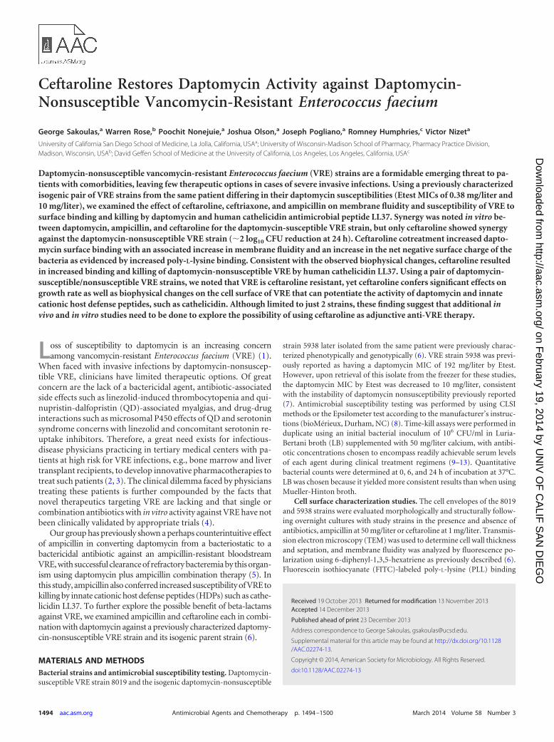
synergy (e.g., 2 log10 reduction in CFU) was seen only with cef-taroline plus daptomycin. Ceftriaxone did not enhance daptomy-cin killing of either strain. Linezolid showed bacteriostatic activityat 8 mg/liter, with CFU counts at 6 and 24 h nearly identical tothose in the starting inoculum.
Ceftaroline alters cell wall thickness. Growth of daptomycin-susceptible and -nonsusceptible VRE resulted in differences in cellwall thickness with subinhibitory antibiotic exposures, as shownin Fig. 2 (top left). There were no differences in cell wall thicknessbetween the 2 strains grown in the absence of antibiotic. Againstdaptomycin-susceptible strain 8019, ampicillin and ceftaroline re-sulted in decreases in cell wall thickness, while the opposite wasseen for daptomycin-resistant strain 5938. We had previouslynoted significant increases in cell division septum formation forstrain 5938 compared to that for strain 8019 (6). Ceftarolinemarkedly reduced septum formation in strain 8019 (36% com-pared to 47%, P � 0.05, chi-square). Against strain 5938, no sig-nificant changes in septum formation were seen with ceftaroline(54%) compared to growth control (58%).
Ceftaroline increases cell membrane fluidity. Previous workhas shown that daptomycin resistance in Enterococcus faecium andEnterococcus faecalis may be accompanied by decreases in mem-brane fluidity (6, 15). Interestingly, our previous characterizationof these 2 strains showed no changes in this property between thestrains under standard growth conditions (6). Growth of strain5938 in ceftaroline at 1 mg/liter showed significant increases inmembrane fluidity, as evidenced by mean polarization indexchanges by ceftaroline. For strain 8019, membrane fluidity wasnot significantly different (Fig. 2, top right).
Ceftaroline decreases net surface charge. Strains 8019 and5938 were grown in antibiotic-free or ceftaroline- or ampicillin-supplemented medium and subjected to PLL binding studies. Asseen in Fig. 3, strain 5938 exhibited significantly less binding toPLL compared to that of strain 8019, as can be expected upon lossof daptomycin susceptibility. Against strain 8019, only ceftarolinebut not ampicillin resulted in an increase in net negative surface
charge. Against strain 5938, both ampicillin and ceftaroline re-sulted in increased PLL binding, reflective of increased negativesurface charge on the bacterial surface.
Ceftaroline increases cell surface daptomycin binding. VREstrains 8019 and 5938 were labeled with 8 mg/liter Bo-DAP, dem-
FIG 1 In vitro antibiotic killing assays performed in calcium-supplementedLB (50 mg/liter) for daptomycin-susceptible VRE strain 8019 (left) and dap-tomycin-nonsusceptible VRE strain 5938 (right) demonstrating log10 CFU/mlat 6 h (top) and 24 h (bottom). Vertical bar denotes the starting inoculum.Concentrations of antibiotics used were daptomycin at 1 mg/liter for the sus-ceptible strain and 8 mg/liter for the nonsusceptible strain, ampicillin at 40mg/liter, ceftaroline at 5 mg/liter, and linezolid at 8 mg/liter. Note only dap-tomycin plus ceftaroline resulted in a net killing of 2 log10 CFU/ml at 24 hagainst the daptomycin-nonsusceptible strain.
FIG 2 Cell wall thickness (top left; mean � SD) and cell membrane polariza-tion fluidity (top right; mean � SD) in daptomycin-susceptible and -nonsus-ceptible VRE when exposed to subinhibitory concentrations of ampicillin at 50mg/liter or ceftaroline at 1 mg/liter. Cell wall thickness measurements wereperformed on 100 to 140 cells for each group. The polarization index is ex-pressed as degree of fluorescence polarization. Membrane fluidity is inverselycorrected to polarization index. *, P � 0.05; **, P � 0.0001 compared to thecontrol by unpaired t test. Bottom panels demonstrate representative electronmicrographs of daptomycin-susceptible (top row) and daptomycin-nonsus-ceptible (bottom row) bacteria grown under different antibiotic conditions asshown. The dark bar at the bottom left denotes 500 nm.
FIG 3 PLL binding of daptomycin-nonsusceptible and isogenic-susceptibleVRE when grown in the presence of ampicillin (AMP) at 40 mg/liter or cef-taroline (CPT) at 5 mg/liter compared to the control (CON) without antibi-otic. Increase binding is indicative of increase in negative charge. *, P � 0.05compared to the control.
Sakoulas et al.
1496 aac.asm.org Antimicrobial Agents and Chemotherapy
on February 19, 2014 by U
NIV
OF
CA
LIF S
AN
DIE
GO
http://aac.asm.org/
Dow
nloaded from
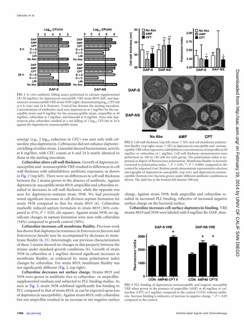
onstrating reduced relative binding of DAP for nonsusceptiblestrain 5938 compared to susceptible strain 8019 (see Fig. S2 in thesupplemental material). To measure the effect of various antibi-otics on Bo-DAP binding, VRE strains 8019 and 5938 were labeledwith 8 mg/liter and 16 mg/liter Bo-DAP, respectively, after growthin LB containing no antibiotic, ampicillin at 40 mg/liter, ceftriax-one at 20 mg/liter, or ceftaroline at 5 mg/liter (Fig. 4). Daptomycinbinding on the cell surface was significantly increased by ceftaro-line for both strains, as evidenced by the number of binding foci/cell, without differences in intensity of the foci. Ampicillin in-creased the number of daptomycin binding foci only for strain5938 and not for strain 8019. We tested ceftriaxone, as many cli-nicians might consider the convenience of once daily dosing, butfound no effect to increase binding of daptomycin.
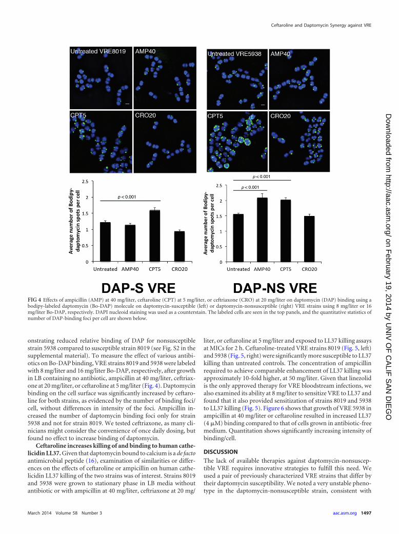
Ceftaroline increases killing of and binding to human cathe-licidin LL37. Given that daptomycin bound to calcium is a de factoantimicrobial peptide (16), examination of similarities or differ-ences on the effects of ceftaroline or ampicillin on human cathe-licidin LL37 killing of the two strains was of interest. Strains 8019and 5938 were grown to stationary phase in LB media withoutantibiotic or with ampicillin at 40 mg/liter, ceftriaxone at 20 mg/
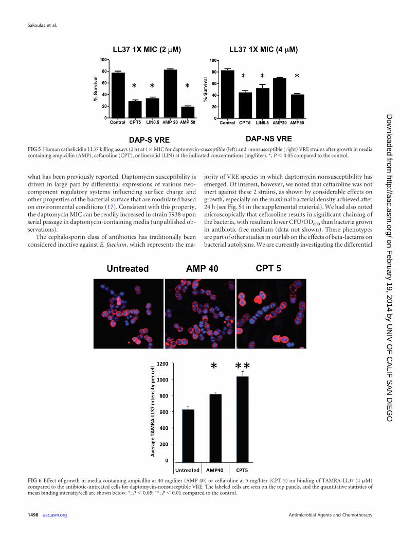
liter, or ceftaroline at 5 mg/liter and exposed to LL37 killing assaysat MICs for 2 h. Ceftaroline-treated VRE strains 8019 (Fig. 5, left)and 5938 (Fig. 5, right) were significantly more susceptible to LL37killing than untreated controls. The concentration of ampicillinrequired to achieve comparable enhancement of LL37 killing wasapproximately 10-fold higher, at 50 mg/liter. Given that linezolidis the only approved therapy for VRE bloodstream infections, wealso examined its ability at 8 mg/liter to sensitize VRE to LL37 andfound that it also provided sensitization of strains 8019 and 5938to LL37 killing (Fig. 5). Figure 6 shows that growth of VRE 5938 inampicillin at 40 mg/liter or ceftaroline resulted in increased LL37(4 �M) binding compared to that of cells grown in antibiotic-freemedium. Quantitation shows significantly increasing intensity ofbinding/cell.
DISCUSSION
The lack of available therapies against daptomycin-nonsuscep-tible VRE requires innovative strategies to fulfill this need. Weused a pair of previously characterized VRE strains that differ bytheir daptomycin susceptibility. We noted a very unstable pheno-type in the daptomycin-nonsusceptible strain, consistent with
FIG 4 Effects of ampicillin (AMP) at 40 mg/liter, ceftaroline (CPT) at 5 mg/liter, or ceftriaxone (CRO) at 20 mg/liter on daptomycin (DAP) binding using abodipy-labeled daptomycin (Bo-DAP) molecule on daptomycin-susceptible (left) or daptomycin-nonsusceptible (right) VRE strains using 8 mg/liter or 16mg/liter Bo-DAP, respectively. DAPI nucleoid staining was used as a counterstain. The labeled cells are seen in the top panels, and the quantitative statistics ofnumber of DAP-binding foci per cell are shown below.
Ceftaroline and Daptomycin Synergy against VRE
March 2014 Volume 58 Number 3 aac.asm.org 1497
on February 19, 2014 by U
NIV
OF
CA
LIF S
AN
DIE
GO
http://aac.asm.org/
Dow
nloaded from
what has been previously reported. Daptomycin susceptibility isdriven in large part by differential expressions of various two-component regulatory systems influencing surface charge andother properties of the bacterial surface that are modulated basedon environmental conditions (17). Consistent with this property,the daptomycin MIC can be readily increased in strain 5938 uponserial passage in daptomycin-containing media (unpublished ob-servations).
The cephalosporin class of antibiotics has traditionally beenconsidered inactive against E. faecium, which represents the ma-
jority of VRE species in which daptomycin nonsusceptibility hasemerged. Of interest, however, we noted that ceftaroline was notinert against these 2 strains, as shown by considerable effects ongrowth, especially on the maximal bacterial density achieved after24 h (see Fig. S1 in the supplemental material). We had also notedmicroscopically that ceftaroline results in significant chaining ofthe bacteria, with resultant lower CFU/OD600 than bacteria grownin antibiotic-free medium (data not shown). These phenotypesare part of other studies in our lab on the effects of beta-lactams onbacterial autolysins. We are currently investigating the differential
FIG 5 Human cathelicidin LL37 killing assays (2 h) at 1� MIC for daptomycin-susceptible (left) and -nonsusceptible (right) VRE strains after growth in mediacontaining ampicillin (AMP), ceftaroline (CPT), or linezolid (LIN) at the indicated concentrations (mg/liter). *, P � 0.05 compared to the control.
FIG 6 Effect of growth in media containing ampicillin at 40 mg/liter (AMP 40) or ceftaroline at 5 mg/liter (CPT 5) on binding of TAMRA-LL37 (4 �M)compared to the antibiotic-untreated cells for daptomycin-nonsusceptible VRE. The labeled cells are seen on the top panels, and the quantitative statistics ofmean binding intensity/cell are shown below. *, P � 0.05; **, P � 0.01 compared to the control.
Sakoulas et al.
1498 aac.asm.org Antimicrobial Agents and Chemotherapy
on February 19, 2014 by U
NIV
OF
CA
LIF S
AN
DIE
GO
http://aac.asm.org/
Dow
nloaded from

effect of class A penicillin binding protein (PBP) inactivation onsusceptibility to daptomycin and antimicrobial peptides andwhether ceftaroline and/or ampicillin exert their effects on VREthrough these targets (15). Additional evidence by others suggeststhat low-molecular-weight PBPs, such as ddcP, which influencesD-alanine-D-alanine carboxypeptidase activity (16), warrant studyas well, particularly with the possible tangential effects on vanco-mycin MIC by ceftaroline.
For daptomycin-nonsusceptible VRE strain 5938, daptomycinat 8 mg/liter, which would be close to the maximum free drugconcentration in vivo (10), allows bacterial growth that is not dif-ferent from growth control. Ceftriaxone showed no appreciablesynergy with daptomycin against both daptomycin-susceptibleand -nonsusceptible strains 8019 and 5938, respectively. Consis-tent with our prior data (5), ampicillin resulted in synergy withdaptomycin against strain 8019, but this synergy was lost uponemergence of daptomycin resistance in strain 5938. Of great inter-est, however, is that ceftaroline showed synergy with daptomycinagainst both strains, achieving �2 log10 CFU killing at 24 h. Thiscombination regimen was considerably more potent than lin-ezolid, the only drug currently approved for VRE bacteremia (13),which was entirely bacteriostatic against both strains. Neverthe-less, we did find that linezolid did sensitize VRE to killing by cathe-licidin LL37, suggesting that in vivo the drug may yield greateractivity than kill curves might suggest. Although not ideal, lin-ezolid has been used to treat VRE bacteremia successfully and isapproved for this indication (13).
In support of the in vitro findings of killing of daptomycin plusceftaroline against VRE, we have shown that ceftaroline (i) in-creases daptomycin surface binding against daptomycin-suscep-tible and -nonsusceptible VRE, with parallel increases in poly-L-lysine binding suggesting an increase in the net negative charge,(ii) increases membrane fluidity for daptomycin-nonsusceptibleVRE, a property that would be expected to increase susceptibilityto daptomycin given that daptomycin-nonsusceptible strainsshow a decreased membrane fluidity phenotype, and (iii) sensi-tizes VRE to killing by the cationic antimicrobial peptide humancathelicidin LL37, via increased LL37 binding to the surface ofVRE. A summary of the changes is presented in Table 2. Note thatampicillin and ceftaroline resulted in decreases in cell wall thick-ness in the daptomycin-susceptible strain and increases in thenonsusceptible strain (Table 2). Collectively, these data provide aninteresting pattern on analysis. First, it appears that enhancementof daptomycin binding by ampicillin may not be consistent acrossVRE strains, as the daptomycin-susceptible strain in this study didnot behave like the strain in our prior study (5). Nevertheless, itappears that marked decreases in cell wall thickness induced byampicillin on this strain may somehow render the organism moredaptomycin susceptible independent of daptomycin binding. Sec-ond, in the setting of increased cell wall thickness that occurs withboth ceftaroline and ampicillin, a concomitant increase in mem-brane fluidity may be necessary to result in daptomycin synergy.Ampicillin, which failed to increase membrane fluidity, failed toshow synergy with daptomycin against the daptomycin-nonsus-ceptible strain. Finally, increased daptomycin binding may not benecessary or sufficient for enhanced daptomycin activity. Dapto-mycin-susceptible VRE showed synergy with ampicillin plus dap-tomycin without an improvement in Bo-DAP or poly-L-lysinebinding. On the other hand, increased Bo-DAP and poly-L-lysine
by ampicillin in the daptomycin-nonsusceptible strain did notresult in synergy.
These data are built upon an increasing line of evidence dem-onstrating that the in vitro effects of antibiotics are not fully ap-preciated by current methods of susceptibility testing. Specifically,beta-lactam antibiotics appear to have profound effects on beta-lactam-resistant Gram-positive organisms. Similar to the effectson VRE by ampicillin that we previously discussed (5), we haveshown that antistaphylococcal beta-lactams increase susceptibil-ity of methicillin-resistant Staphylococcus aureus (MRSA) to dap-tomycin and innate immunity (14, 18). Not all beta-lactams areequal in this effect, as PBP-1 binding beta-lactams are better atpotentiating daptomycin activity than non-PBP-1-specific beta-lactams (19).
The development of daptomycin nonsusceptibility appears tobe multifactorial and complex and, therefore, effects of drugs thatrestore its susceptibility would be expected to be equally complex(5, 20–22). Daptomycin-nonsusceptible enterococci are differentfrom their susceptible counterparts in sensing cell wall stress (vialiaFSR) (15, 16, 18), phospholipid composition (via cls) (20–22),and increased proclivity to septation (via ezrA) (6). While we didnot perform analysis of phospholipid content with ceftarolinetherapy, we noticed changes in septation and cell wall thicknessinduced by ceftaroline. Perhaps ceftaroline, similar to what hasbeen observed with ceftobiprole (23), binds PBP5 and alters thesurface physiology to allow enhanced daptomycin and HDP ac-tivity.
It is important to point out that these data are limited to a singledaptomycin-susceptible/nonsusceptible pair of VRE strains. As it iswell appreciated that clinical strains are quite heterogeneous, ad-
TABLE 2 Summary of changes induced on daptomycin-susceptible and-nonsusceptible VRE by ampicillin or ceftaroline
Characteristic
Resulta
DAP-susceptibleVRE
DAP-nonsusceptibleVRE
Synergy with DAPAMP � CPT � �
Effect on cell wall thicknessAMP 22 1CPT 2 1
Effect on membrane fluidityAMP CPT 1
Poly-L-lysine bindingAMP 1CPT 1 1
Bodipy-DAP bindingAMP 1CPT 1 1
LL37 binding and activityAMP 1 1CPT 11 11
a �, yes; , no;22, more-marked decrease;2, decrease;11, more-markedincrease;1, increase.
Ceftaroline and Daptomycin Synergy against VRE
March 2014 Volume 58 Number 3 aac.asm.org 1499
on February 19, 2014 by U
NIV
OF
CA
LIF S
AN
DIE
GO
http://aac.asm.org/
Dow
nloaded from

ditional in vitro and in vivo studies are needed to explore the effectsof ceftaroline on VRE as potential adjunctive therapy and todetermine whether all the changes we observed in these strains aregeneralizable to other clinical VRE strains. Several of us arecurrently exploring in vitro PK/PD modeling of ceftaroline com-bination therapy against other VRE strains, while others are com-pleting checkerboard studies of daptomycin plus various beta-lactams, including ceftaroline, against a larger collection of VREstrains, some of which are daptomycin nonsusceptible. While wedid not have the opportunity to employ this combination clini-cally, we did recently publish a report showing in vitro synergyusing daptomycin and ceftaroline in the successful treatment of E.faecalis aortic valve endocarditis (24). Optimal dosing of dapto-mycin or ceftaroline, should a clinician choose this combination,remains to be determined, although we might suggest 8 to 10mg/kg of body weight/day of daptomycin given prior studiesshowing increased efficacy of higher doses compared to that of �6mg/kg/day (25).
ACKNOWLEDGMENTS
Ceftaroline powder was obtained from Forest Pharmaceuticals. The re-search described in the manuscript was conducted at the sole discretion ofthe authors without the knowledge or support of Forest Laboratories, Inc.,Forest Research Institute, Inc., or Cerexa, Inc.
G.S. has received speaking honoraria from Cubist, Forest, and PfizerPharmaceuticals, consulting fees from Cubist and Forest Pharmaceuti-cals, and research grant support from Forest Pharmaceuticals. W.R. hasreceived grant funding and speaking honoraria from Cubist and consultsfor The Medicines Company. R.H. has received research funding fromCubist. V.N. is on the Scientific Advisory Board of Trius Therapeutics.
Funding for this research was provided by U54 HD071600-01 09/26/2011-06/30/2016 NICHD on Developmental and Translational Pharma-cology of Pediatric Antimicrobial Therapy (G.S. and V.N.), the GreatLakes Regional Center for Excellence in Biodefense and Emerging Infec-tious Disease Research (AI057153; V.N.), and R01GM073898 (J.P.). Nofunding bodies had any role in study design, data collection and analysis,decision to publish, or preparation of the manuscript.
REFERENCES1. Kelesidis T, Humphries Uslan DZ, Pegues DA. 2011. Daptomycin
nonsusceptible enterococci: an emerging challenge for clinicians. Clin.Infect. Dis. 52:228 –234. http://dx.doi.org/10.1093/cid/ciq113.
2. Orloff SL, Busch AM, Olyaei AJ, Corless CL, Benner KG, Flora KD,Rosen HR, Rabkin JM. 1999. Vancomycin-resistant Enterococcus in livertransplant patients. Am. J. Surg. 177:418 – 422. http://dx.doi.org/10.1016/S0002-9610(99)00083-5.
3. Kamboj M, Chung D, Seo SK, Pamer EG, Sepkowitz KA, JakubowskiAA, Papanicolaou G. 2010. The changing epidemiology of vancomycin-resistant Enterococcus (VRE) bacteremia in allogeneic hematopoieticstem cell transplant (HSCT) recipients. Biol. Blood Marrow Transplant.16:1576 –1581. http://dx.doi.org/10.1016/j.bbmt.2010.05.008.
4. Arias CA, Contreras GA, Murray BE. 2010. Management of multidrug-resistant enterococcal infections. Clin. Microbiol. Infect. 16:555–562.http://dx.doi.org/10.1111/j.1469-0691.2010.03214.x.
5. Sakoulas G, Bayer AS, Pogliano J, Tsuji BT, Yang SJ, Mishra NN, NizetV, Yeaman MR, Moise PA. 2012. Ampicillin enhances daptomycin- andcationic host defense peptide-mediate killing of ampicillin and vancomy-cin-resistant Enterococcus faecium. Antimicrob. Agents Chemother. 56:838 – 844. http://dx.doi.org/10.1128/AAC.05551-11.
6. Humphries RM, Kelesidis T, Tewhey R, Rose WE, Schork N, Nizet N,Sakoulas. G. 2012. Genotypic and phenotypic evaluation of the evolutionof high-level daptomycin non-susceptibility in vancomycin-resistant En-terococcus faecium. Antimicrob. Agents Chemother. 56:6051– 6053. http://dx.doi.org/10.1128/AAC.01318-12.
7. Rose WE, Leonard SN, Sakoulas G, Kaatz GW, Zervos MM, Sheth A,Carpenter CF, Rybak MJ. 2008. Daptomycin activity against Staphylo-
coccus aureus following vancomycin exposure in an in vitro pharmacody-namic model with simulated endocardial vegetations. Antimicrob. AgentsChemother. 52:831– 836. http://dx.doi.org/10.1128/AAC.00869-07.
8. CLSI. 2013. Performance standards for antimicrobial susceptibility test-ing; 23rd informational supplement. CLSI document M100-S23. Clinicaland Laboratory Standards Institute, Wayne, PA.
9. Forest Laboratories Inc. 10 October 2012, accession date. Ceftaroline fosamilpackage insert. Forest Laboratories Inc., New York, NY. http://www.accessdata.fda.gov/drugsatfda_docs/label/2010/200327s000lbl.pdf.
10. Dvorchik BH, Brazier D, DeBruin MF, Arbeit RD. 2003. Daptomycinpharmacokinetics and safety following administration of escalating dosesonce daily to healthy subjects. Antimicrob. Agents Chemother. 47:1318 –1323. http://dx.doi.org/10.1128/AAC.47.4.1318-1323.2003.
11. Foulds G. 1983. Pharmacokinetics of sulbactam/ampicillin in humans: areview. Rev. Infect. Dis. 8(Suppl 5):S503–S511.
12. Patel IH, Chen S, Parsonnet Hackman MR, Brooks MA, Konikoff J,Kaplan SA. 1981. Pharmacokinetics of ceftriaxone in humans. Antimi-crob. Agents Chemother. 20:634 – 641. http://dx.doi.org/10.1128/AAC.20.5.634.
13. Dryden MS. 2011. Linezold pharmacokinetics and pharmacodynamics inclinical treatment. J. Antimicrob. Chemother. 66(Suppl 4):iv7–iv15.
14. Dhand A, Bayer AS, Pogliano J, Yang SJ, Bolaris M, Nizet V, Wang G,Sakoulas G. 2011. Use of antistaphylococcal beta-lactams to increase dap-tomycin activity in eradicating persistent bacteremia due to methicillin-resistant Staphylococcus aureus: role of daptomycin binding. Clin. Infect.Dis. 53:158 –163. http://dx.doi.org/10.1093/cid/cir340.
15. Rice LB, Carias LL, Rudin S, Hutton R, Marshall S, Hassan M, JosseaumeN, Dubost L, Marie A, Arthur M. 2009. Role of class A penicillin-bindingproteins in the expression of beta-lactam resistance in Enterococcus faecium.J. Bacteriol. 191:3649. http://dx.doi.org/10.1128/JB.01834-08.
16. Zhang X, Paganelli FL, Bierschenk D, Kuiper A, Bonten MJM, WillemsRJL, van Schaik W. 2012. Genome-wide identification of ampicillin re-sistance determinants in Enterococcus faecium. PLoS Genet. 8(6):e1002804. http://dx.doi.org/10.1371/journal.pgen.1002804.
17. Kawada-Matsuo M, Yoshida Y, Nakamura N, Konatsuzawa H. 2011.Role of two-component systems in the resistance of Staphylococcus aureusto antibacterial agents. Virulence 2:427– 430. http://dx.doi.org/10.4161/viru.2.5.17711.
18. Sakoulas G, Okumura CY, Thienphrapa, Olson J, Nonejuie P, Dam Q,Dhand A, Pogliano J, Yeaman MR, Hensler ME, Bayer AS, Nizet V. 3December 2013. Nafcillin enhances innate immune-mediated killing ofmethicillin-resistant Staphylococcus aureus. J. Mol. Med. http://dx.doi.org/10.1007/s00109-013-1100-7.
19. Berti A, Sakoulas G, Nizet V, Tewhey R, Rose WE. 2013. �-Lactamantibiotics targeting PBP1 selectively enhance daptomycin activity againstmethicillin-resistant Staphylococcus aureus. Antimicrob. Agents Che-mother. 57:5005–5012. http://dx.doi.org/10.1128/AAC.00594-13.
20. Mishra NN, Bayer AS, Tran TT, Shamoo Y, Mileykovskaya E, DowhanW, Guan Z, Arias CA. 2012. Daptomycin resistance in enterococci isassociated with distinct alterations of cell membrane phospholipid con-tent. PLoS One 7(8):e43958. http://dx.doi.org/10.1371/journal.pone.0043958.
21. Munita JM, Panesso D, Diaz L, Tran TT, Reyes J, Wanger A, MurrayBE, Arias CA. 2012. Correlation between mutations in liaFSR of Entero-coccus faecium and MIC of daptomycin: revisiting daptomycin break-points. Antimicrob. Agents Chemother. 56:4354 – 4359. http://dx.doi.org/10.1128/AAC.00509-12.
22. Arias CA, Panesso D, McGrath DM, Qin X, Mojica MF, Miller C, Diaz L,Tran TT, Rincon S, Barbu EM, Reyes J, Roh JH, Lobos E, Sodergren E,Pasqualini R, Arap W, Quinn JP, Shamoo Y, Murray BE, Weinstock GM.2011. Genetic basis for in vivo daptomycin resistance in enterococci. N. Engl.J. Med. 365:892–900. http://dx.doi.org/10.1056/NEJMoa1011138.
23. Xavier H, Amoroso A, Coyette J, Joris B. 2010. Interaction of ceftobiprolewith the low-affinity PBP 5 of Enterococcus faecium. Antimicrob. AgentsChemother. 54:953–955. http://dx.doi.org/10.1128/AAC.00983-09.
24. Sakoulas G, Noneluie P, Nizet V, Pogliano J, Crum-Cianflone N, HaddadF. 2013. Treatment of high-level gentamicin-resistant Enterococcus faecalisendocarditis with daptomycin plus ceftaroline. Antimicrob. Agents Che-mother. 57:4042– 4045. http://dx.doi.org/10.1128/AAC.02481-12.
25. King EA, McCoy D, Desai S, Nyirenda T, Bicking K. 2011. Vancomycin-resistant Enterococcal bacteremia and daptomycin: are higher doses nec-essary? J. Antimicrob. Chemother. 66:2112–2118. http://dx.doi.org/10.1093/jac/dkr255.
Sakoulas et al.
1500 aac.asm.org Antimicrobial Agents and Chemotherapy
on February 19, 2014 by U
NIV
OF
CA
LIF S
AN
DIE
GO
http://aac.asm.org/
Dow
nloaded from