ce in biotechnology & pharmaceutical€¦ · scientific final program summary ... case study:...
TRANSCRIPT
CE in Biotechnology & Pharmaceutical
Industries: 12th Symposium on the Practical Applications for the Analysis of
Proteins, Nucleotides and Small Molecules
(CE Pharm 2010)
Symposium Chair:
Sarah Kennett, CDER, FDA Cari Sänger - van de Griend, Abbott
Healthcare Products
October 17-21, 2010 Vancouver Marriott Pinnacle
Vancouver, BC Canada
Organized by

Welcome to CE in the Biotechnology and Pharmaceutical Industries: 12th Symposium on the Practical Applications for the Analysis of Proteins, Nucleotides and Small Molecules We are pleased to welcome you to Capillary Electrophoresis in the Biotechnology and Pharmaceutical Industries: 12th Symposium on the Practical Applications for the Analysis of Proteins, Nucleotides and Small Molecules, a symposium devoted to the practical concerns which will strengthen the use of CE within the biotechnology and pharmaceutical industries. The goal of this symposium is to provide a forum for the discussion of recent developments in CE analysis of protein, nucleotide and small molecule pharmaceuticals. The presentations and workshops will be devoted completely for practical concerns to strengthen the use of CE within the biotechnology and pharmaceutical industries. Applications will highlight uses of CE in various areas of product development including high throughput screening, formulation studies, process development, product characterization and validated lot release and stability testing. The Symposium will feature presentations from leading experts within the industries and the regulatory agencies from around world. The symposia will allow for open discussions aimed at improving and increasing the use of CE for analysis of proteins, small molecules, carbohydrates, metabolites, and other molecules, with a focus on validation and qualification, improving sensitivity and technique, new technology, and QbD. Three workshops will focus on instrument qualification, best practices, and the use of QbD for analytical methods. The success of this Symposium will depend not only on the outstanding cast of experienced and knowledgeable speakers and workshop leaders, but also on the interactions and open discussion that take place among the attendees. We encourage you to participate wholeheartedly in the discussion sections that have been designed to stimulate exchange of ideas and information. We would like to thank the speakers who are generously giving their time and resources, and also you for your attendance, which will make this endeavor a success. We gratefully acknowledge the generosity of our exhibitors and program partners: Agilent Technologies, Amgen, Inc., Analis, Beckman Coulter, Inc., Calipers Life Sciences, Convergent Bioscience, Genentech, a Member of the Roche Group, Genetic Engineering & BioTech News, LC/GC North America, Life Science Connect, Novartis, Pfizer, Inc., Picometrics, Inc., Polymicro Technologies, a Subsidiary of Molex, and Royal Society of Chemistry. We are thankful for the expert assistance of CASSS and the audiovisual expertise of Michael Johnstone from MJ Film/Video Productions. Their experience and guidance in the preparation of this Symposium have been invaluable. THE ORGANIZING COMMITTEE François de l'Escaille, Analis Gamze Belin, Novartis Franka Kálmán, University of Applied Sciences Western Switzerland Sarah Kennett, CDER, FDA (Co-chair) Mark Lies, Beckman Coulter, Inc. Stacey Ma, Genentech, a Member of the Roche Group Brian K. Nunnally, Pfizer, Inc. SungAe Suhr Park, Amgen, Inc. Meg Ruesch, Pfizer, Inc. Cari Sänger - van de Griend, Abbott Healthcare Products (Co-chair)
2
Table of Contents Corporate, Exhibitor and Media Program Partners..............................................................4 Scientific Final Program Summary......................................................................................6 CE Pharm Award ...............................................................................................................14 Session Abstracts ...............................................................................................................17 W
orkshop Session I (Monday, October 18) Description...................................................41
orkshop Session II (Tuesday, October 19) Description .................................................42
orkshop Session III (Wednesday, October 20) Description...........................................43
echnical Seminar (Monday, October 18) Abstract ..........................................................44
echnical Seminar (Tuesday, October 19) Abstract..........................................................45
echnical Seminar (Wednesday, October 20) Abstract.....................................................47
oster Abstracts .................................................................................................................49
W W T T T P
3

The Organizing Committee gratefully acknowledges the following program partners for their generous support of this Symposium:
Diamond Program Partners
Agilent Technologies Genentech, a Member of the Roche Group
Platinum Program Partner
Beckman Coulter, Inc.
Silver Program Partners
Amgen, Inc.
Novartis Pfizer, Inc.
Bronze Program Partner
Analis
4

Exhibitors
Agilent Technologies Beckman Coulter, Inc. Caliper Life Sciences
Convergent Bioscience Picometrics, Inc.
Polymicro Technologies, a Subsidiary of Molex
Leading Media Program Partners
LCGC North America Life Science Connect
Media Program Partners
Genetic Engineering & BioTech News
Royal Society of Chemistry
5

CE Pharm 2010 Scientific Final Program Summary
Sunday, October 17, 2010
08:30 – 09:00 Registration and Breakfast (for course attendees ONLY) in Pinnacle Foyer 09:00 – 15:00
Short Course in Shaughnessy I Application of CE to the Analysis of Protein Therapeutics
Short Course Facilitators: Chantal Felten, Alpine Analytical Academy, Vancouver, BC Canada and Oscar Salas-Solano, Seattle Genetics, Bothell, WA USA
10:30 – 11:00 Break in the Shaughnessy I 12:30 – 13:30 Hosted Lunch (for course attendees ONLY) in Pinnacle Foyer
6

Monday, October 18, 2010 07:30 – 08:30 Breakfast in the Pinnacle Foyer 07:00 – 17:00 Registration in the Pinnacle Foyer 08:30 – 08:45 Welcome and Introductory Comments in Pinnacle II & III
Sarah Kennett, CDER, FDA, Bethesda, MD USA
Transitioning to CE/Characterization of Peaks Session in Pinnacle II & III Session Chair: Cari Sänger - van de Griend, Abbott Healthcare Products, Weesp, The Netherlands
08:45 – 09:10 Case Study: Post-Approval Introduction of Capillary Electrophoresis
Methods into a Control System for a Recombinant Human Enzyme Therapeutic Robert Baffi, BioMarin Pharmaceutical Inc., Novato, CA USA
09:10 – 09:35 Characterization of cIEF Charge Variants Using Rotofor®-based Enrichment Amanda Miller, Amgen, Inc., Seattle, WA USA
09:35 – 10:00 Capillary and Microfluidic Electrophoresis Identification of Therapeutic
Recombinant Humanized Monoclonal Antibody Isoforms Jonathan Cooper, MedImmune, Gaithersburg, MD USA 10:00 – 10:15 Discussion 10:15 – 10:45 Break – Visit the Exhibits and Posters in Pinnacle I & Foyer 10:45 – 11:15 CE Pharm Partner Showcase 11:15 – 12:45 Lunch Break – Visit the Exhibits and Posters in Pinnacle I & Foyer 11:30 – 12:30 Technical Seminar/Lunch and Learn Martin Greiner, Agilent Technologies, Waldbronn, Germany The Agilent Electrophoresis Portfolio and How to Combine Agilent 7100 CE with External Detectors (LIF, MS, CCD, UV Area Imaging) Phillip Britz-McKibbin, McMaster University, Hamilton, ON Canada New Advances in Capillary Electrophoresis-Mass Spectrometry for Metabolite Profiling
Sponsored by Agilent Technologies Pinnacle II & III
7

Monday, October 18, 2010 continued
Qualification/Validation Session in Pinnacle II & III Session Chair: Stacey Ma, Genentech, a Member of the Roche Group, South San Francisco, CA USA
12:45 – 13:10 Validation of Capillary - Based IEF Methods for Routine QC of
Biopharmaceuticals Katia Minari, Merck Serono, Montecelio, Italy 13:10 – 13:35 Development and Qualification of a Reverse Mode Capillary Zone
Electrophoresis Assay for Measuring Total Protein in Carryover Studies David A. Michels, Genentech, a Member of the Roche Group, South San Francisco, CA USA
13:35 – 14:00 Heparin Contaminates: The Case for Capillary Electrophoresis Todd Wielgos, Baxter Healthcare Corporation, Round Lake, IL USA 14:00 – 14:15 Discussion 14:15 – 14:45 Break – Visit the Exhibits and Posters in Pinnacle I and Foyer 14:45 – 15:45
Workshop I: Instrument Qualification in Pinnacle II & III Workshop Facilitators:
Gamze Belin, Novartis, Basel, Switzerland and SungAe Suhr Park, Amgen, Inc., Thousand Oaks, CA USA
Assessing Purity of Therapeutic Products in Pinnacle II & III Session Chair: Mark Lies, Beckman Coulter, Inc., Brea, CA USA
15:45 – 16:10 CE as a Powerful Tool for the Separation of Small Molecules Klaus Feige, Merck KGaA, Frankfurt, Germany 16:10 – 16:35 NMR and Computational Studies of Chiral Recognition of AMG 476 by
Capillary Electrophoresis Rick Chiu, Amgen, Inc., Thousand Oaks, CA USA 16:35 – 17:00 The Usefulness of Capillary Electrophoresis in the Separation of Intact
Glycoprotein Glycoforms with Pharmaceutical and Clinical Relevance Mercedes de Frutos, Spanish Council Research (CSIC), Madrid, Spain 17:00 – 17:15 Discussion 17:15 – 19:00 Welcome Reception in the Ambleside Room (4th Floor)
8

Tuesday, October 19, 2010 07:00 – 17:00 Registration in the Pinnacle Foyer 07:30 – 08:30 Breakfast in the Pinnacle Foyer 07:30 – 08:30 Technical Sunrise Seminar Jiaqi Wu, Convergent Bioscience, Toronto, ON Canada Technology Update at Convergent Bioscience Sponsored by Convergent Bioscience Pinnacle II & III
New Technology Session in Pinnacle II & III Session Chair: François de l'Escaille, Analis, Namur, Belgium
08:30 – 08:55 Details of Monoclonal Antibody Reduction and Re-Oxidation From Cell
Culture Through Purification Tim Blanc, ImClone Systems, A wholly-owned subsidiary of Eli Lilly,
Branchburg, NJ USA 08:55 – 09:20 Rapid Analysis of Charge Variants of Monoclonal Antibodies with Capillary
Zone Electrophoresis in Uncoated Capillary Column Yan He, Pfizer, Inc., Chesterfield, MO USA 09:20 – 09:45 Metabolomics for Quantitative Assessment of Oxidative Stress by Capillary
Electrophoresis-Mass Spectrometry: Measuring the Efficacy of N-Acetylcysteine Intervention in Strenuous Exercise Phillip Britz-McKibbin, McMaster University, Hamilton, ON Canada
09:45 – 10:00 Discussion 10:00 – 10:30 Break – Visit the Exhibits and Posters in Pinnacle I and Foyer
Improving Sensitivity Session in Pinnacle II & III Session Chair: Meg Ruesch, Pfizer, Inc., Chesterfield, MO USA
10:30 – 10:55 Sample Preconcentration for Capillary Electrophoresis-Mass Spectrometry Doo Soo Chung, Seoul National University, Seoul, Korea
10:55 – 11:20 Man 5 N-linked Oligosaccharide Labeling Efficiency with APTS by CZE-LIF
for Monoclonal Antibodies Yuling Zhang, Amgen, Inc., Seattle, WA USA
11:20 – 11:45 Rapid Analysis and Sensitive Detection of Clinically Important Compounds Gamze Belin, Novartis, Basel, Switzerland
11:45 – 12:00 Discussion
9

Tuesday, October 19, 2010 continued 12:00 – 13:30 Lunch Break – Visit the Exhibits and Posters in Pinnacle I and Foyer 12:15 – 13:15 Technical Seminar/Lunch and Learn Sponsored by Beckman Coulter Pinnacle II & III
Best Practice/Experience from Long Term Use Session in Pinnacle II & III Session Chair: Gamze Belin, Novartis, Basel, Switzerland
13:30 – 13:55 Applications of CE in Biopharmaceutical Development Zoran Sosic, Biogen Idec, Cambridge, MA USA 13:55 – 14:20 Analytical Method Transfer and Monitoring – Key Elements of the
Analytical Method Life Cycle Management Joerg Herrmann, Genentech, a Member of the Roche Group, South San Francisco, CA USA
14:20 – 14:45 Analysis of Small Ions in the Development of New Pharmaceutical
Substances using CE with Indirect Detection Anna Maria Enlund, AstraZeneca, Sodertalje, Sweden 14:45 – 15:00 Discussion 15:00 – 16:30 Poster Session – Discuss Posters with Presenters and Visit Exhibits in Pinnacle I
& Foyer 16:30 – 17:30
Workshop II: Best Practices in Pinnacle II & III Workshop Facilitators:
Franka Kálmán, University of Applied Sciences Western Switzerland, Sion, Switzerland and Meg Ruesch, Pfizer, Inc. Chesterfield, MO USA
17:30 – 18:30 Exhibit and Poster Reception in the Pinnacle I & Foyer
10
Wednesday, October 20, 2010 07:30 – 08:30 Breakfast in the Pinnacle Foyer 08:00 – 16:00 Registration in the Pinnacle Foyer 08:30 – 08:45 CE Pharm Award Presentation
Regulatory Session in Pinnacle II & III Session Chair: SungAe Suhr Park, Amgen, Inc., Thousand Oaks, CA USA
08:45 – 09:10 Validation of Capillary Electrophoresis Test Procedures: FAQ’s
Stefan Christians, Paul-Ehrlich-Institut, Langen, Germany
09:10 – 09:35 Uses and Limitations of Capillary Electrophoresis for Subsequent Entry Biologics
Michel Girard, Health Canada, Ottawa, ON Canada 09:35 – 10:00 Assay Qualification/Validation – a Reviewer’s Expectations
Sarah Kennett, CDER, FDA, Bethesda, MD USA 10:00 – 10:30 Discussion 10:30 – 11:00 Break – Visit the Exhibits and Posters in Pinnacle I & Foyer
Late Breaking Session in Pinnacle II & III Session Chair: Cari Sänger - van de Griend, Abbott Healthcare Products, Weesp, The Netherlands
11:00 – 11:25 Application of iCIEF Technology Beyond mAbs: A Case Study Kunnel Babu, Bristol-Myers Squibb Company, Pennington, NJ USA
11:25 – 11:50 Enhancing the Coverage of the Urinary Metabolome using Sheathless CE-
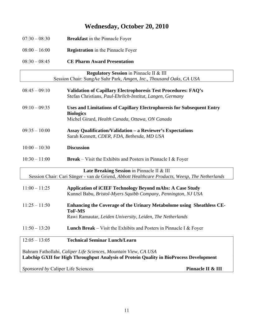
ToF-MS Rawi Ramautar, Leiden University, Leiden, The Netherlands 11:50 – 13:20 Lunch Break – Visit the Exhibits and Posters in Pinnacle I & Foyer 12:05 – 13:05 Technical Seminar Lunch/Learn Bahram Fathollahi, Caliper Life Sciences, Mountain View, CA USA Labchip GXII for High Throughput Analysis of Protein Quality in BioProcess Development Sponsored by Caliper Life Sciences Pinnacle II & III
11

Wednesday, October 20, 2010 continued
QbD Session in Pinnacle II & III Session Chair: Sarah Kennett, CDER, FDA, Bethesda, MD USA
13:20– 13:45 Advanced Applications of Capillary Electrophoresis to Biotech Product
Development: Monitoring Protein Quality From Clone Selection to Final Release Flavie Robert, Merck Serono SA, Corsier Sur Vevey, Switzerland
13:45 – 14:10 Analytical Method Design Space in Biopharmaceutical CE Analysis
Xinfeng Zhang, Amgen, Inc., Fremont, CA USA 14:10 – 14:25 Discussion 14:25 – 15:25
Workshop III: Method QbD in Pinnacle II & III Workshop Facilitators:
Chantal Felten, Alpine Analytical Academy, Vancouver, BC Canada and Sarah Kennett, CDER, FDA, Bethesda, MD USA
15:25 – 15:45 Closing Comments in Pinnacle II & III
Cari Sänger - van de Griend, Abbott Healthcare Products, Weesp, The Netherlands
12

Thursday, October 21, 2010 8:30 – 9:00 Registration in the Pinnacle Foyer 8:30 – 9:00 Breakfast (for course attendees ONLY) in the Pinnacle Foyer 9:00 – 15:00
Short Course Session in Shaughnessy I Method Development, Optimization and Good Working Practice in Capillary Electrophoresis
Short Course Facilitator: François de l'Escaille, Analis, Namur, Belgium and Cari Sänger - van de Griend, Abbott Healthcare Products, Weesp, The Netherlands
10:30 – 11:00 AM Break in the Shaughnessy I 12:30 – 13:30 Hosted Lunch (for course attendees ONLY)
13

CE Pharm Award History
The Organizing Committee of CE Pharm bestows an annual award on behalf of CASSS. The award is based on an individual’s sustained and significant contribution to the application of CE to the analysis of biopharmaceuticals and pharmaceuticals. The award is a peer acknowledgement of the recipient’s contribution and is decided by the CE Pharm committee each year.
Past Recipients of the "CE Pharm Award" include: 2006 - Norberto Guzman - Johnson & Johnson 2007 - Kevin Altria - GlaxoSmithKline 2008 - Anthony Chen and Wassim Nashabeh – Genentech, Inc. 2009 - Stacey Ma – Genentech, Inc. 2010 - Announced Wednesday, October 20 at 8:30AM Do you think we are missing someone influential? Add your suggestion to the list. Suggestions for next year’s award can be submitted on your evaluation.
14

INSERT ANALIS AD HERE
15

INSERT NOVARTIS AD HERE
16

Session Abstracts
Case Study: Post-Approval Introduction of Capillary Electrophoresis Methods into a Control System for a Recombinant Human Enzyme Therapeutic Robert Baffi BioMarin Pharamaceutical Inc., Novato, CA USA Oligosaccharide Profiling by Fluorophore-Assisted Carbohydrate Electrophoresis (FACE) and IsoElectric Focusing (IEF) to monitor charge heterogeneity are utilized as a measure of product consistency and are classified as purity tests for release of one our approved recombinant enzyme therapeutics. BioMarin has completed the implementation of alternate methods for replacement for the FACE and IEF with Capillary Electrophoresis (CE) methods. This presentation will address the technical and regulatory strategies that were successful implemented to revise worldwide licenses for both the methods and corresponding specifications. NOTES:
17

Characterization of cIEF Charge Variants using Rotofor®-based Enrichment Amanda Miller; Himanshu Gadgil Amgen, Inc., Seattle, WA USA Characterizing charge variants of therapeutic proteins is an important step in development. Capillary isoelectric focusing (cIEF) is a high resolution method that is commonly used as a charge-based release assay, but it is not practical to collect peaks from cIEF for further analysis. Rotofor® preparative isoelectric focusing (IEF) was used to enrich minor charge variants in a mAb. Semi-preparative scale allowed enrichment of four hundred milligrams of material which enabled further in-depth biochemical analysis. This technique offered efficiency, high yield, and good correlation to cIEF charge variants. The purified Rotofor® fractions were reanalyzed by cIEF to confirm purity then analyzed using top-down, middle-down, and bottom-up LC-MS analysis to characterize these charge variants. Our data showed the major contribution to charged species was from the conversion of the terminal glutamine to pyroglutamic acid on both the heavy chain and light chain, however we were also able to characterize other distinct modifications such as signal peptide, lysine variants, oxidation, and glycation. Enrichment allowed for identification of signal peptide at levels less than one-tenth of a percent, along with other modifications at levels lower than detected by routine characterization assays. Our results demonstrate that this procedure is not only useful for characterization of charge variants, but it also allows for in-depth characterization and identification of modifications at an early stage in development. NOTES:
18
Capillary and Microfluidic Electrophoresis Identification of Therapeutic Recombinant Humanized Monoclonal Antibody Isoforms Jonathan Cooper MedImmune, Gaithersburg, MD USA Abstract not avaliable at the time of print. NOTES:
19

Validation of Capillary - Based IEF Methods for Routine QC of Biopharmaceuticals Katia Minari Merck Serono, Montecelio, Italy Isoelectric focusing (IEF) is a powerful analytical technique for the separation of different proteins according to charge and to assess the charge heterogeneity of a given protein. Historically, gel-based IEF was mainly employed for characterization purposes. However, with the coming of age of capillary-based systems, these become progressively state of the art in the biopharmaceutical industry for routine QC, stability testing, and characterization of biopharmaceuticals due to their superiority over gel-based methods in terms of resolution, sensitivity, throughput, and the ease of automation. Here, the case studies of two biopharmaceuticals will be reported for which high throughput capillary electrophoresis methods (cIEF and icIEF) were developed and validated. Their advantages over the traditional gel-based ones as well as their performance characteristics obtained during validation will be discussed. NOTES:
20

Development and Qualification of a Reverse Mode Capillary Zone Electrophoresis Assay for Measuring Total Protein in Carryover Studies David A. Michels; Oscar Salas-Solano; Yun Tang; Mansour Jazayri; Martin Vanderlaan Genentech, a Member of the Roche Group, South San Francisco, CA USA A new and highly sensitive capillary zone electrophoresis (CZE) assay was developed to replace ELISA assays for measuring total protein carryover during process validation. Two types of carryover cleaning studies are conducted during process validation to demonstrate cleaning procedures in the purification process – column resin carryover and membrane carryover. These cleaning procedures are designed to regenerate membranes or chromatography resins. Three specific ELISA assays are traditionally used to measure to host cell proteins, Protein A, and Product protein but may have limitations detecting aggregates, fragments, or denatured forms of protein upon treatment with reagents such as concentrated acid or base. In this CZE assay, samples are first labeled with a fluorogenic dye. Primary amine residues are labeled with 3-(2-furoyl)-quinoline-2-carboxaldehyde (FQ) in the presence of nucleophilic cyanide; therefore, lysine-containing protein or peptides become highly fluorescent after the reaction. Upon purification of the labeled material, an aliquot is loaded onto the capillary by hydrodynamic injection, separated by electrophoresis, and monitored by laser-induced fluorescence (LIF) detection. The concentration of total protein is determined by extrapolation from standards of known protein amount. Once optimized, the CZE-LIF assay was shown to quantitate proteins at concentrations ≥120 ng/mL, which meets the sensitivity criteria to demonstrate effective column and membrane cleaning. NOTES:
21

Heparin Contaminates: The Case for Capillary Electrophoresis Todd Wielgos Baxter Healthcare Corporation, Round Lake, IL USA Capillary electrophoresis (CE) played a critical role in the heparin crisis of 2008 as one of two analytical tools that detected the presence of over-sulfated chondroitin sulfate (OSCS) contamination. In 2009, two methods were published using high concentration phosphate buffers (1-2). The methods provide high speed, high resolution, and high precision with detection limits of less than 0.05% and 0.5% for OSCS and DS respectively and were a significant improvement over the early CE method. Baxter has validated this improved method (1). Further improvements allow separation of OSCS, heparin and DS in less than 5 minutes with no pretreatment of the heparin sample other than filtration. Ten laboratories from around the world collaborated in round robin testing of these methods on both Agilent and Beckman CE instruments. Capillary Electrophoresis is sufficient to meet all of the USP requirements for identification; DS limit test (galactosoamine/hexosamine ratio) and the absence of OSCS in minutes rather than tens of hours. References: 1. T. Wielgos, K. Havel, N. Ivanova and R. Weinberger, Determination off impurities in heparin by capillary electrophoresis using high molarity phosphate buffers, J. Pharm. Biomed. Anal. 49 (2009). 2. G. Somsen, Y. Tak, J. Toraño, P. Jongen and G. Jong, Determination of oversulfated Chondroitin sulfate and dermatan sulfate impurities in heparin by capillary electrophoresis, J. Chrom. A. 1216 (2009). NOTES:
22

CE as Powerful Tool for the Separation of Small Molecules Klaus Feige Merck KGaA, Darmstadt, Germany Several practical application examples of capillary electrophoresis (CE) in the analysis of small molecules are presented. All examples given here showing CE as a recognised and established technique in pharmaceutical and fine chemicals industry. Application areas include the analysis of pharmaceuticals, cosmetics, precursor molecules, reagents, and chiral separations are covered. Technical improvements like different detection techniques being contactless capacitively coupled conductivity or indirect UV detection using UV-active background electrolyte systems demonstrate CE as versatile and powerful tool solving different type of problems in an industrial laboratory. NOTES:
23
NMR and Computational Studies of Chiral Recognition of AMG 476 by Capillary Electrophoresis Rick Chiu1; Sophie Wang1; Matthew Hall2; Nina Cauchon1 1Amgen Inc., Thousand Oaks, CA USA; 2Accelrys Inc., Burlington, MA USA A single isomer cyclodextrin - heptakis-6-O-sulfo-cyclomaltoheptaose (HS) were used to achieve separation of the two enantiomers of AMG 476 by capillary electrophoresis. The formation of the enantiomer– HS complexes was mechanistically investigated by proton NMR spectroscopy in association with Accelrys molecular modeling software. The1D NMR results indicated that HS has a stronger affinity with the R-enantiomer of AMG 476 than with its S- enantiomer. A more detailed structure of the complex was obtained using 2D NOESY experiments. The energy calculations from the molecular modeling studies provided predictions of the relative strengths of the interactions between the HS and the AMG 476 enantiomers. The calculated energy results also correlated well with the observed migration order for the enantiomer –HS complexes during the capillary electrophoresis separation. NOTES:
24

The Usefulness of Capillary Electrophoresis in the Separation of Intact Glycoprotein Glycoforms with Pharmaceutical and Clinical Relevance Mercedes de Frutos; Angel Puerta; Sara Ongay; Raul Garrido-Medina; Gabriel Morales-Cid; Jose-Carlos Diez-Masa Institute of Organic Chemistry (CSIC), Madrid, Spain Glycosylation, the main posttranslational modification (PTM) of proteins, besides being cell/tissue or organism-dependent, is also a function of the pathophysiological status. In addition, for recombinant proteins, glycosylation is affected by the culture conditions and purification process. As a result, a glycoprotein is usually present in an organism as a population of different glycoforms. The knowledge of changes in glycosylation for a given glycoprotein is thus of interest in both pharmaceutical and clinical fields; it can be useful for characterization and quality assessment of biopharmaceuticals and, on the other hand, glycosylation can play a role as disease biomarker. From an analytical point of view, there are several approaches to the study of glycoproteins. The protein can be submitted to partial or total hydrolysis, being the resulting fragments analyzed separately. Alternatively, the different forms of the intact glycoprotein can be separated and studied, what provides more holistic information of changes taking place. This has been the approach used in our studies. Different glycans bound to the same polypeptidic change give rise to molecules with different charge and/or size, what makes of capillary electrophoresis (CE) an appropriate technique for the study of intact glycoprotein glycoforms. In this talk the CE analysis of erythropoietin (EPO) and vascular endothelial growth factor A 165 (VEGF A 165) will illustrate the usefulness of this technique to study the glycosylation of proteins of therapeutical interest. Analysis of VEGF A 165, alpha 1-acid glycoprotein (AGP) and prostate specific antigen (PSA) will show how to achieve separation of peaks of glycoproteins to use them as biomarkers of cancer and vascular diseases. The importance of sample preparation, how to enhance sensitivity, and the possibilities of miniaturization will be discussed. NOTES:
25

Details of a Monoclonal Antibody’s Reduction and Re-oxidation from Cell Culture through Purification Tim Blanc ImClone Systems, A wholly-owned subsidiary of Eli Lilly, Branchburg, NJ USA An IgG1 monoclonal antibody was found to be significantly reduced in a limited number of purified bulk lots. The amount of intact IgG for two particular lots was less than 70% by Non-Reducing cSDS while other lots were typically about 97%. During the investigation that these finding prompted, interesting details were uncovered that show that at intermediate steps during manufacture the antibody was considerably more reduced than observed in the final bulk and had re-oxidized in subsequent steps. Analysis of the degree of reduction/oxidation at various intermediate steps allowed for a deeper understanding of how various steps in the downstream process effect reduction and oxidation. NOTES:
26

Rapid Analysis of Charge Variants of Monoclonal Antibodies with Capillary Zone Electrophoresis in Uncoated Capillary Column Yan He; Colleen Isele; Weiying Hou; Margaret Ruesch Pfizer, Inc., Chesterfield, MO USA A capillary zone electrophoresis (CZE) method was developed for fast analysis of charge heterogeneity of IgG monoclonal antibodies (mAbs). The separation was carried out in short (10 cm effective length) uncoated capillary. A number of separation parameters were investigated and optimized, including pH and concentration of separation buffer (e-amino caproic acid), concentration of dynamic coating, triethyltetramine (TETA), capillary internal diameter and field strength. Effect of between-run flushing of capillary and data acquisition rate on separation was also evaluated. Under optimal conditions, fast (less than 5 min), selective and reproducible separation of mAb charge variants was achieved under very high field strength (1000 V/cm), and quick conditioning of capillary was done within 2 min. The method was evaluated in terms of specificity, sensitivity, linearity, accuracy and precision. The same separation conditions were also applied to the rapid separation (2-5 min) of charge variants of multiple mAbs with pI in the range of 7.0-9.5. Compared with other existing methods for charge variants analysis, this method has several advantages including short run time, fast capillary conditioning, and simple sample preparation. NOTES:
27
Metabolomics for Quantitative Assessment of Oxidative Stress by Capillary Electrophoresis-Mass Spectrometry: Measuring the Efficacy of N-Acetylcysteine Intervention in Strenuous Exercise Richard Lee; Philip Britz-Mckibbin McMaster University, Hamilton, ON Canada Despite several decades of active research, the success of large-scale clinical trials involving antioxidants remains confounding given the beneficial and detrimental effects of reactive oxygen/nitrogen species in human health. Herein, we outline a differential metabolomics strategy by capillary electrophoresis-electrospray ionization-mass spectrometry (CE-ESI-MS) for quantitative assessment of exercise-induced oxidative stress. A healthy volunteer was recruited to perform a sub-maximal prolonged ergometer cycling trial until exhaustion with frequent blood collection over a six hour time interval, which included pre-, during and post-exercise periods while at rest. A follow-up study was subsequently performed by the same subject after high-dose oral intake of N-acetylcysteine (NAC) prior to performing the same exercise protocol under standardized conditions. Time-dependent changes in global metabolism of filtered red blood cell lysates by CE-ESI-MS was measured to reveal a significant attenuation of cellular oxidation associated with NAC pretreatment relative to a control when using a cross-over design. Untargeted metabolite profiling allowed for the identification and quantification of several putative early- and late-stage biomarkers that reflected oxidative stress inhibition, including oxidized glutathione (GSSG), reduced glutathione (GSH), 3-methylhistidine (3-MeHis), L-carnitine (C0), O-acetyl-L-carnitine (C2) and creatine (Cre). Our work demonstrates the proof-of-principle that NAC pretreatment is effective at attenuating acute episodes of oxidative stress by unanticipated yet reversible perturbations in global metabolism that can provide deeper insight into the mechanisms of thiol-specific protein inhibition relevant to successful translation of drug intervention for disease treatment/prevention. NOTES:
28

Sample Preconcentration for Capillary Electrophoresis-Mass Spectrometry Jihye Kim1; Kihwan Choi1; Man-Seog Chun2; Asif Riaz1; Ahmed Yacine Badjah Hadj Ahmed Yacine Badjah Hadj3; Zeid ALOthman3; Doo Soo Chung1 1Seoul National University, Seoul, Korea; 2Korea Science Academy of KAIST, Busan, Korea; 3King Saud University, Riyad, Saudi Arabia Sample preconcentration techniques were employed to improve the sensitivity of capillary electrophoresis–mass spectrometry (CE-MS). For a sample in a low conductivity matrix, large volume stacking using an EOF pump (LVSEP), which is one of the most convenient but powerful field enhanced sample stacking methods, was used to stack a long plug of sample after injection (on-line). The difficulty of the CE-MS interface in providing a reverse flow of run buffer during the stacking process was taken care of by using a simple reservoir made of a plastic vial. With LVSEP, 400-fold enrichments were accomplished. For a sample in a high conductivity matrix, two schemes were used. One was single drop microextraction (SDME) directly coupled to CE/MS before injection (in-line). The reservoir, which had been used for LVSEP, provided the reverse flow of the acceptor phase during the drop formation. Sample cleanup and desalting as well as preconcentration were realized with SDME. 40~150-fold enrichments were achieved with 3-min extraction for the analysis of basic drugs. In addition, to improve the concentration effect further, both SDME and LVSEP were coupled to CE/MS, yielding several hundred-fold enrichment factors for anionic analytes including chlorophenols. The other scheme for a sample in a high conductivity matrix was transient isotachophoresis under proper electrolyte configurations. Using a bare fused silica capillary in the normal polarity mode, without off-line desalting, hundred-fold enrichments were obtained for pentachlorophenols and tetrachlorophenols in a highly saline sample. NOTES:
29

Man5 N-Linked Oligosaccharide Labeling Efficiency with APTS by CZE-LIF for Monoclonal Antibodies Yuling Zhang Amgen Inc., Seattle, WA USA High mannose N-linked oligosaccharides, especially Man5, are important portions of N-linked oligosaccharides (glycans) of mAb antibodies and other proteins. The N-linked glycan species are traditionally analyzed by high pH anion exchange chromatography (HPAEC), normal phase chromatography (NP-HPLC), reversed-phase chromatography (RP-HPLC), and capillary zone electrophoresis with laser induced fluorescence detection (CZE-LIF). These analytical techniques are different in terms of separation modes and fluorophore labeling. Each method has its own advantages and disadvantages, and are therefore constantly evolving and being improved upon. When considering cost effectiveness during process development in terms of both manpower and reagents, along with the need for high-throughput capability, CZE-LIF is the method of choice. In the CZE-LIF method, N-linked glycans are enzymatically removed from mAbs/proteins with PNGase F and derivatized with 8-aminopyrene-1,3,6-trisulfonic acid (trisodium salt) (APTS). The derivatized glycans are separated and detected by CZE-LIF. The stoichiometry of the labeling reaction is that a single APTS molecule is attached to each molecule of oligosaccharide, thus enabling a quantitative approach to analysis. It is assumed that the efficiency among a variety of oligosaccharides is equally achieved. However, using the existing APTS labeling conditions, we have observed the labeling efficiency of Man5 and other high mannose species is not proportional when compared with other major glycans. Significantly lower percentages of high mannose species were obtained compared to results generated from other glycan techniques such as 2-aminobenzamide (2AB) labeling followed by normal phase or reversed-phase separation. Extensive studies suggest that it is feasible to achieve higher Man5 labeling efficiency by altering the sample preparation procedures and APTS labeling reaction conditions to reach comparable results obtained from other analytical techniques. These scientific efforts will be reported in this presentation. NOTES:
30
Rapid Analysis and Sensitive Detection of Clinically Important Compounds Gamze Belin Novartis Pharma AG, Basel, Switzerland A laboratory made miniaturized capillary electrophoresis (CE) system was designed and constructed for the analysis of clinically important compounds. This promising CE system was then connected with confocal fluorescence microscopy in order to reach the lowest detection limits. Several parameters controlling the detection limits including focusing effect, laser power and buffer composition were tested and optimized. The applicability of this new system was approved on chiral analysis and direct determination of native proteins. NOTES:
31

Applications of CE in Biopharmaceutical Development Zoran Sosic Biogen Idec, Cambridge, MA USA Capillary electrophoresis (CE)-based methods have been widely used in biopharmaceutical development for analysis of protein therapeutics and process characterization. Automated electrophoretic platforms with different sample throughput and turn-around time targeting specific analytical needs from upstream to downstream development have been utilized. This presentation will review applications of electrophoretic approaches from classical SDS-PAGE and (Beckman PA800) CE-SDS to (Caliperi) chip for recombinant glycoprotein impurity and N-glycan analysis/characterization. Advantages and challenges of using these methods from cell-culture development to GMP release/stability testing and their assay performance characteristics will be discussed and compared. NOTES:
32

Analytical Method Transfer and Monitoring – Key Elements of the Analytical Method Life Cycle Management Joerg Herrmann Genentech, a Member of the Roche Group, South San Francisco, CA USA BLA method validation, typically performed during Phase III of clinical development, is one of the critical steps towards generation of commercial QC test methods and specifications. This, in turn, marks the beginning of the life cycle management for commercial QC methods. Two of the key elements of the life cycle method management are method transfer and method monitoring. Method transfer for commercial products usually occurs when there is a change in manufacturing site for either the drug substance or drug product, or when additional testing sites are established for supply chain needs and/or risk mitigation. While using consistent Quality Guideline and business process, and centrally managed by a dedicated method transfer management group, execution details vary from project to project in order to tailor to the highly variable QC capabilities and quality procedures of each receiving site. Examples of method transfers, specifically challenges in transferring CE methods will be presented. The global QC method monitoring program ensures that the same QC method for the same product performs consistently across all global testing sites. It starts when the first commercial method transfer occurs and continues on a regular basis when commercial testing continues. We will present an overview of the current GNE-ROCHE global QC method monitoring program including the choice of methods and method attributes, as well as monitoring criteria. The current and future utilities of such a monitoring program in supporting Annual Product Review, regulatory inspection, continuous process monitoring, method improvements and QC investigation will be discussed. NOTES:
33

Analysis of Small Ions in the Development of New Pharmaceutical Substances using CE with Indirect Detection; Experiences and Observations Anna Maria Enlund AstraZeneca R&D, Sodertalje, Sweden CE with indirect UV-detection is a convenient method for analyzing small ions that due to poor UV-absorbing properties are hard to detect and are difficult to retain in many chromatographic systems. Background electrolytes (BGE) suitable for analysis of e.g. cations ions are commercially available. This is convenient since the start-up time for an analysis is short and the systems are reliable and simple to handle. However, for analytical methods that are submitted to the authorities it is hazardous to rely on commercial BGE’s with (to us) confidential composition that could go out of production. The story of “Cation buffer for CE pH 4.25” started in 2003 when a method to determine hydroxylamine (HA) in O-methylhydroxylamine (OMHA) was requested. A commercial buffer was tested but in the presence of OMHA the HA peak was distorted. A new in-house BGE was developed based on imidazole and 18-crown-6 with a pH of 4.25. In the end seven amino impurities were identified and determined in OMHA. Since then a number of organic and inorganic cations (as impurities or counter-ions) have been determined in many projects and most often the in-house Cation buffer has been the BGE of choice. The in-house BGE has proven to be stable for at least twelve months in a refrigerator and with the BGE on the shelf the start-up time for cation analysis is very short. Samples may be prepared in water with up to 80 % of acetonitrile or DMSO. Before calibration curves are produced, investigational samples are prepared to indicate the cationic analytes present and to find a suitable sample preparation with regard to solubility and concentration. In-house BGE’s have also been developed for anion analysis. NOTES:
34

Validation of capillary electrophoresis test procedures: FAQ’s Stefan Christians Paul-Ehrlich-Institute, Langen, Germany The introduction of capillary electrophoresis (CE) into the quality control of pharmaceutical products allowed to combine the high resolution power of electrophoretic techniques with the reliability and quantifiability of HPLC techniques. Especially in the case of therapeutic proteins conventional gel electrophoresis techniques are replaced more and more by CE. But as any other analytical test CE methods have to be thoroughly validated before their implementation into quality control. The basic principles of validation studies are summarized in the relevant ICH guideline “ICH Topic Q 2 (R1)”. Instead of reviewing the whole ICH Guideline the scope of the talk will be to highlight some selected aspects, most of which are the FAQ’s in several quality trainings or typical discussing points during inspections or audits. The core challenge of validation studies is the assessment of the analytical method in relation to its intended use. Basically the analytical test procedure is used in quality control to ensure that only products with sufficient pharmaceutical quality are released to the market. Quality requirements of products are always expressed in specifications. These specifications have to be taken into account when the results of the validation study are assessed. Thus the main issue of an analytical test procedure is to provide the analyst with the ability to decide whether certain predefined quality requirements of the product are met or not. NOTES:
35

Assay Qualification/Validation – a Reviewer’s Expectations Sarah Kennett CDER, FDA, Bethesda, MD USA Analytical assay development is a crucial aspect of product development that continues throughout the product lifecycle. It is imperative that the assays used to evaluate therapeutic products are suitable. FDA expects that assays be “qualified” and validated by appropriate stages of clinical development. Compendial methods must also be verified to be suitable. Methods often evolve following initial validation and are frequently transferred to alternate testing sites; in addition, the product itself can be the subject of changes. Each of these scenarios requires additional assessments of the suitability of the analytical methods. Per regulations and guidances, there is a requirement for validation of methods appearing as part of the release and stability specifications of commercial products, but other expectations are not as clearly defined. This presentation is intended to provide a therapeutic protein reviewer’s perspective on these expectations. NOTES:
36
Application of iCIEF Technology Beyond mAbs: a Case Study Kunnel Babu; YingChen Chen; Paul Miller; John Capodici; Jinping Liu; Stephen Hosselet; Reb Russell Bristol-Myers Squibb Company, Pennington, NJ USA Imaged capillary isoelectric focusing (iCIEF) has been recognized by pharmaceutical companies throughout the globe as a powerful separation technology to determine the charge heterogeneity of recombinant proteins especially monoclonal antibodies (rMAbs). iCIEF offers high resolution separation combined with ease of method development and can be used as a platform technology for a variety of bioanalytical applications. However, application of this methodology to highly glycosylated proteins has always been a challenge. We have developed an iCIEF method for the analysis of two glycoproteins with the intention of replacing the traditional gel IEF in a quality control environment. Multi lab robustness testing was performed before moving this method towards validation. In addition to the method development and validation strategies, the promises and challenges of this methodology will be discussed. NOTES:
37

Enhancing the Coverage of the Urinary Metabolome using Sheathless CE-ToF-MS Rawi Ramautar, Leiden University, Leiden, The Netherlands Capillary electrophoresis (CE) coupled to time-of-flight mass spectrometry (ToF-MS) via a sheath liquid interface is a powerful technique for the fast and efficient analysis of ionogenic compounds. The application of CE-ToF-MS to metabolic profiling of body fluids, however, is still hindered by coverage problems as both the sensitivity and resolution are compromised by the dilution effect of the sheath liquid. In this presentation we demonstrate the performance and utility of CE-ToF-MS using a novel sheathless interface for metabolic profiling of body fluids. We show that sheathless interfacing increases sensitivity by an order of magnitude or more while maintaining the high resolution capability provided by CE. Incorporation of in-capillary preconcentration by tITP-stacking results in limits of detection in the high pM to low nM range for amino acids, which is an improvement of two orders of magnitude compared to CE-ToF-MS using a sheath-liquid interface. Such gain in sensitivity allows for the detection of compounds usually missed with a sheath liquid interface and, therefore, improves the coverage of the urinary metabolome significantly. A stable CE performance is accomplished by the use of noncovalent capillary coatings comprised of double and triple layers of charged polymers. These easy-to-produce coatings provide high migration-time reproducibility and good tolerance against sample matrix compounds. Moreover, one capillary with different coatings can be used for both cationic and anionic compounds demonstrating the flexibility of this approach for providing an extended coverage of metabolites in only two CE runs. The feasibility of this novel sheathless CE-ToF-MS method for large scale metabolite detection is shown for urine samples. Using multivariate techniques, it is demonstrated that the CE-ToF-MS platform allows high quality metabolomic data to be obtained. Accordingly, it can be applied for delineation of the urinary metabolome in a clinical setting. NOTES:
38

Advanced Applications of Capillary Electrophoresis to Biotech Product Development: Monitoring Protein Quality from Clone Selection to Final Release Flavie Robert1; Ana Krstanovic1; Francesca Cutillo2; Christian Hunzinger3; Gianni Baer1 1Merck Serono SA, Corsier Sur Vevey, Switzerland; 2Merck Serono, Tirbutina, Italy; 3Merck KGaA, Darmstadt, Germany Developing processes for therapeutic protein production according to the newly-introduced Quality by Design paradigm poses new challenges to the analytical biochemist. In particular, tools for protein quality monitoring have to be more informative, more precise and deliver results at higher rate. We would like to illustrate how our laboratory in Merck Serono Biotech Process Sciences performed this transition using the example of two new technologies used for glycoanalysis. The first is a Capillary Electrophoresis system with an imaging feature and aims at replacing classical gel isoelectrofocusing as a profiling tool for charge isoforms. The second is a Capillary Gel Electrophoresis technique applied to labeled oligosaccharides which has been designed to take over classical normal phase liquid chromatography and to provide a detailed understanding of relationship between production parameters and the glycosylation pattern of proteins. Both technologies have been successfully applied to early and late phases of process development and are in routine use for monoclonal antibodies and other complex recombinant proteins. The throughput, reproducibility and sensitivity of these methods are appropriately matched to the extensive development experiments required to establish Quality by Design. NOTES:
39

Analytical Method Design Space in Biopharmaceutical CE Analysis Xinfeng Zhang Amgen, Inc., Fremont, CA USA Design space concepts as applied to a process unit operation are also applicable to an analytical method. Both have a set of input parameters as well as a set of performance parameters and the design space describes multidimensional space that has been demonstrated to produce acceptable process or method performance. The design space is defined as the multidimensional combination and interaction of input variables and process parameters that have been demonstrated to provide an assurance of quality (ICH Q8). Similarly, an analytical method design space can be defined as the multidimensional combination and interaction of input variables, i.e. method parameters such as flow rate, column/capillary temperature, etc., that deliver acceptable method performance, i.e. analyte specificity, peak resolution, precision, etc. This presentation will discuss the applications of analytical method design space approach using case studies in biopharmaceutical CE analysis. Analytical method design space development using multivariate DOE provides an opportunity to fully identify operating parameter main effects as well as interactions between them that impact method performance leading to the development of a robust response surface model. Consequently, comprehensive statistical verification of the overall method robustness will be achieved. In addition, analytical method design space can provide flexibility so that alternative critical reagents/materials, such as capillaries, can be substituted without having to revalidate the methods. NOTES:
40

Workshop Descriptions
Workshop I
Monday, October 18 14:45-15:45
What is a Perfect CE Instrumentation? Facilitators: SungAe Suhr Park, Amgen, Inc., Thousand Oaks, CA USA Gamze Belin, Novartis Pharma AG, Basel, Switzerland The regulatory landscape has changed with respect to method development and instrumentation qualification using quality by design. A well designed, maintained and properly functioning CE instrument can generate reliable analytical results. In this workshop, we will brainstorm the present issues in CE instrument and discuss how to solve the problem. Build a perfect CE instrument:
1. What is a problem of our current instrument? 2. How can we correct this problem to fulfill the qualification requirement? 3. What are future needs for a basic CE-setup and acceptance criteria for a qualified environment? 4. What new application can we think? 5. Build your own dream CE instrument
Workshop participants will collaborate and share their instrument problem story, lessons learned and their technical solutions. The targeted outcome of the workshop is to identify the issue of current instrument and discuss how to improve the instrument to be robust. NOTES:
41

Workshop II Tuesday, October 19
16:40-17:40
CE Best Practices Facilitators: Franka Kalman, University of Applied Sciences of Western Switzerland, Sion, Switzerland Meg Ruesch, Pfizer Inc., St. Louis, MO USA CE is widely used for characterization, release, and stability testing of many pharmaceutical and biopharmaceutical products in academia and in companies both small and large. However, some organizations may still resist implementing CE methods routinely, at least in certain laboratories. Also, even experienced organizations can encounter issues related to long and time consuming method development, instable test methods, instrument reliability, routine method failures, and problems with method transfers. In this interactive workshop, we will discuss CE best practices and provide an open forum for bringing forth challenges and remaining barriers for wide implementation of CE. While this workshop focuses largely on the pharmaceutical industry, these best practices are useful and applicable to anyone using CE. Included in topics for discussion are: training considerations, instrument maintenance, method development and failure tracking, method robustness, pros and cons regarding the use of commercial kits, development of appropriate standard operating test procedures, and making validation and technology transfers an easy matter. NOTES:
42

Workshop III Wednesday, October 19
14:35-15:35
Method QbD Facilitators: Chantal Felten, Alpine Analytical Academy, Vancouver, BC Canada Sarah Kennett, CDER, FDA, Bethesda, MD USA The benefits of successful application of ICH Q8, Quality by Design (QbD), are often cited: regulatory relief, increased operational flexibility, better product quality, and lower overall manufacturing costs. Since its release in 2005 pharmaceutical development i.e. formulation and process development have shown that the benefits of QbD far outweigh the initial work put in up front. Yet we are still struggling to define what QbD means outside of the “manufacturing world”, and what the actual benefits will be. This workshop describes how analytical methods can be viewed as "processes" and QbD concepts applied. Its goal is to stimulate thinking and discussion on how the analytical method life cycle could evolve as industry increasingly adopts Quality by Design concepts. This will be a 2-part workshop:
1. Current discussions on application of QbD in method validation are primarily focused on the definition of “method design space” by DOE (Design of experiments). We welcome discussion of this aspect but also would like to encourage discussion on the next steps, such as the benefit of QbD to method life cycle management. How can QbD in combination with Q9 and Q10 allow QC methods to stay current with modern technology, without a significant regulatory burden?
2. In addition to application of QbD principles to analytical methods one cannot ignore the
importance of analytical methods on the validity of QbD. QbD for Process/Product development relies heavily on complex data analysis from several sites and functions over a significant time period. The one constant factor in this scenario should be the analytical methods, but we all know that instruments, software and even methods change throughout the lifecycle of a product. How do companies ensure that the data used for QbD analysis is truly “comparable”?
NOTES:
43

Technical Seminar Abstracts
Monday, October 18, 2010
11:30-12:30
Pinnacle II and III Sponsored by Agilent Technologies Martin Greiner, Agilent Technologies, Waldbronn, Germany The Agilent Electrophoresis Portfolio and How to Combine Agilent 7100 CE with External Detectors (LIF, MS, CCD, UV Area Imaging) A short overview on the Agilent instrument portfolio ranging from OFFGel Fractionation to standard Capillary Electrophoresis solutions and to fast Microfluidics Lab-on-a-Chip type Electrophoresis will be followed by technical aspects and data on combining external detectors. Detectors discussed will be Laser-Induced Fluorescence (LIF) by Picometrics SA, UV-imaging detectors for molecule sizing by Paraytec Ltd, Contactless conductivity detectors (CCD) by ISTech GmbH or eDAQ Pty Ltd. as well as combination with the Agilent Mass Spectrometry instruments. Phillip Britz-McKibbin, McMaster University, Hamilton, ON Canada New Advances in Capillary Electrophoresis-Mass Spectrometry for Metabolite Profiling This seminar will present recent advances in CE-MS for the analysis of metabolites in complex biological samples. An introduction into the practical operation of CE with coaxial sheath liquid interfaces in ESI-MS will be presented along with basic models for describing ion migration and ionization behavior of metabolites. Simple methods that allow for in-capillary sample preconcentration with desalting to improve concentration sensitivity for low abundance metabolites with minimal sample handling will be discussed. Recent applications of CE-MS for targeted and/or comprehensive metabolite profiling will also be presented, such as expanded newborn screening of in-born errors of metabolism. NOTES:
44

Tuesday, October 19, 2010 7:30-8:30
Pinnacle II and III Sponsored by Convergent Bioscience Jiaqi Wu, Convergent Bioscience, Toronto, ON Canada Technology Update at Convergent Bioscience Convergent will review new enhancements and applications of its market leading iCE280 IEF Analyzer Platform Technology. NOTES:
45

Tuesday, October 19, 2010 12:25-13:25
Pinnacle II and III Sponsored by Beckman Coulter Speaker and Title TBD
This workshop will focus on applications of Beckman Coulter’s PA 800 plus Pharmaceutical Analysis System and novel CE-MS technology. Selected users will discuss their experiences and results obtained using these technologies. Included in the workshop will be discussion on key assays including CE-SDS, CIEF, and oligosaccharide analysis as they are performed in the biopharmaceutical development and quality environments. Additionally, advantages of using CE-MS for metabolomics research, and results obtained with Beckman Coulter’s new high sensitivity, sheathless porous tip sprayer will be presented. Please join us for lunch and cutting-edge science. NOTES:
46

Wednesday, October 20, 2010 11:50-12:50
Pinnacle II and III Sponsored by Caliper Life Sciences Bahram Fathollahi, Caliper Life Sciences, Mountain View, CA USA Labchip GXII for High Throughput Analysis of Protein Quality in BioProcess Development Protein therapeutics are typically large complex molecules that are often heterogeneous in molecular structures. The control and characterization of the product quality throughout the development process has been a major focus of the biotechnology industry. Design of Experiment (DOE) studies to test the effect of cell culture changes on post translational modifications of protein therapeutics or to monitor its purification process produces large number of samples that can greatly exceed the capacity of modern analytical laboratories. With increased samples comes the demand for high throughput analytical platform with high precision, automation, and ease-of-use. Although capillary electrophoresis SDS (CE-SDS) is now widely used in modern analytical laboratories in place of conventional SDS-PAGE for the analysis of monoclonal antibodies, there are still clear needs for further improvements in performance, reliability, and throughput. In the past several years, microfluidic-based assays for screening protein product quality are also finding wide use because they address the limitations of SDS-PAGE and CE-SDS. Caliper Life Sciences has developed an automated high-throughput integrated platform (LabChip GX) that performs separation and quantification of denatured proteins and profiling of N-glycans. In this presentation we will describe the use of Microchip-CE platform for characterization of monoclonal antibodies under reducing and non-reducing conditions on a reusable microfluidic chip in an automated high throughput mode with analysis time of 40 seconds per sample. In addition, we will describe a high throughput microchip-CE method for profiling N-glycans from recombinant IgG antibodies on the same platform. NOTES:
47

NOTES:
48

Poster Abstracts
P-100 Impact of Lot-To-Lot Differences in the Linearity of the Ampholytes on Capillary IEF Analyses Alan Akiyama; Chi-Ting Huang Acceleron Pharma, Inc., Cambridge, MA USA Highly glycosylated Fc-fusion proteins contain multiple sources of charge heterogeneity ranging from variations in the protein backbone to differences in glycosylation and sialylation. As such, these proteins can exhibit a high degree of heterogeneity when analyzed by capillary IEF and may produce peaks over a wide pI range. The current method developed for the assessment of Fc-fusion protein heterogeneity involved the quantification of the range of pI species and the monitoring of selected distinct peaks in a much narrower range. When transferring the cIEF method for analysis of charge heterogeneity, shifts in the cIEF profiles and the calculated pI values of selected peaks were observed. The variation of different lots of a commercially available ampholytes was investigated and an assessment of the effect on pI calculations was made. Approaches to minimizing the impact of ampholytes on the charge heterogeneity assessment of a highly glycosylated Fc-fusion protein were explored. P-101 N-Linked Oligosaccharide Analysis of a Recombinant Therapeutic Protein using CE-LIF Technology Pratiksha Bhatnagar; Heather Hinds; Kannappan Ragavan Pfizer, Inc., Andover, MA USA Glycosylation is an important product quality attribute for recombinant protein therapeutics. Analysis of the glycosylation profile (N-glycan fingerprint) of a recombinant therapeutic protein is routinely used to monitor batch-to-batch consistency of drug substance during our commercial process. The original approved method for analyzing the glycosylation profile is the High pH Anion Exchange Chromatography with Pulsed Electrochemical Detection (HPAEC-PED). This method has multiple time consuming desalting and glycan separation steps, multiple speed vacuum centrifugation steps and a long enzyme digestion step of 16-18 hours. In addition to a three-day long procedure, the method requires dedicated equipment and detector that requires time consuming and expensive periodic maintenance. The procedure also uses high volumes of hazardous chemicals. Hence, a new method using the Capillary Electrophoresis (CE) with Laser Induced Fluorescence (LIF) detector was developed and validated at Pfizer, Andover. The new method has several advantages, where the sample preparation procedure has been simplified by elimination of the desalting steps, short enzyme digestion step of two hours and a single quick speed vacuum centrifugation step. The entire procedure can be completed in a single day. The LIF detection technology is also more robust than the currently used PED detection method. In addition, the run time of each sample has been reduced from 90 minutes to 10 minutes. Comparability of the glycosylation profiles obtained by the HPAEC-PED and CE-LIF procedures has been demonstrated using several drug substance batches manufactured over a period of thirteen years.
49

NOTES:
50

P-102 Characterization of Isoelectric Focusing Bands and Imaged Capillary Isoelectric Focusing Peaks Using Enzymatic Digestion and Induced Deamidattion Approaches John Capodici; Jinping Liu; Nataliya Afonina; Michael Grace Bristol-Myers Squibb Company, Pennington, NJ USA Isoelectric focusing (IEF) and Imaged Capillary Isoelectric Focusing (iCIEF) are used for monitoring charge variants for in-process, release, and stability analysis of therapeutic protein products. Both analytical techniques separate isoforms based on the charge of each species. Characterization of the separated species can be achieved through additional analytical analysis. Enzymatic digestion and induced deamidation have been used to characterize charge variants profiles of multiple molecules. Treatment with Neuraminidase and extended exposure to elevated pH conditions can be used to produce and confirm major and minor isoform identities related to sialic acid. Treatment with carboxypeptidase B (CPB) can be used to identify C-terminal lysine variants. Enzymatic approaches to cleave N-linked and O-linked glycans are also evaluated. This study will examine N-linked and O-linked sialidation, C-terminal lysine variants and deamidation charge variants that produce the isoform profiles of multiple molecules. P-103 Sensitive Analysis of Trace Metals using Transient Isotachophoresis in Zn Overloaded Samples Asif Riaz; Kihwan Choi; Ju Rung Park; Eun Jae Shim; Su Ji Lee; Doo Soo Chung Seoul National University, Seoul, South Korea In capillary electrophoresis (CE) trace metal analysis combined with online preconcentration often receives interference from concentration disparity of the analytes in the sample. Here we demonstrate that saline samples overloaded with Zn2+ could be masked completely and selectively by adding thioglycolic acid (TGA) in the background electrolyte (BGE). It was observed that high concentrations of TGA (0.1 to 20 mM) in samples was unsuccessful to effectively mask Zn2+. However, contrary to the usual procedure a complete masking could be achieved when Zn-overloaded samples devoid of TGA were analyzed using a BGE containing even a lower concentration (~0.5 mM) of TGA. A selective sweeping mechanism could be described as a means of masking in CE, which includes dynamic complexation and sweeping of Zn2+ selectively from the sample by TGA. While trace metals such as Fe2+, and Ni2+ could be analyzed simultaneously by transient isotachophoresis (TITP) using 4-(2-pyridylazo) resorcinol (PAR) as a complexing agent. Chloride and PAR in the sample acted as the leading electrolyte and the terminating electrolyte, respectively. The optimized BGE was composed of 95 mM N-tris(hydroxymethyl)methyl-3-aminopropanesulfonic acid and 80 mM triethylamine, 0.1 mM PAR, 2 mM TGA and 0.02% FC-PN (a fluorocarbon neutral polymeric surfactant) at pH 9.0. The limits of detection (S/N = 3) were in the subppb range (Fe2+ 0.3 ppb, Ni2+ 0.2 ppb). The method was successfully applied to determine the trace amounts of Fe2+ and Ni2+ in the standard reference urine and human urine samples containing excessive amounts of Zn2+.
51

NOTES:
52

P-104 Highly Sensitive Analysis of Catecholamines by Counter-flow Electrokinetic supurcharging Seo Bong Chang; Yong Oh Jang; Doo Soo Chung Seoul National University, Seoul South Korea Counter-flow electrokinetic supercharging (CF-EKS) is one of the powerful sample stacking methods that combines electrokinetic injection and transient isotachophoresis (TITP). During the sample injection, a constant back pressure is applied to counter-balance the movement of the isotachophoretic boundaries to increase the amount of sample injection and the portion of the capillary available for electrophoresis, resulting in dramatically enhanced sensitivity without loosing separation quality. Analytes were three catecholamines; dopamine, epinephrine, and norepinephrine which are important biomarkers having various roles in biological systems. With TITP of filling 40% of the capillary length with a sample solution containing a terminating electrolyte, we obtained 220-fold sample enrichments. With CF-EKS of applying a counter pressure of 900 mbar during 30-min injection, we obtained 15000-fold sample enrichments. Without any derivatization, the limits of detection for the three catecholamines were about 2 nM monitoring absorbance at 200 nm P-105 Significant Improvement in Sensitivity with CE-ESI-MS Using Ultra Low Flow Rates Jean-Marc Busnel; Jeff Chapman Beckman Coulter Inc., Brea, CA The union of capillary electrophoresis (CE) with electrospray ionization – mass spectrometry has great potential as the fundamental properties of each technique are ideally complimentary to each other. While CE separations are usually performed at very low flow rates the ESI process is known to provide enhanced performance with increased sensitivity and decreased ion suppression at these low flow rates. To take advantage of these fundamental properties, we have designed a robust sheathless interface which through a porous region at the tip allows for the generation of stable electrospray ionization with flow rates ranging from below 10 nL/min to >340 nL/min. This flow range enables the use of CE-MS in either the mass or concentration-sensitive regions of the ESI process. Shealthless coupling further conserves the resolution gained by capillary electrophoresis, by eliminating the analyte dispersion that results from stepping up the low flow generated by electoendosmosis to a substantially higher pressure driven make-up flow utilized in sheathflow approaches. In this presentation we assess the potential of this platform for the analysis of peptide samples of increasing complexity. Particular attention has been dedicated to parameters such as sensitivity and peak capacity. To increase the mass loading abilities of the platform, various online preconcentration methodologies have been integrated, providing concentration sensitivity down to the low picomolar level while achieving peak capacities above 300. We also explore the power of the mass-sensitive region of the ESI process to improve the analysis of phosphorylated and/or heavily sialylated or glycosylated peptides. Examples of using this approach for analysis of intact proteins, protein complexes as well as drugs and metabolites will also be discussed.
53

NOTES:
54

P-106 Method Development and Characterization of Pegylated Protien a using Imaged Capillary Isoelectric Focusion (iCIEF) and Capillary Electorphoresis-Sodium Dodecyl Sulfate (CE-SDS) Ying-Chen Chen; Jinping Liu; Reb Russell; Michael Grace Bristol-Myers Squibb Company, Pennington, NJ USA Pegylation is the procedure for enhancing the therapeutic protein by extending serum half-life, reducing immunogenicity and increasing biological or chemical stability. Protein A is covalently conjugated to a 40 kDa, branched PEG molecule at the C terminus moiety. Isoelectric focusing (IEF) is the traditional method for determining charge heterogeneity for the therapeutic proteins. However IEF is slow, labor-intensive (gel staining/destaining), generally not quantitative. Compared to IEF, imaged capillary isoelectric focusing (iCIEF) has shown great advantages on the automation, high-speed separation, enhanced resolution, and whole column image capture of entire separation using UV detection. Similarly, capillary electrophoresis in polymeric solution incorporated with SDS (CE-SDS) has also been well accepted and applied as sieving matrices for the separation and analysis of a wide variety of protein products in place of traditional SDS-PAGE. This study evaluated the use of iCIEF and CE-SDS technologies for the analysis of pegylated protein A. The method development, optimization, and the challenges of iCIEF and CE-SDS are highlighted in this presentation. The results of iCIEF and CE-SDS methods demonstrate their potential applications in characterization, stability, and GMP sample testing for pegylated protein A. P-107 Separation and Detection of Protein Isoforms Using OFFGEL, Lab-on-Chip Electrophoresis and Mass Spectrometry Suresh Babu CV1; Tobias Preckel2; Christian Wenz2; Andreas Ruefer2 1Agilent Technologies India Pvt. Ltd., Bangalore, India; 2Agilent Technologies GmbH & Co.KG, Waldbronn, Germany Separation and analysis of charge heterogeneity in recombinant protein or monoclonal antibody (mAb) production is a prime quality control step in the biopharmaceutical industry. This step is often carried out by a combination of separation techniques followed by mass spectrometric detection. Two dimensional gel electrophoresis (2D-GE) is unrivalled in terms of resolution but is a tedious procedure. Here we present a combination of two easy methods that separate proteins in analogy to 2D-GE according to pI and molecular weight (kDa) with high reproducibility for the analysis of mAb’s and recombinant proteins followed by mass spectrometry (MS) analysis. For the 1st dimension, OFFGEL electrophoresis was used. This method takes advantage of the impressive resolving power of immobilized pH gradient (IPG) gels but in contrast to conventional IEF delivers sample in liquid-phase. Fractions with charged isoforms in solution can directly be analyzed by MS.
55

For the 2nd dimension, a highly sensitive microfluidic on-chip protein sizing method was employed. This method allows protein separation from 5 to 250 kDa and offers a sensitivity equivalent or better than silver staining and a linear dynamic range across four orders of magnitude. The charge heterogeneity in a mAb sample was evaluated under native conditions and when containing a mild detergent (Tween-20). The difference in focusing patterns between two conditions is clearly visualized by bioanalyzer protein assay. The successful combination of OFFGEL and lab-on-chip high sensitivity protein detection allows separation and identification of isoforms based on pI and molecular weight. In addition, OFFGEL fractionation and lab-on-a- chip sizing on recombinant protein showed that the different charge variants were focused into multiple wells and MS analysis of these fractions shows the enrichment of one of the isoforms in a single fraction. The combination of OFFGEL,Bioanalyzer, and mass spectrometry allows efficient protein/Ab characterization. NOTES:
56

P-108 Application of Capillary Electrophoresis-Mass Spectrometry in Biopharmaceuticals: Glycopeptide Analysis of Monoclonal Antibodies Suresh Babu CV1; Ravindra Gudihal1; Tobias Preckel2; Martin Greiner2 1Agilent Technologies India Pvt. Ltd., Bangalore India; 2Agilent Technologies GmbH & Co.KG, Waldbronn, Germany For enhanced separation efficiency, higher resolution, shorter run times, minimal sample/solvent consumption and flexibility, capillary electrophoresis (CE) has an enormous potential for the analysis of biopharmaceuticals. Further, there is growing interest in exploring CE coupled to mass spectrometry (MS) for the higher sensitivity and better compound identification with accurate mass measurements. Coupling CE with MS sources by sheath liquid interfaces is most predominant due to its robustness and the option to decouple separation and ionization chemistries. Coupling CE to quadrupole- time-of-flight Q-TOF provides high mass accuracy and resolution at high acquisition rate. The feature of seamless interfacing with a range of MS systems, CES provides a versatile and fully automated front end for CE-MS separations. In the present work coupling of an Agilent 7100 CE system to an Agilent 6520 Accurate-Mass Q-TOF was achieved with a coaxial sheath liquid interface. The CE-MS setup equipped with electrospray source and orthogonal sprayer which reduces the risk contamination and improves the MS source cleanness. CE separation was performed with 60 cm polyvinyl alcohol-coated (PVA)-coated capillaries. Sheath liquid was delivered by a Agilent 1200 series isocratic pump. ChemStation software was used for CE instrument and isocratic pump control. MassHunter Workstation software was used to operate the MS instrumentation and data acquisition. The applicability of CE-MS will be demonstrated on monoclonal antibodies glycopeptide separation and to identify the glycan modifications. The tryptic peptide map of the mAb was generated and the glycopeptide was assigned using accurate mass measurement. Subsequently, BioConfirm software was used to map the glycation site on the mAb. The extremely efficient CE separation and superior mass accuracy of the Q-TOF platform, combined with the powerful data processing capabilities of Agilent MassHunter and BioConfirm software, enabled identification of the glycopeptides and glycan moiety attached to the complex protein. NOTES:
57

P-109 Multiplex Gene Expression Analysis Identifies and Characterizes Induced Pluripotent Stem Cells Derived from Human Somatic Cells. Susan Darling1, 2; Jeff Chapman1, 2 1Beckman Coulter, Inc., San Mateo, CA USA; 2Beckman Coulter, Inc, Brea, CA USA Recent methods to reprogram human somatic cells, in order to create induced pluripotent stem cells (iPSCs), intend to provide important tools for drug discovery and models for the study of disease. The ultimate goal is to create an alternative to controversial embryonic stem cells (ESCs) and generate patient-specific pluripotent cells for autologous regeneration. Reprogramming methods include the transfer of genetic material, protein transduction and/or application of chemicals to promote epigenetic modification and the expression of key pluripotency markers. Stable reprogramming results from the upregulation of endogenous pluripotency-associated genes and repression of differentiation-associated genes. However, the mechanistic progression and timing of complete reprogramming are generally accepted as being highly stochastic. Somatic cell reprogramming often results in a heterogeneous population in which some cells remain in a partially reprogrammed state, while others are fully competent iPS cells. A technical challenge faced by researchers is the ability to rapidly and accurately predict the pluripotent capacity of these cells. A novel, multiplex RT-PCR method in combination with capillary electrophoresis laser induced fluorescence (CE-LIF) surmounts this challenge by offering simultaneous, quantitative detection of gene expression for 2 to 40 genes with a minimal requirement for template RNA input. In this study, a multiplex panel of 27 genes was used to generate an expression profile that, when compared to human ESCs, characterizes and defines fully reprogrammed human iPSCs derived from adult fibroblasts. Similar types of gene panels can be used to efficiently monitor the quality of iPSC maintenance cultures and subsequent differentiation into specific cell lineages. Additionally, this method can be effectively applied to all studies that involve gene expression research and biomarker detection. P-110 Strategy to Analyze Aminoglycoside with Capillary Electrophoresis François de l'Escaille; Fabrice Benoit; Philippe Louis; Jean-Bernard Falmagne Analis, Suarlee, Belgium Aminoglycosides antibiotics have increased interest due to recent emergence of resistant Gram-negative bacterial strains. They are composed of amino-modified sugars. While those aminoglycosides do not have a chromophore and are positively charged at low pH, we use a commercial existing kit (Cation Analysis kit, Beckman Coulter) allowing indirect UV detection, normal polarity separation, and dynamic double coating of the capillary. Furthermore some of those aminoglycosides are not direct soluble in water therefore we need to adapt sample preparation as well as modification of the buffer, to avoid precipitation of the sample inside the capillary. Those modifications will imply that instrumental parameters need also to be adapted.
58

To illustrate those strategies examples of separation of Streptomycin, Lincomycin, Troleandomycin, Kanamycin, Amikacin, Tobramycin Erythromycin, and Spectinomycin will be given. NOTES:
59

P-111 Pattern Oriented Approach of Analyzing Herbal Medicines with Capillary Electrophoresis François de l'Escaille; Fabrice Benoit; Philippe Louis; Jean-Bernard Falmagne Analis, Suarlee, Belgium Traditional Chinese Medicine (TCM) and their herbal extract are very present in countries such as China as well as in many other Asian countries. The main difference, when analyzing those medicines, in comparison with the Western drug is the presence of many components and the fact that not all may be readily identified. While a lot of research is ongoing to identify those products, there is a need to control actual lots for efficiency and safety. Those tests should identify differences in origin and quality. The approach we used is to obtain a fingerprint of the extract and to analyze those data with “Multivariate analysis” and to “Principal Component Analysis” graphs. On the analytical side we would like to obtain as many as possible data points or peaks to allow significant differences in breeds and in origin of plants. Our setup is to analyze the aqueous extract with different capillary electrophoresis methods such as MEKC (Micellar Electrokinetic Chromatography), Organic Acids and Carbohydrates and then to combine the data for statistical transformation. To test the feasibility of this method we received different products from Hubei University in Boading (China) such as Radix paeonia Alba, Radix paeonia Rubra, Radix salvia Miltiorrhizae, Radix Scultellariae, and Cortex magnolia Officinalis. However it was very difficult to obtain different breeds and production lots due to Customs concerns. Therefore as model we analyzed Green Tea from different origin. P-112 An Improved Capillary Zone Electrophoresis Method for Analysis of Monoclonal Antibodies Sara Detmer; Alain Balland; Amy Guo1, 2 Amgen Inc., Seattle, WA USA Easy, sensitive and quantitative analytical methods are desired for the characterization and release of recombinant proteins and monoclonal antibodies (mAbs) of therapeutic value. Capillary Zone Electrophoresis (CZE) separates proteins based on charge and therefore provides an essential tool for characterization and release of therapeutic molecules. In this presentation, we discuss the applications of CZE techniques to analyze therapeutic proteins and monoclonal antibodies. Examples of CZE method development and optimization for purity assessment, resolution and quantification of charged variants, molecular characterization and stability studies are shown. The application of CE technologies offers promises and challenges. CE provides unique separation and quantification power; therefore, its use is rapidly expanding as an alternative and useful analytical tool for characterization and release of therapeutic proteins and antibodies.
60

NOTES:
61

P-113 Capillary Electrophoresis with Indirect Ultraviolet Detection for Pharmaceutical Counterion Analysis Cynthia Boardman1; Johannes (Hans) Dewald1; Francois de l'Escaille2; Jean-Bernard Falmagne2 1Beckman Coulter, Inc., Brea CA USA; 2Analis, Namur Belgium Capillary electrophoresis (CE) with indirect UV detection is a proven technology for the analysis of counterions. CE offers short analysis and cycle times, broad tolerance for sample matrices, small sample- and buffer-volume requirements, and low waste production. These traits make it a desirable alternative or complementary analysis method to ion chromatography. We present here the characterization of a counterion analysis method based on CE with indirect UV detection. The method characterization includes determination of the linear range of quantitation and limit of detection for commonly employed organic and inorganic counterions. We additionally demonstrated the ability of the method to accurately analyze samples prepared in several organic solvents commonly used to address drug solubility. P-114 Laser Induced Native Fluorescence Detection of IgG1 using CE-SDS Technology Marcia Santos; Johannes (Hans) Dewald Beckman Coulter Inc., Brea, CA USA Laser induced native fluorescence detection or LINF is an attractive technique in the field of protein characterization using CE-SDS technology due to its lower limit of detection relative to UV absorbance techniques. LINF is uniquely valuable because it does not require any type of sample treatment or labeling which is labor intensive and time consuming. The e-gram obtained with excitation at 224 nm is aligned with the absorbance at 220 which enables accurate quantitation. This work highlights linearity, LOD and LOQ. NOTES:
62

P-115 Determination of trifluoroacetic acid by Capillary Electrophoresis in combination with Capacitively Coupled Contactless Conductivity Detection (CE-C4D) Mohamed El-Attug; Bienvenue Lutumba; Jos Hoogmartens; Erwin Adams; Ann Van Schepdael Katholieke Universiteit Leuven, Leuven, Belgium A method was developed to determine traces of trifluoroacetic acid as impurity in synthetic or semi-synthetic drugs as antibiotics, macro peptides, etc. Capillary electrophoresis in combination with Capacitively Coupled Contactless Conductivity Detection (CE-C4D) was used due to lack of UV absorbance property of trifluoroacetic acid (TFA). The optimized method took less than 1 min with good linearity (R2 = 0.9995) for trifluoroacetic acid concentration from 2 to 100 ppm. It also has a good repeatability expressed by the relative standard deviation (% RSD) which is 1.2 and 2.1 % for intraday and interday precision respectively, and good sensitivity with 0.34 ppm, 1.2 ppm LOD and LOQ respectively. In addition, the content of TFA in synthetic drug was determined using the validated method which gave good linearity (R2 = 0.9996) for trifluoroacetic acid spiked into drug in a concentration range of 2 to 80 ppm, with good intraday repeatability of 2.0 %. The analysis is performed in a background electrolyte composed of 20 mM morpholinoethanesulfonic acid (Mes) and 20 mM L-histidine (L-His) pH 6.15. Cetyltrimethylammonium bromide (CTAB) was added as flow modifier in a concentration (0.2 mM) lower than the Critical Micellar Concentration. Ammonium formate 6 ppm was used as internal standard. The applied voltage was 30 kV in reverse polarity. A fused silica capillary with 75 µm internal diameter and total length 47 cm (31 cm to C4D detector). P-116 The Mystery of Mukaiyama and the strange CoA. Differentiation between Inorganic and Organic Halides with CE Anna Maria Enlund AstraZeneca, Sodertalje, Sweden The Mukaiyama reagent CMPI (2-chloro-1-methylpyridine iodide) is used in synthesis of β-lactam rings. In earlier manufacturing campaigns, CMPI from Company A was used with successful outcome (100 % conversion). For the next campaign a more cost effective supplier was chosen. The reagent was produced and samples were sent to AstraZeneca for analysis and use-tests, however, poor conversion was obtained (70 %). According to the supplier’s certificate of analysis (CoA) the material consisted of 111.6 % CMPI and 22.6 % CMPC (2-chloro-1-methylpyridine chloride) based on a total iodide content of 55.45 % and a total chloride content of 9.78 %. These data were quite puzzling as this meant that the total content was over 134 % (w/w). A number of plausible impurities were listed and work was initiated to identify and quantify these. Titration for halides with AgNO3 gave inconclusive results. Analysis with NMR could differentiate
63

between CMP (2-chloro-1-methylpyridine) and IMP (2-iodo-1-methylpyridine) and it was concluded that the new material had a much higher content of IMP relative to CMP than the old batches. However, it was unclear which counter ion(s) were associated with the pyridinium cations. Methods such as titration or ion chromatography were not able to differentiate between the inorganic and the organic halides. CE, however, could be used to evaluate the material. Inorganic iodide was determined but no inorganic chloride was detected, hence, CMPC and IMPC were excluded as possible impurities. The total content of chloride and iodide measured with CE were in agreement with the content of halides stated on the CoA, hence, the supplier had drawn incorrect conclusions from their results. A recalculation using their halide data corresponded to 70.5 % CMPI and 27.9 % IMPI, which results in a more reasonable total content of 98.4 % w/w. NOTES:
64

P-117 Analysis of Small Ions in the Development of New Pharmaceutical Substances using CE with Indirect Detection using In-house Background Electrolytes Anna Maria Enlund AstraZeneca, Sodertalje, Sweden This poster is about CE with indirect UV-detection that is a convenient method for analyzing small ions that due to poor UV-absorbing properties are hard to detect and are difficult to retain in many chromatographic systems. Background electrolytes (BGE) suitable for analysis of e.g. cations ions are commercially available. This is convenient since the start-up time for an analysis is short and the systems are reliable and simple to handle. However, for analytical methods that are submitted to the authorities it is hazardous to rely on commercial BGE’s with (to us) confidential composition that could go out of production. The story of “Cation buffer for CE pH 4.25” started in 2003 when a method to determine hydroxylamine (HA) in O-methylhydroxylamine (OMHA) was requested. A commercial buffer was tested but in the presence of OMHA the HA peak was distorted. A new in-house BGE was developed based on imidazole and 18-crown-6 with a pH of 4.25. In the end seven amino impurities were identified and determined in OMHA. Since then a number of organic and inorganic cations (as impurities or counter-ions) have been determined in many projects and most often the in-house Cation buffer has been the BGE of choice. The in-house BGE has proven to be stable for at least twelve months in a refrigerator and with the BGE on the shelf the start-up time for cation analysis is very short. Samples may be prepared in water with up to 80 % of acetonitrile or DMSO. Before calibration curves are produced, investigational samples are prepared to indicate the cationic analytes present and to find a suitable sample preparation with regard to solubility and concentration. In-house BGE’s have also been developed for anion analysis. P-118 Evaluation of Monosaccharides using Different Capillary Electrophoresis Platforms Shiao-Yan Fang Biogen Idec, San Diego, CA USA Two CE (capillary electrophoresis) systems, Beckman P/ACE MDQ and Caliper LabChip GX II, were compared for the analysis and characterization of monosaccharides. The monosaccharides were released from glycoproteins via a method using acid hydrolysis. The released sugars were then reacetylated and derivatized with APTS (8-aminopyrene-1,3,6-trisulfonate) in the presence of sodium cyanoborohydride. The resulting fluorescent APTS adducts were detected by a CE-LIF detector with 488 ex/520 em. CE analysis with both systems showed good separation of the main monosaccharides with the Beckman platform showing greater separation and sensitivity and the Caliper platform having greater throughput.
65

NOTES:
66

P-119 Comparison of Capillary Electrophoresis Technologies for Analysis of Purity of a Monoclonal Antibody and an Antibody Fusion Protein Sarah Free1; Sharmila Bagavathinathan3; Matthew Crouch1; Gretchen Kohn1; Covadas McLean1; Damiano Migani2; Joseph Siemiatkoski3; Stacey Traviglia1 1Biogen Idec, Research Triangle Park, NC USA; 2Biogen Idec, San Diego, CA USA; 3Biogen Idec, Cambridge, MA USA Capillary gel electrophoresis is a proven method for analysis of protein purity for which several different platform technologies have been developed. When developing a new protein purity method by capillary electrophoresis, it is important to consider the possible differences between these technologies and to determine which system provides the best results for a given protein. In this poster, we compare the analysis of two proteins, an antibody and an Fc fusion protein, on two specific capillary electrophoresis systems, the Beckman PA800 and the Caliper GXII. The Beckman PA800 utilizes a bare-fused silica capillary onto which a gel-like matrix is injected that allows for electrokinetic separation of proteins based on their size. Separated proteins are then detected by UV absorbance as they are mobilized past a detection window. The Caliper GXII utilizes a microfluidics chip which is filled with a gel-like matrix, also separating proteins electrokinetically by size. Detection of separated proteins is then accomplished by laser-induced fluorescence. The two systems differ not only in their detection method, but also in sample buffer and gel matrix composition. These variations in systems can result in differences in sensitivity and resolution that are not always the same from protein to protein as shown in this poster. P-120 Uses and Limitations of Capillary Electrophoresis for Subsequent Entry Biologics Michel Girard Health Canada, Ottawa, ON Canada Health Canada has recently published an updated guidance document for the preparation and submission of subsequent entry biologics (SEB) (http://www.hc-sc.gc.ca/dhp-mps/alt_formats/pdf/brgtherap/applic-demande/guides/seb-pbu/seb-pbu-2010-eng.pdf). An SEB refers to a biologic drug submission for which authorization is requested based on “demonstrated similarity to a previously approved biologic drug and relies, in part, on prior information regarding that biologic drug in order to present a reduced clinical and non-clinical package as part of the submission”. An eligible SEB will have been thoroughly characterized from a set of modern analytical methods. The demonstration of similarity will also depend upon detailed and comprehensive characterization by comparison with the reference biologic drug. Like many other modern analytical techniques capillary electrophoresis (CE) can be applied to study several critical aspects of a biotherapeutic. It is widely used in the biopharmaceutical industry at various stages of the manufacturing process and it is recognized by the majority of regulatory agencies as a suitable analytical technique for qualitative and quantitative purposes. While reviewing some of the criteria to be included in the submission of an SEB, examples of studies carried out at Health Canada showing the uses and limitations of CE as an analytical tool will be discussed.
67

NOTES:
68
P-121 Separation, Size and Charge Determination of Small Molecules using CE in Combination with UV Area Imaging David Goodall1, 2; Edmund Bergstrom1 1University of York, York, UK; 2Paraytec Ltd, York, UK There is a need within the biopharmaceutical and pharmaceutical industries for techniques which allow size and electrophoretic mobilities of molecules to be determined simultaneously. Knowing the ratio of mobility to hydrodynamic radius allows the charge, or valence, of the molecule to be determined. For antibodies, the higher the valence, the less the tendency for aggregation. In previous work, capillary electrophoresis (CE) has been used in conjunction with Taylor dispersion analysis (TDA) to separate components of a mixture and determine their diffusion coefficients [1]. The aim of the present work is to extend this approach, using the pairing of the Agilent 7100 CE system and Paraytec’s ActiPix D100 UV imaging detector. The analytes lidocaine, phenylmethanol and benzoate are chosen as representative of small molecules of different charge type: cationic, neutral and anionic respectively in the background electrolyte used, phosphate buffer at pH 7.5. A capillary with three windows is used, with detection at the 1st window using the CE system and at the 2nd and 3rd using the UV imaging detector. The separated species are characterised at the 1st window after short-end injection followed by pressure assisted capillary electrophoresis for 3.5 minutes. Hydrodynamic radii of the molecules are determined from the broadening of the bands between the 2nd and 3rd windows during a 15 minute stage of pressure driven flow. The method allows radii to be measured within a single run, with good repeatability (RSDs better than 2.5%, n=9). Charge follows by combining data on radius and mobility. The precision in Taylor dispersion measurements for separated components in a mixture is considerably better than when using single point detection [1]. We will also discuss applications to molecules in other classes, e.g. peptides and proteins. [1] Le Saux, T.; Cottet, H. Anal. Chem. 2008, 80, 1829-1832. P-122 Analysis of Complex Glycoproteins by Capillary Gel Electrophoresis Heather Hinds; Isabelle Ricard; Kannappan Veeraragavan; Jennifer Manfra Pfizer, Inc., Andover, MA USA A capillary gel electrophoresis (CGE) method with laser induced fluorescence detection (LIF) was evaluated to replace the traditional SDS-PAGE (Sodium Dodecyl Sulfate- Polyacrylamide Gel Electrophoresis) for identity and purity of recombinant Factor IX (rFIX). rFIX was derivitized with FQ (3-(2-furoyl) quinoline-2-carboxaldehyde) to increase sensitivity for the detection of less abundant rFIX related species and other protein impurities present in the samples. A large discrepancy (>30kDa) was observed in the apparent molecular weight (MW) of rFIX obtained by the CGE method when compared to SDS-PAGE. However, there seems to be very little or no effect on the MW of other impurities by the CGE method. It was initially thought that the discrepancy in the apparent MW of rFIX might have been due to rFIX aggregation, possibly through disulfide linkages from the FQ derivitization reaction. Therefore the rFIX molecule was alkylated and analyzed by CGE, which minimized aggregation but did
69

not resolve the MW shift. The rFIX molecule was then run without the FQ and analyzed using a PDA detector and the results still indicated that the apparent MW of rFIX was not changed from its original value. Next, the rFIX was deglycosylated and then analyzed by CGE and the results indicated that there was a significant shift in the MW close to the theoretical MW of rFIX. Therefore, it was hypothesized from these results that the abundant glycosylation present in rFIX was interacting with Dextran, a carbohydrate-based polymer used in the Beckman’s CE SDS gel running buffer. This study presents gel and sample buffer alternatives to minimize interactions between the polymer and the protein so that better separation and accurate MW measurements can be achieved. NOTES:
70

P-123 Sheathless Capillary Electrophoresis - Mass Spectrometry: The High Sensitivity Porous Sprayer (HSPS) for the Forensic Analysis of Drugs and Metabolites. John C. Hudson Beckman Coulter Inc., Regina, SK Canada Capillary Electrophoresis - Mass Spectrometry (CE-MS) is a well accepted analytical technique for analysis of charged molecules in separation science. Sheath liquid interfaces in CE-MS offer adequate sensitivity for some applications but sensitivity is compromised along with resolution by the dilution effect of the sheath liquid. Sheathless interfacing increases sensitivity by an order of magnitude or more over the traditional sheath liquid approach while maintaining the high resolution capability provided by capillary electrophoresis. A prototype of a High Sensitivity Porous Sprayer interface for CE-MS, the HSPS, has shown great promise in maintaining resolution and providing the sensitivity necessary for small molecule analysis at sub-therapeutic levels encountered in challenging forensic analysis of whole blood and urine specimens. Examples of analysis of drugs and metabolites of forensic interest will be used to illustrate the usefulness of the HSPS. In Development: The HSPS interface is for Laboratory Use Only; not for use in diagnostic procedures. P-124 Identification and Mitigation of Vial Holder-Related Injection Failures in icIEF Analysis Koman Joe; Ross McDonald; David Michels; Christopher Yu Genentech, a Member of the Roche Group, South San Francisco, CA USA Imaged capillary isoelectric focusing (icIEF) is a standard charge variant method for release and stability testing of monoclonal antibody molecules in early clinical development at Genentech. We recently experienced higher than usual injection failures in a series of assays on multiple instruments in the clinical QC testing group. Initial investigation ruled out the possibility of sample preparation error or defective cartridges. Further analysis indicated that the likely cause was ineffective sample delivery, possibly due to flawed sample vial assemblies not having sufficient pressure during injection operation. Visual inspections showed that the failed vial assemblies shared the same physical flaw – a groove in the vial holder that prevented the formation of a pressure-tight seal. Pressure tests of smooth vs. grooved vial holders confirmed that the smooth vial holders were able to consistently hold pressure, whereas the grooved vial holders consistently leaked. Implementation of visual inspections successfully eliminated injection failures due to flawed vial holders. NOTES:
71

P-125 Development of a Suitable Method for Charge Variant Analysis of an IgG1 Antibody with Low pI Timothy Kaschak; Dan Boyd; Will McElroy; Darren Brown; Amy Que; Judy Chou; David Michels Genentech, a Member of the Roche Group, Oceanside, CA USA A product specific Imaged cIEF method was developed to determine the relative distribution of charge variants for an IgG1 antibody possessing a low pI value. During the method development we identified critical parameter settings that provided for optimal assay performance for this molecule. In particular, key parameters settings included sample buffer composition, use of a narrow range pharmalyte, and careful selection of pI markers. Additionally, instrument settings that controlled balance between inlet and outlet capillary flow were found to be critical during method qualification, especially with respect to the use of narrow range pharmalytes. This imaged cIEF method was assessed for robustness, found to be stability indicating, and was successfully qualified for use as a control system assay to monitor charge heterogeneity. P-126 Characterization of iCIEF Charge Profiles of Monoclonal Antibodies Using Free Flow Electrophoresis (FFE) Charlene Li; Berny Mullappally; Boyan Zhang; Brian Hosken Genentech, a Member of the Roche Group, South San Francisco, CA USA Monitoring the charge-related heterogeneity of therapeutic proteins is required for product characterization and quality control. At Genentech, imaged capillary isoelectric focusing (iCIEF) is used during early stage clinical development due to its high resolution and fast development time. However, characterization of iCIEF charge profiles is challenging because of its nano-scale nature. Therefore, a preparative scale IEF method is necessary for characterization. Using Free Flow Electrophoresis (FFE), we have characterized the acidic and basic variants observed by iCIEF for an IgG1 monoclonal antibody (mAb). The mAb was fractionated using a generic FFE method that was optimized for proteins with pI values between 6.5 and 10. An efficient procedure for removing the separation buffer matrix and concentrating the charge variants was also developed. The protein recovery after FFE separation, purification, buffer exchange and concentration was 79%. Downstream analysis by SEC, MCE-SDS, LC/MS and peptide mapping enabled the identification of charge variants accounting for only 1% of the unfractionated mAb. The methods presented here could easily be applied to other mAbs and therapeutic proteins. NOTES:
72

P-127 Separation of Fucosylated, non-Fucosylated, and Complex Carbohydrates Associated with Monoclonal Antibodies using Capillary Electrophoresis Sushma Rampal1; Lynn Genarro2; Mark Lies1 1Beckman Coulter Inc., Brea, CA USA; 2Genentech, a Member of the Roche Group, South San Francisco, CA USA In order to gain a comprehensive understanding of therapeutic Monoclonal Antibody (MAb) function, it is necessary to critically characterize glycosylation associated with them. Carbohydrates are known to play an important role in the structure, function, and clearance of MAbs and have been shown to be responsible for invoking immune responses in humans. Changes in carbohydrate composition or concentration can significantly impact the overall efficacy of therapeutic MAbs and can also lead to side effects. Because of their link to antibody dependent cellular cytotoxicity (ADCC) and complement dependent cytotoxicity (CDC), accurate analysis of oligosaccharide fucosylation, sialylation, and antennary structure is critical for a complete understanding of MAb microheterogeneity. Capillary electrophoresis (CE) technology has been successfully used to separate major IgG N-linked oligosaccharides G0, G1, and G2 structures from one another. The basis for this separation relies on electrophoresis of oligosaccharides labeled with amino pyrene tri-sulfonic acid (APTS). The complexity of glycans associated with many molecules calls for high resolution separation in order to assess heterogeneity among carbohydrate isomers and co-migrating carbohydrate species. Since CE is already an established technology for automated and quantitative analysis of N-linked oligosaccharides, we set out to develop methodology by which fucosylated, afucosylated, sialylated and complex antennary oligosaccharides can be differentiated from one another. Optimization of chemistry and CE methods enabled separation of these species in a simple, reproducible assay environment. P-128 CE-SDS, a Powerful Tool for the Purity Evaluation of Biopharmaceuticals - Comparison with MCE-SDS Bernd Moritz; Markus Wild; Andrea Heyne; Michele Niess F. Hoffmann-La Roche Ltd, Basel, Switzerland For the analytics of protein pharmaceuticals a widespread standard procedure to visualize and quantify protein fragmentation is the electrophoretic separation after protein denaturation by sodium dodecyl sulphate (SDS). Polymeric solutions are commonly applied as sieving matrices for these separations. For this investigation proteins are labelled by a fluorescent dye and visualized by fluorescence detection. In a side by side study comparing CE-SDS with MCE-SDS it turned out that the separation patterns of both techniques are comparable. Usage of different fluorescent dyes is possible. Sensitivity, reproducibility, overall costs and performance are compared.
73

NOTES:
74

P-129 CE-LIF-TOF Mass Spectrometry of APTS-Labeled Glycans Susanne Moyer; Dat Phan Agilent Technologies, Wilmington, DE USA Analytical characterization of protein-based pharmaceutical drugs often requires characterization of the glycan structures present on the molecule. A standard method for doing so is capillary electrophoresis (CE) with laser induced fluorescence detection (LIF) of APTS-labeled glycans. Recently, there has been increased interest in coupling this method with mass spectrometry (MS) in order to correlate LIF peak detection with mass spectral data. In this work, we demonstrate the use of the Agilent 7100 CE with on-line LIF (Picometrics Zeta-LIF Discovery) and accurate mass detection on an Agilent 6224 time-of-flight (TOF) MS. Improved APTS-labeling procedures incorporating citric acid as a catalyst were used1 and samples were analyzed with and without addition clean-up. A mixture of APTS-labeled NA2, NA2F, NGA2, NGA2F, and NA3 glycans were used to demonstrate the CE separation and the correlation between the LIF and MS glycan signals. 1 Szabo et.al. Electrophoresis. 2010; 31(8):1389-95. P-130 Terpenoids in Plants Sara Poormohammadvali-Behnami Islamic Azad University,Tehran- North Branch, Tehran, Iran This short review focuses on Terpenoids which are a large and diverse class of natural organic chemicals similar to terpenes. Terpenes As the largest classes of natural products have a variety of roles in possessing biological properties, mediating antagonistic, beneficial interactions among organisms, functions in the plant kingdom, traditional herbal and modern remedies in human health and nutrition. Terpenes form structurally and functionally different classes which made from combinations of several 5-carbon-base (C5) units called isoprene. The monoterpene, sesquiterpene, and diterpene synthases of plant origin that use the corresponding C10, C15, and C20 prenyl diphosphates as substrates to generate the enormous diversity of carbon skeletons characteristic of the terpenoid family of natural products. The biosynthesis of the terpenes consists of synthesis of the isopentenyl diphosphate (IPP) precursor, repetitive addition of IPPs to form the prenyldiphosphate precursor of the various classes of terpenes, a terpene containing oxygen is called a terpenoid. The sesquiterpene valencene is a major volatile emitted from flowers of white and red varieties of grapevine. Valencene is also a characteristic fruit flavors and aroma component in some citrus species, and is the likely precursor in the formation of nootkatone, a valuable compound associated with grapefruit aroma. The general role of terpenes in the defense of many types of organisms is widespread in plants, fungi and certain marine organisms. An example of such synergism occurs in conifer resin. Several plant-derived compounds of the terpenoid and phenolic types have commercial use as sweeteners.
75

NOTES:
76

P-131 Analysis of Molecular Characterization of some Iranian Grape Cultivars (Vitis vinifera L.) by using Isozyme Patterns Sara Poormohammadvali-Behnami1, 2; Mohammad Ali Nejatian3; Maryam Peyvandi2; Mehdi Hedayati1 1RIES, Shahid Beheshti University of Medical Scienc, Tehran Iran; 2 Islamic Azad University Tehran North, Tehran Iran; 3Qazvin Agriculture & Natural Resources Research, Qazvin Iran Grapes (Vitis vinifera) have been heralded for their medicinal and nutritional value for thousands of years and it has become imperative to properly identify the different species/cultivars to cite information for further scientific use. In the current study, isozymes were analyzed for a collection of twenty three grape populations from six Vitis vinifera cultivars (Red Rishbaba, White Rishbaba, Red Sahebi, White Sahebi, Red Fakhri, White Fakhri and Yaghooyi) collected from six regions of Iran (West Azarbayjan, Ghazvin, Fars, Tehran, Semnan and Khorasan) in order to analyze isozyme pattern to clarify the appropriate relation between various Vitis vinifera cultivars. These regions are selected in different geographical parts of the country. Fresh leaves extract separations are performed by Poliacrylamide gel electrophoresis, at least four enzyme systems; Peroxidase (PRX), Catalase (CAT), Catechol oxidase (COX) and Superoxide dismutase (SOD) produced consistent, well resolved banding patterns, and are found to be useful for grape cultivar identification. Isozyme markers are proved to be a useful tool for understanding species relationships, identifying species/cultivars. The results of analysis the pattern of cultivars relatedness were calculated by unweighted pair group arithmetic average (UPGMA) method and similarity and dissimilarity matrix, indicated cultivars’ specifications are independent of the geographical factors of the area where the plant is cultivated. Differences between isozyme patterns of each cultivars show that isozymes could provide useful markers for cultivar identification. Presents of Red Sahebi in a separated cluster is a reflection of dissimilarity and existence of genetic distance with other cultivars. Also red and white Fakhri and red and white Rishbaba show more similarities with each others, at last Yaghooti cultivar was classified as a completely separated group. P-132 Development and Qualification of an Imaged CIEF Method for the Early Stage Development of a Monoclonal Antibody X Inkeo Thammavong; Amy Que Genentech, a Member of the Roche Group, Oceanside, CA USA Imaged Capillary Isoelectric Focusing (ICIEF) is a protein characterization method used for the determination of pI and charge variants of protein therapeutics. At Genentech, “Generic” or “Multi-product” versions of ICIEF methods have been developed to serve as platform methods for the evaluation of charge heterogeneity of monoclonal antibodies (MAbs) during early stage development. The platform icIEF method was not suitable for Mab X due the lower pI and higher hydrophobicity of this molecule compared to the typical MAb molecule. This presentation describes assessment of the platform method, method optimization, and Phase I/II method qualification for MAb X. Our method development studies included the assessment of different ampholyte compositions and concentrations, addition of urea, buffer exchange of the sample, assay stability-indicating ability, and the selection of pI
77

markers. Results from the robustness studies and method qualification studies will also be included. Additionally, the observation of cleavage of some of the pI markers after treatment with Carboxypeptidase B and subsequent study on the characteristics of these pI markers will be discussed in this presentation. NOTES:
78

P-134 Capillary Gel Electrophoresis Applications for Analysis of Conjugated Virus-Like Particles (VLPs) Rachel Roberts; Heidi Holovics; Nathan Lacher Pfizer, Inc., Chesterfield, MO USA In vaccine products, Virus-Like Particles (VLPs) can be used as effective carriers to which antigens may be conjugated to elicit an immune response. The Qbeta VLP capsid is an icosahedron composed of 180 copies of the Qbeta phage coat protein monomer which is chemically linked to a selected antigen. The monomer is expressed in E. coli, and during assembly E. coli RNA is packaged within the Qbeta VLP. Capillary gel electrophoresis has proven to be a valuable technique for analysis of these molecules since it may be used for size analysis of two components of conjugated VLPs. First, Qbeta monomers with varying numbers of antigen conjugated may be separated by CE-SDS under reducing conditions. This allows for an evaluation of the average number of antigens conjugated to each monomer, which may impact efficacy of the vaccine. Additionally the E. coli RNA component has a role in both particle formation and the immune response elicited. CGE analysis may be used to assess the average size of the RNA after extraction from the VLP. On stability the average RNA size decreases, however the immunological impact of this is uncertain. This presentation discusses the different techniques that have been evaluated to assess the average number of antigens conjugated to monomer and the average RNA size for conjugated Qbeta VLPs. P-135 Analysis of Fungal Spores of two Model Fungi by Capillary Zone Electrophoresis Saila Cazares-Garcia; Leticia Garcia-Aguilar; Soledad Vazquez-Garciduenas; Gerardo Vazquez-Marrufo; Virginia Robinson Universidad Michoacana de San Nicolas de Hidalgo, Morelia, Mexico The study of surface properties of fungi provides information about cell composition, isoelectric points, rates of uptake of nutrients and antimicrobial drugs, agglutination, and adhesion to surfaces. Fungi are usually detected or determined by their specific host humoral responses, their antigens or specific nucleic acids and distinctive metabolites. Due to the importance of fungi in Biotechnology and Medical research new and fast techniques for fungal identification are necessary. The application of molecular techniques, combined with Capillary Electrophoresis (CE) has been of great aid to obtain faster results in fungal identification. However, studies of fungal cells such as spores using CE are scarce. In this paper Capillary Zone Electrophoresis is used to study the electrophoretic behavior of spores from Trichoderma atroviridae and Agaricus bisporus var Portobello. Conditions such as type of buffer, concentration, pH, voltage, capillary length and internal diameter were studied in order to obtain a single electrophoretic peak for each group of spores. Spores were harvested from the culture media for T. atroviride and directly from the basidiocarps of A. bisporus, washed with distilled water and suspended in the background electrolyte. Electrophoretic bands were obtained under various conditions. Phosphate buffer and Tris provided the best electrophoretic bands for T atroviridae and A. bisporus, respectively. Using phosphate buffer pH 7, 20 mM, a mixture of spores of both fungi was separated. After CE analysis, the spores remained viable and were able to grow again in the appropriate culture media.
79

Conditions used in this work were suitable to group spores in single peaks, were able to separate mixtures of spores and were mild enough for the spores to remain viable. NOTES:
80
P-136 Early Detection of Proteinuria in Dogs by Capillary and Microchip Electrophoresis Idalia Fuentes-Ambriz; Virginia Robinson Universidad Michoacana de San Nicolas de Hidalgo, Morelia, Mexico The dog plays an important role in modern society. It is nowadays the most popular animal in households either as a tool or as a companion because it is able to perform different tasks as accurately as a human being and is also known as the man’s best friend. Consequently, the dog’s health represents a major concern to its owner. Kidney disease represents the second cause of mortality in dogs after accidental death since kidney malfunction is usually detected after 70 to 80% of kidney function has already been lost. Therefore, it is important to provide Veterinarians with tools that help them detect kidney disease at early stages. In this way, the progression can be prevented by the introduction of a new diet or a therapeutic regime. Proteinuria is the most important sign of kidney disease. Several techniques and methods are available to determine proteins in urine and Capillary electrophoresis represents an appropriate tool to determine proteins since proteins can be separated according to different properties such as molecular weight, isoelectric point or in free solution. Besides, sample preparation is very simple. The aim of this paper is to develop a method by capillary electrophoresis to determine several proteins in dogs’ urine. The electrophoretic behavior of albumin, alpha-2 macroglobulin, ceruloplasmin, haptoglobin, fibrinogen, C - reactive protein, transferrin and immunoglobulin G has already been established using capillary zone electrophoresis (CZE). The presence of these proteins in urine of patients with different types of kidney disease is under study by CZE and microchip electrophoresis. P-137 On-line CE-MS Evaluation of N-Linked Glycans Laura Salmeron; Yan Gan Biogen Idec, San Diego, CA USA On line CE-MS technology allows the CE and MS to work as an integrated system, providing characterization and structure identification of known and unknown components in Biogen Idec products. The on-line CE-MS technology has shown promise at Biogen Idec. Characterization and structure identification of N-linked (6) glycans for BIIB products are applications we will explore with this technology. The benefits of implementing the CE-MS approach will be most apparent during characterization and impurity profiling of proteins and include: direct identification and characterization of species analyzed by CE, identification of compounds analyzed in nano- and microscale formats, such as CE is problematic by current methods. Adding the on-line MS detector makes it possible to identify peaks separated by CE. Without an on-line mass analysis option, the species analyzed by CE would have to be collected and characterized by other methods, such as LC/MS. This indirect approach is not always straightforward, it is time consuming and labor intensive (8). Providing structural information that will assist with the identification of unknowns by using unambiguous information on a solute's molecular weight.
81

This technology is intended to support CE based methods for characterization of Biogen Idec products. On-line CE and MS proof of concept will be demonstrated in this poster. There is no commercially available integrated system for CE-MS, therefore integration of CE and MS and implementation for in-house applications requires development and expertise. Initial CE-MS work was started in 2008, and has shown promising results. It is beneficial to continue this work to establish CE-MS as one of the useful tools for protein characterization at Biogen Idec. NOTES:
82

P-138 Comparison of Beckman PA800 vs Caliper GXII Capillary Electrophoresis Method to Determine N-Glycan Distribution of a Monoclonal Antibody Laura Salmeron; Damiano Migani; Tricia Glenn; Joseph Siemiatkoski Biogen Idec, San Diego, CA USA Analytical methods developed for profiling N-linked carbohydrates by Capillary Electrophoresis with Laser Induced Fluorescence (CE-LIF) are used for release and characterization in biotechnology. Due to the need of increase testing throughput BIIB has developed and implemented a high throughput method using the Caliper GXII system and sample preparation using a 96 well plate kit. A comparison was performed using the PA800 and Caliper GXII instruments to evaluate throughput and optimal performance using the 96 well plate deglycosylation Prozyme kit. Samples were compared using 8-aminopyrene-1.3.6-trisulfonic acid (APTS), and Caliper dye plate respectively. Increase throughput using the Caliper GXII was achieved. Implementation of a 96 well plate sample preparation platform was obtained using both instruments. Improved separation of N-linked glycans of different charges was observed on the PA800 due to the possibility of using different capillaries length vs a standard chip length used on the Caliper GXII system. P-139 A Rapid and Sensitive Genetic Identification Method for Detecting Beer-Spoilage Bacteria and Wild Yeast Yong Wu; Manami Saha; Jim Thorn; BeeNa Lee; Lily Nan; Handy Ywanto; Jeff Chapman Beckman Coulter Inc, Brea, CA USA Although beer has been recognized as a beverage with high microbiological stability, many microorganism species have been reported to spoil beer. To improve the quality control of beer products, it is important to rapidly detect the presence of microorganisms and determine their beer spoilage potential. The most commonly used method is to culture the samples under either aerobic or anaerobic conditions. However, this method generally takes a few days for detecting aerobic bacteria and up to a few weeks for detecting anaerobic bacteria. Another drawback of this method is that it cannot determine the beer spoilage ability of these microorganisms. Therefore, protein or nucleic acid based approach that can rapidly and reliably detect the microorganisms and determine their beer spoilage ability has long been desired in the brewing industry. Here we present a multiplexed capillary electrophoresis (MP-CE) method that can simultaneously identify six major genera of beer-spoilage bacteria and their potential to spoil beer by detecting five hop-resistant markers within 24 hours of sampling. This multiplexed approach also detects more than 10 wild yeast species that are commonly discovered during beer brewing. The capacity to detect all major beer-spoilage bacterial genera and wild yeast, along with the information to determine their beer-spoilage ability in a timely manner, will greatly help brewers to improve the quality control procedure.
83

NOTES:
84
P-140 Optimization of CE-ESI-MS parameters for analysis of tryptic peptides in digested Recombinant Tissue-Type Plasminogen Activator (rt-PA) Thuy Vo; Michael Schlüter; Peter Happersberger Boehringer Ingelheim Pharm GmbH Co KG, Biberach/Riss, Germany A tryptic peptide map was used to confirm the primary structure of recombinant Tissue-Type Plasminogen Activator (rt-PA). Recombinant t-PA contains 50 potential sites of tryptic cleavage so that the digestion with trypsin results in a complex mixture of peptides. Liquid Chromatography-Mass Spectrometry (LC-MS) is routinely used for protein structure characterization. However, in many cases, it is impossible to separate peptides containing glycosylated sites by RP-HPLC due to the poor resolution of glycoforms. Since tryptic peptides are amphoteric, they are ideally suited for electrophoresis analysis. In this work, CE conditions and MS source parameters were optimized to find the best conditions for separation and detection. The use of a CE-MS method with optimum parameters allows characterization of tryptic peptide structures in digested rt-PA. P-141 Rapid IgG Glycan Analysis by Labchip GXII Pinger Wang; Laura Jones; Francis Meacle; Mingfang Hong; Sudhir Burman Johnson & Johnson, Raritan, NJ USA A high throughput IgG N-glycan analysis method by Caliper Labhcip GXII was evaluated. IgG glycans were released from antibodies by PGNase and labeled by a fluorescent dye in a 96 well plate. The labeled glycans were analyzed by Labchip GXII. With known glycan standards, the antibody glycans were identified and the relative percent of each glycan species was determined. Repeatability of the analysis showed the RSD was <2% with the major peaks and <20% with minor peaks. The results of the analysis from 7 different therapeutic antibodies showed that the glycan profiles from same cell line exhibited similar glycan profiles. Comparison of FB samples and bioreactor samples analyzed by Labchip, LC/MS and weak anion exchange (WAX)-HPLC methods showed that while more glycan species are detected by Labchip, the percentages of main glycan species analyzed by Labchip GXII were comparable to those analyzed by other two methods. Labchip is an ideal glycan analysis tool for bioreactor cell culture screening as it is capable of performing 96 samples in a day compared to 8 samples per day by LC/MS and 3 samples per 3 days by WAX-HPLC method.
85

NOTES:
86

P-142 A New Platform for High-Throughput N-Glycan Sample Preparation Scott Fulton1; Jo Wegstein2 1BioSystem Development, LLC, Madison, WI USA; 2ProZyme Inc., Hayward, CA USA Glycosylation can critically affect efficacy and pharmacokinetics of biotherapeutics, including antibodies. Glycan profiles are set during cell culture biosynthesis of glycoproteins, and there is a crucial need for high-throughput glycan profile analysis to enable optimization and control of this critical parameter during cell culture processes. N-glycan profile analysis is currently performed by CE, HPLC or LC/MS, but sample preparation is a major bottleneck. We have developed a new, modular N-glycan sample preparation procedure utilizing a high-throughput micro-chromatography platform. The workflow includes target glycoprotein purification from crude samples, rapid enzymatic N-glycan release, instant fluorescent labeling and labeled glycan cleanup. The various chemistry modules can be assembled in different ways to provide a range of target protein purification and labeling methods to handle many target proteins and support all common downstream analytical methods. The procedure allows processing of up to 192 samples in less than 3 hours with manual liquid handling techniques or can be fully automated on standard lab robotic systems. The procedure provides high reproducibility of both total glycan recovery and relative peak area. P-143 Reducing Carrier Ampholytes Background Noise in cIEF Detection Using A Dual-Wavelength Whole-Column UV Absorption Detector Jiaqi Wu Convergent Bioscience Ltd., Toronto, ON Canada Capillary isoelectric focusing (cIEF) requires carrier ampholytes (CAs) to generate a pH gradient. CAs are a mixture of amphoteric compounds numbering in the thousands. CAs were originally designed and developed for slab gel isoelectric focusing (IEF) where protein bands were visualized by staining. For slab gel IEF, the background optical absorption of the CAs was not a consideration. For the majority of cIEF methods, UV absorption detection is used. The absorption spectra of all CAs overlap with the absorption spectra of proteins at their optimal detection wavelength of 280 nm. The CA’s absorption creates background noise in cIEF detection. On the other hand, some brand-name CAs with high UV absorption have better resolution compared to other low-UV absorbing brand name CAs for some protein categories. For these high UV absorbing CAs, using low concentration is an option. However, due to the low concentration, their resolving power is compromised. In this poster, we report a new UV absorption detection method for cIEF – dual wavelength detection. Consistent with Convergent Bioscience’s unique technology, the detection system is a whole-column detector. When the detection is operated at some pre-selected wavelength pairs, the background noise of the CAs can be substantially reduced. The signal-to-noise ratio improvement can be up to 4 times in many cases. The resolving power of CAs can be fully utilized by removing the limitation on their concentrations.
87

NOTES:
88

P-144 Multi-Point pI Calibration in cIEF for Increasing pI Determination Precision Among Different Lots of Carrier Ampholytes Jiaqi Wu Convergent Bioscience Ltd., Toronto, ON Canada When capillary isoelectric focusing (cIEF) is run using Convergent Bioscience’s whole-column detection cIEF instrument – the iCE280 IEF Analyzer, pI calibration can be performed using only two internal standard pI markers. The pI marker peaks should bracket the sample peaks and be as close to each other as possible in pI values. A third pI marker is not useful because its peak can’t be between the peaks of the first and second pI markers since it interferes with sample peaks. This method has a ±0.03 pI STD for an assay run on different iCE280 units and in different laboratories around the world as long as the chemical conditions are identical. However, the calibrated pI values may have bigger STD (up to ±0.15 is observed) when different lots of the same carrier ampholytes are used although all the conditions are identical. The cause is the difference in pH gradient between the two pI marker peaks among different lots of the carrier ampholytes. As noted above, adding another internal pI marker between the first two pI markers interferes with the sample peaks. In this poster, we propose a new calibration method based on multi-points using up to 5 pI markers. This method combines an external pI calibration run using a pI marker blank (up to 5 pI markers) and sample runs with two spiked internal standard pI markers. It requires high reproducibility from run to run and a large number of pI markers in a given pH region. P-145 Precision Monoclonal Antibody Peak Extraction with Full Separation Path Detection cIEF Tiemin Huang; Michelle Huang; Jiaqi Wu Convergent Bioscience Ltd., Toronto, ON Canada A cross channel chip based cartridge was used for high-resolution protein separation and peak extraction of a selected protein peak with full separation path detection cIEF. A monoclonal antibody (mAb, monoclonal anti-�1-antitrypsin was separated into very well resolved isomer peaks. The isomer peaks were manipulated to the cross of the cross channel, and were precisely extracted into the collection capillary connected to the extraction channel of the cross chip. The extracted mAb can be used for further characterization such as amino acid sequence, mAb posttranslational modification investigation, and a mAb interaction study. NOTES:
89

P-146 High Resolution Hemoglobin Variants IEF Separation, Precision Peak Selection and Extraction with Full Separation Path Detection CIEF Tiemin Huang; Michelle Huang; Jiaqi Wu Convergent Bioscience Ltd., Toronto, ON Canada A cross channel chip based cartridge was used for high-resolution protein separation and peak extraction of selected protein peak with full separation path detection cIEF. Hemoglobin (Hb) variants (HbA1c, HbA, HbF, HbS, and HbC) with small pI difference were used as sample proteins. The Hb variants were very well resolved, and base line separation is achieved even for HbA1c and HbA with a pI difference of 0.03. The selected Hb variant was manipulated to the cross of the cross channel, and was precisely extracted into a collection vial. The Hb in the collection vial was mixed with a carrier ampholytes mixture. The purity of the extracted Hb variant was confirmed with analytical cIEF (iCE280 IEF analyzer). The extracted protein can be used for protein characterization such as amino acid sequence, posttranslational modification investigation, and a protein interaction study. P-147 Evaluation of Charge Isoform Enrichment Methods for Use in cIEF Profile Characterization Zac Yates; Jessica Bjorklund; Justin Kim; Charlie Meert; Amanda Miller; Gerd Kleemann Amgen, Inc., Seattle, WA USA Capillary Isoelectric Focusing (cIEF) is emerging as a preferred charge-based release method for therapeutic proteins because of its superior resolution and its potential to be better suited for a platform method, thus requiring less development effort, than other more traditional HPLC charge-based assays. However, one limitation is that there is not a practical way to fractionate sufficient material via cIEF to aid in peak identification and other characterization work. A variety of orthogonal techniques were employed to preparatively enrich charge isoforms of both a high pI monoclonal antibody (mAb), and a low pI, highly glycosylated Fc-fusion protein. These techniques included cation-exchange (CEX) HPLC, chromatofocusing FPLC, and two different solution-based pI separation methods—employing either the Agilent OFFGEL fractionator or the Bio-Rad Rotofor® system. Fractionated material from each of the techniques was analyzed by cIEF to evaluate each method’s effectiveness in producing enriched charge variants. As expected from early development work, the resolving power of CEX was found to be relatively limited for both of the molecules. In contrast, the pI-based separation techniques—chromatofocusing FPLC, OFFGEL fractionation, and Rotofor® fractionation—were able to produce significantly better charge variant purity because their superior resolution allows the separation of charge isoforms that differ by as little as 0.05-0.10 pI units. All three techniques were able to provide at least 80% purity of each charge variant pool of the mAb molecule. The enrichment of the Fc-fusion protein charge isoforms proved to be more challenging, but a 2-step Rotofor® fractionation procedure was also able to provide greater than 80% purity of acidic and basic variants. Each of the techniques has specific advantages and limitations in comparison to each other, which are discussed in the context of the results. NOTES:
90

P-148 Implementation of the Beckman PA800 Plus Systems in Clinical QC Laura Yee; Monica Parker; Emily Liu; Wenni Gao; John De Los Santos; Sarah Du; David A. Michels Genentech, a Member of the Roche Group, South San Francisco, CA USA Multiple CE methodologies have routinely been used for characterization, QC lot release and stability monitoring of therapeutic proteins at Genentech. Although the Beckman PA800 CE instrument provides high sensitivity and excellent separation power, the retest rate is relatively high (~20%) due to hardware-related failures during run sequences. The new Beckman PA800 Plus instrument has demonstrated overall improvements in operational efficiency, ease of use and system robustness in R&D activities. Prior to implementation in QC laboratories, equivalency of the two platforms, in terms of system performance, needs to be demonstrated with QC test methods. This poster presents an overview of the study that was designed to determine the comparability of the new PA800 Plus system with the current PA800 system. P-149 Using CE-SDS and cIEF to Monitor Research Molecules Mei Han; Nancy Sun; Shelton Qu; Philip Liu; Scott Conrad McCarthy; Min Shen; Sophia Siu; Ai Ching Lim; Martin Wolfson Amgen, Inc., Seattle, WA USA Capillary electrophoresis-sodium dodecyl sulfate (CE-SDS) offers a rapid, sensitive and quantitative method for the analysis of biomolecules, in comparison to SDS-PAGE gel. In this work, CE-SDS was used to the eliminate heat and alkylation generated artifacts in SDS-PAGE and to obtain accurate purity data for a monoclonal antibody. Protein impurities can be assessed as well. CE-SDS is also a superior tool to SE-HPLC in monitoring low molecular weight impurities. Capillary Isoelectric focusing (cIEF) is increasingly being used as an identity assay of therapeutic proteins especially antibodies. Accurate pI determination is crucial to any project’s progress. Formulation and manufacturability can hinge on the accurate pI determination of novel therapeutics. Different lead candidates of an antibody campaign were compared, and main peak purity was obtained. Purification efficiency was evaluated using cIEF. Profiles produced by different cell lines were compared. cIEF is also complementary to Ion Exchange Chromatography (IEX) as a charge assay with better resolution for heavily glycosylated proteins. NOTES:
91

P-150 Comparison of Chiral NACE with CD-MECK using a Chiral Acid as Model Compound Cees Slooten; Cari Sänger - van de Griend Abbott Healthcare Products BV, Weesp, the Netherlands. A pharmaceutical company often deals with chiral compounds. The enantiomers of chiral drugs can have differences in terms of, for example, pharmacokinetics, toxicity and activity. To check the chiral purity of a compound, selective analytical methods are needed. According to the strategy applied within Abbott Healthcare Products BV, capillary electrophoresis (CE) is the first technique of choice for enantiomeric separations of chiral compounds. Previously in our laboratory, a cyclodextrin (CD) modified micellar electrokinetic chromatography (MEKC) method was developed for a BCS class 2 chiral acid. This method was successfully validated and used for release and stability of clinical material for phase 1 studies.1 In general, within our company we have to deal with more and more poorly soluble BCS class 2 and 4 drug compounds. Therefore we like to examine the possibilities to extend our chiral CE strategy by the use of organic solvents instead of water in the background electrolyte. The same BCS class 2 chiral acid and CD as above will be evaluated to investigate the usefulness of chiral non-aqueous capillary electrophoresis (NACE) for the enantiomeric separation of poorly solubles, and expand our CE strategy for the chiral compounds. 1 Development and validation of the chiral separation of an acidic pharmaceutical compound and its enantiomer with CD-MEKC, Cees Slooten and Cari Sänger – van de Griend, poster presented at CEPharm2009, Boston P-151 Automation of CESDS-LIF Sample Preparation using Biomek Liquid Handling System and High-Throughput Analysis Betty Chan; David A. Michels; Oscar Salas-Solano Genentech, a Member of the Roche Group, South San Francisco, CA USA Highly sensitive capillary and microchip-based methods have been developed for process development to assess low-level protein impurities as they relate to size heterogeneity. Due to the growing product pipeline, these electrophoretic methods have been increasingly used to support cell culture, purification, and pharmaceutical development. However, to support this increasing number of samples, manual sample preparation with long analysis times can be labor-intensive and time-consuming. To address the sample preparation issue, we developed fully automated methods on a Beckman Biomek FX liquid handling system. To address the lengthy analysis time issue, we developed medium (Agilent 2100 Bioanalyzer) and high-throughput (Caliper GXII Labchip) microchip electrophoresis (MCE) methods providing high-speed separations in under 1 minute.
92

This poster describes the development of sample prep automation for both CESDS-LIF and MCE analysis. Furthermore, we present some optimization work for the new GXII MCE analysis method, which provides reproducible and quantitative results similar to our manual methods. We also show by combining sample prep automation with MCE analysis, we can increase assay productivity (defined as Sample/FTE) by 75-fold while minimizing sample requirements 7-fold compared to the manual method. NOTES:
93

94
NOTES: