cd56 nk cells exhibit potent antitumor · 2018-05-02 · primed cd56bright cells from multiple...
TRANSCRIPT
CD56bright NK cells exhibit potent antitumorresponses following IL-15 priming
Julia A. Wagner, … , Hing C. Wong, Todd A. Fehniger
J Clin Invest. 2017;127(11):4042-4058. https://doi.org/10.1172/JCI90387.
NK cells, lymphocytes of the innate immune system, are important for defense againstinfectious pathogens and cancer. Classically, the CD56dim NK cell subset is thought tomediate antitumor responses, whereas the CD56bright subset is involved inimmunomodulation. Here, we challenge this paradigm by demonstrating that brief primingwith IL-15 markedly enhanced the antitumor response of CD56bright NK cells. Primingimproved multiple CD56bright cell functions: degranulation, cytotoxicity, and cytokineproduction. Primed CD56bright cells from leukemia patients demonstrated enhancedresponses to autologous blasts in vitro, and primed CD56bright cells controlled leukemiacells in vivo in a murine xenograft model. Primed CD56bright cells from multiple myeloma(MM) patients displayed superior responses to autologous myeloma targets, andfurthermore, CD56bright NK cells from MM patients primed with the IL-15 receptor agonistALT-803 in vivo displayed enhanced ex vivo functional responses to MM targets. Effectormechanisms contributing to IL-15–based priming included improved cytotoxic proteinexpression, target cell conjugation, and LFA-1–, CD2-, and NKG2D-dependent activation ofNK cells. Finally, IL-15 robustly stimulated the PI3K/Akt/mTOR and MEK/ERK pathways inCD56bright compared with CD56dim NK cells, and blockade of these pathways attenuatedantitumor responses. These findings identify CD56bright NK cells as potent antitumoreffectors that warrant further investigation as a cancer immunotherapy.
Research Article Immunology Oncology
Find the latest version:
http://jci.me/90387/pdf

The Journal of Clinical Investigation R E S E A R C H A R T I C L E
4 0 4 2 jci.org Volume 127 Number 11 November 2017
IntroductionNK cells are innate lymphoid cells important for host defense against infectious pathogens and cancer (1). The NK cell response to a potential target relies on the integration of activating and inhibitory receptor signals induced by interaction with the tar-get cell (2, 3), memory of prior exposures (4), the cytokine milieu (5), and education during development (6). Human NK cells are identified via surface expression of CD56 and lack of CD3, and can be categorized into 2 developmentally related, but function-ally distinct, subsets based on relative CD56 expression: CD56bright and CD56dim (1, 7, 8). The CD56dim subset comprises 80%–95% of peripheral blood NK cells. CD56dim NK cells represent the final stage of NK cell maturation (stage V), and as a result express maturity-related inhibitory receptors (killer cell immunoglobulin-like receptors [KIRs]), cytotoxic effector proteins (including per-forin and granzyme B) at rest, and high surface levels of CD16
(FcγRIIIa), permitting responses to antibody-opsonized targets. The less mature (stage IV) CD56bright NK cells are a minor subset in the peripheral blood, but predominate in secondary lymphoid tissues. Indeed, CD56bright NK cells are estimated to outnumber CD56dim NK cells overall (9–11). CD56bright NK cells express low-er levels of cytotoxic effector proteins at rest, partially express CD16, rely on CD94/NKG2A receptors rather than KIRs for self- tolerance, and possess distinct chemokine and homing recep-tor repertoires compared with the CD56dim subset. CD56bright NK cells are potent cytokine producers (IFN-γ, TNF, GM-CSF) follow-ing combined cytokine receptor stimulation (7, 12, 13) but, unlike CD56dim NK cells, display minimal antitumor responsiveness at rest. As a result of these properties, CD56bright NK cells are tradi-tionally considered ineffective antitumor responders that instead function primarily in immunomodulation.
Studies from a number of laboratories have established the antitumor potential of NK cells, chiefly in the setting of alloge-neic hematopoietic cell transplantation (HCT) (14) or adoptive NK cell transfer (15, 16), inspiring substantial interest in NK cell–based immunotherapy (17, 18). In addition, reports that donor KIR haplotype can influence allogeneic HCT outcomes pro-vide correlative evidence that NK cells mediate a graft-versus- leukemia effect in vivo (19–21). However, these studies typically do not investigate which NK cell subset is responsible for mediat-ing antitumor responses, and functional analyses have focused
NK cells, lymphocytes of the innate immune system, are important for defense against infectious pathogens and cancer. Classically, the CD56dim NK cell subset is thought to mediate antitumor responses, whereas the CD56bright subset is involved in immunomodulation. Here, we challenge this paradigm by demonstrating that brief priming with IL-15 markedly enhanced the antitumor response of CD56bright NK cells. Priming improved multiple CD56bright cell functions: degranulation, cytotoxicity, and cytokine production. Primed CD56bright cells from leukemia patients demonstrated enhanced responses to autologous blasts in vitro, and primed CD56bright cells controlled leukemia cells in vivo in a murine xenograft model. Primed CD56bright cells from multiple myeloma (MM) patients displayed superior responses to autologous myeloma targets, and furthermore, CD56bright NK cells from MM patients primed with the IL-15 receptor agonist ALT-803 in vivo displayed enhanced ex vivo functional responses to MM targets. Effector mechanisms contributing to IL-15–based priming included improved cytotoxic protein expression, target cell conjugation, and LFA-1–, CD2-, and NKG2D-dependent activation of NK cells. Finally, IL-15 robustly stimulated the PI3K/Akt/mTOR and MEK/ERK pathways in CD56bright compared with CD56dim NK cells, and blockade of these pathways attenuated antitumor responses. These findings identify CD56bright NK cells as potent antitumor effectors that warrant further investigation as a cancer immunotherapy.
CD56bright NK cells exhibit potent antitumor responses following IL-15 primingJulia A. Wagner,1 Maximillian Rosario,1 Rizwan Romee,1 Melissa M. Berrien-Elliott,1 Stephanie E. Schneider,1 Jeffrey W. Leong,1 Ryan P. Sullivan,1 Brea A. Jewell,1 Michelle Becker-Hapak,1 Timothy Schappe,1 Sara Abdel-Latif,1 Aaron R. Ireland,1 Devika Jaishankar,1 Justin A. King,1 Ravi Vij,1 Dennis Clement,2,3 Jodie Goodridge,2 Karl-Johan Malmberg,2,3,4 Hing C. Wong,5 and Todd A. Fehniger1
1Department of Medicine, Division of Oncology, Washington University School of Medicine, St. Louis, Missouri, USA. 2Department of Cancer Immunology, Institute for Cancer Research,
Oslo University Hospital, Radiumhospitalet, Oslo, Norway. 3The KG Jebsen Centre for Cancer Immunotherapy, Institute of Clinical Medicine, University of Oslo, Oslo, Norway. 4Centre for Infectious Medicine, Department of Medicine Huddinge, Karolinska Institutet, Stockholm, Sweden. 5Altor BioScience, Miramar, Florida, USA.
Authorship note: J.A. Wagner and M. Rosario contributed equally to this work.Conflict of interest: H.C. Wong is employed by and has equity interest in Altor BioSci-ence. K.J. Malmberg serves on the Scientific Advisory Board of Fate Therapeutics. All relationships have been reviewed and managed by Oslo University Hospital and Karolinska Institute in accordance with its conflict of interest polices.Submitted: August 31, 2016; Accepted: August 15, 2017.Reference information: J Clin Invest. 2017;127(11):4042–4058. https://doi.org/10.1172/JCI90387.

The Journal of Clinical Investigation R E S E A R C H A R T I C L E
4 0 4 3jci.org Volume 127 Number 11 November 2017
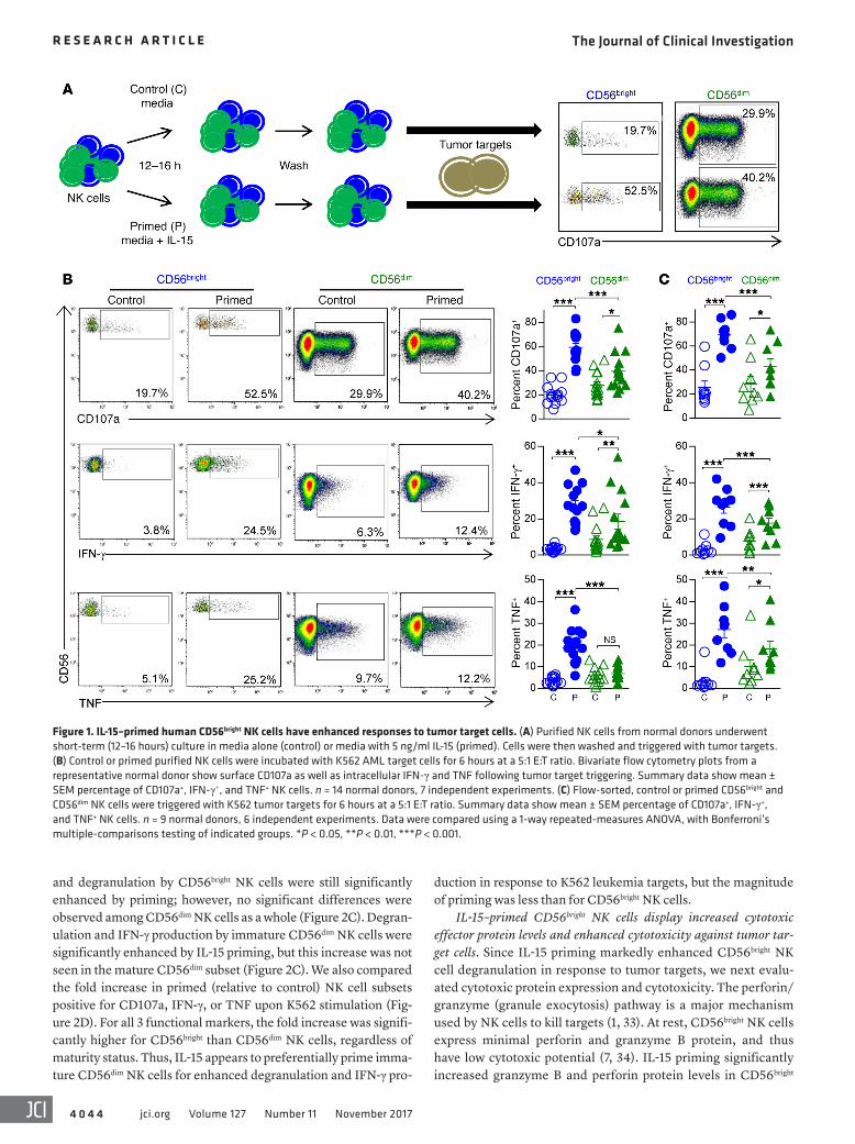
gered with tumor targets for 6 hours, after which degranulation (CD107a surface expression) and cytokine production (intracel-lular IFN-γ and TNF expression) were assessed (Figure 1A). IL-15 priming robustly enhanced degranulation and cytokine production by CD56bright NK cells in response to K562 acute myeloid leukemia (AML) target cells (Figure 1B). Indeed, while IL-15 priming also modestly enhanced CD56dim NK cell antitumor responses, these were surpassed by those of IL-15–primed CD56bright NK cells (Fig-ure 1B). Similar responses to K562 target cells were observed with flow-sorted, control or IL-15–primed CD56bright and CD56dim NK cells (Figure 1C), indicating that IL-15 priming of CD56bright NK cell antitumor responses was not a result of competition for IL-15 or dif-ferential effector-to-target (E:T) ratios. IL-15 priming of CD56bright NK cells could be observed after as little as 1 hour of stimulation (Supplemental Figure 1A; supplemental material available online with this article; https://doi.org/10.1172/JCI90387DS1), and with IL-15 concentrations as low as 1 ng/ml (Supplemental Figure 1B). In addition, a large proportion of IL-15–primed CD56bright NK cells exhibited polyfunctional responses, with simultaneous degranula-tion and IFN-γ and TNF production in response to K562 target cells (Supplemental Figure 2, A and B). Enhanced responses of IL-15–primed CD56bright NK cells were also confirmed against the MHC class I–expressing HL-60 AML cell line (Supplemental Figure 2C).
Since IL-15 is critical for NK cell homeostasis as well as function, we also established that CD56bright NK cell responses immediately following purification were equivalent to those after overnight culture in media alone (control NK cells) from the same donor (data not shown). Additionally, after 16 hours there was no difference in NK cell viability between control and primed conditions. However, IL-15 priming selectively enhanced CD56bright NK cell survival in the setting of cytokine withdrawal (Supplemental Figure 3). Brief IL-15 priming was not sufficient to induce substantial proliferation of CD56bright or CD56dim NK cells (Supplemental Figure 4), suggesting that NK cell proliferation pathways require prolonged IL-15R signals to promote cell divi-sion in CD56bright and CD56dim NK cells.
IL-15 priming of CD56dim NK cells is in part maturity-dependent. While CD56dim NK cells are considered more differentiated than the CD56bright subset, they still exist within a range of maturity states. NK cell maturity can be delineated by the presence or absence of specific surface markers such as KIR, NKG2A, CD57, CD11b, and CD27 (30, 31). We hypothesized that IL-15 priming of CD56dim NK cells produced variable responses because immature NK cells were preferentially being primed. We tested this hypoth-esis using mass cytometry, which permitted simultaneous analysis of more than 30 parameters on each NK cell (Supplemental Table 1). Using viSNE (32), we clustered control or IL-15–primed NK cells stimulated with K562 tumor targets into CD56bright, CD56dim, immature CD56dim, and mature CD56dim subsets based on the expression of maturity markers that were unaffected by priming (KIR, NKG2A, CD57) as well as CD56 (Figure 2, A and B). Imma-ture CD56dim NK cells had an NKG2A+KIR–CD57– profile, whereas mature CD56dim NK cells were primarily NKG2A+KIR+CD57+ or NKG2A–KIR+CD57+. The proportions of control or primed NK cells belonging to each CD56dim subset were similar (Figure 2A). Using mass cytometry to assess functional responses against K562 tumor targets, we found that cytokine (IFN-γ and TNF) production
on KIR+ NK cells, leaving the antitumor potential of CD56bright NK cells relatively unexplored.
IL-15 is a central cytokine in NK cell development, homeosta-sis, and function (1, 22). There are three IL-15 receptor subunits: IL-15Rα, β, and γ. The IL-15 receptor shares its β subunit with IL-2, and its γ subunit (common γ chain) with IL-2, -4, -7, -9, and -21. The IL-15Rβ/γ heterodimer is an intermediate- (nanomolar-)affinity IL-15 receptor constitutively expressed by both CD56bright and CD56dim NK cells that functions in ligand binding and signal transduction. IL-15Rα binds IL-15 with high (picomolar) affinity. IL-15/IL-15Rα can associate with IL-15Rβ/γ either on the same cell (cis-presentation) or on a different cell (trans-presentation) to initiate signaling, although trans-presentation is thought to be the major IL-15R trigger in vivo (22, 23). Three primary pathways are activated downstream of the IL-15R: JAK1/3/STAT3/5, PI3K/Akt/mTOR, and Ras/Raf/MEK/ERK (23, 24). Through these path-ways, IL-15 has been shown to promote the survival and prolifera-tion of CD56bright and CD56dim NK cells, and to enhance the cyto-toxicity of the CD56dim subset (25). Prolonged exposure to IL-2, which also acts through the intermediate-affinity IL-2/15Rβγ, has similarly been shown to increase NK cell cytotoxicity (25). Howev-er, IL-2 also promotes regulatory T cell responses, and may there-by limit antitumor immunity (26, 27). Thus, while IL-15 supports the survival and proliferation of CD56bright NK cells (22), its impact on the antitumor responses of this human NK cell subset has not been well examined.
In a model organism, we and others have previously demon-strated that brief priming of murine NK cells with IL-15 optimizes functional responses to tumor or virus-infected targets, including cytokine production and killing (28, 29). Since in some ways the CD56bright NK cell cytotoxic program resembles that of specific pathogen–free murine NK cells (28), we reasoned that IL-15 may globally enhance CD56bright NK cell antitumor responses. Here, we demonstrate that brief priming of CD56bright NK cells with IL-15 results in markedly increased degranulation, cytotoxicity, and effector cytokine production in response to tumor targets in vitro and in vivo. This priming effect was seen for normal-donor CD56bright NK cells, as well as CD56bright NK cells of patients with hematologic malignancies triggered by autologous tumor cells. Several mechanisms were identified that underlie the enhanced antitumor responsiveness of these polyfunctional IL-15–primed CD56bright NK cells. Moreover, we link preferential induction of PI3K/Akt/mTOR and MEK/ERK signaling by IL-15 in CD56bright, compared with CD56dim, NK cells with enhanced function. Blockade of these pathways attenuated IL-15 priming, identify-ing a molecular mechanism responsible for this priming event. Our findings suggest that in clinical settings of increased IL-15, CD56bright NK cells may exhibit potent multifunctional antitumor responses, and that this NK cell subset could be harnessed for cancer immunotherapy.
ResultsIL-15 primes human CD56bright NK cells for enhanced antitumor responses. Purified NK cells from normal donors were cultured for 12–16 hours with (primed) or without (control) recombinant human (rh) IL-15 at 5 ng/ml (a concentration that induces sig-naling via the IL-2/15Rβγ) (22). Cells were then washed and trig-
The Journal of Clinical Investigation R E S E A R C H A R T I C L E
4 0 4 4 jci.org Volume 127 Number 11 November 2017
duction in response to K562 leukemia targets, but the magnitude of priming was less than for CD56bright NK cells.
IL-15–primed CD56bright NK cells display increased cytotoxic effector protein levels and enhanced cytotoxicity against tumor tar-get cells. Since IL-15 priming markedly enhanced CD56bright NK cell degranulation in response to tumor targets, we next evalu-ated cytotoxic protein expression and cytotoxicity. The perforin/granzyme (granule exocytosis) pathway is a major mechanism used by NK cells to kill targets (1, 33). At rest, CD56bright NK cells express minimal perforin and granzyme B protein, and thus have low cytotoxic potential (7, 34). IL-15 priming significantly increased granzyme B and perforin protein levels in CD56bright
and degranulation by CD56bright NK cells were still significantly enhanced by priming; however, no significant differences were observed among CD56dim NK cells as a whole (Figure 2C). Degran-ulation and IFN-γ production by immature CD56dim NK cells were significantly enhanced by IL-15 priming, but this increase was not seen in the mature CD56dim subset (Figure 2C). We also compared the fold increase in primed (relative to control) NK cell subsets positive for CD107a, IFN-γ, or TNF upon K562 stimulation (Fig-ure 2D). For all 3 functional markers, the fold increase was signifi-cantly higher for CD56bright than CD56dim NK cells, regardless of maturity status. Thus, IL-15 appears to preferentially prime imma-ture CD56dim NK cells for enhanced degranulation and IFN-γ pro-
Figure 1. IL-15–primed human CD56bright NK cells have enhanced responses to tumor target cells. (A) Purified NK cells from normal donors underwent short-term (12–16 hours) culture in media alone (control) or media with 5 ng/ml IL-15 (primed). Cells were then washed and triggered with tumor targets. (B) Control or primed purified NK cells were incubated with K562 AML target cells for 6 hours at a 5:1 E:T ratio. Bivariate flow cytometry plots from a representative normal donor show surface CD107a as well as intracellular IFN-γ and TNF following tumor target triggering. Summary data show mean ± SEM percentage of CD107a+, IFN-γ+, and TNF+ NK cells. n = 14 normal donors, 7 independent experiments. (C) Flow-sorted, control or primed CD56bright and CD56dim NK cells were triggered with K562 tumor targets for 6 hours at a 5:1 E:T ratio. Summary data show mean ± SEM percentage of CD107a+, IFN-γ+, and TNF+ NK cells. n = 9 normal donors, 6 independent experiments. Data were compared using a 1-way repeated-measures ANOVA, with Bonferroni’s multiple-comparisons testing of indicated groups. *P < 0.05, **P < 0.01, ***P < 0.001.

The Journal of Clinical Investigation R E S E A R C H A R T I C L E
4 0 4 5jci.org Volume 127 Number 11 November 2017
is functional and contributes to IL-15–primed CD56bright NK cell cytotoxicity, since killing of TRAIL-sensitive target cells (HL-60 AML and RPMI 8226 myeloma) was partially abrogated in the presence of TRAIL-neutralizing antibodies (Supplemental Fig-ure 5 and refs. 35, 36). However, for relatively TRAIL-insensitive tumor targets such as U266 myeloma (35), TRAIL blockade did not substantially impact killing by IL-15–primed CD56bright NK cells (Supplemental Figure 5).
To further define changes in CD56bright NK cells that may impact cytotoxicity, the granular area and granzyme B content of IL-15–primed CD56bright NK cells were assessed by immuno-fluorescence. At rest, CD56bright NK cells are small lymphocytes
NK cells (Figure 3A). To directly assess the ability of IL-15 to aug-ment cytotoxic function, flow-sorted CD56bright and CD56dim NK cells were primed with IL-15 for 12–16 hours, and then assessed for cytotoxicity against K562 leukemia targets. IL-15 priming led to a substantial increase in the cytotoxicity of CD56bright NK cells at all E:T ratios examined, but had a more modest impact on CD56dim NK cell killing of K562 leukemia targets (Figure 3, B and C). We also assessed expression of alternate mediators of NK cell target killing, such as the death receptor ligands TRAIL and Fas ligand. We observed a marked increase in TRAIL follow-ing IL-15 priming of CD56bright NK cells (Figure 3A), but did not detect cell surface Fas ligand (not shown). The induced TRAIL
Figure 2. Responses of IL-15–primed CD56dim NK cells are influenced by maturity status. Control and primed NK cells were assessed for the expression of 33 markers using mass cytometry following stimulation with K562 tumor targets in a functional assay. (A) Density plots from a representative donor of control and primed NK cells in the tSNE1/2 fields demonstrating the proportion of NK cells that are CD56bright (B), immature CD56dim (Imm), or mature CD56dim (Mat). Percentages of NK cells belonging to the different groups are indicated in parentheses. Cell maturity state was determined according to expression of CD56, NKG2A, KIR, and CD57. Immature CD56dim NK cells were primarily NKG2A+KIR–CD57–, whereas mature CD56dim NK cells were primarily NKG2A+KIR+CD57+ or NKG2A–KIR+CD57+. (B) Median expression of the indicated markers (CD56, NKG2A, KIR2DL2/3, CD57) is shown for the same representative donor, demon-strating that maturity marker expression is unchanged by priming. (C) Summary data show mean ± SEM percentage of CD107a+, IFN-γ+, and TNF+ control or primed CD56bright, CD56dim (immature + mature), immature CD56dim, and mature CD56dim NK cells from n = 8 normal donors, 3 independent experiments. (D) Summary data show mean ± SEM fold change of primed relative to control percentage CD107a+, IFN-γ+, and TNF+ CD56bright, CD56dim (immature + mature), immature CD56dim, and mature CD56dim NK cells from n = 8 normal donors, 3 independent experiments. Data were compared using a 1-way repeated-mea-sures ANOVA with (C) Bonferroni’s multiple-comparisons testing of indicated groups or (D) Tukey’s multiple-comparisons testing. *P < 0.05, ***P < 0.001.

The Journal of Clinical Investigation R E S E A R C H A R T I C L E
4 0 4 6 jci.org Volume 127 Number 11 November 2017
CD56bright NK cells exhibit marked enhancement of multiple anti-tumor responses, including cytotoxic capacity and killing in vitro.
IL-15 priming promotes CD56bright NK cell conjugate formation with tumor targets. The NK cell cytotoxic response to tumor tar-gets involves several events, including cell recognition, conjugate formation, immune synapse formation, and directed delivery of cytotoxic granule contents (33). We evaluated the effect of IL-15 priming on the efficiency of tumor conjugate formation by coin-
that have few cytotoxic granules. IL-15 priming of CD56bright NK cells led to markedly increased CD107a (LAMP1) and granzyme B mean pixel intensity as assessed by confocal immunofluorescence microscopy (Figure 3, D and E). This enhanced granularity of primed CD56bright NK cells was also evident as an increase in mor-phologic cytoplasmic azurophilic granule content (Supplemental Figure 6A) and side scatter (Supplemental Figure 6B), neither of which was changed in CD56dim NK cells. Thus, IL-15–primed
Figure 3. IL-15–primed CD56bright NK cells have enhanced cytotoxicity. (A) Control or primed purified NK cells were assessed for expression of intracel-lular granzyme B and perforin and cell surface TRAIL using flow cytometry. Representative histograms gated on CD56bright NK cells show per-cell protein expression, with gray histograms depicting unstained cells. Summary data show mean ± SEM granzyme B and perforin median fluorescent intensity (MFI), or TRAIL percentage positive CD56bright NK cells. n = 4–6 normal donors, 2–3 independent experiments. (B and C) Control or primed flow-sorted CD56bright and CD56dim NK cells were assessed for cytotoxicity against K562 leukemia cells in a 4-hour flow-based killing assay. (B) Summary data show mean ± SEM percentage specific killing by control or primed NK cell subsets at the indicated E:T ratios. (C) Summary data show mean ± SEM specific killing at the 2.5:1 E:T ratio. n = 4 normal donors, 2 independent experiments. (D) Representative confocal Z-stack images show CD107a (LAMP1) and granzyme B (Gzm B) in flow-sorted, control or IL-15–primed CD56bright NK cells. Representative images were contrast-enhanced for better visualization. (E) Summary data show mean ± SEM LAMP1 and granzyme B single-cell mean pixel intensity × area, quantified from focal planes in the middle of cells (control n = 20–32, primed n = 25–37) from 5 normal donors. Data were compared using (A and E) a paired Student’s t test or (B and C) a 1-way repeated-measures ANOVA, with Bonferroni’s multiple-comparisons testing of indicated groups. *P < 0.05, **P < 0.01, ***P < 0.001.

The Journal of Clinical Investigation R E S E A R C H A R T I C L E
4 0 4 7jci.org Volume 127 Number 11 November 2017
cubating control and primed NK cells with CFSE-labeled K562 cells for 5, 15, or 30 minutes (Figure 4A). A significantly greater percentage of IL-15–primed CD56bright NK cells formed conjugates with tumor targets at each time point investigated compared with controls (Figure 4B). In contrast, there was no significant differ-ence in the number of tumor conjugates formed by control versus IL-15–primed CD56dim NK cells at 5 minutes (28.9% ± 5.7% vs. 31.6% ± 4.6%, P = 0.053), 15 minutes (34.9% ± 8.8% vs. 28.9% ± 5.5%, P = 0.17), or 30 minutes (41.6% ± 9.4% vs. 36.8% ± 9.2%, P = 0.16) from the same donors. Thus, one mechanism underlying the enhanced antitumor response of IL-15–primed CD56bright NK cells involves more effective target recognition or target-cell synapse formation. Therefore, we next examined expression of adhesion molecules and NK cell receptors involved in tumor target recogni-tion with or without IL-15 priming.
IL-15–primed CD56bright NK cells express more activating receptors and adhesion molecules than control cells. NK cells integrate adhe-sion molecule, activating and inhibitory receptor, and costimula-tory signals, among others, to define their functional responses to target cells (3). We evaluated the surface expression of numerous such signaling molecules by flow cytometry. Expression of the fol-
lowing proteins (percentage positive or median fluorescent inten-sity) did not differ between control and IL-15–primed CD56bright NK cells: CD226 (DNAM-1), CD244 (2B4), NKp80, CD94, NKG2A, NKG2C, and CD137 (41BB) (data not shown). In addition, we confirmed no change in KIR (CD158a, CD158b1/2, CD158d, CD158e1/2, and CD158i) or CD57 expression on CD56bright NK cells following IL-15 priming. However, we did observe signifi-cantly increased expression of NKG2D, NKp30, NKp44, CD69, CD2, and CD11a (Figure 5A and Supplemental Figure 7) on IL-15–primed, compared with control, CD56bright NK cells, in agreement with prior reports (37, 38). Because of their role in promoting anti-tumor responses by NK cells, NKG2D, NKp44, NKp30, CD2, and CD11a were thus evaluated for their contribution to IL-15–primed CD56bright NK cell responses to tumor targets. We preincubated IL-15–primed NK cells with blocking mAbs against these recep-tors for 30 minutes, and then triggered the cells with K562 targets. No difference in CD56bright NK cell degranulation or cytokine pro-duction upon NKp44 or NKp30 blockade was observed (data not shown). A small reduction in the priming response was observed when CD11a, CD2, or NKG2D was blocked individually. However, priming was significantly diminished upon simultaneous block-
Figure 4. IL-15–primed CD56bright NK cells more effectively form conjugates with K562 leukemia targets. Control or primed purified NK cells were assessed for conjugate formation via coincubation with CFSE-labeled K562 cells for 5, 15, or 30 minutes at a 1:1 E:T ratio. The NK/K562 conjugate percentage was assessed by the frequency of CD56bright NK cell events that were also CFSE positive. (A) Representative bivariate flow plots show conjugate formation by control or primed NK cells at the 15-minute time point by gating on (i) all live NK and K562 cells, (ii) NK cells (CD56+CD3–), (iii) CD56bright NK cells, and (iv) CFSE-positive cells. (B) Summary data show mean ± SEM percentage of control or primed CD56bright NK cells that formed NK/K562 conjugates after 5, 15, and 30 minutes of coincubation. n = 4 normal donors, 2 independent experiments. Data were compared using a paired Student’s t test. *P < 0.05, **P < 0.01.

The Journal of Clinical Investigation R E S E A R C H A R T I C L E
4 0 4 8 jci.org Volume 127 Number 11 November 2017
ade of these 3 receptors (Figure 5B), indicating that they are col-lectively involved in the enhanced antitumor responsiveness of IL-15–primed CD56bright NK cells.
IL-15 priming promotes integrin activation on CD56bright NK cells. CD11a and CD18 (β2) together constitute the integrin LFA-1. LFA-1 is critical for NK cell cytotoxicity; upon binding its ligands (the
ICAMs), LFA-1 initiates immune synapse formation and tight adhesion to target cells, and provides NK cell–activating signals (33, 39, 40). Integrins typically exist in a bent conformation with low ligand-binding affinity. However, signals from activating recep-tors promote integrin activation via inside-out signaling, leading to high-affinity conformation changes and integrin clustering (41).
Figure 5. IL-15–primed CD56bright NK cells display increased expression of coactivating and adhesion molecules as well as enhanced integrin signaling upon tumor target engagement. (A) Representative histograms gated on control or primed CD56bright NK cells show per-cell surface expression of CD2, CD11a, and NKG2D, with gray histograms depicting unstained cells. Summary data show mean ± SEM CD2, CD11a, and NKG2D MFI on control or primed CD56bright NK cells. n = 4–9 normal donors, 2–4 independent experiments. (B) IL-15–primed purified NK cells were preincubated with isotype control or blocking anti-human mAbs against CD11a, CD2, or NKG2D (or all 3 combined) for 30 minutes prior to coincubation with K562 target cells for 6 hours at 5:1 E:T ratio. Control NK cell responses were also assessed without blocking mAbs. Summary data show mean ± SEM percentage of CD107a+, IFN-γ+, and TNF+ CD56bright NK cells. n = 4–6 normal donors, 2–3 independent experiments. (C) Control or primed purified NK cells were assessed for the expression of open-conformation CD11a (mAb 24) or intracellular phosphorylated ERK (pT202/pY204) following 15 or 3 minutes (respectively) of coincubation with K562 tumor targets (K562, gray), or in the absence of targets (Baseline, blue). Representative histogram plots show per-cell open CD11a on CD56bright NK cells. Summary data show mean ± SEM percentage of control or primed CD56bright NK cells expressing open CD11a (positivity cutoff set on baseline control cells). Representative histogram plots show per-cell pERK expression in CD56bright NK cells. Summary data show mean ± SEM percentage pERKhi control or primed CD56bright NK cells (pERKhi cutoff set on baseline control cells). Data were compared using (A) a paired Student’s t test, (B) a 1-way ANOVA with Bonferroni’s multiple-comparisons testing of indicated groups, or (C) a 1-way repeated-measures ANOVA with Tukey’s multiple-comparisons testing. *P < 0.05, **P < 0.01, ***P < 0.001.

The Journal of Clinical Investigation R E S E A R C H A R T I C L E
4 0 4 9jci.org Volume 127 Number 11 November 2017
enhanced antitumor responses conferred by IL-15 priming extend-ed beyond myeloid leukemia, we assessed responses of CD56bright NK cells against multiple myeloma (MM) targets. Enhanced degranulation and cytokine production by IL-15–primed, com-pared with control, CD56bright NK cells from normal donors against U266 MM cells were observed (Figure 7A). We next tested wheth-er IL-15 was able to prime newly diagnosed MM patient CD56bright NK cells for enhanced responses against autologous CD138+ MM cells (Figure 7B and Supplemental Table 3) or U266 MM targets (Supplemental Figure 8B). Indeed, degranulation and cytokine production were significantly increased in comparison with con-trol CD56bright NK cells. Thus, in vitro responses against MM tar-gets by allogeneic and autologous CD56bright NK cells were signifi-cantly improved after IL-15 priming.
ALT-803 primes CD56bright NK cell antitumor responses in vivo in MM patients. In addition, we investigated whether IL-15 can prime NK cells in vivo using PBMCs from relapsed or refractory (rel/ref) MM patients being treated with ALT-803 (NCT02099539). First, we verified that ALT-803 primed normal-donor CD56bright NK cell antitumor responses in vitro, similar to IL-15 (Supplemen-tal Figure 9). We collected PBMCs from rel/ref MM patients just before, 24 hours after, or 72 hours after a single injection of ALT-803, and immediately assessed NK cell function in response to ex vivo stimulation with U266 myeloma target cells (patient group A) or the traditional NK-sensitive K562 tumor target (patient group B) (Figure 7C). Twenty-four hours after ALT-803 administration, patient CD56bright NK cell degranulation and cytokine production in response to MM and leukemia target cells were significantly increased compared with pretherapy values (Figure 7D). However, this priming effect was relatively transitory in vivo, and responses no longer differed significantly from those observed before ther-apy by 72 hours after ALT-803. Thus, IL-15 primed CD56bright NK cells both in vitro and in vivo for enhanced functional responses against MM target cells.
IL-15 more robustly activates the PI3K/Akt/mTOR and Ras/Raf/MEK/ERK pathways in CD56bright NK cells. Having identified ele-ments responsible for the enhanced antitumor responsiveness of primed CD56bright NK cells, we next examined signaling mecha-nisms that could be responsible for priming of the CD56bright subset by IL-15. In mice, IL-15–induced activation of the PI3K/Akt/mTOR pathway is of particular importance for promoting NK cell cytotox-icity (49, 50), and mTOR activity downstream of the IL-15R has been shown to increase granzyme B expression in both murine and human NK cells (50). We confirmed that CD56bright and CD56dim NK cells both robustly express IL-15Rβ/γ (Figure 8, A and B). Next, the role that each pathway (JAK/STAT, PI3K/Akt/mTOR, and Ras/Raf/MEK/ERK) downstream of the IL-15R plays in CD56bright NK cell priming was investigated. Induction of each pathway (via phos-phorylation of signaling intermediates) upon exposure to IL-15 in CD56bright and CD56dim NK cells was examined (Figure 8, D–F). IL-15 robustly induced phosphorylation of STAT5 in both CD56bright and CD56dim NK cells (Figure 8D). In contrast, IL-15 more selec-tively induced Akt and ERK phosphorylation in CD56bright NK cells, with a minimal fold increase over baseline observed in the CD56dim subset (Figure 8, E and F). This was not a matter of dose respon-siveness, since even concentrations of IL-15 as high as 100 ng/ml did not enhance Akt and ERK phosphorylation in CD56dim NK cells
NKG2D and CD2, among other molecules, can activate LFA-1 on NK cells (39). Thus, in addition to measuring overall LFA-1 (CD11a) expression, we also investigated the proportion of control or IL-15–primed CD56bright NK cells that expressed open-conformation LFA-1 at rest or following K562 target cell engagement. Incubation with IL-15 alone has been shown to only modestly activate LFA-1 on bulk NK cells; however, it enhances subsequent inside-out signal-ing following activating receptor ligation (42). Consistent with this, we observed a modest but significant increase in activated LFA-1 expression on CD56bright NK cells as a result of IL-15 priming alone (Figure 5C). However, the majority of IL-15–primed CD56bright NK cells expressed high-affinity LFA-1 after 15 minutes of incubation with K562 target cells, whereas K562 coincubation induced high-affinity LFA-1 on only a small subset of control CD56bright NK cells (Figure 5C). Signaling through LFA-1 has been shown to activate ERK1/2 (43) (outside-in signaling), and enhanced ERK1/2 phos-phorylation was observed in IL-15–primed CD56bright NK cells trigged with K562 targets compared with control cells (Figure 5C). Thus, IL-15 priming promotes integrin activation on CD56bright NK cells, potentially allowing adhesion to target cells in the absence of signals from other activating receptors (39), and also enhances sub-sequent target-induced inside-out and outside-in signaling.
IL-15 primes CD56bright NK cells from normal donors and AML patients for enhanced responses against primary AML blasts. We next examined whether IL-15 priming enhanced CD56bright NK cell responses against primary leukemic blasts. Patient leukemia blast killing by flow-sorted, control or IL-15–primed CD56bright and CD56dim NK cells from allogeneic normal donors was examined (Figure 6A and Supplemental Table 2). IL-15 priming resulted in a significant increase in AML blast killing by CD56bright, as well as CD56dim, NK cells (Figure 6A). In addition, since IL-15 is cur-rently being tested in clinical trials for cancer therapy (44, 45), we investigated whether IL-15 could prime the endogenous CD56bright NK cells of patients with AML. Control or IL-15–primed periph-eral blood mononuclear cells (PBMCs) from newly diagnosed AML patients were triggered with autologous AML blasts (Figure 6B) or K562 leukemia targets (Supplemental Figure 8A), and NK cell functional responses were assessed. Indeed, we observed significant enhancement of patient CD56bright NK cell antitumor responses against both target cell types following IL-15 priming, with modest enhancement of CD56dim NK cell responses.
Next, the ability of IL-15–primed CD56bright NK cells to con-trol leukemia in vivo was investigated by engrafting of groups of NOD-SCID-IL2Rγ–/– (NSG) mice with either equal numbers of luciferase-expressing K562 cells (K562-luc) and ALT-803–primed CD56bright NK cells, or K562-luc cells alone, via i.v. injection. ALT-803 is an IL-15 superagonist complex exhibiting better pharma-codynamics and pharmacokinetic properties than recombinant IL-15 (46). ALT-803 is currently in clinical trials for cancer immu-notherapy (47, 48). Both groups of mice received ALT-803 after cell engraftment (Figure 6C). We observed significantly lower tumor burden as assessed by whole-body bioluminescence imag-ing in mice treated with ALT-803–primed CD56bright NK cells (Fig-ure 6, D and E), demonstrating in vivo control of tumor cell growth by adoptively transferred, primed CD56bright NK cells.
In vitro IL-15–primed CD56bright NK cells show enhanced function-al responses against multiple myeloma. To investigate whether the

The Journal of Clinical Investigation R E S E A R C H A R T I C L E
4 0 5 0 jci.org Volume 127 Number 11 November 2017
molecule inhibitors at different concentrations for 1 hour before priming with IL-15 and assessed the impact of this inhibition on functional responses to K562 targets (Figure 9, A and B). Inhibi-tion of PI3K significantly impaired IL-15 priming of both degranu-lation and cytokine responses by CD56bright but not CD56dim NK cells in response to K562 leukemia targets (Figure 9A). Notably, impairment of IL-15 priming was not evident after mTOR inhibi-tion by Torin1 at concentrations that selectively inhibit mTORC1
(data not shown). Since the PI3K/Akt/mTOR and Ras/Raf/MEK/ERK pathways were more selectively induced by IL-15 in CD56bright NK cells, we hypothesized that they were important for priming and investigated this through pathway blockade.
The Ras/Raf/MEK/ERK pathway is required for IL-15–primed CD56bright and CD56dim NK cell responses, whereas the PI3K/Akt/mTOR pathway is selectively involved in CD56bright priming. We treated NK cells with PI3K (Ly294002) or MEK (PD98059) small-
Figure 6. IL-15 priming enhances CD56bright NK cell antitumor responses against primary AML blasts in vitro and K562 leukemia in vivo in NSG mice. (A) Flow-sorted, control or IL-15–primed CD56bright and CD56dim NK cells were coincubated with primary AML blasts from newly diagnosed patients. Summary data show mean ± SEM percentage specific killing at a 15:1 E:T ratio. n = 7 normal donors, 4 independent experiments. (B) PBMCs and bone marrow blasts were obtained from newly diagnosed AML patients. Control or IL-15–primed PBMCs were incubated with autologous blasts at a 5:1 E:T ratio for 6 hours and functional responses assessed. Summary data show mean ± SEM percentages of CD107a+, IFN-γ+, and TNF+ NK cells. n = 7 normal donors, 3 independent experiments. (C) Experimental design. Day –1: Flow-sorted CD56bright NK cells primed with 17.5 ng/ml ALT-803. Day 0: Primed CD56bright NK cell injection into NSG mice. Eight mice received K562-luc (0.5 × 10⁶ to 0.66 × 10⁶, K562 + ALT-803) and 8 mice received an equal number of both K562-luc and primed CD56bright NK cells (K562 + primed CD56bright NK). ALT-803 (5 μg) was administered i.v. on day 0 and every 2–3 days thereafter for 30 days. Bioluminescence imaging (BLI) was performed on days 2, 5, 13, 20, and 27, ± 1 day. (D) Representative BLI images from one K562 + ALT-803 and one K562 + primed CD56bright NK mouse at different imaging time points. (E) Summary data of whole-body bioluminescence (photons per second) in the different treatment groups. Data show the mean ± SEM photons per second at each time point. n = 8 mice per group, 2 independent experiments. Data were compared using (A and B) a 1-way repeated-measures ANOVA with Bonferroni’s multiple-comparisons testing of indicated groups or (E) a 2-way ANOVA with Sidak’s multiple-comparisons testing. *P < 0.05, **P < 0.01, ***P < 0.001.

The Journal of Clinical Investigation R E S E A R C H A R T I C L E
4 0 5 1jci.org Volume 127 Number 11 November 2017
fact of differential inhibitor affinity or E:T ratio, we repeated these experiments with flow-sorted CD56bright or CD56dim NK cells pre-treated with both Ly294002 and PD98059. The same pattern of response was observed (Figure 9C and Supplemental Figure 11A). Furthermore, PI3K and MEK inhibition decreased cytotoxic pro-tein expression in CD56bright NK cells following IL-15 priming (Fig-
and mTORC2. However, higher concentrations that nonspecifi-cally inhibit PI3K were able to abrogate IL-15 priming (Supplemen-tal Figure 10 and ref. 51). MEK inhibition significantly impaired functional responses of both IL-15–primed CD56bright and CD56dim NK cells (Figure 9B). To confirm that the functional response abrogation conferred by PI3K or MEK inhibition was not an arti-
Figure 7. MM patient CD56bright NK cells are primed in vitro by IL-15 and in vivo by ALT-803 for enhanced functional responses against autologous myeloma cells and MM cell lines. (A) Control or IL-15–primed normal-donor purified NK cells were incubated with U266 MM target cells for 6 hours at a 5:1 E:T ratio. Summary data show mean ± SEM percentage CD107a+, IFN-γ+, or TNF+ NK cells. (B) PBMCs and enriched CD138+ cells were obtained from newly diagnosed MM patients. Control or IL-15–primed PBMCs were cultured with autologous CD138+ cells at a 5:1 E:T ratio for 6 hours and functional responses assessed. Summary data show mean ± SEM percentage of CD107a+, IFN-γ+, and TNF+ CD56bright or CD56dim NK cells in response to autologous MM cell triggering. (C) Schematic of ALT-803 treatment regimen. Patients with rel/ref MM received 10 μg/kg ALT-803 s.c. (group A) or 3–6 μg/kg i.v. (group B) at the 0-hour time point. Peripheral blood samples were obtained for functional analysis just before ALT-803 administration as well as 24 and 72 hours thereafter. (D) PBMCs were isolated from MM patient peripheral blood samples by Ficoll density centrifugation and immediately incubated with U266 myeloma targets (group A) or K562 leukemia targets (group B) for 6 hours. Summary data show mean ± SEM percentage of CD107a+, IFN-γ+, and TNF+ CD56bright NK cells. n = 2 patients, 3 independent experiments (group A); n = 3 patients, 3 independent experiments (group B). Data were compared using 1-way repeated-measures ANOVA with (A and B) Bonferroni’s multiple-comparisons testing of indicated groups or (D) Tukey’s multiple-comparisons testing. *P < 0.05, **P < 0.01, ***P < 0.001.

The Journal of Clinical Investigation R E S E A R C H A R T I C L E
4 0 5 2 jci.org Volume 127 Number 11 November 2017
stitutively expressed, intermediate-affinity IL-15Rβ/γ markedly enhanced multiple antitumor functional responses (cytotoxicity, degranulation, and cytokine production) of CD56bright NK cells. These enhanced responses were elicited in vitro from allogeneic normal donor– and autologous cancer patient–derived NK cells triggered by primary AML blasts, primary myeloma cells, or a variety of tumor cell lines. IL-15 priming was also evident in vivo in patients with rel/ref MM treated with the IL-15 superagonist complex ALT-803, and in an NSG xenograft model where ALT-803–primed CD56bright NK cells significantly controlled K562 tumor burden. Several mechanisms contributed to the enhanced antitumor function of IL-15–primed CD56bright NK cells, including increased cytotoxic protein levels, tumor conjugate formation, and CD2-, CD11a-, and NKG2D-based triggering. Thus, exposure to IL-15 unleashes the unexpectedly potent antitumor effector function of CD56bright NK cells.
Cytokine activation of NK cells to promote antitumor respons-es has been explored in many clinical settings (52). Here, we exam-ine short-term IL-15 activation, defined as the direct impact of IL-15 on the NK cell, and priming, the impact of IL-15 activation on subsequent antitumor responses. The effects of IL-15 priming on CD56bright NK cells were transient, persisting for less than 72 hours after stimulation in vitro and in vivo in patients. This is dis-
ure 9D), and significantly reduced killing of K562 leukemia target cells (Figure 9E). While the effect of PI3K and MEK inhibition on CD56dim NK cell cytotoxic protein expression was more modest (Supplemental Figure 11B), K562 killing was significantly reduced as a result of treatment with these inhibitors (Supplemental Figure 11C). Thus, both the PI3K/Akt/mTOR and Ras/Raf/MEK/ERK pathways appear to be important for IL-15 priming of CD56bright NK cells, while the PI3K/Akt/mTOR pathway was not required for the functional response of IL-15–primed CD56dim NK cells. Interest-ingly, our phosphorylation studies indicated that phosphorylated Akt (pAkt) expression is similar between unstimulated CD56bright and CD56dim NK cells but pERK is significantly higher in CD56dim NK cells at baseline (Figure 8C). Thus, it is possible that a certain threshold of MEK/ERK pathway activation is required for signifi-cant antitumor responses, which is not achieved in nonprimed CD56bright NK cells or NK cells treated with PD98059.
DiscussionNumerous functional and phenotypic properties have been used to distinguish CD56bright from CD56dim NK cells, including the superior ability of resting CD56dim NK cells to be triggered by tumor targets or activating receptor ligation, and to mediate cyto-toxicity. Here, we identified that brief priming through the con-
Figure 8. IL-15 more robustly activates the PI3K/Akt/mTOR and Ras/Raf/MEK/ERK pathways in CD56bright NK cells. (A) Flow cytometry plot shows gat-ing strategy for CD56bright versus CD56dim NK cells based on relative CD56 and CD16 expression. (B) IL-15Rβ expression was assessed via flow cytometry on purified NK cells. Representative data show percentage IL-15Rβ–positive cells and IL-15Rβ MFI on freshly purified CD56bright and CD56dim NK cells. n = 8 nor-mal donors, 3 independent experiments. (C) Summary data show pAkt and pERK MFI in unstimulated CD56bright and CD56dim NK cells. n = 7 normal donors, 3 independent experiments. (D–F) Purified NK cells were incubated with 5 ng/ml IL-15 for 30 minutes (pSTAT5) or 2 hours (pAkt and pERK) then assessed for signaling molecule phosphorylation. Representative histograms show per-cell expression of pSTAT5 (D), pAkt (E), and pERK (F) in IL-15–stimulated (shaded gray) versus unstimulated (gray line) CD56bright or CD56dim NK cells. Summary data show mean ± SEM fold increase of phosphorylated molecule MFI in IL-15–stimulated CD56bright or CD56dim NK cells relative to unstimulated cells. n = 7 normal donors, 3 independent experiments. Data were compared using a paired Student’s t test. *P < 0.05, ***P < 0.001.

The Journal of Clinical Investigation R E S E A R C H A R T I C L E
4 0 5 3jci.org Volume 127 Number 11 November 2017
would survive preferentially over the CD56dim subset in the setting of repeated ALT-803 administration, where drug concentrations may fall below therapeutic levels between doses. While CD56bright NK cells constitute a minor fraction of NK cells in the peripheral blood, they are the major subset in secondary lymphoid tissues, at sites of inflammation, and in some malignant tumors (9–11, 57, 58). Furthermore, NK cells reside predominantly in the T cell–
tinct from combined cytokine preactivation with IL-12, IL-15, and IL-18 that results in a prolonged enhancement of function (referred to as memory-like NK cell function) (53–56), and suggests that clinical approaches that use IL-15 priming of CD56bright NK cells will likely require repeated IL-15 dosing to maintain primed status. Notably, our results demonstrating that IL-15 confers a selective survival advantage to CD56bright NK cells suggest that these cells
Figure 9. The Ras/Raf/MEK/ERK pathway is required for IL-15 priming of CD56bright and CD56dim NK cell antitumor responses, whereas the PI3K/Akt/mTOR pathway is selectively required for CD56bright NK cell priming. (A and B) Purified NK cells were cultured for 12–16 hours in media alone (control, Con) or media plus 5 ng/ml IL-15 (primed). In addition, 1 hour before addition of IL-15, primed NK cells were treated with small-molecule inhibitors of PI3K (Ly294002) or MEK (PD98059) at various concentrations. Summary data show mean ± SEM CD107a, IFN-γ, or TNF percentage positive control, primed, or primed in the presence of PI3K (A) or MEK (B) inhibitors CD56bright and CD56dim NK cells. n = 8 normal donors, 3 independent experiments. (C–E) Flow-sorted CD56bright NK cells were cultured with or without (no inhibitor, NI) small-molecule inhibitors of PI3K (Ly294002, Pi) and MEK (PD98059, Mi) for 1 hour prior to 12–16 hours of IL-15 prim-ing. Cells were then washed and incubated with K562 tumor targets for 6 hours at a 5:1 E:T ratio. (C) Summary data show mean ± SEM CD107a, IFN-γ, or TNF percentage positive cells. n = 9 normal donors, 6 independent experiments. (D) CD56bright NK cells were assessed for granzyme B and perforin expression via intracellular flow cytometry. Representative histogram plots show per-cell cytotoxic protein expression. Summary data show mean ± SEM granzyme B or per-forin MFI. n = 11 normal donors, 6 independent experiments. (E) CD56bright NK cells were incubated with K562 tumor targets (E:T = 2.5:1) in a 4-hour flow-based killing assay. Summary data show mean ± SEM percentage specific killing. n = 12 normal donors, 8 independent experiments. Data were compared using (A and B) a paired Student’s t test or (C–E) a 1-way repeated-measures ANOVA with Tukey’s multiple-comparisons testing. *P < 0.05, **P < 0.01, ***P < 0.001.
The Journal of Clinical Investigation R E S E A R C H A R T I C L E
4 0 5 4 jci.org Volume 127 Number 11 November 2017
NK cells. The reasons for this differential responsiveness to IL-15 are unclear and warrant further investigation. Possibilities include SHIP1, which dephosphorylates the PI3K product PIP3, and has been shown to have lower expression in CD56bright than in CD56dim NK cells, suggesting that this pathway is subject to less inhibi-tion in the CD56bright subset (66). However, CD56bright NK cells have also been shown to express higher levels of PTEN, another inhibitor of the PI3K/Akt pathway (67). Additional studies linking IL-15R signals to distinct functions are warranted.
This study has translational relevance to several cancer immu-notherapy settings, including the adoptive transfer of allogeneic NK cells (17, 68). Patients who receive allogeneic NK cell infu-sions are typically prepared with lymphodepleting chemotherapy, which has been shown to induce IL-15 protein in the serum (15, 68). Thus, in this situation, CD56bright NK cells that are present in the enriched NK cell product are likely primed in vivo by IL-15. Differences in the NK cell receptor biology of CD56bright NK cells may also influence their antileukemic function. Since only a small fraction of CD56bright NK cells express inhibitory KIRs, most inhibi-tory signals are mediated via CD94/NKG2A (7, 34). Thus, depend-ing on their activation state, the CD56bright subset has the potential to respond to leukemia cells regardless of donor/recipient KIR/KIR-ligand mismatch status. In our experiments, IL-15–primed CD56bright NK cells effectively responded to primary AML blasts without blocking CD94/NKG2A (or other inhibitory receptors). This suggests that IL-15 priming may alter the threshold of acti-vation for CD56bright NK cells, resulting in triggering by leukemia targets regardless of inhibitory signals. These findings are in line with reports of in vitro CD137L/IL-15–expanded bulk NK cells, for which activating receptor signals dominated inhibitory KIR sig-naling (69). Moreover, IL-15 or IL-15/IL-15Rα complexes are being investigated in early-stage clinical studies to support adoptively transferred NK cells (70, 71) or as single agents (45). Our findings suggest that the adoptively transferred or endogenous patient CD56bright NK cells may contribute to the therapeutic efficacy of IL-15 administration. Finally, since ALT-803, unlike recombinant IL-15, accumulates and persists in secondary lymphoid tissues such as lymph nodes and spleen (72), treatment with this agent may be particularly apt to provide sustained priming signals to this lymph node–resident NK cell subset (9).
Another scenario in which IL-15 priming of CD56bright NK cells may be important is early (1–3 months) after allogeneic stem hematopoietic cell transplantation (HCT). Early after an HCT, CD56bright NK cells are the major lymphocyte subset to recover in the peripheral blood (73–78). Functional CD56bright NK cell anti-leukemic responses have not been analyzed in depth in this set-ting (57). In 2 reports, an “activated” phenotype with an intact CD56bright response to combined cytokine activation was observed (76, 78), although responses to leukemia or other target cells were not assessed. In a more recent study focused on KIR+CD56dim NK cells from HCT recipients, IL-15 stimulation led to a significant enhancement of the antileukemia responses of CD56dim NK cells; however, the effects of IL-15 on CD56bright NK cell responses to leu-kemia cells were not reported (77). Based on our findings, elevated (or exogenous) IL-15 in the early postallogeneic HCT period may prime CD56bright NK cells, and thereby promote NK cell clearance of residual AML blasts. Late after HCT, donor NK cells reestab-
and DC-rich parafollicular areas of secondary lymphoid tissues, and may normally be activated by DC-derived IL-15 (9–11). Prior reports demonstrated that CD56bright NK cells from the peripheral blood, tonsils, and spleen were activated by mature DCs to more effectively prevent B cell transformation by EBV virus, compared with CD56dim NK cells (59). These data support the concept that CD56bright NK cells may be primed in humans in vivo by IL-15 on the surface of DCs or monocytes in secondary lymphoid tissue (11), and potentially in other sites of IL-15 production such as the bone marrow and spleen (60, 61), thereby enhancing their functional competence against virally infected or malignantly transformed cells. Previous work has also revealed that exposure to IL-12 or prolonged stimulation with combinations of IL-2, IL-12, and/or IL-15 can increase CD56bright NK cell cytotoxicity (1, 7, 25, 58, 62, 63). This prior work identified that CD56bright NK cells may be cul-tured over days to weeks into lymphokine-activated killer cells that have increased potency against tumor cell lines. Our results are also consistent with a report evaluating CD56bright NK cell responses to autologous activated CD4+ T cells, which were found to be enhanced by 4-day culture with different cytokines, includ-ing IL-15 (64). Here, we report that brief cytokine priming via spe-cific signaling pathways transforms immunoregulatory CD56bright NK cells into robust antitumor effectors exhibiting polyfunctional responses to a diverse set of cancer targets, both in vitro and in vivo. The robust increase in TRAIL expression of IL-15–primed CD56bright NK cells furthermore suggests that these cells may also offer a substantial therapeutic benefit for TRAIL-sensitive tumors. Collectively, these studies highlight the plasticity of the CD56bright NK cell subset, and indicate that cytokine cues from the NK cell microenvironment can profoundly alter its functional capacity.
The signaling mechanisms leading to IL-15 priming of CD56bright NK cells involve the PI3K/Akt/mTOR and Ras/Raf/MEK/ERK pathways. These pathways were robustly induced by IL-15 in the CD56bright NK cell subset, and their blockade signifi-cantly impaired priming. This finding is consistent with studies investigating murine NK cell cytotoxicity (50), whereas reports concerning human NK cell cytotoxicity have been conflicting (38, 50, 65), specifically with regard to the role of mTOR. Similar to reports from another group (38), we found that pretreatment of NK cells with the mTOR inhibitor Torin1 at concentrations that selectively inhibit mTORC1/2 (10 nM) prior to priming did not impact effector responses (CD107a, IFN-γ, TNF) of CD56bright or CD56dim NK cells against K562 targets, or impair cytotoxic protein upregulation. Priming-induced increases in antitumor effector responses and cytotoxic protein levels were, however, impeded by Torin1 at concentrations of 1 μM, similar to what has been used in other studies (65). However, micromolar concentrations result in off-target effects, including inhibition of PI3K (51). This supports a general PI3K/Akt/mTOR requirement for IL-15 prim-ing of CD56bright NK cells. Interestingly, only MEK/ERK pathway inhibition impacted IL-15 priming of CD56dim NK cells. Both the more robust PI3K/Akt/mTOR and MEK/ERK pathway activa-tion in CD56bright compared with CD56dim NK cells by IL-15, and the selective involvement of the PI3K/Akt/mTOR pathway in CD56bright NK cell priming, could then underlie the disproportion-ate enhancement of CD56bright NK cell functional responsiveness by priming, in many situations to a level exceeding that of CD56dim

The Journal of Clinical Investigation R E S E A R C H A R T I C L E
4 0 5 5jci.org Volume 127 Number 11 November 2017
by Ficoll centrifugation from newly diagnosed patients with relatively low peripheral blood disease burden and used fresh or viably cryopre-served. Control or primed patient PBMCs were then triggered with autologous bone marrow AML (bone marrow blast percentage ≥ 50%) or enriched CD138+ MM cells, K562 leukemia cells, or U266 multiple myeloma cells (E:T = 5:1) with function assayed as above.
Proliferation and viability assays. Purified CFSE-labeled or unlabeled NK cells were incubated for 12–16 hours with media alone (control) or media with 5 ng/ml IL-15 (primed). Cytokines were then washed away, and cells were incubated in media alone for 3–5 days. To interrogate pro-liferation, after both 3 and 5 days, NK cells were harvested and surface-stained for NK cell markers, and CFSE dilution or intracellular Ki67 expression (BD Biosciences) was assessed via flow cytometry. To inter-rogate viability, after both 3 and 5 days, NK cells were surface-stained for NK cell markers, then stained for activated caspase-3 and caspase-7 (Cell Event Caspase 3/7 Red, Thermo Fisher Scientific) and annexin V (BD) per the manufacturer’s instructions. Baseline viability was deter-mined using acridine orange and propidium iodide (Nexcelom).
Mass cytometry. Mass cytometry functional assays were per-formed as above. Sample staining and data collection were performed as previously described (56).
Flow-based killing assay. Flow-based killing assays were performed by coincubation of flow-sorted CD56bright or CD56dim NK cells with CFSE-labeled K562 cells or PKH67-labeled (Sigma-Aldrich) primary AML blasts for 4–5 hours and assaying of 7-AAD uptake as previously described (28). Background spontaneous K562 or AML blast death (no effector control wells) was subtracted to yield percentage specific kill-ing, and in all cases this was less than 5% (K562) or 25% (primary AML blasts). E:T ratios of 5:1, 2.5:1, and 1:1 were used for coincubation with K562 targets and 15:1 for primary AML blasts. For experiments inves-tigating the contribution of TRAIL to target cell killing, flow-sorted, IL-15–primed CD56bright NK cells were incubated with CFSE-labeled, TRAIL-sensitive (HL-60, RPMI 8226) or -insensitive (U266) targets for 18 hours (79, 80) at a 5:1 E:T ratio in the presence of 10 μg/ml anti-TRAIL or control IgG antibody. Background spontaneous death was less than 5% (U266, HL60) or 10% (RPMI 8226).
Light microscopy. Slides were prepared from 1 × 10⁵ to 2 × 10⁵ cells (via CytoSpin) and Wright-Giemsa–stained. Micrographs were obtained using Nikon Microphot SA at ×1,000, with no postimage alterations except cropping for image size. Scale bar represents 5 μm.
Confocal immunofluorescence microscopy. Flow-sorted CD56bright NK cells were cultured for 16 hours in X-vivo20 media (Lonza), sup-plemented with 5% human serum with or without rhIL-15 at 5 ng/ml. The cells were adhered to CellTak-coated (Corning) coverslips, fixed with 4% paraformaldehyde, and stained for 4 hours or overnight with rabbit anti–human LAMP1 (Ab24170) and mouse anti–human granzyme B-A647 (GB11), followed by donkey anti-rabbit IgG Alexa Fluor 568 and Hoechst 33342. The stained cells were mounted with ProLong Diamond Antifade on coverslips. Z-stacks were acquired on an LSM710 (Zeiss) confocal microscope equipped with a laser diode 405-30 CW (405 nm), an Ar-Laser Multiline (458/488/514 nm), a DPSS-561 10 (561 nm), and a He-Ne laser (633 nm). A Plan-Apochro-mat 63× NA/1.4 oil DICII (Zeiss) objective was used for the acquisi-tion. All images were acquired using the same confocal settings. Image processing was conducted with Zen2012 software (Zeiss). Single-cell quantification of mean fluorescence intensity × area was performed with Fiji, using the analyze particles function for all particles larger
lish homeostasis after having developed from transplanted stem cells within the host, and are no longer being primed by excessive IL-15. At this time point, the more mature, educated KIR+CD56dim NK cells would be expected to mediate longer-term antileukemia responses, consistent with well-studied associations between donor KIR genotype and transplant outcome (14, 20, 21).
In summary, CD56bright NK cells, traditionally considered to be minimally tumor-responsive, are effectively primed by short-term IL-15 exposure to enable potent cytotoxicity, degranulation, and cytokine production in response to a broad array of tumor targets and primary hematologic tumor cells both in vivo and in vitro. This study provides a novel rationale for developing clinical immuno-therapy approaches that utilize IL-15 to prime CD56bright NK cells, and suggests that IL-15–based therapeutics may act in vivo to enhance the antitumor capacity of this NK cell subset.
MethodsReagents. Antihuman mAbs used in this study are listed in Supple-mental Table 5. Endotoxin-free rhIL-15 was used (Miltenyi Biotec). The following cell lines were maintained in RPMI-1640 plus 10% FCS and supplements: K562 (AML), HL-60 (AML), U266 (MM), and RPMI 8226 (MM). K562 cells were obtained from ATCC (CCL-243) in 2008, viably cryopreserved in liquid nitrogen, thawed for use in these studies, and maintained for less than 2 months at a time in culture according to provider instructions. K562 cells were authenticated in 2015 using SNP analysis and were found to be exact matches to the K562 cells from the Japanese Collection of Research Bioresources, German Collection of Microorganisms and Cell Cultures (DSMZ), and ATCC databases (2015, Genetic Resources Core Facility at Johns Hopkins University). Additional cell lines were obtained from ATCC and provided for our use by Daniel Link (HL-60) and Michael Tomas-son (U266, RPMI 8226).
NK cell purification and cell culture. Anonymous human platelet apheresis donor PBMCs were obtained by Ficoll centrifugation. NK cells were purified using RosetteSep (StemCell Technologies; ≥95% CD56+CD3–) or by flow cytometric cell sorting (BD FACSAria II; ≥97% purity) (53). Cells were cultured in complete RPMI-1640 media con-taining 10% human AB serum (Sigma-Aldrich) for 12–16 hours (unless otherwise stated) with or without 5 ng/ml rhIL-15. ALT-803 in vitro priming of NK cells was performed at 17.5 ng/ml for 12–16 hours. For assays involving IL-15 signaling pathways, NK cells were preincubated with inhibitors of PI3K (Ly294002) or MEK (PD98059; both Cayman Chemical Co.) alone or in combination for 1 hour prior to priming.
Functional assays to assess degranulation and cytokine production. Normal-donor purified or flow-sorted CD56bright and CD56dim NK cells were coincubated with K562 or other cell line targets (E:T ratio of 5:1) for 6 hours, with brefeldin A and monensin (BD Biosciences) added for the final 5 hours. Assessment of CD107a, IFN-γ, and TNF by flow cytometry was performed as previously described (53). In some experiments, NK cells were preincubated for 30 minutes with anti-NKG2D, -CD2, –CD11a/LFA-1, -NKp44, or -NKp30 blocking mAbs (10 μg/ml) before K562 exposure. For ex vivo analysis of peripheral blood samples from MM patients receiving ALT-803, fresh PBMCs were isolated by Ficoll centrifugation and immediately coincubated with U266 myeloma (E:T = 5:1) or K562 leukemia (E:T = 10:1) targets, with functional readouts assayed as above. For assays involving in vitro priming of AML or MM patient NK cells, PBMCs were isolated

The Journal of Clinical Investigation R E S E A R C H A R T I C L E
4 0 5 6 jci.org Volume 127 Number 11 November 2017
group A), and 3 were treated with 3–6 μg/kg ALT-803 i.v. (patient group B). One patient from group A was sampled at 2 independent time points. Peripheral blood samples were drawn before ALT-803 and approxi-mately 24 and 72 hours after the first dose of ALT-803, and functional assays were performed on freshly isolated PBMCs. All patients provided informed consent prior to sample collection and/or treatment.
NSG mice were maintained under specific pathogen–free condi-tions and used in accordance with our animal protocol approved by the Washington University Animal Studies Committee.
Note added in proofWhile this paper was in press, data from a clinical trial further cor-roborated our findings by demonstrating that continuous rhIL-15 infusion enhances cytotoxic CD56bright NK cells in vivo in patients with cancer (83).
Author contributionsJAW, RR, MR, and TAF conceived and designed the study. JAW, RR, MR, MMBE, SES, JWL, RPS, BAJ, MBH, TS, SAL, ARI, DJ, JAK, RV, DC, JG, KJM, and HCW collected, analyzed, and assem-bled the data or provided patient samples. JAW, RR, and TAF wrote the manuscript. All authors reviewed the data and edited and approved the final version of the manuscript.
AcknowledgmentsThis work was supported by grants from the Howard Hughes Med-ical Institute (HHMI; Medical Fellow Award, to JAW), an Ameri-can Society of Hematology Scholar Award (to RR), an American Society of Clinical Oncology Career Development Award (to RR), NIH/National Cancer Institute (NCI) F32CA200253 (to MMBE), NIH T32HL007088 (to JAW and RPS), a Siteman Cancer Center SIP Award (to TAF), an HHMI Physician Scientist Early Career Award (to TAF), a Leukemia SPORE (P50CA171963) Develop-mental Research Award (to TAF), NIH R01AI102924 (to TAF), the Swedish Research Council (to KJM), the Swedish Children’s Cancer Society (to KJM), the Swedish Cancer Center (to KJM), the Norwegian Research Council (to KJM), the Norwegian Can-cer Society (to KJM), the South-Eastern Norway Regional Health Authority (to KJM), and Stiftelsen KG Jebsen (to KJM). The Site-man Cancer Center Flow Cytometry Core and Immunomonitor-ing Laboratory were used for this study, supported by NCI Can-cer Center Support Grant P30CA91842, and the CHiiPs Center at Washington University. The authors thank Washington Uni-versity (P01CA101937 and P50CA171963) for supporting access to primary AML patient samples. We thank the Core Facility of Advanced Light Microscopy and the Flow Cytometry Core Facil-ity at the Oslo University Hospital Institute for Cancer Research. We thank Megan Cooper, Anthony French, Marco Colonna, and Wayne Yokoyama for insightful discussion.
Address correspondence to: Todd A. Fehniger, Division of Oncology, Department of Medicine, Washington University School of Medi-cine, 660 S. Euclid Avenue, Campus Box 8007, St. Louis, Missouri 63110, USA. Phone: 314.362.5654; Email: [email protected].
RPS’s present address is: Novartis Institutes for Biomedical Research, Cambridge, Massachusetts, USA.
than 0.001 μm2. Therefore, binary images were created and local thresholding was applied using the Bernsen algorithm in ImageJ (15-pixel radius, default parameters) and Watershed segmentation.
Tumor cell conjugation assay. Tumor conjugate formation was assessed by coincubation of purified NK cells with CFSE-labeled K562 cells for 5, 15, or 30 minutes. The frequency of CFSE-positive CD56bright NK events was used to calculate tumor conjugate percentages (81).
Integrin (LFA-1) assays. To assess open-conformation LFA-1 expression or signaling (pERK), control or IL-15–primed purified NK cells were coincubated with K562 tumor targets (E:T = 5:1), spun at 400 g for 1 minute, then placed at 37°C for 3 minutes (pERK) or 15 min-utes (open LFA-1). To stain for pERK, cells were fixed in 1.7% parafor-maldehyde, permeabilized in 100% methanol, then stained overnight for NK cell surface markers and pERK1/2 (pT202, pY204). For open-conformation LFA-1 expression, cells were surface-stained for NK cell markers and LFA-1 (clone m24, specific for the open, high-affinity conformation of LFA-1; ref. 82).
NSG xenograft model. CD56bright NK cells were flow-sorted from normal-donor purified NK cells on day –1, then primed with 17.5 ng/ml ALT-803 (equivalent to 5 ng/ml rhIL-15) for 16 hours. Cells were har-vested for i.v. injection (200 μl in PBS via tail vein) into 8- to 12-week-old male and female NOD-SCID-IL2Rγ–/– (NSG) mice (The Jackson Labora-tory) on day 0. Eight mice received an equal number (0.5 × 10⁶ to 0.66 × 10⁶) of K562-luciferase (K562-luc) tumor cells and ALT-803–primed CD56bright NK cells, and 8 mice received the same amount of tumor without NK cells. In addition, 5 μg of ALT-803 was administered via tail vein at the time of adoptive cell transfer and every 2–3 days after for the first 30 days. Mice were imaged by bioluminescence imaging on days 2, 5, 13, 20, and 27 (± 1 day). Bioluminescence imaging was performed as previously described (47) with exposure times of 1–180 seconds.
IL-15 signaling pathway phosphorylation assays. Purified NK cells were incubated with 5 ng/ml IL-15 for 2 hours (pAkt and pERK) or 30 minutes (pSTAT5). Next, cells were fixed in 1.7% paraformaldehyde, permeabilized in 100% methanol, then stained overnight for NK cell surface markers and phospho-signaling molecules.
Flow cytometric analysis. Cell staining was performed as previous-ly described (53), and data were acquired on a Gallios flow cytometer (Beckman Coulter) and analyzed using Kaluza (Beckman Coulter) or FlowJo (Tree Star) software.
Statistics. Statistical comparisons were performed using a paired Student’s t test, 1-way ANOVA, 1-way repeated-measures ANOVA, or 2-way ANOVA with post hoc analyses, where appropriate. A P value less than 0.05 was considered significant (*P < 0.05, **P < 0.01, ***P < 0.001).
Study approval. Patients with newly diagnosed AML provided informed consent under the Washington University IRB–approved protocol 2010-11766 for these studies (Supplemental Table 2). Five patients with newly diagnosed MM provided informed consent under the Washington University IRB–approved protocol 2011-02270 for these studies (Supplemental Table 3). For 1 patient, PBMCs were avail-able only after 1 round of therapy (4 cycles of Revlimid/Velcade/dexa-methasone [RVD] followed by melphalan-conditioned autologous stem cell transplantation). For the above patients, PBMCs or tumor cells were viably cryopreserved and thawed for use in assays.
Five patients with relapsed/refractory MM were treated on a phase I study of ALT-803 and provided informed consent for sample collec-tion under IRB-approved protocol 2011-02270 (Supplemental Table 4). Two of the 5 patients were treated with 10 μg/kg ALT-803 s.c. (patient

The Journal of Clinical Investigation R E S E A R C H A R T I C L E
4 0 5 7jci.org Volume 127 Number 11 November 2017
1. Caligiuri MA. Human natural killer cells. Blood. 2008;112(3):461–469.
2. Lanier LL. NK cell recognition. Annu Rev Immu-nol. 2005;23:225–274.
3. Long EO, Kim HS, Liu D, Peterson ME, Rajago-palan S. Controlling natural killer cell responses: integration of signals for activation and inhibi-tion. Annu Rev Immunol. 2013;31:227–258.
4. Min-Oo G, Kamimura Y, Hendricks DW, Nabeku-ra T, Lanier LL. Natural killer cells: walking three paths down memory lane. Trends Immunol. 2013;34(6):251–258.
5. Vidal SM, Khakoo SI, Biron CA. Natural killer cell responses during viral infections: flexibility and conditioning of innate immunity by experience. Curr Opin Virol. 2011;1(6):497–512.
6. Jonsson AH, Yokoyama WM. Natural killer cell tolerance licensing and other mechanisms. Adv Immunol. 2009;101:27–79.
7. Cooper MA, Fehniger TA, Caligiuri MA. The biol-ogy of human natural killer-cell subsets. Trends Immunol. 2001;22(11):633–640.
8. Freud AG, Caligiuri MA. Human natural killer cell development. Immunol Rev. 2006;214:56–72.
9. Fehniger TA, et al. CD56bright natural killer cells are present in human lymph nodes and are acti-vated by T cell-derived IL-2: a potential new link between adaptive and innate immunity. Blood. 2003;101(8):3052–3057.
10. Ferlazzo G, et al. The abundant NK cells in human secondary lymphoid tissues require activation to express killer cell Ig-like receptors and become cytolytic. J Immunol. 2004;172(3):1455–1462.
11. Ferlazzo G, et al. Distinct roles of IL-12 and IL-15 in human natural killer cell activation by den-dritic cells from secondary lymphoid organs. Proc Natl Acad Sci U S A. 2004;101(47):16606–16611.
12. Fehniger TA, et al. Differential cytokine and chemokine gene expression by human NK cells following activation with IL-18 or IL-15 in combina-tion with IL-12: implications for the innate immune response. J Immunol. 1999;162(8):4511–4520.
13. Cooper MA, et al. Human natural killer cells: a unique innate immunoregula-tory role for the CD56(bright) subset. Blood. 2001;97(10):3146–3151.
14. Ruggeri L, et al. Effectiveness of donor natural killer cell alloreactivity in mis-matched hematopoietic transplants. Science. 2002;295(5562):2097–2100.
15. Miller JS, et al. Successful adoptive transfer and in vivo expansion of human haploidenti-cal NK cells in patients with cancer. Blood. 2005;105(8):3051–3057.
16. Rubnitz JE, et al. NKAML: a pilot study to determine the safety and feasibility of haploi-dentical natural killer cell transplantation in childhood acute myeloid leukemia. J Clin Oncol. 2010;28(6):955–959.
17. Ljunggren HG, Malmberg KJ. Prospects for the use of NK cells in immunotherapy of human can-cer. Nat Rev Immunol. 2007;7(5):329–339.
18. Berrien-Elliott MM, Romee R, Fehniger TA. Improving natural killer cell cancer immu-notherapy. Curr Opin Organ Transplant. 2015;20(6):671–680.
19. Cooley S, et al. Donors with group B KIR haplotypes improve relapse-free survival after unrelated hema-
topoietic cell transplantation for acute myelog-enous leukemia. Blood. 2009;113(3):726–732.
20. Venstrom JM, et al. HLA-C-dependent preven-tion of leukemia relapse by donor activating KIR2DS1. N Engl J Med. 2012;367(9):805–816.
21. Cooley S, et al. Donor selection for natural killer cell receptor genes leads to superior survival after unrelated transplantation for acute myelogenous leukemia. Blood. 2010;116(14):2411–2419.
22. Fehniger TA, Caligiuri MA. Interleukin 15: biol-ogy and relevance to human disease. Blood. 2001;97(1):14–32.
23. Budagian V, Bulanova E, Paus R, Bulfone-Paus S. IL-15/IL-15 receptor biology: a guided tour through an expanding universe. Cytokine Growth Factor Rev. 2006;17(4):259–280.
24. Mishra A, Sullivan L, Caligiuri MA. Molecular pathways: interleukin-15 signaling in health and in cancer. Clin Cancer Res. 2014;20(8):2044–2050.
25. Fehniger TA, Cooper MA, Caligiuri MA. Interleukin-2 and interleukin-15: immuno-therapy for cancer. Cytokine Growth Factor Rev. 2002;13(2):169–183.
26. Koreth J, et al. Interleukin-2 and regulatory T cells in graft-versus-host disease. N Engl J Med. 2011;365(22):2055–2066.
27. Boyman O, Sprent J. The role of interleukin-2 dur-ing homeostasis and activation of the immune system. Nat Rev Immunol. 2012;12(3):180–190.
28. Fehniger TA, et al. Acquisition of murine NK cell cytotoxicity requires the translation of a pre-existing pool of granzyme B and perforin mRNAs. Immunity. 2007;26(6):798–811.
29. Lucas M, Schachterle W, Oberle K, Aichele P, Diefenbach A. Dendritic cells prime natural killer cells by trans-presenting interleukin 15. Immu-nity. 2007;26(4):503–517.
30. Nielsen CM, White MJ, Goodier MR, Riley EM. Functional significance of CD57 expression on human NK cells and relevance to disease. Front Immunol. 2013;4:422.
31. Huntington ND, Vosshenrich CA, Di Santo JP. Developmental pathways that generate natural-killer-cell diversity in mice and humans. Nat Rev Immunol. 2007;7(9):703–714.
32. Amir el-AD, et al. viSNE enables visualization of high dimensional single-cell data and reveals phenotypic heterogeneity of leukemia. Nat Bio-technol. 2013;31(6):545–552.
33. Orange JS. Formation and function of the lytic NK-cell immunological synapse. Nat Rev Immu-nol. 2008;8(9):713–725.
34. Jacobs R, et al. CD56bright cells differ in their KIR repertoire and cytotoxic features from CD56dim NK cells. Eur J Immunol. 2001;31(10):3121–3127.
35. Lincz LF, Yeh TX, Spencer A. TRAIL-induced eradication of primary tumour cells from MM patient bone marrows is not related to TRAIL receptor expression or prior chemotherapy. Leukemia. 2001;15(10):1650–1657.
36. Plasilova M, et al. TRAIL (Apo2L) suppresses growth of primary human leukemia and myelodys-plasia progenitors. Leukemia. 2002;16(1):67–73.
37. Anguille S, et al. Interleukin-15 dendritic cells harness NK cell cytotoxic effector function in a contact- and IL-15-dependent manner. PLoS One. 2015;10(5):e0123340.
38. Keating SE, et al. Metabolic reprogramming sup-ports IFN-γ production by CD56bright NK cells. J Immunol. 2016;196(6):2552–2560.
39. Mace EM, et al. Cell biological steps and check-points in accessing NK cell cytotoxicity. Immunol Cell Biol. 2014;92(3):245–255.
40. Barber DF, Faure M, Long EO. LFA-1 contributes an early signal for NK cell cytotoxicity. J Immu-nol. 2004;173(6):3653–3659.
41. Abram CL, Lowell CA. The ins and outs of leu-kocyte integrin signaling. Annu Rev Immunol. 2009;27:339–362.
42. Urlaub D, Höfer K, Müller ML, Watzl C. LFA-1 activation in NK cells and their subsets: influence of receptors, maturation, and cytokine stimula-tion. J Immunol. 2017;198(5):1944–1951.
43. Perez OD, Mitchell D, Jager GC, Nolan GP. LFA-1 signaling through p44/42 is coupled to perforin degranulation in CD56+CD8+ natural killer cells. Blood. 2004;104(4):1083–1093.
44. Waldmann TA. Interleukin-15 in the treat-ment of cancer. Expert Rev Clin Immunol. 2014;10(12):1689–1701.
45. Conlon KC, et al. Redistribution, hyperprolifera-tion, activation of natural killer cells and CD8 T cells, and cytokine production during first-in-human clinical trial of recombinant human inter-leukin-15 in patients with cancer. J Clin Oncol. 2015;33(1):74–82.
46. Rhode PR, et al. Comparison of the superagonist complex, ALT-803, to IL15 as cancer immuno-therapeutics in animal models. Cancer Immunol Res. 2016;4(1):49–60.
47. Rosario M, et al. The IL-15-based ALT-803 complex enhances FcγRIIIa-triggered NK cell responses and in vivo clearance of B cell lympho-mas. Clin Cancer Res. 2016;22(3):596–608.
48. Zhu X, et al. Novel human interleukin-15 ago-nists. J Immunol. 2009;183(6):3598–3607.
49. Nandagopal N, Ali AK, Komal AK, Lee SH. The critical role of IL-15-PI3K-mTOR pathway in natural killer cell effector functions. Front Immu-nol. 2014;5:187.
50. Marçais A, et al. The metabolic checkpoint kinase mTOR is essential for IL-15 signaling during the development and activation of NK cells. Nat Immunol. 2014;15(8):749–757.
51. Reynolds AM. The Cahn-Hilliard phase separa-tion principle maybe the tip of an iceberg: com-ment on “Phase separation driven by density-dependent movement: a novel mechanism for ecological patterns” by Q.-X. Liu et al. Phys Life Rev. 2016;19:135–136.
52. Romee R, Leong JW, Fehniger TA. Utilizing cytokines to function-enable human NK cells for the immunotherapy of cancer. Scientifica (Cairo). 2014;2014:205796.
53. Romee R, et al. Cytokine activation induces human memory-like NK cells. Blood. 2012;120(24):4751–4760.
54. Cooper MA, Elliott JM, Keyel PA, Yang L, Carrero JA, Yokoyama WM. Cytokine-induced memory-like natural killer cells. Proc Natl Acad Sci U S A. 2009;106(6):1915–1919.
55. Ni J, Miller M, Stojanovic A, Garbi N, Cerwenka A. Sustained effector function of IL-12/15/18-preactivated NK cells against established tumors. J Exp Med. 2012;209(13):2351–2365.
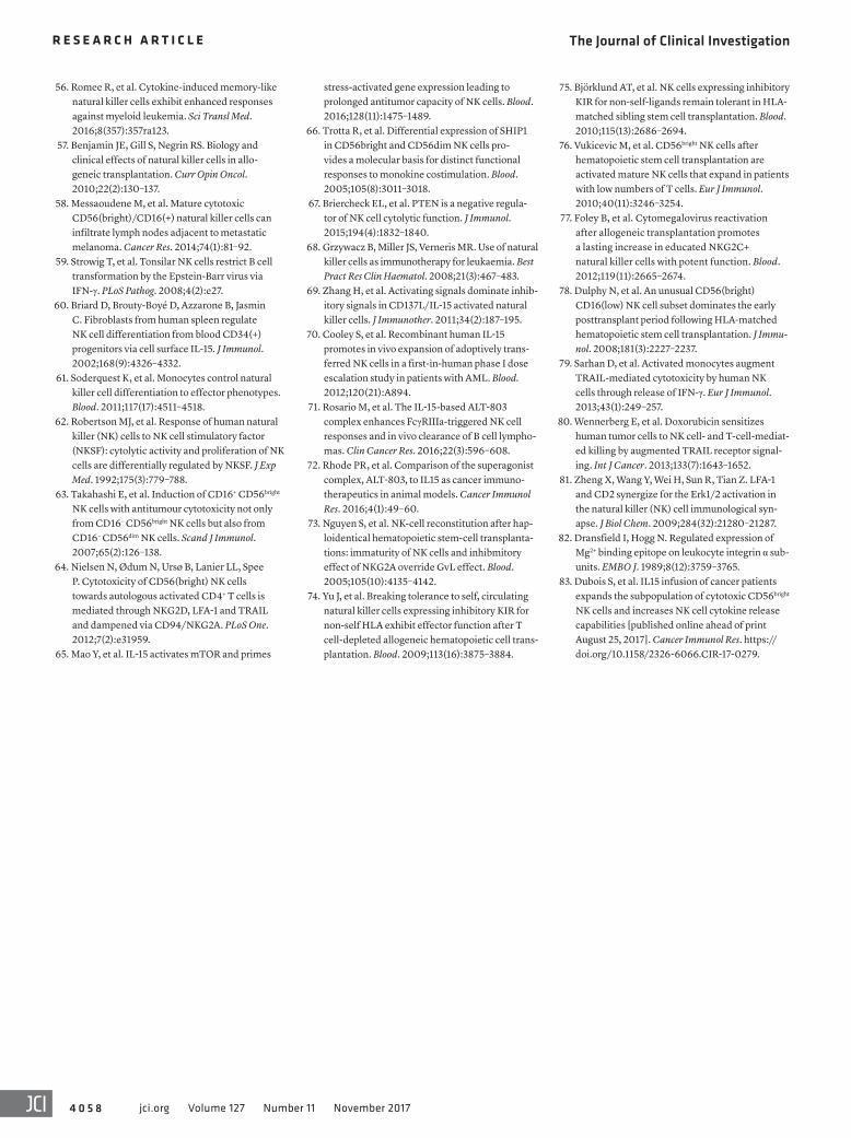
The Journal of Clinical Investigation R E S E A R C H A R T I C L E
4 0 5 8 jci.org Volume 127 Number 11 November 2017
56. Romee R, et al. Cytokine-induced memory-like natural killer cells exhibit enhanced responses against myeloid leukemia. Sci Transl Med. 2016;8(357):357ra123.
57. Benjamin JE, Gill S, Negrin RS. Biology and clinical effects of natural killer cells in allo-geneic transplantation. Curr Opin Oncol. 2010;22(2):130–137.
58. Messaoudene M, et al. Mature cytotoxic CD56(bright)/CD16(+) natural killer cells can infiltrate lymph nodes adjacent to metastatic melanoma. Cancer Res. 2014;74(1):81–92.
59. Strowig T, et al. Tonsilar NK cells restrict B cell transformation by the Epstein-Barr virus via IFN-γ. PLoS Pathog. 2008;4(2):e27.
60. Briard D, Brouty-Boyé D, Azzarone B, Jasmin C. Fibroblasts from human spleen regulate NK cell differentiation from blood CD34(+) progenitors via cell surface IL-15. J Immunol. 2002;168(9):4326–4332.
61. Soderquest K, et al. Monocytes control natural killer cell differentiation to effector phenotypes. Blood. 2011;117(17):4511–4518.
62. Robertson MJ, et al. Response of human natural killer (NK) cells to NK cell stimulatory factor (NKSF): cytolytic activity and proliferation of NK cells are differentially regulated by NKSF. J Exp Med. 1992;175(3):779–788.
63. Takahashi E, et al. Induction of CD16+ CD56bright NK cells with antitumour cytotoxicity not only from CD16– CD56bright NK cells but also from CD16– CD56dim NK cells. Scand J Immunol. 2007;65(2):126–138.
64. Nielsen N, Ødum N, Ursø B, Lanier LL, Spee P. Cytotoxicity of CD56(bright) NK cells towards autologous activated CD4+ T cells is mediated through NKG2D, LFA-1 and TRAIL and dampened via CD94/NKG2A. PLoS One. 2012;7(2):e31959.
65. Mao Y, et al. IL-15 activates mTOR and primes
stress-activated gene expression leading to prolonged antitumor capacity of NK cells. Blood. 2016;128(11):1475–1489.
66. Trotta R, et al. Differential expression of SHIP1 in CD56bright and CD56dim NK cells pro-vides a molecular basis for distinct functional responses to monokine costimulation. Blood. 2005;105(8):3011–3018.
67. Briercheck EL, et al. PTEN is a negative regula-tor of NK cell cytolytic function. J Immunol. 2015;194(4):1832–1840.
68. Grzywacz B, Miller JS, Verneris MR. Use of natural killer cells as immunotherapy for leukaemia. Best Pract Res Clin Haematol. 2008;21(3):467–483.
69. Zhang H, et al. Activating signals dominate inhib-itory signals in CD137L/IL-15 activated natural killer cells. J Immunother. 2011;34(2):187–195.
70. Cooley S, et al. Recombinant human IL-15 promotes in vivo expansion of adoptively trans-ferred NK cells in a first-in-human phase I dose escalation study in patients with AML. Blood. 2012;120(21):A894.
71. Rosario M, et al. The IL-15-based ALT-803 complex enhances FcγRIIIa-triggered NK cell responses and in vivo clearance of B cell lympho-mas. Clin Cancer Res. 2016;22(3):596–608.
72. Rhode PR, et al. Comparison of the superagonist complex, ALT-803, to IL15 as cancer immuno-therapeutics in animal models. Cancer Immunol Res. 2016;4(1):49–60.
73. Nguyen S, et al. NK-cell reconstitution after hap-loidentical hematopoietic stem-cell transplanta-tions: immaturity of NK cells and inhibmitory effect of NKG2A override GvL effect. Blood. 2005;105(10):4135–4142.
74. Yu J, et al. Breaking tolerance to self, circulating natural killer cells expressing inhibitory KIR for non-self HLA exhibit effector function after T cell-depleted allogeneic hematopoietic cell trans-plantation. Blood. 2009;113(16):3875–3884.
75. Björklund AT, et al. NK cells expressing inhibitory KIR for non-self-ligands remain tolerant in HLA-matched sibling stem cell transplantation. Blood. 2010;115(13):2686–2694.
76. Vukicevic M, et al. CD56bright NK cells after hematopoietic stem cell transplantation are activated mature NK cells that expand in patients with low numbers of T cells. Eur J Immunol. 2010;40(11):3246–3254.
77. Foley B, et al. Cytomegalovirus reactivation after allogeneic transplantation promotes a lasting increase in educated NKG2C+ natural killer cells with potent function. Blood. 2012;119(11):2665–2674.
78. Dulphy N, et al. An unusual CD56(bright) CD16(low) NK cell subset dominates the early posttransplant period following HLA-matched hematopoietic stem cell transplantation. J Immu-nol. 2008;181(3):2227–2237.
79. Sarhan D, et al. Activated monocytes augment TRAIL-mediated cytotoxicity by human NK cells through release of IFN-γ. Eur J Immunol. 2013;43(1):249–257.
80. Wennerberg E, et al. Doxorubicin sensitizes human tumor cells to NK cell- and T-cell-mediat-ed killing by augmented TRAIL receptor signal-ing. Int J Cancer. 2013;133(7):1643–1652.
81. Zheng X, Wang Y, Wei H, Sun R, Tian Z. LFA-1 and CD2 synergize for the Erk1/2 activation in the natural killer (NK) cell immunological syn-apse. J Biol Chem. 2009;284(32):21280–21287.
82. Dransfield I, Hogg N. Regulated expression of Mg2+ binding epitope on leukocyte integrin α sub-units. EMBO J. 1989;8(12):3759–3765.
83. Dubois S, et al. IL15 infusion of cancer patients expands the subpopulation of cytotoxic CD56bright NK cells and increases NK cell cytokine release capabilities [published online ahead of print August 25, 2017]. Cancer Immunol Res. https://doi.org/10.1158/2326-6066.CIR-17-0279.